identification, typing and characterization of ... · 61 acnes is well known for its role in acne...
TRANSCRIPT
1
Identification, typing and characterization of Propionibacterium strains from healthy 1
mucosa of the human stomach 2
3
RUNNING TITLE: Propionibacterium strains from human stomach 4
5
Susana Delgado1, Adolfo Suárez
2, and Baltasar Mayo
1 6
7
1Departamento de Microbiología y Bioquímica, Instituto de Productos Lácteos de Asturias 8
(IPLA), Consejo Superior de Investigaciones Científicas (CSIC), Carretera de Infiesto, s/n, 9
33300-Villaviciosa, Asturias, Spain, and 2Servicio de Digestivo, Hospital de Cabueñes, 10
Cabueñes s/n, 33394-Gijón, Asturias, Spain 11
12
13
*Corresponding author: 14
Baltasar Mayo, Instituto de Productos Lácteos de Asturias (CSIC), Carretera de Infiesto s/n, 15
33300-Villaviciosa, Spain 16
Tel.: 34+985 89 21 31 17
Fax: 34+985 89 22 33 18
E-mail address: [email protected] 19
20
*Manuscript with Line NumbersClick here to view linked References
2
ABSTRACT 21
Forty two Propionibacterium isolates were recovered from biopsy samples of the 22
gastric mucosa of eight out of 12 healthy people. Of these, 41 were identified as belonging 23
to Propionibacterium acnes; the remaining isolate was identified as belonging to 24
Propionibacterium granulosum. Repetitive extragenic palindromic (REP)-PCR typing 25
suggested that up to four strains might be present in the mucosa of the same individual. 26
Sequence analysis of either recA, tly or camp5 genes of P. acnes isolates revealed two 27
distinct phylogenetic lineages. As per the recA, most isolates belonged to type I, while the 28
remainder of the isolates belonged to type II. Phenotypic analyses of representative isolates 29
showed the different strains to have diverse biochemical properties. For example, large 30
differences were seen in carbohydrate fermentation patterns, the results of qualitative and 31
quantitative enzymatic profiling, and survival at acidic pH. In contrast, the patterns of 32
resistance/susceptibility to a series of 16 antibiotics were rather similar, with no atypical 33
resistances observed. The examined strains showed limited –if any– enzymatic activities 34
that could be ultimately related to pathogenicity (lipolytic, proteolytic or haemolytic 35
activity). This suggests that, in the gastric ecosystem, some Propionibacterium spp. 36
genotypes and/or phenotypes can be considered true commensals. 37
38
KEY WORDS: Gastric microbiology, human stomach, propionibacteria, 39
Propionibacterium acnes, Propionibacterium granulosum 40
41

3
1. Introduction 42
Propionibacterium species are non-spore-forming, Gram-positive, anaerobic-43
aerotolerant, pleomorphic rod-shaped bacteria, the fermentation end products of which 44
include propionic acid. Besides the classical propionibacteria species from dairy products, 45
Propionibacterium acnes, Propionibacterium granulosum, Propionibacterium avidum, 46
Propionibacterium propionicum, and Propionibacterium lymphophilum are species 47
typically found on the human skin and mucosa (Holland and Bojar, 2002), where they may 48
exert either protective or harmful effects. However, in spite of this importance, scarce data 49
are available for most species, except for P. acnes, the best known of the human-associated 50
propionibacteria. Cutaneous Propionibacterium species might have genetic and 51
biochemical similarity, occupying in consequence similar environments. Not surprisingly, 52
thought at lower numbers, P. granulosum is usually found in close association with P. 53
acnes (Dicksved et al. 2009; Saulnier et al. 2009). 54
P. acnes predominates over other microorganisms inhabiting the pilosebaceous follicles 55
(Miura et al. 2010), and is a member of the normal microbiota of the oral cavity, the 56
nostrils, conjunctiva, the external ear canal, and the upper respiratory and intestinal tracts 57
(Bik et al. 2006; Saulnier et al. 2009). It is usually regarded as a strict anaerobe, although it 58
grows better under low oxygen tension (Gribbon et al. 1994). In fact, P. acnes can tolerate 59
100% oxygen saturation, reflecting its ability to survive in a wide range of environments. P. 60
acnes is well known for its role in acne vulgaris. Some authors suggest it contributes to the 61
inflammatory phase of this condition (Dessinioti and Katsambas, 2010). Damage to host 62
cells and tissues might occur via enzymes with degradative properties, including sialidases, 63
neuraminidases, endoglycoceramidases and lipases (Brüggemann, 2005). The latter 64
enzymes are thought to release free fatty acids, which in the skin irritate the follicular wall 65
4
and surrounding dermis. Immunogenic factors of P. acnes such as surface determinants, 66
heat shock proteins and chemoattractants might then trigger inflammation (Vowels et al. 67
1995). P. acnes is also thought to be an opportunistic pathogen. It has been implicated in 68
postoperative and device-related infections (Harris et al. 2005) as well as in a number of 69
other conditions such as sarcoidosis, synovitis, pustulosis, endocarditis, endophthalmitis, 70
hyperostosis and osteitis (Ishige et al. 2005; Leyden et al. 2001; Perry and Lambert, 2005). 71
Infections generally occur in immunocompromised patients and newborns; they are less 72
common in previously healthy individuals. However, evidence regarding the role of P. 73
acnes as the causal agent of these diseases remains circumstantial. 74
P. acnes is also well known for its immunomodulatory properties. High levels of anti-P. 75
acnes antibodies have been reported in individuals with acne (Ashbee et al. 1997). The 76
induction of pro-inflammatory cytokines, interleukin IL-1a, IL-1b, IL-8 and TNF- by P. 77
acnes has also been reported (Vowels et al. 1995). The genome sequence of the species 78
clearly shows it possesses many proteins involved in the ability to colonise and reside in 79
human skin sites (Bruggemann et al. 2004), although no virulence determinants or 80
pathogenicity factors have been identified. Thus, P. acnes is usually regarded as a disease-81
causing organism, although some authors indicate it to be a harmless commensal (Eady and 82
Ingham, 1994). 83
The human stomach mucosa would appear to harbour a greater microbial diversity than 84
originally anticipated (Bik et al. 2006; Monstein et al. 2000). Different species of 85
lactobacilli and streptococci are among the majority populations seen (Bik et al. 2006; Ross 86
et al. 2005). Although not among the dominant gastric microorganisms, Propionibacterium 87
spp. has also been revealed by different culture-independent approaches (Dicksved et al. 88

5
2009; Monstein et al. 2000). Selected strains of all these bacterial groups could be of 89
greater interest than others of intestinal origin for the design and formulation of probiotics. 90
Their notable resistance to acidic conditions might be of use in maintaining the bacterial 91
viability of probiotics during storage, in improving bacterial survival through the gastric 92
passage, and be advantageous in the formation of biofilms (Holmberg et al. 2009), a key 93
factor in gastric and intestinal colonisation (FAO/WHO, 2002). 94
This work reports the identification and genetic typing of Propionibacterium spp. 95
strains isolated from the stomach of healthy adults. Phenotypic and biochemical tests were 96
undertaken to characterize the gastric strains. In addition, activities related to pathogenicity 97
and resistance to antibiotics were also sought to assess their potential to cause harm. 98
99
100
2. Material and Methods 101
102
2.1. Sampling, media and culture conditions 103
Gastric biopsies were aseptically collected from 12 healthy individuals undergoing 104
routine upper gastrointestinal endoscopy by using an Olympus GIF-Q0165 gastroscope 105
(Olympus Corporation, Tokyo, Japan) and a large (8 Fr) single-use, sterile biopsy forceps 106
with a central bayonet (Boston Scientific Corporatin, Natick, USA). Before exploration, the 107
gastroscope was washed and disinfected by immersion in a detergent solution with 108
proteolytic enzymes (at 7%) and glutaraldehyde (at 2%). Biopsy samples were collected in 109
sterile containers with a reduced saline solution (0.9% NaCl, 0.1% peptone, 0.1% Tween 110
80, and 0.02% cysteine) and transported to the laboratory in less than two hours after 111
intervention. The biopsies were then washed several times in a phosphate buffered saline 112
6
(PBS) solution, drained, weighed, and thoroughly homogenized in Maximum Recovering 113
Diluent (Scharlab, Barcelona, Spain). Dilutions of these samples were spread on agar plates 114
containing the following media, MRS (Merck, Darmstadt, Germany) supplemented with 115
0.25% cysteine (Merck) (MRSC), Columbia Blood medium containing 5% defibrinated 116
horse blood (CB; Merck), and Brain Heart Infusion medium (BHI; Oxoid, Basingstoke , 117
Hampshire, UK). All plates were incubated anaerobically in an anaerobic chamber 118
(Mac500, Down Whitley Scientific, West Yorkshire, UK) at 37°C for up to 5 days. In order 119
to rule out potential bacterial contamination the saline solution used to transport biopsy 120
samples was spread on the same media. Non-inoculated plates, used as a second negative 121
control, were also incubated in the same conditions. 122
123
2.2. Identification and genotyping of isolates 124
Visible colonies were selected, purified by subculturing on the same medium and 125
identified at the species level by a combination of amplified ribosomal DNA restriction 126
analysis (ARDRA) and sequencing of the 16S rRNA amplicons. 127
Total genomic DNA from isolates was purified from overnight cultures using the 128
GenEluteTM
Bacterial Genomic DNA kit (Sigma-Aldrich, St. Louis, MO, USA) following 129
the manufacturer’s recommendations. Electrophoresis was performed in 1% agarose gels, 130
and the DNA visualised by UV light after staining with ethidium bromide (0.5 g ml-1
). 131
For ARDRA, the 16S rRNA genes were almost completely amplified by the polymerase 132
chain reaction (PCR) technique using the universal primers 27F (5’–133
AGAGTTTGATCCTGGCTCAG–3’) and 1492R (5’–GGTTACCTTGTTACGACTT–3’), 134
and a 2 x Taq Master Mix (Ampliqon, Skovlunde, Denmark). Amplicons were purified 135
7
using GenEluteTM
PCR Clean-Up columns (Sigma-Aldrich), digested with the restriction 136
enzymes HaeIII and HinfI (Fermentas, Vilnius, Lithuania), and electrophoresed as above. 137
When required, amplicons were sequenced by cycle extension in an ABI 373 DNA 138
sequencer (Applied Biosystems, Foster City, CA, USA). Sequences were compared to 139
those in the GenBank database using the BLASTN program 140
(http://www.ncbi.nlm.nih.gov/BLAST/), and to those held by the Ribosomal Database 141
Project (http://rdp.cme.msu.edu/index.jsp). 142
All isolates were genotyped by repetitive extragenic palindromic (REP)-PCR 143
fingerprinting using primer BoxA2-R (5’–ACGTGGTTTGAAGAGATTTTCG–3’), as 144
reported by Koeuth et al. (1995). Reproducibility studies of the REP-PCR technique 145
showed a percentage of similarity of around 85%. Similarity of the patterns was expressed 146
using Sorensen’s correlation coefficient after clustering by the unweighted pair group 147
method using arithmetic averages (UPGMA). Analysis of the recA gene with primers PAR-148
1 (5’–AGCTCGGTGGGGTTCGCTCATC–3’) and PAR-2 (5’–149
GCTTCCTCATACCACTGGTCATC–3’) was performed to assign the P. acnes strains to 150
type I or type II, as reported by McDowell et al. (2005). Additionally, DNA sequence 151
analysis of the two putative virulence genes tly and camp5 was conducted using 152
oligonucleotides and PCR conditions as previously described by McDowell et al. (2005) 153
and Valanne et al. (2005), respectively. Sequences were compared to each other and to 154
those deposited in the GenBank database. Similarity analysis was performed with the 155
Neighbor-Joining algorithm using the program MEGA 4 (Tamura et al. 2007). For 156
comparative purposes, representative published sequences from different genetic divisions 157
associated with both health and infections (Lomholt and Kilian, 2010) were also included. 158
159

8
2.3. Fermentation of carbohydrates 160
Carbohydrate fermentation profiles of the different strains were established using the 161
commercial API 50 CH system (bioMérieux, Marcy l’Etoile, France) following the 162
manufacturer’s instructions. 163
164
2.4. Enzymatic activities. 165
Enzyme activities were assessed using the semiquantitative APIZYM system 166
(bioMérieux). Sixty five millilitres of a cellular suspension corresponding to McFarland 167
standard 5 were inoculated into each well of the API-ZYM strips, incubated for 4 h under 168
anaerobic conditions at 37°C, and developed as recommended. Activities were expressed as 169
nmol of substrate hydrolysed under the assay conditions. 170
171
2.5. Acid resistance 172
The ability of the strains to survive under acidic conditions was assessed through 173
exposure of the cells to an acidified solution. Changes on the viability of cell suspensions 174
(108 cells mL
-1) in sterile saline solution (0.5% NaCl) acidified with HCl to pHs ranging 175
from 2.0 to 5.0 were determined by plate counting on MRSC agar after incubation at 37°C 176
for 90 min. 177
178
2.6. Antibiotic resistance/susceptibility profiles. 179
Minimum inhibitory concentrations (MICs) were determined by microdilution in 180
VetMICTM
plates for lactic acid bacteria (National Veterinary Institute of Sweden, Uppsala, 181
Sweden) containing two-fold serial dilutions of 16 antibiotics. Colonies grown on MRSC 182
agar plates were suspended in 2 mL of sterile saline (Oxoid) to obtain a density 183

9
corresponding to McFarland standard 2. This suspension was further diluted 1:15 with 184
Mueller-Hinton medium (Oxoid) containing 0.5% glucose. One hundred microlitres of this 185
were added to each well of the VetMICTM
plate, and the plates incubated at 37ºC for 48-72 186
h. MICs were defined as the lowest antibiotic concentration at which no visual growth 187
(pellet at the bottom of the well) was observed. 188
189
2.7. Presence of virulence factors 190
To determine the pathogenic potential of the strains, the presence of putative 191
virulence-related traits was investigated using classical procedures. The production of 192
haemolysins was analysed by plate assays on CB agar. The presence of haemolysis is 193
indicated by the formation of clear zones surrounding the colonies. Staphylococcus aureus 194
CECT 4520T (Colección Española de Cultivos Tipo, Valencia, Spain) was used as a 195
positive control. The possible interaction of pore-forming Christie-Atkins-Much-Peterson 196
(CAMP) factors from the P. acnes strains with S. aureus sphingomyelinase C was tested as 197
previously described (Valanne et al. 2005) using CB agar plates employing the S. aureus 198
CECT 4520T strain as an indicator. DNase activity was assayed on DNase agar plates 199
(Merck). Proteolytic activity was determined on RCM plates containing 3% skimmed-milk 200
powder (Scharlab). Lipolytic activity was scored on Reinforced Clostridial Medium (RCM; 201
Merck) plates supplemented with 10 g L-1
tributyrin (Sigma-Aldrich); all plates were 202
incubated under anaerobiosis at 37ºC for 3-4 days, the time at which the diameter of the 203
clearing zone was measured. The production of urease was examined by growing the 204
strains in urease test broth consisting of RCM supplemented with urea (20g L-1
) and phenol 205
red (0.01g L-1
) (Sigma-Aldrich). Urease production was indicated by the change in the 206
phenol red indicator. 207

10
208
209
3. Results 210
Viable bacteria of different species were recovered from the MRSC, CB and BHI agar 211
plates for 10 of the 12 stomach mucosa samples examined. Counts ranged from 1x10² to 212
1x10⁵ colony forming units (cfu) per gram of mucosa. Representative isolates from all 213
plates were purified by subculturing and subjected to a polyphasic identification system; the 214
latter included ARDRA profiling, the sequencing of the 16S rRNA gene, and the 215
comparison of the sequences against those in databases. Propionibacterium spp. isolates 216
were recovered in the different media from the mucosa of eight individuals. Surprisingly, of 217
the 42 isolates identified as Propionibacterium spp., 41 belonged to the species P. acnes. 218
The remaining isolate was identified as a Propionibacterium granulosum. 219
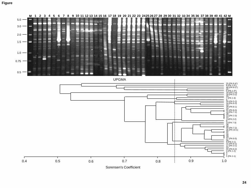
To assess the genetic diversity of the strains, all isolates were subjected to REP-PCR 220
typing (Fig. 1). Statistical analysis of the results showed at least 12 genetic profiles with a 221
similarity level below the percentage of reproducibility (85%) (Fig. 1); these can be 222
considered to belong to different strains. Variability was observed even among isolates 223
from the same individual, suggesting the presence of up to four strains in the stomach 224
mucosa in some subjects. Representative isolates for these 12 profiles were subjected to a 225
further genetic typing and to an exhaustive biochemical characterization to determine their 226
genotypic, phenotypic and safety properties. Isolates with similar profiles but from different 227
mucosa samples were also selected; thus, in total, 24 isolates were characterized, including 228
the P. granulosum strain. The numbering of the strains, as represented in the tables and 229
figures, includes species initials of the species, followed by the subject from which they 230
were obtained and the code of the strain. Sequence analysis of recA, tly and camp5 genes 231
11
from the 23 P. acnes strains showed two consistent and distinct cluster groups (Fig. 2). The 232
number of polymorphic sites varied depending on the gene. Respect to recA gene, 19 233
strains harbour type I-specific sequences, while the remaining four (PA 1-3, PA 1-5, PA 6-234
4, and PA 8-2) contained all type II-specific polymorphisms. Further, a transition mutation 235
(A for G) at position 897 of the recA gene corresponding to type IB (McDowell et al. 236
2008), was observed in nine of the 19 type I isolates. No type III-specific polymorphisms 237
were recorded. Alignment of 777 bp of the tly genes revealed 22 differences in nucleotide 238
positions and four allelic profiles, meanwhile in the camp5 gene 19 polymorphic sites and 239
four different alleles were encountered. Sequences were aligned with those from 240
representative strains of the recent work of Lomholt and Kilian (2010). This allowed us to 241
compare the relatedness at the DNA level of the gastric P. acnes isolates with those from 242
clinical (infections) and non-clinical (environmental) sources. As can be seen in the 243
derivative phylogenetic trees (Fig. 2), the gastric strains were clearly divided into two 244
groups, one of which appears to include reference strains isolated from the oral cavity (such 245
as CCUG45436 and CCUG 50655) and corresponded to recA type II isolates. The second 246
group belonged to recA type I and included 19 strains. This group was further split into two 247
subgroups, containing each a similar number of isolates; one of these subgroups seemed to 248
be related to acne-associated strains, which belonged to recA type IA. The other subgroup, 249
identified as recA type IB, included environmental sequences, no particularly associated 250
with skin infections (Lomholt and Kilian, 2010) 251
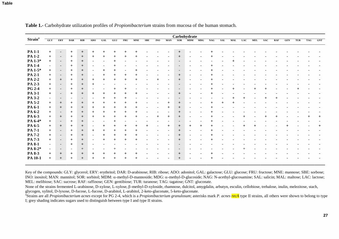
The fermentation of 49 carbohydrates by the 24 selected strains was assayed using the 252
API 50 CH system; Table 1 shows the results obtained. Rather wide phenotypic variation 253
was observed among the different strains. Most fermented glycerol, D-arabinose, ribose, 254
galactose, glucose, fructose, mannose, sorbitol, and N-acethyl-glucosamine, the utilization 255
12
of erythritol, adonitol, and mannitol was variable, and the fermentation of other 256
carbohydrates was sporadic. Strengthening the genetic typing results, strains from the same 257
individual showed different carbohydrate utilization profiles. Strains from different mucosa 258
samples, however, sometimes showed identical or very similar profiles. It should be noted 259
that the carbohydrate fermentation profiles do not always match those supposed specific for 260
P. acnes recA type I or type II, the former –but not the latter– being able to ferment 261
erythritol, ribose and sorbitol. 262
Although less than that observed for the fermentation of carbohydrates, variability 263
among the different strains was also seen in terms of the enzymatic activities assayed using 264
the API-ZYM method (Table 2). In general, the strains showed a limited number of weak 265
enzymatic activities. The majority showed acid phosphatase, naphtol-AS-BI-266
phosphohydrolase and N-acetyl--glucosaminidase activities. Weak alkaline phosphatase, 267
esterase (C4), esterase lipase (C8), -glucosidase and -mannosidase activities were also 268
scored for some strains. 269
The MIC values and antibiotic resistance-susceptibility patterns of 14 representative 270
strains to 16 different antibiotics (all representatives of clinically important antimicrobial 271
agents) were obtained by microdilution with the commercial VetMIC system. Table 3 272
shows the results obtained. Contrary to that reported for clinical P. acnes strains, low 273
resistance levels were observed for most antibiotics, and analysis of the distribution of 274
MICs among the different strains suggests they all are susceptible to most antibiotics. 275
Significant variation was only observed with respect to kanamycin, streptomycin, and 276
trimethoprim. 277
13
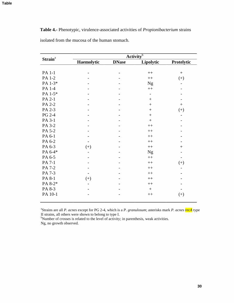
Table 4 shows the results regarding the presence of putative virulence and/or 278
pathogenicity factors in the 24 selected strains. None of the strains showed urease activity 279
in urea broth with phenol red as an indicator. Furthermore, DNase activity in DNase agar 280
plates was never detected. Some slight haemolysis was seen for just two strains of the recA 281
type IB in CB agar. However, haemolysis by either P. acnes or S. aureus was not affected 282
when these were cultivated close to one another, excluding the involvement of CAMP-like 283
factors in this activity. The production of proteases was scant; only three strains showed 284
small (although clear) hydrolysis halos on RCM plates with skimmed milk. In contrast 285
nearly all isolates showed halos of lipolysis in agar plates with tributyrin. Notwithstanding, 286
a few strains showed faint halos, two (PA 1-3, PA 6-4) did not grow in the assay medium, 287
and one (PA 1-5), which did grow, showed no halo. The latter three strains belonged to 288
recA type II. 289
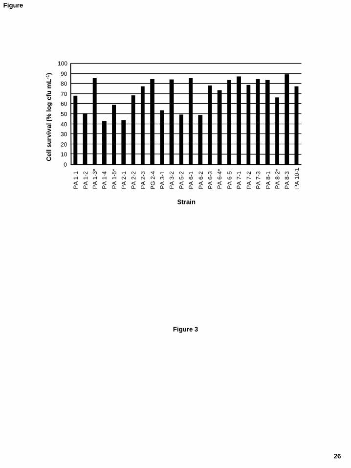
Species inhabiting the stomach ought to be highly resistant to acidic conditions. 290
Analysis of this property was accomplished by incubating the strains in an acidic solution 291
for 90 min and then plating. Counts were expressed as percentage survival compared to that 292
recorded for control cells kept for the same time in a neutral solution. Strains maintained at 293
pH 4.0 or higher showed no noticeable reduction in numbers, while they all were killed 294
during treatment at pH 2.5 and 2.0. Fig. 3 shows large differences among the strains in the 295
tolerance to pH 3.0. Reductions in counts of between 1 and 3 logarithmic units were 296
commonly recorded at this pH. 297
298
4. Discussion 299
In this work, a series of 42 propionibacteria isolates from the stomach mucosa of healthy 300
humans was identified by molecular methods, and then typed by REP-PCR. Of these, 41 301
14
isolates, corresponding to 23 different strains, were assigned to the species P. acnes and 302
one to P. granulosum. These two species are usual members of the resident human 303
microbiota in the sebaceous gland-rich areas of the skin (Miura et al. 2010), but also in 304
conjunctiva, mouth, and intestines (Monstein et al. 2000; Saulnier et al. 2009). The P. 305
acnes strains examined in this work mostly belonged to P. acnes recA type I, four belonged 306
to type II and none to type III. Based on the fermentation of sugars or phenotypic profiling 307
(Brüggemann, 2005; Higaki et al. 2000), the recA type II strains could not be distinguished 308
from those of type I; this has been reported elsewhere (McDowell et al. 2005). Similar 309
numbers of recA type IA and IB strains (as defined by McDowell et al. 2008) were found 310
among the stomach isolates. These two groups corresponded with the clustering derived 311
from tly and camp5 gene analysis (Fig. 2). Sequence analysis of tly and camp5 genes 312
allowed us to allocate the gastric P. acnes strains in genetic divisions related or no with 313
acne, following the recent study by Lomholt and Kilian (2010). According to these authors, 314
the phylogenetic group termed as I-1a, that would include 10 of our strains, is strongly 315
associated with acne, as compared to other divisions such as I-2 and II (which embraced 9 316
and 4 of the gastric strains, respectively). Strains from the latter groups seem to be 317
associated with healthy skin and opportunistic infections. In our study, division II 318
corresponded with the recA type II group. However, in spite of this associations, the role of 319
the different genotypes in health and disease has is far from conclusive (McDowell et al. 320
2005; Lomholt and Kilian, 2010). 321
Wide phenotypic and genetic diversity was found among the strains from the different 322
individuals. Diversity was even encountered between isolates from the same subject, and up 323
to four different strains were identified in the same mucosa sample. REP-PCR analysis 324
matched in general with the clusters derived from sequencing analysis of recA, tly and 325

15
camp5 genes, showing recA type II strains a closer genetic relationship among themselves 326
than to type I strains (Fig. 1). This confirms previous findings suggesting the existence of 327
consistent distinct phylogenetic groups within this species, as revealed by the use of genetic 328
typing methods such as random amplification of polymorphic DNA (RAPD) or multilocus 329
sequence typing (MLST) (Perry et al. 2003; McDowell et al. 2005; Lomholt and Kilian, 330
2010). 331
A gradual worldwide increase in the prevalence of antibiotic resistance in P. acnes has 332
been observed (Nord and Oprica, 2006). This has been attributed to the major role of 333
antibiotics in acne therapy (Eady et al. 2003). In Europe, combined resistance to 334
clindamycin and erythromycin is highly prevalent in P. acnes isolates from patients with 335
acne –from 50 up to 91% (Ross et al. 2003). In contrast, resistance to tetracycline was 336
shown to be in the 0-26% range. Not surprisingly, a strong correlation between antibiotic 337
resistance and the prescribing patterns of different countries exists (Ross et al. 2003). 338
However, strains of this work seem all to be free of atypical resistances, those encoded by 339
dedicated genes (Ammor et al. 2007). Resistance to aminoglycosides (kanamycin, 340
streptomycin) in Gram-positives is thought to be due to intrinsic mechanisms. In fact, the 341
lack of cytochrome-mediated transport is though to be responsible for the strong resistance 342
to aminoglycosides shown by most anaerobic and facultative bacteria (for a review see 343
Ammor et al. 2007). Nevertheless, wide inter-strain variation in resistance levels to these 344
antimicrobial agents, not linked to the presence or absence of dedicated antibiotic resistance 345
genes, has been repeatedly reported in strains from different ecosystems (Ammor et al. 346
2007). 347
A vast array of degrading and host-interactive proteins located at the cell surface has 348
recently been identified from the decoded genome of P. acnes (Brüggemann, 2005; 349
16
Brüggemann et al. 2004). Some of these proteins might be responsible for the phenotypic 350
traits traditionally associated with pathogenesis (Dessinioti and Katsambas, 2010; Higaki et 351
al. 2000). Production of CAMP factors and notable haemolytic and proteolytic activities 352
that could ultimately be related with virulence were never detected by the classical methods 353
used in this study. Moreover, clear differences in these properties were not scored among 354
the different genetic types; only of note, was the scarce or absent lipolytic activity observed 355
in three out of the four recA type II strains as compared with the rest of isolates. 356
Furthermore, resistance to low pH (pH 3.0) was also shown to vary widely within the 357
strains, independently of their genotype. Given that Propionibacterium species have been 358
reported as a majority population in both healthy persons as well as in those suffering from 359
many infections and inflammatory conditions (Eishi et al. 2010; Mihura et al. 2010), it 360
would be of interest to determine the phenotypes and/or genotypes that render this 361
bacterium either a commensal or pathogenic organism (Dessinioti and Katsambas, 2010; 362
Mihura et al. 2010). The availability of the genome sequence of P. acnes, and soon that 363
from other species, will allow post-genomic studies will help clarify whether the existing 364
phenotypic properties are simply colonising traits or true virulence and pathogenic factors 365
involved in the aetiology and pathogenesis of P. acnes-associated diseases. 366
367
368
5. Conclusion 369
Propionibacterium strains, mostly of the P. acnes species, were isolated as part of the 370
dominant anaerobic cultivatable microbiota of mucosa samples from the human stomach. 371
Wide phenotypic and genotypic diversity was encountered among the different strains, even 372
among those isolates from the same individual. No clear pathogenic properties related to 373
17
the infectivity of this species were observed in the strains examined; however, genetic 374
analyses proved some strains to be phylogenetically related with acne-associated strains. 375
Together, the present results suggest propionibacteria species might be habitual, innocuous 376
inhabitants of the gastric environment. Thought further research will be required to 377
determine the precise role(s) of these bacteria in the gastric ecosystem it might be 378
hypothesized about its contribution to the host health and well-being through their 379
immunomodulatory activities. If this were the case, selected strains could be used as 380
probiotics to counteract harmful bacteria such as Helicobacter pylori. 381
382
383
Acknowledgements 384
Research was supported by projects from FICYT (Ref. IB08-005) and INIA (Ref. 385
RM2009-00009-00-00). S. Delgado was supported by a research contract from MICINN 386
(Juan de la Cierva program, JCI-2008-02391). The skillful assistance of N. Arias and C. 387
Hidalgo is greatly acknowledged. Results were partially presented at the 3rd
International 388
Symposium on Propionibacteria and Bifidobacteria: Dairy and Probiotic Applications, held 389
in June 2010, in Oviedo, Spain. 390
391
392
References 393
Ammor, M.S., Flórez, A.B., Mayo, B. 2007. Antibiotic resistance in non-enterococcal 394
lactic acid bacteria and bifidobacteria. Food Microbiology 24, 559-570. 395

18
Ashbee, H.R., Muir, S.R., Cunliffe, W.J., Ingham, E. 1997. IgG subclasses specific to 396
Staphylococcus epidermidis and Propionibacterium acnes in patients with acne 397
vulgaris. British Journal of Dermatology 136, 730-733. 398
Bik, E.M., Eckburg, P.B., Gill, S.R., Nelson, K.E., Purdom, E.A., Francois, F., Perez-Perez, 399
G., Blaser, M.J., Relman, D.A. 2006. Molecular analysis of the bacterial microbiota in 400
the human stomach. Proceedings of the National Academy of Science USA 103, 732-401
737. 402
Brüggemann, H., Henne, A., Hoster, F., Liesegang, H., Wiezer, A., Strittmatter, A., Hujer, 403
S., Durre, P., Gottschalk G. 2004. The complete genome sequence of 404
Propionibacterium acnes, a commensal of human skin. Science 305, 671-673. 405
Brüggemann, H. 2005. Insights in the pathogenic potential of Propionibacterium acnes 406
from its complete genome. Seminars in Cutaneous Medicine and Surgery 24, 67-72. 407
Dessinioti C, Katsambas AD. 2010. The role of Propionibacterium acnes in acne 408
pathogenesis: facts and controversies. Clinical Dermatology 28, 2-7. 409
Dicksved J, Lindberg M, Rosenquist M, Enroth H, Jansson JK, Engstrand L. 2009. 410
Molecular characterization of the stomach microbiota in patients with gastric cancer and 411
in controls. Journal of Medical Microbiology 58, 509-516. 412
Eady, E.A. Ingham, E. 1994. Propionibacterium acnes –friend of foe? Reviews in Medical 413
Microbiology 5, 163-173. 414
Eady, A.E., Cove, J.H., Layton, A.M. 2003. Is antibiotic resistance in cutaneous 415
propionibacteria clinically relevant?: implications of resistance for acne patients and 416
prescribers. American Journal of Clinical Dermatology 4, 813-831. 417
Eishi, Y., Suga, M., Ishige, I., Kobayashi, D., Yamada, T., Takemura, T., Takizawa, T., 418
Koike, M., Kudoh, S., Costabel, U., Guzman, J., Rizzato, G., Gambacorta, M., du Bois, 419

19
R., Nicholson, A.G., Sharma, O.P., Ando, M. 2002. Quantitative analysis of 420
mycobacterial and propionibacterial DNA in lymph nodes of Japanese and European 421
patients with sarcoidosis. Journal of Clinical Microbiology 40, 198-204. 422
Gribbon, E.M., Shoesmith, J.G., Cundliffe, W.J., Holland, K.T. 1994. The microarophily 423
and photosensitivity of Propionibacteriun acnes. Journal of Applied Bacteriology 77, 424
583-590. 425
FAO/WHO. 2002. Report on drafting guidelines for the evaluation of probiotics in food. 426
(ftp://ftp.fao.org/es/esn/food/wgreport2.pdf). London, Ontario, Canada, May 1, 2002. 427
Harris, A.E., Hennicke, C., Byers, K., Welch, W.C. 2005. Postoperative discitis due to 428
Propionibacterium acnes: a case report and review of the literature. Surgical Neurology 429
63, 538-541. 430
Higaki, S., Kitagawa, T., Kagoura, M., Morhashi, M., Yamagishi, T. 2000. Correlation 431
between Propionibacterium acnes biotypes, lipase activity and rash degree in acne 432
patients. Journal of Dermatology 27, 519-522. 433
Holland, K.T., Bojar, R.A. 2002. The cutaneous propionibacteria. In, Molecular Medical 434
Microbiology, M. Sussman (ed.). Chapter 49, pp. 1039-1055. Academic Press, London, 435
UK. 436
Holmberg, A., Lood, R., Mörgelin, M., Söderquist, B., Holst, E., Collin, M., Christensson, 437
B., Rasmussen, M. 2009. Biofilm formation by Propionibacterium acnes is a 438
characteristic of invasive isolates. Clinical Microbiology and Infection 15, 787-795. 439
Ishige, I., Eishi, Y., Takemura, T., Kobayashi, I., Nakata, K., Tanaka, I., Nagaoka, S., Iwai, 440
K., Watanabe, K., Takizawa, T., Koike, M. 2005. Propionibacterium acnes is the most 441
common bacterium commensal in peripheral lung tissue and mediastinal lymph nodes 442

20
from subjects without sarcoidosis. Sarcoidosis, Vasculitis and Diffuse Lung Diseases 443
22, 33-42. 444
Koeuth, T., Versalovic, J., Lupski, J.R. 1995. Differential subsequence conservation of 445
interspersed repetitive Streptococcus pneumoniae BOX elements in diverse bacteria. 446
Genome Research 5, 408-418. 447
Leyden, J.J. 2001. The evolving role of Propionibacterium acnes in acne. Seminars in 448
Cutaneous Medicine and Surgery 20, 139-143. 449
Lomholt, H.B., Kilian, M. 2010. Population genetic analysis of Propionibacterium acnes 450
identifies a subpopulation and epidemic clones associated with acne. PLoS One 5, 451
e12277. 452
McDowell, A., Valanne, S., Ramage, G., Tunney, M.M., Glenn, J.V., McLorinan, G.C., 453
Bhatia, A., Maisonneuve, J.F., Lodes, M., Persing, D.H., Patrick, S. 2005. 454
Propionibacterium acnes types I and II represent phylogenetically distinct groups. 455
Journal of Clinical Microbiology 43, 326-334. 456
McDowell, A., Perry, A.L., Lambert, P.A., Patrick, S. 2008. A new phylogenetic group of 457
Propionibacterium acnes. Journal of Medical Microbiology 57, 218-224. 458
Miura, Y., Ishige, I., Soejima, N., Suzuki, Y., Uchida, K., Kawana, S., Eishi, Y. 2010. 459
Quantitative PCR of Propionibacterium acnes DNA in samples aspirated from 460
sebaceous follicles on the normal skin of subjects with or without acne. Journal of 461
Medical and Dental Sciences 57, 65-74. 462
Monstein, H.J., Tiveljung, A., Kraft, C.H., Borch, K., Jonasson, J. 2000. Profiling of 463
bacterial flora in gastric biopsies from patients with Helicobacter pylori-associated 464
gastritis and histologically normal control individuals by temperature gradient gel 465
21
electrophoresis and 16S rDNA sequence analysis. Journal of Medical Microbiology 49, 466
817-822. 467
Nord, C.E., Oprica, C. 2006. Antibiotic resistance in Propionibacterium acnes. 468
Microbiological and clinical aspects. Anaerobe 12, 207-210. 469
Perry, A.L., Worthington, T., Hilton, A.C., Lambert, P.A., Stirling, A.J., Elliott, T.S. 2003. 470
Analysis of clinical isolates of Propionibacterium acnes by optimised RAPD. FEMS 471
Microbiology Letters 228, 51-55. 472
Perry, A.L., Lambert, P.A. 2005. Propionibacterium acnes. Letters in Applied 473
Microbiology 42, 185-188 474
Ross, J.I., Snelling, A.M., Carnegie, E., Coates, P., Cunliffe, W.J., Bettoli, V., Tosti, G., 475
Katsambas, A., Galvan Peréz del Pulgar, J.I., Rollman, O., Török, L., Eady, E.A., Cove, 476
J.H. 2003. Antibiotic-resistant acne: lessons from Europe. British Journal of 477
Dermatology 148, 467-478. 478
Ross, S., Engstrand, L., Jonsson, H. 2005. Lactobacillus gastricus sp. nov., Lactobacillus 479
antri sp. nov., Lactobacillus kalixensis sp. nov. and Lactobacillus ultunensis sp. nov., 480
isolated from human stomach mucosa. International Journal of Systematic and 481
Evolutionary Microbiology 55, 77-82. 482
Saulnier, D.M., Kolida, S., Gibson, G.R. 2009. Microbiology of the human intestinal tract 483
and approaches for its dietary modulation. Current Pharmaceutical Design 15, 1403-484
1414. 485
Tamura, K., Dudley, J., Nei, M., Kumar, S. 2007. MEGA4: Molecular Evolutionary 486
Genetics Analysis (MEGA) software version 4.0. Molecular Biology and Evolution 24, 487
1596-1599. 488
22
Valanne, S., McDowell, A., Ramage, G., Tunney, M.M., Einarsson, G.G., O’Hagan, S., 489
Wisdom, G.B., Fairley, D., Bhatia, A., Maisonneuve, J.F., Lodes, M., Persing, D.H., 490
Patrick, S. 2005. CAMP factor homologues in Propionibacterium acnes: a new protein 491
family differentially expressed by types I and II. Microbiology 151, 1369-1379. 492
Vowels, B.R., Yang, S., Leyden, J.J. 1995. Induction of proinflammatory cytokines by a 493
soluble factor of Propionibacterium acnes: implications for chronic inflammatory acne. 494
Infection and Immunity 63, 3158-3165. 495
23
FIGURE LEGENDS
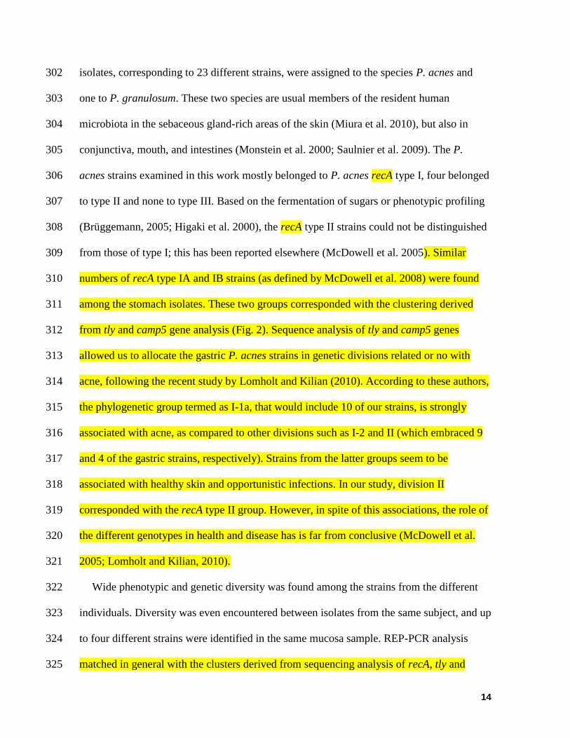
Figure 1.- REP-PCR profiles of the 42 Propionibacterium spp. isolates obtained with
primer BoxA2R. Below, dendogram of similarity of the typing patterns clustered by the
UPGMA method using the Sorensen’s coefficient. Isolates having a coefficient higher than
that of the reproducibility study (85%, indicated by a broken line) were considered to
belong to different strains. Isolates having similar profiles but coming from gastric mucosa
samples of different individuals were also selected for further characterization (indicated by
a code of letters and numbers; see the text). Strains are all P. acnes except for PG 2-4,
which is a P. granulosum. Strains of P. acnes type II are denoted by an asterisk.
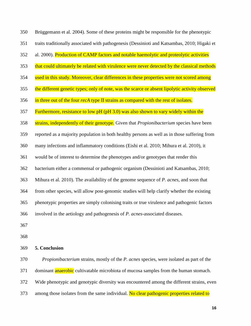
Figure 2.- Neighbour-joining cluster analysis of individual partial DNA sequences of the
recA (A), tly (B) and camp5 (C) genes from 23 gastric Propionibacterium acnes strains.
Published sequences of collection P. acnes strains (NCTC 10390 recA type II reference
strain, NCTC 737 recA type I reference strain isolated from acne, CCUG 50480 from
endocarditis, CCUG 32901 from blood, CCUG 27534 from urinary tract, CCUG 45436
from oral cavity and CCUG 50655 from mandibular gland) were included for comparative
purposes. Strains of P. acnes type II are denoted by an asterisk.
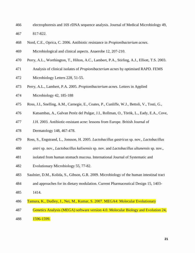
Figure 3.- Survival of Propionibacterium spp. strains at pH 3.0 for 90 minutes. The results
are expressed as the percentage of counts in MRSC as compared to a control dilution of the
same strain maintained under the same conditions at pH 6.5. Strains are all P. acnes except
for PG 2-4, which is a P. granulosum. Strains of P. acnes type II are denoted by an asterisk.
Figure
1 2 3 4 5 6 7 8 9 10 11 12 13 14 15 16 17 18 19 20 21 22 23 24M
0.5
1.0
3.0
2.0
1.5
0.75
5.0
M25 26 27 28 29 30 31 32 33 34 35 36 37 38 39 40 41 42
UPGMA
Sorensen's Coefficient
1 (PA 1-1)243 (PA 1-2)22 (PA 6-2)2317 (PA 3-1)21 (PA 6-1)3 (PA 2-1)2627 (PA 6-5)2930313235 (PA 10-1)34 (PA 7-2)383637(PA 7-3)911(PA 2-2)1213 (PA 2-3)2833 (PA 7-1)41 (PA 8-3)1439 (PA 8-1)1924 (PA 6-3)20 (PA 5-2)57 (PA 1-4)15
16 (PG 2-4)18 (PA 3-2)
6 (PA 1-3*)
8 (PA 1-5*)40 (PA 8-2*)42
25 (PA 6-4*)
0.4 0.5 0.6 0.7 0.8 0.9 1.0
24
Figure

25
Figure 2
CA B
PA8-1
PA8-3
PA7-1
PA6-3
PA6-1
PA5-2
PA3-1
PA2-3
PA2-2
NCTC737
PA7-3
PA7-2
PA6-5
PA3-2
PA2-1
PA1-4
PA1-2
PA10-1
PA1-1
PA6-2
PA1-3*
PA1-5*
PA6-4*
PA8-2*
NCTC10390
0.001
PA6-5
PA10-1
PA1-1
PA1-2
PA6-2
PA3-2
PA2-1
PA1-4
CCUG50480
PA7-2
PA7-3
CCUG32901
PA3-1
PA5-2
PA6-1
PA8-1
PA2-3
PA7-1
PA6-3
PA2-2
PA8-3
CCUG50655
PA1-3*
PA1-5*
PA6-4*
PA8-2*
0.002
PA7-3
PA6-5
PA2-1
PA6-2
PA1-4
CCUG50480
PA1-1
PA3-2
PA10-1
PA1-2
PA7-2
CCUG32901
PA2-2
PA6-1
PA7-1
PA5-2
PA2-3
PA6-3
PA8-1
PA8-3
PA3-1
CCUG45436
PA6-4*
PA8-2*
PA1-3*
CCUG27534
PA1-5*
0.002
Figure
0
10
20
30
40
50
60
70
80
90
100
PA
1-1
PA
1-2
PA
1-3
*
PA
1-4
PA
1-5
*
PA
2-1
PA
2-2
PA
2-3
PG
2-4
PA
3-1
PA
3-2
PA
5-2
PA
6-1
PA
6-2
PA
6-3
PA
6-4
*
PA
6-5
PA
7-1
PA
7-2
PA
7-3
PA
8-1
PA
8-2
*
PA
8-3
PA
10
-1
Strain
Cell s
urv
ival (%
lo
g c
fu m
L-1
)
Figure 3
26
Figure
27
Table 1.- Carbohydrate utilization profiles of Propionibacterium strains from mucosa of the human stomach.
Straina
Carbohydrate GLY ERY DAR RIB ADO GAL GLU FRU MNE SBE INO MAN SOR MDM MDG NAG SAL MAL LAC MEL SAC RAF GEN TUR TAG GNT
PA 1-1 + - + + + + + + + - - - + - - + - - - - - - - - - -
PA 1-2 + - + + + + + + + - - - + - - + - - - - - - - - - -
PA 1-3* + - + + - - + - - - - - - - - - - + - - - - - - - -
PA 1-4 - - + + - - + - - - - - - - - + - - - - - - - - - -
PA 1-5* + - + + - + + - + - - - - - - + - - - - - - - - - -
PA 2-1 + - + + - + + + + - - - + - - + - - - - - - - - - -
PA 2-2 + + + + + + + + + - + - + - - + - - - - - - - - - -
PA 2-3 + - + + + - - - - - - - - - - + - - - - - - - - - -
PG 2-4 + - + + - - + + - - - - - - - + - + - + + - - + - -
PA 3-1 + - + + + + + + + - - - + - - + - - - - - - - - - -
PA 3-2 - - - - - + + + - - - - - - - - - + + - + + - - - -
PA 5-2 + + + + + + + + + - - + + - - + + + - - - - - - - -
PA 6-1 + + + + + + + + + - - + + - - + - - - - - - - - - -
PA 6-2 + - + + + + + + + - - - + - - + - - - - - - - - - -
PA 6-3 + + + + + + + + + - + + + - - + - - + - + + - - + +
PA 6-4* + - + + - - + - - - - - - - - + - - - - - - - - - -
PA 6-5 + + + + - + + + + + - + + + + + - - + + - - + - - +
PA 7-1 + - + + + + + + + - - - + - - + - - - - - - - - - -
PA 7-2 + - + + - + + + + - - - + - - + - - - - - - - - - -
PA 7-3 + - + + - + + + + - - - + - - + - - - - - - - - - -
PA 8-1 - - + + - - - - - - - - - - - - - - - - - - - - - -
PA 8-2* - - - + - - + - - - - - - - - - - - + - - - - - - -
PA 8-3 + + + + + + + + + - - - + - - + - - - - - - - - - -
PA 10-1 + + + + + + + + + - - - + - - + - - - - - - - - - -
Key of the compounds: GLY: glycerol; ERY: erythritol; DAR: D-arabinose; RIB: ribose; ADO: adonitol; GAL: galactose; GLU: glucose; FRU: fructose; MNE: mannose; SBE: sorbose;
INO: inositol; MAN: mannitol; SOR: sorbitol; MDM: -methyl-D-mannoside; MDG: -methyl-D-glucoside; NAG: N-acethyl-glucosamine; SAL: salicin; MAL: maltose; LAC: lactose;
MEL: melibiose; SAC: sucrose; RAF: raffinose; GEN: gentibiose; TUR: turanose; TAG: tagatose; GNT: gluconate.
None of the strains fermented L-arabinose, D-xylose, L-xylose, -methyl-D-xyloside, rhamnose, dulcitol, amygdalin, arbutyn, esculin, cellobiose, trehalose, inulin, melezitose, stach,
glycogen, xylitol, D-lyxose, D-fucose, L-fucose, D-arabitol, L-arabitol, 2-keto-gluconate, 5-keto-gluconate. aStrains are all Propionibacterium acnes except for PG 2-4, which is a Propionibacterium granulosum; asterisks mark P. acnes recA type II strains, all others were shown to belong to type
I; grey shading indicates sugars used to distinguish between type I and type II strains.
Table

28
Table 2.- Enzymatic activities as measured with the API ZYM system of Propionibacterium strains
from mucosa of the human stomach.
Straina
Enzymatic activityb
Alk
ali
n
ph
osp
ha
tase
Est
era
se (
C4
)
Est
era
se l
ipa
se (
C8
)
Aci
d p
ho
sph
ata
se
Na
ph
tol-
AS
-BI-
ph
osp
ho
hy
dro
lase
N-a
ceth
yl-
-
glu
cosa
min
ida
se
-g
luco
sid
ase
-m
an
no
sid
ase
PA 1-1 0 0 0 15 10 10 0 0
PA 1-2 0 0 0 20 20 0 0 0
PA 1-3* 0 0 0 0 0 5 0 0
PA 1-4 0 0 0 10 10 10 0 0
PA 1-5* 5 0 0 35 30 2.5 0 0
PA 2-1 0 0 0 15 15 10 0 0
PA 2-2 2.5 0 0 30 30 10 15 10
PA 2-3 0 0 0 15 2.5 15 10 15
PG 2-4b 35 10 2.5 40 35 0 2.5 0
PA 3-1 2.5 2.5 2.5 30 10 0 0 0
PA 3-2 5 2.5 0 20 0 0 0 0
PA 5-2 0 0 0 20 15 2.5 0 0
PA 6-1 0 0 0 20 15 2.5 0 0
PA 6-2 5 0 2.5 40 15 10 0 0
PA 6-3 0 0 0 15 5 5 0 0
PA 6-4* 10 0 0 40 15 10 20 0
PA 6-5 0 0 0 10 5 5 0 0
PA 7-1 5 0 2.5 40 30 10 0 0
PA 7-2 0 0 0 5 5 2.5 0 0
PA 7-3 2.5 0 0 35 30 10 0 0
PA 8-1 5 0 0 40 40 20 10 15
PA 8-2* 0 0 0 2.5 5 2.5 0 0
PA 8-3 15 0 0 40 40 15 15 15
PA 10-1 5 0 0 35 10 10 0 0
Leucine arylamidase, valine arylamidase, cysteine arylamidase, lipase (C14), trypsin, α-quimiotrypsin, -galactosidase, -
galatosidase, -glucosidase, β-glucuronidase, and -fucosidase activities were never detected. aStrains are all P. acnes except for PG 2-4, which is a P. granulosum; asterisks mark P. acnes recA type II strains, all others
were shown to belong to type I. bUnits of activity are expressed as nmol of substrate hydrolyzed under the assay conditions.
Table

29
Table 3.- Antibiotic resistance susceptibility profiles of representative strains of Propionibacterium strains from mucosa of the human
stomach. MIC (g mL-1
)
Key of antibiotics: AM: ampicillim; PC: penicillin G; GM: gentamicin; KM: kanamycin; SM: streptomycin; NM: neomycin; TC: tetracycline; EM: erythromycin;
CL: clindamycin; CM: chloramphenicol; VA: vancomycin; VI: virginiamycin; LZ: linezolid; TM: trimethoprim; CI: ciprofloxacin; RI: rifampicin. aStrains are all P. acnes except for PG 2-4, which is a P. granulosum; asterisk marks P. acnes recA type II strains, all others were shown to belong to type I.
Straina Antibiotic
AM PC GM KM SM NM TC EM CL CM VA VI LZ TM CI RI
PA 1-1 0.03 0.03 1 4 1 0.5 0.25 0.016 0.06 1 0.5 0.12 0.5 2 1 0.12
PA 1-3* 0.03 0.03 2 8 16 0.5 0.25 0.12 1 4 2 0.5 1 1 0.5 0.12
PA 2-1 0.06 0.03 2 8 4 1 0.5 0.03 0.06 0.5 0.5 0.12 0.5 1 0.5 0.12
PA 2-2 0.12 0.06 2 16 8 2 0.5 0.06 0.12 1 0.5 0.06 1 8 2 0.12
PG 2-4 0.03 0.12 1 8 4 0.5 0.5 0.016 0.03 0.5 0.5 0.06 0.5 32 0.5 0.12
PA 3-1 0.06 0.06 2 2 2 0.5 0.5 0.03 0.06 1 0.5 0.06 0.5 2 1 0.12
PA 3-2 0.12 2 0.5 2 2 0.5 0.25 0.12 0.25 4 1 0.25 1 1 0.25 0.12
PA 5-2 0.03 0.03 2 4 1 0.5 1 0.016 0.06 1 0.5 0.06 1 4 1 0.12
PA 6-1 0.03 0.03 2 4 1 1 0.5 0.03 0.06 1 0.5 0.06 0.5 2 1 0.12
PA 6-3 0.25 0.12 4 16 8 2 0.5 0.06 0.12 1 1 0.12 0.5 4 1 0.12
PA 7-1 0.03 0.03 2 4 0.5 2 0.5 0.016 0.03 0.25 0.5 0.06 0.12 0.25 0.5 0.12
PA 7-3 0.03 0.03 2 8 4 1 0.5 0.25 0.06 1 0.5 0.06 0.5 2 2 0.12
PA 8-2* 0.03 0.03 2 8 0.5 0.5 0.12 0.016 0.03 0.12 0.25 0.06 0.25 2 1 0.12
PA 10-1 0.03 0.03 2 8 2 1 0.25 0.016 0.06 0.5 0.5 0.06 0.25 1 1 0.12
Table
30
Table 4.- Phenotypic, virulence-associated activities of Propionibacterium strains
isolated from the mucosa of the human stomach.
Straina
Activityb
Haemolytic DNase Lipolytic Protelytic
PA 1-1 - - ++ +
PA 1-2 - - ++ (+)
PA 1-3* - - Ng -
PA 1-4 - - ++ -
PA 1-5* - - - -
PA 2-1 - - + -
PA 2-2 - - + +
PA 2-3 - - + (+)
PG 2-4 - - + -
PA 3-1 - - + -
PA 3-2 - - ++ -
PA 5-2 - - ++ -
PA 6-1 - - ++ -
PA 6-2 - - ++ -
PA 6-3 (+) - ++ +
PA 6-4* - - Ng -
PA 6-5 - - ++ -
PA 7-1 - - ++ (+)
PA 7-2 - - ++ -
PA 7-3 - - ++ -
PA 8-1 (+) - ++ -
PA 8-2* - - ++ -
PA 8-3 - - + -
PA 10-1 - - ++ (+)
aStrains are all P. acnes except for PG 2-4, which is a P. granulosum; asterisks mark P. acnes recA type
II strains, all others were shown to belong to type I. bNumber of crosses is related to the level of activity; in parenthesis, weak activities.
Ng, no growth observed.
Table