insect biochemistry and molecular biology - csic upf...
TRANSCRIPT
lable at ScienceDirect
Insect Biochemistry and Molecular Biology 41 (2011) 101e108
Contents lists avai
Insect Biochemistry and Molecular Biology
journal homepage: www.elsevier .com/locate/ ibmb
Citrus, a key insect eggshell protein
Paula Irles, Maria-Dolors Piulachs*
Institut de Biologia Evolutiva (CSIC-UPF), Passeig Marítim de la Barceloneta, 37, 08003 Barcelona, Spain
a r t i c l e i n f o
Article history:Received 22 September 2010Received in revised form5 November 2010Accepted 6 November 2010
Keywords:Blattella germanicaChorionInsect eggCockroachDrosophila
* Corresponding author. Tel.: þ34 93239648; fax: þE-mail addresses: [email protected] (P. Irles), md
(M.-D. Piulachs).
0965-1748/$ e see front matter � 2010 Elsevier Ltd.doi:10.1016/j.ibmb.2010.11.001
a b s t r a c t
Molecular aspects of chorion synthesis in insects have been studied deeply in species with meroisticovaries. Information available in insects with panoistic ovaries is principally structural whereas molec-ular information in these species is scarce. This paper seeks to balance the above situation by describinga novel chorion gene, Citrus, from the cockroach Blattella germanica, a phylogenetically basal hemi-metabolan insect with reproduction regulated by juvenile hormone and with panoistic ovaries. Duringprevious work we discovered a series of novel genes which were specifically expressed during chorionformation in B. germanica. One of them, herein named Citrus, was peculiar due to its high copy numberand its very transient expression. In the present paper we characterize Citrus in terms of structure andfunction. The most prominent structural feature is that the protein contains a motif which is repeated 33times encompassing almost all the sequence. By using RNAi techniques we have demonstrated thatCitrus is a key player in the building of the endochorion of B. germanica eggs.
� 2010 Elsevier Ltd. All rights reserved.
1. Introduction
Chorion in insect eggs plays the essential function of protectingthe embryo from external agents during development, whileallowing gas exchange for respiration and the entry of sperm forfertilization (Woods et al., 2005; Zrubek and Woods, 2006). Ininsects, chorion synthesis is usually under the control of the 20-hydroxyecdysone synthesized by the follicular cells that surroundthe maturing oocyte (Belles et al., 1993). The follicular cells secretethe chorion components and are responsible for the final chorionstructures which may be remarkably complex, as in Lepidopterawhere chorion synthesis involves more than one hundred proteins(Margaritis, 1985a; Regier and Kafatos, 1985; Trougakos andMargaritis, 1998).
Insect chorion is structured into a number of layers which areconserved in their composition as well as in their dispositionthrough diverse insect groups. In the cockroach Blattella germanica(Belles et al., 1993), as in the fly Drosophila melanogaster(Margaritis, 1985b), the vitelline membrane, the inner chorionlayer, the endochorion and the exochorion are well defined layers.The vitelline membrane is the first layer secreted by the follicularcells. It is the most internal and interacts with the oocytemembrane. Covering the vitelline membrane there is the inner
All rights reserved.
chorion layer, which is very faint but clearly distinguishable fromthe other chorion layers. The next to be deposited is the endo-chorion, which is the most complex and formed by an inner-endochorion, a columnar and an outer-endochorion layer. The lastlayer of the eggshell is the exochorion that gives the egg its finalshape. The final step of choriogenesis is completed after oviposi-tion, and consists in the hardening of the eggshell which occursthrough a process of protein crosslinking.
Insect ovaries are classified into panoistic andmeroistic types. Inpanoistic ovaries all oogonia are eventually transformed intooocytes; it is the most common type in phylogenetically basalinsects, such as B. germanica. In meroistic ovaries, oogonia canderive into both oocytes and nurse cells; it is the most common inParaneoptera and Endopterygota, and it has apparently evolvedfrom an ancestral panoistic type (Büning, 1994). Although basicchorion information is available from a varied group of insectsrepresenting both ovary types (Beams and Kessel, 1969; Furneauxet al., 1969; Trougakos and Margaritis, 2002; Gaino et al., 2008),at themolecular level, these studies have greatly focused on Dipteraand Lepidoptera, both having meroistic ovaries (Margaritis, 1985b;Spoerel et al., 1986; Cavaliere et al., 2008; Lecanidou andPapantonis, 2010).
In order to find regular patterns in the data available and tounderstand the molecular basis of the evolution of insect ovaries,molecular information on chorion synthesis in species with pano-istic ovaries should be gathered. In this sense, and usinga Suppression Substractive Hybridization (SSH) library approach(Irles et al., 2009a), we obtained a series of genes that are expressed

P. Irles, M.-D. Piulachs / Insect Biochemistry and Molecular Biology 41 (2011) 101e108102
differentially during the post-vitellogenic period of B. germanica, anhemimetabolan insect with panoistic ovaries, which encapsulatesthe eggs into an egg-case or ootheca, and whose reproduction ismainly regulated by juvenile hormone. From the SSH library, twogenes were especially prominent, Brownie (Bg 30009) and Citrus(Bg30001). They were outstanding due to their remarkable abun-dance, their characteristic pattern of expression and because theyhad no homologues in insect gene databases. The function ofBrownie has been recently described (Irles et al., 2009b) asencoding a protein that plays a key role in the formation of thesponge-like body, a complex structure of the eggshell thatcombines the aeropyle and themicropyle, andwhich is reminiscentof theD. melanogaster egg horns, that permit gas exchange betweenthe egg and the exterior. The function of the other gene, Citrus, isreported in the present paper.
2. Material and methods
2.1. Animal sampling
Freshly emerged adult females of B. germanica were obtainedfrom a colony reared in the dark at 29�1 �C and 60e70% r.h. Thelength of the basal oocyte was used to stage the ovaries from 0- to7-day-old, whereas the stages of choriogenesis (early chorion: EC,mid chorion: MC, and late chorion: LC) were determined accordingto the morphology of the anterior pole of the basal oocyte (Irleset al., 2009b). All dissections and tissue sampling were carriedout on carbon dioxide-anaesthetized specimens.
2.2. Cloning of Citrus cDNA
A fragment of 2693 bp [accession number is FM253359.1] waspreviously obtained from a library of ESTs specific of post-vitello-genic ovaries (Irles et al., 2009b). To complete this sequence, 50-rapid amplification of cDNA ends (RACE) was applied to RNAextracted from ovaries of 7-day-old females at the end of chorionformation, using FirstChoice� RLM-RACE (Ambion, Huntingdon,Cambridgeshire, UK) according to the manufacturer’s instructions.The amplified fragment was analyzed by agarose gel electropho-resis, cloned into the pSTBlue-1 vector (Novagen, Madison, WI,USA) and sequenced. The sequences of all primers used are avail-able on request.
2.3. RNA extraction and retrotranscription to cDNA
For Northern Blot analysis, total RNA was extracted from 7-day-old adult female ovaries at the end of chorion formation. Formonitoring mRNA expression by qRT-PCR, total RNA was isolatedfrom pools of four to six ovary pairs obtained in chosen ages andstages of the first gonadotrophic cycle. RNA extractions were per-formed using the Gen Elute Mammalian Total RNA kit (Sigma,Madrid, Spain). An amount of 400 ng from each RNA extractionwasDNAse treated (Promega, Madison, WI, USA) and reverse tran-scribed with Superscript II reverse transcriptase (Invitrogen,Carlsbad CA, USA) and random hexamers (Promega). RNA quantityand quality was estimated by spectrophotometric absorption at260 nm in a Nanodrop Spectrophotometer ND-1000� (NanoDropTechnologies, Wilmington, DE, USA).
2.4. Northern blot analysis
Total RNA (10 mg) was subjected to electrophoresis in 1% agarosegels containing formaldehyde, following the methodology previ-ously described (Irles et al., 2009b). As a probe, a Citrus fragment of213 bp was amplified by PCR and labelled with fluorescein using
the Gene Images Random Prime-Labelling Module (GE Healthcare,Madrid, Spain).
2.5. Expression studies
Quantitative RT-PCR (qRT-PCR) was used to study Citrusexpression during the first gonadotrophic cycle and to assess theeffect of RNAi over mRNA levels. PCR primers used in qRT-PCRexpression studies were designed using the Primer Express 2.0software (Applied Biosystems, Foster City, CA, USA). The BgActin-5cgene of B. germanica (accession number is AJ862721) was used asa reference. qRT-PCR reactions were performed and analyzed aspreviously reported (Irles et al., 2009a). Statistical analysis of geneexpression values was carried out using the REST 2008 program(Relative Expression Software Tool V 2.0.7; Corbett Research) (Pfafflet al., 2002). This program makes no assumptions about thedistributions, evaluating the significance of the derived results byPair-Wise Fixed Reallocation Randomization Test tool in REST (Pfafflet al., 2002).
2.6. RNAi experiments
A dsRNA encompassing a 213 bp region starting at nucleotide1650 of Citrus sequence (Fig. S1) labelled as dsCitrus-1 wasprepared, then amplified by PCR and cloned into pSTBlue-1 vector.A non-coding sequence from the pSTBlue-1 vector (dsMock) wasused as control dsRNA. The dsRNAs were prepared as previouslydescribed (Ciudad et al., 2006). Five-day-old adult females wereinjected into the abdomen with 1 mg of dsCitrus-1 in a volume of1 ml. Control specimens were injected with the same volume anddose of dsMock. An alternative dsRNA (dsCitrus-2), encompassinga 309 bp region starting at nucleotide 211 of Citrus sequence(Fig. S1), was prepared following the same methodology, and usedto asses the specificity of the effects obtained with dsCitrus-1.
2.7. Scanning electron microscopy (SEM)
Selected eggs from dsCitrus-1- and dsMock-treated femaleswere fixed in 2.5% glutaraldehyde in cacodylate buffer 0.2 M for atleast 2 h. After rinsing twicewith the same buffer, the samples werethen treated with 1% osmium tetroxide (Ted Pella, Inc, Redding,USA) at 4 �C for 1 h. The tissues were dehydrated with increasingconcentrations of alcohol at 15 min intervals. Finally, the sampleswere subjected to critical-point drying in order to complete thedehydration process. The samples were attached to stubs withdouble-stick tape and coated with gold-palladium in a sputter-coating apparatus and observed with a Hitachi S-3500N scanningelectron microscope at 5 kV (Hitachi High-Technologies Corpora-tion, Tokyo, Japan). After fixation, oocytes were gently ripped witha microforceps in order to expose the layers.
2.8. Transmission electron microscopy (TEM)
Ovarian follicles at the end of choriogenetic period, from bothdsCitrus-1 and dsMock-treated females, were fixed with 2.5%glutaraldehyde and 2% paraformaldehyde in 100 mM phosphatebuffer (PB, pH 7.4) for 2 h and rinsed 4 times with 100 mM PB.Samples were then postfixed in 1% osmium tetroxide (TAAB, Berks,UK) containing 0.8% of potassium hexacyanoferrate (III) (Sigma) for2 h andwashed with 100 mM PB. All these steps were made at 4 �C.Samples were dehydrated through a graded acetone series, infil-trated in Epon resin and polymerized for 48 h at 60 �C. Ultrathinsections were mounted in copper grids (200 mesh), contrastedwith uranyl acetate and lead citrate solutions, and observed ina transmission electron microscope Jeol JEM-1400 (Jeol LTD, Tokyo,

P. Irles, M.-D. Piulachs / Insect Biochemistry and Molecular Biology 41 (2011) 101e108 103
Japan) equipped with a Gatan Ultrascan ES1000 CCD camera(Gatan, UK, Oxford, UK) at 80 kV.
3. Results
3.1. Citrus, a novel eggshell protein
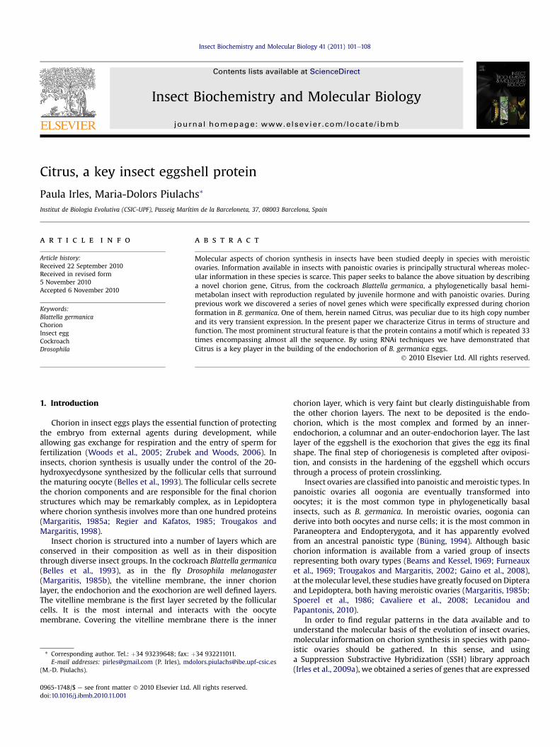
A previously described SSH library of ovarian cDNAs specific tothe post-vitellogenesis period gave the novel sequence Bg30001(Accession number: FM253359) (Irles et al., 2009a). The assembleof 19 ESTs from this library together with 5 sequences obtainedfrom a general ovarian cDNA library also generated in our labora-tory, resulted in a consensus sequence of 2693 bp that wascomplete towards the 30UTR but lacked the 50 end.
50
AD
C
H
KL
MN
QR
T
WA B
C
6 kb
4 kb
Fig. 1. Structural features of Citrus. (A) Northern blot analysis of Citrus mRNA from an extraAmino acid composition of Citrus protein, the percentage of the most abundant is indicatemanually; numbers indicate the position of the repeat into the sequence.
The full sequence (Accession number: FN823078) was obtainedby 50 RACE, which gave a total of 4099 bp, comprising the capsiteand the polyAþ tail (Fig. S1). The size of the mRNAwas assessed byNorthern blot analysis using total RNA from choriogenic ovaries astemplate and a cDNA fragment of 213 bp as probe, resulting ina specific signal around 4000 bp as expected (Fig. 1A).
The mRNA capsite sequence is TCAGT, which matches the mostrepresentative pentamer among arthropods (Cherbas and Cherbas,1993). In frame with the first methionine, which is located 176bases downstream from the capsite, there is a stop codon thatconfirms the ORF (Fig. S1). Additionally, the Kozak sequenceflanking the first ATG is fully conserved. The end of the ORF isdetermined by a stop codon located 3667 nucleotides downstreamfrom the first methionine; this stop codon is followed by 432 bases
Amino acid composition (number)
(13.2%
(11.6%)
(11.7%)
(10.5%)
100 150
EF
G
I
P
S
V
Y
ct of ovaries at the end of chorion formation, showing a single band of c.a. 4000 bp (B)d. (C) Alignment of the 33 repeats from Citrus sequence with ClustalX and modified

Fig. 2. Expression studies of Citrus. (A) Expression of Citrus mRNA in ovaries of adult females during the first gonadotrophic cycle by qRT-PCR; the period of chorion formation wasdivided in three stages, early (EC), mid (MC) and late (LC) choriogenesis; also included are oocyte tissue during oviposition (Op) and embryos of less than 24 h (E0); data representcopies of mRNA per copy of BgActin-5c. (B) mRNA levels of Citrus in LC ovaries from females treated with dsMock or dsCitrus-1 at 5-day-old; qRT-PCR was normalized againstBgActin-5c, data represent normalized values with respect to dsMock expression levels, indicated as the mean� SD (n¼ 3) according to REST software.
P. Irles, M.-D. Piulachs / Insect Biochemistry and Molecular Biology 41 (2011) 101e108104
that constitute the 30 UTR region with the polyadenylation elementplaced 23 bases upstream the polyAþ tail. The resulting sequence(Figs. S1 and S2) encodes a protein of 1163 amino acids witha predicted molecular mass of 130.93 kDa and an isoelectric pointof 4.74. As predicted by SignalP 3.0, this protein would havea peptide signal with a cleavage site between positions 24 and 25.The protein is rich in proline, which represents the 13.2% of totalamino acids, followed by glycine and tyrosine with proportions of11.7 and 11.6%, respectively (Fig. 1B). Of the total residues, 149 havea negative charge (13% of total), where the most abundant is glu-tamic acid (10.5%), whereas, remarkably, the protein lackscysteines.
The protein is peculiar in its structure, containing 33 repeats ofthe same motif. These repeats are 30e40 amino acids in length,contiguous and encompassing practically the entire sequence fromthe amino acid 51 to amino acid 1163 (Fig. 1C). The first 4 repeats inthe N-terminal region are those less conserved. In general, allrepeats are rich in glutamic acid at their 50 end, whereas to the 30
end, they are rich in GYP repetitions. Searching in the databasespossible homologous proteins or motifs we did not find any onesimilar among the different organisms, even when fragmentingsequences into two or more parts. Given the acidic character of theamino acids present at 50 end in all repeats, with glutamic acidmostrepresented, we called the protein “Citrus”, which suggest theacidity of the citrus fruits.
Table 1Measurement of endochorion and exochorion layers in eggs from dsMock- anddsCitrus-1-treated females. Eggs were fixed with glutaraldehyde, and gently rippedto expose the chorion layers in section for SEM. For each egg (n) measures weretaken in 15 different representative points along the chorion layers. Data areexpressed as mean� SD. Asterisks indicated differences statistically significantaccording to the ManneWhitney U test, * (p¼ 0.01), ** (p< 0.0001).
Treatment n Innerendochorionwidth (mm)
Columnsheight(mm)
Columnswidth(mm)
Outer endochorionþ exochorionwidth (mm)
dsMock 10 0.13� 0.07 0.54� 0.32 0.41� 0.11 0.35� 0.08dsCitrus 10 0.14� 0.05 0.81� 0.45* 0.41� 0.08 0.16� 0.05**
3.2. RNAi of Citrus
The maximum expression of Citrus mRNA occurs in ovarianfollicles at MC and LC stages (Fig. 2A), decreasing abruptly at theonset of oviposition, thus suggesting that it could be involved inchorion formation. Furthermore, Citrus was not detected in youngembryos (less than 24 h).
To unveil the function of Citrus, we followed an RNAi approach.Therefore, dsRNA for Citrus (dsCitrus-1) (Fig. S1) was injected ata dose of 1 mg in 5-day-old adult mated females of B. germanica(n¼ 60). As control dsRNA, we used an alien non-coding sequence(dsMock) injected at the same dose (n¼ 29). Ovaries weredissected two days later, at the MC and LC stages. Citrus mRNAexpression in the ovaries of dsCitrus-1-treated specimens was
Fig. 3. Chorion layers of B. germanica oocytes observed at SEM. After fixation oocytes were g(C, D, G, H) dsCitrus-1 groups; ex: exochorion, VM: vitelline membrane, oe: outer-endochomaterial invading the columnar layer; arrows in (E) and (G) indicate the marks left by the fol(A, C, F, H) 2.5 mm, (B, D) 1 mm, (E, G) 10 mm.
reduced 30-fold when compared with the expression in dsMock-treated specimens (Fig. 2B). Furthermore, females treated withdsCitrus-1 did not form the ootheca, the eggs appeared to beextremely fragile, as they easily collapsed when being oviposited.Although the eggs were unviable, these females were mated, asassessed by the presence of spermatozoa in the spermatheca. Werepeated the experiments using an alternative dsRNA for Citrus(dsCitrus-2), encompassing a different region close to the 50 end(Fig. S1). A new batch of females was treated with dsCitrus-2(n¼ 22) and dsMock (n¼ 20), using the same dose and approach.The results were virtually identical to those obtained with dsCi-trus-1: fragile eggs and no ootheca formation. Observed at thelight microscope, the oocytes from females treated with dsCitrus-1 or -2 did not show morphological abnormalities. Moreover, noapparent differences were observed between the follicularepithelium of basal follicles from dsCitrus or dsMock groups, andthe sponge-like body was well formed in both groups (data notshown). However, in spite of this apparent morphologicalnormality, oviposition was impaired in females treated withdsCitrus-1 or -2. We therefore hypothesized that the fragility ofthe eggs of dsCitrus-treated specimens was due to deficiencies inthe fine structure of the chorion.
3.3. Citrus and chorion formation
A new treatment with dsCitrus-1 (n¼ 10) and dsMock (n¼ 10)was carried out following the same methodology, and eggs werecollected as they descended through the oviduct, and they were
ently ripped in order to expose the layers in section. (A, B, E, F) eggs from dsMock andrion, c: columnar layer of endochorion, i.e.: inner-endochorion; the asterisk shows thelicular cells, which are more raised in dsMock than in dsCitrus egg groups. Scale bars¼

P. Irles, M.-D. Piulachs / Insect Biochemistry and Molecular Biology 41 (2011) 101e108 105

Fig. 4. Ultrathin sections of B. germanica oocytes at the end of chorion synthesis. In dsMock oocytes (A), all layers are clearly distributed. The vitellin membrane (VM) form a denseribbon around the oocyte, the endochorion has a thick floor (inner-endochorion: ie) and roof (outer-endochorion: oe), with columns connecting both. The exochorion (ex) spreadsover the endochorion. In dsCitrus oocytes (B), the endochorion lost robustness, the inner- and outer-endochorion (arrowhead) did not form a uniform layer over the endochorion.Oo: oocyte; FC: follicular cell; c: columnar layer; B: bacteriocyte. Scale bars: 2 mm.
P. Irles, M.-D. Piulachs / Insect Biochemistry and Molecular Biology 41 (2011) 101e108106
processed to be examined by SEM. Eggs from dsCitrus-1-treatedfemales were more fragile and most of them collapsed during theprocessing for SEM although the eggs from both, dsCitrus-1 anddsMock groups, showed all the chorion layers. However in thedsCitrus-1 group, the external layers (outer-endochorionþ exo-chorion) were thinner when compared with dsMock group(Table 1) (Fig. 3AeD), and the columns of the endochorion tendedto be higher with respect to dsMock group (Fig. 3B, D) (Table 1).Furthermore, the imprinting left by the follicular cells in theexternal surface of the egg was much less marked in the dsCitrus-1group than in the dsMock eggs (Fig. 3E, G). At high magnification,the eggs surface from dsCitrus-1-treated specimens showeda discontinuous deposition of material with a reticular distribution,which contrasts with the surface of the eggs from the dsMockgroup where it forms a smooth and continuous surface (Fig. 3F, H).
Also outstanding is the presence of material included within thecolumnar layer, suggesting that the inner- and outer-endochorionlayers lost impermeability, which facilitated the leaking of materialfrom the oocyte to the exterior (Fig. 3D).
Ultrathin sections of ovarian follicles from dsCitrus-1 anddsMock groups, observed by TEM confirmed that the greatestdifference between the chorion structures of both groups is foundin the endochorion, where the inner- and outer-endochorion layersare extremely thin (Fig. 4). Moreover, the columns of the endo-chorion layer in the dsCitrus-1-treated females were fewer innumber and lost their regularity in distribution. Apparently, thesedeficiencies in the endochorion structure result in alterations in theexochorion, which show small plaques irregularly deposited overthe outer-endochorion (arrowhead, Fig. 4B) instead of forminga continuous surface.
These defects in the deposition of the external chorion layersmade the eggshell permeable, therefore materials which probably
came from the vitelline membrane were able to invade thecolumnar layer (asterisk, Fig. 4), and even reached the surfacethrough the sponge-like body (Fig. 5).
4. Discussion
The transient expression of high levels of Citrus mRNA inovarian follicles at MC-LC stages, together with its localization inthe follicular epithelium were the first cues to suggest that Citruswas involved in chorion formation (Irles et al., 2009a). The abovefeatures are in fact a characteristic of chorion genes in insect eggs(Orr-Weaver, 1991; Claycomb et al., 2004; Tower, 2004), as it waswell described in D. melanogaster, where the expression of choriongenes in the follicular epithelium is tightly regulated both tempo-rally and spatially, by hormones acting on a complex gene network(Cavaliere et al., 2008).
Citrus protein has a signal peptide and is rich in glycine, tyrosineand proline (35%), as occurs in other insect chorion proteins (Regierand Kafatos, 1985), where the abundance of these amino acids isassociated with structural functions. Insect proteins with thesecharacteristics have been described in the ovary of the locustSchistocerca gregaria (Schoofs et al., 1998) and in chorion proteins ofthe gypsy moth Lymantria dispar (Leclerc and Regier, 1994), as wellas in our own model, the cockroach B. germanica (Irles et al.,2009b).
The most particular feature of Citrus is that it is formed bya motif which is repeated 33 times, encompassing practically theentire protein sequence. From an evolutionary point of view, theserepetitions must have been generated by successive intragenicduplications and homogenizations. However, no data are availableon the possible functional sense of this peculiar feature. There area number of antecedents with respect to repetitive motifs in insect

Fig. 5. Chorion of B. germanica eggs, showing the loss of impermeability. SEMmicrographs of the Sponge-like body from dsMock (A) and dsCitrus-1 (B); the materialleaking out of the egg through the sponge-like body is clearly seen in the latter. Scalebars¼ 50 mm.
P. Irles, M.-D. Piulachs / Insect Biochemistry and Molecular Biology 41 (2011) 101e108 107
chorion proteins. For example, three-amino acids repeats havebeen described in the egg proteins of the kissing bug Rhodniusprolixus (Bouts et al., 2007) and tandem repeats of eight-aminoacids appear in the femcoat protein of D. melanogaster (Kim et al.,2002), and in eggshell proteins Ccs36 and Ccs38 of the Mediter-ranean fruit fly Ceratitis capitata (Aggeli et al., 1991). Nevertheless,the structure of Citrus with such a high number of longer repeats isunique among insect chorion proteins.
The absence of cysteines and the high concentration of tyrosinesof Citrus protein suggest that the cross-linking could occur throughdi- or tri-tyrosine bridges. This kind of cross-linking has beendescribed in the endochorion of higher Diptera (Petri et al., 1976),where an eggshell peroxidase is involved in the bounding. In othergroups of insects, the cross-linking of chorion proteins is mainlymediated by di-sulphide bridges, as occurs in some Lepidoptera,Coleoptera, Orthoptera and Odonata (Regier and Kafatos, 1985).
The absence of cysteines and the high concentration of tyrosinesare also features of the Brownie eggshell protein recently describedin B. germanica (Irles et al., 2009b). Both Brownie and Citrus aregenes with no homologues in insect databases, however, thefunctional difference between both Citrus and Brownie is clear-cut.While Brownie is expressed later in choriogenesis, being localizedin the cells of the anterior part of ovarian follicle, and its function is
restricted to the formation of the sponge-like body, Citrus isexpressed in all follicular cells in earlier stages of chorion forma-tion, and it is involved in the formation of the exochorion. Lookingboth expression profiles, it seems that the genes related with thesponge-like body formation are expressed before those deter-mining the exochorion layer. In any case, the absence of any one ofthese two proteins results in unviable eggs, in the case of Citrusbecause the eggs collapse, and in the case of Brownie because theembryos cannot develop due to gas exchange problems.
The absence of homologue sequences in other insect species isnot surprising given that Citrus and Brownie belong to a cockroachwith panoistic ovaries, and most of the sequences related withchoriogenesis available in databases are from insects withmeroisticovaries, like dipterans and lepidopterans. The discovery of newgenes suggests that the molecular mechanisms underlying cho-riogenesis in panoistic ovaries might be quite different from thoseof meroistic ones.
Acknowledgements
This work was supported by the Ministry of Science and Innova-tion, Spain (project BFU2008-00484 to MDP). PI is recipient of a pre-doctoral research grant (I3P) from the “Consejo Superior deInvestigaciones Cientificas” (CSIC). We are grateful to Prof. XavierBelles for his helpful scientific discussions and critical comments onthe manuscript. We also thank J. M. Fortuño (Centre Mediterranid’Investigacions Marines i Ambientals, CSIC) for help with SEMstudies and A. D. Sanchez-Chardi (Servei de Microscòpia UniversitatAutònomadeBarcelonaCampusde laUAB) forhelpwithTEMstudies.
Appendix. Supplementary data
Supplementary data associated with this article can be found inthe online version, at doi:10.1016/j.ibmb.2010.11.001.
References
Aggeli, A., Hamodrakas, S.J., Komitopoulou, K., Konsolaki, M., 1991. Tandemlyrepeating peptide motifs and their secondary structure in Ceratitis capitataeggshell proteins Ccs36 and Ccs38. Int. J. Biol. Macromol. 13, 307e315.
Beams, H.W., Kessel, R.G., 1969. Synthesis and deposition of oocyte envelopes(vitelline membrane, chorion) and the uptake of yolk in the dragonfly (Odo-nata:Aeschnidae). J. Cell Sci. 4, 241e264.
Belles, X., Cassier, P., Cerda, X., Pascual, N., Andre, M., Rosso, Y., Piulachs, M.D., 1993.Induction of choriogenesis by 20-hydroxyecdysone in the German cockroach.Tissue Cell 25, 195e204.
Bouts, D.M., Melo, A.C., Andrade, A.L., Silva-Neto, M.A., Paiva-Silva Gde, O.,Sorgine, M.H., da Cunha Gomes, L.S., Coelho, H.S., Furtado, A.P., Aguiar, E.C., deMedeiros, L.N., Kurtenbach, E., Rozental, S., Cunha, E.S.N.L., de Souza, W.,Masuda, H., 2007. Biochemical properties of the major proteins from Rhodniusprolixus eggshell. Insect Biochem. Mol. Biol. 37, 1207e1221.
Büning, J., 1994. The Insect Ovary. Ultrastructure, Previtellogenic Growth andEvolution. Chapman & Hall, London.
Cavaliere, V., Bernardi, F., Romani, P., Duchi, S., Gargiulo, G., 2008. Building up theDrosophila eggshell: first of all the eggshell genes must be transcribed. Dev.Dyn. 237, 2061e2072.
Ciudad, L., Piulachs, M.D., Belles, X., 2006. Systemic RNAi of the cockroach vitello-genin receptor results in a phenotype similar to that of the Drosophila yolklessmutant. FEBS J. 273, 325e335.
Claycomb, J.M., Benasutti, M., Bosco, G., Fenger, D.D., Orr-Weaver, T.L., 2004. Geneamplification as a developmental strategy: isolation of two developmentalamplicons in Drosophila. Dev. Cell 6, 145e155.
Cherbas, L., Cherbas, P., 1993. The arthropod initiator: the capsite consensus playsan important role in transcription. Insect Biochem. Mol. Biol. 23, 81e90.
Furneaux, P.J., James, C.R., Potter, S., 1969. The egg shell of the house cricket (Achetadomesticus): an electronmicroscope study. J. Cell Sci. 5, 227e249.
Gaino, E., Piersanti, S., Rebora, M., 2008. Egg envelope synthesis and chorionmodification after oviposition in the dragonfly Libellula depressa (Odonata,Libellulidae). Tissue Cell 40, 317e324.
Irles, P., Belles, X., Piulachs, M.D., 2009a. Identifying genes related to choriogenesisin insect panoistic ovaries by suppression subtractive hybridization. BMCGenom. 10, 206.

P. Irles, M.-D. Piulachs / Insect Biochemistry and Molecular Biology 41 (2011) 101e108108
Irles, P., Belles, X., Piulachs, M.D., 2009b. Brownie, a gene involved in buildingcomplex respiratory devices in insect eggshells. PLoS One 4, e8353.
Kim, C., Han, K., Kim, J., Yi, J.S., Yim, J., Kim, Y.J., Kim-Ha, J., 2002. Femcoat, a noveleggshell protein in Drosophila: functional analysis by double stranded RNAinterference. Mech. Dev. 110, 61e70.
Lecanidou, R., Papantonis, A., 2010. Silkmoth chorion gene regulation revisited:promoter architecture as a key player. Insect Mol. Biol. 19, 141e151.
Leclerc, R.F., Regier, J.C., 1994. Evolution of chorion gene families in lepidoptera:characterization of 15 cDNAs from the gypsy moth. J. Mol. Evol. 39, 244e254.
Margaritis, L.H., 1985a. The egg-shell of Drosophila melanogaster III. Covalentcrosslinking of the chorion proteins involves endogenous hydrogen peroxide.Tissue Cell 17, 553e559.
Margaritis, L.H., 1985b. Structure and physiology of the eggshell. In: Gilbert, L.I.,Kercut, G.A. (Eds.), Comprehensive Insect Physiology, Biochemistry and Phar-macology, vol. 1. Pergamon Press, Oxford, pp. 153e230.
Orr-Weaver, T.L., 1991. Drosophila chorion genes: cracking the eggshell’s secrets.Bioessays 13, 97e105.
Petri, W.H., Wyman, A.R., Kafatos, F.C., 1976. Specific protein synthesis in cellulardifferentiation. III. The eggshell proteins of Drosophila melanogaster and theirprogram of synthesis. Dev. Biol. 49, 185e199.
Pfaffl, M.W., Horgan, G.W., Dempfle, L., 2002. Relative expression software tool(REST) for group-wise comparison and statistical analysis of relative expressionresults in real-time PCR. Nucleic Acids Res. 30, e36.
Regier, J.C., Kafatos, F.C., 1985. Molecular aspects of chorion formation. In:Gilbert, L.I., Kercut, G.A. (Eds.), Comprehensive Insect Biochemistry, Physiologyand Pharmacology, vol. I. Pergamon Press, Oxford, pp. 113e151.
Schoofs, L., Hamdaoui, A., Devreese, B., Van Beeumen, J., De Loof, A., 1998. The ovaryof the desert locust Schistocerca gregaria contains a glycine- and proline-richpeptide that displays sequence similarities with a new class of GPRP proteinsfrom plants. Biochem. Biophys. Res. Commun. 243, 390e394.
Spoerel, N., Nguyen, H.T., Kafatos, F.C., 1986. Gene regulation and evolution in thechorion locus of Bombyx mori. Structural and developmental characterization offour eggshell genes and their flanking DNA regions. J. Mol. Biol. 190, 23e35.
Tower, J., 2004. Developmental gene amplification and origin regulation. Annu. Rev.Genet 38, 273e304.
Trougakos, I.P., Margaritis, L.H., 1998. The formation of the functional chorionstructure of Drosophila virilis involves inercalation of the “middle” and “late”major chorion proteins: a general model for chorion assembly in Drosophilidae.J. Struct. Biol. 123, 97e110.
Trougakos, I.P., Margaritis, L.H., 2002. Novel morphological and physiologicalaspects of insect eggs. In: Hilker, M., Meiners, T. (Eds.), Chemoecology of InsectEggs and Egg Deposition, vol. 1. Blackwell Publishing, Berlin-Viena, pp. 3e36.
Woods, H.A., Bonnecaze, R.T., Zrubek, B., 2005. Oxygen and water flux acrosseggshells of Manduca sexta. J. Exp. Biol. 208, 1297e1308.
Zrubek, B., Woods, H.A., 2006. Insect eggs exert rapid control over an oxygen-watertradeoff. Proc. Biol. Sci. 273, 831e834.