iranian journal of medical...
TRANSCRIPT

Iranian Journal of Medical Physics
ijmp.mums.ac.ir
Evaluation of the Quality Control Program for Diagnostic Radiography and Fluoroscopy Devices in Syria during 2005-2013 M. H. Kharita1, K. M. Wannus1, M. S. Khedr1*
1 Protection and Safety Department, Atomic Energy Commission of Syria, Syria. A R T I C L E I N F O A B S T R A C T
Article type: Original Article
Introduction: Extensive use of diagnostic radiology is the largest contributor to total population radiation doses. Thus, appropriate equipment and safe practice are necessary for good-quality images with optimal doses. This study aimed to perform quality control (QC) audit for radiography and fluoroscopy devices owned by private sector in Syria (2005-2013) to verify compliance of performance of X-ray machines with the regulatory requirements stipulated by the national regulatory body. Materials and Methods: In this study, QC audit included 487 X-ray diagnostic machines, (363 radiography and 124 fluoroscopy devices), installed in 306 medical diagnostic radiology centers in 14 provinces in Syria. We employed an X-ray beam analyzer device (NERO model 8000, Victoreen, USA), which was tested and calibrated at the National Secondary Standard Dosimetry Laboratory traceable to the IAEA Network of Secondary Standard Dosimetry Laboratories. Standard QC tool kits were used to evaluate tube and generator of the X-ray machines, which constituted potential (kVp), timer accuracy, radiation output consistency, tube filtration, small and large focal spot sizes, X-ray beam collimation and alignment, as well as high- and low-resolution and entrance surface dose in fluoroscopy. Results: According to our results, most of the assessed operating parameters were in compliance with the standards stipulated by the National Regulatory Authority. In cases of noncompliance for the assessed parameters, maximum value (28.77%) pertained to accuracy of kVp calibration for radiography units, while the lowest value (2.42%) belonged to entrance surface dose in fluoroscopy systems. Conclusion: Effective QC program in diagnostic radiology leads to obtaining information regarding quality of radiology devices used for medical diagnosis and minimizing the doses received by patients and medical personnel. The findings of this QC program, as the main part of QA program, illustrated that most of the considered diagnostic X-ray devices had acceptable performance and few of them need to be recalibrated for some parameters.
Article history: Received: Nov 27, 2016 Accepted: Mar 13, 2017
Keywords: Diagnostic X-ray Fluoroscopy Quality Control Radiography
►Please cite this article as: Kharita MH, Wannus KM, Khedr MS. Evaluation of the quality control program for diagnostic radiography and fluoroscopy devices in Syria during 2005-2013. Iran J Med Phys 2017; 14: 92-97. 10.22038/ijmp.2017.19712.1186.
Introduction Extensive use of medical diagnostic radiology
represents the largest contributor to total population radiation doses (about 80% from man-made radiation) [1]. Awareness regarding protection of patients undergoing radiation therapy has increased due to the efforts made by the world commissions and organizations. Therefore, the International Commission on Radiological Protection (ICRP) underscored that all medical radiation exposures should be guided by the radiation safety principles of justification and optimization [2]. The International Atomic Energy Agency (IAEA) published in 1996 the International Basic Safety Standards (BSS 115) highlighted the need for radiation protection in medicine [3]. Radiation safety was emphasized by the new BSS issued in 2012 [4].
Quality assurance (QA) in medical diagnostic radiology institutions is an important effort to limit patient and staff radiation doses and ensure optimum quality and maintenance of X-ray unit and its associated equipment [5]. The World Health Organization (WHO) defined QA in medical radiological diagnosis as: " an organized effort by the staff operating a facility, to ensure that the diagnostic images produced by the facility are of sufficiently high quality so they consistently provide adequate diagnostic information at the lowest possible cost and with the least possible exposure of the patient to radiation". As low as reasonably achievable (ALARA) principle [6] and QC comprise the regular testing that must be carried out on each major component of the QA system to ensure its optimum performance within the system of QA program, in medical institutes, including diagnostic and
*Corresponding author: M. S. Khedr. Protection and Safety Department, Atomic Energy Commission of Syria, Syria. Damascus, Syria, P.O.Box 6091,
Tel: +963112132580, Fax: +963116112289.
*E-Mail: [email protected]
Quality Control Program for Diagnostic Radiology Khedr et al
Iran J Med Phys, Vol. 14, No. 2, June 2017
93
interventional radiology, nuclear medicine, and radiotherapy [7].
Atomic Energy Commission of Syria drives an effort collaborated with concerned national institutions and international organizations for monitoring medical exposure supported by developed knowledge and training of qualified staff in the field of diagnostic radiology. All the staffs are involved in radiation safety program supervised by qualified experts from the commission. Additionally, most of the diagnostic X-ray units and their accessories are covered in the QC program [8, 9].
Establishment of effective National Regulatory Authority in Syria in 1998 was supported by issuing the Legislative Decree on Radiation Protection and Safety and Security of Radiation Sources in 2005. Moreover, it continually emphasizes that QC of diagnostic X-ray installations and their maintenance should be prerequisite of the licensing process. Therefore, all the medical institutions in Syria are obligated to comply with the National Regulatory Authority.[9].
The current study evaluated QC program for conventional diagnostic X-ray installations in private hospitals and clinics in Syria to verify compliance with the regulatory requirements stipulated by the National Regulatory Authority [10, 11].
QC testes are the most influential parameters in the conventional diagnostic radiology installations. Compulsive recommendations are officially sent to the responsible institutions to take the maintenance measures for all X-ray units that do not comply with the requirements. This QC audit was carried out for the first time in private radiology hospitals and clinics and it is considered as a base for regular comprehensive QA program in this field.
Materials and Methods QC program tests for radiography and fluoroscopy
devices are carried out by checking a number of technical and physical parameters including kVp accuracy, timer accuracy, output consistency, total filtration, focal spot sizes, congruence of radiation and optical field, and beam alignment. Moreover, the QC audit included high and low resolution, as well as surface dose tests for fluoroscopy X-ray systems.
Potential (kVp) and timer accuracy, radiation output consistency, tube filtration, and entrance surface dose in fluoroscopy were evaluated using NERO Beam Analyzer model 8000. Detector of the device was placed in the path of the X-ray beam at a distance of 65 cm (calibration setup) from the X-ray tube focus [12]. Five kVp stations were selected within the range of 50-100 kVp. Therefore, the measured kVp was compared with setting kVp to assess the kVp accuracy. Moreover, five-time setting of 100-500 msec was checked for each considered X-ray machine and the measured time was compared with time setting to assess timer accuracy [13]. Radiation output consistency was assessed by
using the mean exposure setting values (80 kVp and 20 mAs) and calculating the coefficient of variation using following formula.
1/])([/1 2 nxxxf i (1)
Where f serves as coefficient of variation, x represents output mean value, and n is the number of measurements.
Tube total filtration was assessed by measuring the half value layer (HVL) using the mean exposure setting values (80 kVp and 20 mAs). Then, the total filtration was determined using the relation between total filtration and HVL [14].
Assessment of entrance surface dose was carried out using Plexiglass phantom placed on the X-ray tabletop in the path of fluoroscopy X-ray beam, and the detector of NERO was fixed on the surface of the phantom facing the X-ray tube. Dose rate was measured using automatic operating control system in the X-ray machine [15].
QC kit (Radiation Measurements, INC, USA) was used to check out focal spot sizes (small and large), X-ray beam collimation and alignment, high and low resolution as follows [16].
Focal spot size was checked by using RMI test tool model 112B, It is a bar pattern of 12 groups of slits of size gradually reducing in dimension. The tool was placed over non-screen cassette at table focus distance (TFD ) of 61 cm. We used 70 kVp and 40 mAs setting values for each focal spot. The effective focal spot size was determined from resolution of the pattern image in the film using an appropriate related table [17]. Beam collimation and alignment were checked simultaneously using the RMI test tool models 161A and 162A, respectively. The collimator test tool (model 161A) is a metal rectangular frame of 18 14 cm implant in a plastic plate. Two concentric metal circles with radials of 4 mm and 8 mm are engraved in the center. The tool was placed over the cassette on the horizontal table at 100 cm distance from the tube focus and the light beam was adjusted to cover the metal frame. The beam alignment test tool (model 162A) is a plastic cylinder of 15.2 cm height and 6.3 cm inner diameter. The stainless steel balls are 1.6 mm in diameter, which are coaxially fixed. The beam alignment test tool was placed over the collimation test tool and the steel ball should be at the center of the light beam, where the X-ray table should be leveled. Beam collimation and alignment were assessed by measuring the misalignment between the light and X-ray filed in the image, while the separation between the two steel balls images determines that the central X-ray beam has diverged from the perpendicular position.
Khedr et al Quality Control Program for Diagnostic Radiology
Iran J Med Phys, Vol. 14, No. 2, June 2017
94
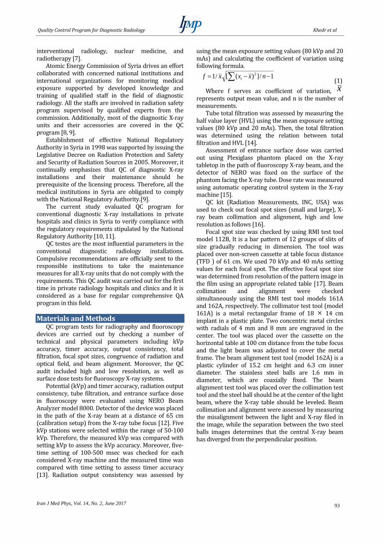
Table 1. Assessment criteria for X-ray machines
Parameter Good Satisfactory Unsatisfactory
kV accuracy ≤ 5% kV (5-10)% kV > 10% kV
Time accuracy ≤ 5% (5-10)% >10%
Large focal spot (f > 1mm) - ≤ 0.5 f 0.5 f >
Small focal spot (f ≤ 1mm) - 0.4 f ≤ 0.4 f >
Radiation output consistency - ≤ 5% > 5%
Filtration (mm Al) > 2.5 2 - 2.5 < 2
Beam collimation ≤ 1% 1 - 2 ) %) > 2%
Beam alignment ≤ 1.5o 1.5o - 3o > 3o
High resolution - 0.8 lp/mm ≥ 0.8 lp/mm ≤
Low resolution - 2 groups ≥ 2 groups ≤
Entrance surface dose - ≤ 50 mGy/min > 50 mGy/min
High resolution in fluoroscopy was checked using
the pattern 07-553 in a lead plate with 0.05 mm thickness and 71 44 mm size. It consists of groups of 4-line pairs with a very slight gradient between the adjacent groups. The pattern was placed on the center of image intensifier surface with relevant operating parameters. High resolution was assessed by a number of resolving groups, which were clearly shown in the image on TV monitor during fluoroscopy and using an appropriate related table [17]. Low-contrast resolution in fluoroscopy was checked using RMI test tool model 151, which is a 0.8 mm thickness sheet of aluminum (17.8 17.8 cm in size). It consists of two sets of 4-holes decrease in diameters (1.5-6.3 mm) and two heavy aluminum plates each one is 1.9 cm thickness. The 0.8 mm aluminum sheet was placed alternatively between the two plates once and with one plate in another, and the test tool placed midway of the focal spot and image intensifier. Low-contrast resolution was assessed (by clearly visible) the number of the image holes on monitor during fluoroscopy using relevant operating parameters. The assessment criteria of QC for X-ray equipment are presented in Table 1. The audit was classified into three categories of good, satisfactory, and unsatisfactory. In addition, the X-ray machines ages are present in Table 2.
Table 2. X-ray machine age values
Statistic assessment X-ray machine age (y) Average 8.7 Minimum 1 Maximum 37 STDEV 6.3
Results QC results for performance of 363 radiography machines are presented in Table 3. Herein, each examined technical parameter categorized to good, satisfactory, and unsatisfactory to total percentage. Performance of most of the considered parameters was within good and satisfactory ranges. The percentage of unsatisfactory performance for all the checked parameters was less than 25.00%, except for high-potential accuracy, which reached a value of 28.77%. The total number of checked parameters for the 363 radiography machines was 5165, 861 parameters of them were unsatisfactory, and quoted a percentage of 16.66% to the total. QC of performance of 124 fluoroscopy machines is illustrated in Table 4. Therefore, performance of most of the considered parameters was in good and satisfactory ranges. Furthermore, the unsatisfactory percentage for all the checked parameters was less than 20.00%, where the maximum value was 16.94% for high resolution and the minimum value was 2.42% for the entrance surface dose. The total number of checked parameters for 124 fluoroscopy machines was 1142 parameters, 132 of which were unsatisfactory, which quoted a percentage of 11.56% to total. In addition, layout of comparatives for unsatisfactory percentage radiography machines and fluoroscopy machines performance is presented in Figure 1.

Quality Control Program for Diagnostic Radiology Khedr et al
Iran J Med Phys, Vol. 14, No. 2, June 2017
95
Figure 1. Layout of comparatives for unsatisfactory percentage radiography machines and fluoroscopy machines performance
Table 3. Assessment of radiography machines’ performance
Parameter Total Good Satisfactory Unsatisfactory Unsatisfactory %
kV accuracy 1689 738 465 486 28.77
Time accuracy 1083 647 215 221 20.41
Large focal spot 338 - 326 12 3.55
Small focal spot 277 - 252 27 9.75
Radiation output consistency
361 - 350 11 3.05
Filtration (mm Al) 359 211 129 19 5.29
Beam collimation OX 350 243 73 34 9.71
Beam collimation OY 355 251 73 31 8.73
Beam alignment 353 271 62 20 5.10
Total 5165 2088 2218 861 16.67

Khedr et al Quality Control Program for Diagnostic Radiology
Iran J Med Phys, Vol. 14, No. 2, June 2017
96
Table 4. Assessment of fluoroscopy machines’ performance
Parameter Total Good Satisfactory Unsatisfactory Unsatisfactory %
kV accuracy 542 302 157 83 15.31
Filtration (mm Al) 114 90 19 5 4.38
Beam collimation OX 57 50 3 4 4.02
Beam collimation OY 57 40 12 5 8.77
High resolution 124 - 103 21 16.94
Low resolution 124 - 113 11 8.87
Entrance surface dose 124 - 121 3 2.42
Total 1142 482 528 132 11.56
Table 5. The percentage of unsatisfactory comparatives between private and general hospitals
Parameter Unsatisfactory %
General hospitals [14] Unsatisfactory %
Private sectors
kV accuracy 20.8 28.77
Time accuracy 11.4 20.41
Large focal spot 46.7 3.55
Small focal spot 63.1 9.75
Radiation output consistency
4.7 3.05
Filtration (mm Al) 18.8 5.29
.
Beam collimation OX 24.4 9.71
Beam collimation OY - 8.73
Beam alignment 10.8 5.10
Average 26.1 10.5
Discussion The current study focused on the performance
of radiography and fluoroscopy devices for private hospitals and clinics. The results showed that most tested parameters of radiography and fluoroscopy devices were in compliance with the comparative standard criteria provided in Table 1. The unsatisfactory percentage values for radiography devices are presented in Table 3. The unsatisfactory percentage values for fluoroscopy devices are provided in Table 4. Radiography and fluoroscopy devices were evaluated in governmental hospitals in previous studies [8]. The comparison between the current study and the previous ones on governmental hospitals is shown in Table 5. As can be noted in this table, all the tested parameters were better in the private hospitals and clinics, except for kVp and time accuracy.
A similar study carried out in Iranian hospitals for conventional radiographic X-ray units [11] showed that kVp accuracy, kVp reproducibility, timer accuracy, timer reproducibility, exposure reproducibility, mA/timer linearity, and half-value layer were not within the acceptable limits in 25%, 4%, 29%, 18%, 11%, 12%, and 7% of the evaluated units, respectively. Another Iranian study evaluated conventional X-ray exposure parameters [18] and showed that the maximum deviation from the standard value for kVp accuracy was 27.52% and for time accuracy the highest extent of the deviation ranged from 36.65% to 133.20%.
Therefore, a comprehensive quality assurance program over the country that includes regular QC audit and establish the local qualified QC teams in far provinces for easy and fast response is essential. In addition, there were some old X-ray machines that need to be replaced.

Quality Control Program for Diagnostic Radiology Khedr et al
Iran J Med Phys, Vol. 14, No. 2, June 2017
97
Conclusion QA program in diagnostic radiology leads to
obtaining information regarding quality of radiology devices used for medical diagnosis and minimizing the doses received by patients and medical personnel. The current study clearly indicated that most considered diagnostic X-ray machines (both radiography and fluoroscopy devices) had acceptable performance. The highest percentage of performance compliance belonged to radiation output (96.95%), while the lowest percentage pertained to kVp (71.23%) in radiography devices. In addition, the maximum value was 97.58% for entrance surface dose and the lowest was 83.06% for high resolution in fluoroscopy devices. Some old X-ray machines need to be replaced, and the X-ray machines in the towns away from cities need further protective maintenance. Additionally, establishing qualified local QC teams near radiology institutes in each province is highly necessary to perform effective QC audits.
Acknowledgment The authors would like to thank Prof. I Othman
and Dr. M S Almasri without whose support this work would not have been possible. Thanks are extended to Ministry of Health and the medical institution owners and their teams for their collaboration.
References
1. United Nations Scientific Committee on the Effects of Atomic Radiation. Sources and effects of ionizing radiation. UNSCEAR 2000 report to the General Assembly, with scientific annexes. UNSCEAR; 2000.
2. International Commission on Radiological Protection. 1990 Recommendations of the Commission on Radiological Protection: ICRP Publication 60, Ann. of the ICRP 21.
3. International Basic Safety Standards for Protection against Ionizing Radiation and for the Safety of Radiation Sources. International Atomic Energy Agency, Vienna. 1996.
4. International Atomic Energy Agency, IAEA 2012 International Basic Safety Standard GSRI part III , Vienna, Austria .
5. Carmichael JH. European guidelines on quality criteria for diagnostic radiographic images. Office for Official Publications of the European Communities; 1996.
6. World Health Organization(WHO), Quality assurance in Diagnostic Radiology, Geneva, 1982.
7. Jomehzadeh A, Shokrani P, Mohammadi M, Amouheidari A. A quality assurance program for an amorphous silicon electronic portal imaging device using in-house developed phantoms: a method development for dosimetry purposes. Int. J. Radiat. Res. 2014 Jul 1;12(3):257-64.
8. Kharita MH, Khedr MS, Wannus KM. A comparative study of quality control in diagnostic radiology.
Radiation protection dosimetry. 2008 Jul 1;130(4):447-51.
9. Syrian Atomic Enrgy Commission (SAEC), Legislative Decree on Radiation Protection and Safety and Security of Radiation Sources , 2005.
10. Recommended Standard For The Routine Performance Testing Of Diagnostic X-Ray Imaging Systems IPEM Report No. 91, UK. 2005.
11. Jomehzadeh Z, Jomehzadeh A, Tavakoli MB. Quality Control Assessment of Radiology Devices in Kerman Province, Iran. Iranian Journal of Medical Physics. 2016 Mar 1;13(1):25-35.
12. Inovision Radiatiom Measurements NERO beam analyzer model 8000 instructions manual. Victoreen, USA.,2001
13. Institute of Physics and Engineering in Medicine. IPEM Report 32, Measurement of the performance characteristic of diagnostic X-ray systems used in medicine X-ray tube and generator. York: IPEM.1996
14. International Commission on Radiological Protection. ICRP Publication 34. Protection of the Patient in Diagnostic Radiology.1983.
15. Criteria And Methods For Quality Assurance In Medical X- Ray Diagnosis. Proceeding Of Scientific Seminar Held In Udine, Italy, 1984.
16. Meier-Duis H, Moedder U. Quality Assurance in Radiology. Radiation Measurements INC. Equipment Catalog, Wisconsin, USA 1984.
17. Rehani MM. Diagnostic imaging: quality assurance. Journal of Medical Physics. 1996 Apr 1;21(2):74.
18. Gholami M, Nemati F, Karami V. The evaluation of conventional X-ray exposure parameters including tube voltage and exposure time in private and governmental hospitals of Lorestan Province, Iran. Iranian Journal of Medical Physics. 2015 Jul 1;12(2):85-92.