ischemic stroke an atlas of investigation and treatment (atlases of investigation and management)
TRANSCRIPT

An Atlas of Investigation and Treatment
ISCHEMIC STROKE
Foreword by LR Caplan
CLINICAL PUBLISHING

For Laurie, Noah and LioraIES
For my husband, Rob, and the Stroke Team at Saint Luke’s HospitalMMR

An Atlas of Investigation and Treatment
ISCHEMIC STROKE
Isaac E Silverman, MDVascular Neurology
Co-Medical Director
The Stroke Center at Hartford Hospital
Hartford, Connecticut
USA
Marilyn M Rymer, MDSaint Luke’s Brain and Stroke Institute
Saint Luke’s Hospital
UMKC School of Medicine
Kansas City, Missouri
USA
Foreword by
Louis R Caplan, MD Professor of Neurology
Department of Neurology Beth Israel Deaconess Medical Center
Boston Massachusetts USA
Special contributions by
Gary R Spiegel, MDCM (Neuroimaging) Jefferson Radiology
Director of Neurointervention Co-Medical Director
The Stroke Center at Hartford Hospital Hartford, Connecticut
USA
Robert E Schmidt, MD, PhD (Neuropathology) Professor, Pathology and Immunology
Washington University School of Medicine St Louis, Missouri
USA
CLINICAL PUBLISHINGOXFORD
Clinical Publishing
an imprint of Atlas Medical Publishing Ltd
Oxford Centre for Innovation
Mill Street, Oxford OX2 0JX, UK
Tel: +44 1865 811116
Fax: +44 1865 251550
Email: [email protected]
Web: www.clinicalpublishing.co.uk
Distributed in USA and Canada by:
Clinical Publishing
30 Amberwood Parkway
Ashland OH 44805, USA
Tel: 800-247-6553 (toll free within US and Canada)
Fax: 419-281-6883
Email: [email protected]
Distributed in UK and Rest of World by:
Marston Book Services Ltd
PO Box 269
Abingdon
Oxon OX14 4YN, UK
Tel: +44 1235 465500
Fax: +44 1235 465555
Email: [email protected]
© Atlas Medical Publishing Ltd 2009
First published 2009
All rights reserved. No part of this publication may be reproduced, stored in a retrieval
system, or transmitted, in any form or by any means, without the prior permission in
writing of Clinical Publishing or Atlas Medical Publishing Ltd.
Although every effort has been made to ensure that all owners of copyright material
have been acknowledged in this publication, we would be glad to acknowledge in
subsequent reprints or editions any omissions brought to our attention.
A catalogue record of this book is available from the British Library
ISBN-13 978 1 84692 017 2
ISBN e-book 978 1 84692 595 5
The publisher makes no representation, express or implied, that the dosages
in this book are correct. Readers must therefore always check the product
information and clinical procedures with the most up-to-date published product
information and data sheets provided by the manufacturers and the most recent
codes of conduct and safety regulations. The authors and the publisher do not
accept any liability for any errors in the text or for the misuse or misapplication
of material in this work.
Project manager: Gavin Smith, GPS Publishing Solutions, Herts, UK
Illustrations by Graeme Chambers, BA(Hons)
Typeset by Phoenix Photosetting, Chatham, Kent, UK
Marston Book Services Ltd, Abingdon, Oxon, UK

Contents
Foreword vii
Preface ix
Acknowledgements x
Abbreviations xi
1 Stroke Basics 1
2 Pathophysiology 17
3 Anterior Circulation 31
4 Posterior Circulation 57
5 Vasculopathies 75
6 Therapies: the management of acute ischemic stroke and secondary stroke prevention 103
Index 121

vii
In neurology, anatomy is very important. Neurologists have a compulsive preoccupation with the anatomy of disease not shared by most non-neurologists, including general practi-tioners and internists. They are taught to ask not only what the disease process is but also where the abnormality is within the nervous system. The nervous system is made up of very different parts. Can one imagine body parts as different as the brain, spinal cord, peripheral nerves and muscles? Each component of the nervous system is very distinct and made of diverse subunits with different appearances, functions and susceptibilities to various diseases.
The brain is especially different from all other organs of the body. Most other internal organs are rather homoge-neous. Parts of the lung, liver, pancreas, spleen and bone marrow all look alike and have identical functions. In these organs, disease is determined by the amount of the organ rendered dysfunctional, not by the location or ‘anatomy’ of the illness. A lesion in one part of the brain results in a loss of speech while a lesion very nearby could cause paralysis, loss of sensation, ataxia or many other functional abnor-malities. The clinical symptoms and signs depend heavily on where the disease process occurs in the brain and on other aspects such as the size and extent of the abnormality and its nature (tumor, infection, vascular occlusion, hemorrhage, traumatic lesion etc.)
In patients with strokes and brain ischemia, anatomy is especially important. In order to effectively plan diag-nosis and management, physicians will need to know not only where the lesion is in the brain, but what, where, and how severe the abnormality is in the blood vessels that sup-ply the affected portion of the brain. They will also want to know if there are any additional abnormalities in other vascular structures. Heart and aortic lesions could be a source of embolism to intracranial blocked arteries. Vascular occlusions in other vessels could affect the body’s ability to develop collateral blood flow to the affected region. Other arterial lesions could represent a threat for future strokes.
Stroke differs from many other neurological conditions in its timing. The term stroke indicates being suddenly ‘stricken’. Doctors must act quickly to try to save brain tis-sue as brain ischemia is a medical emergency. “Time is brain” has become a familiar adage. In order to manage stroke patients, physicians must be very familiar with the anatomy of the blood vessels that supply the brain and know the types of vascular abnormalities and their predilection for different vascular locations. They must also be familiar with the vari-ous modalities that are able to provide images of the brain and the cervical and intracranial arteries and veins.
The good news is that advances in technology have now made it possible to image the brain and its vascular sup-ply quickly and safely. Doctors can now rapidly determine the answers to the ‘what’ and ‘where’ questions if they are familiar with the anatomy, pathology and pathophysiology of brain ischemia and the technology available to show it.
Anatomy is visual: that is why the authors have chosen an atlas format to teach the fundamentals of ischemic stroke. This atlas is written by two very experienced active clini-cians who are involved with the care of stroke patients every day. They succinctly and clearly review the basic aspects of anatomy, pathology and physiology, while illustrating the text with well chosen figures. The figures include anatomi-cal drawings, pathology specimens and many images from modern CT, MRI, echocardiography and angiography. They emphasize the case method of teaching, since each stroke patient is different and their management depends heavily on the specific details of their condition.
I heartily recommend this atlas for anyone who cares for stroke patients. I suggest first reading the entire atlas thor-oughly and then keeping it handy for review of and reference to aspects relating to individual patient scenarios. The writ-ing is clear and easily grasped and the images are superb.
Louis R. Caplan, MDDecember 2008
Foreword

ix
The paradigm for the diagnosis and treatment of stroke has undergone a revolution. It began in the mid-1970s when the first CT scans clearly differentiated acute hemorrhage from ischemia and proved clinicians right and wrong about the anatomical location and size of a stroke, and continued in the mid-1980s with the increased sensitivity of MRI. Then, in the mid-1990s, thrombolytic therapy for stroke was shown to be effective, and it became imperative that the diagnosis was correct and that patients were evaluated and treated urgently. The passive wait-and-see attitude toward acute stroke care was no longer acceptable. Stroke became a Level I medical emergency on a par with acute coronary syndromes and trauma. Interventional neuroradiology also evolved during that decade, providing catheter-based alter-natives to vascular neurosurgery for the treatment of intrac-ranial aneurysms and vascular malformations.
The modern era of stroke clinical trials was ushered in by studies comparing thrombolytic agents with placebo for acute ischemic stroke, carotid endarterectomy versus medical therapy, and antithrombotic agents for secondary stroke prevention. More recently, the treatment of aneu-rysms by neurosurgical clipping versus endovascular coil-ing, and neurosurgery for intracerebral hemorrhage and the malignant middle cerebral artery stroke syndrome have been studied. Now, catheter-delivered agents and devices, novel thrombolytic agents, and neuroprotective strategies, such as hypothermia, promise even longer therapeutic time windows and better outcomes. Advanced CT and MR neuroimaging are directing better patient selection for specific treatments and may also extend the window for revascularization.
These scientific advances have led to palpable changes in the way clinicians are trained and care is organized. Hundreds of hospitals in the USA are becoming certified as Primary Stroke Centers by the Joint Commission. Individual states in the USA are evaluating systems for organizing pre-hospital care. National organizations such as the American Stroke Association and the American Academy of Neurology
are helping to promulgate quality measures and guidelines to improve and standardize the care of stroke patients. The US American Council on Graduate Medical Education has approved fellowship training in vascular neurology and inter-ventional neurology, and the American Board of Psychiatry and Neurology offers vascular neurology subspecialty certifi-cation. There has never been a more exciting time to become an expert in stroke treatment and management. With aging populations and treatment options expanding, the need for stroke specialists is growing rapidly.
Our intent in this atlas is to introduce clinicians, residents in training, and medical and nursing students to the scope of neurovascular disorders. In this volume (Ischemic Stroke) and the companion volume (Hemorrhagic Stroke) we provide a practical visual guide to the emerging field of vascular neurology: neuroimaging and neuropathology organized around the major clinical diagnostic groups. This first volume addresses the evaluation of ischemic stroke, its diverse pathophysiology, and its emerging treatments. The companion volume introduces the causes of intracerebral hemorrhage, including intracranial aneurysms and the various forms of vascular malformations. We are unaware of any reference that offers this broad overview, and we hope that our atlas is an exciting stimulus for fellow clinicians and clinicians-in-training to consider joining our field.
Although the management of stroke and neurovascular care is increasingly driven by evidence-based medicine and formal guidelines, clinicians benefit greatly from the direct experiences of caring for individual patients so we have emphasized case studies.
Enjoy this atlas! The opportunity for effective treatment and improved outcomes for our patients has never been greater.
Marilyn M. Rymer, MDIsaac E. Silverman, MD
December 2008
Preface

x
Acknowledgements
We are indebted to many physicians and other professional colleagues for their comments on the evolving manuscript as well as their contributions to the images included in the book. We would like to thank L. Christy Turtzo, MD, PhD, the first Vascular Neurology Fellow at the University of Con-necticut and Gary R. Spiegel, MD, the lead Interventional Neuroradiologist at Hartford Hospital, for their critical review of certain individual chapters and Naveed Akhtar, MD, Interventional Neuroradiologist at Saint Luke’s Stroke Center, for his review of selected chapters and images. Dr Spiegel contributed to the text, and assisted with the pro-cessing of innumerable angiographic studies.
Our sincere thanks to Robert Schmidt, MD, PhD, Professor of Pathology and Immunology at Washington University School of Medicine. Many of the beautiful pathology photos are his. We also had excellent assistance from Dean Uphoff, MD of the Pathology Department at Hartford Hospital and Louis R. Caplan, MD at the Beth Israel-Deaconess Medical Center in Boston. We are grate-ful to the following contributors from Hartford Hospital: Ethan Foxman, MD for neuroimaging, Inam U. Kureshi, MD, Vascular Neurosurgery, for intra-operative images of hemicraniectomy, and AVM and aneurysm surgeries, Paul Gaudio, MD of Ophthalmology, for retinal fundus photos and Cynthia Taub, MD of Cardiology, for echocardiography studies.
The strength of this project is based on the cases and neuro-vascular pathology that are evaluated on a weekly basis at the multidisciplinary Neurovascular Clinic of Hartford Hospital. Colleagues in this clinic are Donna Avanecean, Neurovascular APRN; Drs Kureshi and Spiegel; and in Vascular Neurology, Nora S. Lee, MD and Louise D. McCullough, MD, PhD.
The other chief source of case studies for this textbook comes from the inpatient service of the Stroke Center at Hartford Hospital. Core members not mentioned above from our stroke team are Dawn Beland, RN, Stroke Center Coordinator; Joao A. Gomes, MD, Neurocritical Care Medicine and Vascular Neurologist; Stephen K. Ohki, MD, Interventional Neuroradiology; Lincoln Abbott, MD, and A. Jon Smally, MD of Emergency Medicine; Michele Landes, RN, database manager; and our clinical trials coordinators, Martha Ahlquist, LPN, and Jennifer Blum.
The members of the stroke team at Saint Luke’s Hospital were a great help in reviewing the work in progress. Irene Bettinger, MD, Charles Weinstein, MD, Steven Arkin, MD, Christine Boutwell, MD, Michael Schwartzman, DO, and Karin Olds, MD, are the neurologists, Naveed Akhtar, MD, is the neurointerventionalist and Debbie Summers, APN, is the coordinator of the stroke team.
A special thanks to Krzysztof Dzialo of the Radiology File Room, and Vladilen Bokotey, Radiology systems manager, both at Hartford Hospital. The input from these two indi-viduals made feasible the digital manipulation of countless MRI, CT, and angiographic studies that ultimately form the heart of this textbook.
Finally, as a case-based, patient-centric endeavor, this atlas would not have been possible without the hundreds of stroke and neurovascular patients evaluated by our Stroke Center groups. Just as our experiences with the clinical presenta-tion, morbidities and neurologic deficits, neuroimaging and neuropathology, and lives of these patients ultimately shape our growth as clinicians, we hope in turn to educate them about their disease and improve their quality of life. The rich breadth of this textbook is a tribute to these patients.

xi
Abbreviations
ACA anterior cerebral arteryAChA anterior choroidal arteryACoA anterior communicating arteryAICA anterior inferior cerebellar arteryAIS acute ischemic strokeAMPA a-amino-3-hydroxy-5-methyl-4-isoxazole
propionic acidAP anteroposteriorBA basilar arteryBI Barthel IndexCADASIL cerebral autosomal dominant
arteriopathy with subcortical infarcts and leukoencephalopathy
CAPRIE Clopidogrel vs Aspirin in Patients at Risk of Ischemic Events (clinical trial)
CHARISMA Clopidogrel for High Atherothrombotic Risk and Ischemic Stabilization, Management, and Avoidance (clinical trial)
CNS central nervous systemCSF cerebrospinal fluidCT computed tomographyCTA computed tomography angiographyDW-MRI diffusion-weighted MRI sequenceECA external carotid arteryEMS Emergency Medical ServicesESPRIT European/Australasian Stroke Prevention in
Reversible Ischaemia Trial (clinical trial)ESPS-2 European Stroke Prevention Study 2
(clinical trial)FDA Food and Drug AdministrationFLAIR MRI fluid-attenuated inversion recovery MRI
sequenceFMD fibromuscular dysplasiaGCA giant cell arteritisGE MRI gradient echo MRI sequenceGOS Glasgow Outcome ScaleHD hospital dayIA intra-arterialICA internal carotid arteryICH intracerebral hemorrhageICU intensive care unit
INR Interventional Neuroradiology/IV intravenousMATCH Management of Atherothrombosis with
Clopidogrel in High-Risk Patients with Recent Transient Ischemic Attacks or Ischemic Stroke (clinical trial)
MCA middle cerebral arteryMELAS mitochondrial encephalopathy, lactic
acidosis, and stroke-like episodesMERCI Mechanical Embolus Removal in Cerebral
Ischemia (endovascular device)MRA magnetic resonance angiographyMRI magnetic resonance imagingmRS modified Rankin ScaleNIHSS National Institutes of Health Stroke ScaleNINDS National Institutes of Neurologic Disease
and StrokeNINDS rt-PA NINDS Recombinant Tissue Plasminogen
Activator (clinical trial)NMDA N-methyl-D-aspartateOS oculus sinister (left eye)PCA posterior cerebral arteryPCoA posterior communicating arteryPFO patent foramen ovalePHQ-9 Patient Health Questionnaire (nine-item)PICA posterior inferior cerebellar arteryPROACT-II Prolyse in Acute Cerebral
Thromboembolism II (clinical trial)PRoFESS Prevention Regimen for Effectively
Avoiding Second Strokes (clinical trial)PT/INR prothrombin time (international
normalized ratio)SAH subarachnoid hemorrhageSCA superior cerebellar arteryTEE transesophageal echocardiogramTIA transient ischemic attackTOAST Trial of ORG10172 in Acute Stroke
Treatment (clinical trial)t-PA tissue plasminogen activatorVA vertebral artery
Chapter 1
Stroke Basics
Introduction
Stroke is the second leading cause of death worldwide and the leading cause of adult disability in many countries. It is imperative that all physicians understand the basics of stroke diagnosis and treatment for three reasons:
Stroke occurs as an acute event, and people will access the closest medical facility or physician’s office for help. Primary care and emergency medicine physicians need to be able to rapidly diagnose and treat patients with acute stroke symptoms.Emerging treatments for stroke are time-dependent. The earlier the initiation of treatment, the better the outcome.Once a stroke has occurred, the risk of a second stroke increases. It is essential that clinicians identify and treat the stroke mechanism and risk factors and institute medi-cations (e.g., antithrombotic and blood pressure-lowering agents) when appropriate in order to prevent recurrence.
The goal of this atlas is to provide clinicians and students with a foundation in the clinical presentation, neuroimag-ing, pathology, pathophysiology, and treatment of stroke syndromes and neurovascular disorders so that they can make an accurate diagnosis and initiate treatment and/or seek vascular neurology consultation when appropriate.
What is a stroke?
A stroke is caused by a disruption in the flow of blood to part of the brain either because of occlusion of a blood vessel in the case of acute ischemic stroke (AIS) or the rupture of a blood vessel causing bleeding in or around the brain: intra-cerebral hemorrhage (ICH) or subarachnoid hemorrhage
(SAH), respectively (1.1). When stroke symptoms resolve and do not cause permanent brain damage, they are called a transient ischemic attack (TIA). Although the historical definition of a TIA is neurological symptoms lasting less than 24 hours, the duration of most TIAs is between 5 and 30 minutes, and can be considered a cerebral equivalent of cardiac angina.1 Some healthcare professionals and patients refer to TIAs as ‘mini-strokes,’ but a TIA is actually a warn-ing that a stroke may occur very soon.
Stroke epidemiology
Stroke incidence and mortality are increasing along with modernization and advancing longevity. Worldwide, 15 mil-lion people suffer a stroke each year. Five million of those die and 5 million are left permanently disabled.2 It is estimated that by 2020, stroke mortality will have almost doubled as a result of an aging population and the future effects of cur-rent smoking patterns.3
Two-thirds of all stroke deaths and 60% of all strokes occur in low and middle income countries.4 As infectious diseases and malnutrition decline in developing countries, stroke incidence rises due to decreased physical activity, increased tobacco use, and dietary changes. By 2040 there will be a bil-lion adults aged 65 years or older at risk for stroke in low and middle income countries. In addition to the aging population, the major stroke risk factors worldwide are hypertension and tobacco use. In most countries, up to 30% of adults suffer from hypertension.2 Men have a slightly higher incidence of stroke than women, but women have higher mortality rates. Black people have a higher stroke mortality rate than white people, and the mortality rate for Hispanic people falls between that of white and black people. There is a high incidence of hemor-rhagic stroke in Asian people.3
1

2 Stroke basics
Strokes occur at any age, but are more common in the elderly. Stroke risk doubles with every decade beyond 50 years of age, but 30% of strokes occur before the age of 65 in the USA.
Types of stroke
Ischemic stroke
Ischemic stroke, the most common type, is caused by an
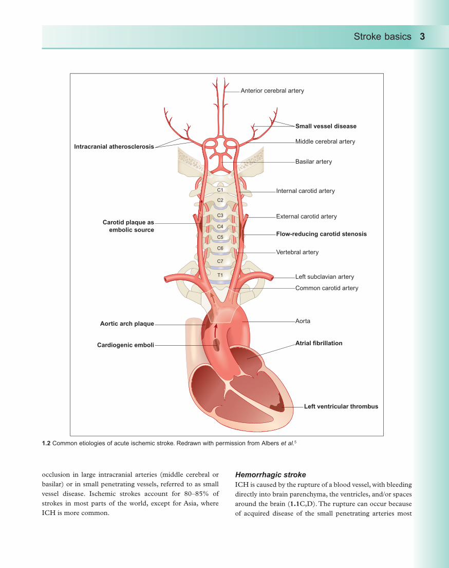
occlusion of an artery in the neck or in the brain, depriv-ing a part of the brain of its nutrients, glucose and oxygen. The etiologies for AIS are diverse (1.2), and are reviewed in Chapter 2. The arterial occlusion is most often caused by a thrombus that has traveled to the brain (embolized) from a more proximal location in the body, such as the heart or from plaque in the wall of a proximal artery, such as the aorta or the internal carotid artery.5 Less often, the etiology is a local thrombus developing immediately at the site of
1.1 Major stroke subtypes. (A) Acute ischemic stroke. Hypodense infarct on head CT scan, with extensive midline (left-to-right) shift.
(B) Subacute ischemic stroke. Gross pathology, at 3–4 days, in the territory of the internal carotid artery (ICA). Note edema and altered
gray–white matter differentiation in the distribution of the MCA and ACA territories (arrows). (C) Primary intracerebral hemorrhage.
A hyperdense lesion on CT scan located in the left putamen is a typical location for hypertensive hemorrhage due to small vessel
disease. (D) Intracerebral hemorrhage. Gross pathology of a frontoparietal intracerebral hemorrhage resulting in subfalcine and
uncal herniation; mass effect likely caused deep midline, punctate lesions, known as Duret hemorrhages (arrows). (E) Subarachnoid
hemorrhage. CT scan reveals hyperdense blood diffusely fi lling the subarachnoid spaces around the Circle of Willis and the Sylvian
fi ssures, bilaterally, and the interhemispheric fi ssure. (F) Intracranial aneurysm: an illustration. (G) Aneurysm of the carotid artery. A
large aneurysm of the cavernous portion of the ICA is measured on a three-dimensional rendering of the conventional angiogram. The
distal branches of the ICA, the MCA (extending laterally, to the left), and the ACA (extending medially) are also shown at the top of this
image. The right side of the image is labeled ‘R.’ Pathology courtesy of Robert Schmidt MD, PhD.
ArteryAneurysm
(A)
(C)
(E)
(F)
(G)
(B)
(D)
Stroke basics 3
occlusion in large intracranial arteries (middle cerebral or basilar) or in small penetrating vessels, referred to as small vessel disease. Ischemic strokes account for 80–85% of strokes in most parts of the world, except for Asia, where ICH is more common.
Hemorrhagic stroke
ICH is caused by the rupture of a blood vessel, with bleeding directly into brain parenchyma, the ventricles, and/or spaces around the brain (1.1C,D). The rupture can occur because of acquired disease of the small penetrating arteries most
1.2 Common etiologies of acute ischemic stroke. Redrawn with permission from Albers et al.5
Intracranial atherosclerosis
Carotid plaque as
embolic source
Aortic arch plaque
Cardiogenic emboli
Anterior cerebral artery
Small vessel disease
Middle cerebral artery
Basilar artery
External carotid artery
Internal carotid artery
Flow-reducing carotid stenosis
Left subclavian artery
Common carotid artery
Aorta
Atrial fibrillation
Left ventricular thrombus
Vertebral artery
C1
C2
C3
C4
C5
C6
C7
T1
4 Stroke basics
commonly related to long-standing hypertension (small vessel disease), degenerative disease of superficial arteries (amyloid angiopathy), or from structural abnormalities of larger intracranial arteries, such as arteriovenous malform-ations. SAH occurs when an intracranial aneurysm ruptures and blood invades the spaces around the brain (1.1E–G). Aneurysms are balloon-shaped outpouchings of an artery where the vessel’s wall has weakened.
Stroke symptoms
A general review of clinical symptoms is provided to help interpret information in the chapters that follow. (See the reference list at the end of this Chapter for textbooks that provide more detail on clinical stroke syndromes.6–8) The hall-mark of stroke is sudden onset of neurologic deficit. Individual stroke symptoms depend entirely on what anatomical area of the central nervous system (brain, spinal cord, or eye) is damaged. Usually, stroke presents as a syndrome, a collec-tion of symptoms that help the examiner localize the region of the central nervous system that is acutely injured.
Headache1 . Sudden severe headache is often associated with ICH and SAH, but is uncommon in ischemic stroke. An exception is ischemic stroke caused by carotid or ver-tebral artery dissection in which headache, facial, or neck pain are typical.9,10
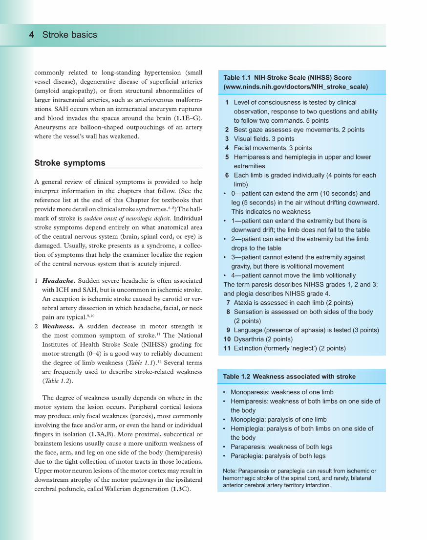
Weakness2 . A sudden decrease in motor strength is the most common symptom of stroke.11 The National Institutes of Health Stroke Scale (NIHSS) grading for motor strength (0–4) is a good way to reliably document the degree of limb weakness (Table 1.1).12 Several terms are frequently used to describe stroke-related weakness (Table 1.2).
The degree of weakness usually depends on where in the motor system the lesion occurs. Peripheral cortical lesions may produce only focal weakness (paresis), most commonly involving the face and/or arm, or even the hand or individual fingers in isolation (1.3A,B). More proximal, subcortical or brainstem lesions usually cause a more uniform weakness of the face, arm, and leg on one side of the body (hemiparesis) due to the tight collection of motor tracts in those locations. Upper motor neuron lesions of the motor cortex may result in downstream atrophy of the motor pathways in the ipsilateral cerebral peduncle, called Wallerian degeneration (1.3C).
Table 1.1 NIH Stroke Scale (NIHSS) Score
(www.ninds.nih.gov/doctors/NIH_stroke_scale)
Level of consciousness is tested by clinical 1
observation, response to two questions and ability
to follow two commands. 5 points
Best gaze assesses eye movements. 2 points2
Visual fields. 3 points3
Facial movements. 3 points4
Hemiparesis and hemiplegia in upper and lower 5
extremities
Each limb is graded individually (4 points for each 6
limb)
0—patient can extend the arm (10 seconds) and
leg (5 seconds) in the air without drifting downward.
This indicates no weakness
1—patient can extend the extremity but there is
downward drift; the limb does not fall to the table
2—patient can extend the extremity but the limb
drops to the table
3—patient cannot extend the extremity against
gravity, but there is volitional movement
4—patient cannot move the limb volitionally
The term paresis describes NIHSS grades 1, 2 and 3;
and plegia describes NIHSS grade 4.
Ataxia is assessed in each limb (2 points)7
Sensation is assessed on both sides of the body 8
(2 points)
Language (presence of aphasia) is tested (3 points)9
Dysarthria (2 points)10
Extinction (formerly ‘neglect’) (2 points)11
Table 1.2 Weakness associated with stroke
Monoparesis: weakness of one limb
Hemiparesis: weakness of both limbs on one side of
the body
Monoplegia: paralysis of one limb
Hemiplegia: paralysis of both limbs on one side of
the body
Paraparesis: weakness of both legs
Paraplegia: paralysis of both legs
Note: Paraparesis or paraplegia can result from ischemic or
hemorrhagic stroke of the spinal cord, and rarely, bilateral
anterior cerebral artery territory infarction.

Stroke basics 5
Most motor recovery from stroke occurs during the ini-tial 1–3 months.13 Incremental gains, however, can be made over the subsequent 9 months or beyond, and may depend on the availability of continuing rehabilitation treatment. Evolving concepts in neuroplasticity may offer hope for improved function years after the initial insult. Frequently, patients with mild residuae are left with minimal lower facial paresis, limited dexterity in the hand (e.g., diminished fi ne fi nger movements) and/or leg (e.g., diminished rate of toe tap), and/or foot drop.
Ataxia3 . Limb ataxia can occur with or without weakness and is a discoordination of movement usually related to infarction in the cerebellar hemisphere (1.4).14 It is tested by evaluating rapid alternating movements and the fi nger-to-nose test in the upper extremities and the heel-to-shin test in the lower extremities. A midline cerebellar lesion may only cause mild vestibular symptoms and gait ataxia
without limb involvement. Long-term recovery from ataxia is usually excellent.Sensory loss4 . Sudden loss of sensation usually occurs in association with weakness in the same distribution, but pure hemibody sensory strokes can occur, usually from occlusions of small vessels supplying the lateral thalamus, pons, or lenticulocapsular region deep in the brain.15–17 The patient usually describes numbness and/or tingling paresthesias on one side of the face or the hemibody, a feeling often likened to ‘novocaine in the dentist’s offi ce’ or ‘having a limb fall asleep.’
Cortical lesions causing sensory defi cits are further dichotomized into those of the insular and opercular areas, affecting primary (primitive) sensation of pain or tempera-ture with intact position sense, versus those involving the postcentral gyrus, resulting in cortical sensory loss affecting position sense, stereognosis, and graphesthesia (1.3B, 1.5).16
1.3 Hemiparesis. (A) Peripheral MCA infarction on diffusion-weighted MRI sequence, involving precentral gyrus (primary motor cortex).
(B) Illustration of motor and sensory strips in the brain cortex. (C) Chronic left MCA-territory stroke, resulting in a wide region of
encephalomalacia on the FLAIR MRI sequence (left). Atrophy of the ipsilateral cerebral peduncle (arrow), demonstrates associated
Wallerian degeneration (right).
Motor cortex Sensory cortex
Auditory cortex Visual cortex
(B)
(A)
(C)

6 Stroke basics
Primary sensation is generally tested with a painful stimulus such as a pin-point or a stimulus of light touch, while corti-cal sensation may be assessed by testing position sense and by having a patient try to identify a number written upon the hand or an object placed into the hand. Inability to interpret the number written on the hand is agraphesthesia and an inability to identify an object such as a key in the hand is astereognosis.
Focal paresthesias, predominantly involving the perioral or finger areas (areas with strong representation within the homunculus), usually result from small distal emboli, to the postcentral gyrus (1.5).16 Rarely, primitive sensory impair-ments evolve into dysesthesias, known as the central post-stroke pain syndrome.16,18
Visual symptoms5 :Amaurosis fugax, which is a term that describes tran-sient blindness in one eye generally lasting 2–10 minutes, is a symptom of a retinal TIA often caused by an embolus from the ipsilateral carotid artery.19,20 Permanent loss of vision in one eye frequently occurs when the central retinal artery is occluded, but this pattern of visual loss is gener-ally not associated with other stroke symptoms (1.6).
1.6 Monocular visual loss. A normal retina of the right eye is
shown (A), contrasted with pallor and decreased vascularity
due to acute central retinal artery occlusion (B). The diffuse
whitening of the ischemic retina leaves only a ‘cherry red spot’
in the foveal center (arrow). (The small circular yellow spots are
scars from past treatment of this retina with a laser.)
1.4 Ataxia. diffusion-weighted MRI sequences show (left) an
acute infarct of the left cerebellar hemisphere, in the territory
of the SCA, and (right) an infarct in the territory of the medial
PICA.
1.5 Cortical sensory loss. Cortical embolic stroke, involving
the postcentral gyrus, caused a loss of cortical sensation. The
emboli originated from atherosclerotic disease of the left ICA.
(A)
(B)
Stroke basics 7
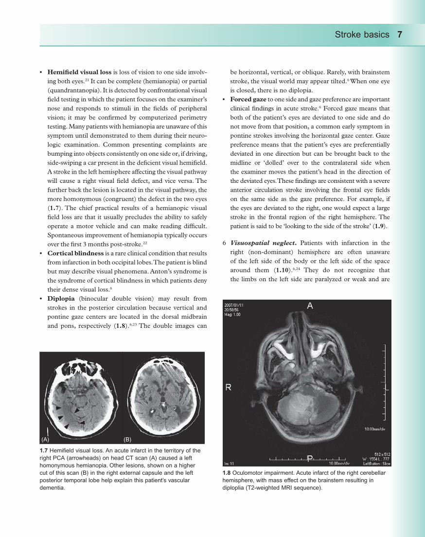
Hemifield visual loss is loss of vision to one side involv-ing both eyes.21 It can be complete (hemianopia) or partial (quandrantanopia). It is detected by confrontational visual field testing in which the patient focuses on the examiner’s nose and responds to stimuli in the fields of peripheral vision; it may be confirmed by computerized perimetry testing. Many patients with hemianopia are unaware of this symptom until demonstrated to them during their neuro-logic examination. Common presenting complaints are bumping into objects consistently on one side or, if driving, side-swiping a car present in the deficient visual hemifield. A stroke in the left hemisphere affecting the visual pathway will cause a right visual field defect, and vice versa. The further back the lesion is located in the visual pathway, the more homonymous (congruent) the defect in the two eyes (1.7). The chief practical results of a hemianopic visual field loss are that it usually precludes the ability to safely operate a motor vehicle and can make reading difficult. Spontaneous improvement of hemianopia typically occurs over the first 3 months post-stroke.22
Cortical blindness is a rare clinical condition that results from infarction in both occipital lobes. The patient is blind but may describe visual phenomena. Anton’s syndrome is the syndrome of cortical blindness in which patients deny their dense visual loss.6
Diplopia (binocular double vision) may result from strokes in the posterior circulation because vertical and pontine gaze centers are located in the dorsal midbrain and pons, respectively (1.8).6,23 The double images can
be horizontal, vertical, or oblique. Rarely, with brainstem stroke, the visual world may appear tilted.6 When one eye is closed, there is no diplopia.Forced gaze to one side and gaze preference are important clinical findings in acute stroke.6 Forced gaze means that both of the patient’s eyes are deviated to one side and do not move from that position, a common early symptom in pontine strokes involving the horizontal gaze center. Gaze preference means that the patient’s eyes are preferentially deviated in one direction but can be brought back to the midline or ‘dolled’ over to the contralateral side when the examiner moves the patient’s head in the direction of the deviated eyes. These findings are consistent with a severe anterior circulation stroke involving the frontal eye fields on the same side as the gaze preference. For example, if the eyes are deviated to the right, one would expect a large stroke in the frontal region of the right hemisphere. The patient is said to be ‘looking to the side of the stroke’ (1.9).
Visuospatial neglect6 . Patients with infarction in the right (non-dominant) hemisphere are often unaware of the left side of the body or the left side of the space around them (1.10).6,24 They do not recognize that the limbs on the left side are paralyzed or weak and are
1.7 Hemifield visual loss. An acute infarct in the territory of the
right PCA (arrowheads) on head CT scan (A) caused a left
homonymous hemianopia. Other lesions, shown on a higher
cut of this scan (B) in the right external capsule and the left
posterior temporal lobe help explain this patient’s vascular
dementia.
1.8 Oculomotor impairment. Acute infarct of the right cerebellar
hemisphere, with mass effect on the brainstem resulting in
diploplia (T2-weighted MRI sequence).
(A) (B)

8 Stroke basics
often unable to identify their left body parts; the patient may deny ownership of a paretic limb. Most commonly due to right parietal infarction, this inability to recognize a deficit is called anosagnosia. In a milder form, when presented with visual or sensory stimuli to both sides of the body at the same time (bilateral simultaneous stimuli), patients may ‘extinguish’ (not perceive) the stimulus on the left. Hemineglect is a predictor for poor rehabilitation and functional outcomes post-stroke.25,26
Language and speech production7 :Dysarthria is slurring or mispronunciation of normal speech. The words and sentences are correct, but the patient may be difficult to understand. Dysarthria can be heard in patients with facial or tongue weakness and also occurs in strokes involving the cerebellum and brainstem. Patients often state that they are ‘speaking like they were drunk.’Aphasia is difficulty with language processing: produc-tion and/or comprehension of speech. The stroke respon-sible occurs in the dominant (usually left) hemisphere.
Broca’s aphasia – (expressive and non-fluent) is a condi-tion in which the patient has difficulty with naming and has very halted, frustrated, effortful speech. There is no difficulty understanding spoken language. The patient may state: ‘I knew what I wanted to say, but just couldn’t find the words.’ Broca’s area is an anatomi-cal site at the base of the motor strip in the dominant hemisphere (1.3B, 1.11A,B). This type of aphasia is often associated with contralateral limb (arm > leg) and facial weakness.
Wernicke’s aphasia – (receptive and fluent) is a con-dition in which the patient cannot understand spoken language, but talks ‘fluently’ in long sentences often devoid of nouns and most meaning. Wernicke’s area is an anatomical site near the angular gyrus in the dom-inant hemisphere (1.3B, 1.11C). Wernicke’s aphasia may exist without motor impairment, and thus, the patient may be misdiagnosed as being simply ‘con-fused,’ or ‘intoxicated.’Global aphasia – is the condition where the patient has both expressive and receptive deficits. Patients are fre-quently mute, and must rely upon visual mimicry to follow along with a neurologic exam.
Aphasia resulting from stroke will frequently evolve and usually improve, such that its basic characteristics fluc-tuate. Patients often will describe that their fluency will seem to break down when they are tired at the end of a long day. Subtle improvements in language-related defi-cits may occur even months to years following a stroke.
1.9 Gaze deviation. A patient with right gaze deviation (A). Note
the far right position of the lenses of the eyes (arrowheads)
on a lower cut of the CT scan. The responsible lesion, a large
right frontoparietal intracerebral hemorrhage (B), impaired the
patient’s ability to drive the eyes to the left, a function of the right
frontal eye fields.
1.10 Hemineglect. Head CT scan demonstrates a subacute
right hemispheric stroke that caused left body hemineglect. The
evolving right hemispheric edema causes mass effect on the
adjacent right lateral ventricle, while a chronic, left MCA infarct
caused atrophy and associated ex-vacuo enlargement of the left
lateral ventricle.
(A) (B)

Stroke basics 9
Cognitive and behavioral deficits8 . Although aphasia and neglect are the most common higher cognitive defi-cits related to stroke, a wider range of neurobehavioral deficits include apraxia, memory loss and dementia, fatigue, depression, and other psychiatric disorders (e.g., emotional incontinence, anger, anxiety) can occur.6,27 Given the advancing age of the general population, strong interest has arisen in the field of cognitive/neurobehav-ioral deficits associated with stroke. Large-scale clinical trials have begun to investigate vascular dementia result-ing from multiple ischemic infarctions (e.g., 1.7) and post-stroke depression.28,29
Stroke outcomes
An excellent predictor of stroke outcomes is stroke severity at presentation. A measure of stroke-related neurologic deficits, the National Institutes of Health Stroke Scale (NIHSS) score has been extensively studied in clinical trials and has been shown to be a very useful predictor of 3-month outcomes. NIHSS scores from 0 to 10 (mild deficits); 11 to 20 (mod-erate); and >20 (severe) have decreasing potential for good outcomes (Table 1.3).30 The NIHSS is an excellent way for clinicians to communicate the severity of a stroke. Physicians and nurses caring for patients with strokes can be certified in the use of the NIHSS by the American Stroke Association or the National Stroke Association on their respective websites. Training materials can be obtained from the National Institutes of Neurologic Disease and Stroke (NINDS) website. The NIHSS score is heavily weighted toward anterior-circulation strokes, such that aphasia and hemiparesis often account for most of the points accumulated by any individual patient. Posterior circulation strokes frequently include neurologic
signs such as nystagmus, dysmetria, gait imbalance, or dys-arthria, which do not contribute significantly to the NIHSS score, such that the score may underestimate the severity of a posterior circulation lesion.31 In addition, a left middle cere-bral artery stroke will have a higher NIHSS than an equally severe right middle cerebral artery stroke because of the scor-ing for language impairment. In practice, it is relatively simple to indicate to patients and their families how severe the deficit appears to be based solely upon the neurologic examination and NIHSS score. The potential for good outcomes is poor in moderate to severe strokes if left untreated.
Other common types of outcomes scales measure differ-ent dimensions of recovery and disability after acute stroke.31 Some were developed specifically for stroke patients, while others look at disability and recovery following any type of acute brain injury:
1.11 Aphasia. A diffusion-weighted MRI
sequence (A) and apparent diffusion coefficient
map (B) of a left MCA lesion, involving an
anterior branch and affecting predominantly the
insular cortex clinically expressed as Broca’s
(non-fluent, expressive) aphasia. A second
acute left MCA infarct, apparent diffusion
coefficient map (C) clinically associated with
Wernicke’s (fluent, receptive) aphasia.
Table 1.3 Admission NIHSS Score predicts short-
term outcomes from acute ischemic stroke*
PROACT-II. Percentage of patients with minimal or no
deficits (Modified Rankin Scale, 0–1), at 90 days post-
stroke:
NIHSS 4–10: 63%
NIHSS 11–20: 24%
NIHSS 21–30: 7%
Adapted from Furlan et al.37
TOAST
NIHSS 6: 80% Excellent or good outcome
NIHSS 16: >85% Severe disability or death
Adapted from Adams et al.30
*Data derived from control groups’ outcomes in two clinical
trials, PROACT-II and TOAST.
(A) (B) (C)

10 Stroke basics
Barthel Index (BI) assesses activities of self-care and mobility.Modified Rankin Scale (mRS) assesses functional independence.Glasgow Outcome Scale (GOS) assesses general level of disability and recovery following acute brain injury.PHQ-932 assesses depression.Stroke-specific Quality of Life scale33 assesses quality of life.
The National Institute of Neurological Disorders and Stroke Recombinant Tissue Plasminogen Activator (NINDS rt-PA) Stroke Trial combined the NIHSS, mRS, BI, and GOS into a single global outcomes measure.34 Most acute stroke clinical trials use the mRS at 90 days as a primary outcome measure.
Diagnosis of stroke
The diagnosis of stroke is made by taking a careful history, performing a neurologic examination, and confirming the clinical diagnosis with an appropriate neuroimaging study. Seizures, hypoglycemia, trauma, and migraine are the most common mimics of the focal neurologic deficits in acute stroke. Brain tumors occasionally present with sudden onset of symptoms due to an associated hemorrhage into the tumor. Global symptoms of stroke such as altered level of conscious can be mimicked by metabolic encephalopathy. In most cases, the clinical diagnosis of stroke is not diffi-cult. However, patients with global encephalopathy due to diagnoses such as venous sinus thrombosis, vasculitis, or multifocal emboli are more challenging; these cases are usu-ally diagnosed by neuroimaging.
Neuroimaging
The evolution of modern neuroimaging has revolutionized the diagnosis and management of stroke. The modalities currently used are listed here and will be shown throughout the text.
Non-enhanced head computed tomography (CT) scans are the most commonly available neuroimaging studies for acute stroke. This modality is excellent in detect-ing ICH and SAH (1.1C,E, 1.9B), but is insensitive to small areas of infarction, especially in the posterior fossa (1.12). In most cases of early infarction (e.g., 1–4 hours after onset), the CT scan is normal. Subsequent scans over the next few hours begin to demonstrate an evolving infarct (1.13).Well-delineated hypodensity on CT indicates infarcted tissue (1.13C).
1.12 Paramedian pontine stroke. (A) Non-contrast head CT scan
shows a faint hypodensity in mid-pons. The hypodense lesion in
the left occipital pole (arrow) is likely a small, old PCA stroke. (B)
The diffusion-weighted MRI study readily delineates the acute
stroke as an area of restricted diffusion in the left medial pons.
1.13 Serial neuroimaging, infarct
in evolution. A right MCA-territory
AIS on head CT scans. (A) Note
blurring of sulcal spaces, and
early hypodensity, at 6 hours. (B)
The hypodense lesion becomes
better delineated, at 24 hours. (C)
The lesion demarcated as a wide
hypodensity, with mass effect along
the midline, at 40 hours.
(A) (B)
(A) (B) (C)
Stroke basics 11
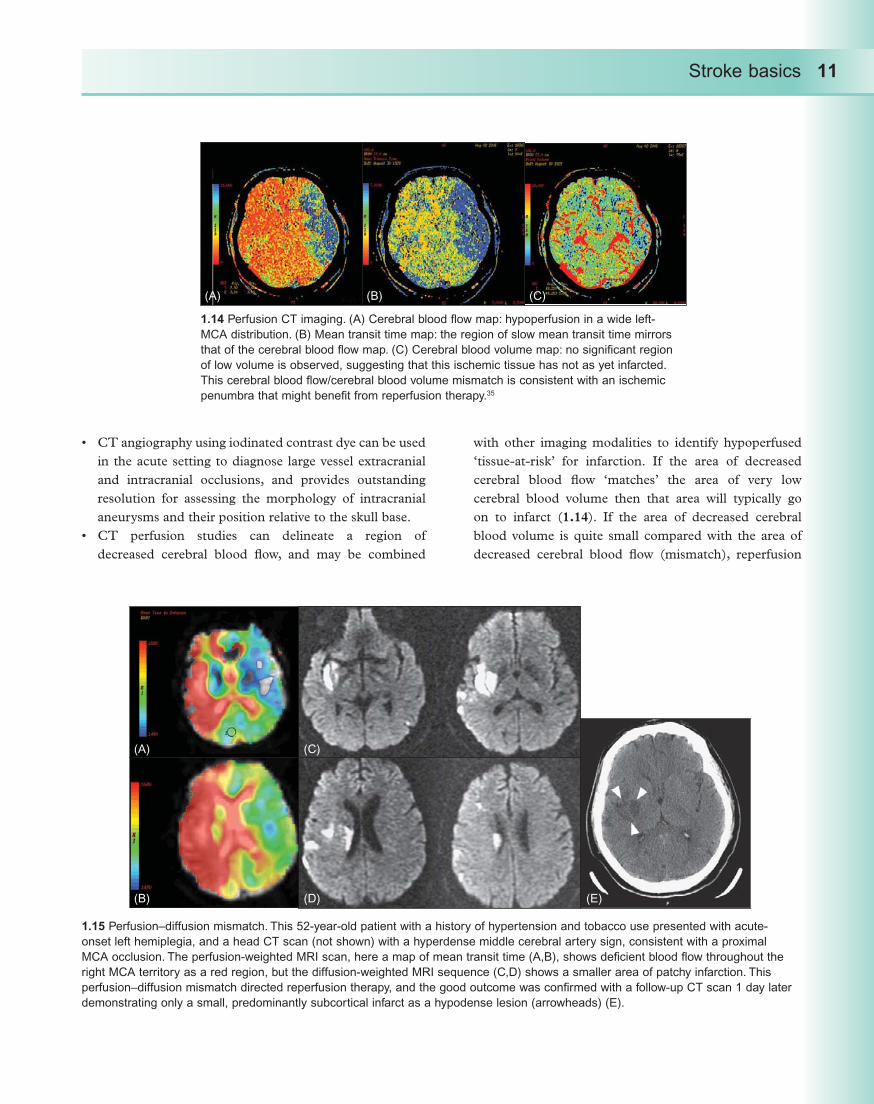
CT angiography using iodinated contrast dye can be used in the acute setting to diagnose large vessel extracranial and intracranial occlusions, and provides outstanding resolution for assessing the morphology of intracranial aneurysms and their position relative to the skull base.CT perfusion studies can delineate a region of decreased cerebral blood flow, and may be combined
with other imaging modalities to identify hypoperfused ‘tissue-at-risk’ for infarction. If the area of decreased cerebral blood flow ‘matches’ the area of very low cerebral blood volume then that area will typically go on to infarct (1.14). If the area of decreased cerebral blood volume is quite small compared with the area of decreased cerebral blood flow (mismatch), reperfusion
1.14 Perfusion CT imaging. (A) Cerebral blood flow map: hypoperfusion in a wide left-
MCA distribution. (B) Mean transit time map: the region of slow mean transit time mirrors
that of the cerebral blood flow map. (C) Cerebral blood volume map: no significant region
of low volume is observed, suggesting that this ischemic tissue has not as yet infarcted.
This cerebral blood flow/cerebral blood volume mismatch is consistent with an ischemic
penumbra that might benefit from reperfusion therapy.35
1.15 Perfusion–diffusion mismatch. This 52-year-old patient with a history of hypertension and tobacco use presented with acute-
onset left hemiplegia, and a head CT scan (not shown) with a hyperdense middle cerebral artery sign, consistent with a proximal
MCA occlusion. The perfusion-weighted MRI scan, here a map of mean transit time (A,B), shows deficient blood flow throughout the
right MCA territory as a red region, but the diffusion-weighted MRI sequence (C,D) shows a smaller area of patchy infarction. This
perfusion–diffusion mismatch directed reperfusion therapy, and the good outcome was confirmed with a follow-up CT scan 1 day later
demonstrating only a small, predominantly subcortical infarct as a hypodense lesion (arrowheads) (E).
(A)
(A) (C)
(B) (D) (E)
(B) (C)

12 Stroke basics
treatment may prevent infarction. The mean transit time also has predictive value.35
Magnetic resonance imaging (MRI) is not as readily available for acute stroke diagnosis in many hospitals. The diffusion-weighted image sequence is sensitive to ischemia within minutes of the onset of symptoms (1.4, 1.15). This technique is helpful when the diagnosis is in question and is excellent for identifying very small strokes causing minimal neurologic deficits. A ‘dark’ or hypointense lesion on the apparent diffusion coefficient map confirms that a diffusion-weighted magnetic resonance imaging lesion is due to infarction (1.11).
1.16 Magnetic resonance angiography, intracranial study.
Proximal occlusion of the left middle cerebral artery, the M1
segment, on coronal (A) and transaxial (B) views (arrow). No
distal, left hemispheric blood flow is observed.
1.17 Conventional angiography, anterior circulation. (A) An
injection of the left common carotid artery, lateral projection
(left), shows the carotid bifurcation, with a tortuous, redundant
cervical ICA and a distal ICA aneurysm, the same lesion shown
in 1.1G. An ICA injection, lateral view (right), again shows
this aneurysm (arrow), as well as flow into the three major
intracranial arteries of the cerebral hemisphere, the PCA, MCA,
and ACA. (B) An anteroposterior injection of the previous image
(A, right) shows clearly the branching of the distal ICA into the
ACA toward the midline and the MCA laterally; as well as a
different view of the aneurysm (arrow). (C) Injection of the right
ICA, anteroposterior projection, of a different patient shows
an acute occlusion of the superior M2 (second-order MCA)
branch (arrowhead), just distal to the prominent (hyperemic)
lenticulostriate system.
(A)
(A)
(B)
(C)
(B)
Stroke basics 13
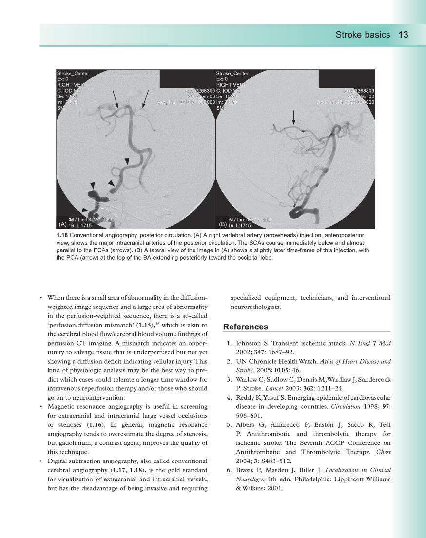
When there is a small area of abnormality in the diffusion-weighted image sequence and a large area of abnormality in the perfusion-weighted sequence, there is a so-called ‘perfusion/diffusion mismatch’ (1.15),36 which is akin to the cerebral blood flow/cerebral blood volume findings of perfusion CT imaging. A mismatch indicates an oppor-tunity to salvage tissue that is underperfused but not yet showing a diffusion deficit indicating cellular injury. This kind of physiologic analysis may be the best way to pre-dict which cases could tolerate a longer time window for intravenous reperfusion therapy and/or those who should go on to neurointervention.Magnetic resonance angiography is useful in screening for extracranial and intracranial large vessel occlusions or stenoses (1.16). In general, magnetic resonance angiography tends to overestimate the degree of stenosis, but gadolinium, a contrast agent, improves the quality of this technique.Digital subtraction angiography, also called conventional cerebral angiography (1.17, 1.18), is the gold standard for visualization of extracranial and intracranial vessels, but has the disadvantage of being invasive and requiring
specialized equipment, technicians, and interventional neuroradiologists.
References
Johnston S. Transient ischemic attack. 1. N Engl J Med 2002; 347: 1687–92.UN Chronicle Health Watch. 2. Atlas of Heart Disease and Stroke. 2005; 0105: 46.Warlow C, Sudlow C, Dennis M, Wardlaw J, Sandercock 3. P. Stroke. Lancet 2003; 362: 1211–24.Reddy K, Yusuf S. Emerging epidemic of cardiovascular 4. disease in developing countries. Circulation 1998; 97: 596–601.Albers G, Amarenco P, Easton J, Sacco R, Teal 5. P. Antithrombotic and thrombolytic therapy for ischemic stroke: The Seventh ACCP Conference on Antithrombotic and Thrombolytic Therapy. Chest 2004; 3: S483–512.Brazis P, Masdeu J, Biller J. 6. Localization in Clinical Neurology, 4th edn. Philadelphia: Lippincott Williams & Wilkins; 2001.
1.18 Conventional angiography, posterior circulation. (A) A right vertebral artery (arrowheads) injection, anteroposterior
view, shows the major intracranial arteries of the posterior circulation. The SCAs course immediately below and almost
parallel to the PCAs (arrows). (B) A lateral view of the image in (A) shows a slightly later time-frame of this injection, with
the PCA (arrow) at the top of the BA extending posteriorly toward the occipital lobe.
(A) (B)

14 Stroke basics
Bogousslavsky J, Hommel M. Ischemic stroke 7. syndromes: clinical features, anatomy, vascular territories. In: Adams H, Jr, ed. Handbook of Cerebrovascular Diseases. New York: Marcel Dekker; 1993: 51–94.Bogousslavsky J, Caplan L. 8. Stroke Syndromes, 2nd edn. New York: Cambridge University Press; 2001.Estol C. Headache: stroke symptoms and signs. In: 9. Bogousslavsky J, Caplan L, eds. Stroke Syndromes, 2nd edn. New York: Cambridge University Press; 2001: 60–75.Schievink W. Spontaneous dissection of the carotid and 10. vertebral arteries. N Engl J Med 2001; 344: 898–906.Pinho e Melo T, Bogousslavsky J. Hemiparesis and 11. other types of motor weakness. In: Bogousslavsky J, Caplan L, eds. Stroke Syndromes, 2nd edn. New York: Cambridge University Press; 2001: 22–33.Goldstein L, Bertels C, Davis J. Interrater reliability of 12. the NIH stroke scale. Arch Neurol 1989; 46: 660–2.Dobkin B. Rehabilitation after stroke. 13. N Engl J Med 2005; 352: 1677–84.Timmann D, Diener H. Cerebellar ataxia. In: 14. Bogousslavsky J, Caplan L, eds. Stroke Syndromes, 2nd edn. New York: Cambridge University Press; 2001: 48–59.Kim J. Sensory abnormality. In: Bogousslavsky J, 15. Caplan L, eds. Stroke Syndromes, 2nd edn. New York: Cambridge University Press; 2001.Kim J. Patterns of sensory abnormality in cortical 16. stroke: evidence for a dichotomized sensory system. Neurology 2007; 68: 174–80.Schmahmann J. Vascular syndromes of the thalamus. 17. Stroke 2003; 34: 2264–78.Bowsher D, Leijon G, Thuomas K-A. Central 18. poststroke pain: correlation of MRI with clinical pain characteristics and sensory abnormalities. Neurology 1998; 51: 1352–8.Benavente O, Eliasziw M, Streifler J,19. et al. Prognosis after transient monocular blindness associated with carotid-artery stenosis. N Engl J Med 2001; 345: 1084–90.Wray S. Visual symptoms (eye). In: Bogousslavsky J, 20. Caplan L, eds. Stroke Syndromes, 2nd edn. New York: Cambridge University Press; 2001: 111–28.Barton J, Caplan L. Cerebral visual dysfunction. In: 21. Bogousslavsky J, Caplan L, eds. Stroke Syndromes, 2nd
edn. New York: Cambridge University Press; 2001: 87–110.Zhang X, Kedar S, Lynn M, Newman N, Biousse V. 22. Natural history of homonymous hemianopia. Neurology 2006; 66: 901–5.Pierrot-Deseilligny C. Eye movement abnormalities. 23. In: Bogousslavsky J, Caplan L, eds. Stroke Syndromes, 2nd edn. New York: Cambridge University Press; 2001: 76–86.Clarke S. Right hemisphere syndromes. In: 24. Bogousslavsky J, Caplan L, eds. Stroke Syndromes, 2nd edn. New York: Cambridge University Press; 2001: 264–72.Beis J-M, Keller C, Morin N,25. et al. Right spatial neglect after left hemisphere stroke: a qualitative and quantitative study. Neurology 2004; 63: 1600–5.Buxbaum L, Ferraro M, Veramonti T,26. et al. Hemispatial neglect: subtypes, neuroanatomy, and disability. Neurology 2004; 62: 749–56.Bogousslavsky J. William Feinberg Lecture 2002: 27. emotions, mood, and behavior after stroke. Stroke 2003; 34: 1046–50.Hackett M, Anderson C, House A. Management 28. of depression after stroke: a systematic review of pharmacological therapies. Stroke 2005; 36: 1092–7.Erkinjuntti T, Roman G, Gauthier S, Feldman H, 29. Rockwood K. Emerging therapies for vascular dementia and vascular cognitive impairment. Stroke 2004; 35: 1010–17.Adams HP Jr, Davis P, Leira E,30. et al. Baseline NIH Stroke Scale score strongly predicts outcome after stroke: a report of the Trial of Org 10172 in Acute Stroke Treatment (TOAST). Neurology 1999; 53: 126–31.Kasner S. Clinical interpretation and use of stroke 31. scales. Lancet Neurol 2006; 5: 603–12.Williams L, Brizendine E, Plue L,32. et al. Performance of the PHQ-9 as a screening tool for depression after stroke. Stroke 2005; 36: 635–8.Williams L, Weinberger M, Harris L, Clark D, Biller J. 33. Development of a stroke-specific quality of life scale. Stroke 1999; 30: 1362–9.National Institute of Neurological Disorders and 34. Stroke rt-PA Stroke Study Group. Tissue plasminogen activator for acute ischemic stroke. N Engl J Med 1995; 333: 1581–7.

Stroke basics 15
Wintermark M, Reichart M, Thiran J-P,35. et al. Prognostic accuracy of cerebral blood flow measurement by perfusion computed tomography, at the time of emergency room admission in acute stroke patients. Ann Neurol 2002; 51: 417–32.Davalos A, Blanco M, Pedraza S,36. et al. The clinical–DWI mismatch: A new diagnostic approach to the brain tissue at risk of infarction. Neurology 2004; 62: 2187–92.Furlan A, Higashida R, Katzan I, Abou–Chebl A. 37. Intra-arterial thrombolysis in acute ischemic stroke. In: Lyden P, ed. Thrombolytic Therapy for Stroke. Totowa, NJ: Humana Press; 2001: 175–95.
Further reading
Bogousslavsky J, Caplan LR. Stroke Syndromes, 2nd edn. New York: Cambridge University Press; 2001.
Brazis PW, Masdeu JC, Biller J. Localization in Clinical Neurology, 4th edn. New York: Little, Brown and Company; 2001.
Kasner S. Clinical interpretation and use of stroke scales. Lancet Neurol 2006; 5: 603–12.
Resources for patients
American Stroke Association: http://www.strokeassociation.orgNational Stroke Association: http://www.stroke.org

Chapter 2
Pathophysiology
Introduction
Ischemic strokes can be classified by anatomic location and by pathophysiologic mechanism. A stroke in the distribution of the middle cerebral artery (MCA; arterial territory: ana-tomic location) could be caused by an embolus originating from the heart (cardioembolic source: pathophysiologic mech-anism). Both types of stroke classifications are important to understand. The symptoms of ischemic stroke (Chapter 1) are entirely dependent upon the arterial territory involved (Chapters 3 and 4). The current chapter addresses the diverse pathophysiology of ischemic stroke.
Stroke pathophysiology is inferred from the patient’s medi-cal history, diagnostic work-up, and the pattern and location of the stroke on neuroimaging. It is critical for the clinician to identify the pathophysiologic mechanism, because the prob-able cause of the stroke informs the medications and proce-dures employed for secondary stroke prevention. For example, a patient with an ischemic stroke in the MCA territory pre-senting with new-onset atrial fibrillation and no other apparent source of thromboembolism likely has a cardiogenic source, and should be started on an anticoagulant such as warfarin.
The pathophysiologic classification system described originally in the TOAST (Trial of ORG 10172 in Acute Stroke Treatment) clinical trial has become the standard for vascular neurology (Table 2.1).1,2 The percentages noted for each classification discussed below are approximate and vary by the population and registry data studied.
Large vessel disease (atherosclerosis)
Large vessel atherosclerotic disease accounts for 30–40% of ischemic strokes. This category includes atherosclerotic disease in the aorta, and major extracranial and intracranial
arteries (2.1). Thrombosis can develop locally at the site of arterial disease, or the diseased arterial segment can be a source of embolism to a more distal segment or branch of the artery. The latter circumstance, artery-to-artery embo-lism, is most commonly observed when atherosclerotic plaque located at the proximal internal carotid artery (ICA) embolizes to the MCA.
The most common sites of atherosclerotic disease are the aortic arch, carotid artery bifurcation, origins of the vertebral and common carotid arteries , the proximal (M1) segment of the MCA (case study 1), the distal vertebral arteries, and the middle and distal sections of the basilar artery. Intracranial stenosis occurs more frequently in Asian and black popula-tions.3 Intracranial MCA disease may cause stroke by several mechanisms: distal atheroemboli, borderzone lesions, and lenticulostriate perforator infarcts (i.e., branch artery occlu-sion) (case study 1).
The diagnosis of large vessel disease is made by imag-ing the blood vessels with CT angiography, MRA, ultra-sound, conventional angiography, and transesophageal echocardiography. This last modality enables visualization of atherosclerotic plaque within the aorta (2.1C,D).4 Plaque thickness >4 mm has been associated with a heightened risk for aortoembolic ischemic stroke.5
17
Table 2.1 TOAST criteria1
1. Large vessel disease (atherosclerosis)
2. Cardiac source (cardioembolic)
3. Small vessel disease (arteriolosclerosis; ‘lacunar
syndromes’)
4. Other determined etiology
5. Undetermined etiology

18 Pathophysiology
Anterior cerebral artery
Middle cerebral artery
Basilar artery
External carotid artery
Internal carotid artery
Subclavian artery
Aorta
Vertebral artery
C1
C2
C3
C4
C5
C6
C7
T1
2.1 Large vessel disease. Illustration of the major
cervical arteries supplying the brain (A). The
common carotid arteries branch into the external
and internal carotid arteries. The vertebral arteries
course through the transverse foramina of the
C6 through C1 vertebral bodies. Adapted with
permission from Osborn.20 The circle of Willis
(B): Gross pathology demonstrates widespread
large vessel atherosclerosis, recognized by
heterogeneous plaque in the arterial walls.
Transesophageal echocardiography: non-mobile
(C) and mobile (D) components of atherosclerotic
plaque (arrows) shown as a thickness in the wall of
the aorta, on transverse images. The mobile plaque
is heterogeneous and irregular, protruding into
the lumen (arrows). Echocardiography courtesy of
Cynthia Taub, MD.
(A)
(B)
(C)
(D)

Pathophysiology 19
Cardioembolic disease (cardiac source
of emboli)
Cardioembolic disease accounts for about 25% of ischemic strokes (case study 2). Atrial fibrillation is, by far, the most common cause of cardioembolic stroke.6 Cardiac sources of emboli causing stroke are stratified by major and minor risk (2.2; Table 2.2).7 Congenital heart conditions with right-to-left shunt, such as patent foramen ovale (2.3) or atrial septal aneurysms, are less frequent causes of cardiogenic emboli.8
The diagnosis of these conditions is made through stand-ard electrocardiography, cardiac monitoring, and echocar-diography. Transesophageal echocardiography (TEE) is more sensitive than is transthoracic echocardiography for identify-ing atrial septal aneurysm and defect, patent foramen ovale,
atrial myxoma, atrial thrombus, atrial appendage thrombus, aortic arch atheromas, and mitral valve vegetations (2.2).7,9
Small vessel disease (arteriolosclerosis)
Arteriolosclerosis accounts for 20% of ischemic strokes. The lesions are small ‘lacunes’ deep in the brain referred to as ‘lacunar infarcts.’10 Local occlusions of small pen-etrating end-arteries, such as the lenticulostriate branches of the MCA, the thalamostriate branches of the posterior cerebral artery, and the pontine perforators from the basilar artery, are the cause (2.4A). Typically, patients with small vessel disease have one or more of the classic cardiovas-cular risk factors: hypertension, diabetes, smoking, and hyperlipidemia. There are specific ‘lacunar syndromes’
2.2 High-risk cardioembolic sources. Echocardiography shows the following high-risk lesions. (A) Atrial myxoma: an irregular
heterogeneous mass involving most of the left atrium, attached to the fossa ovale. (B) Left atrial thrombus in the left atrial appendage
(arrow). (C) Thrombus in the thinned apex of the left ventricle following an acute myocardial infarction (arrow). (D) Left atrial thrombus
in a patient with rheumatic valvular disease: a parasternal long-axis window shows a ‘hockey stick’ appearance of the anterior leaflet
of the mitral valve (arrowhead). Echocardiography courtesy of Cynthia Taub, MD. (E) Gross pathology shows thrombus within the left
ventricle (arrows). Pathology courtesy of Louis Caplan, MD.
(A)
(C)
(B)
(E)
(D)

20 Pathophysiology
2.3 Low-risk cardioembolic source: patent foramen ovale.
A patent foramen ovale is opened (A), closed (B), and with
spontaneous left-to-right shunting on color Doppler mode (C)
between the left and right atria.
Table 2.2 Cardioembolic causes7
High-risk sources
Atrial fibrillation
Rheumatic mitral stenosis
Sick sinus syndrome
Atrial flutter, sustained
Prosthetic valves
Infective endocarditis
Non-bacterial thrombotic endocarditis
Atrial myxoma
Acute myocardial infarction
Aortic atherosclerosis
Low-risk sources
Patent foramen ovale
Atrial septal aneurysm
Mitral valve prolapse
Calcific aortic stenosis and bicuspid aortic sources
Fibroelastomas and Lambl excrescences
Mitral annular calcification
that result from occlusion of these small vessels, including pure motor or pure sensory symptoms, ataxic hemiparesis, dysarthric–clumsy hand syndromes, and simple unilateral sensorimotor deficits, typically presenting without deficits associated with cortical lesions, such as neglect, aphasia, or hemianopia.11
The diagnosis of lacunar infarction is made by determin-ing the pattern of deficits on the clinical exam, the history of risk factors and neuroimaging. These small infarcts are best detected initially by diffusion-weighted magnetic reso-nance imaging (DW-MRI). The acute lesion size of a small vessel infarct is <1.5 cm on T2-weighted MRI sequence and <2.0 cm on DW-MRI sequence (2.4–2.6);12 however, FLAIR (fluid attenuated inversion recovery) MRI sequences are best for grading the severity of chronic small vessel disease (2.4).
Other determined etiology
This category refers to less common causes of strokes, such as arterial dissection and other vasculopathies, hypotension, or hypercoagulable states.13,14 Several of the most common large vessel vasculopathies are surveyed later in this book (Chapter 5).
(A)
(B)
(C)

Pathophysiology 21
2.4 Small vessel disease. (A) Shows the perforator vessels around the circle of Willis from which most lacunar infarcts arise as a result
of arteriolosclerosis. Adapted with permission from Frank Netter, MD. A FLAIR MRI sequence shows severe (B) small vessel disease
in the white matter at the level of the lateral ventricles. The CT scan shows this region as a diffuse hypodensity (C), but this modality is
not sensitive as a diffusion-weighted MRI sequence (D) in detecting an acute infarct within the chronic small vessel disease.
Perforating arteries
Hypothalamic artery
Recurrent artery of Heubner
Lenticulostriate arteries
Superior hypophyseal artery
Inferior hypophyseal artery
Anterior choroidal artery
Anterior thalamostriate artery
Perforating arteries
Posterior thalamostriate artery
Perforating arteries
Internal acoustic (labyrinthine) artery
Anterior communicating artery
Anterior cerebral artery
Ophthalmic artery
Internal carotid artery
Middle carotid artery
Posterior communicating artery
Posterior cerebral artery
Superior cerebellar artery
Basilar artery
Long and short pontine arteries
Anterior inferior cerebellar artery
Vertebral artery(A)
(B) (C) (D)

22 Pathophysiology
2.6 Small vessel disease, posterior circulation. Gross pathology, coronal section (A), shows a small vessel (thalamostriate) infarct in the
thalamus (arrow), adjacent to the third ventricle. Several examples of small vessel strokes seen on diffusion-weighted MRI sequences
involving the left medial thalamus (B), the right lateral thalamus (C), the cerebral peduncle (D, left), and the medial midbrain (arrow in
D, right image). Pathology courtesy of Robert Schmidt MD, PhD.
2.5 Small vessel disease, anterior circulation. A head CT scan (A) shows extensive, chronic small vessel lacunar infarcts involving the
caudate nucleus and external capsules, bilaterally. An AIS involving left putamen (B) on the diffusion-weighted MRI sequences. Single
end-artery strokes in territories of lenticulostriate arteries are seen on diffusion-weighted MRI sequences (C). An old lesion in the head
of the left caudate nucleus (arrow) (D).
(A) (B) (C) (D)
(A) (B)
(C) (D)
Pathophysiology 23
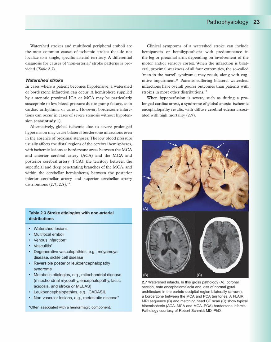
Watershed strokes and multifocal peripheral emboli are the most common causes of ischemic strokes that do not localize to a single, specific arterial territory. A differential diagnosis for causes of ‘non-arterial’ stroke patterns is pro-vided (Table 2.3).
Watershed stroke
In cases where a patient becomes hypotensive, a watershed or borderzone infarction can occur. A hemisphere supplied by a stenotic proximal ICA or MCA may be particularly susceptible to low blood pressure due to pump failure, as in cardiac arrhythmia or arrest. However, borderzone infarc-tions can occur in cases of severe stenosis without hypoten-sion (case study 1).
Alternatively, global ischemia due to severe prolonged hypotension may cause bilateral borderzone infarctions even in the absence of proximal stenoses. The low blood pressure usually affects the distal regions of the cerebral hemispheres, with ischemic lesions at borderzone areas between the MCA and anterior cerebral artery (ACA) and the MCA and posterior cerebral artery (PCA), the territory between the superficial and deep penetrating branches of the MCA, and within the cerebellar hemispheres, between the posterior inferior cerebellar artery and superior cerebellar artery distributions (2.7, 2.8).15
Clinical symptoms of a watershed stroke can include hemiparesis or hemihypesthesia with predominance in the leg or proximal arm, depending on involvement of the motor and/or sensory cortex. When the infarction is bilat-eral, proximal weakness of all four extremities, the so-called ‘man-in-the-barrel’ syndrome, may result, along with cog-nitive impairment.16 Patients suffering bilateral watershed infarctions have overall poorer outcomes than patients with strokes in most other distributions.17
When hypoperfusion is severe, such as during a pro-longed cardiac arrest, a syndrome of global anoxic–ischemic encephalopathy results, with diffuse cerebral edema associ-ated with high mortality (2.9).
Table 2.3 Stroke etiologies with non-arterial
distributions
Watershed lesions
Multifocal emboli
Venous infarction*
Vasculitis*
Degenerative vasculopathies, e.g., moyamoya
disease, sickle cell disease
Reversible posterior leukoencephalopathy
syndrome
Metabolic etiologies, e.g., mitochondrial disease
(mitochondrial myopathy, encephalopathy, lactic
acidosis, and stroke or MELAS)
Leukoencephalopathies, e.g., CADASIL
Non-vascular lesions, e.g., metastatic disease*
*Often associated with a hemorrhagic component.
2.7 Watershed infarcts. In this gross pathology (A), coronal
section, note encephalomalacia and loss of normal gyral
architecture in the parieto-occipital region bilaterally (arrows),
a borderzone between the MCA and PCA territories. A FLAIR
MRI sequence (B) and matching head CT scan (C) show typical
bihemispheric (ACA–MCA and MCA–PCA) borderzone infarcts.
Pathology courtesy of Robert Schmidt MD, PhD.
(A)
(B) (C)

24 Pathophysiology
Multifocal emboli
Small, multifocal emboli typically migrate to distal, border-zone territories (2.10). Watershed and embolic strokes may often coexist (2.8). A low-flow state, most commonly caused by pump (cardiac) failure, may contribute to the poor ‘wash-out’ of microemboli. Patients in the cardiovascular ICU with stroke often have a pattern consistent with both diffuse multi focal emboli and borderzone lesions.18
Undetermined etiology
Even after exhaustive diagnostic studies, a certain number of strokes will have an undetermined cause, a percentage as high as 30% in some studies.19 This category also includes
patients who have more than one feasible stroke etiology. For example, a young woman with a history of tobacco use and complicated migraine, who presents with a stroke and a diagnostic work-up positive for a lower extremity deep vein thrombosis and a patent foramen ovale, has multiple possible contributing etiologies.
2.8 Watershed versus embolic lesions. This patient suffered
cardiogenic shock prior to cardiac valvular replacement. The
lesions on the FLAIR MRI (A,B) and CT scan (C) suggest an
embolic pattern at lower levels (A) but have a more confluent
pattern at higher levels (B,C) consistent with a watershed
pattern.
2.9 Severe anoxic–ischemic encephalopathy. This patient
suffered two cardiac arrests, and had CT scans after each
one. The first scan (left panels) is juxtaposed with the second
(right panels), at several levels: midbrain (A), choroid plexus
calcifications (B), and cerebral hemispheres above the lateral
ventricles (C). Before the second CT scan, a more protracted
arrest caused diffuse, severe cerebral edema seen in the
complete effacement of the sulci in the second study (right
panels).
(A)
(A)
(B)
(C)
(B) (C)

Pathophysiology 25
Case studies
Case study 1. Large vessel disease: intracranial
stenosis causing borderzone infarctions
A 59-year-old woman with a history of tobacco use presents with fluctuating right hemibody symptoms (fluctuating right arm and leg weakness and paresthesias), and was found to have watershed (borderzone) ischemic lesions on diffusion-weighted MRI (DW-MRI) sequence (CS 1.1A), with low perfusion on the cerebral blood flow map (CT perfusion, CS 1.1B).
The etiology was identified as severe left distal M1/ proximal M2 stenosis without hypotension, difficult to visualize on a coronal cervical and intracranial MRA study (CS 1.2A) but magnified on CT angiography (arrow) (CS 1.2B). Angioplasty was performed with a 2-mm
Maverick balloon, immediately before deployment of a 3 9-mm Wingspan™ stent. A post-procedural non-contrast CT scan shows the stent as a hyperdense region coursing into the Sylvian fissure (CS 1.2C).
A second patient with borderzone infarction had a comparable lesion, a right mid-M1 stenosis (CS 1.3) shown
2.10 Distal emboli into borderzone territories. Multifocal
hemispheric emboli, occurring perioperatively during
cardiac valvular surgery, shown on a diffusion-weighted
MRI sequence (A,B). The lesions involve the cerebellar
hemispheres and the superficial–deep MCA–MCA
borderzone of the centrum semiovale. The multifocal lesions
in another case, diffusion-weighted MRI sequence, have
components of embolic as well as borderzone lesions,
with a more confluent, gyriform pattern in the frontoparietal
region (C,D).
CS 1.1
(A)
(B)
(C)
(A) (B)
(D)

26 Pathophysiology
first on MRA (arrowhead) (A). Conventional angiography, (B, C) demonstrates near-occlusive intracranial MCA disease. Elective angioplasty was performed; the lesion is first measured (B); the true lumen of the MCA is >2.0 mm, and the lesion is nearly 5 mm long. Pre-angioplasty (C, left) versus post-angioplasty (C, right) angiography shows improved vessel diameter of the stenotic region. Notice
also the slower filling of the MCA compared with the ACA (C, left) before treatment, and then comparable filling of the two arteries following treatment (C, right; oval).
Comments
Endovascular technology is evolving to transform the management of large vessel intracranial and extracranial disease. Although the experience with angioplasty and stenting for extracranial ICA atherostenosis is extensive, with many randomized clinical trials published,21 clear guidelines are not established for the management of intracranial disease. Some early periprocedural data for the Gateway balloon-Wingspan™ stent system in patients with symptomatic intracranial disease found a high rate of technical success and acceptable periprocedural morbidity.22
CS 1.2
CS 1.3
(A)
(A)
(B)
(C)
(B)
(C)

Pathophysiology 27
These techniques may be useful for elective, secondary prevention as well as emergency revascularization. The risks of endovascular cervical and intracranial procedures include reperfusion injury (e.g., the carotid reperfusion syndrome); perforator infarction; arterial dissection; and distal emboli.23
Case study 2. Cardioembolic disease: calcific
embolism causing middle cerebral artery stroke
History
A 36-year-old man developed a left hemispheric stroke syndrome during a cardiac catheterization procedure. He had a history of a congenital bicuspid aortic valve, and was undergoing catheterization to assess his risk for coronary heart disease and the status of his aortic stenosis. Given femoral access already in place from the cardiac procedure, a distal left ICA occlusion was identified on fluoroscopy, where intra-arterial tissue plasminogen activator, 25 mg, was infused prior to transfer to the regional stroke center.
Clinical course
Hospital day (HD) 1
On arrival, the patient was taken directly to the inter-ventional neuroradiology suite. Conventional angiogra-phy, anteroposterior view, left ICA injection (CS 2.1) shows occlusion of the M1 segment (arrowhead) (A, left). After recanalization efforts using a clot retrieval device, the Mechanical Embolus Removal in Cerebral Ischemia (MERCI) LX, and angioplasty in the first- and second-order MCA (M1 and M2) segments, multiple filling defects are still observed within both the left first-order ACA (A1) and M1 segments (A, right). On a lateral view (B), the guidewire is shown entering the M1 segment, with excellent flow into the A1 (arrow) and A2 (arrowheads) segments (left). With the guidewire removed, flow into the M1 seg-ment remains quite limited, with heterogeneous flow within the distal ICA (arrowhead) and proximal ACA and MCA branches (right). During this procedure, the interventional neuroradiologist noted a large occlusive piece of particulate matter, >2 mm, shifting between the M1 and A1 segments around the distal ICA.
HD 2–3
MCA occlusion (CS 2.2): A calcified ‘hyper-hyperdense’ MCA sign is shown on the non-contrast head CT scan (A,B), and on MRI (C), as a hypointense signal (arrow) on the source images of the intracranial MR angiogram at
the level of the circle of Willis (C, left), and no flow regis-ters into the left M1 segment on the MRA (C, right).Transesophageal echocardiography (CS 2.3): The source of the MCA lesion is confirmed on a long axis view of the aortic valve demonstrating the presence of valvular thickening and calcification (A), and a short axis view of the aortic valve during systole (B).
Follow-up CT scans (CS 2.4) on HD4 (A) and at 6 months post-stroke (B) document the large left MCA stroke.
Comments
Stroke in the setting of cardiac catheterization is not uncom-mon, and is attributed to the dislodgement of clot or athero-matous debris off the aortic arch or directly from the guide catheter tip, and less frequently, valvular debris.24 In this case, the initial head CT scan and echocardiography iden-tified the stroke etiology as particulate thromboembolism emanating from the patient’s calcified aortic valve.
CS 2.1
(A)
(B)

28 Pathophysiology
CS 2.2
(A)
(C)
(B)

Pathophysiology 29
References
Adams H, Jr, Bendixen B, Kappelle L,1. et al. Classification of acute ischemic stroke: definition for use in a multicenter trial. Stroke 1993; 24: 35–41.Publications Committee for the Trial of ORG 10172 in 2. Acute Stroke Treatment (TOAST) Investigators. Low molecular weight heparinoid, ORG 10172 (Danaparoid) and outcome after acute ischemic stroke: a randomized controlled trial. JAMA 1998; 279: 1265–72.Selwa L, Chimowitz M. Atherosclerotic intracranial large 3. artery occlusive disease. Neurologist 1995; 1: 53–64.Amarenco P, Cohen A, Tzourio C, 4. et al. Atherosclerotic disease of the aortic arch and the risk of ischemic stroke. N Engl J Med 1994; 331: 1474–9.
French Study of Aortic Plaques in Stroke Group. 5. Atherosclerotic disease of the aortic arch as a risk factor for recurrent ischemic stroke. N Engl J Med 1996; 334: 1216–21.Ezekowitz M, Levine J. Preventing stroke in patients 6. with atrial fibrillation. JAMA 1999; 281: 1830–5.Schneck MJ, Xu L, Palacio S. Cardioembolic Stroke 7. 2005. (Accessed at http://www.emedicine.com/neuro/topic45.htm/ from eMedicine, last updated February 13, 2008).Mas J-L, Arquizan C, Lamy C, 8. et al. Recurrent cerebrovascular events associated with patent foramen ovale, atrial septal aneurysm, or both. N Engl J Med 2001; 345: 1740–6.Rahmatullah A, Rahko P, Stein J. Transesophageal 9. echocardiography for the evaluation and management of patients with cerebral ischemia. Clin Cardiol 1999; 22: 391–6.Albers G, Amarenco P, Easton J, Sacco R, Teal P. 10. Antithrombotic and thrombolytic therapy for ischemic stroke: The Seventh ACCP Conference on Antithrombotic and Thrombolytic Therapy. Chest 2004; 3: S483–512.Fisher C. Lacunar infarcts: a review.11. Cerebrovasc Dis 1991; 1: 311–20.Wessels T, Rottger C, Jauss M, Kaps M, Traupe H, 12. Stolz E. Identification of embolic stroke patterns by
CS 2.3
CS 2.4
(A)
(A) (B)
(B)

30 Pathophysiology
diffusion-weighted MRI in clinically defined lacunar stroke syndromes. Stroke 2005; 36: 757–61.Blecic S, Bogousslavsky J. Other uncommon 13. angiopathies. In: Bogousslavsky J, Caplan L, eds. Uncommon Causes of Stroke. New York: Cambridge University Press; 2001: 355–68.Bogousslavsky J, Caplan L. 14. Uncommon Causes of Stroke. New York: Cambridge University Press; 2001.Kumral E, Ozdemirkiran T, Alper Y. Strokes in the 15. subinsular territory: clinical, topographical, and etiological patterns. Neurology 2004; 63: 2429–32.Bogousslavsky J, Hommel M. Ischemic stroke 16. syndromes: clinical features, anatomy, vascular territories. In: Adams H, Jr, ed. Handbook of Cerebrovascular Diseases. New York: Marcel Dekker; 1993: 51–94.Gottesman R, Sherman P, Grega M, 17. et al. Watershed strokes after cardiac surgery. Stroke 2006; 37: 2306–11.Caplan L, Hennerici M. Impaired clearance of emboli 18. (washout) is an important link between hypoperfusion, embolism, and ischemic stroke. Arch Neurol 1998; 55: 1475–82.Foulkes M, Wolf P, Price T, Morh J, Hier D. The 19. Stroke Data Bank: design, methods, and baseline characteristics. Stroke 1988; 19: 547–54.
Osborn A. The aortic arch and great vessels. In: 20. Diagnostic Cerebral Angiography, 2nd edn. Philadelphia: Lippincott Williams & Wilkins; 1999: 1–29.Furlan A. Carotid-artery stenting — case open or 21. closed? [editorial]. N Engl J Med 2006; 355: 1726–9.Fiorella D, Levy E, Turk A, 22. et al. US multicenter experience with the Wingspan stent system for the treatment of intracranial atheromatous disease: periprocedural results. Stroke 2007; 38: 881–7.Levy E, Chaturvedi S. Perforator stroke following 23. intracranial stenting: a sacrifice for the greater good? Neurology 2006; 66: 1803–4.Khatri P, Kasner S. Ischemic strokes after cardiac 24. catheterization: opportune thrombolysis candidates? Arch Neurol 2006; 63: 817–21.
Further reading
Adams H, Jr, Bendixen B, Kappelle L, et al. Classification of acute ischemic stroke: definition for use in a multicenter trial. Stroke 1993; 24: 35–41.
Bogousslavsky J, Caplan L. Stroke Syndromes, 2nd edn. New York: Cambridge University Press; 2001.

Chapter 3
Anterior Circulation
Introduction: anatomy
The arterial site of an embolic or thrombotic occlusion determines the anatomic classification of an ischemic stroke. Strokes within the distribution of the carotid arteries are referred to as lesions of the anterior circulation, and strokes within the distribution of the vertebrobasilar arteries are referred to as lesions of the posterior circulation.
The major arteries of the anterior circulation are the internal carotid (ICA), middle cerebrals (MCA), and ante-rior cerebrals (ACA). Eighty percent of blood flow to the brain runs through the carotid system. The major arteries of the posterior circulation are the vertebrals, basilar, and the posterior cerebrals (PCA) (3.1A,B). Neuroimaging studies, either computed tomography (CT) or magnetic resonance imaging (MRI) scans, can identify the arterial territory and size of acute infarcts. Outstanding MRI-based mappings of the anterior and posterior arterial circulations have been published (e.g., 3.1C).1,2
A critical predictor of stroke outcomes is the competency of the collateral circulation to arteries adjacent to the occluded vessel. The primary anastomoses exist through the circle of Willis, connecting the major arteries of the an terior and posterior circulation systems via the anterior and posterior communicating arteries (3.2A; case study 1).3 This circle is complete in approximately 25% of normal individuals. There are also important secondary collateral pathways provided by leptomeningeal vessels linking the territories of the MCA and the ACA and the MCA and the PCA (3.2B).4 During a proximal MCA occlusion, retrograde collateral flow can be supplied from the adjacent ACA leptomeningeal segments that supply the peripheral MCA territory (3.2C,D).
Internal carotid artery territory
Proximal internal carotid artery
The most common site for large vessel atherosclerosis (TOAST mechanism 1) is at the carotid bifurcation (3.3–3.6).5 Other typical locations are in the carotid siphon and the proximal common carotid artery. Most often, symptom-atic carotid atherosclerotic disease results in artery-to-artery embolism to its MCA territory (3.6). Occlusion of the ICA can result in a wide range of lesions, from multifocal small embolic ischemic lesions (3.6A,B) to massive hemispheric stroke (3.7).6,7
Intracranial branches
The ICA has seven segments named for adjacent structures (3.3A). There are no extracranial branches, but typically there are three intracranial branches proximal to the term-inal bifurcation of the ICA into the MCA and ACA.8,9 These branches are the ophthalmic artery, the posterior commu-nicating artery, and the anterior choroidal artery (3.8). The ophthalmic artery chiefly supplies the eye, via its branching into an end-artery, the central retinal artery.10 An embolism to the central retinal artery commonly results in monocular blindness due to infarction of the retina (3.9). Embolism to the anterior choroidal artery is rare (3.10).11
Distal internal carotid artery branches
The ICA terminates with its division into the MCA and ACA. This is called the ‘carotid T’ because of its appearance (3.11A,B). An occlusion of the ICA terminus, a carotid T lesion, is a common cause of severe stroke (3.7, 3.11, 3.12). The thrombus occludes the distal-most part of the ICA, extending into the proximal segments of the MCA (M1)
31
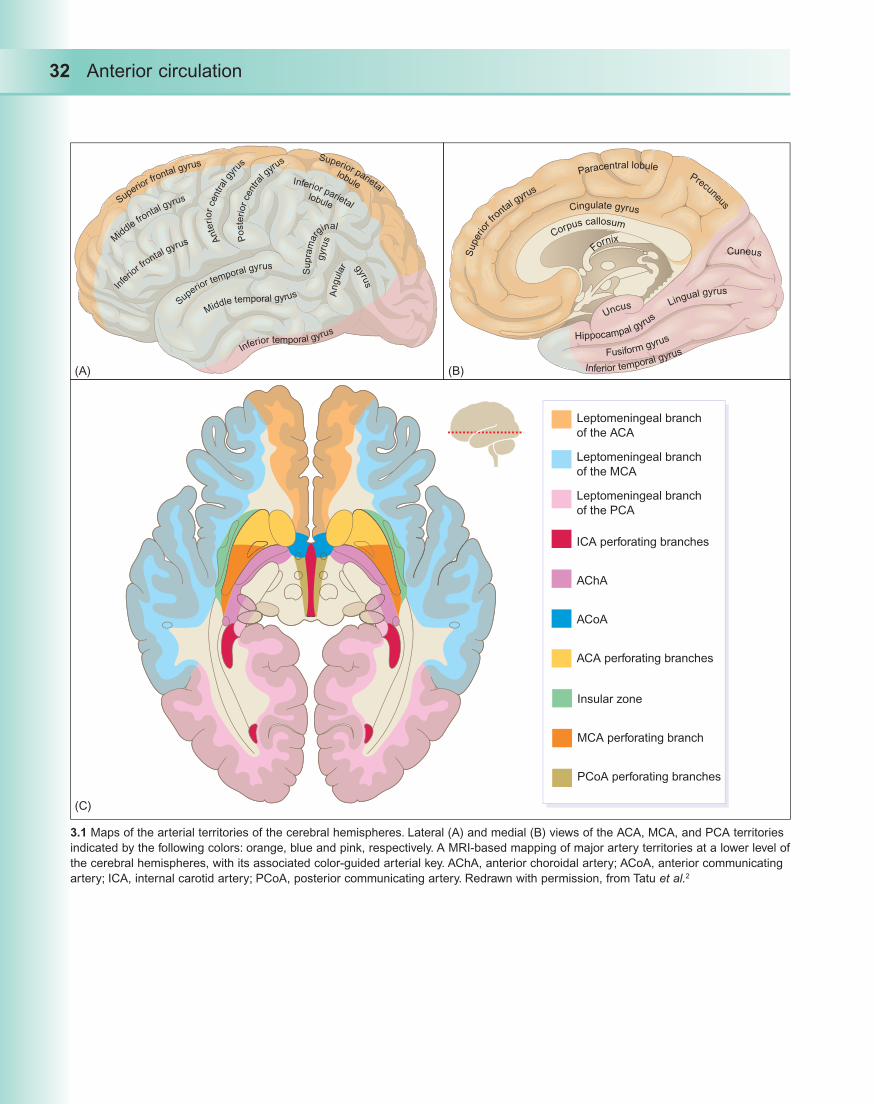
32 Anterior circulation
Superior fr
ontal gyrus
Mid
dle frontal gyrus
In
ferio
r fro
ntal gyrus Ante
rior
central
gyr
us
Poste
rior
cent
ral g
yrus
Inferior parietal
Superior parietal
lobule
lobule
Supra
marg
inal
gyru
sA
ngula
r gyrus
Inferior temporal gyrus
Middle temporal gyrusSuperio
r temporal gyrus
Super
ior f
ronta
l gyrus
Fusiform gyrus
Inferior temporal gyrus
Lingual gyrus
Cuneus
Uncus
Fornix
Precuneus
Paracentral lobule
Cingulate gyrus
Corpus callosum
Hippocampal gyrus
3.1 Maps of the arterial territories of the cerebral hemispheres. Lateral (A) and medial (B) views of the ACA, MCA, and PCA territories
indicated by the following colors: orange, blue and pink, respectively. A MRI-based mapping of major artery territories at a lower level of
the cerebral hemispheres, with its associated color-guided arterial key. AChA, anterior choroidal artery; ACoA, anterior communicating
artery; ICA, internal carotid artery; PCoA, posterior communicating artery. Redrawn with permission, from Tatu et al.2
(A)
(C)
(B)
Leptomeningeal branch
of the ACA
Leptomeningeal branch
of the MCA
Leptomeningeal branch
of the PCA
ICA perforating branches
AChA
ACoA
ACA perforating branches
Insular zone
MCA perforating branch
PCoA perforating branches

Anterior circulation 33
Anterior
communicating
artery Anterior cerebral artery
Internal carotid artery
Middle cerebral artery
Infundibulum
Posterior communicating
artery
Perforating arteries
Superior cerebellar artery
Posterior cerebral artery
Adenohypophysis
(anterior lobe of
pituitary gland)
Ophthalmic artery
Optic nerve
a
ab
b
cc
Anterior circulation Posterior circulation
3.2 Collateral circulation. The confl uence of the anterior and posterior
circulations at the circle of Willis. (A) In addition to providing primary
connectivity between the major intracranial arteries, the circle of
Willis also provides critical perforating end-arteries from the MCA,
ACA, PCA, and the ACoA and PCoA (Chapter 2, 2.4). Adapted
from Osborn.3 Intracranial arterial collateral circulation systems are
shown in lateral and frontal views (B). Common secondary pathways
are the leptomeningeal anastomoses between the ACA and MCA
(a), and between the PCA and MCA (b); as well as tectal plexus
between PCA and SCA (c). Adapted from Liebeskind.4 A conventional
angiogram (anteroposterior view) of a left M1 occlusion (arrow)
(C, left). A later phase image shows some ACA and leptomeningeal
collateral fl ow over the left hemisphere (C, right). Following
recanalization of the M1 occlusion (D), note the balanced ACA and
MCA fl ow in early (D, left) and later (D, right) stages of dye injection.
(A)
(B) (C)
(D)

34 Anterior circulation
and ACA (A1). Rarely, the entire cerebral hemisphere may be at risk, either because the ICA territory also includes the PCA territory due to a fetal PCA circulation (Chapter 4, 4.17), or because rapid uncal herniation creates an occlusive mass effect on the ipsilateral PCA (3.12).
Symptoms of internal carotid artery stroke
When the ICA is occluded, the clinical syndrome is usu-ally one of an MCA occlusion. The anterior communicating artery typically prevents infarction of the ipsilateral ACA by preserving blood fl ow from the contralateral ICA. The next two sections review symptomatology of MCA and ACA infarcts.
Middle cerebral artery territory
The initial segment of the MCA, known as the M1 or fi rst-order division, is a direct continuation of the distal ICA and is larger than its adjacent counterpart in the ACA, the A1 segment (3.13; case study 1). Thromboembolic material in the anterior circulation tends to fl ow into the distribution of the MCA, making it by far the most common territory for ischemic stroke. Strokes within an MCA territory may also be subdivided by the location of the occlusion within the MCA, more proximal M1 and M2 lesions (fi rst- and second-order divisions) versus more distal M3 and M4 lesions (opercular and cortical branches, respectively) (3.13–3.19).12
Ophthalmic
artery
Petrolingual
ligament
Communicating (C7)
Ophthalmic (C6)
Cavernous (C4)
Clinoid (C5)
Lacerum (C3)
Petrous (C2)
Cervical (C1),
carotid bulb
Cervical (C1),
ascending
segment
Carotid canal
3.3 ICA and carotid bifurcation. The course and
branches of the cervical and intracranial ICA, with its
segments, named for adjacent structures: the cervical,
petrous, lacerum, cavernous, clinoid, ophthalmic, and
communicating segments (A). Adapted with permission
from Osborn.8,9 Two conventional angiographic images
of the carotid bifurcation (B). The fi rst study (left) shows
only mild atherosclerosis, while the second (right) shows
typical proximal ICA plaque (arrow). Extensive branching
of the external carotid arteries is seen, while the internal
carotid artery has no cervical branches.
(A)
(B)
Text continued on page 42

Anterior circulation 35
3.4 Normal and abnormal carotid anatomy, Doppler sonography. Normal carotid bifurcation, color Doppler (A). Normal waveform
with peak systolic velocity of 70 cm/s and end diastolic velocity of 25 cm/s is shown (B). Two areas of smooth echogenic plaque are
identified by arrows (C). Calcified plaque creates a bright echogenic region that causes shadowing (arrow) (D). Carotid stenosis <50%
causes minimal change in the baseline velocities (E). This area of smooth plaque (arrows) caused 80–99% stenosis, calculated by
marked velocity elevations (peak systolic velocity of 605 cm/s and end diastolic velocity of 211 cm/s) (F). Images courtesy of Janice
Guica, RN, RVT.
(A)
(C)
(E)
(B)
(D)
(F)

36 Anterior circulation
3.5 Severe carotid atherosclerotic disease. CT angiography, coronal image (A), documents heavy calcified carotid bifurcations,
bilaterally; the hyperdense patches are calcified plaque (arrows). Transaxial cuts of the CT angiogram above the level of the carotid
bifurcation shows the proximal ICA and ECA, documenting calcium, thrombus (arrowhead), and reduced true lumen in the left ICA
(B). Conventional angiography, left common carotid artery injection, documents a high-grade stenosis above the bifurcation of the ICA
(arrow) (C, left), an area magnified and measured for angioplasty and stenting (C, right). Following the placement of two stents, before
(D, left) and during (D, right) carotid injection, demonstrating excellent flow through the formerly near-occlusive lesion on the latter image.
(A)
(C)
(B)
(D)

Anterior circulation 37
3.6 Symptomatic carotid disease: multifocal emboli. Two small, left MCA
territory embolic showers 5 days apart, due to unstable proximal internal
carotid artery plaque, shown on separate diffusion-weighted MRI sequences
(A,B). MRA suggests proximal left ICA disease (arrow) (C), later treated with
an endarterectomy. Gross pathology of a carotid bifurcation plaque, 4 cm
long, removed en bloc during carotid endarterectomy (D). Dissection of the
plaque demonstrated the ulcerated surface (E). Pathology, courtesy of Dean
Uphoff, MD. Gross pathology of dark organized in situ thrombus distal to ICA
bifurcation plaque (F). Pathology, courtesy of Louis Caplan, MD.
(A)
(B)
(D)
(E)
(F)
(C)

38 Anterior circulation
3.7 Right ICA territory stroke in evolution. Non-contrasted CT scan demonstrates a hyperdense distal ICA/proximal MCA sign (arrowheads)
signifying acute thrombus involving these arteries (A). The following three images show the progressively demarcated infarction in the right
ACA and MCA territories, on admission, with loss of sulcal markings consistent with early edema (B), 1 day later (C), and 2 days later (D).
Anterior
falx artery
Recurrent
meningeal artery
Central
retinal artery
Optic
nerve
Ophthalmic
artery
Ciliary
arteries
C5
C6 C7
Anterior
cerebral
artery
Middle
cerebral
artery
Anterior
choroidal
artery
Posterior
communicating
arterySuperior
hypophyseal
arteries
3.8 Intracranial branches of the ICA. A diagram of the intracranial branches of the ICA (A); adapted from Osborn.8 Two angiographic
studies, lateral images, of carotid injections. (B) Distal ICA (carotid T) lesion, just distal to the PCoA (arrow), with most choroidal fl ow
appreciated via the posterior choroidal artery. The distal catheter tip is shown (arrowhead), as is the ophthalmic artery (small arrow)
and the choroidal blush of the eye (small arrowheads); (C) Distal ICA/proximal MCA (M1) occlusion. The lack of visualization of the
MCA, occluded proximally, enables an excellent view of the entire ACA (arrows are on A2 segment).
(A)
(A)
(B) (C) (D)
(B) (C)

Anterior circulation 39
3.9 Retinal stroke. Acute-onset painless vision loss occurs with occlusion of the central retinal artery. Vision was partially recovered due
to maintenance of macular blood supply via a patent cilioretinal artery (arrows), shown here on fundus photograph (A) and fluorescein
angiography (B).
3.10 Infarct of the anterior choroidal artery. Diffusion-
weighted (A) and T2-weighted MRI sequences (arrows) (B).
Note involvement of the choroid plexus of temporal horn of
the lateral ventricle (arrow).
(A)
(A)
(B)
(B)

40 Anterior circulation
3.11 The carotid T lesion. (A) An intact left ‘carotid T’ is shown on the left image, ICA injection, with the distal ICA branching into
the ACA (medial branch) and MCA (lateral branch). A carotid T occlusion is shown on the right. A sharp cut-off of the artery is
marked (arrow), with no flow into the ACA or MCA. Most of the flow is seen posteriorly into the adjacent PCA territory. A composite
transaxial (top-down) view, intracranial MRA (B) shows almost no signal in the right intracranial ICA, in contrast to the normal left side.
A right ICA-distribution stroke on composite CT images (C): note the midline mass effect, squeezing the midbrain and trapping the
contralateral lateral ventricle.
(A)
(C)
(B)

Anterior circulation 41
3.12 Stroke of the entire left hemisphere due to ICA occlusion. The infarct involves the ACA, MCA, and PCA territories. The ACA
territory has infarcted because there is no ACoA and/or due to subfalcine herniation. The PCA territory has infarcted because of mass
effect caused by uncal herniation. Note the spared left-sided territories: midbrain, medial thalamus, rostrum of the corpus callosum, and
some parasagittal cortex.
Internal
carotid artery
Cortical (M3)
segment (MCA)
Cortical (M3)
segment (MCA)
Lateral
lenticulostriate
arteries
Horizontal (M1)
segment (MCA)
Sylvian (M2)
segment (MCA)
3.13 Anatomy of the MCA. A coronal illustration of the proximal MCA and lenticulostriate perforating arteries around the circle of Willis,
as well as the more distal divisions of the MCA.
(A)
(C)
(B)
(D)

42 Anterior circulation
M1 occlusion
M1 occlusions may occur proximal to the lenticulostriate perforating arteries, blocking flow to cortical and subcortical structures. The lenticulostriate arteries supply the internal capsule and other adjacent subcortical structures and are end-arteries (without collateral circulation). The malignant MCA syndrome, in which significant hemispheric edema develops, carries a 70–80% mortality rate with medical treatment only (3.14).13 Uncommonly, downward her-niation may also occlude the ipsilateral PCA, resulting in infarction of medial temporal and occipital lobes (3.15).
Perforating branches
Occlusion of the lenticulostriate small vessels is the most common site for lacunar stroke syndromes. The medial
lenticulostriates (from the proximal MCA near the ICA bifurcation) and the lateral lenticulostriates (from the distal half of the M1 segment) are the largest group of small intracranial perforator vessels. Lacunar syndromes in this region usually affect the basal ganglia, internal capsule, caudate nucleus, lentiform nucleus, as well as the periventricular white-matter (e.g., Chapter 2, 2.4, 2.5). Lacunar strokes can result from intrinsic disease of the perforating artery (TOAST mechanism 3) or from occlusion of the MCA supplying the perforating arteries. Larger branch-artery occlusive disease such as partial occlusion of the M1 segment may result in larger, confluent subcortical lesions, colloquially known as ‘lagunes’ (i.e., for giant lacunes) or ‘macunes’ (i.e., for major lacunes) (3.16).14
3.14 Left M1 occlusions. The time interval between the first (A)
and second (B) CT scan series was 4 days. Note the loss of
sulcal markings, a common early infarct sign, in the left MCA
territory when compared with the right hemisphere (A), and,
later, the severe left-to-right left shift compressing the lateral
ventricle (B). A separate case of an early left M1 occlusion is
shown on transaxial diffusion-weighted MRI sequences (C)
with its apparent diffusion coefficient map (D), to show the wide
hemispheric involvement.
(A)
(C) (D)
(B)

Anterior circulation 43
Cortical branches
M2 occlusions occur distal to the lenticulostriate arteries and will usually produce predominantly cortical infarction. These lesions may be quite large, and a practical clinical differenti-ation is between the superior and inferior division M2 lesions described in the next section on symptoms (3.17, 3.18).
M3 and M4 occlusions occur in one or two of the distal-most branches of the MCA. The area of infarction and symptoms will depend on which individual branch is occluded. Each M2 or M3 segment (stem artery) gives rise
to one to five cortical M4 branches, for a total of 12 major cortical areas (3.19).12 Individual M4 branch infarctions are typically the result of occlusion by small emboli (3.20) or may result after resolution of a more proximal M2 or M3 occlusion (3.21).
Multifocal embolic showers are quite common sequelae of atheroembolism from more proximal sources. Aortic and cardiac sources (TOAST mechanisms 1 and 2) have the potential to cause bilateral hemispheric infarcts, and emboli from the carotid bifurcation (TOAST mechanism 1) may cause multiple ipsilateral hemispheric infarcts (3.6, 3.20, 3.21). Small cholesterol-laden emboli from the ICA and emboli from other sources, such as intracardiac thrombus, carotid dissection, or bacterial endocarditis, may also cause distal occlusions.
As MCA occlusion is the most common location for infarction, high-risk patients may suffer recurrent strokes in the same territory. Both CT and MRI scans can document sequential lesions, to offer a record of acute-upon-chronic strokes (3.22).
Neuroimaging can track the progression of an acute infarct, predicting the eventual lesion size (3.23).
Symptoms of middle cerebral artery stroke6,12,15
Stroke symptoms depend on which section of the MCA is occluded, whether there is total or partial (branch) occlusion, the pathophysiology (large vessel versus small vessel disease), and the laterality of the lesion (right versus left hemisphere).
3.15 MCA plus PCA infarction. An acute MCA occlusion causing supratentorial herniation may occlude the adjacent PCA, resulting in
a combined MCA/PCA infarction. Note the mass effect on the midbrain (A), involvement of lenticulostriate areas and the head of the
caudate nucleus (B,C), and an area of hemorrhagic transformation (arrow) (B). (An alternative mechanism is embolism from an ICA
atherosclerotic disease into both a fetal PCA and an adjacent MCA).
3.16 A ‘Macune.’ A subcortical infarct in the left periventricular
white matter on MRI sequences: a FLAIR (left), and the
gradient echo (GE) sequence (right) demonstrates hemorrhagic
transformation (arrows) of this lesion.
(A) (B) (C)
Text continued on page 48

44 Anterior circulation
3.17 Superior versus inferior M2 territory lesions. Occlusions of the M2 superior and inferior divisions of the MCA are
juxtaposed, beyond or at the level of the MCA bifurcation, on early CT scans, A (superior), B (inferior). Angiography of
ICA injections, showing a superior M2 branch acute occlusion (C, left) and an inferior M2-branch (C, right) (arrowheads).
An inferior M2 division stroke (left) and a superior M2 division stroke (right) shown on transaxial diffusion-weighted MRI
sequences (D); and reconstructed coronal diffusion-weighted MRI (E). Gross pathology, coronal section, shows the
severe loss of tissue from a chronic MCA territory stroke (F). Pathology courtesy of Robert Schmidt MD, PhD.
(A)
(C)
(E)
(B)
(D)
(F)

Anterior circulation 45
3.18 Distal MCA occlusion, recanalized. This lesion, a saddle embolus at the branch point of the distal superior M2 division, is shown
on conventional angiography: pre-treatment (arrowhead) (A); microcatheter injection isolates this lesion (arrow) (B); and post-treatment
(C), with resumption of M2 fl ow into the parietal lobe.
3.19 M3, M4 territory lesions: peripheral middle
cerebral artery branches. (A) Diagram showing how
the inferior and superior distal-most branches of the
MCA travel through the Sylvian fi ssure, wrapping
around the hemispheres to supply their cortical
territories. Adapted from Osborn.12 Conventional
angiography, lateral view, internal carotid artery
injection, demonstrates the multiple distal MCA (B),
as well as a fetal (ICA origin) PCA (arrow). Two
left MCA distal embolic infarctions (C,D), typical
peripheral wedges on the diffusion-weighted MRI
sequences. A gross autopsy specimen (E) shows
a dark area of gliosis and encephalomalacia, with
associated ex vacuo dilatation of the ipsilateral
lateral ventricle. Pathology courtesy of Louis
Caplan, MD.
(A)
(A)
(C)
(D) (E)
(B)
(B) (C)

46 Anterior circulation
3.21 Acute ICA occlusion causing MCA infarction. MRA documents a left cervical ICA occlusion, coronal image (A, left), with poor
intracranial MCA flow (A, right). Though a wide region of hypoperfusion is suggested by the perfusion CT scan (cerebral blood flow (B)
and mean transit time (C) studies), a smaller eventual infarct size is suggested by a relatively smaller area of reduced cerebral blood
volume (arrowhead) (D). Subsequent MRI scan demonstrated a small cortical-branch infarct in the parietal lobe (diffusion-weighted
MRI (E, left) and FLAIR (E, right) sequences). The stroke etiology was likely ‘stump embolism’ from the ICA occlusion.
(A)
(C)
(A) (B) (C)
(D) (E)
(B)
3.20 Distal, peripheral emboli. Bihemispheric emboli (A) indicate
a cardiac or aortic rather than a unilateral carotid source.
Individual small thromboemboli typically create lesions in gyral
patterns (B; and C, left), or watershed territories (C, right) (see
also 3.6A,B).

Anterior circulation 47
3.22 Old versus new stroke on CT scans (A,B). Two cases of acute ischemic stroke in evolution
adjacent to earlier, old strokes. The new lesions (right images, in the anterior right hemispheres)
are less hypodense, with associated early edema, while the older lesions demonstrate
encephalomalacia (same density as cerebrospinal fluid).
3.23 An acute lesion goes chronic. An acute M2 inferior-division middle cerebral artery lesion is shown initially (A) and then
9 months later (B) on serial FLAIR MRI sequences. Notice how the edema of early injured brain parenchyma develops into areas
of cortical laminar necrosis.
(A) (B)
(A)
(B)

48 Anterior circulation
Symptoms of complete M1 occlusion (3.14)
Contralateral hemiplegia/paresis, sensory loss, and gaze palsy (causing gaze preference to the side of the stroke).Left M1 occlusion: global aphasia. Note: rarely, there is right hemisphere dominance for speech.Right M1 occlusion: left hemispatial neglect.Lesions involving >50% of MCA distribution have a high risk for development of the malignant MCA syndrome, with impending herniation during days 2–5 post-onset, heralded by a declining level of consciousness.
Symptoms of superior division M2 occlusion (3.17, 3.18)
Contralateral paresis and sensory loss of face and arm greater than that of the leg. The leg is represented more medially along the motor strip, in the parasagittal terri-tory of the ACA.Contralateral gaze palsy.Left hemisphere: Broca’s (expressive, non-fluent) aphasia.Right hemisphere: spatial neglect.
Symptoms of inferior division M2 occlusion (3.17)
Contralateral superior quadrantic (or hemianopic) visual loss.Left hemisphere: Wernicke’s (receptive, fluent) aphasia.Right hemisphere: spatial neglect.Note the lack of motor or sensory impairment in this distribution, making this a more difficult stroke to diagnose. Patients with Wernicke’s aphasia may be misinterpreted as being confused, but the association with the visual field defect clarifies this as a stroke syndrome.
Symptoms of more distal occlusions (3.19, 3.20, 3.21)
The sensory or motor deficits are patchy, and most often affect the face and/or arm/hand because these body parts have large representation in the homunculus. Conversely, more proximal, subcortical, or brainstem lesions usually cause a more uniform hemiparesis of face, arm, and leg due to the tight collection of descending motor tracts. Diffusion-weighted MRI is the best modality to detect these smaller lesions in the acute phase.
Anterior cerebral artery territory
ACA strokes are much less common than MCA strokes because of the preferred pathway of emboli from the ICA
into its larger and more direct extension, the MCA, and because an intact anterior communicating artery provides a source of collateral circulation to the ACA. The chief deter-minants of lesion size are how proximal within the ACA distribution the occlusion occurs and how wide this artery’s territory extends, based upon the anatomy of the adjacent MCA and the ACA–MCA leptomeningeal collateral circu-lation. Several conventional angiographic studies show the ACA in anteroposterior (3.11A, 3.17C, 3.18A,C) and lateral (3.8C, 3.19B) views.
Perforating branches
The most proximal perforator branches of the A1 and A2 (3.13, 3.24) segments supply the head of the caudate nucleus, part of the basal ganglia and internal capsule, the anterior commissure, and the rostrum of the corpus callosum. The largest and longest ACA penetrating branch, the recurrent artery of Heubner, usually originates from the A2 segment and mostly supplies the head of the caudate (3.13, 3.24; Chapter 2, 2.4A). The medial lenticulostriate arteries and callosal perforating branches originating from A1 and A2 supply the anterobasal forebrain, corpus callosum genu, septum pellucidum, and fornix (3.24, 3.25; Chapter 2, 2.5A,D).
Cortical branches
The orbitofrontal branches supply the olfactory bulb and tract, the gyrus rectus, and the medial orbital gyrus while the frontopolar arteries supply the ventromedial frontal lobe and the corpus callosum (3.26–3.29).
Symptoms of anterior cerebral artery stroke16,17
Contralateral weakness and sensory loss involving the leg to a much greater degree than the arm and face. This is the opposite pattern of the motor impairment seen in MCA strokes. Small emboli to the ACA territory may present with only intermittent ‘TIAs’ with contralateral leg paresis (3.28).Transient expressive aphasia may occur.If bilateral ACA strokes occur, frontal lobe symptoms such as flattened affect and depression, lack of interest and initiative, and cognitive impairment occur in add-ition to bilateral leg weakness (paraparesis) or paralysis (paraplegia) (3.29).
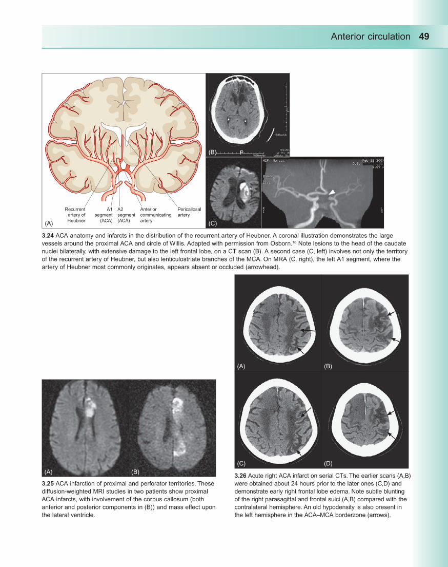
Anterior circulation 49
Pericallosal
artery
Anterior
communicating
artery
A1
segment
(ACA)
A2
segment
(ACA)
Recurrent
artery of
Heubner
3.24 ACA anatomy and infarcts in the distribution of the recurrent artery of Heubner. A coronal illustration demonstrates the large
vessels around the proximal ACA and circle of Willis. Adapted with permission from Osborn.16 Note lesions to the head of the caudate
nuclei bilaterally, with extensive damage to the left frontal lobe, on a CT scan (B). A second case (C, left) involves not only the territory
of the recurrent artery of Heubner, but also lenticulostriate branches of the MCA. On MRA (C, right), the left A1 segment, where the
artery of Heubner most commonly originates, appears absent or occluded (arrowhead).
3.25 ACA infarction of proximal and perforator territories. These
diffusion-weighted MRI studies in two patients show proximal
ACA infarcts, with involvement of the corpus callosum (both
anterior and posterior components in (B)) and mass effect upon
the lateral ventricle.
3.26 Acute right ACA infarct on serial CTs. The earlier scans (A,B)
were obtained about 24 hours prior to the later ones (C,D) and
demonstrate early right frontal lobe edema. Note subtle blunting
of the right parasagittal and frontal sulci (A,B) compared with the
contralateral hemisphere. An old hypodensity is also present in
the left hemisphere in the ACA–MCA borderzone (arrows).
(B)
(A)
(A)
(C)
(B)
(B)
(D)
(C)(A)

50 Anterior circulation
3.28 Acute strokes in cortical branches of the ACA. A small right ACA-distribution embolic stroke (diffusion-weighted MRI sequence
(A, left, and GE MRI sequence, right)), with mild hemorrhagic transformation and calcified falx cerebri (arrowhead) on head CT scan
(B). Restricted diffusion (diffusion-weighted MRI) of a second lesion is shown (C, left), with its accompanying apparent diffusion
coefficient map (C, right). A third case shows punctuate embolic strokes in the left ACA and more lateral MCA territories on diffusion-
weighted MRI (D).
(A) (C)
(B) (D)
3.27 Large ACA infarction. The acute infarct on the diffusion-weighted
MRI sequences involves much of the left hemisphere (A; and B, left).
A T2-weighted sequence shows parasagittal edema, effacing sulci
(B, right). A reconstructed coronal diffusion-weighted MRI shows mass
effect on the frontal horn of the lateral ventricle (C, left). A sagittal
single-shot fast spin echo sequence demonstrates the wide infarct size
(C, right).
(A)
(B)
(C)

Anterior circulation 51
Case studies
Case study 1. Common carotid and M1 occlusions:
intravenous thrombolysis and endovascular
approaches
History
A 59-year-old woman was at home when she presented with new-onset speech difficulty and right hemiparesis. Her daughter witnessed the event, and called ‘911’ within minutes. Her NIHSS score on arrival in the emergency department, 50 minutes after symptoms onset, was 22 points.
Timeline
Hospital day (HD) 1
Emergent management:1
05:30am Onset of symptoms
05:47 Daughter calls EMS.
06:06 Stroke Team pager activated.
06:19 Arrival at Triage Desk:Emergency Department Evaluation.
06:40 Completed CT, CT angiography and CT perfusion studies (CS 1.1).
A hyperdense left MCA sign (indicating the presence of a clot) is present on the initial CT scan (arrow) (A).The region of hypoperfusion is evident on the CT perfusion cerebral blood flow map (B), and prolonged mean transit time (C) as blue regions; however, cerebral blood volume (D) by contrast appears relatively nor-mal, suggesting a cerebral blood flow–cerebral blood volume mismatch and indicating viable penumbra.
07:00 Intravenous tissue plasminogen activator (t-PA) bolus started (0.9 mg/kg, 10% bolus) = 8 mg bolus and 73 mg infusion:
Door-to-drug time = 41 minutes.
3.29 Bilateral ACA infarctions. Two CT scans of chronic, bilateral frontal lobe infarcts,
the first from multifocal cardiogenic emboli (A) and a second due to complications of
the surgical clipping of an intracranial aneurysm of the ACoA (B, and C, a FLAIR MRI
sequence). An old left parietal (MCA territory) infarct is also present (B,C).
CS 1.1
(A)
(B)
(A) (B) (C) (D)
(C)

52 Anterior circulation
07:30 The patient’s daughter was consented for par-ticipation in the Multi-MERCI clinical trial.
Endovascular procedures2 :07–8:00 Mobilization of the Interventional Neuro-
radiology (INR) Team.
08:00 Patient transfer from emergency department to INR Suite.
08:15 Elective intubation, for endovascular proce-dure. (Note: some INR teams routinely use general anesthesia for endovascular proce-dures, while others do not so that the patient’s neurological exam can be clinically monitored during the procedure).
08:30 Diagnostic conventional cerebral angiography (CS 1.2):
Wide left common carotid artery/ICA occlusion (not shown).The primary lesion responsible for this stroke syndrome is a distal left M1 occlusion (arrows), shown on right ICA injection, late phase (A). Note the cross-filling via the anterior communicating artery, the hyperemia within the lenticulostriate
system (arrowheads), and some early leptomeningeal collaterals to the left parietal region from the left ACA circulation (circled region).Other collateralization is appreciated on a left vertebral artery injection, late phase (B), with some left PCA flow supplying left MCA territory (circle).
09:37 The first two attempted Multi-MERCI LX device passes failed to remove any thrombus from within the M1 occlusion. The image shows a non-subtracted view with the MERCI device in the M1 segment, immediately behind the upper margin of the orbit on an antero-posterior projection (CS 1.2C).
10:33 Intra-arterial t-PA infusion (CS 1.3):Traversing the anterior communicating artery, via a right carotid approach (A, unsubtracted; B, subtracted image), with thrombolytic infusion into the local thrombus (arrows) (B). Note how the microcatheter must make a sharp 180-degree turn at the distal right ICA into the adjacent A1 segment (arrowhead) in order to access the anterior communicating artery and then advance into the contralateral, left M1 occlusion.
CS 1.2
(A)
(B) (C)

Anterior circulation 53
10-mg intra-arterial t-PA bolus was injected directly into the M1 occlusion; subsequent 14-mg intra-arterial infusion over 1 hour 10 minutes. Microcatheter infusion demonstrates ongoing, partial recanalization of M1 lesion over time (B,C). A post-infusion injection into the right ICA shows improved flow to much of the left hemisphere (MCA territory) (D). Compare this post-infusion image (CS 1.3D) to the pre-treatment study (CS 1.2A).
12:00 The procedure concluded, and patient is transferred to the ICU.
Hospital course and clinical follow-up.3 HD 2
Extubated.NIHSS = 3 points: expressive aphasia, with right facial and arm paresis.Brain MRI, diffusion-weighted image sequence (CS 1.4),
documents small, predominantly subcortical, patchy lesions within the left MCA territory (A,B).
HD 3
Transferred out of the ICU.Warfarin started to prevent recurrent emboli from the common carotid artery/ICA occlusion.
HD 4
The patient begins to complain of ‘blurry vision’ in her left eye (OS).Her neurologic exam, other than partial loss of vision OS, has normalized.NIHSS = 1 point: monocular visual loss.
HD 5
Transfer to acute rehabilitation.She returns to home 5 days later.
CS 1.3
(A)
(C) (D)
(B)

54 Anterior circulation
30-day outpatient follow-up
Funduscopic photos (CS 1.5) document a normal right eye (A) but multiple branch retinal emboli in the left eye (B), explaining her visual deficit.Much of her visual loss improved, because the fovea (arrow) was largely spared from ischemic injury (B).
Comments
This patient’s NIHSS score of >20 points would carry a high risk for developing a large MCA stroke, with greater than 90% likelihood of post-stroke death or severe disability.18 Factors influencing a favorable outcome included: (1) the early time to arrival at the emergency department and rapid processing, facilitating early exposure to IV t-PA; (2) interventional expertise to access the lesion, via the contralateral ICA, by traversing an intact anterior communicating artery; and (3) the patient’s recruitment of leptomeningeal hemispheric collaterals, suggested by the initial CT perfusion study and subsequent conventional angiography.
This patient’s management was aggressive, in that mul-tiple attempts at endovascular recanalization were made fol-lowing intravenous thrombolysis. Her sole morbidity, partial visual loss OS, was due to thromboembolism into the central retinal artery from the ICA thrombus via its first intracranial branch, the ophthalmic artery. The hemispheric stroke, by contrast, caused no functional limitations.
The emergent treatment of tandem lesions (here, com-mon carotid artery/ICA and MCA occlusions) is evolving. An alternative endovascular approach would have been angioplasty with or without stent placement across the com-mon carotid/ICA occlusion, and then advancing the catheter directly upstream to the MCA occlusion.
References
Tatu L, Moulin T, Bogousslavsky J, Duvernoy H. 1. Arterial territories of human brain: brainstem and cerebellum. Neurology 1996; 47: 1125–35.Tatu L, Moulin T, Bogousslavsky J, Duvernoy H. 2. Arterial territories of the human brain: cerebral hemispheres. Neurology 1998; 50: 1699–708.Osborn A. The Circle of Willis. In: 3. Diagnostic Cerebral Angiography, 2nd edn. Philadelphia, PA: Lippincott Williams & Wilkins; 1999: 105–16.Liebeskind D. Collateral circulation. 4. Stroke 2003; 34: 2279–84.
CS 1.5
(A)
(B)
CS 1.4
(A)
(B)

Anterior circulation 55
Osborn A. Atherosclerosis and carotid stenosis. In: 5. Diagnostic Cerebral Angiography, 2nd edn. Philadelphia, PA: Lippincott Williams & Wilkins; 1999: 359–79.Saver J, Biller J. Superficial middle cerebral artery. In: 6. Bogousslavsky J, Caplan L, eds. Stroke Syndromes. New York: Cambridge University; 1995: 247–59.Bogousslavsky J, Moulin T. Border-zone infarcts. In: 7. Bogousslavsky J, Caplan L, eds. Stroke Syndromes. New York: Cambridge University; 1995: 358–66.Osborn A. The internal carotid artery: cavernous, 8. clinoid, ophthalmic and communicating segments. In: Diagnostic Cerebral Angiography, 2nd edn. Philadelphia, PA: Lippincott Williams & Wilkins; 1999: 83–104.Osborn A. The internal carotid artery: cervical, 9. petrous, and lacerum segments. In: Diagnostic Cerebral Angiography, 2nd edn. Philadelphia, PA: Lippincott Williams & Wilkins; 1999: 57–81.Wray S. Visual symptoms (eye). In: Bogousslavsky J, 10. Caplan L, eds. Stroke Syndromes, 2nd edn. New York: Cambridge University Press; 2001: 111–28.Helgason C. Anterior choroidal artery. In: 11. Bogousslavsky J, Caplan L, eds. Stroke Syndromes. New York: Cambridge University; 1995: 270–6.Osborn A. The middle cerebral artery. In: 12. Diagnostic Cerebral Angiography, 2nd edn. Philadelphia, PA: Lippincott Williams & Wilkins; 1999: 135–52.Vahedi K, Hofmeijer J, Juettler E, 13. et al. Early decompressive surgery in malignant infarction of the middle cerebral artery: a pooled analysis of three randomised controlled trials. Lancet 2007; 6: 215–22.
Phan T, Donnan G, Wright P, Reutens D. A digital 14. map of middle cerebral artery infarcts associated with middle cerebral artery trunk and branch occlusion. Stroke 2005; 36: 986–91.Kumral E, Ozdemirkiran T, Alper Y. Strokes in the 15. subinsular territory: clinical, topographical, and etiological patterns. Neurology 2004; 63: 2429–32.Osborn A. The anterior cerebral artery. In: 16. Diagnostic Cerebral Angiography, 2nd edn. Philadelphia, PA: Lippincott Williams & Wilkins; 1999: 117–35.Sawada T, Kazui S. Anterior cerebral artery. In: 17. Bogousslavsky J, Caplan L, eds. Stroke Syndromes. New York: Cambridge University; 1995: 235–46.Adams HP Jr, Davis P, Leira E,18. et al. Baseline NIH Stroke Scale score strongly predicts outcome after stroke: a report of the Trial of Org 10172 in Acute Stroke Treatment (TOAST). Neurology 1999; 53: 126–31.
Further reading
Bogousslavsky J, Caplan L. Stroke Syndromes, 2nd edn. New York: Cambridge University Press; 2001.
Liebeskind D. Collateral circulation. Stroke 2003; 34: 2279–84.
Osborn A. Diagnostic Neuroradiology, 2nd edn. St Louis, MO: Mosby; 1994.
Tatu L, Moulin T, Bogousslavsky J, Duvernoy H. Arterial territories of the human brain: cerebral hemispheres. Neurology 1998; 50: 1699–708.

Chapter 4
Posterior Circulation
Introduction: anatomy
The posterior circulation, the vertebrobasilar system, receives 20% of the blood fl ow to the brain and supplies the brainstem, cerebellum, and most of the temporal and occipital lobes (4.1). Two vertebral arteries (VA) converge to form the basilar artery (BA). This is the only place in the body where two arteries converge rather than branch,
enabling some redundancy.1,2 If one VA is occluded proximal to the posterior inferior cerebellar artery (PICA), the other vertebral may provide retrograde collateral fl ow to the PICA. Another unusual feature is that, in the neck, the VAs course through the C6 to C1 vertebral transverse foraminae. The bone provides some protection for the arteries; however, degenerative spine disease and traumatic dissection are potential causes for arterial injury (4.1F; Chapter 1, 1.2).
57
Posterior
communicating
artery
Posterior
cerebral artery
Pontine
perforators
Superior
cerebellar artery
Basilar artery
Middle
cerebral artery
Anterior
cerebral artery
4.1 Vertebrobasilar anatomy. The cervical and intracranial vertebrobasilar system is shown on a coronal MRA sequence
(A; Chapter 2, 2.1A). The paired vertebral arteries have extraosseous, foraminal, extraspinal, and intradural segments that run from
the cervical region, through the meninges, to their intracranial termination at the basilar junction.1 The extracranial vertebral arteries
may be diffi cult to fully appreciate on MRA due to the overlying carotid system (A). A view of the posterior circulation ‘draped’ over
the ventral surface of the brainstem and cerebellar hemispheres (B): BA; posterior communicating artery (PCoA); PCA; superior
cerebellar artery (SCA); pontine perforators.
(A) (B)

58 Posterior circulation
Superior cerebellar artery (main trunk)
Anterior inferiorcerebellar artery
Basilar artery
Posterior inferiorcerebellar artery
4.1 (continued) Vertebrobasilar anatomy. The distal vertebral arteries and the pontine perforator branches originating from the BA
are shown on a conventional angiogram (anteroposterior, Townes’ projection) (C); the SCA (arrows) and PCAs (arrowheads) are well
visualized. A lateral view of the cerebellar hemisphere shows the three major cerebellar arteries (D). B and D adapted from Osborn.1
CT scans with contrast: on a sagittal view (E), the relation of the arteries of the posterior circulation to the cerebellum and the proximity
of the BA (arrowheads) to the clivus bone (anterior) are appreciated. Other contrast represents fl ow within the venous sinuses, such as
the straight sinus (arrows). On a coronal view (F), the cervical vertebral arteries are seen to traverse the vertebral foramina.
(C)
(E)
(D)
(F)

Posterior circulation 59
Cerebellum and brainstem
The most common sites for the development of large vessel atherosclerotic disease in the posterior circulation are the branch points of the system at the proximal and distal ends of the VAs and at the distal end of the BA as it divides into the two PCAs (case study 1).1
Three paired cerebellar arteries (PICA anterior inferior, and superior) (4.1D) wrap around the cerebellar hemispheres.3,4 Some smaller cerebellar infarcts are shown (4.2). The largest ischemic strokes in the cerebellar hemispheres are typically in the distribution of the PICA (4.3, 4.4), and, like cerebellar hemorrhage, may require decompressive craniotomy to avoid death of the patient (4.3).
In addition to the cerebellar hemispheres (4.2–4.4), com-mon sites for vertebrobasilar stroke include the paramedian pons (4.5) and the lateral medulla (4.6). The lateral pons (4.7) and the medial medulla (4.8) are uncommon locations. Distal embolic showers through the posterior circu lation often produce multifocal lesions, particularly involving distal branches of the posterior circulation, into the PCA territories. The associated symptoms are often referred to as the ‘top-of-the-basilar’ syndrome (4.9).
Symptoms of vertebrobasilar stroke
The range of symptoms and signs is quite extensive and they can fl uctuate widely over the initial few hours of presen-tation, making diagnosis potentially more complicated than in anterior circulation strokes. The complexity of neurologic
Arterial territories of the
pons
Anteromedial group arising
from the foramen coecum
arteries (basilar artery)
Anterolateral group arising
from the basilar artery
Lateral group arising from
vertebral and anterior
inferior cerebellar arteries
Arterial territories of the
cerebellum
Anterior inferior cerebellar
artery
Lateral branch of the superior
cerebellar artery
Lateral branch of the posterior
inferior cerebellar artery
Medial branch of the posterior
inferior cerebellar artery
4.1 (continued) Vertebrobasilar anatomy. An MRI-based map, transverse section, at the level of the pons (G), demonstrates the
complexity of vascular territories within the posterior fossa. With permission from Tatu et al.2
(G)

60 Posterior circulation
4.2 Cerebellar strokes. Small lesions in the medial and lateral cerebellar hemispheres on diffusion-weighted MRI sequences (A–C). A
chronic infarct in the distribution of the medial branch of the left PICA is a wedge-shaped hyperintense area on MRI (T2WI) (D).
4.3 Cerebellar stroke treated with neurosurgical decompression. Head CT scan (A) and GE MRI sequence (B) show significant
hemorrhagic transformation of a large right PICA distribution stroke, with effacement of the fourth ventricle and mass effect on the
medulla. Postoperative CT scan obtained 7 days later demonstrates the recovery of the basal cisterns (C).
(A)
(C)
(A) (B) (C)
(D)
(B)

Posterior circulation 61
4.3 (continued) Cerebellar stroke treated with neurosurgical decompression. Micropathology of the resected lesion (hematoxylin and
eosin stains) shows hemorrhage (red blood cells, arrows) above and within the pale, infarcted cerebellar tissue (D, 10 ). Magnified
views (40 ) of the tissue show swollen, newly dead Purkinje fibers (arrows) (E) and arteriosclerotic (thickened) arterial walls (arrows)
(F), consistent with long-standing hypertension. Pathology courtesy of Dean Uphoff, MD.
4.4 Infarction of the left PICA.
Diffusion-weighted MRI (A) and
FLAIR MRI (B, right) sequences,
as well as a reconstructed
sagittal diffusion-weighted MRI
sequence (B, left) shows that
PICA supplies the inferior half of
the cerebellar hemisphere.
4.5 Paramedian pontine stroke. A large left pontine infarct is shown on a CT scan (A), and a sagittal diffusion-weighted MRI sequence
(B). Diffusion-weighted MRI sequences of two smaller infarctions in the distribution of the left paramedian pontine perforators (C) affect
primarily the medial motor tracts. (The heightened signal in the bilateral medial temporal lobes (C) is artifactual.)
(D)
(A)
(A) (B) (C)
(B)
(E) (F)

62 Posterior circulation
deficits is amplified when parts of the brainstem are involved in addition to the cerebellum.5 They include:
gait or limb ataxia (patients often fall towards or veer towards the side of the lesion, when ambulating; or have ipsilateral limb ataxia);midline or medial cerebellar lesions may only cause gait imbalance and vestibular symptoms, with no or minimal limb ataxia;6
nystagmus;
diplopia, horizontal or vertical;vertigo, tinnitus, hearing loss;nausea and vomiting;variable patterns of limb weakness and sensory loss;dysarthria and dysphagia.
In general, the positioning of nuclei and tracts within the brainstem dictate that motor (limb and oculomotor) deficits correspond to more medial lesions, while sensory and ataxic symptoms result from more lateral lesions.
4.6 Lateral medullary infarction. A stroke located in the territory of the PICA
is most often related to large vessel vertebral artery disease.17,18 A left lateral
medullary infarct on diffusion-weighted MRI sequence (arrow) (A) was likely
due to an acute left vertebral artery occlusion as seen on MRA sequences,
intracranial (B, left) and subtracted posterior circulation (B, right). The right PICA
is well visualized (arrowheads), while the left PICA is not. Transaxial source
images for this MRA show no signal (flow) in the left vertebral artery, in contrast
to the right vertebral artery (arrow) (C).
4.7 Lateral pontine and cerebellar strokes. An isolated right lateral pontine infarct is shown on diffusion-weighted MRI (A). A second
patient presented with bilateral hearing loss, varying sensory symptoms, and gait imbalance. The responsible embolic lesions primarily
affected bilateral anterior inferior cerebellar artery (AICA) territories in the pons, lateral cerebellum, and cerebellar peduncles on the
diffusion-weighted MRI sequences (B). On a CT scan with contrast, atherostenotic lesions are present in the distal left VA and the
proximal and mid-BA (C). However, on focused sagittal (D, left) and coronal (D, right) windowing, the predominant area of stenosis,
and the likely source of this stroke syndrome, is the mid-basilar artery disease (arrowheads).
(A)
(A)
(B)
(D) (C)
(B)
(C)

Posterior circulation 63
Most important in evaluating the acute stroke patient is simply to localize the patient’s symptoms to the posterior circulation rather than to a specific arterial distribution, as this determines the diagnostic work-up: screening for potential lesions of the VA or BA and embolic sources from the vertebrobasilar tree, the aorta, and the heart.5 Owing to bony artifact in the posterior fossa on CT imaging, MRI
is the preferred modality for identifying infarcts in the posterior circulation.
Basilar artery territory
Occlusion of the BA (4.10, 4.11) are the location of the greatest concern in the posterior circulation. Untreated, BA
4.8 Medial medullary infarction. A CT scan
barely registers the lesion (A), shown readily
on a diffusion-weighted MRI sequence (B). The
etiologies of medial medullary infarction include
large and small vessel atherosclerotic disease
and vertebral artery dissection.17
4.9 Distal emboli: top-of-the-basilar syndrome. A basilar artery occlusion treated with intravenous
and then intra-arterial thrombolysis, with significant recanalization. The diffusion-weighted MRI
sequences (A–C) document patchy, multifocal lesions throughout the posterior circulation, involving
the cerebellar hemispheres, pons, midbrain, occipital lobes, and thalamus.3
(A)
(A)
(B)
(C)
(B)

64 Posterior circulation
occlusion carries a 50–80% mortality rate in case series.7 The high morbidity and mortality reflects the basilar artery’s supply to autonomic centers of the brainstem as well as the paramedian major motor tracts.8,9 If the patient survives, a ‘locked-in’ syndrome is not uncommon. In this situation, the patient may be alert and awake but cannot move with the exception of eye blinking and eye movement. Anatomically, the dorsal brainstem (afferent tracts) is intact and the ventral brainstem (efferent tracts) has infarcted.
By contrast, recanalization of a BA occlusion with mechanical embolectomy or thrombolysis is associated with favorable outcomes in a significant percentage of patients (case study 2). The top-of-the-basilar syndrome occurs when there is occlusion of the distal or rostral portion of the BA usually by an embolus. Clinical presentation includes change in level of consciousness, visual symptoms, includ-ing hallucinations or blindness, oculomotor and pupillary abnormalities, with or without motor symptoms such as decerebrate posturing.
Posterior cerebral artery territory
The PCAs are the distal-most branches of the vertebrobasilar system (4.1). The two most common stroke etiologies are emboli into one or both PCA branches, originating from large vessel atherosclerosis in one of the VAs or within the heart, and small vessel disease affecting the deep perforating branches of the PCAs.3,10,11 In contrast with the middle cerebral artery, proximal PCA atherostenosis is uncommon.
Perforating branches (4.12A)
The first two segments of the PCA, the P1 (pre-communicating) and P2 (ambient) segments, provide local perforator branches to the thalamus, hypothalamus, the posterior limb of the internal capsule, the midbrain, and rostral cranial–nerve nuclei (oculomotor and trochlear nerves).10
The etiology of infarcts involving the central penetrating branches (the thalamoperforating, thalamogeniculate, and peduncular perforating arteries) is small vessel disease
4.10 Basilar artery occlusion. Gross pathology of BA occlusion, extending from the confluence of the VAs and into the left AICA.
Pathology courtesy of Louis Caplan, MD. A case of BA occlusion treated with a multimodal approach. A stent, placed in the BA,
appears as a fence-like structure in the post-procedural intracranial MRA (B). On CT scan (C), the stent is seen as a hyperdense circle
(arrowhead) (left) and metallic artifact with significant subarachnoid hemorrhage (right). Despite excellent recanalization, transaxial
(D, left) and reconstructed coronal diffusion-weighted MRI (D, right) sequences document bilateral pontine infarction.
(C)
(D)(A) (B)

Posterior circulation 65
(Chapter 2, 2.6). Multifocal lesions in this region can also be embolic; e.g., part of a top-of-the-basilar syndrome (4.9).12
Cortical branches (4.12B)
The more proximal occipital artery (lateral branch of the PCA) supplies most of the temporal lobe, particularly the inferomedial temporal region. The more distal medial occipi-tal artery (posterior, medial branch of the PCA) supplies the occipital lobe, including the primary calcarine (visual) cortex and often the adjacent parieto-occipital region. A variety of cortical PCA lesions is shown (4.12–4.16).
The fetal posterior cerebral artery variant (4.17)
Frequently, the PCA does not develop into a distal branch of the BA, but remains a fetal remnant in which
it continues as an extension of the ICA. The so-called ‘fetal PCA’ is the most common variant of the cervical and intracranial arterial territories, and occurs unilater-ally in 5–10% of all individuals.10 With this anatomy, the proximal PCA is larger than the adjacent posterior com-municating artery, and symptomatic carotid atherosclerotic disease may embolize into an ipsilateral fetal PCA as well as middle cerebral artery and anterior cerebral artery ter-ritories (Chapter 3).13
Symptoms of posterior cerebral artery stroke
The most prominent symptom of a cortical PCA stroke is usually a contralateral visual field deficit, a homonymous hemianopia or quadrantanopia. Central vision loss occurs when the occipital pole is involved. If bilateral PCA strokes
4.11 Basilar artery occlusion, following failed recanalization. This 70-year-old patient presented with a rapid progression from
hemiparesis to quadriparesis, diminished level of consciousness, small and poorly reactive pupils, acute respiratory failure, and
bilateral Babinski signs. Urgent angiography documented distal BA occlusion, but endovascular attempts were unsuccessful.
Conventional angiography, right vertebral artery injection (A), shows the final appearance of the occluded mid-BA on anteroposterior
(left) and lateral (right) projections. The MRI study on the following day shows hyperintense signal on the FLAIR sequence within the
BA (arrow) (B). The massive infarct on DW-MRI sequence, transaxial (C,D), coronal (E), and sagittal (F) images, encompasses the
territories of the BA perforators, plus the bilateral SCA and PCA territories: the pons and midbrain, the rostral cerebellum and vermis,
occipital lobes, and thalamus.
(A)
(B)
(C)
(D)
(F)
(E)

66 Posterior circulation
P1 segment
P2 segment and branches
P3 segment
P4 and segments
P1 segment
P2 segment and branches
P3 segment
P4 and segments
4.12 PCA anatomy and ‘complete’ PCA territory infarction. The proximal PCA (A)
wraps around the midbrain, with small vessel infarcts resulting from occlusion
of the perforators off the P1 and P2 segments and the PCoAs. A map of the
cortical branches of the PCA (B), as they originate from the P2 and P3 segments.
Adapted with permission from Osborn.10 An acute infarct of the right P1 segment,
demonstrating involvement of occipital lobe and medial temporal lobes (C,D), the
cerebral peduncle (arrow) (C, right), and the thalamus (D) as well as the splenium
of the corpus callosum (D, right; arrowhead). The lesion on transaxial MRA shows
no fl ow into the right PCA (arrow) (E).
(A)
(C) (D)
(E)
(B)

Posterior circulation 67
involving the occipital lobes occur, there may be cortical blindness.3 However, PCA strokes are noteworthy for a wide range of other associated neurologic defi cits, including memory impairment, visual agnosias, visuospatial processing
diffi culties, and visual hallucinations. When there is thalamic involvement, hemisensory loss, behavioral disorders and higher cognitive processing diffi culties can occur and contribute to vascular dementia (4.16).11,14–16
4.13 Smaller P1 segment infarct. A right P1 occlusion is shown on an isolated intracranial MRA of the posterior circulation
(arrowhead) (A), while the more inferior SCA is intact. The infarct on diffusion-weighted magnetic MRI sequences involves
the anteromedial temporal lobe (B), the choroidal region of the lateral ventricle (C, left), and thalamus (C, right).
4.14 Evolving left PCA infarct. Serial CT scans show an acute occipital infarct (A,B), with effacement of local sulci, and a study done
2 years later (C). The associated encephalomalacia of the chronic lesion results in ex vacuo expansion of the adjacent lateral ventricle (C).
(A)
(A) (B) (C)
(B) (C)

68 Posterior circulation
4.16 Vascular dementia with PCA infarction. Multiple old infarcts,
including a left cortical PCA lesion (A). The thalamus is spared
(A), but extensive encephalomalacia is seen in the frontoparietal
regions, bilaterally (B). A reconstructed sagittal CT image
illustrates how this lesion involves visual cortex in the medial
occipital lobe (C).
(A)
(A)
(C)
(B)
(B)
(C)
4.15 Isolated infarct of the splenium. A 50-year-old woman
presented with the acute onset of higher cognitive difficulties
and memory impairment. The brain MRI scan documents an
acute infarction of the left posterior corpus callosum on DW- (A)
and T2-weighted (B-C) images.

Posterior circulation 69
Case studies
Case study 1. Vertebral artery stenting
This 65-year-old patient had recurrent posterior circulation ischemic events despite an aggressive cardiovascular medical regimen. The distal right VA has an irregular plaque, with a focal >50% stenosis, just proximal to a large PICA on a three-dimensional rendering of the conventional angiography
(CS 1.1A). The lesion was measured (CS 1.1B), and treated with balloon angioplasty and a Wingspan™ stent. On lateral projections (CS 1.1C), right VA injection images are shown before (left) and after stent placement (right), with some increase in the diameter of the lumen. An unsubtracted lateral view shows the relation of the treated VA to the skull base (CS 1.1D); the markers at ends of the stent consist of four radio-opaque points (an arrow is drawn to the proximal end).
4.17 Fetal PCA anatomy. Lateral views of conventional angiography are compared in the same patient. (A) shows a normal left
ICA injection, while the (B) shows a fetal PCA (arrow) originating from the right ICA. Another less common variant is a SCA
(arrowhead) originating from the fetal PCA (B). In another patient, a MRA study demonstrates a left fetal PCA (C): a transaxial
image demonstrates the left PCA (arrowheads) originating from the ICA (left). Note the lack of a P1 segment on that side.
A sagittal MRA (right) isolating the anterior circulation also shows this fetal PCA (arrowheads).
CS 1.1
(A) (B)
(A) (B)
(C)

70 Posterior circulation
Comments
Typically, there is some asymmetry of the VAs. In this case, the dominant, or larger, right VA had severe atheroscle-rotic disease. The decision to treat with stenting was made because of recurrent TIAs as well as the severity of disease in this dominant VA.
Case study 2. Basilar artery occlusion,
with late intra-arterial recanalization
History
A previously healthy 36-year-old woman presented with mild headache radiating to the occipital and cervical region for 2 days. Her only past medical history was of migraine headache and use of oral contraceptive agents. Her admission examination showed mild dysarthria, left facial paresis, and mild left arm ataxia. An urgent CT scan and lumbar puncture were normal, with the exception of a hyperdense BA (arrowhead) (CS 2.1), consistent with local thrombo-occlusive disease. The next morning, she developed worsening dysarthria and dysphagia, gaze paresis, difficulty managing secretions, and inability to generate cough, as well as increased ataxia in the left arm. An initial MRI scan (not shown) showed no major areas of diffusion abnormality. However, with the concern of a progressive posterior-circulation syndrome, the patient and her husband were consented for an endovascular intervention.
Intervention
Hospital day (HD) 2
Diagnostic angiography (CS 2.2) showed a BA occlusion, with an abrupt cut-off in the proximal portion of the BA (arrows), possibly consistent with local dissection of this artery.
Views shown are unsubtracted (A), as well as subtracted –
earlier phase (B, left) and later phase (B, right)
anteroposterior left VA injection, demonstrating reflux back down the right distal VA.Anterior circulation injections of the right and left –
ICAs (shown side by side; C) also indicate that the BA occlusion encompasses much of this artery, as retrograde flow penetrates into only the distal-most portion of the BA (arrow).
Intra-arterial (IA) thrombolysis (CS 2.3): IA tissue plasminogen activator (t-PA) 10 mg bolus with an additional 10 mg infusion over 1 hour.
Angiographic studies, lateral views (A–C), show –
progressive reopening of the local BA thrombus from the mid to distal BA.
CS 2.1
CS 1.1 continued
(D)
(C)

Posterior circulation 71
Hospital course and clinical follow-up
HD3. The post-procedural MRI scan (CS 2.4) the next day showed acute infarcts in the pons and adjacent cerebellar hemispheres, but a low overall lesion volume.
Diffusion-weighted (A; B, left) and FLAIR (B, right) –
MRI sequences are shown.HD5. The patient passed her swallow study.
HD7. Mild left facial paresis and left arm ataxia.Transfer to inpatient acute rehabilitation unit. –
HD10. Returns home.3 weeks. Returns to work.30 days. Outpatient follow-up, Neurovascular Clinic: asymptomatic.90 days. The patient e-mailed her vascular neurologist: ‘Feeling altogether normal! Thank you.’
CS 2.3
(A)
(B)
(C)
CS 2.2
(A)
(B)
(C)

72 Posterior circulation
Comments
This patient received IA thrombolysis starting 30 hours into her stroke syndrome, well beyond the traditional 3-hour window for intravenous t-PA and the 6-hour window for IA t-PA; she made a complete neurologic recovery. The brainstem is quite refractory to ischemia due to the tightly packed arterial system in this region and because it may access nearby extracranial (cervical) arteries for collateral circulation. Hence, case studies of IA thrombolysis for BA occlusion have been anecdotally successful 12–48 hours after symptom onset, particularly when, as in this case, a pre-procedural diffusion-weighted MRI sequence shows minimal-to-no infarction or a diffusion–perfusion mismatch.19,20 In a systematic analysis comparing IA and intravenous thrombolysis for BA occlusion, recanalization rates were higher for the endovascular approach, at 65%, versus 53% for intravenous t-PA alone.7
References
Osborn A. The vertebrobasilar system. In: 1. Diagnostic Cerebral Angiography, 2nd edn. Philadelphia, PA: Lippincott Williams & Wilkins; 1999: 173–94.
Tatu L, Moulin T, Bogousslavsky J, Duvernoy H. 2. Arterial territories of human brain: brainstem and cerebellum. Neurology 1996; 47: 1125–35.Caplan L. Posterior cerebral artery. In: Bogousslavsky J, 3. Caplan L, eds. Stroke Syndromes. New York: Cambridge University; 1995: 290–9.Amarenco P. Cerebellar stroke syndromes. In: 4. Bogousslavsky J, Caplan L, eds. Stroke Syndromes. New York: Cambridge University; 1995: 344–58.Savitz S, Caplan L. Vertebrobasilar disease. 5. N Engl J Med 2005; 352: 2618–26.Amarenco P, Roullet E, Hommel M, Chaine P, 6. Marteau R. Infarction in the territory of the medial branch of the posterior inferior cerebellar artery. J Neurol Neurosurg Psychiatry 1990; 53: 731–5.Lindsberg P, Mattle H. Therapy of basilar artery 7. occlusion: a systematic analysis comparing intra-arterial and intravenous thrombolysis. Stroke 2006; 37: 922–8.Yu W, Binder D, Foster-Barber A, Malek R, Smith 8. W, Higashida R. Endovascular embolectomy of acute basilar artery occlusion. Neurology 2003; 61: 1421–3.Schwarz S, Egelhof T, Schwab S, Hacke W. Basilar 9. artery embolism: clinical syndrome and neuroradiologic patterns in patients without permanent occlusion of the basilar artery. Neurology 1997; 49: 1346–52.Osborn A. The posterior cerebral artery. In: 10. Diagnostic Cerebral Angiography, 2nd edn. Philadelphia, PA: Lippincott Williams & Wilkins; 1999: 153–72.Yamamoto Y, Georgiadis A, Chang H, Caplan L. 11. Posterior cerebral artery territory infarcts in the New England Medical Center Posterior Circulation Registry. Arch Neurol 2000; 56: 824–32.Caplan LR. ‘Top of the basilar’ syndrome. 12. Neurology 1980; 30: 72–9.Pessin M, Kwan E, Scott M, Hedges T. Occipital 13. infarction with hemianopsia from carotid occlusive disease. Stroke 1989; 20: 409–11.Fisher C. Unusual vascular events in the territory of the 14. posterior cerebral artery. Can J Neurol Sci 1986; 13: 1–7.Bogousslavsky J, Regli F, Uske A. Thalamic infarcts: 15. clinical syndromes, etiology, and prognosis. Neurology 1988; 38: 837–48.Schmahmann J. Vascular syndromes of the thalamus. 16. Stroke 2003; 34: 2264–78.
CS 2.4
(A)
(B)

Posterior circulation 73
Kameda W, Kawanami T, Kurita K, 17. et al. Lateral and medial medullary infarction: a comparative analysis of 214 patients. Stroke 2004; 35: 694–9.Sacco R, Freddo L, Bello J, Odel J, Onesti S, Mohr J. 18. Wallenberg’s lateral medullary syndrome: clinical-magnetic resonance imaging correlations. Arch Neurol 1993; 50: 609–14.Brandt T, Kummer R, Muller–Kuppers M, Hacke W. 19. Thrombolytic therapy of acute basilar artery occlusion: variables affecting recanalization and outcome. Stroke 1996; 27: 875–81.Ostrem J, Saver J, Alger J, 20. et al. Acute basilar artery occlusion: diffusion–perfusion MRI characterization of tissue salvage in patients receiving intra-arterial stroke therapies. Stroke 2004; 35: 30e–34e.
Further reading
Bogousslavsky J, Caplan L. Stroke Syndromes, 2nd edn. New York: Cambridge University Press; 2001.
Osborn A. Diagnostic Neuroradiology, 2nd edn. St Louis, MO: Mosby; 1994.
Tatu L, Moulin T, Bogousslavsky J, Duvernoy H. Arterial territories of human brain: brainstem and cerebellum. Neurology 1996; 47: 1125–35.

Chapter 5
Vasculopathies
Introduction
The most common arteriopathies responsible for stroke are large and small vessel atherosclerotic disease, which were discussed in the previous three chapters. However, a substantial percentage of strokes, particularly in younger patients, result from a variety of vasculopathies, in which different arterial wall layers develop pathology (5.1; Table 5.1). These causes fall into the ‘Other determined etiology’ (TOAST mechanism no. 4).1
This chapter surveys four of the most common vascu-lopathies: fi bromuscular dysplasia (FMD), cervical artery dissection, vasculitis, and moyamoya.
Fibromuscular dysplasia
Defi nition
This systemic vasculopathy is caused by a proliferation of fi brous tissue (fi broplasia) in the smooth muscle layer of small- and medium-sized arteries. Smooth muscle cells are
75
Intima Lumen
Media
Adventitia
5.1 Vessel wall layers. The extracranial arterial walls layers are
shown in this diagram. The intracranial arteries differ in their
lack of the external elastic lamina and adventitia. Adapted with
permission from Ross et al.40
Table 5.1 Common vasculopathies, by layer of the
arterial wall involvement
Intima
Atherosclerosis
Cervical artery dissection
Radiation arteriopathy
Media
Cervical artery dissection
Fibromuscular dysplasia
Vasculitis; e.g., isolated CNS angiitis; temporal arteritis
Adventitia
Cervical artery dissection
Tumor invasion
Potential to involve multiple layers
Moyamoya
Sickle cell disease
Cervical artery dissection
Fibromuscular dysplasia
Radiation arteriopathy
Adapted from Osborn.4

76 Vasculopathies
transformed into myofibroblasts, causing variable hyper-plasia and thinning, with the destruction of elastic fibers. The pathology of FMD is entirely different from atherosclerosis (lipid accumulation, calcification, intimal plaque) and arteritis (inflammation and necrosis).2,3 The etiology for FMD is unknown.
FMD most commonly affects medium-sized arteries, especially the renal and the cervical arteries.3–5 The ICA is involved in 95% of all cervical cases, and in 60–85%
of cases the involvement of the ICA is bilateral. Usually, FMD spares the carotid bifurcation and occurs at the C1–C2 vertebral level, 3–4 cm distal to the ICA origin and may extend 0.5–7.0 cm (5.2). VA involvement is uncommon, but is usually opposite the C1 vertebral level, extending 1–2 cm. When complete angiographic studies are available, cerebrovascular and renal FMD coexists in up to 50% of patients.5
Pathogenesis of stroke
FMD is associated with numerous neurovascular disorders, including intracranial aneurysms6 (5.3) and arteriovenous malformations (5.4). Stroke may result from various mechanisms:5
Artery-to-artery thromboembolism: intravascular emboli presumably form within diverticular or pseudoaneurys-mal sacs in the diseased arterial segment.Cervical artery occlusive disease:
arterial occlusion from extensive diverticuli causing –
stasis and subsequent occlusive disease, or dissectionhypoperfusion secondary to tight stenoses. –
Dissection: an estimated 10–20% of all spontaneous cervi-cal arterial dissections are believed to be due to FMD, especially recurrent dissections and those affecting mul-tiple vessels simultaneously.7
Subarachnoid hemorrhage from rupture of intracranial aneurysms.
Diagnosis and differential diagnosis
FMD is a relatively common cause of stroke in young people. It should always be considered in younger stroke patients presenting with unexplained hypertension (which may be due to renal artery involvement (5.5)), cervical artery dis-section, an incidental finding of a ‘beading’ pattern on non-invasive imaging of the ICA, and/or any cases where there is a family history of dissection, intracranial aneurysms, young stroke, and/or renal FMD.
Non-invasive computed tomography and magnetic reso-nance angiography (CTA and MRA) may detect FMD (5.6), but conventional angiography remains the gold standard for delineating the wide range of neurovascular lesions associ-ated with FMD (5.2–5.4) and the angiographic subtypes of FMD (5.7A).8
True ‘beading,’ defined as alternating stenosis and aneu-rysmal dilatation of a cervical artery, is specific to FMD. However, the differential diagnosis for stenosis within the
5.2 Fibromuscular dysplasia of the internal carotid arteries.
This middle-aged patient with cervical bruits was found to have
asymptomatic prominent bilateral ICA fibromuscular dysplasia
on conventional angiography. A coronal MRA study of the
ICAs (A); and on conventional angiography, common carotid
injections, the ICAs are shown side-by-side (B). Note: extensive
‘beading’, regions of both stenosis and dilatation.
(A)
(B)

Vasculopathies 77
cervical and intracranial region is wide (Table 5.2).4 A few arteriopathies closely resemble FMD (5.7B):
Stationary arterial waves1 are regular, evenly spaced tran-sient constrictions during angiographic dye injections that are thought to be a physiologic response to local pressure from a catheter injection (5.8). They may occur quite distant from the injection site and will resolve
spontaneously. The arterial wall diameter is unchanged in intervening segments.Catheter-induced vasospasm2 typically occurs close to the catheter tip, with the potential for alternating stenosis with smooth or fusiform constrictions and dilation resembling FMD (5.9). Unlike FMD and vasospasm associated with subarachnoid hemorrhage, catheter-induced vasospasm is self-limited and rarely precipitates ischemic stroke.
5.3 Intracranial aneurysms associated with fibromuscular dysplasia. A cervical ICA injection, conventional angiography (A),
demonstrates a fusiform aneurysm (arrow) and saccular aneurysm (arrowhead) of the posterior communicating artery, with some
typical beading of the cervical ICA at the level of the catheter tip. A second patient with fibromuscular dysplasia of the VA (B) had a
cavernous ICA aneurysm, shown on a three-dimensional rendering of the conventional angiogram (C).
5.4 Dural arteriovenous fistula associated with fibromuscular dysplasia. This elderly woman presented with several years of right-sided
tinnitus and a pulsatile bruit in the right mastoid region. A large dural arteriovenous fistula based in the right sigmoid sinus was treated
with endovascular embolization to occlude the transverse sinus (arrowheads demarcate some of the coils used in this procedure).
Fibromuscular dysplasia is readily appreciated in both the cervical ICA (A,B) and VA (C).
(A)
(A) (B) (C)
(B) (C)

78 Vasculopathies
5.5 Renal fibromuscular dysplasia.
A 48-year-old patient presented with
previously undiagnosed hypertension and
a right MCA stroke (diffusion-weighted MRI
sequence, composite image; A). Her work-
up showed fibromuscular dysplasia of the
renal arteries, shown on angiography (B),
though not the cervical arteries. Both renal
arteries were treated with angioplasty (C),
after which the stenoses of fibromuscular
dysplasia are largely resolved (D).
5.6 Cervical fibromuscular dysplasia on MRA and CT angiography. Coronal MRA (A) and CT
angiography (B) show fibromuscular dysplasia of the ICA and VA, and a common associated finding,
a dilated carotid bulb on MRA (arrows) (C). Focused segments of the cervical MRA show localized
areas of dilatation and stenosis within the VA (D); on MRA, the ‘waviness’ of these segments
resembles motion artifact.
(A)
(A)
(D)
(B) (C)
(C)
(B)
(D)

Vasculopathies 79
Dissection alone may mimic focal forms of FMD (3 5.7A), and is best distinguished from FMD by natural history. Though stenosis secondary to dissection will often reopen or resolve, a stenosis due to FMD will not improve spontaneously (5.10).
Natural history and management
Minimal longitudinal data exist regarding the natural history of FMD as it relates to stroke risk. There is some documentation of gradual progression of FMD in cervical arterial disease. In a review of the literature, 23% (7 of 31) patients involved in serial cerebral angiographic studies developed mild to moderate degrees of progressive stenosis.3 In patients followed up on medical (antiplatelet) treatment alone, several studies found a range of 0–4% annual risk of incident stroke or transient ischemic attack.3
No formal clinical trials exist regarding effective primary or secondary stroke prevention in patients with known cer-vical FMD. The standard for secondary stroke prevention is antiplatelet treatment, unless patients have high-grade hemodynamically significant stenosis and/or recurrent symptoms despite medical treatment. In this case, balloon
Type 1
89%Type 2
7%
Type 3
4%
(A) (B)
a b c d
5.7 Cervical fibromuscular dysplasia. (A) The morphologic subtypes of cervical fibromuscular dysplasia. This schematic shows typical
fibromuscular dysplasia of the ICA. Type 1 is the most common, but other forms with long areas of stenosis (‘string signs’) (type 2)
and focal disease (type 3) have also been described. Adapted with permission from Osborn.8 (B) Differential diagnosis for cervical
fibromuscular dysplasia. The schematic shows a normal ICA (a), tubular (congenital) stenosis of the cervical ICA (b), stationary arterial
waves (c), and fibromuscular dysplasia (d); see also Table 5.2. Adapted with permission from Osborn.4
Table 5.2 Differential diagnosis of segmental
cervical and intracranial arterial narrowing
Atherosclerosis (B)1.
Cervical artery dissection (B,D)2.
Fibromuscular dysplasia (E,D)3.
Vasculitides (4. Table 5.3):
Takayasu’s arteritis; giant cell arteritis (E)(a)
Primary angiitis of the CNS (I)(b)
Recanalizing embolism (I)5.
Radiation arteriopathy (E)6.
Moyamoya (I)7.
Tumor encasement8.
Meningioma, chordoma, pituitary adenoma (I)(a)
Squamous cell carcinoma (E)(b)
Iatrogenic mimics from cerebral angiography9.
Catheter-induced vasospasm (B)(a)
Stationary arterial waves (E)(b)
Ehlers–Danlos syndrome, type IV (E)10.
Neurofibromatosis (E)11.
Congenital hypoplasia/dysgenesis (E)12.
Predominant area of disease: I, intracranial; E, extracranial; B,
both intracranial and extracranial involvement are common; D,
commonly causes vessel dilatation as well as stenosis.
Adapted from Osborn4 and Zuber.28

80 Vasculopathies
5.9 Catheter-induced vasospasm. Multifocal areas of stenosis (arrows) due to vasospasm of the parent ICA and an intracranial
aneurysm (arrowhead) (A). The vasospasm entirely resolved on a later injection (B). In contrast with fibromuscular dysplasia, there are
no areas of true arterial dilatation.
5.8 Stationary arterial waves. This common carotid injection shows stationary waves within the right cervical ICA (arrowheads) (A,B),
which soon resolved following the dye injection (C).
(A)
(A) (B)
(B) (C)

Vasculopathies 81
angioplasty of cervical arteries may be pursued, and is tech-nically straightforward (case study 1). The lesions of FMD may then be followed periodically with serial non-invasive studies, either MRA or CTA.
Cervical artery dissection
Definition
Dissection is a term derived from the Latin verb, disseco, indicating the separation of anatomic structures along nat-ural lines by the tearing of the connective tissue framework. The resulting vascular lesion depends on which layer(s) of the artery are traumatized. Primary intimal tears, the most common lesion, result in subintimal hematomas. A false lumen develops, which may compromise the true lumen of the artery. More peripheral tears may involve either the media of the vessel wall, or the subadventitial space. The latter may result in aneurysmal dilatation of the vessel wall, which is called a dissecting aneurysm (5.11).9–11
Pathogenesis
The mobile components of the extracranial ICA and VA in the neck make them especially susceptible to dissection from head and neck trauma. Similar-sized vessels, such as the renal and coronary arteries, are much less prone to dis-sect, presumably because of less mobility. The ICA is usually injured within centimeters of the bulb, and the dissection frequently extends up to the entry point of the ICA at the
petrous bone into the skull (5.12). The VA is commonly injured at either the mid-cervical region at entry into the transverse foramen of the C6 vertebra or else at the most distal extracranial portion where the artery courses around the atlas to penetrate the dura mater (5.13).10,11
Several potential mechanisms for stroke are suggested by the common angiographic morphologies of ICA dissection (5.11, 5.14):10,12
Arterial occlusion. An expanding arterial wall may com-promise or occlude the true lumen of the artery, often resulting in a flame-shaped tapering of the cervical ICA (5.15). A low-flow state from severe stenosis (a ‘string sign’) (5.16) may also create local thrombosis via stasis, and/or borderzone infarction.Artery-to-artery embolism:
An intimal tear and medial hematoma may irritate –
the endothelium, precipitating the local formation of thrombi at the site of the tear, with exposure of subintimal thrombogenic elements within the blood-stream.13,14 Multifocal small emboli may result (5.17).Blood may dissect longitudinally and rupture back –
into the true lumen, injecting congealed hematoma to embolize upstream to the brain (5.11, 5.12).The formation of thrombus may occur in mural –
pouches or the lumen of a dissecting aneurysm at a delayed interval, days to weeks following a dissection (5.11, 5.12, 5.18; case study 2).15
5.10 Focal cervical ICA stenosis: dissection versus focal fibromuscular dysplasia. This area of cervical ICA stenosis is shown on
conventional angiography (arrows) (A,B). Small emboli to the left centrum semiovale occurred (FLAIR MRI sequence, C), causing mild
right-sided deficits. The lesion is shown on coronal MRA sequences (D) the day after the angiographic study, showing the stenosis
(arrowhead) (left), that had largely resolved on the repeat study 10 weeks later (right). The improvement in the stenosis is consistent with
a local dissection rather than focal fibromuscular dysplasia.
(A) (B) (C) (D)
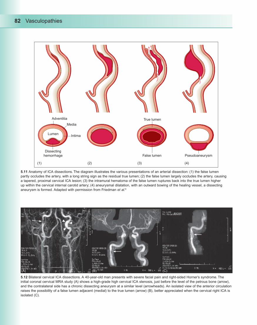
82 Vasculopathies
Lumen
Adventitia
Dissecting
hemorrhage Pseudoaneurysm
True lumen
False lumen
(1) (2) (4)(3)
Media
Intima
5.11 Anatomy of ICA dissections. The diagram illustrates the various presentations of an arterial dissection: (1) the false lumen
partly occludes the artery, with a long string sign as the residual true lumen; (2) the false lumen largely occludes the artery, causing
a tapered, proximal cervical ICA lesion; (3) the intramural hematoma of the false lumen ruptures back into the true lumen higher
up within the cervical internal carotid artery; (4) aneurysmal dilatation, with an outward bowing of the healing vessel, a dissecting
aneurysm is formed. Adapted with permission from Friedman et al.9
5.12 Bilateral cervical ICA dissections. A 40-year-old man presents with severe facial pain and right-sided Horner’s syndrome. The
initial coronal cervical MRA study (A) shows a high-grade high cervical ICA stenosis, just before the level of the petrous bone (arrow),
and the contralateral side has a chronic dissecting aneurysm at a similar level (arrowheads). An isolated view of the anterior circulation
raises the possibility of a false lumen adjacent (medial) to the true lumen (arrow) (B), better appreciated when the cervical right ICA is
isolated (C).
(A) (B) (C)

Vasculopathies 83
Head turned
to right
Left vertebral artery
Subclavian artery
Right vertebral
arteryC1
C2
C3
C4
C5
C6
C7
5.13 VA dissection: anatomy and gross pathology. (A) The
VA is commonly dissected at two areas: (1) around the
transverse foramina from C4 to C6, due to contact and
pressure from osteophytes and from local skeletal muscles
and fasciobends in this proximal segment; and (2) from
stretch injury between the C1 and C2 vertebral transverse
foraminae, when the head is sharply turned. Adapted with
permission from Krueger et al.41 (B) Serial transverse
sections of a dissected vertebral artery are shown from
proximal (P, upper left) to distal (D, lower right). Note
the intramural hematoma (arrows) and varying degrees
of occlusion of the true lumen (arrowheads). Pathology
courtesy of Louis Caplan, MD.
5.14 Common angiographic features of internal carotid artery dissection.
Adapted from Fisher et al.42
Normal Long
dissection
Tell-tale
pouch
Post-sinus
tapering occlusion
Distal
pouch
Early
Late
(B)
(A)

84 Vasculopathies
Aortic dissection. This more proximal arterial dissection normally presents with a distinct history and symptoms, such as blunt trauma to the chest, chest pain, asymmetric blood pressures, and cardiovascular instability, not typical for dissection of the cervical arteries alone. However, cerebral thromboembolism and cervical artery occlusion may occur as a complication of aortic dissection (5.19).
Most cervical artery dissections are spontaneous. A variety of colorful anecdotes of physical activities and head–neck trauma have been reported to cause cervical artery dissections.7,10 An underlying arteriopathy is not usually identified, though several heritable connective tissue disorders have been associated with dissection.7,16–18 A developmental syndrome of neural crest cells, the familial syndrome of arterial dissections with lentiginosis, has been linked to cervical artery dissection.16 Connective tissue and other genetic disorders should be considered when multiple or recurrent dissections occur, or a family history of young stroke or dissection exist.
Epidemiology
Dissection is one of the most common causes of thrombus formation in cervical arteries. It is probably the second leading cause of cervical ICA occlusion, though well behind atherosclerotic disease. The mean age at presentation with cervical artery dissection is only 45 years, perhaps relating to the elasticity and extensibility of these arteries in younger patients.7,16 Cervical artery dissection in patients >65 years of age is rare and is generally caused by significant head–neck trauma.
Clinical syndrome
The triad of facial, head, or neck pain, a Horner’s syndrome, and delayed cerebral or retinal ischemia are strong evidence for cervical artery dissection. In the absence of stroke, dissection may be misdiagnosed as a common or complicated migraine.19 A partial Horner’s syndrome (miosis and ptosis without anhidrosis) results from aneurysmal compression of the oculosympathetic chain in ICA dissection (5.20). In VA dissection, Horner’s syndrome may result from infarction of the lateral medulla. The presence of cerebrovascular symptoms in a person of younger age with any headache or neck pain should prompt a diagnostic evaluation for cervical artery dissection.
5.15 Right ICA occlusion
from dissection. A right MCA
stroke, diffusion-weighted MRI
sequence (A), resulting from
a right cervical ICA occlusion.
The coronal MRA shows an
absence of flow signal through
the entire right cervical ICA
portion (B), while focused
views of the right neck show a
bullet-shaped tapering of the
occluded artery (arrowheads)
(C). Intracranial MRA shows
a healthy proximal right MCA,
supplied via the anterior and
posterior communicating
arteries (D).(A)
(C)
(B)
(D)

Vasculopathies 85
5.16 The ‘halo sign’ of cervical artery dissection. A 53-year-old woman presented with a severe right hemicranial headache, but a
normal examination and no history or imaging findings of stroke. The MRA studies isolating the bilateral ICAs (A) show a smooth
tapering toward a ‘string sign’ (arrows) through much of the mid-upper cervical extent of the right ICA. A classic ‘halo sign’ is
prominently observed on adjacent transaxial T1 fat-saturated sequences (arrowhead) (B). A 35-year-old woman presented with cervical
pain while jogging, and subsequently developed mild gait ataxia and left hemisensory symptoms. The T1 fat-saturated sequence (C)
documented a ‘halo sign’ of the right cervical VA (arrowhead). The diffusion-weighted MRI sequences identify an embolic right PCA-
territory stroke, involving predominantly the thalamus (D).
5.17 Right hemispheric embolic
shower from internal carotid artery
dissection. Diffusion-weighted
sequences show multifocal emboli
to the MCA–MCA borderzone on
adjacent diffusion-weighted MRI
transaxial series (A,B). The source
was cervical ICA occlusion due to
dissection, with absence of flow
void on the transaxial T1-weighted
segment (arrowhead) compared
with the left distal ICA (C). There
was no apparent filling of the distal
right ICA on the intracranial MRA
(D), with diminished flow into the
proximal right MCA.
(A)
(C)
(A)
(C)
(B)
(D)
(B)
(D)

86 Vasculopathies
5.18 Dissecting aneurysm of the cervical ICA. CT angiography, sagittal view (A), in a patient with cervical fibromuscular dysplasia
demonstrates this aneurysmal dilatation close to the skull base (arrows). The lesion is measured at the maximum diameter of the
aneurysmal widening (B, left), and is roughly three times the lumen of the ICA just above the lesion (B, right).
5.19 Aortic dissection extending into the ICA. An
ultrasound study shows a true and false lumen
extending into the innominate artery (brachiocephalic
trunk) (A); the common carotid artery (B,C); and the
ICA (D,E). Three of these images (A,C,E) show the
signal from competing flow, often stagnant or reversed
in the false lumen (B), so the color mode registers blue
versus orange–red for the true lumen. Images courtesy
of Janice Guica, RN, RVT.
(B)(A)
(A)
(C)
(E)
(B)
(D)

Vasculopathies 87
Diagnosis
Non-invasive imaging, particularly transaxial fat-saturated T1-weighted MRI, has supplanted angiography as the urgent diagnostic modality of choice. The pathognomonic ‘halo sign’ of intramural hematoma is often well-visualized as a hyperintense signal early in the course of ICA dissection (5.16B). It is often more difficult to visualize in VA dissections, due to their smaller lumen size (5.16C). Ultrasound is not as useful in diagnosis; this is because most cervical artery dissections occur higher in the neck than can be readily accessed with an external ultrasound probe (5.19).
Natural history
The natural history of stroke due to cervical artery dissection is reasonably benign. In a large prospective series of patients with a first cervical artery dissection (n = 459), there were a total of only four recurrent dissections (0.9%) and four recurrent ischemic strokes (two attributable to the initial dissection, and two from recurrent dissections) in almost 3 years of follow-up.20 Dissections do not typically recur in the same vessel, perhaps due to the local healing process.
Management
Secondary stroke prevention
The most efficacious antithrombotic regimen to best prevent recurrent stroke following cervical artery dissection remains unresolved, but may be tailored toward the individual lesion. Anticoagulation with warfarin could presumably prevent recurrent thromboemboli from a highly stenotic or occluded extracranial cervical artery. Concern about the use of anticoagulation relates to the rare, life-threatening
potential complication of subarachnoid hemorrhage that may occur whenever a cervical artery dissection extends intracranially. Anticoagulation has not been documented to cause propagation or expansion of an intramural hematoma. The consensus approach to treatment in most patients following acute extracranial dissection is anticoagulation for 2–3 months, and then conversion to antiplatelet treatment. Follow-up MRA or CTA at 3 months is used to gauge the artery’s recanalization and any luminal irregularities (5.10D).
Acute interventions
Some data suggest that intravenous thrombolysis does not pose a specific risk to patients with acute cervical artery dissection. In case series, there has been no apparent risk of worsening the primary intramural hematoma, nor additional risk of intracranial extension or aneurysmal dilatation.21,22 Endovascular interventions for acute dissection are technically challenging due to the fluctuating nature of the lesion, the difficulty in differentiating the true from the false lumen, and the risk of rupturing through the arterial wall. In very high-risk situations where there is a compromise of multiple cervical arteries, interventional procedures can be considered, e.g., angioplasty, stenting (case study 3),18,23 neurosurgical trapping, vascular bypass, treatment of an intracranial thromboembolic lesion caused by an extracranial dissection (case study 4), or endovascular coiling with sacrifice of a cervical artery.
Vasculitis
Definition
Systemic vasculitis includes a group of disorders where inflammation and destruction (necrosis) of the vessel wall is the primary event.24–29 Both primary (idiopathic) and sec-ondary (those associated with infectious and non-infectious inflammatory diseases) vasculitides (5.21) can cause cerebral vasculitis (Table 5.3). This section will briefly review giant cell arteritis (GCA) and primary central nervous system (CNS) angiitis.
Clinical presentation and epidemiology
A common cause of headache in patients >50 years of age, GCA is the most common vasculitis to cause cerebrovas-cular symptoms. It is a systemic arteritis of medium- and large-caliber arteries anywhere in the body, with a pre-dilection for the extracranial cervical arteries. The diagnosis should be considered in patients with a variety of symptoms:
5.20 Horner’s syndrome associated with a traumatic left ICA
dissection in a 43-year-old man. Note ptosis and (less evident)
miosis OS. Image courtesy of Robert Lesser, MD.

88 Vasculopathies
headache, visual loss and/or diplopia, pain on continuous chewing (jaw claudication) or swallowing, fever, and other constitutional symptoms (anorexia, weight loss, malaise), and the stiffness of polymyalgia rheumatica, a commonly associated syndrome.29 Serologic markers of inflammation, erythrocyte sedimentation rate and C-reactive protein, are typically elevated. Physical signs of GCA include swollen,
5.21 Tuberculous meningitis causing arachnoiditis and secondary vasculitis.
This patient developed an encephalopathy due to meningitis, obstructive
hydrocephalus, and deep cerebral infarcts caused by proximal intracranial
vasculitis. The first MRI scan shows extensive basal meningitis and
arachnoiditis on adjacent transaxial cuts (A) and coronal (B) T1WI with
gadolinium, lining the region of the circle of Willis. The linear dark structure
on this coronal sequence is an intraventricular catheter (arrows). Infarcts
developed in the cerebellar vermis, as well as the bilateral rostral thalami (C,
left), and 3 weeks later, in the left lenticulostriate territory (C, right). Following
an antitubercular medical regimen, the basal meningitis, with inflammation
lining the lateral ventricles and hydrocephalus (D, left), resolved (D, right).
Table 5.3 Types of vasculitis
Primary CNS vasculitis1. (primary angiitis of the
CNS, granulomatous angiitis)
Systemic vasculitis with CNS involvement2.
Systemic necrotizing vasculitis: polyarteritis (a)
nodosa; Churg–Strauss syndrome
Wegener’s granulomatosis(b)
Behçet’s disease(c)
Giant cell arteritis: Takayasu’s arteritis; giant (d)
cell arteritis
Systemic diseases with secondary CNS angiitis, or 3.
vasculopathy
Scleroderma(a)
Rheumatoid disease(b)
Sjögren’s syndrome(c)
Antiphospholipid antibody syndrome(d)
Systemic lupus erythematosus(e)
CNS disorders with secondary vasculitis4.
Meningitis: pyogenic, tuberculous, fungal, (a)
parasitic, syphilitic
Septic emboli(b)
Non-granulomatous inflammations: (c)
neurosarcoidosis
CNS, central nervous system.
Adapted from Osborn4 and Zuber.28
(A)
(B)
(C)
(D)

Vasculopathies 89
painful superficial temporal arteries, scalp necrosis and ten-derness, ischemic optic neuropathy, and posterior circula-tion ischemic stroke. Longitudinal epidemiological studies note an incidence of 17.0 persons per 100 000 population >50 years of age.29
Primary (or isolated) CNS angiitis has a wide range of presenting symptoms, so it is frequently included in the dif-ferential diagnosis of neurovascular syndromes. CNS angiitis can cause seizures, intracerebral hemorrhage, acute ischemic stroke, headaches, and psychiatric symptoms. Untreated, the disease often results in recurrent brain injury and significant morbidity and mortality. This localized disease of small- and medium-sized intracranial arteries is decidedly rare, with an incidence estimated at <1:2 000 000.28
Pathogenesis and diagnosis
The diagnosis of GCA is typically made by a biopsy of the superficial temporal arteries (5.22). The involvement of the extracranial external carotid and vertebral arteries is appar-ently explained by the disease’s predilection for arteries with a high elastic content in the arterial media and adventitia.27,29 The four vessels most commonly affected in GCA are the superficial temporal, ophthalmic, posterior ciliary, and ver-tebral arteries. Arteritic ischemic optic neuropathy causing blindness, the most common severe morbidity from GCA, is due to local involvement of the posterior ciliary arteries. Visual loss can rapidly evolve in both eyes within days.29 The uncommon association of GCA with ischemic stroke is almost always from local thromboembolic disease develop-ing in vertebral arteries. The local arteritic disease typically extends only 5 mm above the dural perforation of the verte-bral arteries, where the external elastic lamina ends.27
The diagnosis of primary CNS angiitis is also dependent on tissue examination, though it is considered in cases where there are multifocal intracranial lesions, without the presence of a systemic vasculitis. The absence of other organ involvement and normal serologic tests point to a diagnosis of primary CNS angiitis. Neuroimaging (CT and MRI findings) can delineate the extent of CNS involvement, but there are no findings specific to vasculitis. While the cerebrospinal fluid (CSF) is usually abnormal with, at minimum, a lymphocytic pleocytosis and/or elevated protein, the angiographic picture is non-specific and can be seen with other conditions causing vasoconstriction (5.23) or vasospasm, or the commonly seen changes due to atherosclerosis (5.24).30 Brain and/or meningeal biopsy are usually required for a definitive diagnosis, documenting
granulomatous involvement of the small- and medium-sized intracranial arteries (5.25). Given that the treatment is long-term, using immunomodulatory agents, biopsy confirmation of the diagnosis is a critical step in management.
Management
Urgent treatment of GCA with intravenous steroids is indicated when visual loss develops, often to protect the contralateral eye (if the symptomatic eye does not improve). A transition to a long-term tapering regimen with prednisone in much higher
5.22 Temporal arteritis, pathology. Complete occlusion of the
superficial temporal artery, due to an inflammatory process
within the artery’s walls (A) (hematoxylin and eosin stain, 40 ).
The outer layers of the wall, at higher power, demonstrate
giant mononuclear (Langherhans) cells (arrow), diagnostic for
temporal arteritis (hematoxylin and eosin, 100 ) (B). Pathology
courtesy of Robert Schmidt MD, PhD.
(A)
(B)

90 Vasculopathies
5.23 Cerebral vasoconstriction syndrome.30 A healthy 50-year-old woman presented with headache, confusion, and left-sided deficits
from a right lobar parietal hemorrhage (A). Initial conventional angiography shows right ICA injection, lateral view (B); left ICA
injections, lateral (C) and oblique (D) views; and left VA injection (E). Note multifocal stenoses and irregularities, more prominent in
the anterior than the posterior circulation (arrowheads). The working initial diagnosis was primary CNS angiitis, and the patient was
first treated with prednisone and cyclophosphamide. However, the admission cerebrospinal fluid study was unremarkable, as was the
follow-up angiography, obtained 9 months after the first. This second study showed complete resolution of the arteriopathy on a right
ICA injection, oblique view (F).
(B)
(D)
(A)
(C)
(E) (F)
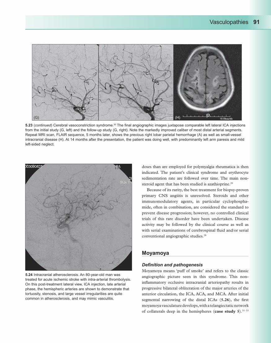
Vasculopathies 91
doses than are employed for polymyalgia rheumatica is then indicated. The patient’s clinical syndrome and erythrocyte sedimentation rate are followed over time. The main non-steroid agent that has been studied is azathioprine.29
Because of its rarity, the best treatment for biopsy-proven primary CNS angiitis is unresolved. Steroids and other immunomodulatory agents, in particular cyclophospha-mide, often in combination, are considered the standard to prevent disease progression; however, no controlled clinical trials of this rare disorder have been undertaken. Disease activity may be followed by the clinical course as well as with serial examinations of cerebrospinal fluid and/or serial conventional angiographic studies.28
Moyamoya
Definition and pathogenesis
Moyamoya means ‘puff of smoke’ and refers to the classic angiographic picture seen in this syndrome. This non-inflammatory occlusive intracranial arteriopathy results in progressive bilateral obliteration of the major arteries of the anterior circulation, the ICA, ACA, and MCA. After initial segmental narrowing of the distal ICAs (5.26), the first moyamoya vasculature develops, with a telangiectatic network of collaterals deep in the hemispheres (case study 5).31–33
5.23 (continued) Cerebral vasoconstriction syndrome.30 The final angiographic images juxtapose comparable left lateral ICA injections
from the initial study (G, left) and the follow-up study (G, right). Note the markedly improved caliber of most distal arterial segments.
Repeat MRI scan, FLAIR sequence, 5 months later, shows the previous right lobar parietal hemorrhage (A) as well as small-vessel
intracranial disease (H). At 14 months after the presentation, the patient was doing well, with predominantly left arm paresis and mild
left-sided neglect.
5.24 Intracranial atherosclerosis. An 80-year-old man was
treated for acute ischemic stroke with intra-arterial thrombolysis.
On this post-treatment lateral view, ICA injection, late arterial
phase, the hemispheric arteries are shown to demonstrate that
tortuosity, stenosis, and large vessel irregularities are quite
common in atherosclerosis, and may mimic vasculitis.
(G) (H)

92 Vasculopathies
Finally, regression of the moyamoya vessels occurs, with concurrent growth of meningeal–pial collaterals from the external carotid artery to supply much of the cerebral hemispheres.32 The basic underlying pathophysiology of moyamoya disease is unknown.
Epidemiology
The moyamoya syndrome can be primary (moyamoya disease) (5.26) or secondary to a variety of underlying diseases that may be due to infectious, inflammatory, vasculopathic, genetic, or hematologic/prothrombotic disorders (moyamoya syndrome).32 About 10% of cases are unilateral and 10% genetic, occurring most frequently in Japanese and other Asian populations.
Natural history and management
The natural history is somewhat different for Asians, who more often have moyamoya disease (5.26), and Caucasians,
who are more likely to have a moyamoya syndrome. Asian children present with ischemic stroke, while hemorrhagic stroke becomes more common in adulthood. In Caucasians, ischemic stroke is most common at any age.34,35
Moyamoya is a progressive disorder with unfavorable outcomes predominating. The progression of the disease as documented by angiography is much more aggressive in childhood than in adulthood, but the prognosis for func-tional status and life expectancy is poor in adults due to recurrent hemorrhages.36
Moyamoya disease is generally treated by a surgical revascularization procedure. The two major types of revascularizations are a superficial temporal artery-to-MCA direct bypass provided that an acceptable cortical branch of the MCA exists for anastomosis and an indirect revascularization procedure known as encephaloduroarteriosynangiosis.32,37–39 In the latter case, a bone window is made to enable the surgeon to directly
5.25 Primary CNS angiitis. Biopsy at low and higher powers show areas of pallor from ischemia within a sample of cerebellar tissue,
including granular layers (A) (hematoxylin and eosin stain, 40 ), as well as multiple granulomas (arrows), localized to the Virchow Robbins
spaces and (not shown) within arteriolar walls (B) (hematoxylin and eosin, 100 ). Pathology courtesy of Robert Schmidt MD, PhD.
(A) (B)

Vasculopathies 93
suture the superficial temporal artery to the pia matter. In successful cases, extracranial-to-intracranial angiogenesis (new collateral vessels) develop over several months that augment hemispheric blood flow (case study 5). Combined direct and indirect revascularizations are also feasible, but controlled clinical trials and long-term follow-up documenting benefits in terms of functional status and cognitive improvement are lacking.36
Case studies
Case study 1. Angioplasty of cervical
fibromuscular dysplasia
The bilateral, severe FMD of the cervical ICA shown (CS 1.1) was measured: right (A) and left (B), and then both sides were treated with angioplasty. The images show progressive dilatation of the left cervical disease (C),
to 4 mmHg balloon pressure (left) and, later, 5 mmHg (right).
Comments
The balloon pressure for progressive arterial dilation dur-ing angioplasty of FMD is only 5–6 atmospheres, versus 15–20 for atherosclerotic stenosis.5 Primary angioplasty alone is quite effective for resolving focal stenoses and may be repeated, if necessary. The risk of employing endo-vascular stents for the treatment of FMD-related stenosis is unknown, but overgrowth of an in situ stent by intimal or medial fibroplasia is theoretically possible.
Case study 2. Vertebral artery dissecting
aneurysm treated with stenting
A middle-aged man presented with a right lateral medullary infarction (not shown) associated with this traumatic right
5.26 Early moyamoya disease on MRA. A 7-year-old boy of Japanese ancestry presented with
mild left-sided deficits. The FLAIR MRI sequence shows a lesion in the anterior limb of the
internal capsule and head of the caudate (A). An oblique coronal MRA study shows prominent
VAs that are comparable or larger than the diminutive atretic-appearing cervical ICAs (B). A
coronal intracranial MRA, with the posterior circulation subtracted (C), shows small distal ICAs,
particularly on the right side, with proximal left MCA stenosis (arrow) and poor or absent flow
through the A1 segments bilaterally and the right M1 segment. By contrast, the A2 and more
distal ACA segments have a strong signal. A focused view of the posterior circulation (D)
shows prominent PCA anatomy, also seen on a transaxial intracranial MRA study (E). This last
study suggests minimal or poor perfusion of MCA branches throughout the right hemisphere,
with possible collateral supply from a lateral PCA branch (arrowhead) and/or extracranial
leptomeningeal supply. This MRA shows occlusive obliteration of the distal ICAs and proximal
ACA and MCA segments typical of early moyamoya disease. A conventional angiogram would
be needed to completely delineate the vasculature, but the telangiectatic moyamoya pattern may
not yet have developed. This patient’s presenting stroke reflects the severe vasculopathy within
the right A1 and M1 segments.
(A)
(E)
(B)
(C)
(D)

94 Vasculopathies
VA dissection. A distal right VA dissection with a small false lumen is shown on conventional angiography (CS 2.1A) and reconstructed three-dimensional images (B,C). The true lumen constitutes only a string sign (arrows) (A).
This lesion did not improve over the initial 3 months, prompting a decision to treat with angioplasty and stenting of the artery to re-establish the lumen and prevent recurrent
thromboembolization. The final image (D) is a CT angio-gram in which the proximal and distal stent margins appear as white markers. The right PICA is seen behind its parent VA. (Note: the left and right sides are reversed on this CTA.)
Case study 3. Bilateral internal carotid artery
dissections with unilateral angioplasty and
stenting
A 36-year-old woman suffered bilateral severe ICA dissec-tions while horseback-riding (CS 3.1). The initial cervical MRA shows flame-shaped tapering of both cervical ICAs, with possible occlusions (A). A borderzone stroke, shown on the diffusion-weighted MRI sequence (B), caused mild left hemiparesis, predominantly affecting the hand.
Conventional cerebral angiography (CS 3.2) showed a tapered left ICA that appears completely occluded (arrows), with the catheter tip at the beginning of the tapering (A), as well as a long severe string sign of the right ICA, with reconstitution at the level of the petrous bone (arrow) (B).
Two stents were placed (CS 3.3) in order to expand the true lumen, as shown on the unsubtracted (A, left) and subtracted (A, right) lateral views, with excellent result. A post-stent ICA injection (B) shows excellent filling of the right hemisphere, including the right fetal posterior cerebral artery (PCA) and also, via the anterior communicating artery, the left ACA. The patient remained without further symptoms on subcutaneous enoxaparin and clopidogrel.
A follow-up MRA study (CS 3.4) 3 months from the initial dissections showed apparent recanalization in the left cervical ICA (A, left; reconstructed coronal image; note the artifact produced in the region of the right ICA stents (arrow-heads). An intracranial MRA (A, right) shows normal ICA siphon flow bilaterally. The final infarct is shown on T2WI at 3 months (B); the patient had only mild slowing of fine finger movements in the left hand, and anticoagulation was discontinued.
Comments
For a number of reasons a decision was made to treat the right ICA lesion first (versus the left ICA) with angioplasty and stenting. The right carotid dissection appeared more acces-sible, with a more viable lumen. The periprocedural risk might be lower because the right ICA supplies the non-dominant hemisphere. The right hemispheric flow demonstrated (with stroke) a more critical hemodynamic insufficiency than the left hemisphere. Finally, the right ICA also supplied the fetal PCA, putting a larger area of the brain at risk for stroke.
CS 1.1
(A)
(B)
(C)

Vasculopathies 95
CS 2.1
(A)
(C)
(B)
(D)

96 Vasculopathies
CS 3.1
CS 3.2
CS 3.3
(A)
(A)
(A)
(B)
(B)
(B)

Vasculopathies 97
Case study 4. Urgent stenting of middle cerebral
artery occlusion due to internal carotid artery
dissection
The patient was treated for acute subcortical right MCA ischemic stroke, which had been caused by right cervical ICA dissection embolizing into the MCA territory; this was preceded by several hours of monocular right-sided blind-ness, due to previous embolism in the central retinal artery (not shown). The diffusion-weighted MRI sequence 1 day later is shown (CS 4.1).
Conventional angiography performed on admission shows a flame-shaped right ICA dissection (CS 4.2). On common carotid artery injection, the lesion appears occlusive (A, left), but an ICA injection shows a string sign (arrowheads) with some reconstitution of the lumen before the petrous level (A, right; arrow). Treatment of the ICA terminus thrombus extending into the MCA with intra-arterial thrombolysis was unsuccessful. The MCA lesion is shown before (B, left) and
immediately after (B, right) infusion of 12 mg intra-arterial tissue plasminogen activator; however, this recanalization was only transient.
Endovascular angioplasty and stenting of the MCA was pursued next. Two NeuroformTM stents were placed in a bifurcative fashion into the superior and inferior divisions. The initial stent (3 mm 20 mm, width by length), placed within the superior branch, is shown on a local injection, anteroposterior view (C, left), and then, local injection with a guidewire into the inferior branch before the second stent (C, right).
Next, two stents were placed more proximally within the petrous ICA lesion with initial restoration of antegrade flow through the cervical ICA (not shown). However, on a follow-up CTA study 2 months later (CS 4.3), a transaxial cut shows the right cervical ICA stent (arrowhead) (A) has become occluded, in contrast to the hyperdense normal flow signal within the contralateral ICA (arrow). The intracranial MCA stents on non-contrast CT scans create local artifacts (B). Intact flow through the stents is evident on the source images of the MRA study; compare the right and left M1 and M2 segments coursing into the Sylvian fissures (C).
Comments
This patient had a complex tandem lesion, with a typical cervical ICA dissection that embolized to both the lenticulostriate arteries and the central retinal artery (e.g., see also Chapter 3, case study 1). The lesion with the highest risk, the right distal ICA and M1 thrombus, was treated with stenting after intra-arterial thrombolysis failed. Given the subsequent thrombosis in the cervical ICA stenting,
CS 3.4
CS 4.1
(A)
(B)

98 Vasculopathies
collateral flow from the anterior communicating artery likely played a major part in limiting the eventual size of the right hemispheric infarct.
At 2 years post-stroke, the patient’s sole deficit is monocular blindness from the retinal lesion, with no residual deficits associated with the cerebral lesion. The old right MCA territory stroke, with ex vacuo enlargement of the frontal horn of the lateral ventricle, is shown (CS 4.4).
CS 4.2 CS 4.3
(A)
(A)
(B)
(C)
(B)
(C)

Vasculopathies 99
Case study 5. Moyamoya disease, treated with
the encephaloduroarteriosynangiosis procedure
A 25-year-old woman had cerebral palsy due to a perinatal right hemispheric stroke shown on CT scan (CS 5.1), and her MRA (not shown) suggested bilateral moyamoya vasculature.
Conventional angiography (CS 5.2)
The early phase study (left ICA injection) (A) shows prominent P1 and PCA anatomy (arrowhead) and an enlarged ophthalmic artery (arrows), with small arterial, telangiectatic anatomy in the region of the proximal MCA (encircled). The later phase (B) shows large proximal ACA anatomy, as well as several collateral routes directly connecting the distal ACA and PCA branches (arrowheads). Extensive extracranial external carotid artery–intracranial collateral segments have developed into the anterior circulation (right-hand side of images). An anteroposterior
CS 4.4
CS 5.2CS 5.1
(A)
(A)
(B)
(C)(B)

100 Vasculopathies
view (C) documents markedly abnormal, small and large telangiectatic arteries encompassing the left ACA and MCA territories. A CT scan on the first postoperative day (CS 5.3) shows the region of the encephaloduroarteriosynangiosis, with air into the subdural and subgaleal spaces as well as the left anterior frontal lobe (arrows).
Follow-up angiography (CS 5.4)
Obtained 6 months following encephaloduroarteriosynan-giosis, this study shows the development of external carotid artery (ECA)–MCA neovascular collateral blood supply.
The ECA injections shown are: (A) a lateral view of the left hemisphere and also anteroposterior views: subtracted (B, left) and unsubtracted (B, right) views; an ECA–MCA collateral pathway is marked (arrow). (The nasal turbines and orbital bones are present in the lower half of the images, marking the midline.)
Comments
This patient had bilateral moyamoya disease, presumed idiopathic, with a significant perinatal right hemispheric stroke. The decision to treat the left hemisphere with an encephaloduroarteriosynangiosis procedure was based on the plan to preserve the normal status of this dominant hemisphere, and the lack of an intact MCA recipient artery for a direct bypass procedure.
References
Adams H, Jr, Bendixen B, Kappelle L,1. et al. Classification of acute ischemic stroke: definition for use in a multicenter trial. Stroke 1993; 24: 35–41.Luscher T, Lie J, Stanson A, Houser O, Hollier L, 2. Sheps S. Arterial fibromuscular dysplasia. Mayo Clin Proc 1987; 62: 931–52.Healton E, Mohr J. Cerebrovascular fibromuscular 3. dysplasia. In: Barnett H, Mohr J, Stein B, Yatsu F, eds. Stroke: Pathophysiology, Diagnosis, and Management, 3rd edn. New York: Churchill Livingstone; 1998: 833–44.Osborn A. Nonatheromatous vasculopathy. In: 4. Diagnostic Cerebral Angiography, 2nd edn. Philadelphia, PA: Lippincott Williams & Wilkins; 1999: 341–58.
CS 5.3
CS 5.4
(A) (B)

Vasculopathies 101
Slovut D, Olin J. Fibromuscular dysplasia. 5. N Engl J Med 2004; 350: 1862–71.Cloft H, Kallmes D, Kallmes M, Goldstein J, 6. Jensen M, Dion J. Prevalence of cerebral aneurysms in patients with fibromuscular dysplasia: a reassessment. J Neurosurg 1998; 88: 436–40.Schievink W. Spontaneous dissection of the carotid and 7. vertebral arteries. N Engl J Med 2001; 344: 898–906.Osborn A, Anderson R. Angiographic spectrum of 8. cervical and intracranial fibromuscular dysplasia. Stroke 1977; 8: 617–26.Friedman W, Day A, Quisling J, 9. et al. Cervical carotid dissecting aneurysms. Neurosurgery 1980; 7: 207–14.Saver J, Easton J. Dissections and trauma of 10. cervicocerebral arteries. In: Barnett H, Mohr J, Stein B, Yatsu F, eds. Stroke: Pathophysiology, Diagnosis, and Management, 3rd edn. New York: Churchill Livingstone; 1998: 769–86.Mokri B. Cervicocephalic arterial dissections. In: 11. Bogousslavsky J, Caplan L, eds. Uncommon Causes of Stroke. New York: Cambridge University Press; 2001: 211–29.Fisher C, Ojemann R, Robertson G,12. et al. Spontaneous dissection of cervicocerebral arteries. Can J Neurol Sci 1978; 5: 9–19.Molina C, Alvarez-Sabin J, Schonewille W,13. et al. Cerebral microembolism in acute spontaneous internal carotid artery dissection. Neurology 2000; 55: 1738–41.Baumgartner R, Arnold M, Baumgartner I,14. et al. Carotid dissection with and without ischemic events: local symptoms and cerebral artery findings. Neurology 2001; 57: 827–32.Touze E, Randoux B, Meary E, Arquizan C, Meder 15. J-F, Mas J-L. Aneurysmal forms of cervical artery dissection: associated factors and outcome. Stroke 2001; 32: 418–23.Schievink WI, Mokri B, O’Fallon WM. Recurrent 16. spontaneous cervical-artery dissection. N Engl J Med 1994; 330: 393–7.Schievink WI, Mokri B, Piepgras DG, Kuiper JD. 17. Recurrent spontaneous arterial dissections: risk in familial versus nonfamilial disease. Stroke 1996; 27: 622–4.Volker W, Besselmann M, Dittrich R,18. et al. Generalized arteriopathy in patients with cervical artery dissection. Neurology 2005; 64: 1508–13.
Silverman IE, Wityk RJ. Transient migraine-like 19. symptoms with internal carotid artery dissection. Clin Neurol Neurosurg 1998; 100: 116–20.Touze E, Gauvrit J-Y, Moulin T, Meder J-F, Bracard 20. S, Mas J-L. Risk of stroke and recurrent dissection after a cervical artery dissection: a multicenter study. Neurology 2003; 61: 1347–51.Derex L, Nighoghossian N, Turjman F,21. et al. Intravenous tPA in acute ischemic stroke related to internal carotid artery dissection. Neurology 2000; 54: 2159–61.Georgiadis D, Lanczik O, Schwab S,22. et al. IV thrombolysis in patients with acute stroke due to spontaneous carotid dissection. Neurology 2005; 64: 1612–14.Lylyk P, Cohen J, Ceratto R, Ferrario A, Miranda C. 23. Angioplasty and stent placement in intracranial atherosclerotic stenoses and dissections. Am J Neuroradiol 2002; 23: 430–6.Nadeau S. Diagnostic approach to central and 24. peripheral nervous system vasculitis. Neurol Clin 1997; 15: 759–77.Biller J, Grau R. Cerebral vasculitis. In: Adams HP Jr, 25. ed. Handbook of Cerebrovascular Diseases. New York: Marcel Dekker, Inc.; 2005: 653–72.Calabrese L. Therapy of systemic vasculitis. 26. Neurol Clin 1997; 15: 973–91.Weyand C, Goronzy J. Medium- and large-vessel 27. vasculitis. N Engl J Med 2003; 349: 160–9.Zuber M. Isolated angiitis of the central nervous 28. system. In: Bogousslavsky J, Caplan L, eds. Uncommon Causes of Stroke. New York: Cambridge University Press; 2001: 1–9.Davis S. Temporal arteritis. In: Bogousslavsky J, 29. Caplan L, eds. Uncommon Causes of Stroke. New York: Cambridge University Press; 2001: 10–17.Singhal A, Koroshetz W, Caplan L. Cerebral 30. vasoconstriction syndromes. In: Bogousslavsky J, Caplan L, eds. Uncommon Causes of Stroke. New York: Cambridge University Press; 2001: 114–23.Reardon M, Mathews K. Moya moya disease. In: 31. Adams HP Jr, ed. Handbook of Cerebrovascular Diseases. New York: Marcel Dekker, Inc.; 2005: 783–4.Adams H, Jr. Moya-moya. In: Bogousslavsky J, 32. Caplan L, eds. Uncommon Causes of Stroke. New York: Cambridge University Press; 2001: 241–57.

102 Vasculopathies
Yilmaz EMP, Bruno A, Lopez-Yunez A, Biller J. 33. Moyamoya: Indiana University Medical Center experience. Arch Neurol 2001; 58: 1274–8.Chiu D, Shedden P, Bratina P, Grotta J. Clinical 34. features of moyamoya disease in the United States. Stroke 1998; 29: 1347–51.Kuroda S, Hashimoto N, Yoshimoto T, Iwasaki Y. 35. Radiological findings, clinical course, and outcome in asymptomatic moyamoya disease: results of multicenter survey in Japan. Stroke 2007; 38: 1430–5.Masuda J, Ogata J, Yamaguchi T. Moyamoya disease. 36. In: Barnett H, Mohr J, Stein B, Yatsu F, eds. Stroke: Pathophysiology, Diagnosis, and Management, 3rd edn. New York: Churchill Livingstone; 1998: 815–32.Suzuki Y, Negoro M, Shibuya M, Yoshida J, Negoro T, 37. Watanabe K. Surgical treatment for pediatric moyamoya disease: use of the superficial temporal artery for both areas supplied by the anterior and middle cerebral arteries. Neurosurgery 1997; 40: 324–30.Kinugasa K, Mandai S, Kamata I, Sugiu K, Ohmoto 38. T. Surgical treatment of moyamoya disease: operative technique for encephalo-duro-arterio-myo-synangiosis, its follow-up, clinical results, and angiograms. Neurosurgery 1993; 32: 527–31.Whitaker J. Management of moyamoya syndrome. 39. Arch Neurol 2001; 58: 132.Ross R. Atherosclerosis—an inflammatory disease. 40. N Engl J Med 1999; 340: 115–26.Krueger B, Okazaki H. Vertebral-basilar distribution 41. infarction following chiropractic cervical manipulation. Mayo Clin Proc 1980; 55: 322–32.Fisher C, Ojemann R, Robertson G. Spontaneous 42. dissection of cervico-cerebral arteries. Can J Neurol Sci 1978; 5: 9–18.
Further reading
Bogousslavsky J, Caplan LR, eds. Uncommon Causes of Stroke. Cambridge University Press; 2001.
Osborn A. Nonatheromatous vasculopathy. In: Diagnostic Cerebral Angiography, 2nd edn. Philadelphia, PA: Lippincott Williams & Wilkins; 1999: 341–58.
Schievink W. Spontaneous dissection of the carotid and vertebral arteries. N Engl J Med 2001; 344: 898–906.
Slovut D, Olin J. Fibromuscular dysplasia. N Engl J Med 2004; 350: 1862–71.
Cervical Artery Dissection in Stroke Study. An international randomized clinical trial comparing anticoagulation to antiplatelet therapy for secondary stroke prevention in patients with cervical artery dissection has begun a feasibility stage. http://www.dissection.co.uk/
Resources for patients
Fibromuscular dysplasia
The Fibromuscular Dysplasia Society of America. http://www.fmdsa.orgFibromuscular Dysplasia, NINDS. http://www.ninds.nih.gov/disorders/fibromuscular_dysplasia/fibromuscular_dysplasia.htm
Moyamoya disease
Moyamoya Disease, NINDS. http://www.ninds.nih.gov/disorders/moyamoya/moyamoya.htm
Temporal arteritis
Vasculitis, including Temporal Arteritis, NINDS. http://www.ninds.nih.gov/disorders/vasculitis/vasculitis.htm

Chapter 6
Therapies: The Management of Acute Ischemic Stroke and Secondary Stroke Prevention
Introduction
The rapidly evolving and increasing ability to treat AIS effectively is one of the most exciting topics in medicine. Acute stroke treatment is time-dependent so it is important to stress the need for stroke victims to reach the hospitals most able to offer the best combination of drug and device treatment(s). This means that regional stroke networks and effi cient transfer processes will be key to increasing the number of people treated. This chapter addresses the state of acute treatments and secondary prevention for ischemic stroke.
Overview
Key variables infl uencing the viability of brain tissue during AIS include: time, hemodynamics, tissue, and interven-tion (Table 6.1).1 Most of the effort to improve outcomes in ischemic stroke has focused on decreasing the time to treatment and pharmacologic or mechanical interventions for re perfusion or neuroprotection. More sophisticated as-sessment of hemodynamic issues, particularly collateral circulation, may infl uence what interventions are recom-mended at what point in time. Maximizing tissue viability through tight control of serum glucose and hypothermia is being evaluated.
The challenges of providing optimal treatment for every patient to improve clinical outcomes are signifi cant:
Time1 . Although the healthcare systems in many countries are evolving to address acute stroke as a highest-level medical emergency, public awareness of stroke symptoms remains low. If stroke symptoms are not recognized or
patients not rapidly transported to ‘stroke-ready’ hospi-tals, then any efforts to develop treatments to improve stroke outcomes during the critical initial minutes-to-hours will be wasted. In the USA, Emergency Medical Services (EMS) are organized at the community and/or state-wide level making the early response to acute stroke patients highly variable. Benchmarks for hospital performance have been established to encourage rapid treatment on arrival.2 The recommended time from door-to-computed tomography (CT) scan is 25 minutes, and from door-to-drug (intravenous (IV) tissue plasminogen activator (t-PA)) 60 minutes.Healthcare resources2 . The capacity of individual hospitals to care for acute stroke varies widely. In the USA, national and regional quality improvement initiatives have led to formal certifi cation/identifi cation of acute ‘stroke-ready’ medical centers;3 however, many hospitals lack adequate infrastructure and medical staff to meet all of the needs of acute stroke patients. Most likely, a two-tiered system will emerge with Primary Stroke Centers capable of rapid tri-age and administration of IV thrombolysis partnering with regional Comprehensive Stroke Centers that offer subspe-cialty neurovascular care, clinical trials, physician training programs, and endovascular (IA) treatments. The Joint Commission began certifying Primary Stroke Centers in the USA in 2004, and criteria for Comprehensive Stroke Centers have been published.4
Stroke clinical trials3 . The modern era of acute stroke clinical trials is in its infancy in regard to drug and device development and clinical trial design. Patient selection and outcomes can be based on clinical criteria alone or may include neuroimaging criteria. The pathophysiologic heterogeneity of stroke makes the assessment of outcomes even more challenging.5
103
104 Therapies
Complexity of cerebral ischemia4 . Understanding of the biol-ogy of cerebral ischemia, which is the basis for new drug and device development, continues to evolve. The time-dependent cascade of acute focal ischemia is illustrated (6.1).6 Drugs that interrupt or slow the ischemic cascade may interact with only one of the various steps. Ischemia affects not only neurons (constituting <5% of the cells in cerebral gray matter) but also astrocytes and other glial cells supporting neurons, the axons of neurons (which relay signals to other cells), and the microvessels that supply oxygen and nutrients.7 Treatments that target this entire ‘neurovascular unit’ rather than simply neurons may be more effective in treating acute ischemic brain injury (6.2).7
Treatment for acute ischemic stroke
The modern era of treatments for AIS was ushered in by clinical trials that began in the 1980s and early 1990s. Two major therapeutic approaches have been studied since the early trials were conducted:
Reperfusion treatments include IV or IA drugs and endovascular devices aimed at the recanalization of occluded intracranial and extracranial arteries. IV t-PA was approved for the treatment of AIS within 3 hours of onset of symptoms in the USA in 1996.8 The MERCI Retriever was approved for the removal of clots from intracranial and extracranial cerebral arteries within 8 hours of stroke symptoms onset in 2004 (6.3; case study 1).9 A second endovascular thrombectomy device, the Penumbra Stroke System, was similarly approved by the Food and Drug Administration (FDA) in 2008 for its aspiration mechanism.10
Neuroprotective agents or devices are meant to main-tain optimal tissue viability in the ischemic penumbra. No pharmacologic agent has been shown to be effective. Trials of hypothermia and infrared laser to test potential for neuroprotection are currently in progress.11
The scope of established and putative treatments for AIS is shown in Table 6.2.11 The stage is set for an explosion in treatment modalities during the upcoming decades.6,7,12
Reperfusion treatment and time
Time is brain! For every minute stroke is left untreated, an estimated 1.9 million neurons are destroyed.13 The benefit of IV t-PA has been demonstrated to wane through the 3-hour window.14 However, subsequent pooled analysis of three major clinical trials suggests that the time window of effec-tiveness for IV t-PA may actually be as long as 4.5 hours.15
Recent clinical trials evidence from the European study group subsequently went on to prove that IV t-PA is efficacious for AIS within this 3 to 4.5-hour time window.16
IA treatments, initially catheter-delivered thrombolytic agents, such as pro-urokinase17,18 directly address the arterial clot responsible for AIS. In addition to drugs, the catheter itself and other devices such as balloon angioplasty, flexible stents, and clot retrieval devices can be used to manipulate thrombus and recanalize vessels. Successful outcomes with IA treatment may occur well beyond the traditional 3-hour
Table 6.1 Factors for tissue viability in acute
ischemic stroke
Time1.
Acute Stroke Team, protocols reduce time-to-
treatment
Public awareness use of ‘911’ EMS systems
Impact: speed time to starting acute therapies
Hemodynamics2.
Collateral circulation: circle of Willis;
leptomeningeal
Extracranial and intracranial stenoses
Measure: neuroimaging to define, perfusion
CT/MRI studies
Impact: manipulate systemic blood pressure,
acute occlusive lesions
Tissue3.
Local metabolic, hemostatic, vascular, structural
changes secondary to ischemia
Measure: age, gender, serum glucose,
temperature, blood pressure, oxygenation
Impact: correcting treatable variables that
may impact upon brain tissue viability (e.g.,
hyperthermia, hyperglycemia, coagulability)
Intervention4.
Reperfusion tactics, neuro/cytoprotection
Impact: IV/IA thrombolysis, devices for vessel
recanalization
Adapted from Warach.1

Therapies 105
time window for IV t-PA. Timeframes of 6–8 hours for ante-rior circulation strokes and 12–48 hours for posterior circu-lation strokes have been reported in case series and formal clinical trials (Chapter 4, case study 2).12
The two leading endovascular approaches are IA throm-bolysis and the MERCI clot retrieval device (this chapter, case study 1; Chapter 3, case study 1). IA thrombolysis has limited randomized clinical trial data to support its
Leukocyte
Release of
mediators of
inflammation
ISCHEMIA
Axon terminalNMDA
receptor
Na+
Ca2+
Na+
Ca2+
Na+
Ca2+
AMPA
receptor
Kainate
receptor
Failure of
ionic pumps;
increased
cellular Na+,
Cl–, and Ca2+
Mitochondrial
injury
NeuronProstaglandins
Leukotrienes
Prostaglandins
Leukotrienes
Injury to cell membrane
Nucleus
Gene regulation
of injury
Stimulation
of proteases
Stimulation of
phospholipases
Increased free
fatty acids
Arachidonic acid
Breakdown of
cytoskeleton
and DNA
Generation
of oxygen
radicals
Decreased
energy
production
Blood vessel
Release of
glutamate and
other excitotoxins
6.1 The molecular events initiated in brain tissue by acute cerebral ischemia. Interruption of cerebral blood fl ow results in decreased
energy production, which in turn causes failure of ionic pumps, mitochondrial injury, activation of leukocytes (with release of mediators
of infl ammation), generation of oxygen radicals, and release of excitotoxins. Increased cellular levels of sodium, chloride, and calcium
ions result in stimulation of phospholipases and proteases, followed by generation and release of prostaglandins and leukotrienes,
breakdown of DNA and the cytoskeleton, and ultimately, breakdown of the cell membrane. Alteration of genetic components regulates
elements of the cascade to alter the degree of injury. AMPA denotes -amino-3-hydroxy-5-methyl-4-isoxazole propionic acid and NMDA
N-methyl-D-aspartate. Adapted with permission from Brott et al.6

106 Therapies
effi cacy.18,19 Current guidelines recommend IA thrombolysis for selected patients who have major stroke of <6 hours dura-tion due to occlusions of the MCA and who are otherwise not candidates for IV t-PA and for patients with contraindi-cations (e.g., recent surgery) to the use of IV thrombolysis;20 however, criteria for patient selection and evaluation of opti-mal IA treatments are subjects of ongoing investigation.19
The potential advantages of IA thrombolysis over IV t-PA include: direct angiographic visualization of the lesion responsible for AIS; the ability to mechanically disrupt the thrombus with guidewire and catheter; improved recanalization rates for selected proximal lesions of higher thrombus burden (e.g., basilar,21 proximal MCA,22 and distal ICA occlusions23); lower dose of thrombolytic agent (e.g., 5–20 mg IA t-PA, versus a maximal IV t-PA dose of 90 mg); and the longer time windows. The MERCI clot retrieval device also enables direct analysis of the pathogenesis of AIS, when thromboembolic material is retrieved for pathologic study (6.3).9,24 The potential barriers to the widespread use of endovascular approaches are their limited availability at acute care hospitals in most countries and the additional time, often 1–2 hours, to prepare such interventional procedures.
Neuron
Basal
lamina
Tight
junction
Microvessel
lumen
Microglial
cell
Astrocyte
Endothelial
cell
Pericyte
6.2 The neurovascular unit. A conceptual framework, the
neurovascular unit is comprised of neurons, the microvessels that
supply them, and their supporting cells. Cerebral microvessels
consist of the endothelium (which forms the blood–brain
barrier), the basal lamina matrix, and the end-feet of astrocytes.
Microglial cells and pericytes may also participate in the unit.
Communication has been shown to occur between neurons and
microvessels through astrocytes. Adapted from del Zoppo.7
6.3 The MERCI clot retrieval device.9
The earlier-generation clot retrieval
device consists of a fl exible helical
snare (arrowhead) that uncoils
beyond and into the thrombo-
occlusive lesion (see inset), and a
balloon (arrow) that enables proximal
protection, to prevent blood fl ow
from fragmenting and embolizing the
ensnared clot up into the cerebral
vasculature. Image published with
permission, courtesy of Concentric
Medical, Inc.

Therapies 107
Combination modalities
Some of the combined modalities for treatment studied to date are listed in Table 6.2. The development of multimodal approaches combining drugs and devices is not dissimilar from those employed for acute coronary syndromes.11,25 Key challenges with acute pharmacologic and endovascular interventions in the brain versus the heart are: (1) greater technical difficulty in accessing intracranial arteries with endovascular devices due to smaller, more tortuous vessels; (2) the susceptibility to reperfusion hemorrhage in the brain; and (3) limited accessibility to interventional specialists to
perform endovascular procedures for AIS. In the USA, for example, hundreds of hospitals are capable of performing IA treatments for acute coronary syndromes, while only a few dozen are well-staffed with neurointerventionalists to treat AIS around the clock.
Neuroimaging
Modern neuroimaging has revolutionized acute stroke treatment by helping clinicians to more accurately and rapidly select patients best suited for emergent interventions.
Multimodal MRI and CT imaging protocols, developed only in the past decade, help identify patients most likely to benefit from reperfusion and direct treatments based on lesion-specific data. Neuroimaging differentiates AIS from acute hemorrhage, detects very early ischemia, quanti-fies and delineates irreversibly infarcted core tissue from salvageable penumbra, demonstrates areas of hemorrhagic transformation, and identifies large vessel occlusions and stenoses. All of this information can be rapidly obtained in 5–20 minutes.12 The resultant data indicate the degree of collateral circulation, a critical variable in eventual infarct size. Strong collateral circulation (direct, via the circle of Willis, and indirect, via adjacent leptomeningeal supply) can keep a large area of penumbral tissue viable for an extended period of time, while patients with poor collaterals are less likely to do well even with early recanalization. The neuroimaging data enable patient selection based upon an individualized, physiologic ‘tissue clock’ rather than a fixed chronologic clock.12
The concept of the ‘mismatch model’ discussed in Chapter 1 to assess areas of brain viability based on perfu-sion MRI and CT techniques is gaining credibility. In this model, a core (irreversible) infarct is surrounded by a region of salvageable ischemic penumbra, and an adjacent region of benign oligemia.26 Modern clinical trials have begun to incorporate real-time neuroimaging data into their patient selection and outcomes data. Current research is utiliz-ing functional MRI and CT imaging in an effort to select patients with viable ischemic tissue who could benefit from recanalization beyond 3 hours. These newer neuroradiologic techniques may identify those patients who may be assisted by reperfusion and other treatments as well as those who cannot be helped (case study 2) and who could even poten-tially be harmed.
Table 6.2 Therapeutic modalities for reperfusion
therapy during acute ischemic stroke
Fibrinolytic agents
Established: IV t-PA
Novel, experimental: tenecteplase, desmoteplase,
reteplase, microplasmin
Glycoprotein IIb/IIIa antagonists
Abciximab, tirofiban
Combined pharmacological approaches
Lytics and antithromboticsa.
Examples (in preliminary trials): IV t-PA + (i)
tirofiban; reteplase + abciximab; eptifibatide +
t-PA; argatroban (direct thrombin inhibitor) +
t-PA
Lytics and neuroprotectantsb.
Hypothermia(i)
Magnesium (in field), with in-hospital (ii)
reperfusion
Lytics and vasoprotectantsc.
NXY-059 (spin trap agent) ± IV t-PA(i)
Sonothrombolysisd.
2-MHz continuous ultrasound monitoring + t-PA(i)
Endovascular mechanical treatments
Intra-arterial fibrinolysis
Mechanical disruption of occlusive material
Endovascular thrombectomy
Note: The above list includes drugs and devices that have
been trialed, rejected, and/or approved in clinical studies.
t-PA, tissue plasminogen activator.
Adapted from Molina et al.12

108 Therapies
Management of acute ischemic stroke
Acute stroke is ‘brain shock.’ The part of the brain that is affected lacks blood supply and, therefore, oxygen and glucose. The goal is to reperfuse as quickly as possible and resuscitate ‘shocked (stunned) brain.’ Guidelines on the standard of care for patients with AIS are published per-iodically by the American Stroke Association (Table 6.3).20 Ample clinical trials evidence also has demonstrated that stroke patients’ outcomes are improved with care in dedi-cated stroke units, largely by averting post-stroke medical complications (e.g., deep vein thrombosis, aspiration).27
In cases of massive hemispheric stroke, most typically involving the entire territory of the MCA, the development of malignant edema results in herniation syndromes with high early mortality rates. Hemicraniectomy (removal of the skull on one side of the head, with duraplasty) is some-times recommended as a life-saving tactic (case study 3).28 Recently, a pooled analysis of three randomized controlled European trials documented a strong benefit of this approach in patients <60 years of age with malignant MCA territory infarction.29 Patients with large infarcts of the cerebellar hemisphere, usually in the territory of the posterior inferior
cerebellar artery, may similarly face a high early mortality from brainstem compression, yet also have encouraging outcomes with surgical decompression of the posterior fossa (Chapter 4, 4.3).
It is likely that acute stroke treatment will always be time-dependent, with the best outcomes resulting from the earliest interventions paired with meticulous medical management in dedicated stroke units. It is also likely that treatment decisions will be based more and more on an individual patient’s cerebrovascular physiology rather than strict time limits. The approach will be to permanently open the occluded vessel with drugs and/or devices as rapidly as possible while protecting vulnerable cells with neuroprotective drugs or devices.
Secondary prevention of stroke
Once an ischemic stroke has occurred, the patient is at an increased risk for a second event, especially over the ensuing days or weeks.30 The first step in secondary prevention is to determine the cause of the stroke, if possible. If there is a mechanical cause that can be corrected such as significant
Table 6.3 Basic management of patients with acute ischemic stroke20
Body position: the patient should be supine in the bed.
Oxygen: all stroke victims should initially be placed on supplemental oxygen.
Blood pressure: both elevated and low blood pressure are associated with poor outcomes after stroke.41 There are
no definite guidelines for blood pressure levels immediately after AIS. Ischemic brain loses its autoregulation so the
oxygen available for tissue becomes completely dependent on perfusion pressure which in turn is dependent on
mean arterial blood pressure. Prior to reperfusion, blood pressure should remain high. Blood pressure no higher
than 185/110 is the recommended level for administration of IV t-PA, and 180/105 for the initial 24 hours after IV
t-PA treatment. After reperfusion, there is the risk of reperfusion injury (i.e., hemorrhage and edema), particularly
at higher blood pressures. If pressures drop, particularly with worsening of clinical status, normotonic fluids without
glucose should be used for resuscitation. Pressors can be considered in some cases, especially to encourage
collateral blood flow in patients with defined proximal occlusive disease.
Hyperglycemia: patients with elevated blood glucose following AIS have poorer outcomes.42 In general, the desired
blood glucose levels are 80–140 mg/dl.
Temperature: fever should be treated to normothermia. Hypothermia is being tested to see if it improves outcomes
from AIS.
Antithrombotic agents: the standard timing of antithrombotic agents is 24 hours after the administration of IV t-PA
or other reperfusion modalities. Patients who are not candidates for thrombolysis or other reperfusion treatments
should be administered aspirin within 48 hours after onset of the stroke.
AIS, acute ischemic stroke; t-PA, tissue plasminogen activator.

Therapies 109
carotid stenosis or patent foramen ovale (PFO), then procedures to correct the lesion should be considered in some cases. There are established data to support carotid endarterectomy over medical management in surgical candidates who have a >70% stenosis of the symptomatic carotid artery.31 The benefits and risks of carotid stenting versus carotid endarterectomy are being evaluated in clinical trials.32 Transcatheter devices and increasingly less-invasive open surgical approaches to close PFO are available, but have not yet been shown to be superior to medical management.33,34 Vertebral and intracranial artery stenoses can be treated with angioplasty and stenting if the patient remains symptomatic on optimal medical treatment, and clinical trials will address the proper role of the use of stents in this setting.35,36
Identification of stroke risk factors is the next impor-tant step in secondary prevention. Hypertension, diabetes, smoking, and hyperlipidemia are the major risk factors, but obesity, inactivity, excess use of alcohol, and stress may also be important. Table 6.4 summarizes recom-mended goals for risk factor management. The ‘big three’ of secondary stroke prevention are antithrombotic treat-ment, antihypertensive agents, and lipid-lowering treat-ment.31 Recent randomized clinical trials have extensively addressed the importance of controlling these risk factors. Efforts to further drive down levels of blood pressure and low-density lipoprotein appear to be increasingly effective in stroke prevention.37,38
The last step in effective secondary prevention is selection of antithrombotic treatment.39 In general, treatment with warfarin is the most effective way to prevent recurrent cardioembolic stroke. In most cases the International Normalized Ratio (INR) should be 2–3, with the exception of patients with artificial cardiac valves in which the INR should be 2.5–3.5. Warfarin is no better than aspirin in preventing recurrent stroke in patients with intracranial disease, and has a higher risk.40 For all other types of strokes, including those caused by small vessel disease, antiplatelet treatment is recommended. Table 6.5 summarizes the options for antiplatelet treatment and the trials supporting their use.
Case studies
Case study 1. Endovascular recanalization of a
carotid T lesion
History
A 51-year-old woman with a history of hypertension and dyslipidemia presented on the morning of admission with right-sided hemiparesis. She was last seen well at 05:30, and subsequently found down by family member at 07:00.
Timeline
Emergency department
05:30–7:00, symptoms onset.07:43, EMS dispatched.08:10, Emergency Department arrival.
Exam – : she could follow some commands, but was densely dysarthric with non-fluent aphasia, left gaze preference, right hemiparesis and neglect.NIHSS score = 14 points. –
Table 6.4 Recommended goals for stroke risk
factors31,43
Blood pressure <120/70
Total cholesterol <200
Low-density lipoprotein <100 (in diabetics, <70)
Smoking cessation
Maximum alcohol 2 drinks per day for males;
1 drink per day for females
Exercise 5 days per week
Normal body weight
Table 6.5 Options for antiplatelet therapy31,39
Aspirin alone at doses of 81–325 mg daily has been
shown to be effective in multiple trials
Clopidogrel at 75 mg daily is effective (CAPRIE)44
The combination of clopidogrel and Aspirin is
not more effective than either agent alone, and
the combination produces excess bleeding
(CHARISMA45, MATCH46).
The combination of low dose aspirin and extended
release dipyridamole is more effective than Aspirin
alone (ESPS-247, ESPRIT48).
The combination of low dose aspirin and extended
release dipyridamole is as effective as clopidogrel in
secondary stroke prevention (PRoFESS49).’

110 Therapies
08:30–8:40, head CT and CT angiography were obtained (CS 1.1):
a hyperdense left MCA sign was observed consistent –
with left M1 occlusion (A)a normal left ICA bifurcation (B), but coronal intra- –
cranial images (C) show diminished flow in the distal left ICA as well as the M1 and A1 segments consistent with a carotid T occlusionthe CT perfusion study was incompletely processed, –
but a time-to-peak map suggests substantial left hemi-spheric ischemia (blue region) (D)owing to the uncertainty of time from symptoms onset –
and the large clot located at the carotid T (likely to be unresponsive to IV t-PA alone), consent was obtained from the patient’s husband for IA revascularization procedures.
09:10, anesthesia was started, with elective intubation.
Endovascular procedure
Diagnostic angiography1 (CS 1.2):left ICA injection, posterior lateral views, shows mild –
cervical FMD (A,B), with a tortuous, redundant proxi mal cervical segment (A, arrows) as well as an acute intracranial occlusion (arrowheads), just above the level of the anterior choroidal arteryright ICA injection, anteroposterior (AP) projection, –
shows good cross-filling via the anterior communicat-ing artery into the left A1 segments and distal ACA branches, with some leptomeningeal collateralization into the left MCA territory (arrows) (C).
Interventional treatment2 (CS 1.2, continued):10:30, angiography during the procedure shows progres-sive reopening of the distal M1/proximal M2 occlusion, AP view (D).
A single pass of the MERCI LX device resulted in –
excellent recanalization; the post-procedure AP (E) and a comparison of lateral views, pre-procedure (F, left) versus post-procedure (F, right).
Pathology and post-treatment MRI scan3 (CS 1.3):The procedure aspirated a <2-cm thrombus, shown attached to the MERCI LX device (A), and alone (B). The pathologist’s evaluation of the clot identified only fibrin and red blood cells.12:16, transfer to Neuroscience ICU.15:15, extubated.16:45, brain MRI shows a small lesion on diffusion-weighted (arrowhead) (C) and FLAIR (D) sequences, with a normal intracranial MRA (E).
CS 1.1
(A)
(B) (C) (D)

Therapies 111
Hospital course
Hospital day (HD) 2, NIHSS score = 0 points.HD3, a TEE study showed a PFO with an interatrial septal aneurysm, and spontaneous right-to-left shunting. Anticoagulation was started.HD4, a lower-extremity venous Duplex ultrasound scan showed acute thrombus involving one posterior tibial vein and one peroneal vein in the left leg.
Laboratory studies were significant for elevated serum –
low-density lipoprotein, 116 mg/dl; and elevated C-reactive protein, 1.46 (normal range, 0–0.49 mg/dl).
HD5, discharge to rehabilitation, on warfarin and statin treatment with no neurologic deficits.
Comments
This patient made an outstanding recovery from early endovascular recanalization. Her relatively low NIHSS score for a carotid T lesion probably reflected excellent native collateral circulation. The stroke etiology was likely paradoxical thromboembolism from a deep vein thrombosis,
via a PFO. Secondary stroke prevention in this setting is controversial, with newer transcatheter and less invasive open surgical methods to close the PFO.33,34 With identification of a deep vein thrombosis, warfarin for the first several months is an appropriate antithrombotic agent. Another risk factor to consider over the long-term is this patient’s cervical FMD.
Case study 2. Intravenous thrombolysis with a
‘matched’ computed tomography perfusion study
History
A 75-year-old man with a history of tobacco use and hyper-tension presented to a local hospital with a left hemispheric stroke syndrome.
Timeline and hospital course
08:30, symptoms onset.0 h 30 min, arrival at local hospital.0 h 50 min, non-contrast head CT scan (not shown).1 h, contact regional stroke center facility via 1-800 Stroke Hotline.
CS 1.2
(A)
(D)
(B) (C)
(E) (F)

112 Therapies
2 h, bolus and infusion of IV t-PA started at local hospital.3 h, arrival at regional stroke center:
initial NIHSS score = 22 points. –
3 h 50 min, head CT scan (CS 2.1A):CT angiography documents an M3 occlusion of the –
left MCA, shown on reconstructed three-dimensional
study (B) (pink arrow) and maximum intensity projection (C) (yellow arrow).
4 h, CT perfusion (CS 2.2):processing of the CT scan, and decision, with the neu- –
rointerventional team, to not pursue an endovascular procedure. The low-density lesion most prominent on the cerebral blood flow map (a black–dark blue region)
CS 1.3
CS 2.1
(A)
(C)
(A) (B) (C)
(D) (E)
(B)
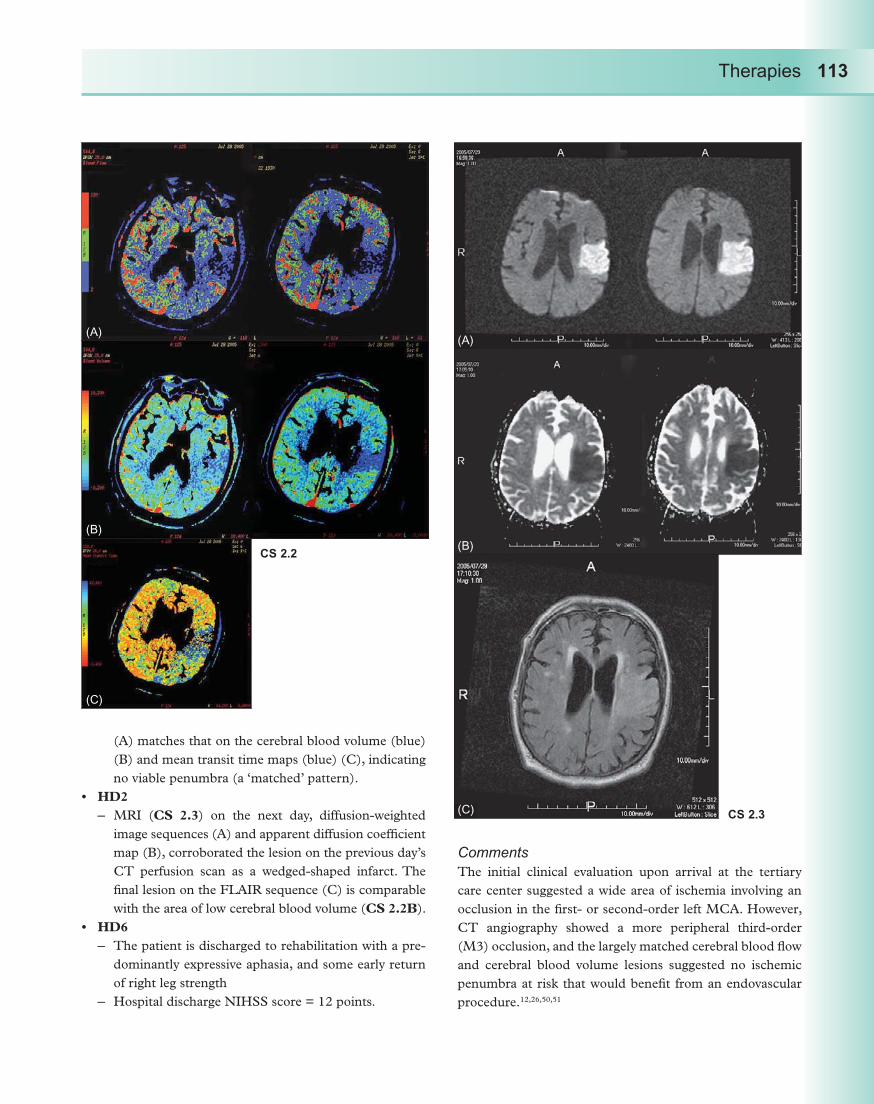
Therapies 113
(A) matches that on the cerebral blood volume (blue) (B) and mean transit time maps (blue) (C), indicating no viable penumbra (a ‘matched’ pattern).
HD2MRI ( – CS 2.3) on the next day, diffusion-weighted image sequences (A) and apparent diffusion coefficient map (B), corroborated the lesion on the previous day’s CT perfusion scan as a wedged-shaped infarct. The final lesion on the FLAIR sequence (C) is comparable with the area of low cerebral blood volume (CS 2.2B).
HD6The patient is discharged to rehabilitation with a pre- –
dominantly expressive aphasia, and some early return of right leg strengthHospital discharge NIHSS score = 12 points. –
Comments
The initial clinical evaluation upon arrival at the tertiary care center suggested a wide area of ischemia involving an occlusion in the first- or second-order left MCA. However, CT angiography showed a more peripheral third-order (M3) occlusion, and the largely matched cerebral blood flow and cerebral blood volume lesions suggested no ischemic penumbra at risk that would benefit from an endovascular procedure.12,26,50,51
CS 2.2
CS 2.3
(A)(A)
(B)
(C)
(B)
(C)

114 Therapies
Despite a relatively high initial NIHSS score (>20 points), the Stroke Team decided not to offer additional treatments beyond IV thrombolysis. The subsequent MRI scan confirmed a peripheral frontoparietal lesion. The patient’s early recovery during the acute hospitalization was encouraging and validated this clinical decision.
Case study 3. Hemicraniectomy for the malignant
middle cerebral artery syndrome
History and exam
A 56-year-old man was hospitalized for pneumonia. Two days later, he developed a right hemispheric stroke syndrome, with dysarthria, left hemiplegia, and hemineglect.
Timeline
HD1 Acute stroke symptoms onset.
HD3 Transfer to the regional stroke center.Serial head CT scans (CS 3.1):
HD4 Admission head CT scan shows an early large right MCA stroke (A,B).
Exam: alert and conversant; left hemianopia and hemineglect; dense left hemiplegia. NIHSS score = 16 points.Neurosurgery consult to consider hemicraniectomy.
HD6 Deteriorating mental status prompted repeat head CT scan (C–E):
Extensive right uncal herniation has markedly obscured the entire posterior fossa, with pressure on the midbrain and trapping of the contralateral left ventricle (C), midline shift of the calcified pineal gland (arrow) (D), and well-delineated MCA-territory infarct with subfalcine herniation (E).
HD6 Hemicraniectomy (CS 3.2). The patient’s wife was consented for this procedure.
Following a large curvilinear incision from the anterior tragus back toward the occipital bone and to the midline of scalp (A, left), burr holes are made (A, right), and a wide skull bone window is removed (B).The exposed brain shows signs of elevated intracranial pressure, with engorged superficial veins (C). Following this craniotomy, the dura is closed with the addition of fascia latae from the scalp (arrows) (D).
HD7 Postoperative day 1, Neuroimaging (CS 3.3):head CT scan shows severe edema, pushing brain beyond the former skull margin, with some normalization toward midline. A single transaxial view (A) and composite views (B,C) demonstrate how the infarcted brain extends beyond the skull margin. A postoperative lateral skull film shows the stapled wound (D), and three-dimensional CT scan shows a postoperative AP view (E).
HD15 Transfer to acute rehabilitation.Day 60 Recovery:
Walking 80–100 feet, holding a quad cane in the right hand.Left shoulder subluxation on plain X-ray.
Day 84 Cranioplasty procedure (CS 3.4).The skull bone is replaced 3 months later (A): reconstruc-tive surgery to encompass the region where the temporalis muscle was removed.Post-cranioplasty FLAIR MRI sequence (B,C). Note that there are small areas of apparently intact brain tissue within the large right MCA stroke.
CS 3.1
(A)
(B)
(D)
(C)
(E)

Therapies 115
The patient following cranioplasty, with a widened pal-pebral fi ssure and mild lower facial paresis on the left (D).NIHSS score = 7 points, largely from residual left hemiparesis.
Day 104 Returns home from assisted living facility. Ambulating with quad cane.
A second patient, aged 51, underwent craniotomy for a malignant right MCA syndrome, and returned to the stroke clinic 1 year later, ambulatory, but with signifi cant left hemiparesis (CS 3.4E). Note the left ankle–foot orthosis, and fl exor posturing of the left hand.
CS 3.2
(A)
(B)
(D)
(C)

116 Therapies
CS 3.3
(A)
(B)
(C)
(D) (E)

Therapies 117
Comments
Hemicraniectomy works. In an analysis of three randomized clinical trials, the procedure benefited not only survival but also short-term outcomes.27 The time-to-randomization in this analysis was 45 hours, in order to begin the surgical procedure within 48 hours. Patient selection included an NIHSS score >15 points, with a diminished level of con-sciousness. The short-term survival was improved from 29% in the medical (placebo) arm, versus 78% in the treatment (surgical) arm. The number needed to treat to result in a single favorable outcome at 12 months, defined as modified Rankin Scale of 4 points, was two patients.
Key observations about these data are: (1) most case series and clinical trials work restrict the procedure to patients 60 years of age; (2) an excellent outcome from
this life-threatening stroke may be defined by the ability to return to an ambulatory state, as the patients presented here did; and (3) the decision to intervene should be made within the first 12–48 hours, soon after acute treatments (e.g., IV t-PA and endovascular approaches) have been attempted yet before significant progression toward uncal herniation. In practice, hemicraniectomy is typically recommended for the treatment of non-dominant hemisphere strokes, but can also be considered for highly selected patients with left hemispheric stroke.
Some of the functional recovery seen with hemicran-iectomy may relate to the salvaging of brain tissue within the ischemic penumbra (CS 3.4B,C). The patches of spared brain presumably avert pressure necrosis against the adja-cent skull when that tissue acutely swells.
CS 3.4
(A)
(D) (E)
(B)
(C)

118 Therapies
References
Warach S. Tissue viability thresholds in acute stroke: 1. the 4-factor model. Stroke 2001; 32: 2460–1.Tilley B, Lyden P, Brott T, Lu M, Levine S, Welch K. 2. Total quality improvement method for reduction of delays between emergency department admission and treatment of acute ischemic stroke. Arch Neurol 1997; 54: 1466–74.Alberts M, Hademenos G, Latchaw R,3. et al. Recommendations for the establishment of primary stroke centers. JAMA 2000; 283: 3102–19.Alberts M, Latchaw R, Selman W,4. et al. Recommendations for comprehensive stroke centers: a consensus statement from the Brain Attack Coalition. Stroke 2005; 36: 1597–618.Fisher M. Recommendations for advancing 5. development of acute stroke therapies: Stroke Therapy Academic Industry Roundtable 3. Stroke 2003; 34: 1539–46.Brott T, Bogousslavsky J. Treatment of acute ischemic 6. stroke. N Engl J Med 2000; 343: 710–22.del Zoppo G. Stroke and neurovascular protection. 7. N Engl J Med 2006; 354: 553–5.National Institute of Neurological Disorders and 8. Stroke rt-PA Stroke Study Group. Tissue plasminogen activator for acute ischemic stroke. N Engl J Med 1995; 333: 1581–7.Smith W, Sung G, Starkman S,9. et al., for the MERCI Trial Investigators. Safety and efficacy of mechanical embolectomy in acute ischemic stroke: results of the MERCI Trial. Stroke 2005; 36: 1432–40.McDougall C, Clark W, Mayer T, 10. et al. The Penumbra Stroke Trial: safety and effectiveness of a new generation of mechanical devices for clot removal in acute ischemic stroke [abstract]. International Stroke Conference (New Orleans) 2008; LB4.Konstas A-A, Choi J, Pile-Spellman J. Neuroprotection 11. for ischemic stroke using hypothermia. Neurocrit Care 2006; 4: 168–78.Molina C, Saver J. Extending reperfusion therapy 12. for acute ischemic stroke: emerging pharmacological, mechanical, and imaging strategies. Stroke 2005; 36: 2311–20.Saver J. Time is brain-quantified. 13. Stroke 2006; 37: 263–6.
Marler JR, Tilley BC, Lu M,14. et al. Early stroke treatment associated with better outcome: The NINDS rt-PA Stroke Study. Neurology 2000; 55: 1649–55.Hacke W, Donna G, Fieschi C,15. et al. Association of outcome with early stroke treatment: pooled analysis of ATLANTIS, ECASS, and NINDS rt-PA stroke trials. Lancet 2004; 363: 768–74.Hacke W, Kaste M, Bluhmki E, 16. et al. Thrombolysis with alteplase 3 to 4.5 hours after acute ischemic stroke. N Engl J Med 2008; 359: 1317–29.Furlan A, Higashida R, Katzan I, Abou-Chebl A. 17. Intra-arterial thrombolysis in acute ischemic stroke. In: Lyden P, ed. Thrombolytic Therapy for Stroke. Totowa, NJ: Humana Press; 2001: 175–95.Furlan A, Higashida R, Wechsler L,18. et al. Intra-arterial prourokinase for acute ischemic stroke. The PROACT II Study: a randomized controlled trial. JAMA 1999; 282: 2003–11.Mattle H. Intravenous or intra-arterial thrombolysis? 19. It’s time to find the right approach for the right patient [editorial]. Stroke 2007; 38: 2038–40.Adams H, Jr, del Zoppo G, Alberts M,20. et al. Guidelines for the early management of adults with ischemic stroke: a guideline from the American Heart Association/American Stroke Association Stroke Council, Clinical Cardiology Council, Cardiovascular Radiology and Intervention Council, and the Atherosclerotic Peripheral Vascular Disease and Quality of Care Outcomes in Research Interdisciplinary Working Groups. Stroke 2007; 38: 1655–711.Lindsberg P, Mattle H. Therapy of basilar artery 21. occlusion: a systematic analysis comparing intra-arterial and intravenous thrombolysis. Stroke 2006; 37: 922–8.Agarwal P, Kumar S, Hariharan S,22. et al. Hyperdense middle cerebral artery sign: can it be used to select intra-arterial versus intravenous thrombolysis in acute ischemic stroke? Cerebrovasc Dis 2004; 17: 182–90.Zaidat O, Suarez J, Santillan C,23. et al. Response to intra-arterial and combined intravenous and intra-arterial thrombolytic therapy in patients with distal internal carotid artery occlusion. Stroke 2002; 33: 1821–7.Marder V, Chute D, Starkman S,24. et al. Analysis of thrombi retrieved from cerebral arteries of patients with acute ischemic stroke. Stroke 2006; 37: 2086–93.

Therapies 119
Rogalewski A, Schneider A, Ringelstein E, Schabitz 25. W-R. Toward a multimodal neuroprotective treatment of stroke. Stroke 2006; 37: 1129–36.Kidwell CS, Alger JR, Saver JL. Beyond mismatch: 26. evolving paradigms in imaging the ischemic penumbra with multimodal magnetic resonance imaging. Stroke 2003; 34: 2729–35.Stroke Unit Trialists Collaboration. How do stroke 27. units improve patient outcomes? A collaborative systematic review of the randomized trials. Stroke 1997; 28: 2139–44.Schwab S, Steiner T, Aschoff A,28. et al. Early hemicraniectomy in patients with complete middle cerebral artery infarction. Stroke 1998; 29: 1888–93.Vahedi K, Hofmeijer J, Juettler E,29. et al. Early decompressive surgery in malignant infarction of the middle cerebral artery: a pooled analysis of three randomised controlled trials. Lancet 2007; 6: 215–22.Rothwell P, Warlow C. Timing of TIAs preceding 30. stroke: time window for prevention is very short. Neurology 2005; 64: 817–20.Sacco R, Adams R, Albers G,31. et al. Guidelines for prevention of stroke in patients with ischemic stroke or transient ischemic attack. Stroke 2006; 37: 577–617.Furlan A. Carotid-artery stenting—case open or 32. closed? [editorial]. N Engl J Med 2006; 355: 1726–9.Adams H, Jr. Cardiac disease and stroke: will history 33. repeat itself? Mayo Clin Proc 2006; 81: 597–601.Kizer J, Devereux R. Patent foramen ovale in young 34. adults with unexplained stroke. N Engl J Med 2005; 353: 2361–72.Fiorella D, Levy E, Turk A,35. et al. US multicenter experience with the Wingspan stent system for the treatment of intracranial atheromatous disease: periprocedural results. Stroke 2007; 38: 881–7.Zaidat O, Klucznik R, Chaloupka J, 36. et al. An NIH-funded multicenter registry on the use of the Wingspan intracranial stent for high-risk patients with symptomatic intracranial arterial stenosis [abstract]. International Stroke Conference (San Francisco) 2007; LB3.PROGRESS Group. Randomised trial of a perindopril-37. based blood-pressure-lowering regimen among 6105 individuals with previous stroke or transient ischaemic attack. Lancet 2001; 358: 1033–41.
The Stroke Prevention by Aggressive Reduction in 38. Cholesterol Levels (SPARCL) Investigators. High-dose atorvastatin after stroke or transient ischemic attack. N Engl J Med 2006; 355: 549–59.Albers G, Amarenco P, Easton J, Sacco R, Teal 39. P. Antithrombotic and thrombolytic therapy for ischemic stroke: The Seventh ACCP Conference on Antithrombotic and Thrombolytic Therapy. Chest 2004; 3: S483–512.Chimowitz MI, Lynn MJ, Howlett-Smith H,40. et al. Comparison of warfarin and aspirin for symptomatic intracranial arterial stenosis. N Engl J Med 2005; 352(13): 1305–16.Castillo J, Leira R, Garcia M, Serena J, Blanco M, 41. Davalos A. Blood pressure decrease during the acute phase of ischemic stroke is associated with brain injury and poor stroke outcome. Stroke 2004; 35: 520–6.Baird T, Parsons M, Phanh T,42. et al. Persistent poststroke hyperglycemia is independently associated with infarct expansion and worse clinical outcome. Stroke 2003; 34: 2208–14.Chobanian A, Bakris G, Black H,43. et al. Seventh report of the Joint National Committee on prevention, detection, evaluation, and treatment of high blood pressure (JNC 7—Complete Version). Hypertension 2003; 42: 1206–52.CAPRIE Steering Committee. A randomised, 44. blinded, trial of clopidogrel versus aspirin in patients at risk of ischaemic events (CAPRIE). Lancet 1996; 348: 1329–39.Bhatt D, Fox K, Hacke W,45. et al. Clopidogrel and aspirin versus aspirin alone for the prevention of atherothrombotic events. N Engl J Med 2006; 354: 1706–17.Diener H-C, Bogousslavsky J, Brass L,46. et al. Aspirin and clopidogrel compared with clopidogrel alone after recent ischaemic stroke or transient ischaemic attack in high-risk patients (MATCH): randomised, double-blind, placebo-controlled trial. Lancet 2004; 364: 331–7.Diener H, Cunha L, Forbes C, Sivenius J, Smets P, 47. Lowenthal A. European Stroke Prevention Study 2. Dipyridamole and acetylsalicylic acid in the secondary prevention of stroke. J Neurol Sci 1996; 143: 1–13.The ESPRIT Study Group. Aspirin plus dipyridamole 48. versus aspirin alone after cerebral ischaemia of arterial origin (ESPRIT): randomised controlled trial. Lancet 2006; 367: 1655–73.

120 Therapies
Sacco R, Diener H-C, Yusuf S, 49. et al. Aspirin and extended-release dipyridamole versus clopidogrel for recurrent stroke. N Engl J Med 2008; 359: 1238–51.Lev M, Segal A, Farkas J,50. et al. Utility of perfusion-weighted CT imaging in acute middle cerebral artery stroke treated with intra-arterial thrombolysis: Prediction of final infarct volume and clinical outcome. Stroke 2001; 32: 2021–8.Wintermark M, Reichhart, Thiran JP,51. et al. Prognostic accuracy of cerebral blood flow measurement by perfusion computed tomography, at the time of emergency room admission in acute stroke patients. Ann Neurol 2002; 51: 417–32.
Further reading
Adams H, Jr, del Zoppo G, Alberts M, et al. Guidelines for the early management of adults with ischemic stroke:
a guideline from the American Heart Association/American Stroke Association Stroke Council, Clinical Cardiology Council, Cardiovascular Radiology and Intervention Council, and the Atherosclerotic Peripheral Vascular Disease and Quality of Care Outcomes in Research Interdisciplinary Working Groups. Stroke 2007; 38: 1655–711.
del Zoppo G. Stroke and neurovascular protection. N Engl J Med 2006; 354: 553–5.
Lyden P. Thrombolytic Therapy for Stroke. Towana, NJ: Humana Press; 2001. This textbook provides a superb overview of the development of IV and IA thrombolysis for AIS, along with case studies.
Sacco RL, Adams R, Albers G, et al. Guidelines for prevention of stroke in patients with ischemic stroke or transient ischemic attack. Stroke 2006; 36: 577–617.
Warach S. Tissue viability thresholds in acute stroke: The 4-factor model. Stroke 2001; 32: 2460–1.

121
Note: page numbers in bold refer to Figures, and those in italics refer to Tables.
abciximab 107acute ischemic stroke (AIS) 1, 2–3
causes 3CT appearance 2management 108
adventitia 75agraphesthesia 6alcohol consumption 109amaurosis fugax 6aneurysms 2, 4
dissecting 82fibromuscular dysplasia 76, 77
angiographycatheter-induced vasospasm 77, 80conventional 12, 13, 26, 27CT 11, 26
carotid atherosclerosis 36fibromuscular dysplasia 79internal carotid artery dissection 83,
84MRI 12, 13, 28stationary arterial waves 77, 80
angioplasty, case studies 25, 26, 27cervical fibromuscular dysplasia 93,
94anosagnosia 8anoxic–ischemic encephalopathy 23, 24anterior cerebral artery (ACA) 18, 49anterior cerebral artery infarcts 49–51
symptoms 48anterior cerebral artery territory 32, 48anterior choroidal artery 38anterior choroidal artery infarct 39anterior circulation 31
internal carotid artery territory 31, 34
middle cerebral artery territory 34, 42–3
anticoagulationafter cervical artery dissection 87in secondary prevention 109
antiplatelet therapy 109Anton’s syndrome 7
aortic atherosclerosis, transoesophageal ultrasound 18
aortic dissection 84extension into ICA 86
aphasia 8MCA lesions 9
arterial occlusion, dissection 81, 82arterial territories
cerebellum 59cerebral hemispheres 32pons 59
arteriolosclerosis 19–20, 21, 22arteriovenous fistula, fibromuscular
dysplasia 77artery-to-artery embolism 17
in dissection 81in fibromuscular dysplasia 76
aspirinin acute ischemia stroke 108in secondary prevention 109
astereognosis 6ataxia 5, 62
cerebellar infarct 6atherosclerosis 17, 18
case study 25–7intracranial 91
atrial fibrillation 19atrial myxoma 19atrial thrombus 19auditory cortex, location 5
balloon angioplasty, fibromuscular dysplasia 93, 94
Barthel Index (BI) 10basilar artery (BA) 18, 57, 58
top-of-the-basilar syndrome 59, 63basilar artery occlusion 64, 65
case study 70–2basilar artery territory 63–4‘beading’ pattern, fibromuscular
dysplasia 76, 77behavioural deficits 9, 67blood pressure
in acute ischemic stroke 108recommended goal 109
body position, acute ischemic stroke 108
‘brain shock’ 108brain tumours 10Broca’s aphasia 8, 48Broca’s area 5, 9
cardiac catheterization 27cardioembolic disease 19, 20
case study 27–9carotid arteries 18
see also internal carotid artery (ICA)carotid atherosclerosis
CT angiography 36multifocal emboli 37
carotid bifurcation 34Doppler sonography 35
carotid endarterectomy 37, 109carotid stenting 109carotid T 31carotid T occlusion 40
endovascular recanalization 109–12case studies
basilar artery occlusion 70–2bilateral internal carotid
dissections 94, 96cardioembolic disease 27–9carotid T lesion, endovascular
recanalization 109–12cervical fibromuscular dysplasia,
angioplasty 93, 94common carotid and M1
occlusions 51–4IV thrombolysis with ‘matched’ CT
perfusion study 111–14large vessel disease
(atherosclerosis) 25–7malignant MCA syndrome,
hemicraniectomy 114–17MCA occlusion due to ICS
dissection 97–8moyamoya disease 99–100vertebral artery stenting
atherosclerosis 69–70dissecting aneurysm 93–4, 95
catheter-induced vasospasm 77, 79, 80central post-stroke pain syndrome 6central retinal artery occlusion 6, 39, 97cerebellar arteries 58, 59
INDEX

122 Index
cerebellar hemisphere infarction 5, 6cerebellar stroke 59, 60–1, 62, 108
micropathology 61cerebellum, arterial territories 59cerebral ischemia
complexity 104molecular events 105
cerebral vasoconstriction syndrome 90–1
cervical arterial dissection 79, 81, 82, 84angiographic features 83clinical syndrome 84diagnosis 87embolic shower 85epidemiology 84extension from aorta 86in fibromuscular dysplasia 76, 86‘halo sign’ 85management 87natural history 87pathogenesis 81, 84
cholesterol, recommended goal 109choroidal arteries 38circle of Willis 31, 33
atherosclerosis 18perforator vessels 21
clinical trials 103clopidogrel 109cognitive deficits 9, 67, 68collateral circulation 31, 33, 52, 104,
107combination therapeutic
modalities 107common carotid occlusion, case
study 51–4computed tomography (CT) 10
CT angiography 11, 26carotid atherosclerosis 36
CT perfusion studies 11–12old versus new strokes 47
conventional angiography 12, 13, 26, 27
cortical blindness 7cranioplasty 114, 117
decompression, cerebellar stroke 59, 60desmoteplase 107diagnosis 10
neuroimaging 10–13diffusion-weighted MRI 6, 11, 12digital subtraction angiography 12, 13,
26, 27diplopia 7, 62dipyridamole 109
dissection 79, 81fibromuscular dysplasia 76see also cervical arterial dissection
Doppler sonography, carotid artery 35Duret haemorrhages 2dysarthria 8, 62dysphagia 62
Ehlers–Danlos syndrome 79emboli 46
artery-to-artery 17, 76, 81from internal carotid dissection 85,
97multifocal 24, 25, 37, 43retinal 54
Emergency Medical Services 103encephaloduroarteriosynangiosis 92–3,
100endarterectomy 37, 109endovascular procedures 104–6
angioplasty 25, 26, 27cervical fibromuscular
dysplasia 93, 94basilar artery occlusion, IA
thrombolysis 70–2carotid T lesion,
thrombectomy 109–12cervical artery dissection 87M1 occlusion, IA thrombolysis 52–3see also stenting
false lumen, ICA dissections 82fetal PCA 65, 69fever 108fibrinolytic agents 107
see also tissue plasminogen activator (t-PA) therapy
fibromuscular dysplasia 75–6angioplasty, case study 93, 94diagnosis and differential
diagnosis 76–9of internal carotid arteries 76
dissection 86morphologic subtypes 79natural history and management 79,
80pathogenesis of stroke 76renal 78resources for patients 102
forced gaze 7, 8, 48frontal lobe symptoms 48
gaze deviation 7, 8, 48giant cell arteritis (GCA)
clinical presentation and epidemiology 87–9
management 89, 91pathogenesis and diagnosis 89resources for patients 102temporal arteritis 89
Glasgow Outcome Scale (GOS) 10global aphasia 8, 48glycogen IIb/IIIa antagonists 107
haemodynamics, factors in tissue viability 104
haemorrhagic stroke 1, 2, 3–4, 8see also subarachnoid haemorrhage
‘halo sign’, cervical arterial dissection 85, 87
headache 4healthcare resources 103hearing loss 62hemicraniectomy 108
case study 114–17, 115hemifield visual loss 7, 65hemineglect 7–8, 48hemiparesis 4hemiplegia 4herniation syndromes 108
case study 114–17Horner’s syndrome, ICA dissection 84,
87hyperglycemia 108hypophyseal arteries 38hypotension, watershed stroke 23, 24hypothermia 107, 108
incidence of stroke 1INR (International Normalized
Ratio) 109internal carotid artery (ICA) 34
Doppler sonography 35fibromuscular dysplasia 76, 78, 79intracranial branches 38see also carotid arteries
internal carotid artery dissectionanatomy 82angiographic features 83, 84bilateral, case study 94, 96clinical syndrome 84diagnosis 87embolic shower 85epidemiology 84extension from aorta 86in fibromuscular dysplasia 86‘halo sign’ 85management 87

Index 123
MCA occlusion, case study 97–8natural history 87pathogenesis 81, 84‘string sign’ 85
internal carotid artery stroke 38, 41symptoms 34
internal carotid artery territory 31, 32, 34intima 75intra-arterial thrombolysis 104–6, 107
basilar artery occlusion 70–2M1 occlusion 52–3
intracerebral haemorrhage (ICH) 1, 2, 3–4, 8
see also subarachnoid haemorrhageintracranial atherosclerosis 91intravenous thrombolysis 104, 106, 107
case study 111–14ischemia, molecular events 105
lacunar infarcts 19–20, 21, 22, 42‘lagunes’ 42language problems 8large vessel disease
(atherosclerosis) 17, 18case study 25–7
late intra-arterial thrombolysis 70–2lateral medullary infarction 62lenticulostriate arteries 42, 48, 49leptomeningeal anastomoses 31, 33locked-in syndrome 64low-density lipoprotein, recommended
goal 109
‘macunes’ 42, 43magnesium 107magnetic resonance imaging (MRI) 6,
11, 12–13, 28angiography 37
malignant MCA syndrome 42, 48hemicraniectomy 114–17
‘man-in-the-barrel’ syndrome 23‘matched’ CT perfusion, case
study 111–14media 75medullary infarction 62, 63memory impairment 67, 68MERCI Retriever 52, 104, 106
case study 110–12microplasmin 107middle cerebral artery (MCA) 18, 41
M1 34M1 occlusion 42
case study 51–4symptoms 48
M2 occlusion 43, 44recanalization 45symptoms 48
M3, M4 occlusions 43, 45middle cerebral artery infarction 5, 9,
10, 46combination with PCA infarction
43due to ICA dissection, case
study 97–8symptoms 43, 48
middle cerebral artery territory 32, 34, 42–3
embolic showers 37mismatch model 11, 13, 107Modified Rankin Scale (mRS) 10monoparesis 4monoplegia 4mortality rates 1motor cortex, location 5motor recovery 5moyamoya 79
definition and pathogenesis 91–2epidemiology 92natural history and
management 92–3moyamoya disease 92, 93
case study 99–100resources for patients 102
moyamoya syndrome 92multifocal emboli 24, 25
National Institutes of Health Stroke Scale (NIHSS) 4
prediction of outcome 9nausea 62neurofibromatosis 79neuroimaging 10–13, 107
see also computed tomography (CT); magnetic resonance imaging (MRI)
neuroprotection 104, 107neurovascular unit 104, 106NXY-059 107nystagmus 62
oculomotor impairment 7old versus new strokes, CT 47ophthalmic artery 38outcomes 9–10oxygen therapy 108
paraparesis 4paraplegia 4
patent foramen ovale 19, 20pathophysiology 17
cardioembolic disease 19, 20etiologies with non-arterial
distributions 23large vessel disease 17, 18multifocal emboli 24, 25small vessel disease
(arteriolosclerosis) 19–20, 21, 22
undetermined etiology 24watershed stroke 23
Penumbra Stroke System 104perforating branches, posterior cerebral
artery 64–5perforator vessels 21perfusion–diffusion mismatch 11, 13,
107perfusion-weighted MRI 11, 13PHQ-9 10pons, arterial territories 59pontine perforators 57, 58pontine stroke 10, 61, 62posterior cerebral artery (PCA) 32, 64,
66cortical branches 65fetal variant 65, 69perforating branches 64–5
posterior cerebral artery infarction 7, 66–8
combination with MCA infarction 43
symptoms 65, 67posterior ciliary arteries, involvement in
GCA 89posterior circulation 31, 57posterior communicating artery
(PcoA) 57posterior communicating artery
territory 32posterior inferior cerebellar artery
(PICA) 58, 59posterior inferior cerebellar artery
infarcts 60, 61, 62primary CNS angiitis
biopsy appearance 92clinical presentation and
epidemiology 89management 91pathogenesis and diagnosis 89
progression of lesions 47psychiatric disorders 9
quadrantanopia 7, 48, 65

124 Index
radiation arteriopathy 79recanalization
basilar artery 64carotid T, case study 109–12M2 45
recurrent artery of Heubner 48, 49renal fibromuscular dysplasia 78reperfusion treatments 104
therapeutic modalities 107timeframes 104–5see also angioplasty; stenting;
thrombolysisreteplase 107retinal emboli 54retinal stroke 38revascularization, moyamoya
disease 92–3rheumatic valvular disease 19risk factors, recommended goals 109
secondary prevention 108–9segmental arterial narrowing,
differential diagnosis 79sensation testing 6sensory cortex, location 5sensory loss 5–6small vessel disease
(arteriolosclerosis) 19–20, 21, 22sonothrombolysis 107speech problems 8
MCA lesions 9splenium, isolated infarct 68stationary arterial waves 77, 79, 80stenting 25, 26, 109
basilar artery 64bilateral ICA dissections 94, 96middle cerebral artery 97, 98vertebral artery 69–70
dissecting aneurysm 93–4, 95steroid therapy
in giant cell arteritis 89, 91in primary CNS angiitis 91
‘string signs’fibromuscular dysplasia 79internal carotid dissection 85
strokeclassification 1epidemiology 1–2see also acute ischemic stroke (AIS)
‘stroke-ready’ medical centres 103Stroke-specific Quality of Life scale 10subacute ischemic stroke, gross
pathology 2
subarachnoid haemorrhage (SAH) 1, 4CT appearance 2fibromuscular dysplasia 76
superior cerebellar artery (SCA) 57symptoms
of anterior cerebral artery stroke 48ataxia 5of cervical arterial dissection 84cognitive and behavioural deficits 9headache 4of internal carotid artery stroke 34language and speech problems 8of middle cerebral artery stroke 43,
48of posterior cerebral artery
stroke 65, 67sensory loss 5–6of vertebrobasilar stroke 59, 62–3visual 6–7visuospatial neglect 7–8weakness 4–5
temperature 108temporal arteritis
pathology 89resources for patients 102see also giant cell arteritis (GCA)
tenecteplase 107thrombectomy 104, 106, 107thrombolysis 104–6, 107
basilar artery occlusion 70–2in cervical artery dissection 87intra-arterial 105–6intravenous, case study 111–14M1 occlusion 52–3
time to treatment 103, 104–5, 104tinnitus 62tirofiban 107tissue plasminogen activator (t-PA)
therapy 51, 104, 106, 107basilar artery occlusion 70–2
tissue viability, influencing factors 103, 104
TOAST criteria 17top-of-the-basilar syndrome 59, 63,
64, 65transient ischemic attack (TIA) 1transoesophageal echocardiography
(TEE) 17, 18, 19, 27, 29tuberculous meningitis 88tubular stenosis, ICA 79
undetermined etiology 24
vascular dementia 67, 68vasculitis 87
classification 88clinical presentation and
epidemiology 87–9management 89, 91pathogenesis and diagnosis 89secondary to tuberculous
meningitis 88vasculopathies 75
cervical arterial dissection 81–7fibromuscular dysplasia 75–81moyamoya 91–3vasculitis 87–91
vasospasm, catheter-induced 77, 79, 80
ventricular thrombus 19vertebral arteries 18, 57, 58
fibromuscular dysplasia 78involvement in GCA 89
vertebral artery dissection 81, 83diagnosis 87see also cervical arterial dissection
vertebral artery stenting, case studiesatherosclerosis 69–70dissecting aneurysm 93–4, 95
vertebrobasilar stroke, symptoms 59, 62–3
vertebrobasilar system 57–8arterial territories of pons and
cerebellum 59vertigo 62vessel wall layers 75visual cortex, location 5visual field testing 7visual symptoms 6–7
in giant cell arteritis 89in middle cerebral artery stroke
48in posterior cerebral artery
stroke 65, 67visuospatial neglect 7–8vomiting 62
Wallerian degeneration 4, 5warfarin
after cervical artery dissection 87in secondary prevention 109
watershed stroke 23, 24weakness 4–5
NIH Stroke Scale (NIHSS) 4Wernicke’s aphasia 8, 48Wernicke’s area 5, 9