issn 1809 - 3736 cardiovascular sciences...
TRANSCRIPT

JAN. / MAR. 2009 - VOL. 4 - NUMBER 1 �
ISSN 1809 - 3736
EDITORIAL COORDINATIONAlexandre Ciappina Hueb
Carlos Henrique Marques SantosOsvaldo Sampaio Netto
ASSOCIATED EDITORSAlfredo I. Fiorelli, Elias Kallás José Carlos Dorsa V. Pontes
Rafael Diniz Abrantes Danton R. Rocha - Loures,
EDITORIAL SECRETARY: Otoni Moreira Gomes
CARDIOVASC SCI FORUM Jan. / Mar. 2009 Vol. 4/ NUMBER �
CARDIOVASCULAR SCIENCES FORUMCARDIOVASCULAR SCIENCES FORUM
Sponsored by:
President: Elaine Maria Gomes (OAB)Scientific Coordination: Otoni M. GomesClinic Director: Eros Silva GomesEvents Administration: Elton S. Gomes
Scientific Co-sponsorship by: International College of Cardiovascular Sciences, South American Section of the International Academy of Cardiovascular Sciences (IACS - SAS), Department of Experimental Research of the Brazilian Society of Cardiovascular Surgery (DEPEX - SBCCV), SBCCV Department of Extracorporeal Circulation and Mechanical Assisted Circulation (DECAM - SBCCV), SBCCV Department of Clinical Cardiology, Brazilian Association of Intensive Cardiology, Brazilian Academy of Cardiology for the Family, SBCEC - Brazilian Society of Extracorporeal Circulation.
Fundação Cardiovascular São Francisco de Assis Verdade é JesusSão Francisco de Assis Truth is Jesus Cardiovascular Foundation
Fundación Cardiovascular San Francisco de Assis Jesus es la Verdad
Alberto J. Crottogini (Argentina)Borut Gersak (Slovenia)
Celina Morales (Argentina)Daniel Bia (Uruguay)
Calogerino Diego B. Cuzumano (Venezuela)Diego A. Borzelino (Venezuela)
Domingos S. R. Souza (Sweden)Eduardo Armentano (Uruguay)Eduardo R. Migliaro (Uruguay)
Michael Dashwood (England)Pascal Dohmen (Germany),
Patrícia M. Laguens (Argentina) Pawan K. Singal (Canadá) Ricardo Gelpi (Argentina)
Ruben P. Laguens (Argentina)Sylvain Chauvaud (França)Tofy Mussivand (Canadá)Tomas A. Salerno (EE.UU)
Verônica D’Annunzio (Argentina)
International Scientific Board

CARDIOVASCULAR SCIENCES FORUM2
Adalberto Camim (SP)Aguinaldo Coelho Silva (MG)Alan Tonassi Paschoal (RJ)Alcino Lázaro da Silva (MG)Alexandre Ciappina Hueb (SP)Alexandre Kallás (MG)Antônio Alves Coelho (DF)Antônio A. Ramalho Mota (MG)Antônio de Pádua Jazbik (RJ)Antônio S. Martins (SP)Bruno Botelho Pinheiro (GO)Carlos Alberto M. Barrozo (RJ)Carlos Henrique M. Santos (MS)Carlos Henrique V. Andrade (MG)Cláudio Pitanga M. Silva (RJ)Cristina Kallás Hueb (MG)Danton R. Rocha Loures (PR)Domingos J. Moraes (RJ)Edmo Atique Gabriel (SP)Eduardo Augusto Victor Rocha (MG)Eduardo Keller Saadi (RS)Elmiro Santos Resende (MG)Eduardo Sérgio Bastos (RJ)Eros Silva Gomes (MG)Evandro César V. Osterne (DF)Fábio B. Jatene (SP)Francisco Diniz Affonso Costa (PR)Francisco Gregory Jr. (PR)Geraldo Martins Ramalho (RJ)
Geraldo Paulino S. Filho (GO)Gilberto V. Barbosa (RS)Gladyston Luiz Lima Souto (RJ)Guaracy F. Teixeira Filho (RS)Hélio Antônio Fabri (MG)Hélio P. Magalhães (SP)Henrique Murad (RJ)Jandir Ferreira Gomes Junior (MS)João Bosco Dupin (MG)João Carlos Ferreira Leal (SP)João Jackson Duarte (MS)Jorge Ilha Guimarães (RS)José Dondici Filho (MG)José Ernesto Succi (SP)José Francisco Biscegli (SP)José Teles de Mendonça (SE)Juan Alberto C. Mejia (CE)Kleber Hirose (SP)Liberato S. Siqueira Souza (MG)Luiz Antonio Brasil (GO)Luiz Boro Puig (SP)Luis Carlos Vieira Matos (DF)Luiz Fernando Kubrusly (PR)Luiz Paulo Rangel Gomes Silva (PA)Mário Ricardo Amar (RJ)Marcelo Sávio Martins (RJ)Marcio Vinicius L. Barros (MG)Marcílio Faraj (MG)Maria José Compagnole (MG)
Mario Coli J. Moraes (RJ)Mario Oswaldo V. Peredo (MG)Melchior Luiz Lima (ES)Miguel Angel Maluf (SP)Neimar Gardenal (MS)Noedir A. G. Stolf (SP)Osvaldo Sampaio Netto (DF)Pablo Maria A. Pomeratzeff (SP)Paulo Antônio M. Motta (DF)Paulo de Lara Lavítola (SP)Paulo Rodrigues da Silva (RJ)Pedro Rocha Paniagua (DF)Rafael Haddad (GO)Rodrigo Mussi Milani (PR)Ronald Sousa Peixoto (RJ)Rika Kakuda Costa (SE)Roberto Hugo Costa Lins (RJ)Ronaldo D. Fontes (SP)Ronaldo M. Bueno (SP)Rubio Bombonato (SC)Rui Manuel S. A. Almeida (PR)Sérgio Luis da Silva (RJ)Sérgio Nunes Pereira (RS)Sinara Silva Cotrim (MG)Tânia Maria A. Rodrigues (SE)Victor Murad (ES)Walter José Gomes (SP)Walter Labanca Arantes (RJ)Wanewman Lins G. Andrade (BA)
CARDIOVASC SCI FORUM Jan. / Mar. 2009 Vol. 4/ NUMBER �
EDICOR Ltda.
“Truth is Jesus the Word of God”
John 1.1; 14.6; 17.17
S C I E N T I F I C B O A R D - B R A Z I L
CARDIOVASCULAR SCIENCES FORUMCARDIOVASCULAR SCIENCES FORUM
International College of Cardiovascular Research

JAN. / MAR. 2009 - VOL. 4 - NUMBER 1 �
ISSN 1809 - 3736
EDITORIAL SECRETARYFundação Cardiovascular São Francisco de Assis Verdade é Jesus
R. José do Patrocínio, 522 - Santa Mônica, Belo Horizonte / MG - Brazil
CEP: 31.525-160 - Tel./ Fax: (55) 31 3439.3004e-mail: [email protected]
Site: www.servcor.com
DATA PROCESSING CENTERCoordination:
Elton Silva GomesCover:
Elton Silva Gomes, Joselito Pacheco Barbosa
Tiping: Maristela de Cássia Santos Xavier
Lay-out:Elton S. Gomes ADVERTISING
Advertising inquiries should be addressed to ServCor - Division of Events,
R. José do Patrocínio, 522 - Santa MônicaBelo Horizonte / MG - Brazil - CEP: 31.525-160
Tel./ Fax: (55) 31 [email protected]
Copyrights:
EDICOR Ltda.“Truth is Jesus the Word of God”
John 1.1; 14.6; 17.17
Home Page: www.servcor.com
CARDIOVASCULAR SCIENCES FORUMCARDIOVASCULAR SCIENCES FORUM

CARDIOVASCULAR SCIENCES FORUM4
CONTENTSEDITORIAL
La Investigación Basica y Clínica en Cardiologia: Premios y Castigos
H.E. Cingolani
ARTIGO ESPECIAL / SPECIAL ARTICLE
The Criation Evolution
Otoni M. Gomes
ORIGINAL ARTICLE / ARTIGOS ORIGINAIS
1 - Assessments of the Inflamatory Reaction in Placing a Polypropylene Mesh at Different Distances from the Aponeurotic Border.Avaliação da Reação Inflamatória na Colocação da Tela de Polipropileno com Diferentes Afastamen-tos da Borda Aponeurótica.
Maria Raquel B. Massoti, Alexandre Ciappina Hueb, Eduardo Chibeni F. Ramos, Rogério Mendes Grande, Félix Carlos Ocáriz Bazzano.
2 - Short -Term Analysis of Heart Rate Variability in Diabetic Patients.
E. R. Migliaro, P. Contreras.
CASE REPORT / RELATO DE CASO
Supraventricular Tachyarrhythmia in Fetus Experience and Treatment
G. C. Puentes.
UPDATING ARTICLE / ARTIGO DE ATUALIZAÇÃO
Life Style importance for Cardiovascular diseases preventionA Importância do Estilo de Vida na Prevenção das Doenças Cardiovasculares
Pereira, S.N.; Pacheco; L. S.; Dias, D.; Barbosa, V.A.; Portela, L.O.C.;
HOW TO DO / COMO FAZER
Modified De Vega Tricuspid Valve RepairValvoplastia Tricúspide De Vega Modificada
Otoni M. Gomes, Márcio Pitchon
INSTRUCTIONS FOR AUTORS
UPCOMING MEETINGS
Cardiovasc Sci Forum Jan. /Mar. 2009 Vol. 4 / Number �
5
9
21
33
39
43
48
51
53

JAN. / MAR. 2009 - VOL. 4 - NUMBER 1 �
ISSN 1809 - 3736
EDITORIALEDITORIAL
Aunque la mayor parte de los médicos aceptamos que la investigación Básica es la hecha con animales y la Clínica la realizada en pacientes, esta clasificación ha sido cuestionada. Para algu-nos especialistas en el tema, la especie utilizada para probar una hipótesis no define qué clase de investigación se realiza. Una clasificación mejor sería posiblemente dividir la investigación en Bá-sica y Aplicada. La investigación Básica está des-tinada a describir mecanismos aún no conocidos sin pretender aplicación o rédito alguno (los inves-tigadores saben que si el mecanismo es nuevo y la investigación bien hecha, tarde o temprano, ten-drá aplicación práctica). Esta investigación Básica puede hacerse en animales o seres humanos.
¿Y la investigación Clínica? Alfredo Lana-ri decía que si hacíamos experimentos en la rata diabética o hipertensa hacíamos investigación clí-nica, puesto que pretendíamos resolver problemas de aplicación Clínica.
Debemos reconocer, con las salvedades anteriores, que generalmente se denomina inves-tigación Clínica a la realizada en pacientes; puede ser ésta Básica o Aplicada, pero en la mayoría de los casos es Aplicada puesto que busca encontrar una aplicación a un mecanismo muchas veces an-teriormente descripto. Cuando se describe un me-canismo nuevo la investigación es Básica aunque haya sido realizada en pacientes.
Bernardo Houssay escribía: “... la investi-gación Básica persigue hallar verdades nuevas aún desconocidas, las cuales en general son inespera-das y tendrán consecuencias no siempre previsi-bles al principio...”.
Comroe y Drips analizaron retrospectiva-mente durante 1971-1977 qué hizo posible lo que ellos consideraron los mayores diez adelantos de la Ciencia Médica durante los últimos años. Ana-lizaron 6.000 publicaciones científicas. Estos diez adelantos fueron:
*LA INVESTIGACIÓN BÁSICA Y CLÍNICA EN CARDIOLOGIA “Premios y Castigos”“The Basic and Clinical Investigation in Cardiology: Prize and Punishment”
H. E. CingolaniDirector del Centro de Investigaciones Cardiovasculares - CONICET - UNLP
1. Antibióticos.2. Prevención de la Poliomielitis.3. Cirugía Cardíaca.4. Transplantes.5. Tratamiento de la hipertensión arterial.
6. Resucitación Cardíaca.7. Tratamiento de las Arritmias.8. Quimioterapia.9. Unidades intensivas.10. Tratamiento del infarto de miocardio.
* Republished from LA Arch Cardiovasc Sci 2001; 2(1): 5-8

CARDIOVASCULAR SCIENCES FORUM�
Luego de este meticuloso estudio llega-ron estos autores a la conclusión que los estudios “clave” que posibilitaron estos diez adelantos pro-venían 62% de la investigación Básica y 38% de la investigación Aplicada (observemos que no se especifica la especie usada). Del 62% de investi-gación Básica, un 42% no pretendían solucionar ningún problema ni tenían relación alguna con el tema en cuestión que posibilitaron.
La investigación cardiológica (Básica y Clínica) en Latinoamérica está subdesarrollada. Así lo muestra el escaso número de publicaciones o presentaciones en ambientes científicos en los cuales existe una selección rigurosa. Hoy en día en que todo parece que hubiese que convertirlo en cifras y medirlo, también se han mensurado las pu-
blicaciones científicas. Hay dos parámetros usados comunmente para evaluar una publicación cientí-fica: 1) El “impacto”; este representa el porcentaje de los trabajos publicados por esta revista que son citados en los dos años posteriores a la publicaci-ón; 2) La “vida media” de los trabajos publicados. Es decir un índice de por cuánto tiempo son cita-dos estos trabajos luego de publicados. El primero es el más conocido y usado de ellos. Combinando los dos se obtiene el “doble producto”. Cuando examinamos algunas pocas publicaciones cientí-ficas con este parámetro observamos que apare-cen primero Circulation Research seguida luego de Circulation, Journal of the American College of Cardiology, Cardiovascular Research y Journal Molecular and Celular Cardiology.
Fig. 1: Se examina con este parámetro, “doble producto”, cinco publicaciones científicas cardiológicas durante 1996, 1997, 1998 y 1999. La primer barra (la más alta) correspondió a Circulation Research, seguida de Circulation durante los 4 años examinados.

JAN. / MAR. 2009 - VOL. 4 - NUMBER 1 �
ISSN 1809 - 3736
Si tomamos la primera de estas publicacio-nes, Circulation Research, vemos que se recibie-ron para ser considerados para su publicación, des-de 1-7-99 al 30-6-00, 1114 manuscritos; sólo 11 de ellos fueron de Latinoamérica. Brasil, Argentina, México y Chile fueron los países que enviaron manuscritos. La figura 2 muestra la distribución geográfica de los manuscritos recibidos por esta publicación en el lapso anteriormente menciona-
do. Sería necesario aclarar que estos no fueron los manuscritos publicados sino los que fueron envia-dos para ser considerados. Sólo un 20%, apróxi-madamente, de los manuscritos recibidos son pu-blicados. Observemos que sólo un 1% provenían de Latinoamérica. Cálculos similares podrían ha-cerse posiblemente en relación a otras prestigiosas revistas científicas o a la presentación de trabajos anuales a la American Heart Association.
Fig. 2: Distribución geográfica de los 1114 manuscritos recibidos para ser considerados para su publicación en Circulation Research desde 1-7-99 al 30-6-00. Se observa que sólo el 1% de ellos provenía de Latinoamérica. Note que éstos son los manuscritos recibidos para ser considerados para su publicación; sólo 20% de ellos fueron publicados
Las causas de este subdesarrollo en la in-vestigación cardiológica hay que buscarla posible-mente en la falta de “premios y castigos”. Por otro lado, el porcentaje del PBI que Latinoamérica de-dica a la investigación está muy por debajo del de los países desarrollados del resto del mundo.
Dado que la función principal de la cardio-
logía no sea posiblemente la de publicargeográfica de los manuscritos recibidos por esta publicación en el lapso anteriormente menciona trabajos sino la de curar enfermos cardiológicos, uno podría plantearse el interrogante si las otras dos “patas” del trípode Asistencia – Docencia – Investigación ayudan o son contraproducentes para la Asistencia.

CARDIOVASCULAR SCIENCES FORUM�
Esto se ha mensurado y se llegó a establecer que en aquellas instituciones académicas en las cuales se hace investigación y docencia, a los pacientes les va mejor (Do “America´s best hospitals” perform better for acute myocardial infarction? Jersey Chen, et al. New England Journal of Medicine 340:286-292, 1999; Effects of admission to a teaching hos-
pital on the cost and quality of care for medicare beneficiaries. Donald H Taylos, et al. New England Journal of Medicine 340:293-2999, 1999.)
La Prestigiosa Universidad Norteamerica-na Jonhs Hopkins posee un logotipo que trata de resumir estos tres principios en los cuales descan-sa la Educación Médica.
Fig. 2: Distribución geográfica de los 1114 manuscritos recibidos para ser considerados para su publicación en Circulation Research desde 1-7-99 al 30-6-00. Se observa que sólo el 1% de ellos provenía de Latinoamérica. Note que éstos son los manuscritos recibidos para ser considerados para su publicación; sólo 20% de ellos fueron publicados
Volviendo a los “premios y castigos”, esto aunque discutible para quiernes piensan en que la Investigación Científica es una actividad creadora que no puede estar sujeta a “premios y castigos”, me ha hecho reflexionar en cuál sería la produc-ción científica sa la Educación Médica en países
desarrollados si no hubiera exigencias sobre ésta para subsistir académicamente. Por lo tanto, una política agresiva, invirtiendo fondos y aplicando “premios y castigos” ayudaría a impulsar la Inves-tigación Cardiológica Latinoamericana.
*

JAN. / MAR. 2009 - VOL. 4 - NUMBER 1 9
ISSN 1809 - 3736
SPECIAL ARTICLESSPECIAL ARTICLESTHE CREATION EVOLUTION
Otoni M. Gomes
The antagonism between the concepts of creation and evolution of the human being is fa-mous all over the world. However, at this moment, we will review the undisputed evidence of the Cre-ation evolution, an inescapable witness of the Fa-ther with His Son and the Holy Spirit’s work in the beginning of time confronted with the scientific facts that Darwin has used to compose his agnostic conception of man’s evolution. Darwin attested in his book “Descent of Man”, published in Februa-ry, 1871, (not in the book “The Origin of Species”, published in 1859) that ‘man is subject to the same mutation laws as all other species are’, according to “The Origin of Species” under the subtitle “by Means of Natural Selection, or the Preservation of Favoured Races in the Struggle for Life”.
It is important to recall Saint John’s, the Baptist, saying “A man can receive nothing un-less it has been given him from heaven.” (John 3:27) . Even the positivist philosopher, Emmanuel Kant, having no Christian bonds whatsoever said, in 1750, ‘human beings don’t possess innate ide-as’. Darwin also quotes Milne-Edwards’ assertion “nature is prodigal in variety, but niggard in inno-
vation”. Therefore, the Holy Spirit’s presence and will is embedded in all new ideas conceived for the good.
Charles Darwin has done a priceless ser-vice to humankind culture when he presented his findings and basic concepts of human development that otherwise would be buried in time. Darwin defied conventional thinking and approached the problem with what at the time seemed an unscien-tific method. Although his concepts are pertinent and incontestable to the adaptation phenomenon of individuals from a particular species, they do not apply to the transformation of one species into another.
The first correlation between human body development and animal development is attributed to Karl Von Baer, in 1827. Defending the Theory of Recapitulation, he said that ontogeny makes him think of phylogeny. (Von Baer. Apud in Bru-ggen WW, Cardiac design in lower vertebrates: what can phylogeny? Experientia 1988; 44: 919-39 - Rodrigues TMA. Contribution to the compa-red study of the vascularization of the coronary ar-teries in vertebrate species with vinyl acetate tech-
1New American Standard Bible (1995)

CARDIOVASCULAR SCIENCES FORUM�0
nique (vinilite) – Master Thesis/ São Francisco de Assis Cardiovascular Foundation - ServCor, Belo Horizonte, MG, 1998.)
Ontogeny describes the origin (genesis) and development of a certain individual. Phylo-geny describes the origin of beings that appeared before the formation of the individual. This asser-tion defines Von Baer’s ingenious and comprehen-sive scientific knowledge because it entails a vast knowledge about human being embryology and about the anatomy and physiology of numerous and different animals in the biological evolution scale.
The development of human blood circula-tion study presents an enlightening example of this phenomenon. Human conception has its origin in the union of a spermatozoid, the male sex cell, with an egg from the female ovary. The egg or zygote forms the morula - a solid mass of blastomeres formed by the zygote splitting - that is implanted in the uterine mucosa. In that place, the maternal blood feeds the morula that becomes an embryo with a tube-shaped heart, namely the cardiac tube. By the 21st day, it begins to contract as if starting the future heartbeats. The cardiac tube assumes the heart shape with the aorta that carries the blood all over the body and the pulmonary artery that carries the blood to the lungs where oxygen enriches it.
These are the phases that can be identified in the process: the morula and the trophoblasts phases when the nutrition is done by nutrients di-ffusion; the cardiac tube phase, the primitive heart phase with its longitudinal chambers; the forma-tion of a primitive heart with no separation betwe-en venous and arterial blood; the rising of septa or partitions inside the heart and the separation be-
tween the aorta and the pulmonary artery.This is the ontogeny description of our cir-
culatory system formation. Ontogeny actually re-minds phylogeny, this is why the aquamarine and the sea anemones are examples of nutrition similar to that of the trophoblasts. A cardiac tube with no beatings exists in lancelets and in earthworms and fishes themselves possess a beating cardiac tube. There are no internal divisions in the anurans he-arts and the reptiles have incomplete internal divi-sions. Having less evident variations, the hearts of birds and mammals possess human like structure.
Therefore, even if environmental adversity contributed to the acquisition or the loss of the pro-per physical resources for survival, as Wallace and Darwin claimed, it would not be possible for a par-ticular species heart to develop into another species without Jesus’ will, hence that species either survi-ve as the lancelets and the sharks or disappear.
In the Origin of Species2, page 158, Da-rwin claims, “If it could be demonstrated that any complex organ existed, which could not possibly have been formed by numerous, successive, slight modifications, my theory would absolutely break down. But I can find out no such case. No doubt many organs exist of which we do not know the transitional grades, more specially if we look to much isolated species, round which, according to my theory, there has been much extinction.”
Here we find a great scientific contradic-tion: how can one affirm the existence of some-thing never proved to be, that is, the existence of transitional forms by alleging they are extinct, if his own theory claims that the transitional species would be more capable to survive than the original forms?
2On the Origin of Species - By Means of Natural Selection or The Preservation of Favoured Races in the Struggle for Life, London, John Murray, Albermale Street, 1859 - New York, Bantam Books Bantam Books, New York 1996.
JAN. / MAR. 2009 - VOL. 4 - NUMBER 1 ��
ISSN 1809 - 3736
The passage from one kingdom to another (crystal that can regenerate in form, plants that mul-tiply and seek out better biological environments for survival, intelligent animals, man that is able to speak) is done by leaps. The remnants of all spe-cies are multiplying and there are no intermediate forms between them. Creation has evolved by Our Lord Jesus’ will and not by any accident (catastro-phe) or by an environmental demanding; on the contrary, evolution has evolved proportionally to life better conditions in our planet. The adaptation of the species to the environment, a real thing, ha-ppens according to different principles and evo-lution purposes, that is, the creation evolution is very different from the adaptation evolution. No thinking animal has appeared on Earth after man.
In Darwin’s own book title, “Origin of Spe-cies by Means of Natural Selection, or the Preser-vation of Favoured Races in the Struggle for Life”, shows the verification on the evolution basic mis-take, as proposed by Darwin himself, because the species that came before us in the phylogenesis are still surviving and multiplying themselves. One can mention the algae, the lancelets that exist for over 500 million years, the Sharks that exist for over 350 million years, the frogs, the reptiles, the birds and mammals, not to mention the ants, the invertebrates, the plants and bacteria that appea-red before all animals and exist in great numbers until our days. De Baer’s scientific verification about our gestation on how our circulatory system evolves from an inferior animal structure while we coexist with the survival of these same animals is something crystal clear. That is, our ancestries are still existing in the same form in which they were
created and reproducing in much more quantity than we are.
Why couldn’t The Father Jesus and the Holy Spirit create us isolatedly without links with other living beings on the planet? I was discussing this at home, years ago, while our book “Emmaus Road” was being written, when my son Eros rea-soned, “Dad, this is God’s greatest gift to our mor-tal life; we coexist emerged in all nature as His most perfect work; if it wasn’t like that we would live mechanically as robots, having only a superfi-cial perception of all wonders of Creation without the sublime poetry of its existence”. All my doubts went away!
However, how can one reconcile the per-fect description of Creation, in Genesis, with the scientific universe evolution concepts and the li-ving beings on Earth? Since the cathecism of our childhood, the Church has impressive information about the creation of the world in seven days, as said in Genesis, explaining that God has a diffe-rent time from ours and that one day may represent a billion years.
Saint John (21:25) teach us, “And there are also many other things which Jesus did, which if they were written in detail, I suppose that even the world itself would not contain the books that would be written.”
How could one describe the universe cre-ation to the human race in the early days of the scientific culture if until nowadays we do not even know the basic natural phenomena like the bla-ck holes that hide more than 99% of the universe substance we can observe with the most sophis-ticated telescopes? Including Einstein’s, all theo-
3New American Standard Bible (©1995)

CARDIOVASCULAR SCIENCES FORUM�2
4A type of lottery in Brazil.5The New Revised Standard Version (Anglicized Edition), copyright 1989, 1995.
ries agree with an initial explosion (the Big Bang theory) that projected the energy that formed the celestial bodies. These theories are falling apart before the fact of the speed in which the splitting of the most distant galaxies are increasing. It can be compared to a stone tossed in the air that grows in speed, going far, and far instead of falling down caused by the loss of the energy of the initial im-pulse, WITH AN IMPREDICTABLE DESTINY!
About Adam and Eve’s metaphor, how to initiate humankind culture in the world creation successfully, making it accessible to all peoples including the illiterate and semi-illiterate persons all over the world with different accounts about Genesis?
On the other hand, about the universe crea-tion, the solar system, the Earth and life all around the Earth, all sciences only confirm the Holy Spi-rit presence instructing St Moses in the writing of Genesis, which we will discuss later step by step. Before that, it is necessary to confirm the evidence that is impossible to the human beings to enume-rate just 13 unknown facts in a perfect and conti-nuous sequence, once the odds are 1 to 5 billions! It is a million of times more difficult to guess the right numbers of the “quina” lottery as said by the Christian scientist, Hugh Ross. (The Fingerprint of God, 2ª Ed. Promise Publishing Co, California, 1991)
St. Moses’ intelligence knocks down all agnostic theory of evolution versus creation by Wallace, Edwards and Darwin’s own argumen-tation on the fact that nature makes no leaps! St. Moses was found in a river and created as a prince in Egypt. When he was 40, he began living in the
desert struggling to the survival of God’s chosen people, having no investigation instruments or related literature to help him. How could he des-cribe with scientific precision – over 3.500 years ago! – all phases about life on Earth and all phases sequences of the Creation of the universe?! This kind of knowledge was proved right only in the last decades with the use of special ultra-telesco-pes help! Without the Divine Holy Spirit’s wisdom presence, it would be impossible for St.Moses to describe the sequences of the physical facts of the universe, of life and of the humankind creation perfectly.
This is what Genesis says: 1.1 - “In the beginning when God created the heavens and the earth; 1.2 - the earth was a formless void and da-rkness covered the face of the deep, while a wind from God swept over the face of the waters. 1.3 - Then God said, ‘Let there be light’; and there was light. 1.4 - And God saw that the light was good; and God separated the light from the darkness. 1.5 - God called the light Day, and the darkness he called Night. And there was evening and there was morning, the first day. 1.6 - And God said, ‘Let there be a dome in the midst of the waters, and let it separate the waters from the waters. 1.7 - So God made the dome and separated the waters that were under the dome from the waters that were above the dome. And it was so. 1.8 - God called the dome Sky. And there was evening and there was morning, the second day. 1.9 - And God said, ‘Let the waters under the sky be gathered together into one place, and let the dry land appear.’ And it was so. 1.10 - God called the dry land Earth, and the waters that were gathered together he cal-

JAN. / MAR. 2009 - VOL. 4 - NUMBER 1 ��
ISSN 1809 - 3736
led Seas. And God saw that it was good. 1.11 - Then God said, ‘Let the earth put forth vegetation: plants yielding seed, and fruit trees of every kind on earth that bear fruit with the seed in it.’ And it was so. 1.12 - The earth brought forth vegeta-tion: plants yielding seed of every kind, and trees of every kind bearing fruit with the seed in it. And God saw that it was good. 1.13 - And there was evening and there was morning, the third day. 1.14 - And God said, ‘Let there be lights in the dome of the sky to separate the day from the night; and let them be for signs and for seasons and for days and years, 1.15 - and let them be lights in the dome of the sky to give light upon the earth.’ And it was so. 1.16 - God made the two great lights - the greater light to rule the day and the lesser light to rule the night - and the stars. 1.17 - God set them in the dome of the sky to give light upon the earth, 1.18 - to rule over the day and over the night, and to separate the light from the darkness. And God saw that it was good. 1.19 - And there was evening and there was morning, the fourth day”. Fifty day: God created the great sea monsters and every winged bird of every kind. Sixth day: First God made the animals of the earth of every kind and finally he created man.
Scientific events sequence: All advanced scientific studies state that in its formation phase a cosmic dust blocked the passage of all lights to the Earth surface (“darkness covered the face of the deep”). They also state that in the beginning all the planet surface was covered by water and volcanic lava; (“a wind from God swept over the face of the waters”). Next moment the entire Earth surface was split into two parts: a single ocean and a single continent, which geology calls Pangaea; (“Let the
waters under the sky be gathered together into one place, and let the dry land appear”.) This single continent existed until around 225 million years ago. After that (200 – 150 million years ago) three continents were formed and in 120 – 80 million of years ago four continents appeared and began to split one from another until they formed the conti-nents as we know nowadays. (Lane TR. Life, The Individual, The Species. Saint Louis, C.V. Mosby Company, 1976).
This is the sequence established: 1- Crea-tion of the physical universe (space, time, matter, energy, galaxies, stars, planets, etc.). 2- The crea-tion of the Milk Way. 3- The creation of the Earth’s atmosphere from opaque into translucent. 4- The formation of a continuous water cycle. 5- The splitting up of the oceans and continents. 6- The production of plants in the continent. 7- The at-mosphere transformation from opaque into trans-lucent, turning the sun, the moon and the stars vi-sible for the first time. 8- The production of small sea creatures. 9- The creation of the sea mammals. 10- The creation of the birds. 11- The formation of the earth animals (wild, domestic and rodents ani-mals). 12- Humankind creation. (Human species - Adam).
Recently, in his book “The Universe in a Nutshell”, Stephen Hawkins highlighted some as-tronomy facts that appointed to the existence of several universes undergoing a different evolution from ours. It is sublime that St.Moses knew this fact and pointed to it in his first verses on the book of Genesis, using the word heavens in the plural form, “In the beginning when God created the HE-AVENS and the earth, …”
Many of these universes have already im-
6The Universe in a Nutshell (Published by Bantam Books, Copyright 2001, Stephen Hawking.)

CARDIOVASCULAR SCIENCES FORUM�4
7 e 8 New American Standard Bible. (©1995)
ploded and others, like ours, are transforming and expanding progressively. This fact casts a spe-cial mystery upon the present conceptions of the physical laws about the universe creation (or the universes), because, according to all formulation, including Einstein’s theory, the galaxies and cons-tellations should be slowing the speed in which they are splitting up in relation to the initial explo-sion power of the Big Bang, but this splitting up is increasing instead! Now, we are informed that the Earth and the stars are going somewhere in the space of which we have no idea. Our Lord Jesus words comfort us, “I am the Light of the world; he who follows Me will not walk in the darkness, but will have the Light of life. … “he who walks in the darkness does not know where he goes’. (Saint John 8:12 and 12:35.)
The special issue now is to know when in the Creation evolution time we have become God’s image and likeness. The answer is in Jesus’ identity as witnessed by Saint John (1: 1-5), “In the begin-ning was the Verb, and the Verb was with God, and the Verb was God. He was in the beginning with God. All things came into being through Him, and apart from Him nothing came into being that has come into being. In Him was life, and the life was the Light of men. The Light shines in the darkness, and the darkness did not comprehend it”. We were created in God’s likeness at the very moment in which we were physically prepared and received from Him the Verb power, the greatest perfection of evolution in all Universe. No other living being possess it and without it, we would have no notion about the existence of eternity and could not know God. The Verb knowledge is the greatest leap in animal intelligence, there are no antecedents of it and it is perfect and immutable in humankind his-
tory. Our first ancestries discovered fire and repro-duced it, discovered the wheel and used it; there is no intelligence surmounting but culture chan-ges through the millenniums to our days. Darwin did not realize that his intelligence on studying the past and the future, therefore the Verb of Creation, was his own agnosticism disqualification.
We shall remember that the Lord do traps into the astuteness of the wises, and transforms their wisdom into madness. (Saint Paul, Corin-thians 3: 19, 20) It is possible to conclude that the evolutionist theory apply to the circumstantial and regional adaptation phenomenon of living beings on Earth and that science confirms the existence and perfect coherence of all phenomena described in the universe creation, including the Earth and everything above it, mainly the gift of our creation in God’s likeness and image with the Word Grace that leads us to eternity. The Verb, Creation subli-me leap, raised us above everything that appea-red before us and made us into God’s image and likeness making it possible for us to participate with Him for all eternity. Without the Life from the Verb, we would be like animal that perishes, knowing nothing and understanding nothing about the wonderful universe we live as the Priest Anto-nio Francisco da Silva highlighted on the Sunday Mass, in Santa Monica Church, “He who does not know the past cannot understand the present and is not able to conceive the future”. So without the Verb it would not be possible to understand and be-lieve in Lord Jesus saying: “He who has seen Me has seen the Father.” “I and the Father are one.” – (The Lord Jesus in Saint John 10:30 and 14:9)

JAN. / MAR. 2009 - VOL. 4 - NUMBER 1 ��
ISSN 1809 - 3736
A EVOLUÇÃO DA CRIAÇÃO
Otoni M. Gomes
É de fato mundialmente notório o antago-nismo entre os conceitos da criação e de evolução do ser humano, contudo, neste momento, estare-mos revendo as evidências incontestáveis da Evo-lução da Criação, que são testemunho inescusável da obra do Pai com o Filho e o Espírito Santo no princípio dos tempos e na inspiração dos fatos científicos que Darwin usou para compor sua con-cepção agnóstica da evolução do homem, isto por-que ele afirma, não no livro “A Origem das Espé-cies”, publicado em 1859, mas no livro “Descent of Man”, publicado em fevereiro de 1871, que” o homem está sujeito às mesmas leis de mutações de todas as espécies” como na “Origem das Espécies” que tem como subtítulo: “Por meio da seleção na-tural ou A sobrevivência das raças favorecidas na luta pela vida”.
Antes de prosseguirmos é importante re-cordarmos a afirmação de São João Batista de que “O homem não pode gerar nenhum bem que não tenha antes recebido de Deus (São João 3- 27)” e até Emanuel Kant, filósofo positivista sem vínculo cristão afirmava em 1750 que “O ser humano não possui idéias inatas”. Darwin também cita Mine Edwards, afirmando que “a natureza é pródiga em variedades, mas medíocre em inovações”. Assim,
em toda idéia nova concebida para o bem está a presença e vontade do Divino Espírito Santo.
Charles Darwin prestou serviço inestimá-vel para a cultura da humanidade trazendo a públi-co descobertas e conceitos fundamentais do desen-volvimento humano que poderiam ficar sepultados por séculos. Mas os conceitos de Darwin abordam com vista desarmada e pouco científica a questão da evolução sendo mais pertinentes, e nisto incon-testáveis, aos fenômenos adaptativos de indivídu-os de espécies já definidas e não da transformação de uma espécie em outra, atributos próprios da criação.
A primeira correlação entre o desenvolvi-mento do corpo humano em relação com o desen-volvimento dos animais coube Karl von Baer, em 1827, quando afirmou que a ontogenia recorda a filogenia, defendendo a Teoria da Recapitulação. (Von Baer. Apud In Bruggen WW, Cardiac de-sign in lower vertebrates: what can phylogeny?. Experientia 1988; 44: 919-39 - Rodrigues TMA. Contribuição ao estudo comparativo da vasculari-zação das artérias coronariana em espécies de ver-tebrados com técnica em acetato de vinil (vinilite) – Tese de Mestrado/Fundação Cardiovascular São Francisco deAssis-ServCor, Belo Horizonte, MG,
CARDIOVASCULAR SCIENCES FORUM��
1998.Ontogenia é a descrição da formação ou
gênese de um determinado indivíduo. Filogenia é a descrição da origem dos seres familiares que an-tecederam a formação do indivíduo em questão.
Esta afirmação, por si mesma, define a ge-nialidade e a extensão da cultura científica de Von Baer, porque pressupõe conhecimentos vastos da embriologia do ser humano e da anatomia e fisio-logia de inúmeros e diferentes animais da escala de evolução biológica.
O estudo do desenvolvimento da circu-lação sanguínea humana apresenta um exemplo muito esclarecedor desse fenômeno.
A concepção humana tem início na união do espermatozóide, célula sexual masculina, com o óvulo proveniente do ovário encontrado nas mu-lheres. Forma-se o ovo ou zigoto, que sofre multi-plicação de suas células até constituir uma estrutu-ra denominada mórula que se implanta na mucosa uterina, nos primeiros dias após a fecundação. Ali, alimentada pelo sangue materno, a mórula trans-forma-se no embrião que no 21°. dia já possui um coração de forma tubular, ou tubo cardíaco que começa a contrair, como iniciando os futuros bati-mentos do coração.
O tubo cardíaco, por sucessivas dobras vai assumindo a forma definitiva do coração com a aorta, que leva o sangue para todo o corpo, e a artéria pulmonar, que levará o sangue para os pulmões onde será enriquecido com o oxigênio. As seguintes fases podem ser identificadas: a fase da mórula e do trofoblasto onde a nutrição é feita pela simples difusão dos nutrientes; a fase do tubo cardíaco, a fase do coração primitivo com suas câ-maras dispostas longitudinalmente, a formação de um coração primitivo, onde não há separação entre sangue venoso e arterial em suas câmaras; o apare-
cimento de septos ou divisões dentro do coração e a separação entre a aorta e a artéria pulmonar.
Esta é, pois a descrição da ontogenia da formação de nosso sistema circulatório.
E a ontogenia de fato recorda a filogenia, por isso encontramos na água marinha, ou anê-mona, o exemplo de nutrição semelhante ao trofo-blasto. O tubo cardíaco sem batimentos encontra-mos nos anfioxos e nas minhocas. O tubo cardíaco com batimentos encontramos nos peixes. Os ba-tráquios possuem corações sem as divisões inter-nas, e nos répteis a divisão é incompleta. Já nas aves e mamíferos o coração é normalmente com estrutura semelhante ao humano, com variações menos evidentes, evoluindo esta semelhança nos mamíferos.
Assim, por mais que as adversidades am-bientais tenham influído na aquisição ou perda de recursos físicos de sobrevivência, como descrito por Wallace e Darwin, não seria possível o coração de uma espécie evoluir para outra sem a vontade do Pai Jesus e a espécie ou sobreviveria como o Anfioxo e o Tubarão de nossos dias, ou desapare-ceria.
Na página 158 de Origem das Espécies (On the Origin of Species - By Means of Natural Selection or The Preservation of Favoured Races in the Struggle for Life, London, John Murray, Al-bermale Street, 1859 - New York, Bantam Books Bantam Books, New York 1996), Darwin afirma: “ If it could be demonstrated that any complex or-gan existed, which could not possibly have been formed by numerous, successive, slight modifi-cations, my theory would absolutely break down. But I can find out no such case. No doubt many organs exist of which we do not know the transi-tional grades, more specially if we look to much isolated species, round which, according to my

JAN. / MAR. 2009 - VOL. 4 - NUMBER 1 ��
ISSN 1809 - 3736
theory, there has been much extinction” (Se fos-se possível demonstrar a existência de qualquer órgão complexo, que não tenha sido formado por seqüência numerosa de pequenas modificações a minha teoria não teria sentido. Mas eu não encon-trei nenhum caso. Não há dúvidas de que existem muitos órgãos dos quais não conhecemos os graus de transição, porque, segundo minha teoria –con-tinua Darwin - tem ocorrido muita extinção). E aqui está uma grande contradição científica: como afirmar a existência do que nunca pode ser pro-vado, ou seja, das formas de transição, simples-mente alegando que foram extintas, sendo que pela própria teorias especies transicionais seriam mais capazes para sobreviverem do que as formas originais?
A passagem de um para outro reino (cris-tais que são regenerados na forma, plantas que se multiplicam e se orientam buscando melhores condições de sobrevivência biológicas, animais com inteligência, homem com a fala e o verbo) é toda feita por saltos. Os remanescentes de todas as espécies estão em franca multiplicação e não exis-temas formas intermediárias entre elas. A Criação evoluiu e evolui pela vontade do Senhor Jesus e não por acidentes (catástrofes) ou exigências do meio, ao contrário, a evolução progrediu propor-cionalmente com a melhora das condições de vida de nosso planeta. A adaptação de espécies ao meio, que é real, atende a princípios e a finalida-des de evolução absolutamente diferentes, ou seja, a evolução da criação é absolutamente diferente da evolução da adaptação. Nenhum animal novo surgiu depois do homem.
No próprio título “Origem das Espécies - Por meio da seleção natural ou a sobrevivência das raças favorecidas na luta pela vida” está a consta-tação de erro básico na evolução como proposta
por Darwin, porque as espécies que nos antecede-ram na filogênese estão praticamente todas sobre-vivendo e multiplicando-se, desde algas marinhas, o Anfioxo com 500 milhões de anos, os tubarões com 350 milhões, os sapos, répteis aves e mamífe-ros, sem falar que ainda tem mais formiga e inver-tebrados do que nós na terra, e plantas e bactérias que precederam a todos os animais e ainda predo-minam mais que todos. É cristalina a constatação científica de De Baer de que em nossa gestação nosso sistema circulatório evolui passando pela estrutura dos animais inferiores enquanto convive-mos com a sobrevivência dos mesmos animais na natureza ao nosso lado. Ou seja, nossos ancestrais estão sobrevivendo igual quando foram criados e reproduzindo-se muito mais do que nós.
Mas por que Jesus Pai Filho e Espírito San-to não nos criou isoladamente, sem vínculo com outros seres no planeta? Estava comentando em casa sobre esta dúvida durante a redação de nosso livrinho “Estrada de Emaús”, anos atrás, quando meu filho Eros argumentou: Pai, esta é a maior dádiva de Deus para nossa vida mortal; nós con-vivemos inteiramente imersos em toda a natureza sendo sua mais perfeita obra; se assim não fosse viveríamos mecanicamente, como robôs, com per-cepção apenas superficial de toda a maravilha da criação sem a poesia sublime de sua existência. Dissiparam-se todas as minhas dúvidas!
Mas como conciliar a descrição perfeita da criação, no gênesis, com os conceitos científicos progressivos da evolução do universo e dos seres na terra?
Uma informação de grande impacto está no ensinamento da Igreja, desde nosso primeiro catecismo de infância, sobre a criação do mundo em sete dias, como no Gênesis, explicando que o tempo para Deus é diferente, e que um dia pode ser

CARDIOVASCULAR SCIENCES FORUM��
um bilhão de anos. São João (21, 25) nos ensina: “Jesus fez
ainda muitos outros sinais, que se fossem escritos um por um, penso que nem o mundo inteiro pode-ria conter os livros que se deveriam escrever”.
Como poderia ser descrita, então, a criação do universo, para a raça humana, nos primórdios de sua cultura científica, se até hoje não conhecemos nem fenômenos naturais básicos, como os chama-dos buraco negros, que escondem mais de 99% da matéria do universo que podemos ver, mesmo com os mais sofisticados telescópios? Todas as teorias, inclusive a de Einstein, concordando com uma explosão inicial (o chamado Big Bang) proje-tando a energia que constituiu os corpos celestes, estão ruindo diante do fato de que a velocidade de separação das galáxias mais distantes está aumen-tando. É como se uma pedra fosse atirada no ar, e em vez de cair pela perda progressiva da energia do impulso inicial, ela ganhasse cada vez mais ve-locidade e distância no espaço, COM DESTINO IMPREVISÍVEL!
Então, sobre a metáfora de Adão e Eva, como iniciar a cultura da humanidade na criação do mundo, acessível a todos os povos em todas as culturas, também analfabetas e semi-analfabetas e em toda a face da terra, com tanto sucesso, com um relato diferente do gênesis?
Por outro lado, sobre o fato da criação do universo, do sistema solar, da terra e da vida na terra, toda a ciência só faz confirmar a presença do Divino Espírito Santo instruindo São Moisés na redação do Gênesis, e isto veremos passo a passo. Mas antes, precisamos confirmar a evidência loté-rica de que é impossível ao ser humano enumerar 13, isto mesmo, “treze” fatos desconhecidos, em seqüência perfeita e contínua, pois a chance é de 1 em 5 bilhões! Milhões de vezes mais difícil de
acertar a seqüência do que o necessário para ga-nhar a “quina lotérica” como afirmou o cientista cristão Hugh Ross ( The Fingerprint of God, 2ª Ed. Promise Publishing Co,, Califórnia, 1991).
A inteligência de São Moisés derruba toda a teoria agnóstica da evolução x criação pelo pró-prio argumento de Wallace, Edwards e Darwin de que a natureza não dá saltos! São Moisés foi achado no rio e criado como príncipe no Egito. Aos 40 anos de idade passou a viver no deserto lutando arduamente pela sobrevivência do povo escolhido, sem nenhum instrumento de investiga-ção nem literatura correlata. Como escreveu com precisão científica - há mais de 3.500 anos atrás!- todas as fases da vida sobre a terra e todas as fases da seqüência de criação do universo e da terra?! Conhecimentos que só puderam ser comprovados nas últimas décadas com os ultratelescópios espa-ciais!! Sem a presença da sabedoria do Divino Es-pírito Santo em São Moisés, seria impossível sua descrição absolutamente perfeita da seqüência dos fatos físicos da criação do universo, da vida e da humanidade.
Assim está no Gênesis: PRIMEIRO DIA - 1.1 - No princípio. Deus criou os céus e a terra. 1.2 - A terra estava informe e vazia; as trevas co-briam o abismo e o Espírito de Deus pairava sobre as águas. 1.3 - Deus disse: “Faça-se a luz!” E a luz foi feita. 1.4 - Deus viu que a luz era boa, e separou a luz das trevas. 1.5 - Deus chamou à luz DIA, e às trevas NOITE. Sobreveio a tarde e de-pois a manhã: foi o primeiro dia. SEGUNDO DIA - 1.6 - Deus disse: “Faça-se um firmamento entre as águas, e separe ele umas das outras” 1.7 - Deus fez o firmamento e separou as águas que estavam debaixo do firmamento daquelas que estavam por cima. 1.8 - E assim se fez. Deus chamou ao firma-mento CÉUS. Sobreveio a tarde e depois a manhã:

JAN. / MAR. 2009 - VOL. 4 - NUMBER 1 �9
ISSN 1809 - 3736
foi o segundo dia. TERCEIRO DIA - 1.9 - Deus disse: “Que
as águas que estão debaixo dos céus se ajuntem num mesmo lugar, e apareça o elemento árido”. E assim se fez. 1.10 - Deus chamou ao elemento ári-do TERRA, e ao ajuntamento das águas MAR. E Deus viu que isso era bom. 1.11 - Deus disse: “Pro-duza a terra plantas,ervas que contenham semente e árvores frutíferas que dêem fruto segundo a sua espécie e o fruto contenha a sua semente”. E assim foi feito. 1.12 - A terra produziu plantas, ervas que contêm semente segundo a sua espécie,contendo o fruto a sua semente. E Deus viu que isso era bom. 1.13 - Sobreveio a tarde depois a manhã: foi o ter-ceiro dia. QUARTO DIA - 1.14 - Deus disse: “Fa-çam-se luzeiros no firmamento dos céus para sepa-rar o dia da noite; sirvam eles de sinais e marquem o tempo, os dias e os anos; 1.15 - e resplandeçam no firmamento dos céus para iluminar a terra”. E assim se fez. 1.16 - Deus fez os dois grandes lu-zeiros: o maior para presidir o dia, e o menor para presidir à noite; e fez também as estrelas. 1.17 - Deus colocou-os no firmamento dos céus para que iluminassem a terra, 1.18 - presidissem ao dia e à noite, e separassem a luz das trevas. E Deus viu que isso era bom. 1.19 - Sobreveio a tarde e depois a manhã: foi o quarto dia. QUINTO DIA – Foram criados os seres do mar e as aves. SEXTO DIA – Foram criados primeiro os animais sobre a terra e finalmente o homem.
SEQUÊNCIA CIENTÍFICA DE EVEN-TOS: Todos os estudos científicos, mais avança-dos hoje, atestam que na fase de sua formação a poeira cósmica impedia a passagem de qualquer luz para a superfície da terra (“as trevas cobriam o abismo”!). Também é afirmado que no princípio, toda a superfície do planeta era coberta pelas águas e larvas vulcânicas (“E o Espírito de Deus pairava
sobre as águas’!). Num momento seguinte toda a superfície da terra ficou dividida em apenas duas partes: Um só oceano e um só continente gigantes-cos, e ao continente único a Geologia denomina de Pangea (“Reúnam-se as águas em um só lugar e apareça a terra”!). Esse continente persistiu único até cerca de 225 milhões de anos atrás. Depois (200 – 150 milhões de anos atrás) formaram três continentes, e, há 120 – 80 milhões de anos, a divisão completou os quatro continentes, que fo-ram separando-se uns dos outros, até definirem a forma que conhecemos hoje (Lane TR. Life,The Individual, The Species.Saint Louis, C.V.Mosby Company, 1976). Constitui-se, então a seqüência: 1- Criação do universo físico (espaço, tempo, ma-téria, energia, galáxias, estrelas, planetas, etc.). 2- Criação da via látea. 3 - Criação da atmosfera ter-restre de opaca em translúcida. 4- Formação de um ciclo estável de águas. 5- Separação entre oceanos e continentes. 6- Produção de plantas no continen-te. 7- Transformação da atmosfera de translúcida para transparente, com o sol, a lua e as estrelas ficando visíveis pela primeira vez. 8- Produção dos animais marinhos pequenos. 9- criação dos mamíferos marinhos. 10- Criação dos pássaros. 11- Formação dos animais terrestres (Selvagens, domésticos e roedores). 12- Criação da humanida-de (Espécie humana – Adão)
Mais recentemente, Stephen Hawkins em seu livro “O Universo numa Casca de Noz” (Edi-tora Mandarim, São Paulo, 2001) ressalta fatos da astronomia, que apontam para a existência de vá-rios outros universos, com histórias de evolução diferentes do nosso. É sublime, que São Moisés soubesse desse fato e nos anunciasse, já nas pri-meiras palavras suas no primeiro versículo do Gê-nesis, quando afirma o termo céus, no plural: “No princípio criou Deus OS CÉUS e aterra”

CARDIOVASCULAR SCIENCES FORUM20
Muitos desses universos já implodiram e outros, como o nosso, estão em transformações com expansão em velocidade progressiva. Este fato lança um mistério especial nas concepções atuais das leis físicas para a criação do universo (ou dos universos), porque segundo todas as for-mulações, inclusive pela teoria de Eistein, as ga-láxias e constelações deveriam estar diminuindo a velocidade de separação uma das outras, em res-peito à força de uma explosão inicial do Big Bang, mas a velocidade de separação continua aumen-tando! Agora, estamos sendo informados de que a terra e as estrelas estão caminhando no espaço, e para onde vamos não sabemos. Neste particular as palavras do Senhor Jesus chegam confortantes: “Eu sou a luz do mundo. Todo o que crer em mim não andará nas trevas, mas ao contrário terá a luz da vida... Porque quem anda nas trevas não sabe para onde vai – Jesus, em S. João 8, 12 e 12, 35”.
Agora a questão especial do quando na evolução da criação nos tornamos imagem e se-melhança de Deus. A resposta está na identidade de Jesus testemunhada por São João (1. 1-5): “No princípio era o Verbo. O Verbo estava com Deus. Todas as coisas foram feitas por seu intermédio e nada do que se fez sem ele se fez. A vida estava nele e a vida é a luz dos homens. A luz resplandece nas trevas e as trevas não prevaleceram sobre ela”. Fomos criados à semelhança de Deus no momento em que fisicamente por Ele preparados recebemos o poder do Verbo, maior perfeição da evolução de todo o Universo. Nem um outro ser vivo o pos-sui e sem ele não teríamos noção da existência da eternidade e não poderíamos conhecer a Deus. O conhecimento do verbo é o maior salto da inteli-gência animal, não têm antecedentes e é perfeito e imutável na história da humanidade. Nosso pri-meiro ancestral que descobriu o fogo e o reprodu-
ziu, que descobriu a roda e a utilizou, não tem su-peração de inteligência, mas apenas de cultura nos milênios até hoje. Darwin não percebeu que sua inteligência estudando passado e futuro, portanto o Verbo da Criação era a própria desqualificação de sua agnose.
Sem esquecermos de que o Senhor faz ar-madilhas na astúcia dos sábios e transforma em loucura a sua sabedoria (São Paulo, Coríntios 3-19,20), é possível concluir pelo fato de que a teo-ria evolucionista aplica-se comprovadamente aos fenômenos de adaptação circunstancial e regional dos seres sobre a terra e de que a ciência confirma a existência e perfeita coerência de todos os fenô-menos descritos da Criação do universo, da terra e de tudo que existe sobre ela, principalmente da dádiva de nossa criação à imagem e semelhança de Deus com a Graça do Verbo que nos conduz à eternidade. Verbo, salto sublime da Criação que nos elevou acima de tudo quanto nos precedeu, fa-zendo-nos à imagem e semelhança de Deus para com ele participarmos em toda a eternidade. Sem a Vida do Verbo seríamos como os animais que perecem, nada conhecendo e nada entendendo do maravilhoso universo em que vivemos porque, como recordou e acentuou o Revdo. Padre Antônio Francisco da Silva na Missa de domingo da Igreja de Santa Mônica: “Quem não conhece o passado não compreende o presente e não pode conceber o futuro”. Portanto, sem o Verbo seríamos abso-lutamente incapazes de entender e crer no Senhor Jesus dizendo: “Quem me vê, vê o Pai que me en-viou. Eu e o Pai somos um – O Senhor Jesus em São João 10,30 e 14,9).

JAN. / MAR. 2009 - VOL. 4 - NUMBER 1 2�
ISSN 1809 - 3736
ORIGINAL ARTICLESORIGINAL ARTICLESASSESSmENTS Of THE INfLAmmATORY REACTION IN PLACING A POLY-PROPYLENE
mESH AT DIffERENT DISTANCES fROm THE APONEUROTIC BORDER.Avaliação da Reação Inflamatória na Colocação da Tela de Polipropileno com Dife-rentes
Afastamentos da Borda Aponeurótica.
Maria Raquel B. Massoti 1, Alexandre Ciappina Hueb 2, Eduardo Chibeni F. Ramos 3, Rogério Mendes Grande 4, Félix Carlos Ocáriz Bazzano 3.
SummaryObjective: assess the acute and chronic
inflammatory response in experimental animals implanted with polypropylene meshes at different distances from the aponeurotic border.
material: 48 Rattus novergicus albinus specimens that were grouped as acute (24) and chronic (24). They were subdivided into control (8), 1 cm from the aponeurotic border (8) and 2 cm from the aponeurotic border. After preparation the aponeurosis was sectioned and a polypropyle-ne mesh used at different distances from the apo-neurotic border. The histological assessment of the aponeurosis was made after 3 or 28 days.
Results: no histological or microscopic al-terations were observed in the acute group. Gre-ater adhe-rence was observed in the acute group with 2 cm distance than in the control (p= 0.026). There was greater presence of lymphomononucle-
ar cells in the chronic 1 cm distance group than in the 2 cm distance group (p<0.001). The presence was observed of neutrofil cells in the acute control (p= 0.010). A significant presence was observed of gigantic cells and collagen in the chronic group (p< 0.001). When the acute 2 cm distance group was compared with the chronic 2 cm distance group, the presence of lymphomononuclear cells was observed in the acute group (p< 0.001) and the presence of collagen in the chronic group (p= 0.026).
Conclusions: There was an inflammatory response with the use of polypropylene mesh. The initial phase was identified by the presence of neu-trofil and lymphomononuclear cells the late pha-se by collagen and granulation tissue. Adherence occurred with greater distance and time of mesh use. Abdominal wall hernia was not observed at greater distance from the aponeurotic border.
Trabalho Realizado no Departamento de Técnica Operatória da Faculdade de Medicina da Universidade do Vale do Sapu-caí - UNIVAS1- Residente de Cirurgia Geral do Hospital das Clínicas “Samuel Libânio” da Universidade do Vale do Sapucaí - UNIVAS2- Cirurgião do Hospital das Clínicas “Samuel Libânio” da Universidade do Vale do Sapucaí – UNIVAS3- Professor da Disciplina de Técnica Cirúrgica e Cirurgia Experimental da Faculdade de Ciência da Saúde “Dr. José An-tonio Garcia Coutinho” da Universidade do Vale do Sapucaí – UNIVAS4- Patologista do Hospital das Clínicas “Samuel Libânio” da Universidade do Vale do Sapucaí – UNIVAS

CARDIOVASCULAR SCIENCES FORUM22
ResumoObjetivo: avaliar a resposta inflamatória
aguda e crônica em animais de experimentação submetidos ao implante de malhas de polipropile-no com diferentes afastamentos da borda aponeu-rótica.
material: 48 espécimes de rattus novergi-cus albinus que foram agrupados em agudo (24) e crônico (24). Foram subdivididos em controle (8), afastamento da borda aponeurótica 1cm (8) e afas-tamento 2cm (8). Após preparo a aponeurose foi seccionada e utilizado tela de polipropileno com diferentes afastamentos da borda aponeurótica. Após 3 ou 28 dias houve avaliação histológica da aponeurose.
Resultados: Não foram observadas altera-ções histológicas e macroscópicas no grupo agudo. Obser-vou-se maior aderência no grupo agudo com afastamento de 2cm que no controle (p= 0,026). Houve presença mais intensa de linfomononucle-ares no crônico 1cm que no 2cm (p<0,001). Ob-servou-se presença de aderência nos grupos com afastamento 1 e 2cm (p= 0,007 e p= 0,026). Obser-vou-se pre-sença de neutrófilos no controle agudo (p= 0,010). Observou-se presença significativa de células gigantes e colágeno no grupo crônico (p< 0,001). Comparando agudo 2cm com o crônico 2cm, observou-se presença de linfomononuclea-res no agudo (p< 0,001) e presença de colágeno no crônico (p= 0,026).
Conclusões: Existe resposta inflamatória com a utilização de tela de polipropileno. A fase inicial é identificada pela presença de neutrófilos e linfomononucleares a fase tardia por colágeno e o tecido de granulação. Aderência ocorreu com maior afastamento e tempo de utilização da tela. Não obser-vou-se hérnia de parede com maior afastamento da borda aponeurótica.
INTRODUCTIONIncision and suture of the abdominal wall is
one of the most common exercise in surgical prac-tice (1). Ideally the main purpose in closing the abdominal wall is to restore its functions after ope-rational intervention and the ideal suture should be sufficient to unite the parts, disappearing as soon as healing starts, be free from infection and non-irritating, principles that until today guide the se-arch for suitable techniques and materials for the abdominal wall synthesis (2).
The abdominal wall suture in addition to maintaining coaptation of the operational wound borders should resist the extrinsic tensions until the scar acquires its own tension forces (3).
The use of mesh prostheses that help ab-dominal wall synthesis has increased greatly re-cently, its main objective is to minimize tension in the wall, relieve postsurgical pain and decrease the occurrence of deiscences or abdominal wall hernias (1,2,4).
Polypropylene meshes stand out because they are inert in the presence of infection becau-se of their tensile force characteristics and their capacity to incorporate into the tissue. However, these incorporation qualities cause a strong stimu-lus to the inflammatory response, and to adheren-ce formation (5). Polypropylene was introduced to Brazil in 1969 and is the material most used by physicians, mainly when it is required to prevent excessive tension in the stitching line at the apo-neurotic border (6).
Most authors recommend the use of absor-bable threads to fix the mesh in the aponeurosis, although these cause problems in some patients, such as foreign body granuloma and palpable thre-ads in thin patients (7). To prevent complications related to closing the wall technical faults should

JAN. / MAR. 2009 - VOL. 4 - NUMBER 1 2�
ISSN 1809 - 3736
be avoided at the moment of synthesis such as inadequate use of absorbable threads, breaking or looseness in the suture thread and laceration of the tissue that supports the thread (8).
Regardless of the technique or material used, all sutures cause variable degrees of inflam-matory reaction in the tissues in which they are implanted, partly because of the trauma of inser-tion and partly because of their physical-chemical properties. Furthermore, improper aponeurosis co-apatation influences both the type and the intensity of the inflammatory response (9).
Based on these assumptions, the objective of this study was to assess the acute and chronic inflammatory response in experimental animals implanted with polypropylene meshes at different distances from the aponeurotic border.
mETHODForty-eight specimens were used of male
rattus novergicus albinus, Rodentia mammalia, Wistar line, average age 3 months and weighing from 200 to 300 grams (average 230±30g); sup-plied by the Animal House at the “Dr. José Antô-
nio Garcia Coutinho“ College of Medical Sciences at the University of Vale do Sapucaí (UNIVÁS) - Pouso Alegre, Minas Gerais. The project was carried out according to Federal Law 6.638 follo-wing the norms of the Brazilian College of Animal Experimentation (COBEA). The project was ap-proved by the Ethics Committee at UNIVÁS. A pilot project was carried out on three specimens to minimize the learning curve.
The procedures took place in the surgical center of the UNIVÁS Animal House, in an en-vironment with continuous air flow, without noi-se and at room temperature, following the ethical principles of animal experimentation.
The animals were first divided into two groups with 24 specimens each to characterize the acute and chronic processes. The acute group was subdivided into three further groups characterized as the control group (8 specimens), group 1 cm distance (8 specimens) and group 2 cm (eight spe-cimens). The chronic group was subdivided in the same way with the allocation of the same number of specimens (Figure 1).
Fig. 1 - Organogram of the project.
CARDIOVASCULAR SCIENCES FORUM24
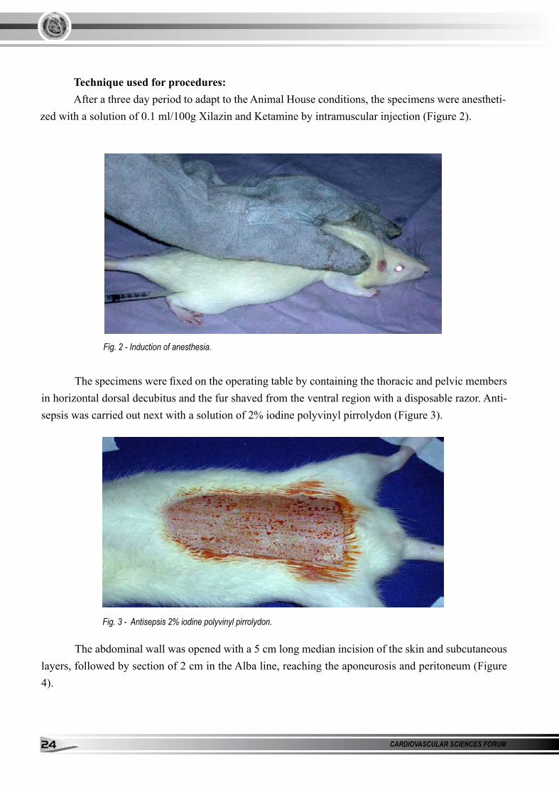
Technique used for procedures:After a three day period to adapt to the Animal House conditions, the specimens were anestheti-
zed with a solution of 0.1 ml/100g Xilazin and Ketamine by intramuscular injection (Figure 2).
Fig. 2 - Induction of anesthesia.
The specimens were fixed on the operating table by containing the thoracic and pelvic members in horizontal dorsal decubitus and the fur shaved from the ventral region with a disposable razor. Anti-sepsis was carried out next with a solution of 2% iodine polyvinyl pirrolydon (Figure 3).
Fig. 3 - Antisepsis 2% iodine polyvinyl pirrolydon.
The abdominal wall was opened with a 5 cm long median incision of the skin and subcutaneous layers, followed by section of 2 cm in the Alba line, reaching the aponeurosis and peritoneum (Figure 4).

JAN. / MAR. 2009 - VOL. 4 - NUMBER 1 2�
ISSN 1809 - 3736
Control groupIn the control group, the aponeurosis synthesis was performed with nylon non-absorbable mo-
nofilament threads with atraumatc needles using single separate stitches. Four stitches and three knots each were used and then the skin and the subcutaneous cells were closed with single separate stitches, using nylon monofilament 5.0 atraumatic needles.
1 cm distance groupIn the 1 cm distance group, the aponeurosis was performed with non-absorbable nylon mono-
filament 5.0 atraumatic needles with separate single stitches, but a Marlex® polypropylene mesh was placed the aponeurotic-peritoneal plane, taking care to keep the borders of the aponeurosis at 1 cm dis-tance from each other after closing the skin and subcutaneous cells. (Figure 5).
Fig. 4 - Median incision
Fig. 5 - Aponeurosis synthesis with polypropylene mesh.

CARDIOVASCULAR SCIENCES FORUM2�
2 cm distance groupIn the 2 cm distance group, the procedure for the aponeurotic border synthesis was similar, Mar-
lex® polypropylene mesh was placed over the aponeurotic border peritoneal plane, taking care to keep the aponeurosis borders with 2 cm distance between each other after closing the skin and subcutaneous cell. (Figure 6).
Fig. 6 - Aponeurosis synthesis with polypropylene mesh and 2 cm distance.
In the post surgery period, the specimens of each group were kept in separate cages, with chow and water ad libitum until the end of the ob-servation period. Those that died or that presented complications in the operation site were excluded from the experiment.
The specimens in the acute group were euthanized three days after the surgical procedure and the chronic group was euthanized 28 days af-ter the procedure. Euthanasia was carried out with a solution of 10% potassium chloride in intercar-diac injection after anesthesia.
A fragment of the abdominal wall was ob-tained including the whole laparotomy area are, that included the peritoneum, aponeurosis, mus-cle, subcutaneous cells and skin. This material was removed for histological study. (Figure 7). The fragments obtained from the abdominal wall were fixed in ALFAC solution (80% alcohol + 40%
formaldehyde + glacial acetic acid), at the ratio of 8.5: 1:2 for 24 hours. They were then dehydrated, diaphanized and blocked in paraffin. The blocks were cut with 0.5 mm thickness and stained with hematoxylin-eosin. Three cuts from each block were assessed by the same observer.
Fig. 7 - Obtaining the abdominal wall fragment.

JAN. / MAR. 2009 - VOL. 4 - NUMBER 1 2�
ISSN 1809 - 3736
The histological analysis of the control group was performed in the aponeurosis tissue in the location that surrounded the suture wound, while the in other groups it was carried out at the intersection of the aponeurosis with the Marlex mesh. The histological analysis assessed the follo-wing cell elements: neutrofil filtrate, lymph-mo-noplasmocytic infiltrates, collagen deposit, gigan-tocyte deposit, foreign body type cell deposits and granulation tissue. The data obtained according to the pathologist´s assessment were reported in the following manner:
Absence: absence or traces of the cell ele-ment assessed;
Presence: moderate or intense presence of the cell element assessed.
Statistical AnalysisThe various parame-
ters analyzed were compared by the analysis of variance of one Factor, and the differen-ces between the groups were discriminated by the exact Fi-sher Test.
The data are presented in mean and with and without the standard deviation. The level of significance was set at 5%.
RESULTSTo analyze the data
obtained, the specimens from the acute group (24 speci-mens) and the chronic group (24 specimens) were placed in the following groups: acute
control (A control) 8 specimens, 1 cm distance (A 1 cm distance) 8 specimens and 2 cm distance (A 2 cm distance) 8 specimens where the following histological variables were assessed: neutrofils, lymphomononuclear cells, gigantic cells, collagen fibers and granulation tissue. Macroscopic varia-bles related to the wound were also assessed such as adherence and abdominal wall hernias.
The same variables were assessed in the chronic group designated as the following groups: chronic control (C control), 8 specimens, 1 cm distance (C 1 cm distance) 8 specimens and 1 cm distance (C 1 cm distance), eight specimens.
The variables of the acute and chronic groups were compared considering the control group, the 1 cm distance group and the 2 cm dis-tance group. The data are shown in descriptive and graphic form. Table 1.

CARDIOVASCULAR SCIENCES FORUM2�
A control: Control Acute Group, A 1 cm: Acute Group with 1 cm distance, A 2 cm: Acu-te Group with 2 cm distance, C control: Control Chronic Group, C 1 cm: Control Group 1 cm dis-tance, C 2cm: Control Group With 2 cm distance, LMN: lymphomononuclear cells.
Acute groupThe animals in the A control, A 1 cm dis-
tance and A 2 cm distance groups were assessed. Histological and macroscopic alterations were not observed between the groups A control and A 1 cm distance, nor between A 1 cm distance and A 2 cm distance. (Figure 8). When A control group was compared with A 2 cm distance, greater adherence was observed in the A 2 cm distance group (p= 0.026).
Chronic group Animals from the C control, C 1 cm distance and C 2 cm distance groups were assessed. His-
tological difference was observed with greater presence of lymphomononuclear cells in the group C 1 cm distance compared to C 2 cm distance (p<0.001). (Figure 9). Adherence was observed in C control group with statistical significance (p= 0.007 and p= 0.026, respectively).(Figure 10).
Fig. 9 - Presence of these cell elements according to the group.
Fig. 10 - Presence of adherence according to group

JAN. / MAR. 2009 - VOL. 4 - NUMBER 1 29
ISSN 1809 - 3736
Acute x chronic groupThe acute and chronic groups were corre-
lated. When the A control group was compared with the C control group, the presence of neutrofils was more significant in the A control group (p=
0.010). A more significant presence was observed of gigantic cells and collagen in C control than in A control (p< 0.001 and p< 0.001, respectively). (Figure 11).
Fig. 11 - Presence of cell elements according to group. (Gigantic C: gigantic cells)
When A 2 cm distance was compared with C 2 cm distance, a greater presence of lymphomono-nuclear cells was observed in the acute group (p< 0.001) and greater presence of collagen in the chronic group (p= 0.026). (Figure 12).
. 12 - To presence of cell elements according to group. (LMPH: Lymphomononuclear cells)

CARDIOVASCULAR SCIENCES FORUM�0
When the A 1 cm distance group was com-pared to the C 1 cm distance group, significant presence of granulation in the chronic group was observed compared to the acute group (p= 0.007), and the presence of adherences that was greater in C 1 cm distance (p= 0.041).
DISCUSSIONClosing the abdominal wall is a simple
anatomic technique, but there are situations whe-re the tension under which this suture is made damages the surgical result. Polypropylene mesh is frequently used in peritoneostomies and in the repair of abdominal hernias. The use of the mesh prevents abdominal hypertension, facilitates inter-vention, prevents evisceration and minimizes the damage to the abdominal wall (10).
Polypropylene is a synthetic material that produces little tissue reaction and has good tensi-le resistance, a resistance that is maintained over several years after its use in live organisms (6,11). In spite of the low tissue reaction, polypropylene mesh can trigger a granular reaction all of the fo-reign body type, chronic inflammatory reaction and fibrosis(9), because the mesh surface is filled with adipose tissue and a inflammatory response triggered by operational trauma.
Experimental models of incision hernia have been reported in several experimental ani-mals(6,11). Abdominal hernias in rats are perfor-med by a removing a fragment of the abdominal wall and repairing it with prosthesis. This model of abdominal hernia in rats has been used by seve-ral authors to study the correction of incision her-nia with different types of processes, immediately after the defect was produced (12).
Polypropylene mesh was introduced at the end of the 1950s by Usher et al (13) and was a
great advance in a hernia treatment and continues to be the mesh most used today. Its characteris-tics include all properties necessary for biomate-rials, because it is less reactive, resistant to trac-tion, non-absorbable and microporous. This last characteristic gives greater resistance to infection and greater incorporation of the mesh to the host tissue.
The objective of the present study was to assess the type of inflammatory response and the intensity of this response in experimental animals, where situations of clinical difficulty were simu-lated such as those caused by acute abdominal surgery or abdominal infection with increase in the intra-abdominal pressure. For this we implan-ted the experimental animals with polypropylene mesh at different distances, 1 cm or 2 cm, from the aponeurotic border.
The histological assessment of the tissue was extremely important, because it identified the type of inflammatory response and its intensity. The presence of young cells such as neutrofils, lymphomononuclear cells and gigantic cells sug-gested an acute inflammatory response while the presence of collagen and granulation tissue identi-fied a late healing reaction.
When the specimens in the acute groups were compared amongst each other a greater pre-sence of young cells was observed in those eutha-nized three days after the procedure, especially neutrofils and lymphomononuclear cells.
Differences were not observed when the control group was compared to the group with 1 cm distance and 2 cm distance from the aponeu-rotic border. In the chronic group The presence of young and repair cells was observed in speci-mens euthanized 28 days after the procedure. The presence of late response cells was evident in the

JAN. / MAR. 2009 - VOL. 4 - NUMBER 1 ��
ISSN 1809 - 3736
three chronic groups, the control and 1 cm distan-ce and 2 cm distance groups, but was greater in the chronic 2 cm distance group. It is interesting to note the increase in the lymphomononuclear cells in the 1 cm distance group, compared to the chronic 1 cm distance and 2 cm distance groups. The lower tension may indicate a less intense late healing response.
The within-group analysis, either the acu-te or chronic groups, showed that in the control group the variable neutrofil cell presence was greater in the acute group while that of gigan-tic cells and collagen was greater in the chronic group. The same pattern of the presence of young or repair cells was observed when the acute group with 1 cm distance was compared with the chronic group with the same distance. A greater presence of lymphomononuclear cells was observed in the acute group and a greater presence of collagen in the chronic group.
Another interesting fact, that can be obser-ved macroscopically, was the presence of adheren-ce. The presence of adherence was observed in the acute group 2 cm distance compared to the control group, in the chronic group adherence was obser-ved intensely in the two chronic groups compared to the control and in the acute group 1 cm distance acute group compared to the chronic group with the same distance. These data suggested that the more intense inflammatory response may be rela-ted to time and distance from the aponeurotic bor-der and use of polypropylene mesh.
Regarding the various publications on ab-dominal wall hernia and the use of polypropylene mesh, this study assessed the type and intensity of the inflammatory response in an experimental mo-del with different distances from the aponeurotic border.
CONCLUSIONIt was concluded that there was an inflam-
matory response with the use of polypropylene mesh and that in the initial phases this response was identified by the more intense presence of neutrofil and lymphomononuclear cells and in the later phase, collagen and granulation tissue were greater. The adherences were more frequent in the groups with greater distance from the aponeurotic borders and longer time of polypropylene mesh use. Prevalence of abdominal wall hernias was not observed in the groups with greater distance from the aponeurotic border.
REfERENCES1. Goligher JC, Irvin TT, Johnston D, De Dombal
FT, Hill GL, Horrocks JC. Acontrolled clinical trial of three methods of closure of laparotomy wounds. Br. J. Surg. 1975;62:823-9.
2. Tognini JRF, Goldenberg S. Síntese da parede abdominal: sutura contínua ou com pontos se-parados? revisão da literatura. Acta Cir. Bras. 1997;v.13 n.2.
3. Goldenberg A, Matone J, Marcondes W, Her-bella FA, Farah JF. Comparative study of in-flammatory response and adhesions formation after fixation of different meshes for inguinal hernia repair in rabbits. Acta Cir Bras. 2005; 20(5):347-52
4. Metzger J, Lutz N, Laidlaw I. Guidelines for inguinal hernia repair in everyday practice. Ann R Coll Surg Engl. 2001;83:209-14.
5. Bellón JM, Buján J, Contreras LA, Carrera-San

CARDIOVASCULAR SCIENCES FORUM�2
Martín A, Jurado F. Comparison of a new type of polytetrafluorethilene patch and polypropylene prosthesis for repair of abdominal wall defects. J Am Coll Surg. 1996;183:11-8.
6. Gianlupi A, Trindade, MRM. Comparação en-tre o uso de fio inabsorvível (polipropileno) e fio absorvível (poliglactina 910) na fixação de prótese de polipropileno em correção de defei-tos músculo-aponeurótico da parede abdominal: estudo experimental em ratos. Acta Cir. Bras 2004; v. 19, n. 2.
7. Condon RE. Ventral Abdominal Hernia. In: Fis-cher JD, editor. Mastery of Surgery. Thirth ed: Little, rown and Company; 1997. p. 1877-81.
8. Sanders RJ, DiClementi D. Principles of abdo-minal wound closure. Arch. Surg. 1977;112:118-91.
9. Mazzini DL; Mantovani M. Suture of the ab-dominal wall with partial approximation of the borders of the aponeurosis using vicryl or marlex meshes in rats. Acta Cir Bras 1999; Jan Mar; 14:28-34.
10. Dinsmore RC, Calton WC, Harvey SB, Blaney MW. Prevention of adhesions to polyproylene mesh in a traumatized bowel model. J Am Coll Surg 2000; 191:131-6.
11. Danilo NSP, Pereira FEL, Mata RF, Dauad FR, Isabel CALP. Experimental models of longitu-dinal abdominal incisional hernia in rats. Acta Cir. Bras.1997 Dec; 12(4): 235-239.
12. Torres Neto JR, Barreto AP, Prudente ACL,
Santos AM, Santiago RR. Uso da peritoneosto-mia na sepse abdominal. Rev Bras. Colo-Proc-tol 2007 Sep; 27(3):278-283.
13. Usher FC, Cogan JE, Lowry TI. A new tech-nique for the repair of inguinal and incisional hernias. Arch Surg 1960; 81:847-54.

JAN. / MAR. 2009 - VOL. 4 - NUMBER 1 ��
ISSN 1809 - 3736
SHORT-TERm ANALYSIS Of HEART RATE VARIABILITY IN DIABETIC ATIENTS
E. R. Migliaro, P. Contreras.Departamento de Fisiologia. Facultad de Medicina. Montevideo, URUGUAY.
Summary The aim of this work was to assess the
value of a short-term routine to measure the Heart Rate Variability (HRV) and its parasympathetic and sympathetic components. We studied a group that was supposed to have low values of HRV owing to their long-lasting type I diabetes (n=15). None of these patients had symptoms of autonomic neuropathy. This group was compared with 13 healthy volunteers with similar gender and age profile. An ECG was obtained from an electrocardiograph and fed into a computer by means of an analog to digital converter. R waves were detected and visual inspection allowed the elimination of the non-sinusal beats. The normal R-R intervals (N-N) were measured and stored. Using the N-N lists we calculated two time-domain indexes: the standard deviation of intervals (SDNN) and the square root of the mean of the squared differences between adjacent intervals (rMSSD). Spectral analysis was done to study HRV in the frequency domain. The spectrum obtained was divided in different components: the high frequency band (HF) from 0.15 to 0.4 Hz and the low frequency band (LF) from 0.04 to 0.15 Hz. The HF component is considered dependent on the
parasympathetic activity and the LF component reflects both parasympathetic and sympathetic drive. The total power of the spectrum and the LF/HF ratio were also calculated. The results show a clear diminution of HRV in the diabetic patients in comparison with the control group. Such a reduction seems to reflect a parallel decrease of the sympathetic and parasympathetic flows.
Key Words: Heart rate variability, diabetic neuropathy, autonomic nervous system.
IntroductionThe duration of normal heart beat intervals
display subtle variations which lead to changes in heart rate known as Heart Rate Variability (HRV). The main determinants of HRV in healthy people are the respiratory changes, resting heart rate and age (1). All of them are related with the autonomic nervous system (ANS) activity. The former is the well-known respiratory sinus arrhythmia, the normal variation of heart rhythm coupled with respiration which is more evident in young people (2). Resting heart rate expresses the sympatho-vagal balance, when it is shifted towards increments of resting heart rate, HRV diminishes. Influence
ORIGINAL ARTICLESORIGINAL ARTICLES

CARDIOVASCULAR SCIENCES FORUM�4
of age could be related with autonomic aging (HRV diminishes as age increases) (3). Besides such physiological changes, many pathological states also affect HRV. Among them,the most wildly studied are: coronary disease (4)(5), heart failure (6),(7), anxiety or phobias (8), and diabetes mellitus (9),(10),(11). Diabetes delays neural conduction as a consequence of either vascular alteration (microangiopathy of the vasa nervorum) or of metabolic or autoimmune changes in the myelin sheath (12). Sensitive-motor dysfunctions (somatic neuropathy) are found in the spinal nerves of diabetic patients, while in the ANS this illness causes alterations related to the denervation of the viscera (autonomic neuropathy).
The cardiovascular autonomic neuropathy may play a main role in causing arrhythmias and sudden death (13),(14). The interest of studying the cardiovascular modifications in the autonomic neuropathy of diabetic patients is based on their early appearance. Compared with other tests used to diagnose autonomic neuropathy, HRV is preferred by its simplicity, which explains why its analysis has represented an important tool in the study of this pathology (15).
methods for HRV analysis. The introduction of microcomputers
allowed the improvement of the recording method and the measurement of HRV. In brief, HRV can be measured in short-term recordings (few minutes) or in long lasting ones (24 h Holter monitorization). In each case, a list of successive R-R intervals is obtained. Noise and non-sinusal beats are removed and the resulting normal intervals (N-N) can be processed in many ways, (statistical methods, non-linear analysis, frequency domain studies). Here we chose two of them:
Time-domain analysis.The numeric expression of the dispersion
of data is the standard deviation of the N-N intervals (SDNN) or the indexes that derive from it (16). Another useful time-domain index is the square root of the mean of the squared differences between adjacent intervals (rMSSD) that evaluates more accurately the beat to beat changes (17),(5).
frequency-domain analysis.The succession of intervals over time
(tachogram) can be analyzed as a new complex signal, then its power spectral density can be estimated.. In this work we computed the values between 0.0033 and 0.50 Hz (referred as total power). The analysis of HRV in the frequency domain introduces a main aspect that refers to the possibility of separating influences of both branches of the ANS. Most researchers agree that the high frequency band power (HF, 0.15 to 0.40 Hz) is related to the modulation of the parasympathetic branch (18),(11). The low frequency band power (LF, from 0.04 to 0.15 Hz) represents both autonomic branches and other non-autonomic influences (11). The basis of the components of frequencies below 0.04 Hz (“ultra low frequency” and “very low frequency” bands) still remain unclear (19). In addition, our short-term study do not assess properly those frequencies.
The aim of the present work was the evaluation of a short-term study as a diagnostic tool for HRV in a group of patients that is supposed to have low values of HRV owing to their long-lasting type I diabetes.
3. material and methods.We studied 15 diabetic patients (7 men and
8 women) aged between 44 and 66 years (mean

JAN. / MAR. 2009 - VOL. 4 - NUMBER 1 ��
ISSN 1809 - 3736
[± SD] 53.8 ± 7.4). All of them were insulin-dependent and had had diabetes mellitus for a long time (mean [± SD] diabetes duration 38.33 ± 8.5 years). They were allowed to receive their usual medication (digitalis, calcium antagonists, converting enzyme inhibitors).
The control group was composed by 13 healthy volunteers (5 men and 8 women) aged between 45 and 70 years (mean [± SD] 55.8 ± 7.8 years). The volunteers were non-smokers and were not receiving any medication. We excluded for the study people who had arrhythmia in the clinical examination or in the ECG or Holter recording.
HRV was assessed measuring the intervals between heart beats recorded in a conventional ECG. The procedure for recording was the following: Subjects were asked not to perform heavy exercise or take coffee or alcohol 24 h prior to the study. Electrodes for the ECG recording were placed on the chest, two of them were used to obtain a bipolar derivation. We connected the ECG with the computer by means of an analog to
digital (A/D) converter. Such connection allowed the acquisition of the entire ECG. The sampling frequency was 500 Hz. In order to achieve a good relaxation the subject lied for 20 min in supine position in a quiet room. After that, we began the recording period which lasted for 10 min. All the studies were done between 4:00 and 6:00 PM.
In an off-line analysis, a special program detected each R wave. A visual inspection routine allowed the validation of the detected R waves, correcting those related with premature beats and movements (false positives). After that, we obtained a train of N-N (normal to normal) intervals. Another program measured the N-N intervals in ms. With the lists obtained, a software calculated the indexes for time domain HRV analysis: SDNN and rMSSD. For the frequency domain analysis, graphs of the total spectrum were plotted (Figure 1); then, we computed the spectral density and calculated: total power, LF power, HF power and LF/HF ratio 1 .
The statistical comparisons were done with a non-parametric test (Mann-Whitney). Values of p<0.05 were considered significant.
Fig 1 - Spectral analysis of two subjects matched by age and sex. The control has higher values of spectral density than the diabetic patient in all frequencies.
1 - The original software for acquisition, visual inspe-ction, N-N measurements and calculus of HRV indexes were developed by El. Eng. R. Canetti and El..Eng. M. Hackas from the Instituto de Ingeniería Eléctrica. Facultad de Ingeniería Montevideo, Uruguay.

CARDIOVASCULAR SCIENCES FORUM��
4. ResultsAs we mentioned earlier both groups are
similar in age and gender distribution. The values of their mean (± SD) resting heart rate are: 73.9 ± 9.4 beats/min in the diabetic group and 69.7 ± 5.2 beats/min for the control group. Such figures are not statistically different.
Time-domain analysis.Mean values of SDNN and rMSSD in each
group, are shown in Figure 2. As we can see, values for the diabetic group are significantly lower than those for the controls.
frequency-domain analysis.The average values of spectral density are
shown in Table 1. The diabetic patients show a significant reduction of the mean values of the total power as well as HF and LF bands. Nevertheless, values of LF/HF ratio are not statistically different.
Fig 2 - Time-domain indexes of HRVBar graphs show the comparison of SDNN (upper graph) and rMSSD (lower graph) between control group and diabetic patients. Figures inside the boxes correspond to the mean values ± S.D. The symbol (*) indicates p<0.05. See text for discussion.
TABLE 1 Diabetic ControlMean SD Mean SD P
TPSD 558.67 637.24 1801.50 1263.40 0.0004LF 138.44 187.70 501.48 410.53 0.0004HF 86.11 124.92 263.68 311.88 0.007
LF/HF 2.695 1.838 2.849 2.056 N.S.Table 1 - Frequency-domain indexes of HRVMean values (± S.D.) of: Total power spectrum density (TPSD), LF band, HF band LF/HF ratio. All values but the LF/HF ratio. are statistically significant See text for discussion.
5. CommentsHRV is often studied by means of Holter
monitorization. This procedure has the advantage that circadian variations of HRV are recorded. On the other hand, its acquisition frequency is lower than the one we use (128 vs. 500 Hz). In addition, Holter studies are usually done using tape recorders
and small variations in tape speed can induce abnormal HRV measurements.
On the contrary, short-term studies are performed in control conditions, i.e. all subjects are in the same environment without external influences. The acquisition is performed with a computer through an A/D converter, in this way

JAN. / MAR. 2009 - VOL. 4 - NUMBER 1 ��
ISSN 1809 - 3736
tape speed changes are avoided and the acquisition frequency can be increased. Using this short-term procedure, our results agree with those of other authors which point out the affectation of the ANS in diabetes mellitus.
The concomitant reduction of LF and HF bands and therefore, the non-significant variation of the LF/HF ratio seems to show that the decrease of HRV in the diabetic group is based on a similar affectation of the sympathetic and parasympathetic branches. Other authors have obtained evidence about an early affectation of both branches of the ANS in this pathology (20)(11). Meanwhile, there are reports that states an increase (21) or a decrease (22) in LF/HF ratio in diabetic compared with controls.
In the earliest studies of diabetic neuropathy, a rest taquicardia was described, which would imply an premature parasympathetic affectation (23). In our groups, the differences between the mean values of heart rate is not significant, which agrees with the parallel reduction of the HF and LF components.
6. ConclusionsOur short-term procedure is useful enough
to the diagnosis of the HRV impairment in diabetes. In the present study, the diabetic patients show a clear reduction of their HRV when compared with the control volunteers. Such a reduction seems to reflect a parallel affectation of the sympathetic and parasympathetic branches.
REfERêNCIAS01. Tsuji, H., Venditti, F. J., Manders, E. S., Evans, J.
C., Larson, M. G., Feldman, C. L., and Levy, D. Determinants of Heart Rate Variability. J.Am.Coll.Cardiol. (1996) 28:1539-1546.
02. Marriot, H. J. L. and Myerburg, R. J. Recognition and tratment of cardiac arrhythmias and conduction disturbances. In: Hurst, J.W. eds.The Heart. Fourth Edition. New York. McGraw Hill Company. 1978. 637-694.
03. Pfeifer, M. A., Weinberg, C. R, Cook, D., Reenan, A., and Halter, J. B. Variaciones diferenciales de la función del sistema nervioso autonómico con la edad del varón. Am J Med (Spanish edition) (1983) 18:39-44.
04. Odemuyina, O., Malik, M., Farrell, T., Bashir, Y., Poloniecki, J., and Camm, J. Comparison of the predictive characteristics of HRV index and left ventricular ejection fraction for all cause mortality, arrhytmic events and sudden death after acute myocardial infarction. Am J Cardiol (1991) 68:434-439.
05. Bigger, J. T., Kleiger, R. E., Fleiss, J. L., Rolnitzky, L. M., Steinman, R. C., Miller, J. P. Multicenter post-infarction research group,. Components of heart rate variability measured during healing of acute myocardial infarction. Am J Cardiol (1988) 61:208-215.
06. Stein, P. K., Rich, M. W., Rottman, J. N., and Kleiger, R. E,. Stability of index of heart rate variability in patients with congestive heart failure. Am Heart J (1995) 129:975-981.
07. Woo, M. A., Stevenson, W. G., Moser, D. K., Trelease, R. B., and Harper, R. M. .Patterns of beat-to-beat heart rate variability in advanced heart failure. Am Heart J (1992) 123:704-710.
08. Kawachi, I., Sparrow, D., Vokonas, P. S., and Weiss, S. T. Decreased heart rate variability in men with phobic anxiety (data from the Normative Aging Study). Am J Cardiol (1995) 75:882-885.

CARDIOVASCULAR SCIENCES FORUM��
09. Malpas, S. C. and Maling, T. J. Heart rate variability and cardiac autonomic function in diabetes. Diabetes (1990) 39:1177-1181.
10. Thomaseth, K., Cobelli, C., Bellavere, F., Balzani, I., De Masi, G., Bax, G., and Carenza, P. Heart rate spectral analysis for assesing autonomic regulation in diabetic patients. J Auton Nerv Syst (1990) 30:S169-S172.
11. Malik, M. Heart Rate Variability. In: Zipes D., Jalife J. Eds. Cardiac Electrophysiology. Third. Edition. Philadelphia. W.B.Saunders Company. 1999. 753-762.
12. Cotran , R. S., Kumar, V., and Robbins, S. T. El páncreas. In: Robbins S.T. Ed. Patología estructural y funcional. Madrid. McGraw-Hill - Interamericana. 1990. 1033-1062.
13. Rathmann, W., Ziegler, D., Jahnke, M., Haastert, B., and Gries, F. A. .Mortality in diabetic patients with cardiovascular autonomic neuropathy. Diabet Med (1993) 10:820-48.
14. Ong, J. J., Sarma, J. S., Venkataraman, K., Levin, S. R., and Singh, B. N.. Circadian rhythmicity of heart rate and QTc interval in diabetic autonomic neuropathy: implications for the mechanism of sudden death.. Am Heart J (1993) 125:744-752.
15. Howorka, K., Pumprla, J., and Schabmann, A. Optimal parameters of short-term heart rate spectrogram for routine evaluation of diabetic cardiovascular autonomic neuropathy. J Auton Nerv Syst. (30-4-1998) 69:164-172.16 Kleiger, R., Stein, P., Bossner, M., and Rottman, J. Time domain measurements of heart rate varibility. Cardiol Clin (1992) 10:487-498.
17. von Neumann, J., Kent, R. H., Bellinson, R. H., and
Hart, B. I. The mean square succesive difference. Ann Math Stat (1941) 12:153-162.
18. Appel, M. L., Berger, R. D., Saul, J. P., Smith, J. M., and Cohen, R. J.,.Beat to beat variability in cardiovascular variables: Noise or music? J Am Coll Cardiol (1989) 14 nº 5:1139-1148.
19. Task Force of the European Society of Cardiology and the North American Society of Pacing and Electrophysiology. Heart rate Variability. Standards of measurement, physiological interpretation, and clinical use. Circulation (1996) 93:1043-1065.
20. Weise, F. and Heydenreich, F. A non-invasive approach to cardiac autonomic neuropathy in patients with diabetes mellitus. Clin Physiol (1990) 10:137-145.
21. Weston, PJ., James, MA., Panerai, RB., McNally, PG., Putter, JF., and Thurson, E. Evidence of defective cardiovascular regulation in insuline.dependant diabetic patients without clinical autonomic dysfunction. Diabetes Res Clin.Pract (1998) 43:141-148.
22. Singh, JP, Larson, M. G., O´Donnell, CJ, Wilson, PF, Tsuji, H., Lloyd-Jones, DM, and Levy, D. Association of hyperglicemia with reduce heart rate variability. (The Framinham heart study). Am J Cardiol. (2000) 86:309-312.
23. Lloyod-Mostyn, R. and Watkins, P. Defective inervation of heart in diabetic autonomic neuropathy. BMJ (1975) 3:15.

JAN. / MAR. 2009 - VOL. 4 - NUMBER 1 �9
ISSN 1809 - 3736
SUPRAVENTRICULAR TACHYARRHYTHmIA IN fETUS - EXPERIENCE AND TREATmENT
Glicerio C. Puentes, MD.
CASE REPORTCASE REPORT
SummaryIntroduction: Cardiac arrhythmias in the
feturs are not seen frecuently; one can see premature atrial beats (PAB) or premature ventricular beats (VBP) isolated; others as sinus bradycardia (SB) and sinus tachycardia (ST) are seen very often without any consequence for the fetus. Among supraventricular arrhythmias, supraventricular tachycardia (SVT) and atrial flutter (AF) are the arrhythmias that need some treatment as several authors have described.
General Objectives: To report a case of total tachyarrhythmia:
And (1): To recognize the behaviour of SVT with echocardiographic follow up in the fetus (ECO) after having done the diagnosis and to recognize what will happen once the patient will be born.
And (2): To recognize what happened with the treatment with digoxin and to support the opinion that echocardiographic test is the best diagnostick
tool in face of all cardiac arrhythmias.
method: We used a Combinson 310, Kretz (M and B mode), in outpatients clinic, in order to study all pregnants women with risk of heart defects in their babies from the municipality of East Havana.
Results: There was a good response after the arrthythmia wa diagnosed and treated with digoxin by maternal way.
We make a comment of what happened after the baby was born.
Conclusions: It is emphasized that ECO is the best diagnostic method among others. Digoxin is the drug of first line and the maternal route, is a good way.
Key Words: Cardiac Arrhythmia, Paediatric Cardiology, Fetal Arrhythmia.
From Cardiocenter William Soler Hospital, Havana city Piti Fajardo Hospital, Havana, Cuba

CARDIOVASCULAR SCIENCES FORUM40
IntroductionThe use of ultrasound in the fetal world has
allowed to discover great unknown things of the behaviour of the fetus and the world around it during the nine months of pregnancy. Cardiac arrhythmias are not isolated of this acquired knowing, and there are a lot of authors which since a few years have begun to speak about their experiences in Fetal Medicine.
Fetal arrhythmias have received different clasifications, one of them is Dr. Oberhansli’s(1):
Fetal Arrhythmias.- Simple - Premature atrial contraction (ACP) - Premature ventricular contraction (VCP) - Sinus tachycardia (ST) - Sinus bradycardia (SB) - Premature atrial contraction blocked.- Potencially Mortal - Supraventricular tachycardia (SVT) - Atrial flutter (AF) - Nodal tachycardia - Atrioventricular block (A-V) of 2nd grade - Atrioventricular complete block - Complex arrhythmias
Echocardiography in their different forms has permited to diagnose all of them. M mode is the one that has been more used (2, 3, 4, 5).
The different treatments of choice in arrhythmias depend on many factors but Digoxin is the drug of first choice; several authors have confirmed its benefit (6, 7, 8). Hydropis is a bad sign and when it happens, it will determine the prognosis of the fetus (9).
General Objectives:To compare our findings with others
diagnostic methods used to diagnose arrhythmias in fetal life and in the afterbirth period.
Specific Objetives:To recognize the benefit of the treatment
with digoxin (slow treatment) and the value of its use by maternal route.
methodOur country has a programme of Prenatal
Diagnostic. The province of Havana inserted Fetal echocardiography in 1989, as a pilot study, in the east of the province. Since the beginning we have used a Combinson 310 Kretz (M and B mode ) equipment.
Pregnants women are sent to receive our service when they have some risk:
maternal risk factors: Extreme ages in life (<18 years and > 36
years). Some illness as: Sistemic Eritematous Lupus, IDDM etc. Treatment with some drugs: Dilatin, Ethanol, Retinoic acid etc.
Obstetrical risk factors: Abnormal biometric profile. Amniotic liquid
abnormalities orchanges, etc.Fetal risks factors: Fetal arrhythmias. Four
chambers abnormal view.Pregnants women are selected according
to the risks by: the Family doctors, Obgyn. doctors Genetist and others who treat the patients.
Case report:MDF,38 years, white. G2P1. who was sent
to our Fetal echocardiographic service because she was 36 years old.The first test was not possible because of the position of the fetus. At 24 5/7

JAN. / MAR. 2009 - VOL. 4 - NUMBER 1 4�
ISSN 1809 - 3736
weeks, we reoeated the test and found a fetal cardiac frequency around of >200 BPM (M mode) without atrial-ventricular(A-V) dissociation and without heart defects and with a normal cardiotocographic (CTG) test (Fig. 1). We began to treat the feto by maternal route with digoxin (slow treatment)(0,125 mg/day), step by step and in the 3 er day, we raised the treatment to 0,25 mg/day.
After 15 days with 0,25 mg/day, cardiac rate in the fetus was normal (Fig. 2).
An echocardiographic control test was done just as far as the cardic rate was normalized and after that, every two weeks was done an echocardiographic control until pregnancy was finished. We never found any arrhythmia during the treatment.
Since the beginning of the treatment and until the end of pregnancy, the pregnant received a close control by Obgyn. department. The fetus was born without any problem (Apgar 9-9). They were discharged from the hospital after 72 hours.
When the baby was 2 weeks old, he began with tachycardia, polynea and cyanosis (Fig. 3). We understood what happened when we knew that the treatment was stopped when they went to his house. We began again with digoxin 10 mcg/kg/day and after 72 hours the symptoms and signs
disappeared.The baby did not have any other problem
in the first year of his life (ECO and ECG, normal).Now, he is 1.2/1.3 year old and he is doing well without any treatment.
Discussion:The treatment with dogoxin in fetus with
SVT was reported in 1980 by Lingman(10), he recommended digoxin, 0,25 mg every 12 hours in the first day and later on digoxin 0,25 mg/day and he also found no-correspondence between the CTG test and transabdominal ECG in the fetus. All the authors agree with the good response to digoxin.
In our case it was the first patient that we have treated in 3000 test with 15 simple arrhythmias which did not need any treatment.
We used the slow treatment with digoxin because we could not dosify digoxin in blood. We considered that there was a good response without any bad secondary effects, so much to the mother as to the fetus. The baby proved with his illness that he needed digoxin all the time when he repeated the arrhythmia. He finished the treatment 3 months ago and he is very well.
In face of a fetal arrhythmia we should always do an echocardiographic test.
Fig. 1 Fig. 2

CARDIOVASCULAR SCIENCES FORUM42
REfERENCES1. Oberhanli-Weis, I. Afecciones cardiacas del feto.
Schweiz. Med. Wochenschr.1995, 125; 294-303.
2. Allan,LD. Manual of fetal Echocardiography, 1986. MTP Press Limited, Falcon House, Lancaster, En-gland.
3. DeVore, G.R. et. al. Fetal echocardiography III. The diagnosis of cardiac arrhythmias. Am.J.Obstet.Gyne-col.1983. 146;792-99.
4. Friedman, D.M. et. al Benign fetal bradycar-dias diagnosed by echocardiography. Am.J .Perin. 1995,12;2,295-99.
5. Kleimman, C.S. et. al. Fetal echocardiography. A tool for evaluation of in utero cardiac arrhythmias and monitoring of utero therapy. Am.J.Cardiol. 1983,51; 2,237-43.
6. Allan, L.D.Fetal tachycardia. Cardiol Young, 1996,6;197.
7. Hallak, M. et. al. Fetal supraventricular tachycardia and hydrops fetalis: Combined intensive, direct and
transplacental therapy. Obstet. Gynecol. 1991,78; 523-25.
8. Perry,J.C. Fetal arrhythmias, pediatric arrhythmias, and pediatric electrophysiology. Curr. Opin. Cardiol. 1995,10;52-57.
9. Copel, J.A. et.al. Management of fetal cardiac arrhyth-mias. Fet.Diag. Therap. 1997, 24; 1,201-11.
10. Lingman, G. et.al. Intrauterine digoxin treatment of fetal paroxismal tachycardia case report. Brit. J. Obs-tet.Gynecol. 1980,87; April, 340-43.
Fig. 3

JAN. / MAR. 2009 - VOL. 4 - NUMBER 1 4�
ISSN 1809 - 3736
LIfE STYLE ImPORTANCE fOR CARDIOVASCULAR DISEASES PREVENTION
A Importância do Estilo de Vida na Prevenção das Doenças Cardiovasculares
Pereira, S.N.; Pacheco; L.S.; Dias, D; Barbosa, V.A.; Portela, L.O.C.
UPDATING ARTICLE UPDATING ARTICLE
A importância das doenças cardiovasculares tanto do ponto de vista epidemiológico como econômico é reconhecida em todo mundo. No início do século XX elas representavam cerca de 10% do total de óbitos no mundo, porém, no inicio do século XXI, já eram responsáveis por 50% das mortes nos países desenvolvidos e 25% nos países em desenvolvimento1. Este índice vem decaindo em vários países, principalmente a partir da década de 1960, no entanto, em paises em desenvolvimento a taxa encontra-se em ascensão (3-9).
No Brasil, a Doença Cardiovascular (DCV) é responsável por 32% dos óbitos e por um elevado custo tanto para o paciente quanto para o sistema de saúde (9). No Rio Grande do Sul e no municipio de Santa Maria as DCV representam a principal causa de óbito, atingindo percentuais de 31 a 33% respectivamente (4,5,6).
Como bem documentado na literatura, principalmenta após o estudo Framingham, o desenvolvimento das DCV relaciona-se com a presença de seus fatores de risco, sejam eles não-
modificáveis (idade, sexo e história familiar) ou potencialmente modificáveis (tabagismo, hipertensão arterial sistêmica - HAS, dislipidemia, sedentarismo, Diabetes Mellitus - DM, sobrepeso e obesidade) (1-10) . Estratégias de prevenção primária em cardiologia baseadas em mudança de estilo de vida, quando adequadamente executadas, mostraram-se bastante eficazes (12), e aliados aos grandes avanços de seus tratamentos são, provavelmente, os responsáveis pela recente redução da taxa de mortalidade por DCV observada em alguns paises.
Em um trabalho de revisão, Castro e et al. (13), evidenciaram como o exercício pode reduzir vários fatores de risco, além de trazer outros benefícios para o organismo em geral e para o aparelho circulatório em particular. Entre estes benefícios estão a redução do peso, elevação do HDL colesterol, redução do LDL e triglicerídeos e dos níveis de tensão arterial (14-20). Metanálises de estudos de reabilitação cardíaca demonstraram redução de 20-25% no risco de morte (14), podendo
Correspondência: Sérgio Nunes PereiraHospital Universitário de Santa Maria/Centro de Educação Física e Desporto - UFSM, Santa Maria, RS, Brasil.Rua Venâncio Aires, 2086 / 803 - Santa Maria - RS - CEP: 97010-004 / BrazilE-mail: [email protected]

CARDIOVASCULAR SCIENCES FORUM44
chegar a cerca de 35 %, como aconteceu no estudo das enfermeiras (7) .
Estudos epidemiológicos nos Estados Unidos demonstraram aumento de cerca de 30% de indivíduos com sobrepeso e obesidade, associados com aumento também na prevalência de Diabete Mellitus (DM) (11).
Em contraste, houve diminuição de outros fatores de risco, tais como: o percentual de indivíduos com hipercolesterolemia baixou de 34 para 17%, hipertensos de 31 para 15% e tabagistas de 39 para 26% (11).
Em 1992 a AHA publicou as suas recomendações sobre o exercício (21), partindo do princípio de que a inatividade é um reconhecido fator de risco para doença coronária. Baseou-se também em metanálises que demonstraram que o exercício supervisionado pode reduzir as taxas de mortalidade por doença das coronárias e recomenda um mínimo de 30-60 minutos por 3-4 vezes por semana. Adverte que os médicos têm a responsabilidade de promover o assunto o exercício para toda a sociedade.
Em maio de 2001 foi publicado o relatório III NCEP/2001 com as diretrizes do Programa Nacional de Educação sobre o Colesterol dos Estados Unidos (22). Entre estas recomendações está a orientação sobre a Sindrome Metabólica, com resitência à insulina. Nela há excesso de gordura corporal, principalmente às custas de obesidade abdominal e iantividade física. Nestas recomendações comentou-se que o sobrepeso e a obesidade estão associados à resistência insulínica e a síndrome metabólica. O manejo da síndrome metabólica tem dois objetivos principais: Reduzir a obesidade e inatividade física e tratar os fatores de risco lipídicos e não lipídicos associados. Demonstrou-se que a prática de exercícios físicos
contribui para:aumentar as taxas de HDL-colesterol e reduzir os níveis de LDL-colesterol e VLDL, e além disto, reduz a pressão arterial, a resistência insulínica e melhora o desempenho cardiovascular. Com base nestes fatos o NCEP recomendou a prática regular de atividades físicas em pacientes com hipercolesterolemia.
Estudos mais recentes demonstraram a relação da inatividade física com o aumento das doenças cardiovasculares (23-26) e também os benefícios do exercício na prevenção e na redução da morbi-mortalidade por estas doenças (27-30) .
Em nosso país a Sociedade Brasileira de Cardiologia desenvolveu um grande estudo epidemiológico sobre doenças cardiovasculares e seus fatores de risco. Este projeto denominado Corações do Brasil foi desenvolvido a partir de 2004, avaliando 2550 pessoas, de 74 cidades, da cinco regiões geográficas do País. Este estudo demonstrou taxas de mortalidade de 27,5% por doenças cardiovasculares no Brasil e de 32 % na região Sul (9).
Em 2006 realizamos um estudo sobre o perfil clínico e laboratorial de 103 servidores da Universidade Federal de Santa Maria, participantes voluntários de um projeto de prevenção em cardiologia baseado na mudança de estilo de vida. Este grupo era constituído por 38 homens e 65 mulheres. Este estudo evidenciou uma prevalência de 51,96 % de sobrepeso/obesidade. Encontramos dislipidemia, com colesterol total maior do que 200 mg/dl em 58,42% e colesterol LDL superior a 130 mg/dl em 47,42% dos participantes. A prevalência de hipertensão arterial foi de 28 %. O tabagismo representou apenas 4,85% e a hiperglicemia (> 100 mg/dl) ocorreu em 5,82 dos participantes.
Nesta avaliação inicial dividimos os participantes em ativos e sedentários conforme seu

JAN. / MAR. 2009 - VOL. 4 - NUMBER 1 4�
ISSN 1809 - 3736
desempenho no teste ergoespirométrico, tomando como referência a tabela de aptidão física da AHA (baseada na medida do VO2 Max). Os resultados desta análise demonstraram diferença significativa nos seguintes indicadores: cintura masculina superior a 94 cm, HDL feminino inferior a 50 mg/dl e masculino inferior a 40 mg/dl, com prevalência maior nos indivíduos sedentários em relação aos ativos (p<0,02). Os indicadores colesterol total, LDL, triglicerídeos e glicemia não mostraram diferença significativa.
Estes resultados, embora com as limitações do tamanho da amostra, sugeriram benefício da atividade física na redução de fatores de risco. exercício na redução de alguns fatores de risco conhecidos. Estes dados concordam com os da literatura revisada e chamam a atenção para a necessidade de se estimular com grande empenho a mudança do estilo de vida na população.
É particularmente preocupante o amumento da prevalência de sobrepeso e obesidade, dislipidemia, síndrome metabólica, diabete e tabagismo em população cada vez mais jovem e também o aumento destes indicadores nos países sub-desenvolvidos e em desenvolvimento, que tem levado ao fenômeno denominado transição epidemiológica com aumento das doenças crônico-degenerativas, em especial a doença cardiovascular e redução das doenças infecciosas, antes mais freqüentes.
Esta situação agrava-se ainda mais se considerarmos os elevados custos dos tratamentos de ponta, para os hospitais, a rede pública e, especialmente, para os doentes, que se deparam com tratamentos dispendiosos, muitas vezes inacessíveis para a maioria da população que caminha, inexoravelmente, para um grande aumento de sua morbi-mortalidade por este tipo de doença.
Assim, cabe a nós, médicos a missão de nos conscientizarmos para a gravidade da situação e nos transformarmos em grandes divulgadores da idéia, especialmente válida para as doenças cardiovasculares, que mais vale prevenir do que remediar.
Talvez o maior desafio desta atitude seja a dificuldade na mudança de estilo de vida, pois a doença cardiovascular se desenvolve lenta e silenciosamente, de forma assintomática e desapercebida, e quando se manifesta geralmente já traz um dano muitas vezes irreparável. Mas gradualmente estão surgindo as evidências de que as medidades de mudança de estilo de vida tem se mostrado não só benéficas, mas tambem com custo/benefício favoravel em relação às atuais medidas terapêuticas existentes. Talvez, com a maior divulgação destes resultados se possa mudar, nos países em desenvolvimento, a tendência do perfil evolutivo da doença cardiovascular.
REfERêNCIAS1. Gus I, Zielinsky P. As cardiopatias no Brasil. In: Fer-
reira C; Póvoa R. Cardiologia para o clínico geral. Rio de Janeiro: Atheneu; 1999. p. 131-43.
2. Kuulasmaa K, Tunstall-Pedoe H, Dobson A, Fort-mann S, Sans S, Tolonen H et al. Estimation of contribution of changes in classic risk factors to trends in coronary-event rates across the WHO MONICA Project populations. Lancet 2000 Feb 26;355(9205):675-87.
3. McGovern P G, Pankow J S, Shahar E, Doliszny K M, Folsom A R, Blackburn H et al. Recent trends in acute coronary heart disease--mortality, morbidity, medical care, and risk factors. The Minnesota He-art Survey Investigators. N Engl J Med 1996 Apr
CARDIOVASCULAR SCIENCES FORUM4�
4;334(14):884-90.
4.Capewell S, Morrison C E, McMurray J J. Contri-bution of modern cardiovascular treatment and risk factor changes to the decline in coronary heart dise-ase mortality in Scotland between 1975 and 1994. Heart 1999 Apr;81(4):380-6.
5.Capewell S, Beaglehole R, Seddon M, McMurray J. Explanation for the decline in coronary heart di-sease mortality rates in Auckland, New Zealand, between 1982 and 1993. Circulation 2000 Sep 26;102(13):1511-6.
6. Cooper R, Cutler J, Desvigne-Nickens P, Fortmann S P, Friedman L, Havlik R, Hogelin G et al. Trends and disparities in coronary heart disease, stroke, and other cardiovascular diseases in the United States: findings of the national conference on cardiovascu-lar disease prevention. Circulation, Dec 2000; 102: 3137 - 3147.
7. McGovern P G, Jacobs D R Jr, Shahar E, Arnett D K, Folsom A R, Blackburn H et al. Trends in acute coronary heart disease mortality, morbidity, and me-dical care from 1985 through 1997: the minnesota heart survey. Circulation 2001 Jul 3;104(1):19-24.
8.Rothwell P M, Coull A J, Giles M F, Howard S C, Sil-ver L E, Bull L M et al. Change in stroke incidence, mortality, case-fatality, severity, and risk factors in Oxfordshire, UK from 1981 to 2004 (Oxford Vascu-lar Study). Lancet 2004 Jun 12;363(9425):1925-33.
9.Ministério da Saúde [homepage na Internet]. Secre-taria Executiva. Datasus [citado 2005]. Informações de Saúde. Morbidade e informações epidemiológi-cas. Disponível em: url: http://www.datasus.gov.br
10.Gus I; Fischmann A; Medina C. Prevalence of Risk Factors for Coronary Artery Disease in the Brazilian
State of Rio Grande do Sul. Arq Bras Cardiol, volu-me 78 (5): 484 -90, 2002.
11.Gregg E W, Cheng Y J, Cadwell B L, Imperatore G; Williams D E, Flegal K M et al. Secular tren-ds in cardiovascular disease risk factors according to body mass index in US adults. JAMA 2005 Apr 20;293(15):1868-74.
12.Rezende F A C, Rosado L E F P L, Ribeiro R C L, Vidigal F C, Vasques A C J, Bonard I S et al. Índi-ce de massa Corporal e circunferência Abdominal: Associação com Fatores de Risco Cardiovasculares. Arq. Brás Cardiol 2006;87(6):728- 734.
13.Castro, I; Bueno, N, Ramos, AM; Martini, R; Har-ter, K. - Importância do exercício físico na preven-ção da aterosclerose e da cardiopatia isquêmica. Ver. Med. IC-FUC, 2000; 3:248-259.
14.Miller, TD, Baladt, GJ, Fletcher GF – Exercise and its role in the prevention and rehabilitation of car-diovascular disease. Ann. Behav Med. 1997: 19(3): 220-9
15.Manson, JE; Stampfer, MJ; Willett, WC; Colditz, GA; Spelzer, FE; Hennekens, CH – Physical activity and incidence of coronary heart disease and stroke in women. Circulation 1995; 9:927
16.Stefanick ML, Mackey S, Sheehan M, Elsworth N, Haskell WL, Wood PD – Effects of diet and exer-cises in men and postmenopausal women with low levels of HDL cholesterol and high levels of LDL Cholesterol. N. Engl. J. Med. 1998 2; 339(1): 12-20
17. King, AC; Haskell, WL; Young, DR; Oka, RK; Ste-fanick, ML – Long term effects, of varying intensities and formats of physical activity on participation ra-tes, fitness, and lipoproteins in men and women aged 50 to 65 years - Circulation. 1995; 91:2596-2604

JAN. / MAR. 2009 - VOL. 4 - NUMBER 1 4�
ISSN 1809 - 3736
18. Paffenbarger, RS; Wing, AL; Hyde, RT. – Work ac-tivity and coronary heart mortality. N. Eng. J. Med. 292: 545, 1975
19.Wood, PD; Stefanick, ML; Dreon, DM; Frey-Hewitt, Garay, SC; Williams, PT; Superko, HR; Fortmann, SP; Albers, JJ; Vranizan, KM; Ellsworth, NM; Ter-ry, RB; Haskell, WL. - Changes in plasma lipids, and lipoproteins in overweight men during weight loss through dieting and compared with exercise. New Engl. J. Med. 1988,319(18): 1173-9
20. Wood PD, Stefanick ML, Williams PT, Haskell WL - The effects on plasma lipoproteins of a prudent weight-reducing diet, with or without exercise, in overweight men and women. New England J. Med 325 (7): 461-66
21.Wood PD, Stefanick ML, Williams PT, Haskell WL - The effects on plasma lipoproteins of a prudent weight-reducing diet, with or without exercise, in overweight men and women. New England J. Med 325 (7): 461-66
22. Fletcher, GF; Blair, SN; Blumenthal, J; Caspersen, C; Chaitman, B; Epstein, S; Falls, H; Froelicher, ESS; Froelicher, VF; Pina, IL. - Statement on Exer-cise - Benefits and Recommendations for physical activity programs for all americans - A statement for heath professionals by the Committee on Exercise and Cardiac Rehabilitation of the Council on Clini-cal Cardiology, American Heart Association. Circu-lation 1992; 86(1): 340-343
23.III NCEP/2001 - Executive Summary on the Third Report on National Cholesterol Education Program (NCEP) Expert Panel on detection, Evaluation, and Treatment of High Blood Cholesterol in Adults (Adult Treatment Panel III). JAMA 2001, 285 (19):2486-2497
24.Evenson, KR et. al. - Women’s Health Initiative Ob-servational Cohort Study. Vigorous leisure activity through womens’s adult life: the Women’s Health Initiative Observational Cohort Study. Am. J. Epi-demiol. 2002, 156 (10): 945-53.
25. Willams, PT - Phisical fitness and activity as sepa-rate heart disease risk factors: a metaanalysis. Med. Sci. Sports Exerc. 2001, 33(5): 754-61.
26. Paffenbarger Jr., RS et. al. -– Physical activity, all-cause mortlitiy, and logevity of college alumni. N. Engl. J. Med. 1986, 314 (10):605-13.
27. Manson, JE et al. - A prospective study of walking as compared with vigorous exercise in the preven-tion of coronary heart disease in women. N. Engl. J. Med. 1999, 341 (9):650-8.
28. Manson, JE et al. - Walking compared with vi-gorous exercise in the prevention of cardiovascu-lar events in women. N. Engl. J. Med. 1999, 347 (10):716-25.
29. Leon, AS et. al. - Leisure-time physical activity levels and risk of coronary heart disease and dea-th. The Multiple Risk Factor Interventional Trial. JAMA 1987, 258 (17): 2388-95.
30.Nobre, MRC, Santos, LA; Fonseca, VR - Epide-miologia do risco cardiovascular associado à ativi-dade física. In Cardiologia do Exercício - do atleta ao cardiopata, Negrão CE & Barreto, ACP, Manole, Barueri, SP, 2005, p.1.

CARDIOVASCULAR SCIENCES FORUM4�
mODIfIED DE VEGA TRICUSPID VALVE REPAIRValvoplastia Tricúspide De Vega Modificada
Otoni Moreira Gomes, Márcio Pitchon
HOW TO DOHOW TO DO
INTRODUÇÃODe Vega, em 1972, demonstrou que o
tratamento da insuficiência valvar tricuspídea poderia ser feito satisfatoriamente pela simples constrição do contorno antero-lateral do anel da valva tricúspide, praticamente excluindo a cirurgia de implante valvar artificial nesta posição, mais sujeita a tromboembolismo e endocardite que nas demais.
O presente estudo descreve simplificação da técnica original de De Vega, caracterizada pela plicatura total apenas da metade posterior do contorno ântero-lateral do anel valvar tricuspídeo.
CASUÍSTICA E mÉTODOForam estudados três pacientes portadores
de Cardiopatia Reumática, com dados gerais descritos no Quadro I.
QUADRO I - DADOS GERAIS DOS PACIENTES
OBS. N° NOME REG. IDADE
/ ANOS SEXO DIAGNÓSTICO GRUPO NYHA DÉFICIT MIOCÁRIDIO
1 RNP 160077 25 M EM + INSUF. TRICÚSPIDE IV ++++ / 4
2 RAO 154200 14 F DLM + INSUF. TRICÚSPIDE III +++ / 4
3 SLS 190926 30 M DLM + INSUF. TRICÚSPIDE III +++ / 4
Endereço para correspondência: Otoni M. GomesFundação Cardiovascular São Francisco de AssisRua José do Patrocínio, 522 – Santa Mônica – 31525-160 – Belo Horizonte – MG / BrasilE-mail: [email protected]

JAN. / MAR. 2009 - VOL. 4 - NUMBER 1 49
ISSN 1809 - 3736
HOW TO DOHOW TO DOAs operações foram realizadas sob anestesia geral, toracotomia mediana anterior e com circulação
extracorpórea hipotérmica (Quadro II).
QUADRO II - DADOS GERAIS DA OPERAÇÃO
OBS. N° NOME DATA
OPERAÇÃOCECmm T°C CARDIOPLEGIA OPERAÇÃO PRÓTESE
1 RNP 20/04/83 39 29 HC UFMG-II ANULOP. TRICÚSPIDE
COMISSUROTOMIA MITRAL
____
2 RAO 12/09/84 101 26 HC UFMG-II ANULOP. TRICÚSPIDETROCA MITRAL
PERICÁRDIO
FLVMEN
3 SLS 13/09/84 150 26 HC UFMG-II ANULOP. TRICÚSPIDE PERIACÁRDIOFLVMEN
Estando os pacientes em circulação extracorpórea, aberto o átrio direito e apresentada a valva tricúspide, procedeu-se à plicatura da metade posterior do contorno anterior do anel valvar, empregando-se fio de poliester 3-0 com almofada de dacron (Fig.1).
A correção da disfunção disfunção tricúspide foi assim obtida, durante o tempo de esfriamento corpóreo na CEC, estando o coração ainda batendo.
Terminada a anuloplastia tricúspide e
atingida a temperatura corpórea desejada, a aorta foi ocluída, a solução cardioplégica perfundida e as demais valvas doentes tratadas.
RESULTADOSO quadro III expõe os resultados obtidos,
concernentes à correção de valva tricúspide, conforme observado na alta hospitalar e no último controle ambulatorial.
Os pacientes RAO e SLS evoluíram sem intercorrência e o paciente RNP recebeu alta do CTI no 3º dia de pós-operatório, mas no 8º dia de pós-operatório apresentou quadro compatível com embolia pulmonar, complicada com pneumonia por gram-negativo e necessitando de traqueostomia no 16º dia de pós-operatório. Recebeu alta no 45º dia de pós-operatório em condições clínicas satisfatórias.
Além da disfunção das outras válvulas corrigidas, notou-se completa recuperação clínica das manifestações relativas à insuficiência tricúspide, desaparecendo o pulso hepático, o sopro sistólico característico e reduzindo-se significativamente o éstase jugular.
Fig.1

CARDIOVASCULAR SCIENCES FORUM�0
QUADRO III - RESULTADOS
PRÉ-OPERATÓRIO PÓS-OPERATÓRIO
OBS. N° SS-FT GRUPO
NYHA.FÍGADO
RCDINGURGITAMENTO
JUGULAR SS-FT GRUPO NYHA. FÍGADO RCD INGURGITAMENTO
JUGULAR
1 4/VI IV 4,0cm ++++ /4 - II NÃO PALP. ____
2 4/VI III 7,0cm +++ /4 - I 4,0cm ____
3 4/VI III 7,5cm +++/4 1/VI II 4,0cm ____
COmENTÁRIOSA correção da Insuficiência Tricúspide,
conforme proposta por De Vega, oferece resultados eficientes, comprovados em vários centros cardiológicos. Contudo, a preocupação com o grau de constrição obtida, para evitar insuficiência importante residual ou estenose por correção excessiva, provavelmente tenha motivado as modificações empregadas por CARPENTIER e por DURAN, usando dispositivos semi-rígidos para delimitar o diâmetro final da correção.
A técnica ora descrita, sendo eficaz, torna mais simples e segura a operação, posto que a plicadura sendo total, na extensão de 1/4 do anel, prescinde de avaliações intra-operatórias do diâmetro remanescente do orifício valvar.

JAN. / MAR. 2009 - VOL. 4 - NUMBER 1 ��
ISSN 1809 - 3736
1 - Objectives: The Cardiovascular Sciences Fo-rum aims to serve all the Cardiovascular Sci-ences fields of investigation to hold together multiprofessional experience to optimize the generation of new ideas, improving mankind resources in the prevention and treatment of cardiovascular diseases.
2 - Advertising: Cardiovascular Sciences Forum does not hold itself responsible for statements made by any authors. Statements or opinions expressed in Cardiovascular Sciences Forum of the authors(s) and do not represent official policy of the Sponsor Institutions unless so especified.
No responsability is assumed by the Cardio-vascular Sciences Forum Sponsor Institutions any neither by it’s Publising Enterprises, for any injury and/or damage to persons or pro-pertty as a matter of products liability, negli-gence or from any use or operation of any me-thods, products, instructions or ideas contai-ned in the material herein. No suggest test or procedure should be carried out unless, in the reader’s judgment, its risk is justified. Becau-se of rapid advances in the medical sciences we recommend that the independent verifica-tion of diagnosis and drug dosages should be made. Discussions, views and recommendati-ons as to medical procedure, choice of drugs and drug dosages are the responsability of the authors.
Although all advertising material published in Cardiovascular Sciences Forum is expected to conform to ethical (medical) standards, inclusion in this publication does not constitute
a guarantee or endorsement by it’s Sponsor Institutions or the Publisher of the quality or value of such product or of the claim made by its manufacture.
3 - Papers sent for publication in Cardiovascular Sciences Forum (Editorials, original articles, conferences, case reports, actualizations brief communications) should be related to cardio-vascular sciences and unpublished.
4 - Although the stem language of the Archives, happens to be English, articles may also be published in spanish or portuguese. When published in spanish or portuguese for original reports an abstract version in english must be included.
5 - They should be typed in double spacing on foolscap paper, with 3 cm margins all around and in 3.5” diskettes, Word 6.0 or superior. Illustrations do not need to be printed in diskettes.
6 - Manuscripts should be arranged as follow: a) title page consisting of concise and informative title, full name of authors, b) The Service or Institution name should be displayed in the bottom of the first page. Folowing that, the name of the corresponding author, together with the address, phone, fax and e-mail. c) abstract not exceeding 250 words and three key words that can be called in www.decsbvs.br and/or www.nlmnih.gov/mesh, d) Introduction, e) material and methods, f) results, g) comments, h) conclusions,
INSTRUCTIONS FOR AUTHORSINSTRUCTIONS FOR AUTHORS

CARDIOVASCULAR SCIENCES FORUM�2
9 - The Cardiovascular Sciences Forum adopt the Vancouver Norms (www.icmje.org).
10.1 - Bibliographic references: listed in the order
in which they are first mentioned in the text. Identify references in text in arabic numerals within parenthesis marks. Titles of journals are abbreviated according to the Index Medicus / Medline. References should be numbered sequentially, as per appearance in the text, References cannot have indented paragraphs, but lined up on the left. Personal communications and data that have not been published, should not be included in the list of references, but just mentioned in the text and in the footnotes on the page where mentioned.
10.1 1 - Journals: Author (s) name (s) based on rule
explained in item 10.1) - Article title. Journal title (see item 10.1). year; Volume: first page - last page.
10.1 2 - Books: Authors (s) name (s) - Title. Edition
(if not the first). City: Publisher, Year: Number of pages (or that specific for reference).
10.1 3 - Chapter in a book: Author (s) name (s) of
the chapter. Title of the chapter. In: Author (s) name (s) of the book, eds. Title of the book. Edition (if not the first). City: Publisher, Year: first and last pages of the referred chapter.
10.1 4 - Thesis: Autor’s Name, Title (Thesis de-
gree), City, University, Year.
10.1 5 - Annals of Congress: Name of the author (s)
- Title of the paper published. In: Annals of the... name of the Congress. City: Promoter Society of Institution, Year: page.
11 - “Unpublished observations” and “personnal communications” should not be used as references. They are included in the text, within parenthesis marks, or, if extensive, appear as footnotes. Include among references: papers accepted but not yet published, designating the journal and adding “In press” (within parenthesis marks).
i) bibliographic references, j) name of the Service or Institution where the study was performed, k) address for correspondence.
7 - All articles should be sent together with a Submission Letter, mentioning the Section in wich the article is to be part of (see list above), statement from the author and co-authors to the fact that all are in agreement with the contents mentioned in the material, making it clear presence or not of conflict of interest and the absence of ethical problem related. That letter must by all means be sent by fax (55 - 31.3452.6514) or by mail.
8 - Illustrations and Tables should be printed in separated pages, with their numbers and legends.

JAN. / MAR. 2009 - VOL. 4 - NUMBER 1 ��
ISSN 1809 - 3736
UPCOMING MEETINGSUPCOMING MEETINGS

CARDIOVASCULAR SCIENCES FORUM�4

JAN. / MAR. 2009 - VOL. 4 - NUMBER 1 ��
ISSN 1809 - 3736

CARDIOVASCULAR SCIENCES FORUM��

JAN. / MAR. 2009 - VOL. 4 - NUMBER 1 ��
ISSN 1809 - 3736

CARDIOVASCULAR SCIENCES FORUM��

JAN. / MAR. 2009 - VOL. 4 - NUMBER 1 �9
ISSN 1809 - 3736

CARDIOVASCULAR SCIENCES FORUM�0
EDICOR Ltda.
“Truth is Jesus the Word of God”
John 1.1; 14.6; 17.17