itmill - harvard...
TRANSCRIPT

ELSEVIER Hearing Research 114 (1997) 21 34
Itmill
Localization of pH regulating proteins H+ATPase and C1-/HCO 3 exchanger in the guinea pig inner ear
Kons tan t ina M. Stankovid a,., Dennis Brown b, Seth L. Alper c, Joe C. Adams d
Harvard-Massachusetts Institute of Technology Division of Health Sciences and Technology, and Eaton-Peabody Laboratory, Massachusetts" Eye and Ear Infirmary, 243 Charles Street, Boston, MA 02114, USA
1~ The Renal Unit, Massachusetts General Hospital, and Department of Pathology, Harvard Medical School Boston, MA 02129, USA ' Molecular Medicine and Renal Units, Beth Israel-Deaconess Medical Center, and Departments of Medicine and Cell Biology,
Harvard Medical School, Boston, MA 02115, USA d Department of Otology and Laryngology, Harvard Medical School and Massachusetts Eye and Ear Infirmary, Boston, MA 02214, USA
Received 12 March 1997; accepted 18 March 1997
Abstract
Mechanisms that regulate endolymphatic pH are unknown. It has long been recognized that, because of the large positive endolymphatic potential in the cochlea, a passive movement of protons would be directed out of endolymph leading to endolymphatic alkalization. However, endolymphatic pH is close to that of blood, suggesting that H + is being secreted into endolymph. Since the kidney and the inner ear are both actively engaged in fluid and electrolyte regulation, we attempted to determine whether proteins responsible for acid secretion in the kidney also exist in the guinea pig inner ear. To that end, a monoclonal antibody against a 31 kDa subunit of a vacuolar vH+ATPase and a polyclonal, affinity purified antibody against the AE2 C1-/HCO 3 exchanger (which can also recognize AE1 under some conditions) were used. In the cochlea, the strongest immunoreactivity for the vH+ATPase was found in apical plasma membranes and apical cytoplasm of strial marginal cells. These cells were negative for the C I - / H C O 3 exchanger. Certain cells of the inner ear demonstrated both apical staining for vH+ATPase and basolateral staining for the C1-/HCO 3 exchanger; these included interdental cells and epithelial cells of the endolymphatic sac. Cochlear cell types with diffuse cytoplasmic staining for vH+ATPase and a basolaterally localized C1 /HCO 3 exchanger included inner hair cells, root cells and a subset of supporting cells in the organ of Corti. Hair cells of the utricle, saccule and cristae ampullaris also expressed both vH+ATPase and the CI /HCO 3 exchanger, but immunostaining for the vH+ATPase was less intense and less polarized than in the cochlea. These immunocytochemical results support a role for the vH+ATPase and C1-/HCO 3 exchanger in the regulation of endolymphatic pH and suggest that certain cells (including strial marginal cells and epithelial cells of the endolymphatic sac) may be specialized for this regulation.
© 1997 Published by Elsevier Science B.V. All rights reserved
Key words." vH+ATPase; C I - / H C O 3 exchanger; Endolymph; pH
I. Introduction
Unders tanding the regulation o f endolymphat ic p H has been a long-s tanding problem in physiology. In the cochlea, the large positive endolymphat ic potential should direct a passive flux o f positively charged H +
* Corresponding author. Tel.: +1 (617) 573-4235; Fax: +l (617) 720-4408; E-mail: [email protected]
0378-5955/97/$17.00 © 1997 Published by Elsevier Science B.V. All rights P I I S 0 3 7 8 - 5 9 5 5 ( 9 7 ) 0 0 0 7 2 - 5
out o f endolymph, leading to endolymphat ic alkaliza- tion. However , the pH o f endo lymph is not substan- tially different f rom the pH o f blood (Misrahy et al., 1958; Sterkers et al., 1984) or o f peri lymph (Sterkers et al., 1984), suggesting that H + is being secreted into endolymph, against its electrochemical gradient. The pH of mos t other body fluids is normal ly maintained within a very nar row range, as small changes in pH can signal impor tan t metabolic and developmental transi- tions (reviewed by Busa and Nuccitelli, 1984). Thus,
reserved

22 K.M. Stankovi[' et al . /Hearing Research 114 (1997) 21 34
the regulation of endolymphatic pH is likely important for normal cochlear function.
The ototoxic and nephrotoxic side effects of drugs such as loop diuretics have long suggested that the in- ner ear and the kidney may possess similar mechanisms of fluid and electrolyte homeostasis. Based on physio- logical measurements, earlier authors (Sterkers et al., 1984, 1988; Ikeda et al., 1987) have suggested that the inner ear may have pH-regulatory mechanisms re- sembling those present in the kidney. The present report examines whether two proteins contributing to pH reg- ulation in the kidney, the electrogenic vacuolar H+ATPase (vH+ATPase) and the C I - / H C O 3 exchang- er, are present in the inner ear and could, therefore, contribute also to regulation of the endolymphatic pH.
In the kidney, one of the major acid-secreting cell types is the type A intercalated cell that has a polarized distribution of a vH+ATPase and a C I - / H C O 3 ex- changer ('anion exchanger'). Carbonic anhydrase II in the cytosol generates H + and HCO 3 by catalyzing hy- dration of CO2. Apically located vH+ATPase actively pumps H + across the lumenal plasma membrane. Ba- solaterally located C I - / H C O y exchanger facilitates passive extrusion of HCOy from the cell in exchange for extracellular C1-. The vH+ATPase is expressed at high levels on plasma membranes only in cells special- ized for active transepithelial H + secretion (reviewed by Forgac, 1989; Brown and Breton, 1996). The vH+AT - Pase is also present at lower levels in the vacuolar mem- branes of eucaryotic cells where it acidifies intracellular compartments. Insertion of the vH+ATPase into apical plasma membranes of epithelial cells is regulated by exocytotic insertion of vesicles in whose membranes this proton pump is densely packed in ordered arrays (Cannon et al., 1985; Brown and Breton, 1996).
The present immunostaining results indicate that both the vH+ATPase and C1 / H C O 3 exchanger are found in some epithelial cells that are in contact with endolymph. This report, therefore, identifies specific cells of the inner ear as likely candidates for acid-secret- ing cells that participate in keeping endolymphatic pH within normal values.
A preliminary report of this work has been presented (Stankovid et al., 1995).
2. Materials and methods
Fifteen inner ears were obtained from 14 albino guinea pigs. Animals anesthetized with intraperitoneally delivered urethane (1.5 g/kg) were exsanguinated by transcardial perfusion with 20 ml warm physiological saline containing 0.1% sodium nitrite, followed by 100 ml of fixative. The fixative was either a 10% solution of formalin in saline containing 0.5°/,, zinc dichromate with the pH adjusted to 5.0 with NaOH (n = 8 inner ears), or
2% acetic acid in 10% formalin (n = 7 inner ears). Each bulla was quickly opened, the stapes removed, the round window perforated and 0.2-0.4 ml of fixative gently injected into the scala vestibuli through the oval window. Specimens were then placed in fixative for 30 min at room temperature, or 1-24 h at 4°C. The excess fixative was removed by flushing specimens with saline. Specimens fixed in acetic acid in formalin, rather than formalin alone, showed better overall tissue preservation, which facilitated identification of cell types.
The fixed inner ears were decalcified by immersion in 0.12 M ethylenediaminetetraacetic acid (EDTA, pH 7.0) with gentle stirring for 10-30 days at room temperature. The EDTA solution was changed every 2-3 days. The tissues were then dehydrated through a graded series of alcohols, cleared in xylene and embedded in paraffin.
The paraffin-embedded inner ears were cut serially in 6 p,m thick sections. Every 25th section was stained with hematoxylin and eosin. Sections from selected regions were deparaffinized, hydrated, rinsed in distilled water and stained using a biotin-amplified avidin-biotin com- plex method of indirect immunostaining (Adams, 1992).
Two primary antibodies were used. The first anti- body was a mouse monoclonal antibody against the 31 kDa subunit of the bovine renal vacuolar proton pump (vH+ATPase). This antibody recognizes the 31 kDa subunit only when it is abundant, as the antibody does not cross-react with lysosomes in paraffin-em- bedded cells of the kidney (Nelson et al., 1992). The second antibody was an affinity purified rabbit polyclo- nal antibody against the C-terminal amino acids 1224- 1237 of mouse AE2 C1- /HCO 3 exchanger. This anti- body also recognizes the homologous C-terminus of the AE1 C1 / H C O 3 exchanger polypeptide when present at high levels. Therefore, the CI /HCO~ exchanger lo- calized in this study is described as 'AE2' by virtue of the antigen against which it was raised. The character- ization and affinity purification of the primary antibod- ies has been published previously (Yurko and Gluck, 1987; Stuart-Tilley et al., 1994; Brosius et al., 1995). Most inner ears (n = 13) were stained with both anti- bodies; either one or the other antibody was used on the remaining two inner ears. A large number of cases was used in this study to ensure that observations on different anatomical subsections of the inner ear were based on at least five cases (since certain parts of certain inner ears were not well preserved, thus precluding identification of every cell type in every inner ear).
The sites of non-specific antibody binding were pre- absorbed by exposing the sections to a 5% solution of normal horse serum (NHS) in phosphate buffered saline (PBS) for 30 60 min at room temperature. The sections were then incubated with the primary monoclonal anti- body against the vH+ATPase or with the primary anti- body against the CI / H C O 3 exchanger for 12 h. The

K.M. StankoviO et al./Hearing Research 114 (1997) 21-34 23
optimal dilution of the primary antibody was deter- mined by staining adjacent tissue sections with increas- ingly more dilute solutions of the primary antibody (anti-vH+ATPase antibody was diluted in 1% NHS- PBS in six steps from 100× to 1000×, and anti-AE2 antibody was diluted in 1% NHS-PBS in 11 steps from 1000X to 1 000000X). The primary antibody concen- tration that gave a clearly cell-confined pattern of stain- ing, without diffuse staining of the extracellular tissue, was considered optimal. At higher than optimal pri- mary antibody concentrations, diffuse background staining appeared; at lower than optimal primary anti- body concentration, staining disappeared. The sections were rinsed with PBS and incubated with a 1:400 dilu- tion of a biotinylated donkey anti-mouse or anti-rabbit antibody (Jackson ImmunoResearch) for 1 h. The sec- tions were then rinsed in PBS, incubated with Vecta- stain ABC reagent (Vector Laboratories, Burlingame, CA) for 1 h, rinsed in PBS, and then incubated with a biotinylated tyramine (BT) solution (1:100) with 0.01% H202 for 10 min. The sections were subsequently rinsed in PBS and incubated with Vectastain ABC re- agent for 30 min. Sites of bound primary antibodies were visualized by exposure to 3,3'-diaminobenzidine- 0.01% H202 substrate medium in 0.1 M phosphate buffer for 4-10 min. Selected slides were counterstained with azure. Some slides were immunostained with a rabbit anti-Na+/K+-ATPase diluted by 1:300000 to identify dark cells of the semicircular canals and the utricle, so that the cells positive for pH regulating pro- teins could be compared to the locations of the Na+/ K+-ATPase-rich dark cells of these organs (Spicer et al., 1990; Ichimiya et al., 1994).
To enhance credibility of experimental results, some slides were stained using an alternative immunostaining technique - immunofluorescence (as in Alper et al., 1989). The two techniques, i.e. immunofluorescence and immunoperoxidase, gave the same general pattern of staining. A major advantage of the biotin-amplified avidin-biotin complex immunoperoxidase technique is that the primary antibody can be greatly diluted and still give a strong signal (Adams, 1992). This, in turn, leads to a greater signal-to-noise ratio with minimal background staining.
Positive controls for the two antibodies used in this study have already been published by the coauthors (e.g. Alper et al., 1989; Stuart-Tilley et al., 1994; Bro- sius et al., 1995). Specifically, using the same technique that was applied to some inner ear sections in this study (i.e. immunofluorescence) and using the same antibod- ies as in this study, the coauthors have shown that the two antibodies recognize specific sites on kidney epithe- lial cells.
Negative control sections were processed in parallel with those showing positive staining. Control sections were treated as described above, except 1% NHS was
used instead of the primary antibody. Specificity of the monoclonal antibody was additionally tested by com- paring the staining pattern it produced with patterns produced by two other irrelevant monoclonal antibod- ies: a monoclonal antibody against gentamicin at 1:40 000, and a monoclonal antibody against neurofila- ment at 1:50000. An additional control for the poly- clonal antibody against the C1 - /H CO 3 exchanger was pre-absorption of antibody with an excess of the puri- fied peptide against which the antibody was raised (Stuart-Tilley et al., 1994).
Localization of immunostaining was determined by light microscopy. Sections stained with the optimal con- centration of the primary antibody (that somewhat var- ied across animals from 1:100 to 1:300 for vH+AT - Pase, and from 1:10000 to 1:35000 for the C1 / H C O 3 exchanger) were used to compare relative staining intensities of different cells. Staining intensity was arbitrarily categorized into four main groups: ++++ was assigned to the most and + to the least intensely labeled structures, with two intermediate grades of +++ and ++. Whereas no attempt was made to ascribe quantitative grades to the qualitative marks, grades + and ++++ differed by roughly an or- der of magnitude, as defined by primary antibody dilu- tion series. For example, anti-vH+ATPase antibody di- luted 100× recognized both strial marginal cells (assigned ++++) and pillar cells (assigned +), whereas at 800 x dilution, the antibody recognized only strial marginal cells.
Sections were first inspected at a low magnification and a staining intensity grade was ascribed to all struc- tures that could be identified at that magnification. Then, a high magnification was used to identify the subcellular localization of staining. Membrane-bound staining was differentiated from cytoplasmic staining by inspecting how the staining pattern within individual cells varied with adjustments of the fine focusing that allowed inspection over the entire thickness of a section. It is recognized that a degree of uncertainty is associ- ated with subcellular localization of staining, due to limitations of light microscopy. These limitations will likely be overcome in future studies by the use of elec- tron microscopy.
Inspections of sections at high magnification also allowed reevaluation of equal grades given to different structures. Specifically, optical effects due to proximity of two structures with widely varying staining inten- sities are minimized at a high magnification where one can focus only on a few cells at a time. Inner ear tissue was graded from one animal at a time as, for a given antibody dilution, the absolute staining intensity varied somewhat from animal to animal (presumably because of differences in a degree of tissue fixation or other processing variables). Whenever possible, slides that contained parts of both the cochlea and the vestibular

24 K.M. Stankovi?" et al. / Hearing Research 114 (1997) 21 34
par t o f the inner ear were evalua ted . This ensured that , for a pa r t i cu la r an imal , ass ignment o f in tensi ty grades was ca l ib ra ted across the ent ire inner ear. The final in tensi ty g rade ( summar ized in Table 1) given to a spe- cific cell type was the average intensi ty g rade for tha t cell type across different animals . Comple t e g rad ing was p e r f o r m e d on at least three separa te occas ions (at least 2 m o n t h s apar t ) by two observers (K.M.S . and J .C.A.) .
Light m ic rog raphs o f results were p r o d u c e d using a H a m a m a t s u C C D c a m e r a on a Zeiss Universa l micro- scope, af ter s ta ining intensi ty grades were assigned. Im- age 1 d a t a acquis i t ion system (Universa l Imaging , West Chester , PA) was used to cap ture images. Picture Pub- l isher sof tware (Microgra fx , R i cha rdson , TX) was then used to sharpen and u n s h a r p mask the images to opt i - mize con t r a s t and reso lu t ion for pub l i ca t ion purposes .
The care and use o f the an imals r epor t ed on in this s tudy were a p p r o v e d by the ins t i tu t ional A n i m a l Care and Use Commi t tee .
3. Results
Specific cells o f the inner ear tha t are l ikely to par- t ic ipate in t ransepi the l ia l H + secret ion, and thus in reg-
Table 1 Summary of main immunostaining results
Location Cell type H ~ ATPase C1 /HCO 3 exchanger
Cochlea (n = 11) Interdental ++ a(c) +/++ b Inner hair cell ++++ c +++ b Pillar + ++ b Deiters' ++ c(a) +++ b Outer hair cell ++ c(a) - * Hensen's ++ c(a) /+ Claudius +++ b Outer sulcus ++ c(a) ++++ h Root ++ c ++++1 Reissner's /+ c ++++ b Strial marginal ++++ a(c) Type I fibrocyte +/++ c
Saccule (n = 9) Hair cell +++ c ++++ b Vestibular membrane + ++ b
Utricle (n = 5) Hair cell ++ c +++ b Ampulla (n = 8) Hair cell ++ e ++ h
Transitional cell - /+ ++++ b Dark cell + ~ - /+ h
E. sac (n = 5) Epithelial ++++ a ++ b E. tube (n = 5) Epithelial ++ a +++ b
Symbols indicate staining intensity: ++++ very heavy, +++ heavy+ ++ moderate, + light, absent, - /+ equivocal. Values listed in the table are average values based on cases (n) that were stained with both antibodies. Letters indicate cellular localization (as evaluated with light microscopy): a apical plasma membrane and apical cytoplasm, b basolateral plasma membrane, c cytoplasm; 0: less common. ~Membrane-bound staining (root cells are not polarized). *As dis- cussed in text, a possibility of some staining in the very basal pole cannot be ruled out using light microscopy. E, sac = endolymphatic sac; E. tube= Eustachian tube.
u la t ion o f e ndo lympha t i c pH, were identif ied using anti- bodies aga ins t the v H + A T P a s e and C 1 - / H C O y ex- changer . Resul ts were consis tent across an imals and are summar ized in Table 1. It is evident f rom the table tha t cer tain cells o f the inner ear resemble in te rca la ted cells o f the k idney in d e m o n s t r a t i n g s taining for bo th the v H + A T P a s e and CI / H C O f exchanger ; o ther cell types show immunoreac t iv i ty for ei ther one or the o ther prote in . Resul ts o f con t ro l exper iments will first be summar ized and then immunos t a in ing results in ana- tomica l ly dis t inct par t s o f the inner ear will be de- scribed.
3.1. C o n t r o l e x p e r i m e n t s
Cont ro l exper iments worked well thus enabl ing inter- p re ta t ion o f results. The s ta ining pa t t e rn for the m o n o - c lonal a n t i - v H + A T P a s e a n t i b o d y was dis t inct ly differ- ent f rom staining pa t te rns ob ta ined with o ther i r re levant monoc lona l an t ibod ies descr ibed in methods . The s taining pa t t e rn for the polyc lona l , affinity purif ied a n t i b o d y agains t the AE2 CI / H C O ~ exchanger was comple te ly abol i shed when the an t ibody was p reab- sorbed with the pep t ide agains t which the an t ibody was raised. As a posi t ive cont ro l , the s ta ining pa t t e rn was unaffected by p r e a bso rp t i on o f the an t ibody with an i r re levant pept ide. Nega t ive con t ro l slides (ob ta ined by omi t t ing the p r ima ry a n t i b o d y from the s taining p ro toco l ) showed no s ta ining o f inner ear tissue. This d e m o n s t r a t e d that the secondary an t ibodies used in this s tudy exhibi ted min ima l cross- reac t iv i ty with tissues o f the inner ear.
3.2. Coch lea
3.2.1. vH+A T P a s e
The mos t intensely s ta ined cochlear cells were mar - ginal cells o f the str ia vascular is (Fig. 1A) and inner ha i r cells (Fig. 1C). Sta in ing o f strial marg ina l cells was no tab le for intense immunoreac t iv i ty associa ted with the apical p l a sma m e m b r a n e and apical cy toplasm. This feature is i l lus t ra ted at higher magni f ica t ion in Fig. lB. There is a b u n d a n t apical s ta ining and less intense punc ta te s ta ining in the rest o f the cy toplasm. In con- t ras t to such po la r i zed staining, reac t ion p roduc t in in- ner hair cells a p p e a r e d evenly d is t r ibu ted t h roughou t the cy top la sm (Fig. 1C).
Othe r cochlear sites tha t were clearly stained, a l though less intensely than strial marg ina l and inner hair cells (see Table 1), were in terdenta l cells (Fig. ID) , Dei ters ' cells (Fig. 1C), outer hair cells (Fig. 1C), cells o f the ou te r sulcus, roo t cells (Fig. 1A), and type I f ibrocytes o f the spiral l igament (Fig. I A). Differences in s ta ining intensi t ies o f different cell types are unl ikely to be an ar t i fac t due to imperfec t t issue f ixat ion as these differences were consis tent a m o n g animals .

K.M. StankoviO et al./Hearing Research 114 (1997) 21 34 25
t
7 . " _
O • rrrl
s f ~
, . i x rrl b u
B
D
S
E
4 31i;!;i ! ! ! I I ! ! 2 !
Fig. I. Immunostaining of the cochlea with antibody against the 31 kD subunit of the vH+ATPase. A: Strial marginal cells are the most in- tensely stained cochlear site (double arrows). Reaction product is also found in inner hair cells (single small arrowhead) and outer hair cells (double arrowhead). Punctate reaction product is found in type I fibrocytes of the spiral ligament (feathered arrow) and, somewhat more densely, in root cells of the spiral ligament (large arrowhead). B: Intense apical (arrows) and punctate cytoplasmic (arrowheads) immunoreac- tivity in strial marginal cells. '*' indicates a blood vessel lumen. C: Intense staining throughout the cytoplasm of an inner hair cell (double ar- rows), and moderate immunoreactivity in the cytoplasm of outer hair cells (feathered arrow) and Deiters' cells (arrowheads). Deiters' cells were identified by demonstrating a reaction product below the basal pole of outer hair cells. D: Reaction product in interdental cells is concentrated in apical, supranuclear poles (arrows), but is also found in the rest of the cytoplasm. ' rm': Reissner's membrane. Primary antibody dilution 1:300. Scale bars=70 ~tm (A); 15 gm (B, C, D). E: A schematic drawing of a cochlear cross section. Boxed areas are shown in Fig. 1 (solid boxes) and Fig. 2 (dotted and dashed boxes). Box 1 corresponds to Fig. 1B, box 2 to Fig. 2D, box 3 to Figs. I C and 2A, dashed box inside box 3 to Fig. 2B, dotted box inside box 3 to Fig. 2C, and box 4 to Figs. 1D and 2F.

26 K.M. Stankovi( et al./Hearing Research 114 (1997) 21 34
A B
¢
, D E
(3
Fig. 2. Immunosta in ing of the cochlea with antibody against C1 / H C O 3 exchanger. A: Intense staining in the region of basolateral surfaces of an inner hair cell and support ing cells immediately adjacent to it (feathered arrow). Prominent staining is also present in the interface region where Deiters' cells 'cup' basal poles of outer hair cells (arrows). B: A magnified view of the interface region between Deiters' and outer hair cells allows localization of the reaction product to Deiters' cells (the arrow points to a Deiters' cell process that extends from the intensely stained 'cup' toward cuticular plate) rather than outer hair cells. Six such interface regions are evident in this picture because the tissue was cut at an angle relative to the mid-modiolar plane. C: Basolateral surfaces of both an inner hair cell (feathered arrows) and an inner phalangeal cell (simple arrows) are positive. We interpret dark color of the entire inner hair cell as intense basolateral staining, or possible intracellular vesicle staining, that shows through cytoplasmic layers of this, relatively thick (6 gm), tissue section. D: Basolateral staining of Claudius cells (simple arrows) and intense staining of root cells (feathered arrows). E: A tangential cut through a cochlear hook region provides a surface view of root cells. This view allows localization of immunostaining to membranes of root cells (arrow). F: Basolateral staining of interdental cells of the spiral l imbus (double arrows) and of epithelial cells of Reissner's membrane (feathered arrow). G: Intense basolateral staining of ep- ithelial cells of Reissner's membrane. Primary antibody dilution is 1:30000. Scale b a r s = 2 0 gm (A), 5 gm (B), 10 gm (C), 50 ~tm (D), 30 ~tm (E), 15 gm (F), 5 gm (G).

K.M. StankoviO et al. / Hearing Research 114 (1997) 21-34 27
k
A
k
B
Fig. 3. Immunostaining of the saccule (A) and crista ampullaris (B) with antibody against the 31 kDa subunit of the H+ATPase. A: Diffuse cy- toplasmic staining in a saccular hair cell (simple arrows). Staining in an adjacent supporting cell (identified by its shape and basal pole below the level of hair cells) is also evident (feathered arrow). B: Cytoplasmic staining in hair cells of the crista ampullaris. By increasing the dilution of the primary antibody, we observed a gradation of cytoplasmic staining from faint in the apical pole to more intense in the subnuclear region (arrow). 'n': cell nucleus; 'k': kinocilium. Primary antibody dilution is 1:100 (A) and 1:300 (B). Scale bars = 5 ~am (A, B).
Interdental cells showed polarized immunostaining, with the majority of the reaction product located api- cally (Fig. 1D). Often, less intense staining was present throughout the cytoplasm. Immunostaining of outer hair cells, Deiters' cells and Hensen's cells had a cyto- plasmic pattern (that sometimes appeared apically po- larized) and was considerably less intense than staining in inner hair cells (Fig. 1C).
Type I fibrocytes of the spiral ligament (Fig. 1A) were also positive for the vH+ATPase. Immunoreact iv- ity in these cells was of light to moderate intensity and showed a punctate, cytoplasmic pattern.
As the pr imary ant ibody was systematically diluted in five steps f rom 1:100 to 1:800, the last cells that remained positive for vH+ATPase were strial marginal and inner hair cells. This pointed to strial marginal and inner hair cells having the most abundant and/or the most easily accessible epitope recognized by the anti- vH+ATPase antibody.
3.2.2. CI-/HCOj exchanger Several epithelial cell types surrounding the cochlear
endolymph demonstrated intense immunoreactivity for the C 1 - / H C O 3 exchanger. Immunoreact ivi ty in these cells appeared to be associated with basolateral plasma membranes. These cells included inner hair cells (Fig. 2A,C), Deiters' cells (Fig. 2A, B), root cells (Fig. 2D,E), cells of the external sulcus (Fig. 2D,E), Claudius cells (Fig. 2D), interdental cells (Fig. 2F), and epithelial cells of Reissner's membrane (Fig. 2F,G). Fig. 2A shows prominent staining of basolateral surfaces of an inner
hair cell and supporting cells immediately adjacent to it. Fig. 2A also demonstrates staining in the interface re- gion where Deiters' cells 'cup' basal poles of outer hair cells. Inspection of this interface region at a higher magnification (Fig. 2B) allowed identification of baso- lateral surfaces of Deiters' cells, rather than outer hair cells, as sites of intense immunoreactivity (as illustrated by staining of a Deiters' cell process extending from the intensely stained membrane that cups the basal pole of outer hair cells toward cuticular plate). Note, however, that because of close apposition of cell membranes in the interface region, and because of limitations of light microscopy, a possibility of some staining in the very basal pole of outer hair cells cannot be definitively ruled out. Even though staining in Deiters' cells was often most intense in the interface region, it was by no means restricted to that region, but encompassed the rest of the basolateral membrane. A higher-power view of the inner hair cell region shows staining in both an inner hair cell and an inner phalangeal cell (Fig. 2C).
An abundance of the reaction product was found in root cells of the spiral ligament, whose distinct mor- phology is not apparent in hematoxylin and eosin stained sections. Fig. 2D,E shows a magnified view of root cells within numerous branching immunostained root processes. The surface view of root cells in Fig. 2E (from a tangential cut through a cochlear hook re- gion) demonstrates that the reaction product is heavily concentrated on basolateral membranes (arrow). Clau- dius cells that are adjacent to cells of the outer sulcus and root cells were notable for staining of basolateral

28 K.M. Stankovi/" et al./Hearing Research 114 (1997) 21 34
B
t ~
G D
G
Fig. 4. lmmunos ta in ing of the vestibular part of the inner ear with antibody against C1 / H C O 3 exchanger. A: Intense immunoreactivity asso- ciated with the sensory epithelium of the saccule (triple arrows), and moderate immunoreactivity associated with the vestibular membrane (feathered arrow). B: A high-power view of saccular hair cells shows a dark reaction product associated with basolateral (arrows), but not api- cal membranes. 'k ' : kinocilium. C: The most intensely stained cells in the ampulla were transitional cells (double arrows). Moderately stained cells included hair cells (arrowhead) and epithelial cells lining the ampullae (feathered arrow: these cells extend beyond the level of dark cells). D: Immunoreactivity in epithelial cells lining the ampullae is associated with basolateral membranes (arrows). E: Staining in basolateral mem- branes of hair cells of the crista (arrows). F: Basolateral staining in hair cells of the utricle. G: Staining of basolateral membranes in transition- al cells of the utricle. Primary antibody dilution is 1:30000 (B, F), and 1:35000 in other pictures. Scale ba r s= 50 gm (A, C); 5 gm (B, D, G); 10 ~tm (E, F).
plasma membranes (Fig. 2D). A similar basolateral dis- tribution of immunoreactivity was found in interdental
cells (Fig. 2F), and epithelial cells of the Reissner's membrane (Fig. 2F,G).

K.M. Stankovi~ et al./Hearing Research 114 (1997) 21-34 29
/ "
B
W
D Zs
l
|
Fig. 5. Immunostaining of the endolymphatic sac with antibody against the 31 kDa subunit of the vH÷ATPase (A, B) and with antibody against CI-/HCO 3 exchanger (C, D). A: A dark reaction product is associated with apical surfaces of epithelial cells of the endolymphatic sac (arrows). The asterisk is within the sac lumen. B: A high-power view of epithelial cells demonstrates a striking polarization of immunoreactivity on the apical pole (arrows). C: A low-power view of the endolymphatic sac shows staining of epithelial cells. In this example, the staining is discontinuous, at least in part due to imperfect cell preservation. The asterisk indicates the sac lumen which, in this case, is filled with unstained cells. D: A high power view of epithelial cells demonstrates a dark reaction product associated with basolateral (double arrows), but not apical (feathered arrow) membranes. As in Fig. 2C, we interpret dark color of the entire epithelial cell as intense basolateral staining, and possible in- tracellular membrane staining, that shows through cytoplasmic layers of this, relatively thick (6 ~tm), tissue section. Primary antibody dilution is 1:100 (A, B); 1:35000 (C, D). Scale bars=60 ~tm (A), 5 ~tm (B), 50 ~tm (C), 10 ~tm (D).
3.3. Vestibular system
3.3.1. vH+ATPase
N o cell in the vest ibular par t of the inner ear was s tained as s t rongly as strial marg ina l cells - the most intensely stained cochlear cells. Vest ibular cells with the heaviest react ion p roduc t for the v H + A T P a s e were hair
cells of the saccule (Fig. 3A). They often demons t ra ted non-polar ized cytoplasmic s ta ining similar to (but less intense than) cochlear inner hair cells. Reac t ion product was also consistent ly found in suppor t ing cells of the saccule (Fig. 3A). Hai r cells of the utricle and cristae ampul lar is (Fig. 3B) showed modera te immunoreac t iv - ity for the vH+ATPase . Sta ining in these cells often had

30 K.M. Stankovii' et al./ Hearing Research 114 (1997) 21 34
cytoplasmic distribution which sometimes appeared more intense in the subnuclear cytoplasmic portion (Fig. 3B).
In contrast to intensely stained cochlear strial mar- ginal cells, vestibular dark cells showed only light im- munoreactivity for the vH+ATPase.
3.3.2. CI-/HCO? exchanger In the vestibular system, the strongest immunoreac-
tivity was comparable in intensity to the most intensely stained cochlear cells (i.e. root cells) and was found in hair cells of the saccule (Fig. 4A,B), as well as in transi- tional cells of the cristae ampullaris (Fig. 4C). Staining in saccular hair cells appeared to be limited to baso- lateral plasma membranes (Fig. 4B). Transitional cells of the cristae ampullaris also showed basolateral stain- ing (Fig. 4C).
Several other vestibular sites showed significant, but less intense immunoreactivity, often associated with ba- solateral plasma membranes. These sites included the vestibular membrane in the saccule (Fig. 4A), which is a continuation of the cochlear Reissner's membrane, as well as hair cells (Fig. 4E) and epithelial cells lining ampullae (these cells are distinct from and extend be- yond dark cells; Fig. 4C,D). In the utricle, immuno- reactivity in hair cells (Fig. 4F) was similar to that in transitional cells (Fig. 4G), whereas in the ampullae, transitional cells were more heavily stained than hair cells (Fig. 4C).
3.4. Endolymphatic sac
3.4.1. vH+ATPase Epithelial cells of the endolymphatic sac showed
prominent apical staining. This polarized staining is il- lustrated in a low-magnification view of the endolym- phatic sac (Fig. 5A), and in a high-magnification view of a single epithelial cell (Fig. 5B). The staining inten- sity of these cells was approximately the same as that of the strial marginal cells that were the most intensely stained cochlear cells.
3.4.2. CI-/HCO~ exchanger Staining of epithelial cells of the endolymphatic sac
was of moderate intensity and appeared limited to ba- solateral plasma membranes, as illustrated in Fig. 5D. Staining of epithelial cells sometimes appeared discon- tinuous, as shown in Fig. 5C. This discontinuity may, at least in part, be an artifact of imperfect cell preserva- tion.
3.5. Eustachian tube
Epithelial cells of the Eustachian tube showed strong immunoreactivity for the C1 / H C O 3 exchanger on their basolateral membranes (Fig. 6), and moderate im- munoreactivity for the vH+ATPase on their apical sur- faces. Such polarized staining for the two pH regulating proteins suggests that epithelial cells of the Eustachian tube likely secrete acid.
Fig. 6. Immunostainmg of the Eustachian tube with antibody against CI / H C O 3 exchanger. Prominent immunoreactivity is found in basolateral (arrows), but not apical (feathered arrow), membranes of epithelial cells. The asterisk is within the lumen. Primary anti- body dilution is 1:35 000. Scale bars = 25 gm.
4. Discussion
The present results show that specific cell types of the guinea pig inner ear stain for vH+ATPase and the AE2 C I - / H C O 3 exchanger or a related epitope. The major- ity of these cells are epithelial cells facing the endo- lymph (Table 1), which suggests their participation in endolymphatic pH regulation.
4.1. Comparison between the arrangement of acid-transport proteins in the inner ear and kidney
The kidney and the inner ear appear to express key pH-regulating proteins in common. However, the present results suggest that the organization of pH reg- ulation in the cochlea is in some respects different from that in the kidney. This may not be surprising given that the two tissues have distinctly different cellular organization that reflects their respective functions. Per- haps some of the differences between the tissues are related to the presence of the endolymphatic potential, which is unique to the cochlea and whose presence likely places constraints on ion controlling mechanisms within the cochlea.

K.M. Stankovib et al./ Hearing Research 114 (1997) 21-34 31
One similarity between some putative acid-secreting cells of the inner ear and type A acid-secreting interca- lated cells of the kidney is in apical staining for the vH+ATPase and a basolateral staining for the C 1 - / H C O y exchanger. The clearest examples of such inner ear cells are interdental cells of the cochlea and epithelial cells of the endolymphatic sac.
Another similarity is that both the putative acid-se- creting cells of the inner ear and the acid-secreting in- tercalated cells of the kidney display a cytoplasmic pat- tern of staining for the vH+ATPase, in addition to membrane-associated staining. In the kidney, the cyto- plasmic staining pattern arises from an intracellular pool of vH+ATPase-containing vesicles that can be in- serted into plasma membranes in a regulated fashion, as well as from free soluble subunits. The main regulatory stimulus for this insertion is hypercapnia (reviewed by Brown and Breton, 1996). By analogy, the intracellular vH+ATPase in the inner ear may undergo a similar regulatory process. Consistent with this idea, Prazma (1978) found that acetazolamide can prevent hypercap- nia-induced increases in the endocochlear potential. Acetazolamide is a blocker of CA, and CA plays a major role in generating H +. In the ear, electrogenic pumping of H + into endolymph is thought to contrib- ute a few mV to the endocochlear potential (Sterkers et al., 1984).
It is possible that cytoplasmic stores of vH+ATPase in the inner ear may also play a role in intracellular pH regulation. Alterations of intracellular pH are known to influence cellular signaling; e.g. Ca 2+ binding by cal- modulin strongly depends on pH (reviewed by Busa and Nuccitelli, 1984). In addition, changes in intracel- lular pH are known to directly influence electrochemical coupling, via gap junctions, between supporting cells of the organ of Corti (Sato and Santos-Sacchi, 1994).
One difference between the putative acid-secreting cells of the inner ear and known acid- and base-secret- ing cells of the kidney is the presence of CA, an enzyme that accelerates the generation of H + and HCOy. In the kidney, all subtypes of acid- and base-secreting cells (Alper et al., 1989) have large amounts of a specific CA isoform - CA II (Kim et al., 1990). In the guinea pig inner ear, however, large amounts of CA II have been described for cells other than the putative acid secreting cells identified in this study (Ichimiya et al., 1994; see also Spicer and Schulte, 1991 for results in the gerbil). It is possible that the putative acid-secreting cells of the inner ear express isoform(s) of CA (reviewed by Sly and Hu, 1995) that have not yet been identified in the cochlea. Indeed, a recent histochemical localiza- tion of CA in the inner ear (Okamura et al., 1996) demonstrated that CA reactivity can be found even in cell types that do not have large amounts of CA II (as assessed using immunocytochemistry with antibodies specific to different CA isoforms).
4.2. Issues specific to parts of the inner ear
4.2.1. vH+ATPase and C I - / H C O ~ exchanger in the cochlea
The present results indicate that cochlear cells can be divided into several classes depending on the presence of and relative staining intensity for the vH+ATPase and C 1 - / H C O y exchanger. This heterogeneity in im- munostaining is unlikely to arise from tissue processing as it was repeatable among animals. Instead, the heter- ogeneity probably reflects different functional special- izations. The C1-/HCO~- exchanger always appeared to be associated with basolateral surfaces of epithelial cells. The vH+ATPase, on the other hand, was immu- nolocalized to both apical surfaces of endolymph-facing epithelial cells, and to the cytoplasmic region of other epithelial and non-epithelial cells (Table 1).
The cochlear cells that most clearly resembled type A intercalated cells of the kidney in having the vH+AT - Pase apically and the C1- /HCO~ exchanger basolater- ally were interdental cells. These results are interesting in light of results of Kronester-Frei (1979) and Freeman et al. (1996) who showed that the dimensions of the tectorial membrane depend on extracellular pH. As in- terdental cells are thought to secrete and maintain the tectorial membrane, their putative acid secretion could influence cochlear mechanics by changing the size of the rectorial membrane.
Other cell types that also resemble intercalated cells in having both the vH+ATPase and C1-/HCO~- ex- changer include inner hair cells and root cells with cells of the external sulcus. However, a distinctive feature of these cells is diffuse cytoplasmic, as opposed to polar- ized apical, staining for vH+ATPase. The cytoplasmic staining in these cells suggests that they have a large storage pool of cytoplasmic vH+ATPase (sub)units. Al- ternatively, cytoplasmic staining for vH+ATPase in these cells may indicate an unusually high concentration of acidic organelles that are required for cellular func- tion. Staining for C1- /HCO~ exchanger in cells of the organ of Corti is consistent with an earlier report (Ne- grini et al., 1995) that identified an isoform of AE2 in the organ of Corti cDNA library.
The cochlear cells that are likely to be the main acid- secreting cells bordering endolymph were marginal cells of the stria vascularis. These cells showed the heaviest staining and a striking apical distribution for the vH+ATPase. The apparent absence of the C1- /HCO~ exchanger in basolateral membranes of these cells sug- gests that they express a bicarbonate efflux pathway that is neither AE1 nor AE2, or express a masked AE2 epitope (Brown et al., 1996). It is possible that electrogenic H + secretion by the apical vH+ATPase, unaccompanied by a shunting chloride conductance, might contribute to the endocochlear potential (EP). This idea is consistent with earlier results (Prazma,

32 K.M. StankoviO et al./Hearing Research 114 (1997) 21 34
1978; Sterkers et al., 1984; Ikeda et al., 1987) that the CA blocker, acetazolamide, can alter EP. In the kidney, CA is known to participate in the process of active H + secretion. While electrogenic pumping of H + into endo- lymph is unlikely to substantially contribute to EP (i.e. more than a few mV, Sterkers et al., 1984; Salt et al., 1987; Wangemann et al., 1995), the contribution may be physiologically significant. For example, a few mV change in EP is sufficient to alter the spontaneous dis- charge rate of auditory nerve fibers (Sewell, 1984; Guinan and Gifford, 1988).
Interestingly, Spicer et al. (1995) found that in adult gerbils, gastric K+/H + ATPase, another proton secret- ing pump, was immunolocalized mostly to basolateral membranes of epithelial cell types that, in the present study, showed either absent or minimal immunostaining for the vH+ATPase and C1- /HCO 3 exchanger. In that study, the only cells with apical immunoreactivity for the K+/H+ATPase were outer hair cells. These results, when compared with our results, suggest that the vH+ATPase and K+/H+ATPase have different, perhaps complementary, roles in regulating pH of cochlear flu- idsl Given the high K + of the cochlear endolymph, api- cally located K+/H+ATPase might contribute consider- ably to secretion of H + into endolymph. However, the electroneutral K+/H+ATPase could not contribute to the positive endolymphatic potential.
In the present study, outer hair cells did not exhibit immunostaining for AE2 C 1 - / H C O f exchanger, de- spite physiological evidence for C1- /HCO 3 exchange activity (Ikeda et al., 1992). Instead, the C I - / H C O 3 exchanger was localized to basolateral surfaces of Dei- ters' cells (as discussed before, however, a possibility of some staining in the very basal pole of outer hair cells cannot be definitively ruled out with light microscopy because of close apposition of outer hair cell and Dei- ters' cell membranes). Perhaps this apparent lack of immunostaining in our results means that a different isoform of the C I - / H C O 3 exchanger, such as AE3 (or AE1, as indicated by Kalinec et al., 1993), may be present in outer hair cells, or that AE2 is present in such low amounts or with its epitope so masked that it was undetectable with our methods.
4.2.2. vH÷ATPase and CI-/HCO~ exchanger in the vestibular system
Accumulating evidence suggests that vestibular dark cells and cochlear strial marginal cells are analogous morphologically (Kimura, 1969), biochemically (e.g. Marcus et al., 1994; Wangemann et al., 1996) and func- tionally (e.g. Wangemann et al., 1995). The present im- munocytochemical results, however, suggest that the two cell types are different with regard to distribution of the pH regulating protein vH+ATPase. In particular, staining for vH+ATPase in vestibular dark cells was light and appeared diffuse, but heavy and polarized in
strial marginal cells. This suggests that strial marginal cells are substantially more involved with transepithelial H + secretion than vestibular dark cells. If strial margin- al and vestibular dark cells indeed have analogous func- tions in regulating and maintaining endolymphatic composition, the present results suggest that apparent differences between the two cell types (with regard to transepithelial H + secretion) reflect differences in elec- trical properties of endolymph that these cells face. Spe- cifically, the above reported immunocytochemical re- sults are consistent with physiological data that a driving force for endolymphatic alkalization is far greater in the cochlea (where endolymphatic potential is ,~ +90 mV, e.g. Morgenstern et al., 1982) than in the vestibular system (where endolymphatic potential is +4 mV in the utricle and +1 mV in the saccule, Smith et al., 1958; Morgenstern et al., 1982).
The functional significance of the pH regulating pro- teins vH+ATPase and C1- /HCO 3 exchanger in the ves- tibular tissue is unclear. However, abundant immuno- staining for the CI /HCO 3 exchanger in transitional cells of the ampullae is consistent with the notion that the C1- /HCO 3 exchanger in these cells participates in intracellular pH regulation, and perhaps in cell volume regulation, as well. Transitional cells are thought to mediate significant salt transport from endolymph and there is evidence that they have a basolateral Na+/H + exchange activity (Wangemann et al., 1993). The coor- dinated activities of basolateral C I - / H C O y and Na+/ H + exchangers could regulate cell volume during trans- epithelial salt transport. Such volume regulation has been demonstrated in Xenopus oocytes expressing het- erologous AE2 in concert with the endogenous Na+/H + exchange activity (Jiang et al., 1997).
In the maculae of the utricle and saccule, coordi- nated activity of vH+ATPase and CI /HCO~ exchang- er may play a role in otoconial remodeling. Formation and dissolution of renal calcifications is highly depend- ent on tubular pH.
4.2.3. vH+ATPase and CI-/HCO~ exchanger in the endolymphatic sac
The endolymphatic sac is anatomically continuous with other endolymphatic spaces of the inner ear, but its ionic composition is distinctly different from that of the cochlear and vestibular endolymph (Silverstein, 1966; Morgenstern et al., 1982). The function of the endolymphatic sac has not been well established but it is widely held that it resorbs endolymph and thereby controls cochlear fluid volume (Guild, 1927). Epithelial cells of the endolymphatic sac are excellent candidates for an acid-secretory function because of their strong immunoreactivity for the vH+ATPase apically along with moderate immunoreactivity for the C1 /HCO 3 exchanger basolaterally. The staining intensity for the vH+ATPase in the endolymphatic sac was comparable
K.M. Stankovik et aL /Hearing Research 114 (1997) 21-34 33
endolymphatic sac vestibular cochlea system
C pH6.6,11mV 1 3 P H m 7 ¢ ~ ~ ) /
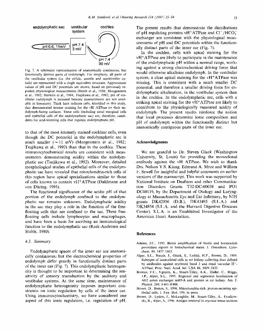
90 mV Fig. 7. A schematic representation of anatomically continuous, but functionally distinct parts of endolymph. For simplicity, all parts of the vestibular system (i.e. the utricle, saccule and semicircular ca- nals) are represented with a single equivalent structure. Approximate values of pH and DC potentials are shown, based on previously re- ported physiological measurements (Smith et al., 1958; Morgenstern et al., 1982; Sterkers et al., 1984; Tsujikawa et al., 1992; pH of ves- tibular endolymph is assumed because measurements are not avail- able in literature). Thick lines indicate cells, identified in this study, that demonstrated intense staining for the vH+ATPase on their en- dolymph-facing surfaces. These cells (including strial marginal cells and epithelial cells of the endolymphatic sac) are, therefore, candi- dates for acid-secreting cells that regulate endolymphatic pH.
to that of the most intensely stained cochlear cells, even though the DC potential in the endolymphatic sac is much smaller ( ~ 1 1 mV) (Morgenstern et al., 1982; Tsujikawa et al., 1992) than that in the cochlea. These immunocytochemical results are consistent with meas- urements demonstrat ing acidity within the endolym- phatic sac (Tsujikawa et al., 1992). Moreover, detailed morphological studies of epithelial cells of the endolym- phatic sac have revealed that mitochondria-rich cells of this region have apical specializations similar to those of cells known to contain vH+ATPase (Dahlmann and von Diaring, 1995).
The functional significance of the acidic pH of that port ion of the endolymph confined to the endolym- phatic sac remains unknown. Endolymphat ic acidity in the sac may play a role in the function of the free- floating cells that are confined to the sac. These free- floating cells include lymphocytes and macrophages, and have been a basis for ascribing an immunological function to the endolymphatic sac (Rask-Andersen and Stahle, 1980).
4.3. S u m m a r y
Endolymphat ic spaces of the inner ear are anatomi- cally continuous; but the electrochemical properties of endolymph differ greatly in functionally distinct parts of the inner ear (Fig. 7). This endolymphatic heterogen- eity is thought to be important in determining the sen- sitivity of sensory transduction by the auditory and vestibular systems. At the same time, maintenance of endolymphatic heterogeneity imposes important con- straints on ionic regulation by cells of the inner ear. Using immunocytochemistry, we have considered one aspect of this ionic regulation, i.e. regulation of pH.
The present results that demonstrate the distributions of pH regulating proteins vH+ATPase and C 1 - / H C O 3 exchanger are consistent with the physiological meas- urements of pH and DC potentials within the function- ally distinct parts of the inner ear (Fig. 7).
In the cochlea, cells with apical staining for the vH+ATPase are likely to participate in the maintenance of the endolymphatic pH within a normal range, work- ing against a strong electrochemical driving force that would otherwise alkalinize endolymph. In the vestibular system, a clear apical staining for the vH+ATPase was missing. This is consistent with a much smaller DC potential, and therefore a smaller driving force for en- dolymphatic alkalization, in the vestibular system than in the cochlea. In the endolymphatic sac, cells with a striking apical staining for the vH+ATPase are likely to contribute to the physiologically measured acidity of endolymph. The present results reinforce the notion that local processes determine ionic composition and pH of endolymph within the functionally distinct but anatomically contiguous parts of the inner ear.
Acknowledgments
We are grateful to Dr. Steven Gluck (Washington University, St. Louis) for providing the monoclonal antibody against the vH+ATPase. We wish to thank Drs. Nelson Y.S. Kiang, Edmund A. Mroz and William F. Sewell for insightful and helpful comments on earlier versions of the manuscript. This work was supported by National Institute on Deafness and other Communica- tion Disorders Grants T32-DC-00038 and PO1 DC00119, by the Depar tment of Otology and Laryng- ology at Massachusetts Eye and Ear Infirmary, by N I H grants DK42956 (D.B.), DK43495 (S.L.A.) and DK34854 (S.L.A. and the Harvard Digestive Diseases Center). S.L.A. is an Established Investigator of the American Hear t Association.
References
Adams, J.C., 1992. Biotin amplification of biotin and horseradish peroxidase signals in histochemical stains. J. Histochem. Cyto- chem. 40, 1457-1463.
Alper, S.L., Natale, J., Gluck, S., Lodish, H.F., Brown, D., 1989. Subtypes of intercalated cells in rat kidney collecting duct defined by antibodies against erythroid band 3 and renal vacuolar H +- ATPase. Proc. Natl. Acad. Sci. USA 86, 5429-5433.
Brosius, F.C., Nguyen, K., Stuart-Tilley, A.K., Hailer, C., Briggs, J.P., Alper, S.L., 1995. Regional and segmental localization of AE2 anion exchanger mRNA and protein in rat kidney. Am. J. Physiol. 269, F461 F468.
Brown, D., Breton, S., 1996. Mitochondria-rich, proton-secreting epi- thelial cells. J. Exp. Biol. 199, in press.
Brown, D., Lydon, J., McLaughlin, M., Stuart-Tilley, A., Tyszkow- ski, R., Alper, S., 1996. Antigen retrieval in cryostat tissue sections

34 K.M. Stankovig" et al./Hearing Research 114 (1997) 2134
and cultured cells by treatment with sodium dodecyl sulfate (SDS). Histochem. Cell Biol. 105, 261 267.
Busa, W.B., Nuccitelli, R., 1984. Metabolic regulation via intracellular pH. Am. J. Physiol. (Regul. Integr. Comp. Physiol. 15) 246, R409 R438.
Cannon, C., Adelsberg, v., J., Kelly, S., A1-Awqati, Q., 1985. Carbon- dioxide-induced exocytotic insertion of H ~ pumps in turtle-blad- der lumenal membrane: role of cell pH and calcium. Nature 314, 443446.
Dahlmann, A., von DUring, M., 1995. The endolymphatic duct and sac of the rat: a histological, ultrastructural, and immunocyto- chemical investigation. Cell Tissue Res. 282, 277-289.
Forgac, M., 1989. Structure and function of vacuolar class of ATP- driven proton pumps. Physiol. Rev. 69, 765 796.
Freeman, D,M., Hattangadi, S.H., Weiss, T.F., 1996. Osmotic re- sponses of the isolated mouse rectorial membrane to changes in pH. Assoc. Res. Otolaryngol. Abstr. 19, 232.
Guild, S.R., 1927. The circulation of the endolymph. Am. J. Anat. 39, 57-81.
Guinan, J.J., Jr., Gifford, M.L., 1988. Effects of electrical stimulation of efferent olivocochlear neurons on cat auditory-nerve fibers. II. Spontaneous rate. Hear. Res. 33, 115-127.
Ichimiya, I., Adams, J.C., Kimura, R.S., 1994. Immunolocalization of Na +, K+-ATPase, calcium-binding proteins, and carbonic anhy- drase in the guinea pig inner ear. Acta Otolaryngol. (Stockh.) 114, 167-176.
Ikeda, K., Kusakari, J., Takasaka, T., Saito, Y., 1987. Early effects of acetazolamide on anionic activities of the guinea pig endolymph: Evidence for active function of carbonic anhydrase in the cochlea. Hear. Res. 31,211 216.
Ikeda, K., Saito, Y., Nishiyama, A., Takasaka, T., 1992. Intracellular pH regulation in isolated cochlear outer hair cells of the guinea- pig. J. Physiol. 447, 627 648.
Jiang, L.W., Chernova, M.N., Alper, S.L., 1997. Secondary regula- tory volume increase conferred on Xenopus oocytes by heterolo- gous expression of anion exchanger AE2. Am. J. Physiol. (Cell) 272, C191-C202.
Kalinec, F., Jaeger, R.G., Kachar, B., 1993. Mechanical coupling of the outer hair cell plasma membrane to the cortical cytoskeleton by anion exchanger and 4.1 proteins. In: Duifhuis, H., Horst, J.W., Van Dijk, P., Van Netten, S.M. (Eds.), Biophysics of Hair Cell Sensory Systems, pp. 175 181.
Kim, J., Tisher, C.C., Linser, P.J., Madsen, K.M., 1990. Ultrastruc- tural localization of carbonic anhydrase II in subpopulations of intercalated cells of the rat kidney. J. Am. Soc. Nephrol. 1, 245 256.
Kimura, R.S., 1969. Distribution, structure, and function of dark cells in the vestibular labyrinth. Ann. Otol. Rhinol. Laryngol. 78, 542 561.
Kronester-Frei, A., 1979. The effect of changes in endolymphatic ion concentrations on the tectorial membrane. Hear. Res. 1, 81-94.
Marcus, D.C., Liu, J., Wangemann, P., 1994. Transepithelial voltage and resistance of vestibular dark cell epithelium from the gerbil ampulla. Hear. Res. 73, 101 108.
Misrahy, G.A., Hildreth, K.M., Clark, L.C., Shinabarger, E.W., 1958. Measurement of the pH of the endolymph in the cochlea of guinea pigs. Am. J. Physiol. 194, 393 395.
Morgenstern, C., Amano, H., Orsulakova, A., 1982. Ion transport in the endolymphatic space. Am. J. Otolaryngol. 3, 323 327.
Negrini, C., Rivolta, M.N., Kalinec, F., Kachar, B., 1995. Cloning of an organ of Corti anion exchanger 2 isoform with a truncated C- terminal domain. Biochim. Biophys. Acta 1236, 207-211.
Nelson, R.D., Guo, X.L., Masood, K., Brown, D., Kalkbrenner, M., Gluck, S., 1992. Selectively amplified expression of an isoform of the vacuolar H+-ATPase 56-kilodalton subunit in renal interca- lated cells. Proc. Natl. Acad. Sci. USA 89, 3541 3545.
Okamura, H., Sugai, N., Suzuki, K., 1996. Enzyme-histochemical localization of carbonic anhydrase in the inner ear of the guinea pig and several improvements of the technique. Histochem. Cell Biol. 106, 425430.
Prazma, J., 1978. Carbonic anhydrase in the generation of cochlear potentials. Am. J. Physiol. (Renal Fluid Electrolyte Physiol. 4) 235, F317 F320.
Rask-Andersen, H., Stahle, J., 1980. Immunodefence of the inner ear? Acta Otolaryngol. 89, 283 294.
Salt, A.N., Melichar, I., Thalmann, R., 1987. Mechanisms of endo- cochlear potential generation by stria vascularis. Laryngoscope 97, 984~991.
Sato, Y., Santos-Sacchi, J., 1994. Cell coupling in the supporting cells of Corti's organ: Sensitivity to intracellular H + and Ca 2+. Hear. Res. 80, 21 24.
Sewell, W.F., 1984. The relation between the endocochlear potential and spontaneous activity in auditory nerve fibers of the cat. J. Physiol. 347, 685-696.
Silverstein, H., 1966. Biochemical and physiological studies of the endolymphatic sac in the cat. Laryngoscope 76, 498 512.
Sly, W.S., Hu, P.Y., 1995. Human carbonic anhydrase and carbonic anhydrase deficiencies. Annu. Rev. Biochem. 64, 375-401.
Smith, C.A., Davis, H., Deatherage, B.H., Gessert, C.F., 1958. DC potentials of the membranous labyrinth. Am. J. Physiol. 193, 203 206.
Spicer, S.S., Schulte, B.A., 1991. Differentiation of inner ear fibrocytes according to their ion transport related activity. Hear. Res. 56, 53 64.
Spicer, S.S., Schulte, B.A., Adams, J.C., 1990. Immunolocalization of Na +, K+-ATPase and carbonic anhydrase in the gerbil's vestibular system. Hear. Res. 43, 205 218.
Spicer, S.S., Smolka, A.J., Schulte, B.A., 1995. Distribution of H,K- ATPase in the adult gerbil inner ear and changes in its expression during development. Assoc. Res. Otolaryngol. Abstr. 18, 559.
Stankovi6, K.M., Adams, J.C., Brown, D., 1995. Localization of H~ ATPase and C1 , HCO~ exchanger in the guinea pig cochlea. Assoc. Res. Otolaryngol. Abstr. 18, 87.
Sterkers, O., Saumon, G., Tran Ba Huy, P., Ferrary, E., Amiel, C., 1984. Electrochemical heterogeneity of the cochlear endolymph: effect of acetazolamide. Am J. Physiol. (Renal Fluid Electrolyte Physiol. 15) 246, F47-F53.
Sterkers, O., Ferrary, E., Amiel, C., 1988. Production of inner ear fluids. Physiol. Rev. 68, 1083 1129.
Stuart-Tilley, A., Sardet, C., Pouyssegur, J., Schwartz, M.A., Brown, D., Alper, S.L., 1994. Immunolocalization of anion exchanger AE2 and cation exchanger NHEI in distinct, adjacent cells of gastric mucosa. Am. J. Physiol. (Cell) 266, C559-C568.
Tsujikawa, S., Yamashita, T., Amano, H., Kumazawa, T., Vosteen, K.H., 1992. Acidity in the endolymphatic sac fluid of guinea pigs. Otorhinolaryngology 54, 198-200.
Wangemann, P., Shiga, N., Marcus, D.C., 1993. The Na~/H + ex- changer in transitional cells of the inner ear. Hear. Res. 69, 107- 114.
Wangemann, P., Liu, J., Marcus, D.C., 1995. Ion transport mecha- nisms responsible for K + secretion and the transepithelial voltage across marginal cells of stria vascularis in vitro. Hear. Res. 84, 19- 29.
Wangemann, P., Liu, J., Shiga, N., 1996. Vestibular dark cells contain the Na~/H t exchanger NHE-I in the basolateral membrane. Hear. Res. 94, 94 106.
Yurko, M.A., Gluck, S., 1987. Production and characterization of a monoclonal antibody to vacuolar H+ATPase of renal epithelia. J. Biol. Chem. 262, 1577015779.