j comp neurol 2014 yokota s
TRANSCRIPT

Respiratory-Related Outputs of Glutamatergic,Hypercapnia-Responsive Parabrachial Neuronsin Mice
Shigefumi Yokota,1,2,3 Satvinder Kaur,1,2 Veronique G. VanderHorst,1 Clifford B. Saper,1,2,4
and Nancy L. Chamberlin1,2*1Department of Neurology, Beth Israel Deaconess Medical Center and Harvard Medical School, Boston, Massachusetts 022152Division of Sleep Medicine, Beth Israel Deaconess Medical Center and Harvard Medical School, Boston, Massachusetts 022153Department of Anatomy and Morphological Neuroscience, Shimane University School of Medicine, Izumo 693-8501, Japan4Program in Neuroscience, Beth Israel Deaconess Medical Center and Harvard Medical School, Boston, Massachusetts 02215
ABSTRACTIn patients with obstructive sleep apnea, airway obstruc-
tion during sleep produces hypercapnia, which in turn
activates respiratory muscles that pump air into the
lungs (e.g., the diaphragm) and that dilate and stabilize
the upper airway (e.g., the genioglossus). We hypothe-
sized that these responses are facilitated by glutamater-
gic neurons in the parabrachial complex (PB) that
respond to hypercapnia and project to premotor and
motor neurons that innervate the diaphragm and genio-
glossus muscles. To test this hypothesis, we combined
c-Fos immunohistochemistry with in situ hybridization for
vGluT2 or GAD67 or with retrograde tracing from the
ventrolateral medullary region that contains phrenic pre-
motor neurons, the phrenic motor nucleus in the C3–C5
spinal ventral horn, or the hypoglossal motor nucleus.
We found that hypercapnia (10% CO2 for 2 hours) acti-
vated c-Fos expression in neurons in the external lateral,
lateral crescent (PBcr), and K€olliker-Fuse (KF) PB subnu-
clei and that most of these neurons were glutamatergic
and virtually none g-aminobutyric acidergic. Numerous
CO2-responsive neurons in the KF and PBcr were labeled
after retrograde tracer injection into the ventrolateral
medulla or hypoglossal motor nuclei, and in the KF after
injections into the spinal cord, making them candidates
for mediating respiratory-facilitatory and upper-airway-
stabilizing effects of hypercapnia. J. Comp. Neurol.
000:000–000, 2014.
VC 2014 Wiley Periodicals, Inc.
INDEXING TERMS: phrenic; diaphragm; respiratory; hypoglossal; c-Fos; carbon dioxide; AB_518147; AB_631250;
AB_2341545
Patients with obstructive sleep apnea typically have
small, collapsible pharyngeal airways. During wakeful-
ness airway patency is maintained by enhanced airway
dilator muscle activity, mainly of the genioglossus mus-
cle (GG), a tongue protruder innervated by motoneurons
residing in the hypoglossal motor nucleus. Loss of this
neuromuscular compensation during sleep leads to par-
tial or complete block of airflow. The consequent hyper-
capnia, hypoxia, and negative intrapharyngeal airway
pressure created by inspiratory effort against a blocked
airway produce graded and progressive activation of
respiratory muscles and often arousal from sleep. The
neural circuitry that mediates these responses is not
well understood.
The parabrachial complex (PB) is a key component of
the central respiratory network (Chamberlin, 2004;
Dutschmann and Dick, 2012) and thus is well posi-
tioned to monitor and respond to physiological signals
of respiratory difficulty. The PB contains at least 11 dis-
tinct subnuclei with different chemical and connectional
phenotypes. The respiratory portions of the PB, which
include the external lateral (PBel), lateral crescent
(PBcr), and K€olliker-Fuse (KF) subnuclei, receive intense
inputs from the respiratory part of the nucleus of the
solitary tract (NTS) and the retrotrapezoid nucleus
Grant sponsor: National Institutes of Health; Grant numbers: P01HL095491; NS079623.
*CORRESPONDENCE TO: Nancy L. Chamberlin, PhD, Department ofNeurology, Beth Israel Deaconess Medical Center, 330 Brookline Ave.,Boston, MA 02215. E-mail: [email protected]
Received October 6, 2014; Revised November 20, 2014;Accepted November 21, 2014.DOI 10.1002/cne.23720Published online Month 00, 2014 in Wiley Online Library(wileyonlinelibrary.com)VC 2014 Wiley Periodicals, Inc.
The Journal of Comparative Neurology | Research in Systems Neuroscience 00:00–00 (2014) 1
RESEARCH ARTICLE
(Ricardo and Koh, 1978; Herbert et al., 1990; Rosin
et al., 2006) that collectively carry both peripheral and
central respiratory chemoreceptor information as well
as breathing-related mechanical sensation. Lateral PB
and KF neurons express c-Fos in response to hypercap-
nia or hypoxia (Berquin et al., 2000), and PB lesions
produce decrements in hypercapnic and hypoxic venti-
latory responses in awake (Mizusawa et al., 1995) and
anesthetized (Song and Poon, 2009) rats. However, lit-
tle is known about the projections of the PB neurons
that are engaged by hypercapnia, particularly those that
contribute to respiratory drive. The KF projects directly
to respiratory motoneurons in the brainstem and spinal
cord (Yokota et al., 2001, 2004, 2011; Ezure and
Tanaka, 2006), and the KF and PBcr send intense pro-
jections to the ventrolateral medullary region that con-
tains both neurons that generate the respiratory rhythm
(the pre-B€otzinger complex) and premotor neurons for
the diaphragm and accessory breathing muscles (the
rostral ventral respiratory group; Herbert et al., 1990;
Yokota et al., 2004, 2007; Ezure and Tanaka, 2006).
We hypothesized that, during hypercapnia, these PBcr
and KF neurons augment the motor output of the respi-
ratory pump (diaphragm) and upper airway dilator (GG)
muscles.
Therefore, we sought to identify PB neurons that 1)
respond to hypercapnia and 2) project to respiratory
motor and premotor neurons. To match active neurons
with their downstream targets, we combined immuno-
histochemistry for c-Fos, a marker for neural activation,
with retrograde tracing. We chose to examine
hypercapnia-induced c-Fos expression in neurons pro-
jecting to three major PB targets that contain respira-
tory motor and premotor neurons and therefore could
affect the strength of ventilatory responses to hyper-
capnia, the ventrolateral medulla/rostral ventral respira-
tory group (diaphragm premotor neurons), the phrenic
motor nucleus, and the hypoglossal motor nucleus (GG
and other tongue motoneurons; Herbert et al., 1990;
Yokota et al., 2001, 2004, 2011).
MATERIALS AND METHODS
Experiments were performed on adult male C57BL/6
mice (Jackson Laboratories, Bar Harbor, ME; n 5 79)
weighing between 23 and 33 g. Mice were maintained
under standard laboratory conditions, including 12:12-
hour light:dark cycle (lights on at 0700) and
21�C 6 1�C room temperature. The mice had ad libitum
access to food and water. Care of the mice met
National Institutes of Health standards, as set forth in
the Guide for the care and use of laboratory animals,
and all protocols were approved by the BIDMC Institu-
tional Animal Care and Use Committees.
The mice were anesthetized with isoflurane (2%) and
9–15 nl of a 0.2% solution of cholera toxin b subunit
(CTb; List, Campbell, CA) were injected into the
intended target following craniotomy or laminectomy as
described previously (Lu et al., 2000; VanderHorst and
Ulfhake, 2006). Wounds were closed and mice given
postoperative analgesia (meloxicam; 5 mg/kg, sc; once
per day for 2 days). After the surgical procedure, the
mice were maintained undisturbed in the housing room
for at least 14 days. Mice were acclimated to the
experimental environment for 629 hours per day for 4
days prior to hypercapnia exposure. Mice in their home
cages were moved from the animal housing room to
the laboratory and placed inside a secondary box with
a light inside. Home cages were fitted with a special
airtight top, and mice were exposed to a hypercapnic
mixture of compressed air (21% O2, 10% CO2, 69% N2)
or a control mixture (21% O2, 0% CO2, 79% N2) for 2
hours. Air or hypercapnic exposures were performed
between 1400 and 1900 hours.
Mice were then deeply anesthetized with chloral
hydrate (500 mg/kg, ip) and transcardially perfused with
saline, followed by 10% formalin. Brains were removed,
postfixed overnight, immersed in 20% sucrose, and cut
into four alternate series of 30-lm frozen sections.
Histology: antibody characterizationAntibodies used in this study (see Table 1) were char-
acterized as follows. Tissue staining with rabbit anti-a-
calcitonin gene-related peptide (CGRP) was eliminated by
preadsorption with rat a-CGRP (Wang et al., 2006). The
c-Fos antibody from Oncogene Sciences stained a single
band of 55 kDa on Western blots from rat brain (manu-
facturer’s technical information). The c-Fos antibody from
Santa Cruz Biotechnology (Santa Cruz, CA) was raised
against amino acids 3216 near the N-terminus of c-Fos
of human origin: FSGFNADYEASSSR and affinity purified.
Both c-Fos antibodies stained only neuronal nuclei in the
same patterns as previously reported at this time of day
for rats that were untreated (Scammell et al., 2000) or
exposed to CO2 (Berquin et al., 2000). The staining pat-
terns with the CTb antibody depended solely on the site
of CTb injection; this antibody stains nothing in animals
not injected with CTb.
Histology: immunohistochemistrySections were processed for detection of c-Fos
alone, or c-Fos in combination with CTb or CGRP, or c-
Fos in combination with in situ hybridization for vGluT2
or GAD67 mRNA (see below). All incubations were per-
formed on free-floating tissue sections at room
S. Yokota et al.
2 The Journal of Comparative Neurology |Research in Systems Neuroscience

temperature. Sections were first incubated overnight in
c-Fos antibody. When c-Fos staining was to be com-
bined with CGRP, we used goat anti-c-Fos antibody
(AB_631250; diluted 1:500 in PBS with 0.2% Triton X-
100 and 2.5% normal donkey serum). After rinsing, sec-
tions were incubated in Alexa 488 (green fluorescence)-
conjugated donkey anti goat-IgG (Invitrogen, Carlsbad,
CA; A11055) at 1:500 in PBS containing 0.2% Triton-X
and 2.5% normal donkey serum for 3 hours. After rins-
ing in PBS, sections were incubated overnight with rab-
bit anti-CGRP (AB_518147; diluted 1:1000 in PBS with
0.2% Triton X-100 and 2.5% normal donkey serum).
After rinsing on the next day, sections were incubated
in Cy3 (red fluorochrome)-conjugated donkey anti-rabbit
IgG (Jackson Immunoresearch, West Grove, PA; code
111–165-003) at 1:500.
When c-Fos was to be visualized alone or in combina-
tion with CTb, tissue sections were first incubated over-
night with rabbit anti-c-Fos (diluted 1:20,000 in PBS
with 0.2% Triton X-100 and 2.5% normal donkey serum).
After being rinsed, sections were incubated in biotinyl-
ated donkey anti-rabbit IgG (Jackson Immunoresearch;
diluted 1:500) for 3 hours and then ABC elite (Vector,
Burlingame, CA; 1:1,000) for 1 hour. After being rinsed
again, sections were reacted with 0.05% diaminobenzi-
dine hydrochloride (DAB; Sigma, St. Louis, MO) and
0.01% H2O2 in PBS containing 0.01–0.02% nickel sul-
fate, which renders a black reaction product. For immu-
nohistochemical visualization of CTb, sections were
next incubated overnight with goat anti-CTb
(AB_2341545; diluted 1:10,000 in PBS with 0.25% Tri-
ton X-100). After being rinsed, sections were incubated
in biotinylated donkey anti-goat IgG (Jackson Immunore-
search; diluted 1:500) for 3 hours and then with ABC
Elite (Vector, 1:1,000) for 1 hour. After a rinse, sections
were reacted with 0.05% DAB and 0.01% H2O2.
Histology: in situ hybridizationFree-floating sections were incubated overnight with
hybridization buffer containing probes for vGluT2 [1 mg/
ml; template graciously donated by Dr. Qinchung Tong
(Tong et al., 2007)] or fragmented probes by alkaline
hydrolysis for GAD67, 0.5 mg/ml, full-length cDNA
encoding rat GAD67 (3.2 kb; Erlander et al., 1991) from
Dr. A.J. Tobin (University of California, Los Angeles, CA)
at 60�C. Subsequently, the sections were washed twice
in 23 standard saline citrate (SSC) at 60�C for 60
minutes, treated with RNase A (20 mg/ml), washed in
23 SSC at 60�C for 60 minutes, and then washed twice
in 13 SSC at 60�C for 60 minutes. After being washed
in Tris-buffered saline (pH 7.6), the sections were incu-
bated overnight in an alkaline phosphatase-conjugated
anti-digoxigenin antibody (1:500; Roche, Indianapolis, IN).
After being washed, the sections were developed with 5-
bromo-4-chloro-3-indolyl phosphate/nitroblue tetrazolium
(BCIP/NBT) solution (Roche) for 6 hours. Subsequently,
c-Fos protein was visualized as described above. Briefly,
the sections were incubated overnight with an anti-Fos
antibody (1:10,000), with a biotinylated donkey antibody
to rabbit IgG, and then with ABC Elite for 1 hour. The
sections were reacted with 0.05% DAB and 0.01% H2O2.
Data analysis: PB subnuclear anatomyCTb- and c-Fos-immunoreactive (-ir) cells in the PB
were first plotted from six sections in each series of
every fourth 30-mm-thick section. After the locations of
all labeled neurons were mapped, the coverslips were
removed from slides and the tissue stained for Nissl
substance with thionin. The borders of subnuclei were
drawn based on cytoarchitecture, with the exception of
the PBcr (Fig. 1). The PBcr is not distinct cytoarchitec-
tonically (Fulwiler and Saper, 1984) but is clearly out-
lined by retrograde tracing from the ventrolateral
medulla in rats (Herbert et al., 1990; Chamberlin and
Saper, 1992), in which the lateral crescent neurons are
seen along the lateral margin of the unlabeled PBel
(Chamberlin and Saper, 1992). Because we saw a simi-
lar retrograde labeling pattern in our mice, we consid-
ered all of the retrogradely labeled neurons along the
border of the PBel after medullary injections to be part
TABLE 1.
Table of Primary Antibodies Used
Antigen Description of immunogen Source, host species, catalog No., RRID
Concentration
used
CGRP (a-calcitoningene-related peptide)
Amino acids 83–119 of the CGRPprecursor protein
Peninsula Labs, rabbit polyclonal,T4032, AB_518147
1:1,000
c-Fos Amino acids 4–17 of human c-Fos Oncogene Sciences, rabbit polyclonal, Ab5,none (this antibody is no longer commerciallyavailable)
1:20,000
c-Fos Amino acids 3–16 near the N-terminusof c-Fos of human origin:FSGFNADYEASSSR
Santa Cruz Biotechnology, goat polyclonal,sc-52-G, AB_631250
1:500
CTb Cholera toxin b subunit List Biological, goat polyclonal, 703, AB_2341545 1:10,000
Outputs of CO2-responsive parabrachial neurons
The Journal of Comparative Neurology | Research in Systems Neuroscience 3

of the PBcr (see below). Some of these neurons
wrapped around the dorsomedial border of PBel into
what had been considered the central lateral subnu-
cleus (PBcl). Because there is no clear cytoarchitectonic
boundary at the interface between the PBcr and PBcl,
but the vast majority of hypercapnia-activated PB neu-
rons projecting to the medulla were continuous with
the PBcr, in our cell counts of c-Fos-ir neurons for prac-
tical purposes we considered these two structures
together as a single unit. The borders of the KF were
drawn on the basis of cytoarchitecture. KF neurons are
larger than other PB neurons, have a pyramidal shape,
and have distinct nucleoli, as has been previously
reported in rats (Fulwiler and Saper, 1984). It is worth
noting that in both rats and mice the ventral spinocere-
bellar tract provides a convenient tissue landmark for
the dorsolateral border of KF (Figs. 1, 3, 6–9).
Results were expressed as mean 6 SEM. Differences
between groups were assessed by one-way ANOVA and
groups, with P< 0.05 taken as significant.
Figure 1. Distribution of c-Fos-ir neurons in the parabrachial nucleus (PB) in animals breathing normal air or hypercapnic air (10% CO2). Each
red dot indicates one c-Fos-ir neuronal nucleus. Shown are three representative sections through the PB from rostral to caudal (A-C and D-
F). Note the increase in c-Fos expression in the hypercapnic condition vs. control in the KF nucleus (A vs. D) and in the outer portion of
external lateral (el), lateral crescent (cr; large arrowheads in B,E), and central lateral (cl) PB subnuclei. The smaller arrowheads in B and E
show the area where we found hypercapnia-activated, medullary-projecting neurons (see Fig. 6). cl, Central lateral; cr, lateral crescent; dl,
dorsal lateral; el, external lateral; exm, external medial; LC, locus coeruleus; m, medial; Mo5, motor trigeminal; Pr5, principal sensory trigemi-
nal; scp, superior cerebellar peduncle; sl, superior lateral; vl, ventral lateral; vsct, ventral spinocerebellar tract. Scale bars 5 200 lm.
S. Yokota et al.
4 The Journal of Comparative Neurology |Research in Systems Neuroscience

RESULTS
c-Fos distribution in the PB followinghypercapnia
We examined the distribution of c-Fos-ir nuclei in the
PB in control animals breathing air, and then compared
that with the pattern of c-Fos expression after hyper-
capnia (Fig. 1). We then quantified the numbers of c-
Fos-expressing neurons in PB subnuclei. Under control
conditions, c-Fos-ir nuclei appeared sparsely throughout
the PB (Fig. 1A–C). After hypercapnia, we found numer-
ous c-Fos-ir nuclei in the KF, PBcr, PBcl, and PBel sub-
nuclei (Fig. 1D,E). At rostral levels the most prominent
group of activated neurons was located in the KF, nes-
tled against the medial edge of the ventral spinocere-
bellar tract (vsct; Fig. 1D). At mid-PB levels just caudal
to the KF, c-Fos immunoreactivity was seen in the lat-
eral part of PBel and in the PBcr just lateral to it. c-
Fos-ir neurons extended medially over the surface of
the PBel into the PBcl subnucleus (Fig. 1E, small arrow-
head). Counts of c-Fos-ir neurons verified the observa-
tions of increased labeling following hypercapnia in the
KF and in the PBel and PBcr/cl (Fig. 2).
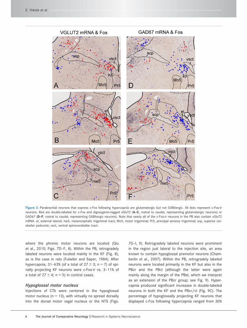
Neurochemical phenotype ofhypercapnic-responsive PB neurons
Because most PB neurons contain either glutamate or
GABA, we determined whether PB neurons responding to
hypercapnia contained these neurotransmitters. Tissue
from two groups of mice exposed to hypercapnia was
labeled for vGluT2 (n 5 4) or GAD67 (n 5 4) mRNA in com-
bination with c-Fos immunoreactivity (Figs. 3, 4). Nearly
every c-Fos-ir neuron also contained vGluT2 mRNA; 1,255
of the 1,353 (93%) PB cells exhibiting c-Fos immunoreac-
tivity following hypercapnia also expressed vGluT2 mRNA.
By contrast, only a few PB cells (3%; 35 of 1,360 cells)
exhibiting c-Fos immunoreactivity also expressed GAD67
mRNA (n 5 4 mice; six sections for each mouse).
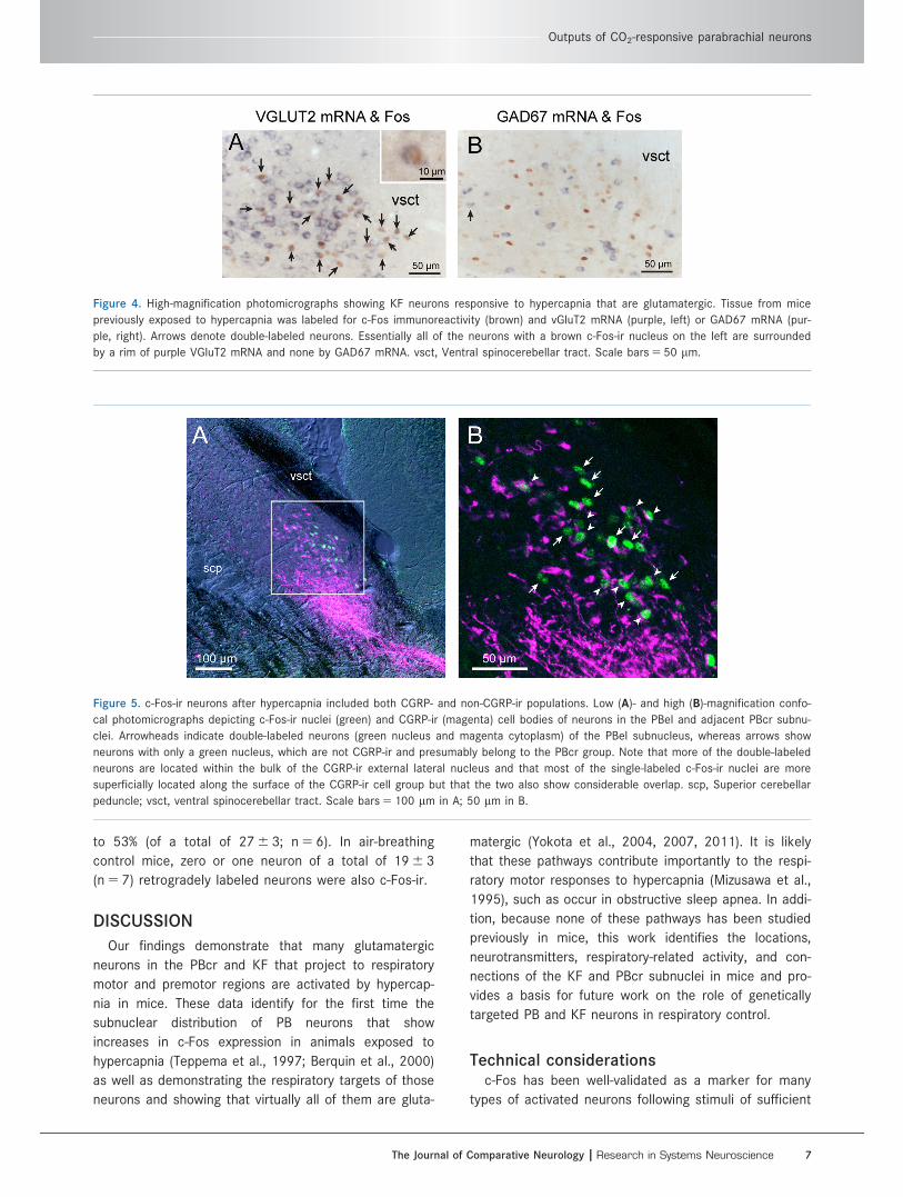
After hypercapnia, many c-Fos-ir neurons straddled
the border between the outer portion of the PBel and
the PBcr (large arrowhead, Fig. 1E). To determine the
cell group to which these neurons belonged, tissue sec-
tions from two hypercapnia-exposed mice were doubly
stained immunohistochemically with antibody to CGRP
(which is found in most PBel neurons) in combination
with c-Fos antibody (Fig. 5). In each case, c-Fos labeling
was seen in both CGRP-ir and CGRP-negative neurons,
indicating that neurons of both types were CO2 respon-
sive. Most of the c-Fos-ir cells that were also CGRP-ir
were located in the PBel and most of the CGRP-negative
ones in a thin layer beyond the PBel (Fig. 5), but there
was a mixture of both types along the outer edge of the
PBel (i.e., they were not cleanly separated anatomically).
Projections of hypercapnia-responsivePB neuronsVentrolateral medullaTo determine whether PB neurons projecting to the ven-
trolateral medulla expressed c-Fos following hypercapnia
exposure, we combined retrograde tracing with c-Fos
immunohistochemistry (n 5 12). After injections of CTb
into the ventrolateral medulla at the level where the NTS
just touches the floor of the fourth ventricle rostral to the
obex, retrogradely labeled neurons were found mainly in
the KF, PBcl, PBcr, and medial PB subnuclei in a pattern
similar to that previously described for rats (Fig. 6; Her-
bert et al., 1990; Chamberlin and Saper, 1992). After
hypercapnia, 31–40% (of a total of 110 6 12; n 5 5) of
retrogradely labeled neurons in the KF were also c-Fos-ir,
indicating that hypercapnia activated neurons projecting
to the ventrolateral medulla (Figs. 6, 7A–C). By contrast,
in controls, only 2–6% (of a total of 140 6 14; n 5 7) neu-
rons in the KF were double labeled. In addition to the KF,
double-labeled neurons were located in the PBcr area
and wrapped around the medial edge of the PBel into the
PBcl (Fig. 6J,K). There were no double-labeled neurons in
the PBcl subnucleus aside from this group just medial to
PBel. Based on these observations, we consider this
group of double-labeled neurons to be an extension of
the PBcr (see Discussion), and, within the PBcr defined in
this way, 32.9% 6 2.85% (n 5 5) of retrogradely labeled
neurons were c-Fos-ir
Phrenic motor nucleusSpinal cord injections (n 5 12) of CTb were made into
the ventral horn of the spinal cord at the C3–C5 level,
Figure 2. Mean numbers of c-Fos-ir neurons in the PB of mice
breathing normal air or hypercapnic air (10% CO2). The gray bars
illustrate the average numbers of c-Fos-ir neurons in each PB
subnucleus (6SEM). *P< 0.05, **P< 0.001. cl, Central lateral;
cr, lateral crescent; dl, dorsal lateral; el, external lateral; exm,
external medial; il, internal lateral; m, medial; sl, superior lateral;
vl, ventral lateral.
Outputs of CO2-responsive parabrachial neurons
The Journal of Comparative Neurology | Research in Systems Neuroscience 5
where the phrenic motor neurons are located (Qiu
et al., 2010; Figs. 7D–F, 8). Within the PB, retrogradely
labeled neurons were located mainly in the KF (Fig. 8),
as is the case in rats (Fulwiler and Saper, 1984). After
hypercapnia, 31–63% (of a total of 27 6 3; n 5 7) of spi-
nally projecting KF neurons were c-Fos-ir vs. 3–11% of
a total of 27 6 4; n 5 5) in control cases.
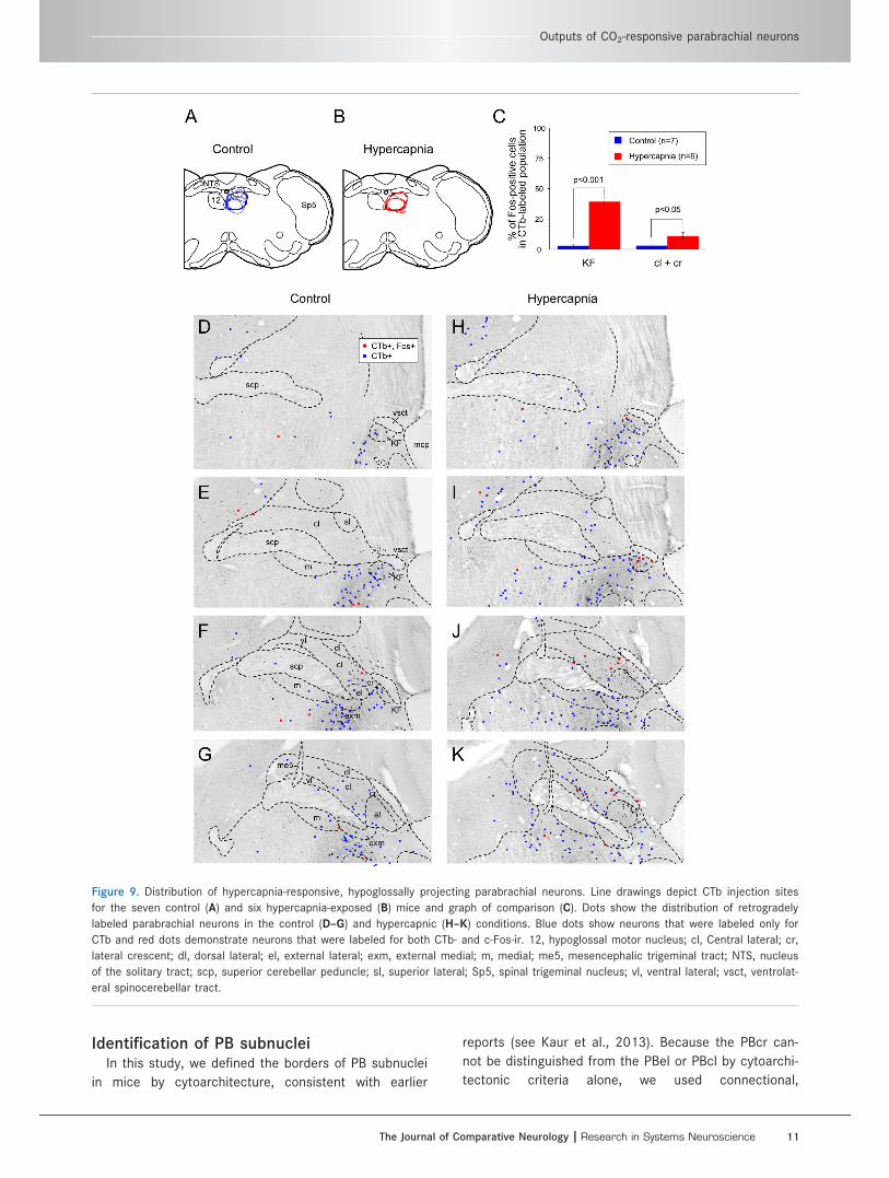
Hypoglossal motor nucleusInjections of CTb were centered in the hypoglossal
motor nucleus (n 5 13), with virtually no spread dorsally
into the dorsal motor vagal nucleus or the NTS (Figs.
7G–I, 9). Retrogradely labeled neurons were prominent
in the region just lateral to the injection site, an area
known to contain hypoglossal premotor neurons (Cham-
berlin et al., 2007). Within the PB, retrogradely labeled
neurons were located primarily in the KF but also in the
PBcr and the PBcl (although the latter were again
mainly along the margin of the PBel, which we interpret
as an extension of the PBcr group; see Fig. 9). Hyper-
capnia produced significant increases in double-labeled
neurons in both the KF and the PBcr/cl (Fig. 9C). The
percentage of hypoglossally projecting KF neurons that
displayed c-Fos following hypercapnia ranged from 30%
Figure 3. Parabrachial neurons that express c-Fos following hypercapnia are glutamatergic but not GABAergic. All dots represent c-Fos-ir
neurons. Red are double-labeled for c-Fos and digoxygenin-tagged vGluT2 (A–C, rostral to caudal, representing glutamatergic neurons) or
GAD67 (D–F, rostral to caudal, representing GABAergic neurons). Note that nearly all of the c-Fos-ir neurons in the PB also contain vGluT2
mRNA. el, external lateral; me5, mesencephalic trigeminal tract; Mo5, motor trigeminal; Pr5, principal sensory trigeminal; scp, superior cer-
ebellar peduncle; vsct, ventral spinocerebellar tract.
S. Yokota et al.
6 The Journal of Comparative Neurology |Research in Systems Neuroscience
to 53% (of a total of 27 6 3; n 5 6). In air-breathing
control mice, zero or one neuron of a total of 19 6 3
(n 5 7) retrogradely labeled neurons were also c-Fos-ir.
DISCUSSION
Our findings demonstrate that many glutamatergic
neurons in the PBcr and KF that project to respiratory
motor and premotor regions are activated by hypercap-
nia in mice. These data identify for the first time the
subnuclear distribution of PB neurons that show
increases in c-Fos expression in animals exposed to
hypercapnia (Teppema et al., 1997; Berquin et al., 2000)
as well as demonstrating the respiratory targets of those
neurons and showing that virtually all of them are gluta-
matergic (Yokota et al., 2004, 2007, 2011). It is likely
that these pathways contribute importantly to the respi-
ratory motor responses to hypercapnia (Mizusawa et al.,
1995), such as occur in obstructive sleep apnea. In addi-
tion, because none of these pathways has been studied
previously in mice, this work identifies the locations,
neurotransmitters, respiratory-related activity, and con-
nections of the KF and PBcr subnuclei in mice and pro-
vides a basis for future work on the role of genetically
targeted PB and KF neurons in respiratory control.
Technical considerationsc-Fos has been well-validated as a marker for many
types of activated neurons following stimuli of sufficient
Figure 5. c-Fos-ir neurons after hypercapnia included both CGRP- and non-CGRP-ir populations. Low (A)- and high (B)-magnification confo-
cal photomicrographs depicting c-Fos-ir nuclei (green) and CGRP-ir (magenta) cell bodies of neurons in the PBel and adjacent PBcr subnu-
clei. Arrowheads indicate double-labeled neurons (green nucleus and magenta cytoplasm) of the PBel subnucleus, whereas arrows show
neurons with only a green nucleus, which are not CGRP-ir and presumably belong to the PBcr group. Note that more of the double-labeled
neurons are located within the bulk of the CGRP-ir external lateral nucleus and that most of the single-labeled c-Fos-ir nuclei are more
superficially located along the surface of the CGRP-ir cell group but that the two also show considerable overlap. scp, Superior cerebellar
peduncle; vsct, ventral spinocerebellar tract. Scale bars 5 100 lm in A; 50 lm in B.
Figure 4. High-magnification photomicrographs showing KF neurons responsive to hypercapnia that are glutamatergic. Tissue from mice
previously exposed to hypercapnia was labeled for c-Fos immunoreactivity (brown) and vGluT2 mRNA (purple, left) or GAD67 mRNA (pur-
ple, right). Arrows denote double-labeled neurons. Essentially all of the neurons with a brown c-Fos-ir nucleus on the left are surrounded
by a rim of purple VGluT2 mRNA and none by GAD67 mRNA. vsct, Ventral spinocerebellar tract. Scale bars 5 50 lm.
Outputs of CO2-responsive parabrachial neurons
The Journal of Comparative Neurology | Research in Systems Neuroscience 7

Figure 6. Distribution of hypercapnia-responsive, ventrolateral medullary-projecting parabrachial neurons. Line drawings depicting CTb
injection sites for the seven mice exposed to room air (control; A) and five hypercapnia-exposed mice (B) and graph of comparison (C).
Dots show the distribution of retrogradely labeled parabrachial neurons in one representative control (D–G) and hypercapnia-treated (H–K)
mouse. Blue dots illustrate neurons that were labeled only for CTb, and red dots show neurons that were labeled for CTb- and c-Fos
immunoreactivity. Note the thin layer of double-labeled PBcr neurons in J,K. The arrowhead in J shows the double-labeled neurons that we
take as PBcr neurons intruding into PBcl. 12, hypoglossal motor nucleus; cl, Central lateral; cr, lateral crescent; dl, dorsal lateral; el, exter-
nal lateral; exm, external medial; m, medial; me5, mesencephalic trigeminal tract; NTS, nucleus of the solitary tract; scp, superior cerebel-
lar peduncle; sl, superior lateral; Sp5, spinal trigeminal nucleus; vl, ventral lateral; vsct, ventral spinocerebellar tract.
S. Yokota et al.
8 The Journal of Comparative Neurology |Research in Systems Neuroscience
duration and magnitude (Sagar and Sharp, 1993). How-
ever, some limitations should be considered in data inter-
pretation. First, absence of c-Fos cannot be taken as
absence of activity because not all activated neurons dur-
ing a specific physiological response may express c-Fos.
Second, c-Fos-ir neurons in our experiments might have
responded to other stimuli that covary with hypercapnia.
For example, PB neurons in the PBel outer subdivision
respond to raised blood pressure (Miller et al., 2012).
Hypercapnia is typically accompanied by hypertension, so
our data do not allow us to distinguish between responses
driven by hypercapnia and by subsequent responses such
as hypertension. Nevertheless, the c-Fos-ir neurons dur-
ing hypercapnic stimulation provide a starting point for
understanding the physiological responses that are seen
during hypercapnia. By combining this with retrograde
labeling from sites involved with respiratory control, we
can identify candidate pathways by which elevated CO2
invokes respiratory responses, which can then be studied
by genetically targeted manipulations in mice.
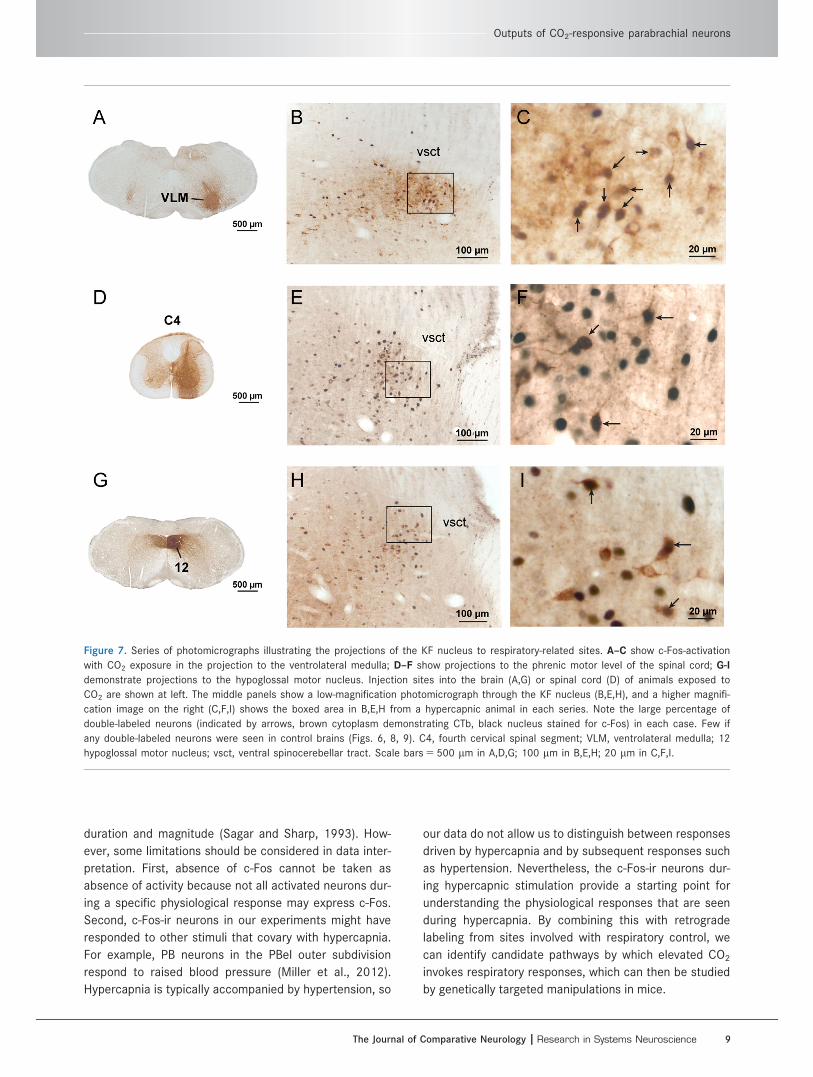
Figure 7. Series of photomicrographs illustrating the projections of the KF nucleus to respiratory-related sites. A–C show c-Fos-activation
with CO2 exposure in the projection to the ventrolateral medulla; D–F show projections to the phrenic motor level of the spinal cord; G-I
demonstrate projections to the hypoglossal motor nucleus. Injection sites into the brain (A,G) or spinal cord (D) of animals exposed to
CO2 are shown at left. The middle panels show a low-magnification photomicrograph through the KF nucleus (B,E,H), and a higher magnifi-
cation image on the right (C,F,I) shows the boxed area in B,E,H from a hypercapnic animal in each series. Note the large percentage of
double-labeled neurons (indicated by arrows, brown cytoplasm demonstrating CTb, black nucleus stained for c-Fos) in each case. Few if
any double-labeled neurons were seen in control brains (Figs. 6, 8, 9). C4, fourth cervical spinal segment; VLM, ventrolateral medulla; 12
hypoglossal motor nucleus; vsct, ventral spinocerebellar tract. Scale bars 5 500 lm in A,D,G; 100 lm in B,E,H; 20 lm in C,F,I.
Outputs of CO2-responsive parabrachial neurons
The Journal of Comparative Neurology | Research in Systems Neuroscience 9

Figure 8. Distribution of hypercapnia-responsive, spinally projecting parabrachial neurons. Line drawings depict CTb injection sites for
the five control (A) and seven hypercapnia-exposed (B) mice and graph of comparison (C). Dots show the distribution of retrogradely
labeled parabrachial neurons in the control (D–G) and hypercapnic (H–K) conditions. Blue dots illustrate neurons that were labeled only
for CTb and red dots show neurons that were labeled for both CTb- and c-Fos-ir. cl, Central lateral; cr, lateral crescent; dl, dorsal lat-
eral; el, external lateral; exm, external medial; m, medial; me5, mesencephalic trigeminal tract; scp, superior cerebellar peduncle; sl,
superior lateral; vl, ventral lateral; vsct, ventral spinocerebellar tract.
S. Yokota et al.
10 The Journal of Comparative Neurology | Research in Systems Neuroscience
Identification of PB subnucleiIn this study, we defined the borders of PB subnuclei
in mice by cytoarchitecture, consistent with earlier
reports (see Kaur et al., 2013). Because the PBcr can-
not be distinguished from the PBel or PBcl by cytoarchi-
tectonic criteria alone, we used connectional,
Figure 9. Distribution of hypercapnia-responsive, hypoglossally projecting parabrachial neurons. Line drawings depict CTb injection sites
for the seven control (A) and six hypercapnia-exposed (B) mice and graph of comparison (C). Dots show the distribution of retrogradely
labeled parabrachial neurons in the control (D–G) and hypercapnic (H–K) conditions. Blue dots show neurons that were labeled only for
CTb and red dots demonstrate neurons that were labeled for both CTb- and c-Fos-ir. 12, hypoglossal motor nucleus; cl, Central lateral; cr,
lateral crescent; dl, dorsal lateral; el, external lateral; exm, external medial; m, medial; me5, mesencephalic trigeminal tract; NTS, nucleus
of the solitary tract; scp, superior cerebellar peduncle; sl, superior lateral; Sp5, spinal trigeminal nucleus; vl, ventral lateral; vsct, ventrolat-
eral spinocerebellar tract.
Outputs of CO2-responsive parabrachial neurons
The Journal of Comparative Neurology | Research in Systems Neuroscience 11

chemoarchitectonic, and functional information as well
to deduce the subnuclear location of hypercapnia-
activated PB neurons. In rats, the PBcr neurons project
to the ventrolateral medulla, whereas the PBel neurons
project to the amygdala; the combination of retrograde
transport from these two targets clearly distinguishes
the two cell populations (Chamberlin and Saper, 1992).
In mice, the PBcr and PBel can also be visualized by
tracing from the ventrolateral medulla and central
nucleus of the amygdala, respectively. Neurons retro-
gradely labeled from the ventrolateral medulla can be
followed along the lateral margin of the PBel, into the
PBcl (Fig. 6). In both rats and mice, ventrolateral medul-
lary projecting neurons can be found throughout the
mediolateral extent of PBcl. By contrast, the distribution
of those medullary projecting neurons that are hyper-
capnia activated (c-Fos positive) is confined to the por-
tion of PBcl just adjacent to PBel (Fig. 6J, red dots,
small arrowhead). Therefore, we conclude that these
neurons belong to PBcr. Recent work has demonstrated
a distinct group of neurons in rats expressing the tran-
scription factors FoxP2 and Lmx1b and having a distri-
bution identical to the pattern we have established for
the PBcr 1 KF (Miller et al., 2012), i.e., extending from
the KF dorsally into the lateral PB along the margins of
PBel. It would be interesting to know whether the PBcr
in mice would be defined by neurons expressing these
same two transcription factors.
Sources of excitatory drive to PBhypercapnia-responsive neurons
Although there are no published data to indicate
whether PB neurons are directly CO2 or acid sensitive,
our preliminary data from in vitro slices indicate that PB
neurons do not respond to either (Arrigoni and Cham-
berlin, unpublished observations). Therefore, it is likely
that PB neurons are driven by other CO2-sensitive neu-
rons. There are several pathways that could contribute
to PB activation following stimulation of peripheral and
central CO2 chemoreceptors. Afferents from peripheral
CO2 chemoreceptors in the carotid body terminate in
the dorsolateral and caudal commissural parts of the
NTS (Panneton and Loewy, 1980; Finley and Katz,
1992), which in turn project to the lateral PB and KF
(Herbert et al., 1990). Neurons along the ventral medul-
lary surface in the retrotrapezoid nucleus are CO2 sen-
sitive; also receive excitatory inputs from the CO2-
sensitive parts of the NTS (Takakura et al., 2006); and
project to the PBel, PBcl, PBcr, and KF (Rosin et al.,
2006; Bochorishvili et al., 2012). Finally, the PB may
receive CO2 chemoreceptor information from medullary
serotonin neurons or lateral hypothalamic orexin neu-
rons, both of which are also directly CO2 sensitive
(Wang et al., 2001; Rosin et al., 2006; Williams et al.,
2007) and project to the lateral PB and KF.
Functional roles of hypercapnia-responsivePB neurons
Our previous work demonstrated that one function of
hypercapnia-activated PB neurons is likely to be causing
the arousal that occurs when breathing is compromised
during sleep, as in obstructive sleep apnea. We found
that deletion of vGluT2 from neurons in the PBel and
adjacent PBcr attenuated the EEG arousal to hypercap-
nia (Kaur et al., 2013). We did not detect any changes
in ventilatory responses to hypercapnia in those experi-
ments, although they were not designed to examine
this question specifically. The rapidly rising CO2 levels
in those experiments awakened the control mice in 10–
15 seconds, leaving us too little time with the animals
asleep with CO2 to examine the ventilatory response.
We also did not have sufficient experiments involving
the KF to examine the role of that subnucleus in driving
ventilation. It will be important in future studies to
examine more thoroughly the role of the KF and PBcr
vs. PBel in ventilatory responses using more cell-type
specific interventions.
Pathways mediating effects ofhypercapnic-responsive PB neurons
Our data suggest that, when PBcr and KF neurons
are engaged by hypercapnia, they contribute to activa-
tion of ventrolateral medullary respiratory pattern gener-
ator neurons as well as phrenic and hypoglossal motor
neurons. Because the hypercapnia-responsive neurons
in the PB that project to the ventrolateral medulla and
hypoglossal and phrenic motor nuclei are glutamatergic,
they would be expected to increase the firing frequency
of their target neurons. Activation of neurons in the ros-
tral ventral respiratory group increases breathing rate
and depth (McCrimmon et al., 1986), whereas activa-
tion of phrenic motor neurons would cause stronger
diaphragmatic contraction that would increase negative
intrathoracic pressure and the rate of air flow in the
upper airway. This would tend to collapse the airway,
but the activation of hypoglossal inputs to the genio-
glossus muscle would protrude the tongue, thus stiffen-
ing and dilating the oropharyngeal airway to counteract
the collapsing force of negative airway pressure. There-
fore, the glutamatergic, hypercapnia-responsive PB neu-
rons that we have identified would be expected to
increase both respiratory rate and tidal volume while
keeping the airway open, all reflexes that are critical
S. Yokota et al.
12 The Journal of Comparative Neurology | Research in Systems Neuroscience

under conditions of asphyxia, particularly in patients
who suffer from sleep apnea.
By contrast to hypercapnia-activated PBcr and KF
neurons, which have brainstem targets, PBel neurons
that respond to hypercapnia drive forebrain targets,
including the lateral hypothalamus, substantia innomi-
nata, central nucleus of the amygdala, and bed nucleus
of the stria terminalis (Schwaber et al., 1988; Yasui
et al., 1989; Bernard et al., 1993; Kaur et al., 2013).
One or more of these regions is likely to mediate hyper-
capnic EEG arousal. It will be important in future work
to take advantage of genetic differences in different PB
neurons to disrupt selectively PBel vs. PBcr and KF neu-
rons, structures that lie cheek-by-jowl in the parabra-
chial complex (Chamberlin and Saper, 1992), as well as
to determine both the forebrain and the brainstem sites
activated by the PB during hypercapnia and their inter-
actions in supporting arousals that re-establish
adequate ventilation.
CONFLICT OF INTEREST STATEMENT
We have no conflicts of interest to report.
ROLE OF AUTHORS
All authors had full access to all the data in the
study and take responsibility for the integrity of the
data and the accuracy of the data analysis. SY, VV, and
SK performed experiments. SY and NC analyzed and
interpreted data. NC and CS obtained funding and con-
tributed to the conception and design of the project, to
data analysis and interpretation, and to writing the
article.
LITERATURE CITEDBernard JF, Alden M, Besson JM. 1993. The organization of
the efferent projections from the pontine parabrachialarea to the amygdaloid complex: a Phaseolus vulgarisleucoagglutinin (PHA-L) study in the rat. J Comp Neurol329:201–229.
Berquin P, Bodineau L, Gros F, Larnicol N. 2000. Brainstemand hypothalamic areas involved in respiratory chemore-flexes: a Fos study in adult rats. Brain Res 857:30–40.
Bochorishvili G, Stornetta RL, Coates MB, Guyenet PG. 2012.Pre-Botzinger complex receives glutamatergic innervationfrom galaninergic and other retrotrapezoid nucleus neu-rons. J Comp Neurol 520:1047–1061.
Chamberlin NL. 2004. Functional organization of the parabra-chial complex and intertrigeminal region in the control ofbreathing. Respir Physiol Neurobiol 143:115–125.
Chamberlin NL, Saper CB. 1992. Topographic organization ofcardiovascular responses to electrical and glutamatemicrostimulation of the parabrachial nucleus in the rat.J Comp Neurol 326:245–262.
Chamberlin NL, Eikermann M, Fassbender P, White DP,Malhotra A. 2007. Genioglossus premotoneurons and thenegative pressure reflex in rats. J Physiol 579(Part 2):515–526.
Dutschmann M, Dick TE. 2012. Pontine mechanisms of respi-ratory control. Compr Physiol 2:2443–2469.
Erlander MG, Tillakaratne NJ, Feldblum S, Patel N, Tobin AJ.1991. Two genes encode distinct glutamate decarboxyl-ases. Neuron 7:91–100.
Ezure K, Tanaka I. 2006. Distribution and medullary projectionof respiratory neurons in the dorsolateral pons of therat. Neuroscience 141:1011–1023.
Finley JCW, Katz DM. 1992. The central organization ofcarotid body afferent projections to the brainstem of therat. Brain Res 572:108–116.
Fulwiler CE, Saper CB. 1984. Subnuclear organization of theefferent connections of the parabrachial nucleus in therat. Brain Res Rev 7:229–259.
Herbert H, Moga MM, Saper CB. 1990. Connections of theparabrachial nucleus with the nucleus of the solitarytract and the medullary reticular formation in the rat.J Comp Neurol 293:540–580.
Kaur S, Pedersen NP, Yokota S, Hur EE, Fuller PM, Lazarus M,Chamberlin NL, Saper CB. 2013. Glutamatergic signalingfrom the parabrachial nucleus plays a critical role inhypercapnic arousal. J Neurosci 33:7627–7640.
Lu J, Greco MA, Shiromani P, Saper CB. 2000. Effect oflesions of the ventrolateral preoptic nucleus on NREMand REM sleep. J Neurosci 20:3830–3842.
McCrimmon DR, Feldman JL, Speck DF. 1986. Respiratorymotoneuronal activity is altered by injections of pico-moles of glutamate into cat brain stem. J Neurosci 6:2384–2392.
Miller RL, Knuepfer MM, Wang MH, Denny GO, Gray PA,Loewy AD. 2012. Fos-activation of FoxP2 and Lmx1bneurons in the parabrachial nucleus evoked by hypoten-sion and hypertension in conscious rats. Neuroscience218:110–125.
Mizusawa A, Ogawa H, Kikuchi Y, Hida W, Shirato K. 1995.Role of parabrachial nucleus in ventilatory responses ofawake rats. J Physiol 489:877–884.
Panneton WM, Loewy AD. 1980. Projections of the carotidsinus nerve to the nucleus of the solitary tract in thecat. Brain Res 191:239–244.
Qiu K, Lane MA, Lee KZ, Reier PJ, Fuller DD. 2010. The phre-nic motor nucleus in the adult mouse. Exp Neurol 226:254–258.
Ricardo JA, Koh ET. 1978. Anatomical evidence of direct pro-jections from the nucleus of the solitary tract to thehypothalamus, amygdala, and other forebrain structuresin the rat. Brain Res 1553:1–26.
Rosin DL, Chang DA, Guyenet PG. 2006. Afferent and efferentconnections of the rat retrotrapezoid nucleus. J CompNeurol 499:64–89.
Sagar SM, Sharp FR. 1993. Early response genes as markersof neuronal activity and growth factor action. Adv Neurol59:273–284.
Scammell TE, Estabrooke IV, McCarthy MT, Chemelli RM,Yanagisawa M, Miller MS, Saper CB. 2000. Hypothalamicarousal regions are activated during modafinil-inducedwakefulness. J Neurosci 20:8620–8628.
Schwaber JS, Sternini C, Brecha NC, Rogers WT, Card JP.1988. Neurons containing calcitonin gene-related peptidein the parabrachial nucleus project to the central nucleusof the amygdala. J Comp Neurol 270:416–426, 398–419.
Song G, Poon CS. 2009. Lateral parabrachial nucleus medi-ates shortening of expiration and increase of inspiratorydrive during hypercapnia. Respir Physiol Neurobiol 165:9–12.
Takakura AC, Moreira TS, Colombari E, West GH, StornettaRL, Guyenet PG. 2006. Peripheral chemoreceptor inputs
Outputs of CO2-responsive parabrachial neurons
The Journal of Comparative Neurology | Research in Systems Neuroscience 13

to retrotrapezoid nucleus (RTN) CO2-sensitive neurons inrats. J Physiol 572(Part 2):503–523.
Teppema LJ, Veening JG, Kranenburg A, Dahan A,Berkenbosch A, Olievier C. 1997. Expression of c-Fos inthe rat brainstem after exposure to hypoxia and to nor-moxic and hyperoxic hypercapnia. J Comp Neurol 388:169–190.
Tong Q, Ye C, McCrimmon RJ, Dhillon H, Choi B, Kramer MD,Yu J, Yang Z, Christiansen LM, Lee CE, Choi CS, ZigmanJM, Shulman GI, Sherwin RS, Elmquist JK, Lowell BB.2007. Synaptic glutamate release by ventromedial hypo-thalamic neurons is part of the neurocircuitry that pre-vents hypoglycemia. Cell Metab 5:383–393.
VanderHorst VG, Ulfhake B. 2006. The organization of thebrainstem and spinal cord of the mouse: relationshipsbetween monoaminergic, cholinergic, and spinal projec-tion systems. J Chem Neuroanat 31:2–36.
Wang H, Wei F, Dubner R, Ren K. 2006. Selective distributionand function of primary afferent nociceptive inputs fromdeep muscle tissue to the brainstem trigeminal transitionzone. J Comp Neurol 498:390–402.
Wang W, Tiwari JK, Bradley SR, Zaykin RV, Richerson GB.2001. Acidosis-stimulated neurons of the medullaryraphe are serotonergic. J Neurophysiol 85:2224–2235.
Williams RH, Jensen LT, Verkhratsky A, Fugger L, Burdakov D.2007. Control of hypothalamic orexin neurons by acidand CO2. Proc Natl Acad Sci 104:10685–10690.
Yasui Y, Saper CB, Cechetto DF. 1989. Calcitonin gene-related peptide immunoreactivity in the visceral sensorycortex, thalamus, and related pathways in the rat.J Comp Neurol 290:487–501.
Yokota S, Tsumori T, Ono K, Yasui Y. 2001. Phrenic motoneur-ons receive monosynaptic inputs from the Kolliker-Fusenucleus: a light- and electron-microscopic study in therat. Brain Res 888:330–335.
Yokota S, Tsumori T, Ono K, Yasui Y. 2004. Glutamatergicpathways from the K€olliker-Fuse nucleus to the phrenicnucleus in the rat. Brain Res 995:118–130.
Yokota S, Oka T, Tsumori T, Nakamura S, Yasui Y. 2007. Glu-tamatergic neurons in the Kolliker-Fuse nucleus projectto the rostral ventral respiratory group and phrenicnucleus: a combined retrograde tracing and in situhybridization study in the rat. Neurosci Res 59:341–346.
Yokota S, Niu JG, Tsumori T, Oka T, Yasui Y. 2011. Glutama-tergic Kolliker-Fuse nucleus neurons innervate hypoglos-sal motoneurons whose axons form the medial(protruder) branch of the hypoglossal nerve in the rat.Brain Res 1404:10–20.
S. Yokota et al.
14 The Journal of Comparative Neurology | Research in Systems Neuroscience