litio y taquicardias - docvadis · litio, ondas “u” y taquicardias relaciÓn entre niveles de...
TRANSCRIPT
- 1 -
LITIO, ONDAS “U” Y TAQUICARDIAS
RELACIÓN ENTRE NIVELES DE LITEMIA PRESENCIA DE ONDAS “U” y TAQUICARDIA PAROXÍSTICA.
Prof. BREIJO MÁRQUEZ. FR. Md.PhD. BERNÁ. A. SERRANO. PAGÁN A.Hospital Morales Messenger. Centro de Urgencias Abanilla. Murcia. España.Correspondencia: Prof. BREIJO MÁRQUEZ.MD.PhD PZA MATEO VÍLLORA 1-3. 02001. ESPAÑA. TFNO: +34.967247867. E-MAIL: [email protected].
El presente Estudio no presenta en la actualidad ningún conflicto de Intereses.
PALABRAS CLAVE: Taquicardia Paroxística. LITEMIA. Ondas U.
ABSTRACTO:
La disminución de los niveles sanguíneos de Litio en sangre, pueden alterar la respuesta de permeabilidad de membrana miocitica y permitir mayor efecto de los neurotransmisores habituales del corazón, provocando una respuesta de hiperestimulación en la despolarización miocárdica con la consiguiente aparición de fenómenos de Ritmos Rápidos tipo Taquicardia Paroxística.
HIPÓTESIS.
Se intenta demostrar la asociación existente de posibles factores de riesgo con la aparición de episodios de Rítmos Rápidos Cardiacos del tipo Taquicardia Paroxística. (Niveles de Litio en sangre por debajo de 0.8 mEq/Litro. Presencia de Ondas “U” en trazados EKG´s previos) en una Población de Riesgo Adulta y con Hipertensión Arterial mantenida que- inicialmente- no hubiese padecido ningún acceso de Taquicardia Paroxística.Se practicó un Estudio Prospectivo a 12 meses. De cada uno de los pacientes se recogieron variables diversas al comienzo del Estudio: Niveles de LITEMIA.Presencia de Ondas”U”.electrocardiográficas. Antecedentes Familiares.Sexo. Edad.

- 2 -
CARACTERÍSTICAS DE LITIO
Nombre Litio
Número atómico 3
Valencia 1
Estado de oxidación +1
Electronegatividad 1,0
Radio covalente (Å) 1,34
Radio iónico (Å) 0,60
Radio atómico (Å) 1,55
Configuración electrónica 1s22s1
Primer potencial de ionización (eV) 5,41
Masa atómica (g/mol) 6,941
Densidad (g/ml) 0,53
Punto de ebullición (ºC) 1330
Punto de fusión (ºC) 180,5
Descubridor George Urbain en 1907
El Litio encabeza la familia de los metales alcalinos en la tabla periódica. En la naturaleza se encuentra como una mezcla de los isótopos Li6 y Li7. El compuesto principal del litio es el hidróxido de litio. En la Tabla Periódica se encuentra junto al Sodio (Na) y Potasio (K). Iones claves conocidos en la polarización celular.La FDA lo aprobó en 1970 como fármaco para el tratamiento de los trastornos maniaco-depresivos.
Concentraciones Normales de Litio en Sangre: 0.8 a 1.2 mEq/L.
Los iones de litio son absorbidos por completo del tracto gastrointestinal y se alcanzan concentraciones séricas picos después de 3 a 5 horas de su administración. Su unión a las proteínas séricas es mínima y su volumen de distribución es de 0.9L/Kg. sin embargo, su distribución dentro del compartimento tisular y fuera de él puede tardar hasta 25-30 horas con dosis terapéuticas de los preparados de liberación sostenida y más aún cuando se ingiere una sobredosis del fármaco. El litio no es metabolizado sino que se elimina prácticamente por entero por vía renal. La vida media inicial es de 6 a 12 horas; de ahí en adelante, a medida que el compartimento tisular libera el fármaco en él almacenado, la vida media se puede prolongar a 24 horas o más. Los túbulos proximal y distal reabsorben alrededor del 80% del litio filtrado y la reabsorción aumenta en forma notoria en los estados de hiponatremia.
Los principales efectos clínicos corresponden al SNC, los riñones y el corazón.[1]

- 3 -
Sus efectos cardiovasculares se asocian a cambios en el potasio intracelular, con interacciones con el metabolismo del magnesio y del calcio. Se han informado cambios reversibles en la función del nódulo sinusal, taquicardia sinusal, bradicardia, bloqueos aurículo-ventriculares, extrasístoles ventriculares, fibrilación ventricular, etc.En pacientes con patología cardiovascular hay que realizar monitoreos más frecuentes y recordar su mayor riesgo de intoxicación por trastornos hidroelectrolíticos.
En concentraciones superiores a 1.6 mEq/L. s puede observar hipotensión, arritmias y alteraciones electrocardiográficas consistentes en aplanamiento de la onda T, prolongación del QT y ondas U. (Conocidas hasta la actualidad)
El Litio puede causar cambios en el EKG asociados con la repolarización y, con menor frecuencia, a trastornos de conducción, agravamientos de arritmias o nuevas arritmias.
El LITIO bloquea el desarrollo de hipersensibilidad de los receptores dopaminérgicos, adrenérgicos o colinérgicos. Al bloquear el desarrollo de hipersensibilidad de los receptores dopaminérgicos, adrenérgicos y colinérgicos, su déficit, aumenta la sensibilidad de los receptores de la membrana celular a dichos componentes afectando a su densidad a los neurotransmisores:
COLINERGICO SISTEMA: Según algunos autores, existe un incremento en la concentración, en la síntesis y en el turnover de la acetilcolina en el cerebro.SISTEMA CATECOLAMINERGICO: Disminuye la concentración, almacenamiento y liberación de dopamina y noradrenalina en el SNC.SISTEMA SEROTONINERGICO: Inicialmente se produce un incremento de la captación del triptófano en las sinapsis. Luego, aumenta la síntesis de serotonina. Algunos autores sugieren que existe una relación entre el efecto antiagresivo del litio y los niveles incrementados de serotonina, evidenciada por un aumento de los niveles de 5-HIAA (Ácido 5-hidroxi-indolacetico) en LCR.
En este sentido, se sabe que el Litio es capaz de influir sobre una serie de procesos fisiológicos y muchos de estos efectos se han intentado relacionar con su acción terapéutica o tóxica. Existe la hipótesis de que el litio modifica los niveles absolutos de la Proteína G (PG), que tiene un importante papel en la modulación de señales receptoras entre los neurotransmisores
El mecanismo directo por el cual el litio actuaría estabilizando a la PG en su forma inactiva es aún desconocido; sin embargo, se cree que modula, de forma alostérica, las vías de neurotransmisión que se encuentren alteradas, respetando las que funcionan normalmente.
La Vida Media de eliminación del Litio se encuentra comprendida entre 14 y 30 horas. Por lo que alcanza el estado estacionario alrededor de los cinco días que es cuando se realizan las pruebas sanguíneas necesarias para constatar los niveles sanguíneos del mismo.

- 4 -
OBJETIVO DE ENSAYO CLÍNICO. [2] [3] [4]
Mediante el presente Ensayo, se desea analizar la asociación de cada factor riesgo con la aparición de Taquicardias Paroxísticas (TP) en una población de riesgo de Hipertensos (controlados o no) adultos.
Para ello se realizó un Estudio Prospectivo de 12 meses en 167 pacientes, inicialmente sin aparición de crisis de TP previa.
De cada paciente se recogieron diversas variables como edad, sexo, antecedentes familiares, presencia o no de Ondas “U” en EKG y niveles de LITEMIA al comienzo del estudio.
Se desea analizar la Asociación de cada Factor de Riesgo con Taquicardias Paroxísticas y establecer su Significación estadística (RR y OR) independientemente de los demás factores.
Las variables utilizadas en este estudio fueron dicotómicas, excepto la Edad.
DEFINICIÓN DE VARIABLES UTILIZADAS.
ID.- Identificación de paciente.
TP: 1: Si. 2: No.
LITEMIA: 1: Menos de 0.8 mEq/Litro. 2: En rangos normales.
PRESENCIA DE ONDA “U”. 1: Si. 2: No.
ANTECEDENTES FAMILIARES: 1: Si. 2: No.
EDAD:
SEXO: 1: Hombre. 2: Mujer.
Se utilizaron técnicas básicas bivariantes en Epidemiología sin tener en cuenta posibles efectos de interacción entre variables, ni se tuvieron en cuenta los posibles efectos que sobre la respuesta puedan tener las variables de confusión.
En posteriores Ensayos, si utilizamos Técnicas Multivariantes de Regresión Logística.

- 5 -
MATERIAL y MÉTODOS.
Ramdomizado. Prospectivo a 1 año. Entorno Hospitalario.
Universo Total: 348 pacientes.
Universo Seleccionado: 167 pacientes.
Controles Analíticos cada 2 meses incluyendo LITEMIA en todos ellos.
Controles Electrocardiográficos cada 2 meses.
CRITERIOS DE INCLUSIÓN:
Hipertensión Arterial previa (> 140/ 90 mm de Hg.)
No haber referido ningún signo/ síntoma de Taquicardia Paroxística al inicio del Estudio.
Grado de adhesión constatado de 4-5 / 5.
CRITERIOS DE EXCLUSIÓN:
No cumplir alguno criterio de inclusión.
MÉTODOS
Se obtuvieron Frecuencias Absolutas y Relativas de los Factores implicados y la aparición de Taquicardia Paroxística durante 12 meses.
Se calculó el “Riesgo Relativo” y el “Odss Ratio”. Como medidas de Fuerza de Asociación Estadística.
Se consideró Factor Desencadenante con RR y OR > 1.
Se consideró Factor Protector con RR y OR < 1.
(Para variables dicotómicas).
Con la obtención del Intervalo de Confianza pudimos conocer la precisión de dichas medidas.

- 6 -
La Significación de las Medidas de Efecto se realizó mediante el cálculo de Chi-Cuadrado.
RESULTADOS
FRECUENCIAS ABSOLUTAS y RELATIVAS DE FACTORES.
TP Frecuencias Porcentajes1_si 51 30.54 2_no 116 69.46Total 167 100.00LITIO1_si 53 31.74 2_no 114 68.26Total 167 100.001_hombre 91 54.492_mujer 76 45.51Total 167 100.00ONDA “U”,2_no 92 55.091_si 75 44.91Total 167 100.00.

- 7 -
ESTUDIO DEL FACTOR RIESGO LITIO
TABLA DE FRECUENCIAS DE TSV (FILAS) POR LITIO (COLUMNAS)
Número de Casos: 167
NIVELES DE LITEMIA < 0.8 mEq/L
TSVP. SI NO TOTAL
N % N % N %
SI 29 54.72 22 19.30 51 30.54NO 24 45.28 92 80.70 116 69.46TOTAL 53 100 114 68.26 167 100
* La ocurrencia de Taquicardia Paroxística en pacientes con LITEMIA disminuida (< 0.8 mEq/L.) fue de un 54.72 % versus al 19.30 % de pacientes con LITEMIA en rangos.
MEDIDAS DE ASOCIACIÓN EN ESTUDIOS EPIDEMIOLÓGICOS
Variable Resolución: TP
Variable Explicativa: LITIO
Número de Casos : 167
Asumiendo Explicativa en Columnas y Respuesta en Filas con:
Localización RR EE [LnRR] IC95.00% inf. IC95.00% sup.
(+,+) En la celda 1 2.8353 0.2287 1.8111 4.4388
(+,+) En la celda 2 0.3527 0.2287 0.2253 0.5521
(+,+) En la celda 3 0.5611 0.1578 0.4119 0.7645
(+,+) En la celda 4 1.7822 0.1578 1.3081 2.4280
Asumiendo Explicativa en Columnas y Respuesta en Filas con:
Localización OR EE [LnOR] IC95.00% inf. IC95.00% sup.
(+,+) En la celda 1 5.0530 0.3640 2.4759 10.3125
(+,+) En la celda 2 0.1979 0.3640 0.0970 0.4039

- 8 -
(+,+) En la celda 3 0.1979 0.3640 0.0970 0.4039
(+,+) En la celda 4 5.0530 0.3640 2.4759 10.3125.
El factor positivo (+) junto a resolución positiva (+) se encuentra en celda 1.
SIGNIFICACIÓN DE MEDIDAS DE EFECTO
CHI-Cuadrado DE TSV (FILAS) POR LITIO (COLUMNAS)
Tamaño Muestral: 167
Estadístico De Contraste Chi-Cuadrado: 21.3961
G.L.: 1
P-valor: <0.0001
Nº de celdas con frecuencias absolutas esperadas < 5: 0 de 4, un 0.0000%
Nº de celdas con frecuencias absolutas esperadas < 1: 0 de 4, un 0.0000%.
RESULTADOS: Riesgo Relativo (RR) 2.83 con un I.C. del 95% y un Odds Ratio (OR) de 5.053. A un valor de p < 0.0001, altamente significativo para este factor, se RECHAZA HIPÓTESIS NULA para el mismo.
Se alcanza una Asociación de Fuerza Estadística SUFICIENTE para deducir que valores bajos de Litio en sangre se acompañan de ocurrencia de Taquicardias Paroxísticas.

- 9 -
ESTUDIO DEL FACTOR DE RIESGO “ONDAS U”
Tabla de Frecuencias de TSV (filas) por ONDAS U (columnas)
Número de Casos: 167
ONDAS U 1_si 2_no Total TSV Fila
22 29 51 1_si 55.00 22.83 30.54
18 98 11645.00 77.17 69.46
2_ no
40 127 167 Columna 23.95 76.05 100.00
La ocurrencia en 12 meses de TP en pacientes con Onda “U” previa en el EKG fue de un 55.0 % versus al 22.83 % en pacientes si Onda “U” previa.
El RR fue de 2.40 y el OR de 4.13 con un I.C.95 %.
Chi-Cuadrado de TP (filas) por ONDAS U (columnas)
Tamaño Muestral: 167
Estadístico de contraste Chi-Cuadrado: 14.8364
G.L.: 1
P-valor: < 0.0001
Nº de celdas con frecuencias absolutas esperadas < 5: 0 de 4, un 0.0000%
Nº de celdas con frecuencias absolutas esperadas < 1: 0 de 4, un 0.0000%.
También se rechaza HIPÓTESIS NULA a valor-p de < 0.0001. La presencia previa de Ondas “U” en los EKG´s de los pacientes es un FACTOR DESENCADENANTE DEMOSTRADO.

- 10 -
ÍNDICES DIAGNÓSTICOS
Variable Estado de la Naturaleza: TP
Variable Test: LITIO
Prevalencia = 0.3054
Sensibilidad = 0.5686 con I.C. al 95.00% [0.4225, 0.7065]
Especificidad = 0.7931 con I.C. al 95.00% [0.7080, 0.8627]
Proba (TSV=1_si | LITIO=1_si) = 0.7332 si Prev = 0.5000 con I.C. al 95.00% [0.6304, 0.8360]
Proba (TSV=1_si | LITIO =2_no) = 0.3523 si Prev = 0.5000 con I.C. al 95.00% [0.2324, 0.4722]
Likelihood Ratio (LITIO=1_si) = 2.7484 con I.C. al 95.00% [1.7895, 4.2209]
Likelihood Ratio (LITIO = 2_no) = 0.5439 con I.C. al 95.00% [0.3916, 0.7554]
OR = 5.0530 con I.C. al 95.00% [2.4759, 10.3125].
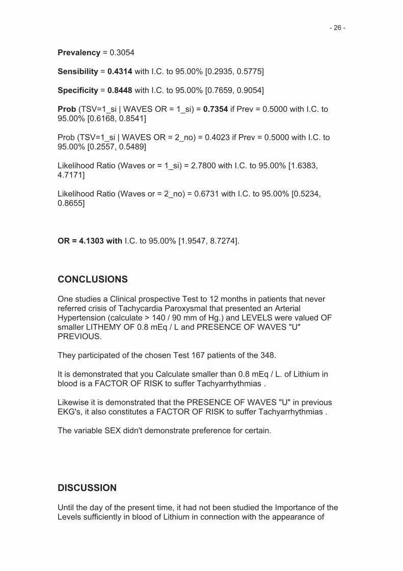
Variable Estado de la Naturaleza: TP
Variable Test: ONDAS U
Prevalencia = 0.3054
Sensibilidad = 0.4314 con I.C. al 95.00% [0.2935, 0.5775]
Especificidad = 0.8448 con I.C. al 95.00% [0.7659, 0.9054]
Prob (TSV=1_si | ONDAS U = 1_si) = 0.7354 si Prev = 0.5000 con I.C. al 95.00% [0.6168, 0.8541]
Prob (TSV=1_si | ONDAS U= 2_no) = 0.4023 si Prev = 0.5000 con I.C. al 95.00% [0.2557, 0.5489]

- 11 -
Likelihood Ratio (ONDAS U= 1_si) = 2.7800 con I.C. al 95.00% [1.6383, 4.7171]
Likelihood Ratio (ONDAS U= 2_no) = 0.6731 con I.C. al 95.00% [0.5234, 0.8655]
OR = 4.1303 con I.C. al 95.00% [1.9547, 8.7274].
CONCLUSIONES
Se estudio un Ensayo Clínico prospectivo a 12 meses en pacientes que nunca refirieron crisis de Taquicardia Paroxística, que presentaban una Hipertensión Arterial (cifras > 140/ 90 mm de Hg.) y se valoraron NIVELES DE LITEMIA MENORES DE 0.8 mEq/L y PRESENCIA DE ONDAS “U” PREVIAS.
Tomaron parte del Ensayo 167 pacientes de los 348 escogidos.
Se demuestra que Cifras menores de 0.8 mEq/ L. de Litio en sangre es un FACTOR DE RIESGO para sufrir Taquiarrítmias.
Así mismo se demuestra que la PRESENCIA DE ONDAS “U” en EKG´s previos, también constituye un FACTOR DE RIESGO para sufrir Taquiarrítmias.
La variable SEXO no demostró preferencia por uno determinado.
DISCUSIÓN
Hasta el día de la actualidad, no se había estudiado suficientemente la Importancia de los Niveles en sangre de Litio en relación con la aparición de Rítmos Rápidos (Taquicardias Paroxísticas, Aleteo Atrial y Ventricular, Fibrilación Atrial y Ventricular). Tampoco el papel que tenía la presencia de Ondas “U”.
A falta de estudios de Regresión Logística en Ensayos con Multivariantes que, publicaremos posteriormente, con este Ensayo basado en Epidemiología Básica, se demuestra que ambas variantes actúan de manera Significativa y con Fuerza Estadística suficiente como FACTORES DE RIESGO o PREDISPONENTES.

- 12 -
Los Autores del presente Ensayo, proponen como determinación rutinaria dentro del Perfil de Iones, la inclusión junto a Sodio, Potasio, Calcio, Cloro de ion LITIO.
ANEXOS GRÁFICOS
Serie 1
Serie 2
Serie 3
f(x)=15.514396*0.35270468^x; R²=1
f(x)=0.45085663*x-0.27817854; R²=1
f(x)=1.724*x+0.682; R²=1
0.5 1 1.5 2 2.5 3 3.5 4 4.5 5 5.5
1
2
3
4
5
x
y
(1,5.472)
(2,1.930)
(2.835,1)
(5.053,2)
(1,2.406)
(2,4.130)RR Y OR (ONDA U/TP)
PORCENTAJE LITIO-TAQUICARDIAOR y RR (LITIO/TP)

- 13 -
LITIO Y TSVP
54,72 19,3
0
10
20
30
40
50
60
+/+ +/-
PO
RC
EN
TA
JE
S
POSIT.
NEGAT.
ASOCIACIÓN ESTADÍSTICA
2,83
5,05
0
1
2
3
4
5
6
VA
LO
RE
S
RR
OR
ÍNDICES DIAGNÓSTICOS
30,43
56,86
79,31
0
20
40
60
80
100
PO
RC
EN
TA
JE
S
SENSIB.
PREVA.
ESPECIF.

- 14 -
BIBLIOGRAFÍA
[1]/Eutimia.psicofarmacos.litio.
[2]Toivonen LMore light on QT interval measurement. Heart 2002;87:193-194. Noviembre 2001.
[3] Revista Electrónica de Medicina Intensiva. Imagen nº 14. Vol. 4 nº 8, agosto 2004.
[4] IMPORTANCIA DELEKG EN TOXICOLOGÍA.Lina Maria Peña Acevedo. Residente III de Toxicología Clínica .Departamento de farmacología y Toxicología .Facultad de Medicina. Universidad de Antioquia.
1. Perales N Manual de RCP avanzada. Plan Nacional de RCP. Madrid: Arán Ediciones, 1989. 2. Chamberlain D Peri-arrest arrhythmias. Br J Anaesth 1997; 79: 198-202. 3. Kern KB, Paraskos JA Task Force 1: cardiac arrest. JACC 2000; 35: 832-846. 4. Kloeck W, Cummins RO, Chamberlain D, Bossaert L, Callanan V, Crali P et al The universal advanced life support algorithm. An advisory statement from the advanced life support working group of the International Liaison Committee on Resuscitation. Circulation 1997; 95: 2180-2182. 5. Zipes DP Specific arrhythmias: Diagnosis and treatment. En: Braunwald E, editores. Heart disease. A textbook of cardiovascular medicine. Filadelfia: WB Saunders, 1992; 667-725. 6. American Heart Association Arritmias. En: American Heart Association, editores. Reanimación cardiopulmonar avanzada. Barcelona: Medical Trends, 1996; 3.1-3.24. 7. Ruano M, Tormo C, Cuñat J Arritmias. En: Comité Español de RCP, editores. Manual y juego de diapositivas para el instructor de Soporte Vital Avanzado. Barcelona: Masson, S.A., 1996; 69-80. 8. Wlaker MJA, Curtis MJ, Hearse DJ, Campbell RWF, Janse MJ, Yellon DM et al The Lambeth Conventions: guidelines for the study of arrhythmias in ischaemia, infarction, and reperfusion. Cardiovasc Res 1988, 22: 447-455.9. Cummins R, Chamberlain DA, Abramson NS, Allen M, Baskett PJ, Becker L et al Recomended guidelines for uniform reporting of data from out-of-hospital cardiac arrest: The Utstein Style. Circulation 1991; 84: 960-975. 10. Myerburg RJ, Kessler KM, Castellanos A Recognition, clinical assessment, and management of arrhythmias and conduction disturbances. En: Schlant RC, Alexander RW, editores. The Heart. Nueva York: McGraw-Hill, 1994; 705-758. 11. Benchimol A, Desser KB, Schumacher J Blood flow velocity during ventricular fibrillation in man measured with the Doppler flowmeter technique. Chest 1971; 60: 265-267. 12. Capella G, Brugada P, Wellens HJJ Ventriculoatrial conduction and atrial activation during ventricular flutter and fibrillation. Am J Cardiol 1988; 61: 916-918. 13. Tchervenkov CI, Wynands EJ, Symes JF, Malcolm ID, Dobell AR, Morin JE Persistent atrial activity during cardioplegic arrest: a possible factor in the etiology of postoperative su-14. Clayton RH, Murray A, Higham PD, Campbell RW Selft-terminating ventricular tachyarrhythmias-a diagnostic dilemma? Lancet 1993; 341: 93-95. 15. Robertson C, Steen P, Adgey J, Bossaert L, Carli P, Chamberlain D et al The 1998 European Resuscitation Council guidelines for adult advanced life support. Resuscitation 1998; 37: 81-90. 16. Coma-Canella I, García-Castillo Riesgo L, Ruano Marco M, Loma-Osorio Montes A, Malpartida de Torres F, Rodríguez García JE Guía de actuación clínica de la Sociedad Española de Cardiología en resucitación cardiopulmonar. Rev. Esp Cardiol 1999; 52: 589-603. 17. Ruano Marco M, Tormo Caladín C, Cuñat de la Hoz J Arritmias. En: Consejo Español de RCP, editores. Manual de soporte vital avanzado. Barcelona: Masson, S.A., 1999; 95-116. 18. SEMES-1999 Recomendaciones en reanimación cardiopulmonar avanzada. Madrid: Edicomplet, 1999. 19. Patt MV, Podrid PJ, Friedman PL, Lown B Spontaneous reversion of ventricular fibrillation. Am Heart J 1988: 115: 919-923. 20. Denes P, Gabster A, Huang SK Clinical, electrocardiographic and follow-up observations in patients having ventricular fibrillation during Holter monitoring. Am J Cardiol 1981; 48: 9. 21. Cafri C, Ilia R, Battler A Selft-terminating ventricular fibrillation during variant angina. Angiology 1998: 49: 581-584. 22. Tye KH, Desser KB, Benchimol A Survival following spontaneous ventricular flutter-fibrillation associated with QT syndrome. Arch Intern Med 1980; 140: 255. 23. Dubner SJ, Gimeno GM, Elencwajg B, Leguizamon J, Tromgé JE, Quinteiro R Ventricular fibrillation with spontaneous reversion on ambulatory ECG in the absence of heart disease. Am Heart J 1983; 105: 691. 24. Moskowitz RM, Schwartz AB Spontaneous termination of prolonged ventricular fibrillation after acute myocardial infarction. Arch Intern Med1987: 147: 171-172. 25. Ring ME, Huang SK Spontaneous termination from prolonged ventricular fibrillation. Am Heart J 1987: 113: 1226-1228. 26. Kontny F, Dale J Self-terminating idiopathic ventricular fibrillation presenting as syncope: a 40-year follow-up report. J Intern Med 1990; 227: 211-213. 27. Van Hemel NM, Kingma JH A patient in whom self-terminating ventricular fibrillation was a manifestation of myocardial reperfusion. Br Heart J 1993: 69: 568-571. 28. Myerburg RJ, Kessler KM, Mallon SM, Cox MM, DeMarchena E, Interian A et al Life-threatening arrhythmias in patients with silent myocardial ischemia due to coronary artery spasm. N Engl J Med 1992; 326: 1451-1455. 29. Schwarz LS, Goldfischer J, Sprague GJ, Schwartz SP Syncope and sudden death in aortic stenosis. Am J Cardiol 1969; 23: 647-658. 30. Fidler GI, Campbell RWF, Pottage A, Godman MJ Varicella myocarditis presenting with unusual ventricular arrhythmias. Br Heart J 1977; 39: 1150-1153. 31. Selzer A, Wray HW Quinidine syncope. Circulation 1964; 30: 17-26. 32. Frieden J Quinidine effects due to disopyramide. N Engl J Med 1978; 298: 975.

- 15 -
33. Monoach M Factors influencing maintenance and spontaneous termination of ventricular fibrillation. Int Cardiol 1984; 5: 398-402. 34. Fabiato A, Coumel P Torsades de pointes, un cuarto de siglo más tarde: un tributo al doctor F. Dessertenne. Cardiovasc Drug Ther (ed. esp.) 1992; 2: 137-157. 35. Kloeck W, Cummins RO, Chamberlain D, Bossaert L, Callanan V, Crali P et al Special resuscitation situations. An advisory statement from the International Liaison Committee on Resuscitation. Circulation 1997; 95: 2196-2210. 36. Viskin S Long QT syndromes and torsade de pointes. Lancet 1999; 354: 1625-1633. 37. Ahmed R, Sager PT Evaluación y tratamiento de la torsade de pointes inducida por fármacos. Cardiovasc Drug Ther (ed. esp.) 1994; 15: 301-306. 38. Faber TS, Zehender M, Van de Loo A, Hohnloser S, Just H Torsade de pointes complicating drug treatment of low-malignant forms of arrhythmia: four cases reports. Clin Cardiol 1994; 17: 197-202. 39. Bayés de Luna A, Coumel P, Leclercq JF Ambulatory sudden cardiac death: mechanisms of production of fatal arrhythmia on the basis of data from 157 cases. Am Heart J 1989; 117: 151-159
© DR. BREIJO. 2.007.

- 16 -
RELATION AMONG LEVELS DE LITEMIA PRESENCE OF WAVES "u" AND TACHYCARDIA
PAROXYSMAL.
Prof. BREIJO MÁRQUEZ. FR. Md.PhD. BERNÁ. A. SERRANO. PAGÁN A.
Hospital´s Morales Messenger. Center of Urgencies Abanilla. Murcia. Spain.
Correspondence: Prof. BREIJO MÁRQUEZ.MD.PhD
PZA MATEO VÍLLORA 1-3. 02001. SPAIN.
TFNO: +34.967247867.
E-MAIL: [email protected].
The present Study doesn't present any conflict of Interests at the present time.
KEY WORDS: Tachycardia Parosysmal. LITHEMY. Waves U.
ABSTRACT:
The decrease of the sanguine levels of Lithium in blood, they can alter the answer of permeability of membrane miocitic and to allow bigger effect of the habitual neurotransmitters of the heart, provoking a hyperstimulation answer in the depolarization miocardic with the rising appearance of phenomena of Rhythms Quick type Tachycardia Paroxysmal.
HYPOTHESIS.

- 17 -
It is tried to demonstrate the existent association of possible factors of risk with the appearance of episodes of Quick Heart Rhythm of the type Tachycardia Paroxysmal. (Levels of Lithium in blood below 0.8 mEq / Liter. Presence of Waves "U" in having traced previous EKG's) in a Mature Population of Risk andwith Arterial Hypertension maintained that - initially - has not suffered any access of Tachycardia Paroxysmal.
He was practiced a Prospective Study to 12 months.
Of each of the patients diverse variables were picked up at the beginning of the Study: Levels of LITHEMY.
Presence of Waves "U ".electrocardiographics .
Family Antecedents.
Sex. Age.
CHARACTERISTIC OF LITHIUM
Name Lithium
Atomic number 3
Valencia 1
State of oxidation +1
Electronegative 1,0
radiate covalente (Å) 1,34
radiate ionic (Å) 0,60
radiate atomic (Å) 1,55
Electronic configuration 1s22s1
First ionization (eV) potential 5,41
Atomic (g / mol) mass 6,941
Density (g / ml) 0,53
Point of boil (ºC) 1330
Coalition (ºC) point 180,5
Discoverer George Urbain in 1907

- 18 -
Lithium heads the family of the alkaline metals in the periodic chart. In the nature it is like a mixture of the isotopes Li6 and Li7. The main compound of the lithium is the lithium hydroxide. In the Periodic Chart it is next to the Sodium (Na) and Potassium (K). Key well-known ions in the cellular polarization.
FDA approved it in 1970 as fármaco for the treatment of the maniac-depressive dysfunctions.
Normal Concentrations of Lithium in Blood: 0.8 to 1.2 mEq / L
.
The lithium ions are absorbed completely of the gastrointestinal tract and concentrations in serum picks are reached after 3- 5 hours of their administration. Its union to the proteins serum is minimum and its distribution volume is of 0.9L /Kg. however, their distribution inside the compartment tisular and outside of him it can take up to 25-30 hours with therapeutic dose of the preparations of sustained liberation and stiller when an overdose of the fármaco is ingested. The litio is not metabolized but rather it is eliminated practically entirely for renal route. The half initial life is from 6 to 12 hours; of there from now on, as the compartment tisular liberates the fármaco in him stored, the half life can be prolonged at 24 hours or more. The túbulos proximal and distal reabsorb around 80% of the filtered litio and the reabsorption increases in notorious form in the hiponatremia states.
The clinical main effects correspond to SNC, the kidneys and the heart. [1]
Their cardiovascular effects associate to changes in the potassium intracelular, with interactions with the metabolism of the magnesium and of the calcium. Reversible changes have been informed in the function of the nodule sinusal, taquicardia sinusal, bradicardia, aurículo-ventricular blockades, ventricular extrasístoles, ventricular fibrillation, etc.In patient with cardiovascular pathology is to carry out more frequent monitoreos and to remember their biggest intoxication risk for dysfunctions hidroelectrolíticos.
In superior concentrations to 1.6 mEq / L. s can observe hypotension, arrhythmias and alterations consistent electrocardiográficas in leveling of the wave T, continuation of QT and waves U. (Well-known until the present time)
Lithium can cause changes in EKG associated with the repolarización and, with smaller frequency, to conduction dysfunctions, worsenings of arrhythmias or new arrhythmias.

- 19 -
LITHIUM blocks the development of hypersensitivity of the receiving dopaminérgycs, adrenérgics or colinérgics. When blocking the development of hypersensitivity of the receiving dopaminérgics, adrenérgics and colinérgics, their deficit, the sensibility of the receivers increases from the cellular membrane to this components affecting to its density to the neurotransmitters:
COLINERGIC SYSTEM: According to some authors, an increment exists in the concentration, in the synthesis and in the turnover of the acetilcolin in the brain.CATECOLAMINERGIC SYSTEM: It diminishes the concentration, storage and dopamina liberation and noradrenaline in SNC.SEROTONINERGIC SYSTEM: Initially an increment of the reception of the triptófano takes place in the sinapsis. Then, the serotonin synthesis increases. Some authors suggest that a relation exists between the effect antiagresiv of the lithium and the increased levels of serotonin, evidenced by an increase of the levels of 5-HIAA (Acid 5-hidroxi-indolacetico) in LCR.
In this sense, it is known that Lithium is able to influence on a series of physiologic processes and many of these effects have been tried to relate with its therapeutic or toxic action. It exists the hypothesis that the lithium modifies the absolute levels of the Protein G (PG) that has an important paper in the modulation of signs receptors among the neurotransmitters
The direct mechanism for which the litio would act stabilizing PG in its inactive form is even unknown; however, it is believed that it modulates, in way alostéryc, the neurotransmission roads that are altered, respecting those that usually work.
The Half Life of elimination of Lithium is understood between 14 and 30 hours. For what reaches the stationary state around the five days that it is when they are carried out the sanguine necessary tests to verify the sanguine levels of the same.
OBJECTIVE OF CLINICAL TEST. . [2] [3] [4]
By means of the present Test, it is wanted to analyze the association of each factor risk with the appearance of Taquicardias Paroxísticas (TP) in a population of risk of Hipertensos (controlled or not) adults.
For he was carried out it a Prospective Study of 12 months in 167 patients, initially without appearance of crisis of previous TP.
Of each patient diverse variables were picked up as age, sex, family antecedents, he witnesses or not of Waves "U" in EKG and levels of LITHEMY at the beginning of the study.

- 20 -
It is wanted to analyze the Association of each Factor of Risk with Tachycardias Paroxysmal and to establish their statistical (RR and OR) Significance independently of the other factors.
The variables used in this study were dicotómycs, except the Age.
DEFINITION OF USED VARIABLES.
ID.- Identification of patient.
TP: 1: If. 2: No.
LITHEMY: 1: Less Than 0.8 mEq / Liter. 2: In normal ranges.
PRESENCE OF WAVE "U." 1: If. 2: No.
FAMILY ANTECEDENTS: 1: If. 2: No.
AGE:
SEX: 1: Man. 2: Woman.
Basic technical bivariants was used in Epidemiology without keeping in mind possible interaction effects among variables, neither they were kept in mind the possible effects that it has more than enough the answer they can have the variables of confusion.
In later Tests, if we use Technical Multivariantes of Logistical Regression.
MATERIAL AND METHODS.
Ramdomized. Prospective to 1 year. Half-close Hospital.
Total Universe: 348 patients.
Selected Universe: 167 patients.
Control Analytic every 2 months including LITHEMY in all them.
Control Electrocardiográphics every 2 months.
APPROACHES OF INCLUSION:
Arterial hypertension previous (> 140 / 90 mm of Hg.)

- 21 -
Not to have referred any sign / symptom of Tachycardia Paroxysmal to the beginning of the Study.
Verified grade of adhesion of 4-5 / 5.
APPROACHES OF EXCLUSION:
Not to complete some inclusion approach.
METHODS
Absolute and Relative Frequencies of the implied Factors and the appearance of Tachycardia Paroxysmal was obtained during 12 months.
It was calculated the Relative Risk and Odss Ratio. As measures of Force of Statistical Association.
It was considered Unchaining Factor with RR and OR > 1.
It was considered Protective Factor with RR and OR < 1.
(For variable dicotómics).
With the obtaining of the Interval of Trust we could meet the precision of this measures.
The Significance of the Measures of Effect was carried out by means of the calculation of Chi-square.
RESULTS
ABSOLUTE AND RELATIVE FREQUENCIES OF FACTORS.
TP Frequencies Percentages1_si 51 30.54 2_no 116 69.46Total 167 100.00LITHIUM 1_si 53 31.74 2_no 114 68.26Total 167 100.001_hombre 91 54.492_mujer 76 45.51Total 167 100.00WAVE"U",2_no 92 55.091_si 75 44.91Total 167 100.00.

- 22 -
STUDY OF THE FACTOR RISK LITHIUM
CHART OF FREQUENCIES DE TSV (LINES) FOR LITHIUM (COLUMNS)
Number of Cases: 167
LEVELS DE LITHEMI < 0.8 mEq / L
TSVP. IF NO TOTAL
N % N % N %
IF 29 54.72 22 19.30 51 30.54
NO 24 45.28 92 80.70 116 69.46
TOTAL 53 100 114 68.26 167 100
* The occurrence of Tachycardia Paroxysmal in patient with diminished LITHEMIA (< 0.8 mEq / L.) it was of 54.72% versus to 19.30% of patient with LITHEMIA in ranges.
MEASURES OF ASSOCIATION IN EPIDEMIC STUDIES
Variable Resolution: TP
Explanatory Variable: LITHIUM
Number of Cases: 167
Assuming Explanatory in Columns and Answer in Lines with:
Localization RR EE [LnRR] IC95.00% inf. IC95.00% sup.
(+,+) In the cell 1 2.8353 0.2287 1.8111 4.4388
(+,+) In the cell 2 0.3527 0.2287 0.2253 0.5521

- 23 -
(+,+) In the cell 3 0.5611 0.1578 0.4119 0.7645
(+,+) In the cell 4 1.7822 0.1578 1.3081 2.4280
Assuming Explanatory in Columns and Answer in Lines with:
Localization OR EE [LnOR] IC95.00% inf. IC95.00% sup.
(+,+) In the cell 1 5.0530 0.3640 2.4759 10.3125
(+,+) In the cell 2 0.1979 0.3640 0.0970 0.4039
(+,+) In the cell 3 0.1979 0.3640 0.0970 0.4039
(+,+) In the cell 4 5.0530 0.3640 2.4759 10.3125.
The positive (+) factor next to positive (+) resolution is in cell 1.
SIGNIFICANCE OF MEASURES OF EFFECT
CHI-square DE TSV (LINES) FOR LITHIUM (COLUMNS)
Size Muestral: 167
Statistical of Contrast Chi-square: 21.3961
G.L.: 1
P-value: < 0.0001
Nº of cells with absolute prospective frequencies < 5: 0 of 4, a 0.0000%
Nº of cells with absolute prospective frequencies < 1: 0 of 4, 0.0000%.
RESULTS: Relative (RR) Risk 2.83 with An I.C. of 95% and an Odds Ratio (OR) 5.053. To a value of p < 0.0001, highly significant for this factor, NULL HYPOTHESIS is REJECTED for the same.
An Association of Force ENOUGH Statistic is reached to deduce that you value first floor of Lithium in blood they accompany of occurrence of Tachycardias Paroxysmal.

- 24 -
STUDY OF THE FACTOR OF RISK "WAVES U"
Chart of Frequencies of TP (lines) for WAVES U (columns)
Number of Cases: 167
WAVES U 1_si 2_no Total TP Line
22 29 51 1_si 55.00 22.83 30.54
18 98 11645.00 77.17 69.46
2_ not
40 127 167 Column 23.95 76.05 100.00
The occurrence in 12 months of TP in patient with Wave "U" previous in EKG it was of 55.0% versus to 22.83% in patient without Wave "U" previous.
RR was of 2.40 and OR 4.13 with an I.C.95%.
Chi-square of TP (lines) for WAVES U (columns)
Size Muestral: 167
Statistical of contrast Chi-square: 14.8364
G.L.: 1
P-value: < 0.0001

- 25 -
Nº of cells with absolute prospective frequencies < 5: 0 of 4, a 0.0000%
Nº of cells with absolute prospective frequencies < 1: 0 of 4, 0.0000%.
NULL HYPOTHESIS is also rejected to value-p of < 0.0001. The previous presence of Waves "U" in EKG's of the patients it is an UNCHAINING DEMONSTRATED FACTOR.
DIAGNOSTIC INDEXES
Variable State of the Nature: TP
Variable Test: LITHIUM
Prevalency = 0.3054
Sensibility = 0.5686 with I.C. to 95.00% [0.4225, 0.7065]
Specificity = 0.7931 with I.C. to 95.00% [0.7080, 0.8627]
Proba (TSV=1_si | LITIO=1_si) = 0.7332 if Prev = 0.5000 with I.C. to 95.00% [0.6304, 0.8360]
Proba (TSV=1_si | LITIO = 2_no) = 0.3523 if Prev = 0.5000 with I.C. to 95.00% [0.2324, 0.4722]
Likelihood Ratio (LITIO=1_si) = 2.7484 with I.C. to 95.00% [1.7895, 4.2209]
Likelihood Ratio (LITIO = 2_no) = 0.5439 with I.C. to 95.00% [0.3916, 0.7554]
OR = 5.0530 with I.C. to 95.00% [2.4759, 10.3125].
Variable State of the Nature: TP
Variable Test: WAVES U
- 26 -
Prevalency = 0.3054
Sensibility = 0.4314 with I.C. to 95.00% [0.2935, 0.5775]
Specificity = 0.8448 with I.C. to 95.00% [0.7659, 0.9054]
Prob (TSV=1_si | WAVES OR = 1_si) = 0.7354 if Prev = 0.5000 with I.C. to 95.00% [0.6168, 0.8541]
Prob (TSV=1_si | WAVES OR = 2_no) = 0.4023 if Prev = 0.5000 with I.C. to 95.00% [0.2557, 0.5489]
Likelihood Ratio (Waves or = 1_si) = 2.7800 with I.C. to 95.00% [1.6383, 4.7171]
Likelihood Ratio (Waves or = 2_no) = 0.6731 with I.C. to 95.00% [0.5234, 0.8655]
OR = 4.1303 with I.C. to 95.00% [1.9547, 8.7274].
CONCLUSIONS
One studies a Clinical prospective Test to 12 months in patients that never referred crisis of Tachycardia Paroxysmal that presented an Arterial Hypertension (calculate > 140 / 90 mm of Hg.) and LEVELS were valued OF smaller LITHEMY OF 0.8 mEq / L and PRESENCE OF WAVES "U" PREVIOUS.
They participated of the chosen Test 167 patients of the 348.
It is demonstrated that you Calculate smaller than 0.8 mEq / L. of Lithium in blood is a FACTOR OF RISK to suffer Tachyarrhythmias .
Likewise it is demonstrated that the PRESENCE OF WAVES "U" in previous EKG's, it also constitutes a FACTOR OF RISK to suffer Tachyarrhythmias .
The variable SEX didn't demonstrate preference for certain.
DISCUSSION
Until the day of the present time, it had not been studied the Importance of the Levels sufficiently in blood of Lithium in connection with the appearance of

- 27 -
Rhythms Rapids (Tachycardias Paroxysmal, Flapping Atrial and Ventricular, Fibrillation Atrial and Ventricular). Neither the paper that had the presence of Waves "U."
For lack of studies of Logistical Regression in Tests with Multivariantes that, we will publish later on, with this Test based on Basic Epidemiology, it is demonstrated that both variants behave in a Significant way and with Force enough Statistic as FACTORS OF RISK or UNDERLYING.
The Authors of the present Test, propose as routine determination inside the Profile of Ions, the inclusion next to Sodium, Potassium, Calcium, ion Chlorine LITHIUM.
GRAPHIC ANNEXES
Serie 1
Serie 2
Serie 3
f(x)=15.514396*0.35270468^x; R²=1
f(x)=0.45085663*x-0.27817854; R²=1
f(x)=1.724*x+0.682; R²=1
0.5 1 1.5 2 2.5 3 3.5 4 4.5 5 5.5
1
2
3
4
5
x
y
(1,5.472)
(2,1.930)
(2.835,1)
(5.053,2)
(1,2.406)
(2,4.130)RR Y OR (ONDA U/TP)
PORCENTAJE LITIO-TAQUICARDIAOR y RR (LITIO/TP)

- 28 -
LITIO Y TSVP
54,72 19,3
0
10
20
30
40
50
60
+/+ +/-
PO
RC
EN
TA
JES
POSIT.
NEGAT.
ASOCIACIÓN ESTADÍSTICA
2,83
5,05
0
1
2
3
4
5
6
VA
LO
RE
S
RR
OR
ÍNDICES DIAGNÓSTICOS
30,43
56,86
79,31
0
20
40
60
80
100
PO
RC
EN
TA
JES
SENSIB.
PREVA.
ESPECIF.

- 29 -
BIBLIOGRAPHY
[1] /Eutimia.psicofarmacos.litio.
[2] Toivonen LMore light on QT interval measurement. Heart 2002;87:193-194. November 2001.
[3] it has Electronic of Intensive Medicine. Image nº 14. Vol. 4 nº 8, I wither 2004.
[4] IMPORTANCE DELEKG IN TOXICOLOGÍA.Lina María Peña Acevedo. Resident III of Clinical Toxicology pharmacology .Departamento and Toxicology .Facultad of Medicine. University of Antioquia.
1. Pear trees Manual N of advanced RCP. National Plan of RCP. Madrid: Arán Editions, 1989. 2. Chamberlain D Peri-arrest arrhythmias. Br J Anaesth 1997; 79: 198-202. 3. Kern KB, Paraskos JA Task Forced 1: cardiac arrest. JACC 2000; 35: 832-846. 4. Kloeck W, Cummins RO, Chamberlain D, Bossaert L, Callanan V, Crali P et to The universal advanced life support algorithm. An advisory statement from the advanced life support working group of the International Liaison Committee on Resuscitation. Circulation 1997; 95: 2180-2182. 5. Zipes DP Specific arrhythmias: Diagnosis and treatment. In: Braunwald AND, editors. Heart disease. To textbook of cardiovascular medicine. Philadelphia: WB Saunders, 1992; 667-725. 6. American Heart Association Arrhythmias. In: American Heart Association, editors. Cardiopulmonary advanced Reanimación. Barcelona: Medical Trends, 1996; 3.1-3.24. 7. Ruano M, Tormo C, Cuñat J Arrhythmias. In: Spanish committee of RCP, editors. Manual and game of slides for the instructor of Vital Advanced Support. Barcelona: Masson, CORP., 1996; 69-80. 8. Wlaker MJA, Curtis MJ, Hearse DJ, Campbell RWF, Janse MJ, Yellon DMK et to The Lambeth Conventions: guidelines for the study of arrhythmias in ischaemia, infarction, and reperfusion. Cardiovasc Head 1988, 22: 447-455. 9. Cummins R, Chamberlain GIVES, Abramson NS, Allen M, Baskett PJ, Becker L et to Recomended guidelines for uniform reporting of dates from out-of-hospital cardiac arrest: The Utstein Style. Circulation 1991; 84: 960-975.10. Myerburg RJ, Kessler KM, Castellano TO Recognition, clinical assessment, and management of arrhythmias and conduction disturbances. In: Schlant RC, Alexander RW, editors. The Heart. New York: McGraw-Hill, 1994; 705-758. 11. Benchimol TO, Desser KB, Schumacher J Blood flow velocity during ventricular fibrillation in man measured with the Doppler flowmeter technique. Chest 1971; 60: 265-267. 12. Capella G, Brugada P, Wellens HJJ Ventriculoatrial conduction and atrial activation during ventricular flutter and fibrillation. Am J Cardiol 1988; 61: 916-918. 13. Tchervenkov CI, Wynands EJ, Symes JF, Malcolm GOES, Dobell AR, Morin JE Persistent atrial activity during cardioplegic arrest: to possible factor in the etiology of postoperative its-14. Clayton RH, Murray TO, Higham PD, Campbell RW ventricular Selft-terminating tachyarrhythmias-to diagnostic dilemma? Lancet 1993; 341: 93-95. 15. Robertson C, Steen P, Adgey J, Bossaert L, Carli P, Chamberlain D et to The 1998 European Resuscitation Council guidelines for adult advanced life support. Resuscitation 1998; 37: 81-90. 16. Coma-Canella I, García-castle Risk L, Ruano Marco M, Hill-Osorio Mounts TO, Malpartida of Torres F, Rodríguez García JE Guide of clinical performance of the Spanish Society of Cardiology in cardiopulmonary resuscitation. Rev. Esp Cardiol 1999; 52: 589-603. 17. Ruano Marco M, Tormo Caladín C, Cuñat of the Sickle J Arrhythmias. In: Spanish Council of RCP, editors. Manual of vital advanced support. Barcelona: Masson, CORP., 1999; 95-116. 18. SEMES-1999 Recommendations in cardiopulmonary advanced reanimación. Madrid: Edicomplet, 1999. 19. Patt MV, Rot PJ, Friedman PL, Lown B Spontaneous reversion of ventricular fibrillation. Am Heart J 1988: 115: 919-923. 20. Denes P, Gabster TO, Huang SK Clinical, electrocardiographic and follow-up observations in patients having ventricular fibrillation during Holter monitoring. Am J Cardiol 1981; 48: 9. 21. Cafri C, Ilia R, Battler TO Selft-terminating ventricular fibrillation during variant angina. Angiology 1998: 49: 581-584. 22. Tye KH, Desser KB, Benchimol TO Survival following spontaneous ventricular flutter-fibrillation associated with QT syndrome. Arch Intern Med 1980; 140: 255. 23. Dubner SJ, Gimeno GM, Elencwajg B, Leguizamon J, Tromgé JE, Quinteiro R Ventricular fibrillation with spontaneous reversion on ambulatory ECG in the absence of heart disease. Am Heart J 1983; 105: 691. 24. Moskowitz RM, Schwartz AB Spontaneous termination of prolonged ventricular fibrillation after acute myocardial infarction. Arch Intern Med1987: 147: 171-172. 25. Ring ME, Huang SK Spontaneous termination from prolonged ventricular fibrillation. Am Heart J 1987: 113: 1226-1228. 26. Kontny F, Give him/her J Self-terminating idiopathic ventricular fibrillation presenting ace syncope: to 40-year follow-up report. J Intern Med 1990; 227: 211-213. 27. Van Hemel NM, Kingma JH TO patient in whom self-terminating ventricular fibrillation was to manifestation of myocardial reperfusion. Br Heart J 1993: 69: 568-571. 28. Myerburg RJ, Kessler KM, Mallon SM, Cox MM, DeMarchena AND, Interian TO et to Life-threatening

- 30 -
arrhythmias in patients with silent myocardial ischemia due to coronary artery spasm. N Engl J Med 1992; 326: 1451-1455. 29. Schwarz LS, Goldfischer J, Sprague GJ, Schwartz SP Syncope and sudden death in aortic stenosis. Am J Cardiol 1969; 23: 647-658. 30. Fidler GI, Campbell RWF, Pottage TO, Godman MJ Varicella myocarditis presenting with unusual ventricular arrhythmias. Br Heart J 1977; 39: 1150-1153. 31. Selzer TO, Wray HW Quinidine syncope. Circulation 1964; 30: 17-26. 32. Frieden J Quinidine effects due to disopyramide. N Engl J Med 1978; 298: 975. 33. Monoach M Factors influencing maintenance and spontaneous termination of ventricular fibrillation. Int Cardiol 1984; 5: 398-402. 34. Fabiato TO, Coumel P pointes Torsades, a century room later: a tribute to the doctor F. Dessertenne. Cardiovasc Drug Ther (ed. esp.) 1992; 2: 137-157. 35. Kloeck W, Cummins RO, Chamberlain D, Bossaert L, Callanan V, Crali P et to Special resuscitation situations. An advisory statement from the International Liaison Committee on Resuscitation. Circulation 1997; 95: 2196-2210. 36. Viskin S Long QT syndromes and pointes torsade. Lancet 1999; 354: 1625-1633. 37. Ahmed R, Sager PT Evaluation and treatment of the pointes torsade induced by fármacos. Cardiovasc Drug Ther (ed. esp.) 1994; 15: 301-306. 38. Faber TS, Zehender M, Van of I Laud TO, Hohnloser S, Just H Torsade of pointes complicating drug treatment of low-malignant forms of arrhythmia: four marries reports. Clin Cardiol 1994; 17: 197-202. 39. Bayés of Moon TO, Coumel P, Leclercq JF Ambulatory sudden cardiac death: mechanisms of production of fatal arrhythmia on the basis of dates from 157 you marry. Am Heart J 1989; 117: 151-159
© DR. BREIJO. 2.007.

- 31 -
.

- 32 -
.