mechanisms and genes controlling the signalling network for biotic
TRANSCRIPT
Mechanisms and genes controlling the signalling network
for biotic and abiotic stress defences
in Arabidopsis thaliana (L.) Heyhn.
Functional cross-talk between photo-produced reactive oxygen species,
photosynthesis and plant disease defence responses
張寂禎
Christine Chi-Chen Chang Stockholm University, Department of Botany
Doctoral thesis, 2005

To my father, Wen-Kui Chang 張文貴 and my mother, Yen-Hua Chen 陳炎華

1
PREFACE This thesis is based on the following published papers and manuscripts, which will be referred to in the text by their Roman numerals. I. Chang, C.C.-C., Ball, L., Fryer, M.J., Baker, N.R., Karpinski, S., and Mullineaux, P.M.
(2004). Induction of ASCORBATE PEROXIDASE 2 expression in wounded Arabidopsis leaves does not involve known wound-signalling pathways but is associated with changes in photosynthesis. Plant J. 38, 499-511.
II. Mateo, A., Muhlenbock, P., Rusterucci, C., Chang, C.C.-C., Miszalski, Z., Karpinska, B.,
Parker, J.E., Mullineaux, P.M., and Karpinski, S. (2004). LESION SIMULATING DISEASE 1 is required for acclimation to conditions that promote excess excitation energy. Plant Physiol. 136, 2818-2830.
III. Chang, C.C.-C., Slesak, I., Sotnikow, A., Mullineaux, P. M., Karpinski, S., and
Karpinska, B. (2005). Functional characterization of the chloroplastic glutathione peroxidases (cpGPXs) in Arabidopsis thaliana: its role in light acclimatory mechanisms. (manuscript).
IV. Chang, C.C.-C., Jorda, L., Slesak, I., Melzer, M., Moritz, T., Miszalski, Z., Mullineaux,
P. M., Parker, J., Karpinska, B., Karpinski, S. (2005). Functional analysis of chloroplastic glutathione peroxidases (cpGPXs) in Arabidopsis thaliana provides a direct link between photo-oxidative stress and basal pathogen resistance mechanisms. (manuscript).
Reprints and Figures from paper I and II are copyrighted by the Blackwell Publishers and American Society of Plant Biologists respectively and are reproduced with their kind permission. My contributions to the papers were: (I) Planning, performing major part of the experiments and writing. (II) Analysis of salicylic acid effects. (III) Planning, performing major part of the experiments and writing. (IV) Planning, performing major part of the experiments and writing.

2
ABSTRACT Excess excitation energy, mechanical injury and defence against pathogens, each
trigger rapid production of reactive oxygen species (ROS) in Arabidopsis thaliana leaves. ROS, such as hydrogen peroxide (H2O2), are required for the induction of systemic acquired acclimation and may lead to redox changes in photosynthetic electron transport (PET). On one hand, enhanced ROS production during stress can destroy cells, and on the other, ROS can also act as signals for the activation of stress responsive and defensive pathways.
In this work, physiological and molecular analyses of Arabidopsis mutants and transgenic lines were applied to investigate the signalling network controlling biotic and abiotic stress responses. A key enzyme of the antioxidant network is encoded by ASCORBATE PEROXIDASE 2 (APX2). Wounded leaves showed low induction of APX2 expression and when exposed to excess light, APX2 expression was increased synergistically. Signalling pathways dependent upon jasmonic acid, chitosan and abscisic acid were not involved in the wound-induced expression of APX2, but PET was required, and APX2 induction was preceded by a depressed rate of CO2 fixation.
Analysis of lsd1 (LESION SIMULATING DISEASE 1) strongly suggests that light acclimatory processes and pathogen defences are genetically and functionally linked. It is important to know that LSD1 type of mutants have mainly been studied with regard to pathogenesis. From this work, it reveals that association of LSD1 with hypersensitive response may only be supplementary.
GLUTATHIONE PEROXIDASES (GPXs) are another major family of ROS scavenging enzymes. Analysis of the Arabidopsis genome database revealed a new open-reading frame, thus increasing the total number of AtGPX gene family to eight (AtGPX1-AtGPX8). Arabidopsis thaliana transgenic lines with reduced expression of both putative chloroplastic isoforms (AtGPX1 and AtGPX7) and AtGPX7 knock-out mutant (ko-GPX7) were more sensitive to photo-oxidative stress but had a reduced bacterial growth rate when inoculated with virulent strains Pseudomonas syringae pv. tomato DC3000 and P.s.t. maculicola strain ES4326, indicating increased resistance to pathogenesis. This, to our knowledge, is the first functional and genetic analysis of chloroplastic GPXs in plants, and confirms that light and chloroplastic ROS metabolism is important for basal resistance against virulent pathogens.
The above results confirm that light sensing, light acclimatory processes and photo-produced ROS also govern pathogen defence pathways. This has a great ecological relevance for Darwinian fitness of plants growing in the natural environment, where simultaneous pathogen attack and fluctuations in light, temperature and other environmental factors make rapid acclimation a constant necessity. Molecular, biochemical and physiological analysis of pathogen responses in mutants impaired in light sensing, EEE-dissipatory mechanisms, and similar analysis of light acclimatory processes in mutants impaired in pathogen defences may prove to be seminal. Chang, C.C.-C. 2005. ISBN 91-7155-002-X, pp. 1-44. Mechanisms and genes controlling the signalling network for biotic and abiotic stress defences in Arabidopsis thaliana (L.) Heyhn. : Functional cross-talk between photo-produced reactive oxygen species, photosynthesis and plant disease defence responses. Department of Botany, Stockholm University, S-106 91 Stockholm, Sweden [email protected] © Christine Chi-Chen Chang 2005, Printed at PrintCenter, Stockholm University
3
ABBREVIATIONS ABA Abscissic acid ACC 1-aminocyclopropane-1-carboxylic acid ACO acyl coA oxidase gene APX Ascorbate peroxidase Avr pathogen avirulence proteins BTH benzothiadiazole CAT Catalase CHIB Basic chitinase CRY cryptochrome DHAR Dehydroascorbate reductase DPI Diphenylene iodonium Eds enhanced disease susceptibility EEE Excess excitation energy EL Excess light ET Ethylene GPX Glutathione peroxidase GSH Reduced glutathione H2O2 Hydrogen peroxide HR Hypersensitive response INA 2,6-dichloroisonicotinic acid ISR induced systemic resistance JA Jasmonic acid Jr1 jasmonate-responsive gene 1 Jr2 jasmonate-responsive gene 2 MDAR Monodehydroascorbate reductase MeJA methyl jasmonate NPQ Non-photochemical quenching OGA Oligosaccharide Pad Phytoalexin deficient PCD programmed cell death PET Photosynthetic electron transport PHOT phototropin PHY phytochrome PR1 Pathogenesis related protein 1 PSII Photosystem II qP Photochemical quenching R gene resistance gene rcd Runaway cell death ROS Reactive oxygen species SA Salicylic acid SAA Systemic acquired acclimation SAR Systemic acquired resistance SO*- Superoxide SOD Superoxide dismutase VSP vegetative storage protein WR wound-responsive genes Wr3 wound-responsive gene 3
4
TABLE OF CONTENTS Abstract……………………………………………………………................................. 2 Abbreviations………………………………………………………................................ 3 1. Introduction………………………………………………………............................. 5
1.1 Light perception …………………………………………………………………. 5 1.1.1 Photo-oxidative stress and excess excitation energy……………………... 6 1.1.2 Light acclimation responses…………………………………………......... 7 1.1.3 Reactive oxygen species protection responses………………………......... 7
1.2 Signalling network and cross-talk…………………………............................... 10 1.2.1 Wounding responses……………………………….................................... 11 1.2.2 Mechanisms of the plant ‘immune’ response…………………………….. 11 1.2.3 Excess excitation energy and defence against pathogens .……………….. 12
2. Aim of the present study……………………………………………………………. 13 3. Results and Discussion……………………………………………............................ 14
3.1 The role of chloroplasts derived redox signalling and reactive oxygen species in regulation of wounding responses…………....................................................
14
3.1.1 Synergistic effect of wounding and excess light on ASCORBATE PEROXIDASE 2 expression……………………………………..………...
14
3.1.2 Photosynthetic electron transport is required for wound-induced ASCORBATE PEROXIDASE 2 expression………………………...……...
16
3.1.3 Wound and excess light-induced ASCORBATE PEROXIDASE 2 expression is not controlled by known wound signalling pathways in Arabidopsis thaliana………………………………..……………………..
16 3.1.4 Sources of hydrogen peroxide for induction of ASCORBATE
PEROXIDASE 2 expression….……………………………………………
17 3.2 LESION SIMULATING DISEASE 1 controls acclimation to excess
light……………………………………………..............................................
19 3.2.1 Role of stomata and photorespiration in development of LESION
SIMULATING DISEASE 1 phenotype………….........................................
19 3.2.2 Attenuation of LESION SIMULATING DISEASE 1 phenotype in non-
photorespiratory conditions………………………………………………..
19 3.2.3 Salicylic acid, photo-oxidative stress and stomatal
conductance………………………………………………………………..
20 3.3 Functional characterization of the chloroplastic glutathione peroxidases
(cpGPXs) in Arabidopsis thaliana ……………………………………….........
22 3.3.1 Arabidopsis plants with reduced expression of chloroplastic
GPXs………………………………..……………………………………..
24 3.3.2 Enhanced photo-inhibition in chloroplastic glutathione peroxidases
deficient lines………………………………...............................................
24 3.3.3 Photo-produced reactive oxygen species and resistance against pathogen
infection…………………….......................................................................
26 4. Concluding remarks and future perspectives…………………………………….. 30 5. Material and Methods…………………………………………...……………......... 31 6. References…………………………………………………………............................ 33
5
1. INTRODUCTION “Leaves in the dark see the light”
(Foyer and Noctor, 1999) The sessile nature of plants implies that they must be able to adjust metabolic processes to constantly fluctuating light and other changing environmental factors. In full sunlight and optimal conditions, only a small fraction of absorbed light energy is used for CO2 fixation (Foyer and Halliwell, 1976; Foyer et al., 1994; Asada, 1999). The amount of absorbed light energy in excess of that needed by plants for photosynthetic metabolism is termed as EEE or excess excitation energy (Karpinski et al., 1997; Karpinski et al., 1999; Niyogi, 2000; Fryer et al., 2003). During full sunlight, EEE is an ever-present problem for plants and light acclimation processes have several major compensatory functions: first, to optimise the energy status of the cell (NADPH/ATP ratio) for its metabolic needs; second, to optimise the use of absorbed light for photosynthesis and reactive oxygen species (ROS) formation; and third, to use changes in the light spectra and light energy as a source of information about diurnal or seasonal shifts (circadian or seasonal check points). All these objectives can be pivotal for the Darwinian fitness of plants and other photosynthetic organisms, which cannot escape from stressful conditions (Kulheim et al., 2002).
The above strategy of plants’ acclimation processes necessitates accurate sensing and
simultaneous processing of the incoming environmental signals such as changes in light intensity and quality, fluctuations in temperature and relative humidity, photoperiod length, water availability, nutritional status, wounding, pathogen attack and herbivores, etc. All these changes must be simultaneously conveyed to the plant cell in order to generate optimal acclimatory and defence responses, which will allow successful propagation.
The reaction of a plant cell to a stimulus is assumed to be composed of at least three
elementary processes: signal perception, signal amplification and transduction, and primary and secondary responses. Signal perception includes physical and chemical interactions between the stimulus and a receptor. Subsequently, signal amplification and transduction of changes in the receptor generates specific effectors and downstream signalling cascades. The ultimate responses are specific changes in gene expression or enzyme activities and subsequent changes in the cellular metabolism. These interactions may be quite complex and are likely to involve both plant hormones such as jasmonic acid, ethylene, and abscisic acid (ABA), as well as other elicitors (Mullineaux and Karpinski, 2002). 1.1 Light perception The plant cell is equipped with sophisticated light-sensing mechanisms that are localized in the plasma-membrane, the cytosol, within chloroplasts and in the nucleus (Mullineaux et al., 2000). There are three families of photoreceptors that have been identified to date, phytochromes (PHY), cryptochromes (CRY), and phototropins (PHOT) (Clack et al., 1994; Briggs et al., 2001; Lin, 2002; Schäfer and Bowler, 2002). Only PHY are active in the long-wavelength region of the light spectrum: they absorb predominantly red-light and far-red-light, whereas CRY and PHOT monitor the blue-light spectra and the ultraviolet-A region
Introduction
6
respectively (Ahmad et al., 1998). Presumably CRY functions by mediating a light-dependent redox reaction (Yang et al., 2001). PHY mediates the light control of all major phases of plant development from seed germination, through seedling establishment (de-etiolation, leaf expansion and chloroplast maturation) and determination of the architecture of the mature plant, to the induction of flowering and dormancy (Schäfer and Bowler, 2002). PHOT controls three distinct types of light-induced movement responses that are thought to optimize photosynthesis: phototropism, chloroplast relocation and stomatal opening. In Arabidopsis thaliana, there are two CRY genes (Lin, 2002), two PHOT genes (Lin, 2002) and five PHY photoreceptor genes (Schäfer and Bowler, 2002). 1.1.1 Photo-oxidative stress and excess excitation energy Diurnal and seasonal fluctuations in light intensity, as well as environmental stresses such as cold, drought, salinity, and nutrient deficiency limit CO2 fixation and can result in the absorption of more light energy than can be utilized productively for photosynthetic metabolism (Karpinski et al., 1999; Mullineaux and Karpinski, 2002). Since acclimation responses that require alterations in gene expression are often too slow, plants utilize several “safety valves” to ensure that the harvested light energy does not inadvertently lead to ROS formation and cellular damage (Niyogi, 2000). Failure to dissipate EEE results in over-reduction of photosynthetic electron transport components and increased production of ROS in the chloroplast and peroxisomes. ROS include singlet oxygen(1O2), superoxide anion(O2
.-), hydrogen peroxide(H2O2), and hydroxyl radical(OH.). Hyper-accumulation of ROS leads to photo-inhibition, photo-oxidative damage and eventually death of the cell, manifested as bleaching or chlorosis of the photosynthetic tissues (Karpinski et al., 1999; Kasahara et al., 2002).
Dissipation of EEE in plants is achieved by a combination of non-photochemical and
photochemical quenching processes. Non-photochemical quenching processes include the transfer of excitation energy from antenna chlorophyll to zeaxanthin instead of the reaction centres. The extended NPQ is regulated by conformational changes in the PSII antenna and the reversible conversion of violaxanthin to zeaxanthin in the xanthophylls cycle. Both processes are dependent on the pH gradient across the thylakoid membrane. Additionally, carotenoids can scavenge 1O2 that emerges when excited chlorophyll converts to the triplet state and then induces a spin conversion in triplet O2 (Havaux and Niyogi, 1999; Niyogi, 1999; Niyogi, 2000; Muller et al., 2001).
Photochemical quenching of EEE is the collective term for processes that increase the
consumption of photosynthetic electrons by the deployment of additional metabolic sinks. These include the reduction of O2 at PSII or PSI with subsequent return of the electrons to water by antioxidant enzymes (water-water cycle) and an increased photosynthetic and photo-respiratory metabolism. Photorespiration starts from the oxygenase reaction of Rubisco and results in the energy consuming release of CO2 from fixed carbon (Kozaki and Takeba, 1996; Willekens et al., 1997; Wingler et al., 2000). Both the water-water cycle and photorespiration have a duplicated dissipation value, because they dissipate excess electrons and at the same time provide NADP+ as electron acceptor.

Introduction
7
1.1.2 Light acclimation responses Light acclimation processes are aimed to optimize light harvesting and CO2 fixation. Plants will dissipate EEE or minimize its formation by diminishing the capacity of the leaf to capture light energy (Walters and Horton, 1994; Niyogi, 2000; Bailey et al., 2001).
Among the acclimatory pathways, a short-term chromatic adaptation known as state-
1/ state-2 transition is required when the flux of electron excitation of PSII and PSI is unequal (Allen, 1995; Haldrup et al., 2001; Wollman, 2001; Allen, 2003). In oxygenic photosynthesis, two separate reactions centres named PSI and PSII, which have distinct light absorption properties, are coupled in sequence by a chain of electron carriers. PSI is enriched in chlorophyll a, absorbing in the far-red region (absorption maximum λ ≈ 700 nm) whereas PSII is enriched in chlorophyll b, with absorption maximum at shorter wavelengths in the orange/red region (λ ≈ 650 - 680 nm) (Wollman, 2001). Thus, spectral imbalances of the exciting light may result in an uneven excitation of the two photosystems leading to lower or higher ROS production (Karpinski et al., 1999; Karpinska et al., 2000; Wollman, 2001; Mullineaux and Karpinski, 2002; Fryer et al., 2003). During state transitions, a part of the PSII antenna is moved towards PS I or back to PS II to level out imbalanced excitation of the two photosystems.
Short-term processes are subsequently complemented by long-term light acclimation
processes if unbalanced excitation pressure persists (Karpinski et al., 2003). Common long term acclimation responses include increasing the capacity for photosynthetic electron transport and CO2 fixation, as well as decreasing the size of the light-harvesting antennae that are associated with photosystems (Noctor and Foyer, 1998; Pfannschmidt et al., 1999). This acclimation responses involve the regulation of gene expression (nuclear and chloroplastic) by redox potential, and/or proteolytic degradation of existing light harvesting complexes and other proteins (Allen, 1993; Durnford and Falkowski, 1997; Bailey et al., 2001; Pfannschmidt et al., 2001a; Pfannschmidt et al., 2001b; Pfannschmidt et al., 2003).
Although potentially damaging, ROS are also needed to trigger other protective
responses, such as the down-regulation of PSII activity, as well as to induce cellular and systemic signals that can promote redox changes in the proximity of PSII in unstressed chloroplasts, thus inducing protective mechanisms in remote chloroplasts (Karpinski et al., 1999; Mullineaux et al., 2000; Kovalchuk et al., 2003). 1.1.3 ROS protection responses In addition to light-dependent regulatory systems used to avoid the formation of ROS, plants are equipped with enzymatic (mainly water soluble) and non-enzymatic (water and lipid soluble) ROS scavenging systems (Foyer and Halliwell, 1976; Foyer et al., 1994; Jimenez et al., 1997; Mittler, 2002; Mittler et al., 2004). Enzymatic antioxidant systems consist of superoxide dismutase (SOD, EC : 1.15.1.1) ascorbate peroxidase (APX, EC : 1.11.1.11), catalase (CAT, EC : 1.11.1.6), monodehydro- and dehydro-ascorbate reductase (MDAR, EC : 1.6.5.4 and DHAR, EC : 1.8.5.1) glutathione reductase (GR, EC : 1.6.4.2) and glutathione

Introduction
8
peroxidase (GPX, EC : 1.11.1.9). Detailed information on the isoforms encoded in the Arabidopsis genome is listed in Table 1. Table 1. Summary of ROS scavenging enzymes and corresponding gene family in the Arabidopsis genome Name Reaction Gene family in
Arabidopsis Accession number
Localization
References
FSD1 At4g25100 chl (Camp et al., 1990) FSD2 At5g51100 chl (Kliebenstein et al.,
1998) FSD3 At5g23310 chl (Kliebenstein et al.,
1998) CSD1 At1g08830 cyt (Hindges and
Slusarenko, 1992) CSD2 At2g28190 chl (Kliebenstein et al.,
1998) CSD3 At5g18100 per (Kliebenstein et al.,
1998) MSD1 At3g10920 mit (Kliebenstein et al.,
1998)
SOD
2O2
-+2H+→H2O2+O2
MSD-like At3g56350 - - APX1 At1g07890 cyt (Kubo et al., 1993a) APX2 At3g09640 cyt (Kubo et al., 1992) APX3 At4g35000 per (Zhang et al., 1997) APX4 At4g09010 chl (Schubert et al., 2002) APX5 At4g35970 cyt - APX6 At4g32320 chl - thylakoidAPX At1g77490 chl (Shigeoka et al., 2002)
APX
H2O2+2Asc→2H2O+2MDA
stromal APX At4g08390 chl (Shigeoka et al., 2002) MDAR At1g63940 chl/cyt - MDAR At3g09940 cyt - MDAR At3g27820 cyt/mit - MDAR At3g52880 cyt -
MDAR
NADH + H+ + 2 MDA → NAD+ + 2Asc
MDAR At5g03630 cyt - DHAR At5g16710 chl/cyt - DHAR At5g36270 cyt - DHAR At1g75270 cyt - DHAR At1g19550 cyt -
DHAR
DHA+ 2GSH→Asc+GSSG
DHAR At1g19570 cyt - GR At3g24170 cyt -
GR 2GSH+NADP+→glutathione disulfide + NADPH + H+ GR At3g54660 chl (Kubo et al., 1993b)
CAT 1 At1g20630 per (Frugoli et al., 1997) CAT 2 At4g35090 per (Chevalier et al., 1992)
CAT
2H2O2→2H2O+O2
CAT 3 At1g20620 per (Frugoli et al., 1997) Localization is based on sequence analysis and computational predictions unless stated elsewhere in the cited reference. “-“ means there is no specific citation at the moment, however one can find the locus information in Arabidopsis complete genome sequence analysis from “The Arabidopsis Information Resource (TAIR)”, www.arabidopsis.org/aboutarabidopsis.html, on www.arabidopsis.org, November 23, 2004 (Rhee et al., 2003). SOD-superoxide dismutase, FSD-Fe SOD, CSD-Cu/Zn SOD, MSD-Mn-SOD, APX-ascorbate peroxidase, MDAR, monodehydroascorbate redutase, DHAR, dehydroascorbate reductase, GR, glutathione reductase, CAT-catalase, chl-chloroplast, cyt-cytosol, per-peroxisome, mit-mitochondria.

Introduction
9
Non-enzymatic antioxidants, such as ascorbate (vitamin C), glutathione, α-tocopherol (vitamin E), pigments and phenolic components, prevent or interrupt cascades of uncontrolled oxidation of cellular compounds (Asada, 1994; Noctor and Foyer, 1998; Bailly, 2004).
GPXs catalyse the reduction of H2O2 or different kinds of lipid peroxides by using reduced glutathione (GSH) as an electron donor (Ursini et al., 1995; Eshdat et al., 1997). Similar to APXs, GPXs comprise of eight isozymes (seven were previously found and one was newly identified by our present analysis) that protect cells against oxidative damage
generated by ROS. Several groups have reported the presence of GPXs in plants, but the specific role of each GPX isoform in plants has not been well documented. The putative subcellular localizations of the encoded GPX proteins are the cytosol, chloroplast,
Figure 1. The scavenging of active oxygen species in the chloroplast in both the lipid membrane phase and the stroma, linked to redox cycles for ascorbate and glutathione and the oxidation of α-tocopherol (Vitamin E). Abbreviations are as follows: P-LIPID -OO, phospholipid peroxy radical; P-LIPID-OOH, phospholipid peroxide; P-LIPID-OH, phospholipid alcohol; VIT-E (OH), α-tocopherol (vitamin E); VIT-E (O*), α-chromanoxyl radical; PHGPX, phospholipid hydroperoxide-dependent glutathione peroxidase; GSH, reduced glutathione; GSSG, glutathione disulfide; GR, glutathione reductase; DHAR, dehydroascorbate reductase; ASC, ascorbic acid; DHA, dehydroascorbate; MDA, monodehydroascorbate free radical; MDAR, monodehydroascorbate free radical reductase; APX, ascorbate reductase; SOD, superoxide dismutase; O2
.-, superoxide ion. Reaction 1 is the non-enzyme-catalyzed spontaneous dismutation of two MDA molecules to one ASC and one DHA, respectively. From (Mullineaux et al., 2000).

Introduction
10
mitochondria, and endoplasmic reticulum (Milla et al., 2003). There are a plethora of antioxidants, all of which are worthy of individual study. However, it is likely that it is the network of reactions that is important in vivo and which provides comprehensive control of ROS in cells, rather than the action of any single reaction. The first antioxidant network to be proposed for plants was the ascorbate-glutathione cycle of the chloroplast stroma (Figure 1) (Foyer and Halliwell, 1976; Foyer and Mullineaux, 1994; Asada, 1999). While the individual enzymes described in the ascorbate-glutathione cycle still receive a considerable amount of attention (eg. SOD, APX), it is now apparent that many more complex schemes can be created that involve a wider range of enzymes and molecules. These networks now include the movement of antioxidant compounds by specific transporters between and within cells, as well as the use of membrane-spanning redox couples (Jimenez et al., 1997; Polle, 1997; Horemans et al., 2000; Foyer and Noctor, 2003). Such networks have been described for a range of tissues and organs, including photosynthetic tissues of the leaf, the developing embryo, roots, ripening fruit and the germinating seed (Jimenez et al., 2002; Mittler et al., 2004). In addition, beyond the ascorbate glutathione cycle, further compounds, proteins and enzymes have since been demonstrated to be important contributors to antioxidant defences. These include potent antioxidant phenolic compounds (Gray et al., 1997; Grace and Logan, 2000; Horemans et al., 2000), small protein molecules such as (2-cys) peroxiredoxins, glutaredoxins and thioredoxins (Meyer et al., 1999; Horling et al., 2003), enzymes which turn out to have potent ROS scavenging potential such as glutathione-S-transferases/peroxidases (Marrs, 1996; Cummins et al., 1999), and the repair enzymes of oxidatively damaged proteins such as peptide methione sulfoxide reductases (Bechtold et al., 2004).
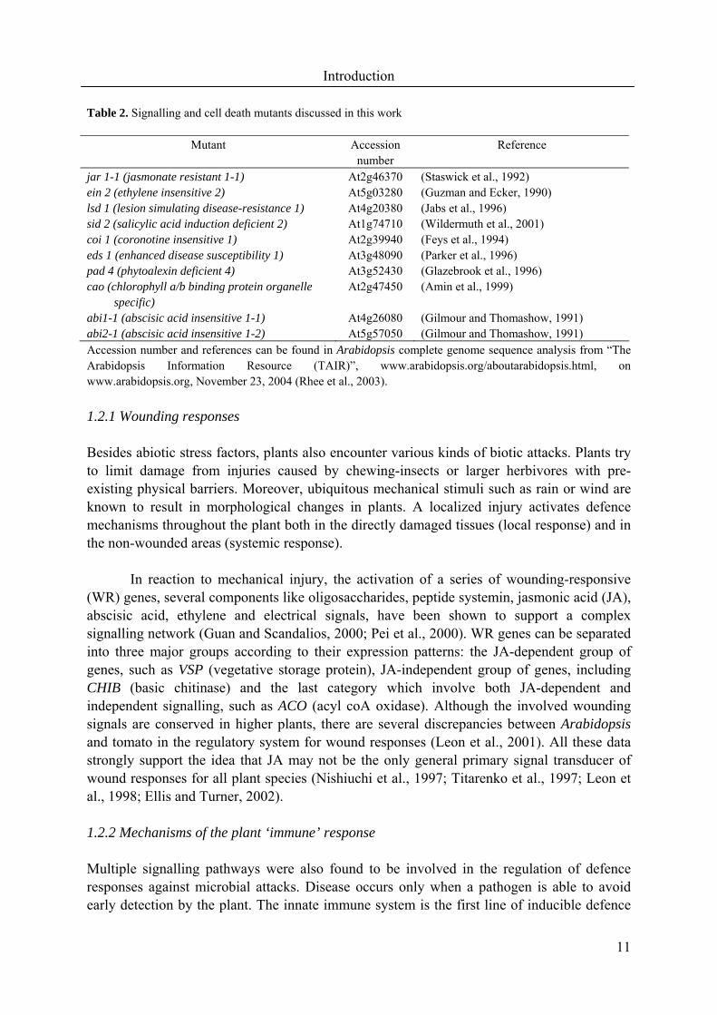
1.2 Signalling network and cross-talk Due to the complexity of the antioxidant network, it is easily concluded that the network coordinating the antioxidant defence with the basic metabolism and other defence responses has to be even more intricate. Mutant analysis has contributed greatly to our understanding of the signalling transduction network in plants. Table 2 summarizes the mutants discussed in this thesis, which are valuable for studies of the signals involved in cross-talk between biotic and abiotic stresses. Widespread analysis of different biotic and abiotic stress responses has led to the conclusion that considerable "horizontal" interactions exist between many components of different signalling pathways (Gomez et al., 2004).
So-called "cross-talk" is now a subject of intense scrutiny, especially in laboratories using Arabidopsis as their experimental species (Rojo et al., 1999; Schenk et al., 2000; Swidzinski et al., 2002; Mahalingam et al., 2003; De Paepe et al., 2004). The application of post-genomic techniques and the analysis of considerable amounts of microarray data and mutants give an impression of a complex network of responses from the sub-cellular to whole plant level. Such integrated "systems biology" is seen by many as the way forward in studying plant-environment interactions (Raikhel and Coruzzi, 2003). Current research has barely begun to scratch the surface of how a response to multiple stresses is organised and why such signalling networks have evolved. Thus the reality of how signalling pathways integrate their responses to environmental stimuli is akin to the complex behaviour of neural networks and attempts to describe how such networks might operate have just begun (Genoud and Métraux, 1999; Pastori and Foyer, 2002).
Introduction
11
Table 2. Signalling and cell death mutants discussed in this work
Mutant Accession number
Reference
jar 1-1 (jasmonate resistant 1-1) At2g46370 (Staswick et al., 1992) ein 2 (ethylene insensitive 2) At5g03280 (Guzman and Ecker, 1990) lsd 1 (lesion simulating disease-resistance 1) At4g20380 (Jabs et al., 1996) sid 2 (salicylic acid induction deficient 2) At1g74710 (Wildermuth et al., 2001) coi 1 (coronotine insensitive 1) At2g39940 (Feys et al., 1994) eds 1 (enhanced disease susceptibility 1) At3g48090 (Parker et al., 1996) pad 4 (phytoalexin deficient 4) At3g52430 (Glazebrook et al., 1996) cao (chlorophyll a/b binding protein organelle
specific) At2g47450 (Amin et al., 1999)
abi1-1 (abscisic acid insensitive 1-1) At4g26080 (Gilmour and Thomashow, 1991) abi2-1 (abscisic acid insensitive 1-2) At5g57050 (Gilmour and Thomashow, 1991) Accession number and references can be found in Arabidopsis complete genome sequence analysis from “The Arabidopsis Information Resource (TAIR)”, www.arabidopsis.org/aboutarabidopsis.html, on www.arabidopsis.org, November 23, 2004 (Rhee et al., 2003). 1.2.1 Wounding responses Besides abiotic stress factors, plants also encounter various kinds of biotic attacks. Plants try to limit damage from injuries caused by chewing-insects or larger herbivores with pre-existing physical barriers. Moreover, ubiquitous mechanical stimuli such as rain or wind are known to result in morphological changes in plants. A localized injury activates defence mechanisms throughout the plant both in the directly damaged tissues (local response) and in the non-wounded areas (systemic response).
In reaction to mechanical injury, the activation of a series of wounding-responsive
(WR) genes, several components like oligosaccharides, peptide systemin, jasmonic acid (JA), abscisic acid, ethylene and electrical signals, have been shown to support a complex signalling network (Guan and Scandalios, 2000; Pei et al., 2000). WR genes can be separated into three major groups according to their expression patterns: the JA-dependent group of genes, such as VSP (vegetative storage protein), JA-independent group of genes, including CHIB (basic chitinase) and the last category which involve both JA-dependent and independent signalling, such as ACO (acyl coA oxidase). Although the involved wounding signals are conserved in higher plants, there are several discrepancies between Arabidopsis and tomato in the regulatory system for wound responses (Leon et al., 2001). All these data strongly support the idea that JA may not be the only general primary signal transducer of wound responses for all plant species (Nishiuchi et al., 1997; Titarenko et al., 1997; Leon et al., 1998; Ellis and Turner, 2002).
1.2.2 Mechanisms of the plant ‘immune’ response Multiple signalling pathways were also found to be involved in the regulation of defence responses against microbial attacks. Disease occurs only when a pathogen is able to avoid early detection by the plant. The innate immune system is the first line of inducible defence
Introduction
12
against disease. A key function of innate immunity is the detection of pathogen-associated molecular patterns produced by infectious agents but not by host cells (Asai et al., 2002). Strong disease resistance of plants often is conditioned by gene-for-gene interactions: when a pathogen has an avirulence (avr) gene and a plant has a corresponding resistance (R) gene, the plant can rapidly recognize the pathogen and effectively deploy defence responses (Dangl and Jones, 2001). According to this model, the specific recognition of an avr gene product by a plant resistance gene product leads to a defence response (so called hypersensitive response or HR) that is characterized by localized cell lesion of the infected tissue and induction of certain plant defence-related genes (Lamb et al., 1989). Recognition of an avirulent pathogen triggers the rapid production of ROS, and nitric oxide, which also participates as a signal in the immune and vascular systems in plant disease resistance (Delledonne et al., 1998).
When a plant survives the infection of a pathogen at one site, it often develops
increased resistance to subsequent attack at sites throughout the plant and enjoys protection against a wide range of pathogen species. This phenomenon, called systemic acquired resistance (SAR), develops over a period of several days following initial infection (Chen et al., 1993; Dietrich et al., 1994). Arabidopsis MAP kinase 4 negatively regulates systemic acquired resistance and thus potentially enhances protection against diverse pathogen types (Petersen et al., 2000). 1.2.3 EEE and defence against pathogens Several common features characterize the response of plants to both EEE and infection by incompatible pathogens. They are characterized by a rapid increase in the foliar concentrations of ROS, the depletion of antioxidant pools, the chlorosis and necrosis of leaves, local and systemic defence responses (systemic acquired acclimation and systemic acquired resistance) and induction of defence gene expression.
However, in cells infected by an avirulent pathogen, the sub-cellular origin of the
ROS burst is thought to be NADPH oxidases at the plasma membrane, which is different from EEE that stimulates ROS production mainly in chloroplasts and peroxisomes. It has been proposed that ROS produced during the oxidative burst act as the central regulator of the HR (Levine et al., 1994). The manipulation of endogenous ROS alters the outcome of cell death signals and the induction of cell death requires the controlled attenuation of ROS detoxification (Lamb and Dixon, 1997). In situations where ROS are actively involved in the induction of cell death, there are constitutive and induced ROS detoxification systems that could inhibit cell death. Insight into the ROS dependent regulation of cell death came from analysis of the lesion simulating disease 1 (lsd1) mutant, which was initially characterized for its O2
.--dependent spreading of chlorotic/necrotic lesions (a phenotype termed runaway cell death, rcd) that develop under long or continuous photoperiods or after infection with an avirulent pathogen (Dietrich et al., 1994; Jabs et al., 1996). LSD1 has been suggested to be a negative regulator of cell death by acting as a ROS rheostat. Short day photoperiods and low photosynthetically active photo flux density are permissive conditions for the growth of lsd1. These observations strongly imply the relationship between prevailing light conditions and the initiation of the runaway cell death in lsd1. Therefore, the lsd1 mutant is a good candidate
Aim of the present studies
13
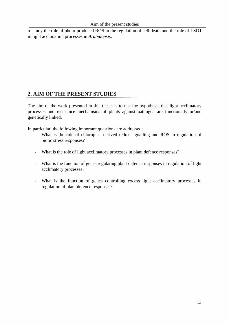
to study the role of photo-produced ROS in the regulation of cell death and the role of LSD1 in light acclimation processes in Arabidopsis. 2. AIM OF THE PRESENT STUDIES The aim of the work presented in this thesis is to test the hypothesis that light acclimatory processes and resistance mechanisms of plants against pathogen are functionally or/and genetically linked. In particular, the following important questions are addressed:
- What is the role of chloroplast-derived redox signalling and ROS in regulation of biotic stress responses?
- What is the role of light acclimatory processes in plant defence responses? - What is the function of genes regulating plant defence responses in regulation of light
acclimatory processes? - What is the function of genes controlling excess light acclimatory processes in
regulation of plant defence responses?

14
3. RESULTS AND DISCUSSION 3.1 The role of chloroplast-derived redox signalling and ROS in regulation of wounding responses Previously published observations showed that the expression of the firefly luciferase gene (APX2LUC) fused to the APX2 promoter in transgenic Arabidopsis (Karpinski et al., 1999) was enhanced in the veins closest to the wounded tissue of detached leaves partially exposed to excess light (Mullineaux et al., 2000). This raised the possibility that the signalling pathway controlling the expression of APX2 could be integrated into one or more wound-signalling pathways or could share common components, or is responsive to the same effectors molecules. However, further investigations reported here led to a different and unexpected conclusion. Presented results suggest that in wounded leaves exposed to light, there is an increased diversion of photosynthetic electron flux to O2 in the vascular regions leading to redox changes in photosynthetic electron transport and an increased production of H2O2 that, in turn, triggers induction of APX2 expression. This has important implications for how “wound-specific” gene expression is viewed. 3.1.1 Synergistic effect of wounding and excess light on APX2 expression The induction of APX2 expression in wounded leaves could be detected by induction of expression of APX2LUC reporter system in transgenic Arabidopsis lines (Karpinski et al., 1999). Luciferase activity could be imaged in the central vein and apical region of both the wounded leaf and adjacent unwounded leaves (Figure 2a). APX2 transcript could be detected as little as 45 minutes after wounding (Figure 2b). Under these conditions, the wounded plants expressed the JA-dependent wound-inducible VEGETATIVE STORAGE PROTEIN 1 (VSP1) and JA-independent wound-inducible BASIC CHITINASE (CHIB) genes (Rojo et al., 1999; Ellis and Turner, 2001). APX2 expression has been shown to be induced in leaves subjected to excess light treatments at times as little as 7 minutes after the onset of the challenge (Karpinski et al., 1997). To determine whether the regulation of APX2 expression shared any similarities in wounded leaves compared with those subjected to excess light stress, the effect on expression of these combined stresses was compared with undamaged leaves subjected to a 5-fold excess light stress. Using RNA gel blots, APX2 transcripts was detected 15 minutes earlier and they accumulated to a >10-fold higher level in wounded, excess light-stressed leaves than in their undamaged counterparts (Figure 2b). It should be noted that in wounded leaves, induction of APX2 transcript could only be detected using a RT-PCR and was undetectable on RNA gel blots (data not shown and Figure 2b). Therefore, it was concluded that the combined effect of wounding and excess light had a synergistic effect on the level of induced APX2 transcript, compared with either excess light or wounding stress alone. Both VSP1 and CHIB expression were induced further by excess light treatment, but no synergistic effect could be discerned of the combined stresses on their transcript levels (Figure 2c).

Results and Discussion
15
The observation that wounded leaf tissue showed enhanced expression of APX2 under
ambient and excess light conditions (Figure 2) raised the possibility that wound signalling pathways in Arabidopsis might share common components. This data presented here indicates that neither the JA-dependent nor JA-independent wound signalling pathways known in Arabidopsis (McConn et al., 1997; Nishiuchi et al., 1997; Titarenko et al., 1997; Rojo et al., 1998; Rojo et al., 1999; Leon et al., 2001) are implicated in APX2 expression in wounded leaves (paper I), despite both pathways being operative in our experimental conditions as evidenced by the induction of VSP1 and CHIB expression, respectively (Figure
Figure 2. Induction of APX2 expression in wounded leaves.
(a) Imaging of luciferase activity from an APX2LUCtransgenic Arabidopsis line (Karpinski et al., 1999)2 h after wounding (right panel). The wounding was carried out by crimping with forceps at the points shown by red bands in the right panel. Wounded tissue can also be seen in the left panel at the start of the experiment as slightly darkened bands (arrowed).
(b) Effect of combined excess light and wounding on levels of APX2 transcript. Individual plants, undamaged or wounded as in (a), were subjected to a fivefold excess light treatment for one of the time points indicated, and their RNAs were extracted and analysed by blotting of RNA gels and probing with an APX2-specific cDNA. The ACTIN probe was used as a loading control. For early detection of APX2transcript after wounding, 3' RACE PCR was used to amplify cDNA followed by Southern blotting, gene-specific probes and detection using a phosphorimager. Here cDNA derived from APX3 transcript is used as a loading control and to check for presence of cDNA in early time points.
(c) Levels of VSP1 and CHIB transcripts in wounded, excess light treated, and combined treatments. The levels of transcripts were assayed using a quantitative RT-PCR procedure, normalized for loading using 18S RNA standard and expressed relative to untreated controls.

Results and Discussion
16
2b). While not examined in detail, it was interesting to note that high light – mediated induction of VSP1 expression was not JA-dependent (Figure 2b), adding further weight to the argument that the JA signalling pathway plays no direct role in governing responses of gene expression to high light stress. 3.1.2. Photosynthetic electron transport is required for wound-induced APX2 expression The highest levels of APX2 expression were always observed in leaves subjected to both wounding and excess light (Figure 2c). Wounded leaves treated with the photosynthetic inhibitor DCMU did not show any induction of APX2 expression (Figure 3). This led us to hypothesize that the mechanism of APX2 induction in wounded Arabidopsis leaves was qualitatively the same as that described previously for excess light-stressed leaves, depending on redox changes in photosynthetic electron transport that generate H2O2 and augmented by
ABA (Karpinski et al., 1999; Fryer et al., 2003). This hypothesis would mean that induction of APX2 expression would differ between wounded and excess light-stressed leaves only in the degree of response. The synergistic effect of excess light and wounding on APX2 expression and its inhibition by DCMU were consistent with this hypothesis.
3.1.3 Wound and excess light-induced APX2 expression is not controlled by known wound signalling pathways in Arabidopsis Arabidopsis has at least two wound signalling pathways, JA-dependent and JA-independent (Titarenko et al., 1997; Rojo et al., 1999; Ellis and Turner, 2002). Communication between these pathways is mediated by ethylene generated in damaged tissues (Rojo et al., 1999). The mutant coronatine-insensitive 1-1 (coi 1-1) (Feys et al., 1994) is blocked in the perception of JA and COI 1-1 may play a role in cross–talk between the JA pathway and those directed by ethylene and ABA (Feys et al., 1994; Ellis and Turner, 2002). The coi1-1 mutant showed no difference from wild-type plants in the abundance of APX2 transcript when wounded or subjected to excess light (paper I). VSP1 transcript induction was blocked in wounded, but not in high light-stressed coi1-1 plants (paper I). Similarly, wounded or high light treated
Figure 3. The inhibition of wound-induced APX2LUC expression by the photosynthesis inhibitor DCMU.
Leaves were vacuum infiltrated for 3 min with 10 µM DCMU and then incubated in the same solution for 3 h. At the end of this period, the leaves were wounded, incubated on wetted blotting paper at growth PPFD for 2 h and sprayed with luciferin, and the luciferase activity was imaged. The color bar code gives the range of luciferase activity from background to 1000 RLU (blue) to 1800 RLU (yellow).

Results and Discussion
17
mutants impaired in their perception of ethylene, ethylene insensitive 2-1 (Guzman and Ecker, 1990), and ethylene resistant 1-3 (Bleecker et al., 1988), or JA (Staswick et al., 1992) showed no inhibition of APX2 expression (paper I).
APX2LUC Arabidopsis rosettes sprayed with either JA or chitosan, an elicitor of the
JA-independent pathway (Rojo et al., 1999), did not induce luciferase activity under ambient light conditions (paper I). The effects of JA and chitosan in the leaves were confirmed by CHIB and VSP1 gene induction (paper I). Taken together these data indicate that the wound induction of APX2 expression was not regulated by either of the known JA-dependent or JA–independent pathways in Arabidopsis and in contrast to wounding, VSP1 induction of expression in excess light stressed leaves also was not JA-dependent. 3.1.4 Sources of H2O2 for induction of APX2 expression It has been proposed that NADPH oxidase(s) could be responsible for the generation of H2O2 in the peri-veinal tissue of wounded leaves (Allan and Fluhr, 1997; Orozco-Cardenas et al., 2001; Bolwell et al., 2002). This suggestion was based on the observation that the wound-induced accumulation of H2O2 can be inhibited by pre-treatment of leaves with diphenylene iodonium (Orozco-Cardenas and Ryan, 1999; Orozco-Cardenas et al., 2001). Therefore, the effect of DPI was tested on the wound-induced APX2 expression and on photosynthetic electron transport.
DPI inhibited the wound-induced expression of APX2LUC in fully expanded leaves
by >80 % after 6 hours (Figure 4a). This inhibition of APX2LUC expression by DPI was associated with a decline in the PSII operating efficiency indicative of decreases in the rate of photosynthetic electron transport (Figure 4b). These data suggest that DPI is a potent inhibitor of photosynthetic electron transport in wounded leaves. This conclusion was further supported by the inhibition of the rate of photosynthetic O2 evolution over a range of PPFDs (photosynthetically active photon flux density) in DPI-treated leaves (Figure 4c). Furthermore, the rate of O2 evolution in DPI-treated leaves saturated above a PPFD of 400 µmols m-2s-1 in DPI-treated leaves, whereas control leaves required in excess of 800 µmols m-2s-1 for saturation.
This observation has led to the proposal that wound-induced H2O2 accumulation may
be catalysed by a bundle sheath located NADPH oxidase (Orozco-Cardenas et al., 2001). Similar conclusions about the origins of H2O2 have been made from DPI treatments of stomatal guard cells, wounded fruit mesocarp tissue and elicitor-treated suspension culture cells (Levine et al., 1994; Tenhaken et al., 1995; Watanabe and Sakai, 1998; Murata et al., 2001; Zhang et al., 2001). There is considerable biochemical and molecular genetic evidence for the existence of at least the gp96phox subunit of plant NADPH oxidases, homologous to that in mammalian phagocytes (Tenhaken et al., 1995; Keller et al., 1998; Torres et al., 1998). However, DPI has been shown to inhibit other haem-containing enzymes such as xanthine oxidase, nitric oxide synthase (Delledonne et al., 1998; Moulton et al., 2000) and cell wall peroxidases (Bolwell et al., 1999). Some of these enzymes have been implicated as sources of ROS at least for the HR (Allan and Fluhr, 1997; Bolwell et al., 1999; Bolwell et al., 2002).
Results and Discussion
18
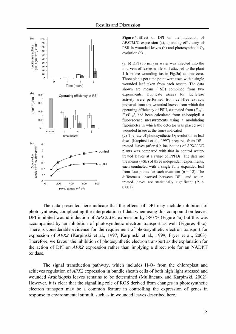
The data presented here indicate that the effects of DPI may include inhibition of
photosynthesis, complicating the interpretation of data when using this compound on leaves. DPI inhibited wound induction of APX2LUC expression by >80 % (Figure 4a) but this was accompanied by an inhibition of photosynthetic electron transport as well (Figures 4b,c). There is considerable evidence for the requirement of photosynthetic electron transport for expression of APX2 (Karpinski et al., 1997; Karpinski et al., 1999; Fryer et al., 2003). Therefore, we favour the inhibition of photosynthetic electron transport as the explanation for the action of DPI on APX2 expression rather than implying a direct role for an NADPH oxidase.
The signal transduction pathway, which includes H2O2 from the chloroplast and
achieves regulation of APX2 expression in bundle sheath cells of both high light stressed and wounded Arabidopsis leaves remains to be determined (Mullineaux and Karpinski, 2002). However, it is clear that the signalling role of ROS derived from changes in photosynthetic electron transport may be a common feature in controlling the expression of genes in response to environmental stimuli, such as in wounded leaves described here.
Figure 4. Effect of DPI on the induction of APX2LUC expression (a), operating efficiency of PSII in wounded leaves (b) and photosynthetic O2
evolution (c). (a, b) DPI (50 µm) or water was injected into the mid-vein of leaves while still attached to the plant 1 h before wounding (as in Fig.3a) at time zero. Three plants per time point were used with a single wounded leaf taken from each rosette. The data shown are means (±SE) combined from two experiments. Duplicate assays for luciferase activity were performed from cell-free extracts prepared from the wounded leaves from which the operating efficiency of PSII, estimated from (F m' -F')/F m', had been calculated from chlorophyll afluorescence measurements using a modulating fluorimeter in which the detector was placed over wounded tissue at the times indicated (c) The rate of photosynthetic O2 evolution in leaf discs (Karpinski et al., 1997) prepared from DPI-treated leaves (after 4 h incubation) of APX2LUCplants was compared with that in control water-treated leaves at a range of PPFDs. The data are the means (±SE) of three independent experiments, each conducted with a single fully expanded leaf from four plants for each treatment (n = 12). The differences observed between DPI- and water-treated leaves are statistically significant (P <0.001).
Results and Discussion
19
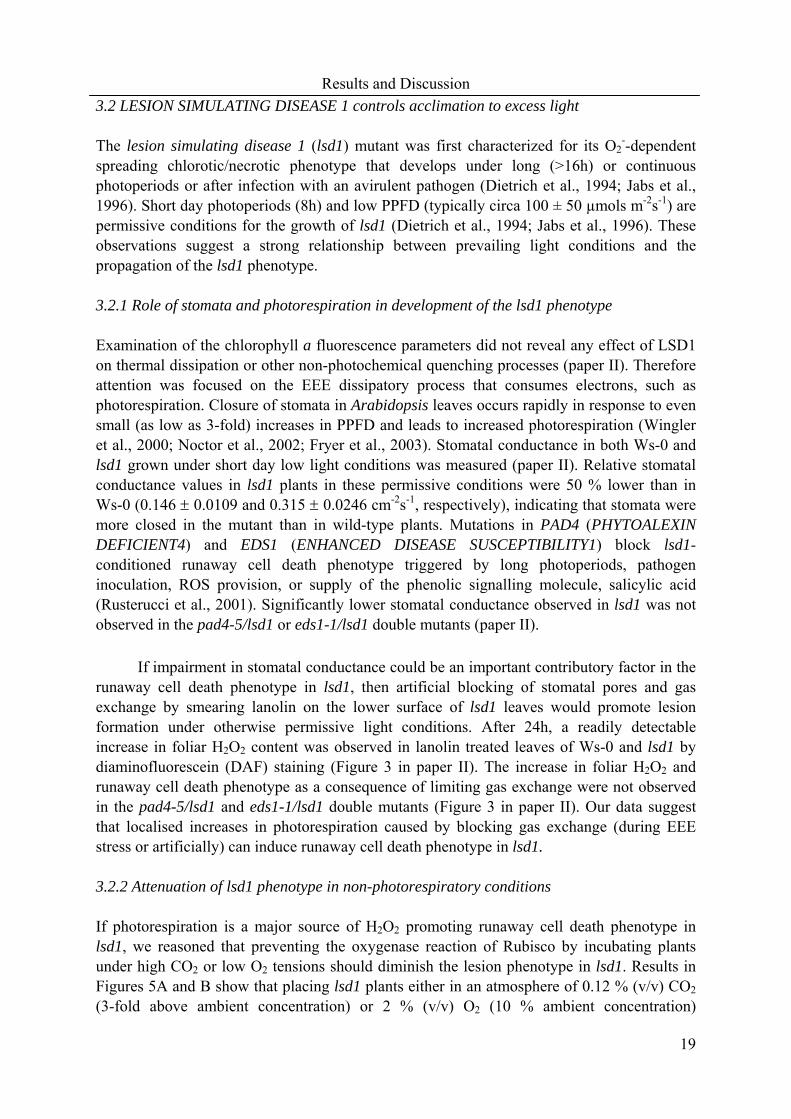
3.2 LESION SIMULATING DISEASE 1 controls acclimation to excess light The lesion simulating disease 1 (lsd1) mutant was first characterized for its O2
--dependent spreading chlorotic/necrotic phenotype that develops under long (>16h) or continuous photoperiods or after infection with an avirulent pathogen (Dietrich et al., 1994; Jabs et al., 1996). Short day photoperiods (8h) and low PPFD (typically circa 100 ± 50 µmols m-2s-1) are permissive conditions for the growth of lsd1 (Dietrich et al., 1994; Jabs et al., 1996). These observations suggest a strong relationship between prevailing light conditions and the propagation of the lsd1 phenotype.
3.2.1 Role of stomata and photorespiration in development of the lsd1 phenotype Examination of the chlorophyll a fluorescence parameters did not reveal any effect of LSD1 on thermal dissipation or other non-photochemical quenching processes (paper II). Therefore attention was focused on the EEE dissipatory process that consumes electrons, such as photorespiration. Closure of stomata in Arabidopsis leaves occurs rapidly in response to even small (as low as 3-fold) increases in PPFD and leads to increased photorespiration (Wingler et al., 2000; Noctor et al., 2002; Fryer et al., 2003). Stomatal conductance in both Ws-0 and lsd1 grown under short day low light conditions was measured (paper II). Relative stomatal conductance values in lsd1 plants in these permissive conditions were 50 % lower than in Ws-0 (0.146 ± 0.0109 and 0.315 ± 0.0246 cm-2s-1, respectively), indicating that stomata were more closed in the mutant than in wild-type plants. Mutations in PAD4 (PHYTOALEXIN DEFICIENT4) and EDS1 (ENHANCED DISEASE SUSCEPTIBILITY1) block lsd1-conditioned runaway cell death phenotype triggered by long photoperiods, pathogen inoculation, ROS provision, or supply of the phenolic signalling molecule, salicylic acid (Rusterucci et al., 2001). Significantly lower stomatal conductance observed in lsd1 was not observed in the pad4-5/lsd1 or eds1-1/lsd1 double mutants (paper II).
If impairment in stomatal conductance could be an important contributory factor in the
runaway cell death phenotype in lsd1, then artificial blocking of stomatal pores and gas exchange by smearing lanolin on the lower surface of lsd1 leaves would promote lesion formation under otherwise permissive light conditions. After 24h, a readily detectable increase in foliar H2O2 content was observed in lanolin treated leaves of Ws-0 and lsd1 by diaminofluorescein (DAF) staining (Figure 3 in paper II). The increase in foliar H2O2 and runaway cell death phenotype as a consequence of limiting gas exchange were not observed in the pad4-5/lsd1 and eds1-1/lsd1 double mutants (Figure 3 in paper II). Our data suggest that localised increases in photorespiration caused by blocking gas exchange (during EEE stress or artificially) can induce runaway cell death phenotype in lsd1. 3.2.2 Attenuation of lsd1 phenotype in non-photorespiratory conditions If photorespiration is a major source of H2O2 promoting runaway cell death phenotype in lsd1, we reasoned that preventing the oxygenase reaction of Rubisco by incubating plants under high CO2 or low O2 tensions should diminish the lesion phenotype in lsd1. Results in Figures 5A and B show that placing lsd1 plants either in an atmosphere of 0.12 % (v/v) CO2 (3-fold above ambient concentration) or 2 % (v/v) O2 (10 % ambient concentration)

Results and Discussion
20
substantially attenuated the lesion phenotype under non-permissive long day conditions. Treatment of lsd1 leaves with 20 mM H2O2 under permissive light conditions also caused spreading lesions after 48 h (Figure 5C). These were not observed on H2O2 treated Ws-0 leaves or in lsd1 leaves kept in the dark (Figure 5C).
Acclimation to EEE in higher plants is signalled by significant increases in H2O2
levels (Karpinski et al., 1997; Karpinski et al., 1999; Karpinska et al., 2000; Fryer et al., 2003). It can be concluded from the above experiments that LSD1 promotes the effectiveness of photorespiration by reducing the damaging effects of H2O2 accumulation during light acclimation. 3.2.3 Salicylic acid, photo-oxidative stress and stomatal conductance Salicylic acid treatment induces rcd in lsd1 under otherwise permissive conditions (Jabs et al., 1996). Interestingly, rapid closure of stomata and an increase in foliar H2O2 have been shown to occur upon SA treatment of leaves (Manthe et al., 1992; Chen et al., 1993; Rao et al., 1997; Shirasu et al., 1997; Mori et al., 2001). This may be associated with SA-dependent inhibition of catalase (CAT) activity (Sanchez-Casas and Klessig, 1994; Conrath et al., 1995; Durner and Klessig, 1996; Chen et al., 1997). If peroxisomal CAT levels prior to a change in the light environment are crucial to successful acclimation or recovery, it can be anticipated that wild-type leaves treated with SA would generally behave in a manner similar to lsd1.
Figure 5. Reactive oxygen species (ROS) originating from photorespiration are critical for runaway cell death (rcd) in lsd1. (A) Four-week-old lsd1 plants cultivated in LD and which had initiated lesion formation were fumigated either with a 0.12 % CO2 and 21 % O2
(high CO2) atmosphere LD (16 h) for a period of 2 weeks or (B) with a 2 % O2 and 0.04 % CO2
(low O2) atmosphere in continuous light (CL) for a period of 1 week. (C) Pictures of representative leaves of Ws-0 and lsd1 after 2 d incubation either in light or dark in a solution of 20 mM H2O2 Control leaves were incubated in water.
Results and Discussion
21
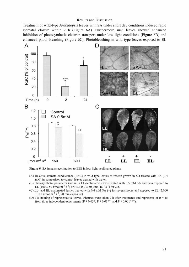
Treatment of wild-type Arabidopsis leaves with SA under short day conditions induced rapid stomatal closure within 2 h (Figure 6A). Furthermore such leaves showed enhanced inhibition of photosynthetic electron transport under low light conditions (Figure 6B) and enhanced photo-bleaching (Figure 6C). Photobleaching in wild type leaves exposed to EL
Figure 6. SA impairs acclimation to EEE in low light-acclimated plants. (A) Relative stomata conductance (RSC) in wild-type leaves of rosette grown in SD treated with SA (0.4
mM) in comparison to control leaves treated with water. (B) Photosynthetic parameter Fv/Fm in LL-acclimated leaves treated with 0.5 mM SA and then exposed to
LL (100 ± 50 µmol m–2 s–1) or HL (450 ± 50 µmol m–2 s–1) for 2 h. (C) LL- and HL-acclimated leaves treated with 0.4 mM SA (+) for several hours and exposed to EL (2,000
± 100 µmol m–2 s–1, 90 min exposure). (D) TB staining of representative leaves. Pictures were taken 2 h after treatments and represents of n = 15
from three independent experiments (P 0.05*, P 0.01**, and P 0.001***).

Results and Discussion
22
was characterised by the appearance of a delimited area of cell death revealed by lactophenol-trypan blue (TB) staining (Figure 6D). These effects were strongly accelerated by a combined effect of SA-treatment and EL exposure (Figures 6B,C,D). It is important to note here that stomatal gas exchange in SA-treated wild-type leaves before EL exposure was reduced approximately by a factor of 4 (Figure 6A), while in combined treatment of wild type leaves by a factor of 12 (0.027 ± 0.0185 cm-2s-1). SA treatment of lsd1 leaves in low light caused reduction of stomatal gas exchange by a factor of 14 (0.024 ± 0.0161 cm-2s-1) in comparison to wild-type control values. The data suggest that SA induces an increase in EEE under low light intensities such that failure to tolerate EL treatment ensued. This notion was reinforced by the observation that high light acclimated leaves, already dissipating or limiting EEE, were tolerant to SA treatment (Figure 6D) and have higher foliar SA levels (Karpinski et al., 2003).
It can be concluded that the effectiveness of photorespiration in dissipating EEE may
be limited by the efficiency of H2O2 scavenging. The above results show that the Arabidopsis mutant lsd1 failed to acclimate to light conditions that promote photo-oxidative stress, and that LSD1 function was required for the expression of CATALASE1, encoding a subunit of the peroxisomal catalase (paper II). Through this regulation LSD1 can control the effectiveness of photorespiration in dissipating EEE absorbed by photosystem II. It can also be concluded that lsd1 had reduced stomatal conductance in permissive conditions and induced H2O2 accumulation together with runaway cell death when stomatal gas exchange was impeded. Previously H2O2 had been excluded as a molecular trigger of lsd1 runaway cell death phenotype (Jabs et al., 1996). These light sensitive lsd1 phenotype depended on the defence regulators EDS1 and PAD4 and PSII light harvesting antenna size. Salicylic acid, which induces stomatal closure, inhibited catalase activity and triggered the runaway cell death phenotype of lsd1, also impaired acclimation of wild type plants to conditions that promote EEE. It can be proposed that the roles of LSD1 in light acclimation and regulation of stomatal conductance, as well as in restricting pathogen-induced cell death are functionally linked (paper II). 3.3 Functional characterization of the chloroplastic glutathione peroxidases (cpGPXs) in Arabidopsis thaliana Glutathione peroxidases (GPXs) are commonly considered to be important ROS scavengers
because their broader substrate specificities and stronger affinity for H2O2 than catalases (Brigelius-Flohe and Flohe, 2003). Several plant ROS scavenging proteins are encoded by families of closely related genes that typically display specific structural and regulatory features (Ferrari et al., 2003). Sub-cellular compartmentation of ROS scavenging systems is crucial for efficient removal/control of ROS at their sites of generation (Grene, 2002). DNA and derived amino acid sequence analysis suggests that Arabidopsis GPXs also belongs to this category of enzymes with putative localization in the cytosol, chloroplast, mitochondria and endoplasmic reticulum (ER). Analysis of the Arabidopsis genome sequence revealed one additional open-reading frame encoding protein closely related to seven previously described GPXs (Milla et al., 2003), thus increasing the total number of genes to eight (AtGPX1-AtGPX8; Figure 7A). Phylogenetic analysis of Arabidopsis GPXs suggests three pair-group - AtGPX1 and AtGPX7 (chloroplastic group); cytosolic AtGPX4 and ER-AtGPX5; cytosolic

Results and Discussion
23
AtGPX6 and cytosolic AtGPX8; whereas cytosolic AtGPX2 and mitochondrial AtGPX3 are on single branches of the tree (Figure 7B). The presence of multiple genes may reflect the evolutionary advantage of functional redundancy, which is likely to ensure a higher level of protection and confer a selective advantage, or it may be a consequence of the acquisition of new substrate specificity, such as the partitioning of the task of an ancestral protein into separate gene products (Lynch et al., 2001).
Figure 7. Amino acid sequence analysis of the AtGPX family. (A) Alignment of the predicted amino acid sequences of AtGPX family. Homologous sequences were obtained from the National Centre for Biotechnology Information, and the alignment was performed with WORKBENCH software using the Clustal W method. Amino acids are given with standard single-letter designation and dashes (-) indicate no consensus. Residues shown in white letters with black background indicate identification to the consensus sequence at the aligned position (*), residues shown in black letters with grey background indicate conservation of strong groups (:) and residues shown in white letters with grey background as (.) indicate conservation of weak groups. Numbers indicate protein length in amino acids. (B) Phylogenetic tree of the AtGPX family. The amino acid sequences of the above eight AtGPX proteins were organized into a phylogenetic tree with Treeview software (Page, 1996). The length of the branch indicates the extent of divergence according to the bar at the bottom.

Results and Discussion
24
3.3.1 Arabidopsis plants with reduced expression of chloroplastic GPXs Most plant GPXs have been analysed only partially and the biological function of these enzymes have not been sufficiently elucidated. In order to analyse the role of two chloroplastic isoforms of GPXs, we have generated Arabidopsis thaliana transgenic lines with reduced cpGPX activity. Results presented here suggest that cpGPXs play important protective and regulatory roles during acclimation to photo-oxidative stress conditions.
Arabidopsis plants were transformed with a construct consisting of the cDNA encoding
GPX1 in antisense orientation under the control of the cauliflower mosaic virus (CaMV) 35S. Twelve independent transformed of AS-cpGPX lines were thereby produced. For analyses, homozygous T4 plants from lines carrying single insertions were used. As revealed by RT-PCR and protein level analysis, AS-cpGPX lines (71-90, 71-92 and 71-93) showed significantly lower expression of both AtGPX1 and AtGPX7 whereas knockout (ko)-GPX7 suppress AtGPX7 only (Figure 8A). Furthermore, GPX activity measured in intact chloroplasts was significantly lower in AS-cpGPX lines than in control plants, while ko-GPX7 mutant was only slightly reduced (Figure 8B). We also employed enzyme-linked immunosorbent assay (ELISA) to quantify the content of cpGPX and 71-93 line showed the most reduced among AS-cpGPX lines compared to control plants (Figure 8C). The different cpGPX suppression levels in AS-cpGPX lines could be due to the positional effects that depend on the insertion place of the transgene into the Arabidopsis genome.
3.3.2 Enhanced photo-inhibition in chloroplastic glutathione peroxidases deficient lines When two-week-old AS-cpGPX lines were challenged with continuous high light combined with chilling stress (continuous high light 650 ± 50 µmol s-1m-2 at 4 oC), seedlings did not
Figure 8. Selected transgenic AS-cpGPX and ko-GPX7 of the chloroplastic AtGPXs transgenic plants have reduced cpGPX activity (A) Transcript abundance analysis for
AtGPX1 and AtGPX7 in selected transgenic Arabidopsis lines (90, 92, 93 and ko-GPX7) by using RT-PCR quantitative assay and 18S RNA as an internal standard as described in Material and Methods. (B) GPX activity measured in intact chloroplast extracts. t-butyl peroxide and hydrogen peroxide were used as substrates and enzyme unit expressed as µM/min/mg-1FW (C) cpGPX protein level analysis by enzyme-linked immunosorbent assay (ELISA) and expressed as ratio compared to control plants (ck).

Results and Discussion
25
show more visible photo-bleaching or photo-damage but did reveal different levels of photo-inhibition of photosynthetic electron transport (Figure 9 and paper III). Characteristic picture of young Arabidopsis plants exposed to such stress can be divided into four major categories; green, red, half-bleached and fully bleached leaves (Figure 9A). Photo-bleached leaves can be used as an indicator of the level of photo-oxidative stress. Percentages of the previous four categories are represented in Figure 9B. AS-cpGPX lines demonstrated enhanced inhibition of photosynthetic electron transport (Figure 9C). However, maximum photochemical efficiency (Fv/Fm) recovered equally as in control plants after four days of such a stress (Figures 9B,C). It is important to know that under this excessive stress condition, utilisation of light energy through photosynthetic carbon assimilation is decreased more than the rate of electron transport (Foyer et al., 1994). Therefore this could result in considerable increase in the sensitivity of PSII to photo-inhibition as observed in AS-cpGPX lines (Figure 9C).
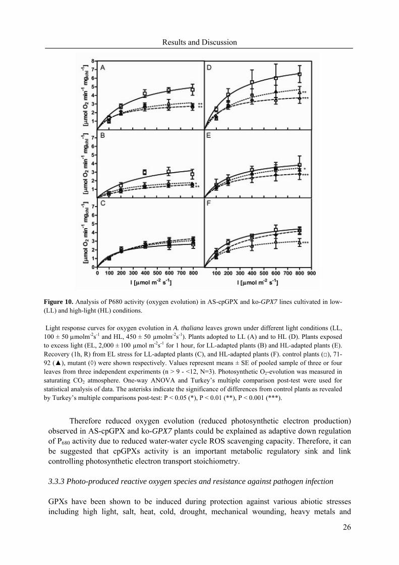
Significant differences between low light (LL) and high light (HL) acclimated AS-cpGPX plants were observed for light response curves for oxygen evolution (Figure 10). Increases in photosynthesis-irradiance (PI) curves observed in Figure 10 reflect an increased capacity for electron transport, which in turn requires the provision of increased electron sink capacity, principally through the CO2 fixation via Calvin cycle, water-water cycle or photorespiration (Kozaki and Takeba, 1996; Asada, 1999; Mullineaux and Karpinski, 2002).
Figure 9. Antisense chloroplastic AtGPXs transgenic lines were more sensitive to photo-oxidative stress. (A) Image of two-week-old Arabidopsisseedlings from low-light cultivation conditions followed by continuous high-light (700 µmol s-
1m-2) combined with chilling temperature (at 4 oC) for four days. (B) Relative numbers (in percentage) of leaves of
four different categories (green, red, half-bleached and fully bleached) which were shown in (A). Leaves number of twenty rosettes for each line were counted after four days of photo-oxidative stress and three days after recovery from such a stress. (C) Photosynthetic maximal electron transport efficiency parameter (Fv/Fm) during photooxidative stress and recovery period (n > 20 +SD).
Results and Discussion
26
Therefore reduced oxygen evolution (reduced photosynthetic electron production) observed in AS-cpGPX and ko-GPX7 plants could be explained as adaptive down regulation of P680 activity due to reduced water-water cycle ROS scavenging capacity. Therefore, it can be suggested that cpGPXs activity is an important metabolic regulatory sink and link controlling photosynthetic electron transport stoichiometry. 3.3.3 Photo-produced reactive oxygen species and resistance against pathogen infection GPXs have been shown to be induced during protection against various abiotic stresses including high light, salt, heat, cold, drought, mechanical wounding, heavy metals and
Figure 10. Analysis of P680 activity (oxygen evolution) in AS-cpGPX and ko-GPX7 lines cultivated in low-(LL) and high-light (HL) conditions. Light response curves for oxygen evolution in A. thaliana leaves grown under different light conditions (LL, 100 ± 50 µmolm-2s-1 and HL, 450 ± 50 µmolm-2s-1). Plants adopted to LL (A) and to HL (D). Plants exposed to excess light (EL, 2,000 ± 100 µmol m-2s-1 for 1 hour, for LL-adapted plants (B) and HL-adapted plants (E). Recovery (1h, R) from EL stress for LL-adapted plants (C), and HL-adapted plants (F). control plants (□), 71-92 (▲), mutant (◊) were shown respectively. Values represent means ± SE of pooled sample of three or four leaves from three independent experiments (n > 9 - <12, N=3). Photosynthetic O2-evolution was measured in saturating CO2 atmosphere. One-way ANOVA and Turkey’s multiple comparison post-test were used for statistical analysis of data. The asterisks indicate the significance of differences from control plants as revealed by Turkey’s multiple comparisons post-test: P < 0.05 (*), P < 0.01 (**), P < 0.001 (***).

Results and Discussion
27
osmotic stress (Sugimoto and Sakamoto, 1997; Depege et al., 1998; Avsian-Kretchmer et al., 1999; Roxas, 2000; Swidzinski et al., 2002; Yoshimura et al., 2004). Involvement of GPXs in biotic stress responses has also been demonstrated (Kuniak and Skodowska, 2001) and GPX has become a robust molecular marker for biotic stress responses (Rao and Davis, 1999). However, when plant GPX was indexed for the first time as molecular marker for biotic stress responses, Levine and colleagues (1994) did not recognize that cDNA employed in their experiments encoded the putative chloroplastic isoform of selenium-independent GPX (Mullineaux et al., 1998).
Both ROS and lipid peroxide play dual functions in cellular metabolism. On one hand,
they are highly toxic and must be kept under tight control (Noctor and Foyer, 1998). On the other hand, ROS and lipid peroxides serve as substrates in metabolism and as signals for regulation of environmental stress responses (Foyer and Noctor, 2000). The chloroplast envelope is suggested as a main source of phospholipid peroxides which serve as substrates for oxylipin and jasmonates biosynthesis (La Camera et al., 2004). These in turn regulate many plant defence responses including infection with pathogens. Thus, ROS produced in chloroplast, especially hydrogen peroxide that can cross the membranes together with phospholipid peroxides may contribute to the defence network under pathogen infection. ROS- and lipoxygenase-mediated production of fatty acid hydroperoxides in chloroplasts are specific features of the hypersensitive response in plants and show the major role of this organelle in signalling during incompatible plant-pathogen interaction (Hammond-Kosack and Parker, 2003).
Our current results with five-week-old AS-cpGPX lines acclimated to both low light
and high light conditions showed higher levels of hydrogen peroxide visualised by 3-3 diaminobenzidine (DAB) precipitate after excess light stress treatment (Figure 11A). Furthermore, quantification of foliar hydrogen peroxide levels was consistent with the above observation (Figure 11B). A readily detectable increase in foliar H2O2 was observed in 71-93 from low light conditions both in leaf and petiole without any pre-treatment when we used more sensitive dichlorofluorescein staining, another H2O2 accumulation index (Figure 11C). Since AS-cpGPX lines (71-92 and 71-93) showed more H2O2 accumulation in both untreated and adapted to high light or exposed to excess light leaves, the above results confirm that reduction of cpGPX activity could be associated with higher foliar H2O2 accumulation.
Furthermore, higher levels of ROS (Figure 11) in AS-cpGPX lines might lead to the possibility of cell death, and increase the plants’ susceptibility to infection by avirulent strains. A visible HR can be demonstrated by lactophenol-trypan blue (TB) staining (Figure 12A). AS-cpGPX lines showed more extensive HR regions than the control plants after 72 hrs of avirulent pathogen strain (avrRPM1) infection. Ion leakage was used as a quantitative indicator of cell death (O'Donnell et al., 2001). Consequently, ion leakage increased more in AS-cpGPX lines than in control plants in four days after the inoculation with avirulent strain (avrRPM1) (Figure 12B). However, in later times (after four days), the pathogen growth was not significantly different in transgenic and control plants (data not shown). These results suggest that the observed changes in HR were not able to restrict the pathogen growth and influence the plant resistance to avirulent

Results and Discussion
28
Figure 11. Hydrogen peroxide accumulates to higher levels in AS-cpGPX transgenic lines.
(A) Five-week-old Arabidopsis control (ck) and 71-93 line leaves were taken from low light-adapted plants (LL; 100 ± 50 µmolm-2s-1) and high light –adapted plants (HL; 450 ± 50 µmolm-2s-1), exposed to excess light (EL; 2,000 ± 100 µmol m–2 s–1) for one and half hour respectively and followed by 3,3-diaminobenzidine (DAB) staining. (B) Quantification of foliar hydrogen peroxide concentration from LL growth plants after one hour EL exposure, and after one hr recovery from such stress. Concentration is expressed in nmol of H2O2 per gram fresh weight (gFW-1). (C) 2’,7’-dichlorodihydrofluorescein diacetate (H2DCF-DA) staining of three-week-old seedlings. Data are representative for three independent experiments and several repetitions in one single experiment (N =3 ±SD).
Figure 12. Transgenic lines with reduced cpGPX activity displayed enhanced basal resistance towards virulent pathogen infection. (A) Leaves of 4-week-old plants were inoculated with a bacterial suspension containing 105 CFU (colony forming units) mL-1. (A) Tryphan blue (TB) staining pictures were obtained after 0 (T0), 24 (T24) and 72 (T72) hrs of rosette leaves infiltration with avirulent P. syringae pv tomato DC3000 carrying AvrRpm1 avirulence gene. (B) Ion leakage was measured after 0, 2 and 4 days followed by infiltration of leaves with avirulent P. syringae strain (avrRpm1), or with 10 mM MgCl2 used as a control infiltration-induced wounding at 0, 2 and 4 days. (C) Bacterial growth at four days post inoculation of virulent strains P. syringae pv maculicola ES4326 and P. syringae pv tomato DC3000. Mean values ± SD of three or four different plants are depicted. Similar results were obtained in three independent experiments. The asterisks indicate the significance of differences from the control plants at the level at least P < 0.05).

Results and Discussion
29
pathogen attack. On the other hand, higher levels of ROS could possibly help to develop the basal resistance and defence against virulent pathogen attack (Figure 12C). To test this possibility, the bacterial growth of virulent pathogen strains P. syringae pv maculicola ES4326 and P. syringae pv tomato DC3000 was further monitored in AS-cpGPX lines and control plants. The initial rate of bacterial growth was similar in all tested lines (data not shown). However, at four days post inoculation, the bacterial titre was approximately 10-fold (significantly) lower in AS-cpGPX lines as compared to control plants, which indicates enhanced resistance against both virulent pathogens (Figure 12C). These results are in agreement with recent observations made in the analysis of vitamin C deficient mutants (vtc1, vtc2), virulent bacterial growth (P. syringae pv maculicola ES4326) was reduced in those mutants due to higher foliar ROS level (Barth et al., 2004). Both examples illustrate that photo-produced ROS play an important role in basal defence mechanisms. In addition, the above results also suggest that AtGPX1 but not AtGPX7 might be involved in defence against virulent pathogen infection. This hypothesis is supported by the observation that virulent pathogen growth was significantly reduced in AS-cpGPX lines, with the inhibition of both AtGPX1 and AtGPX7 (Figure 12C), while virulent pathogen growth in infected ko-GPX7 line was not significantly reduced (data not shown).

30
4. CONCLUDING REMARKS AND FUTURE PERSPECTIVES
In this work, physiological and molecular analyses of Arabidopsis mutants and transgenic lines were applied to investigate the signalling network controlling biotic and abiotic stress responses. The signalling role of photo-produced ROS resulting from redox changes in the photosynthetic electron transport may be a common feature in controlling the expression of genes in response to environmental stimuli, such as wounding. ASCORBATE PEROXIDASE 2 (APX2) encodes a key enzyme of the antioxidant network. In excess light-stressed Arabidopsis leaves, H2O2 and abscisic acid regulate APX2 expression. Wounded leaves showed low induction of APX2 expression and when exposed to excess light, APX2 expression was increased synergistically. Signalling pathways dependent upon jasmonic acid, chitosan and abscisic acid were not involved in the wound-induced expression of APX2, but was shown to require PET and was preceded by a depressed rate of CO2 fixation.
Analysis of lsd1 in different mutant backgrounds puts forward a convincing argument
for a loss of photorespiratory control in the LSD1 type of mutant and strongly suggests that light acclimatory processes and pathogen defences are genetically and functionally linked. It is important to know that LSD1 type of mutants have mainly been studied with regard to pathogenesis. From this work, it reveals that association of LSD1 with hypersensitive response may only be supplementary.
GLUTATHIONE PEROXIDASES (GPXs) are considered important ROS scavenging
enzymes. Analysis of the Arabidopsis genome database revealed one additional open-reading frame encoding cytosolic protein closely related to seven previously described GPXs, thus increasing the total number of AtGPX genes to eight (AtGPX1-AtGPX8). Two putative chloroplastic isoforms showed differential expression patterns in various Arabidopsis organs in flower and in breaking of the ABA-dependent dormancy during seed germination. Arabidopsis thaliana transgenic lines with reduced expression of putative chloroplastic isoforms (AtGPX1 and AtGPX7) and AtGPX7 knock-out mutant (ko-GPX7) were more sensitive to photo-oxidative stress but manifested reduced bacterial growth rate when virulent strains Pseudomonas syringae pv. tomato DC3000 and P.s.t. maculicola strain ES4326) were inoculated. Functional analysis of cpGPXs confirms that light and chloroplastic ROS metabolism is important for basal resistance against virulent pathogens. Accordingly, to our knowledge, this is the first genetic analysis of chloroplastic GPXs in plants.
The presentation and discussion of the above results confirm that light sensing, light
acclimatory processes and photo-produced ROS also govern pathogen defence pathways. This has a great ecological relevance for Darwinian fitness of plants growing in the natural environment, where simultaneous pathogen attack and fluctuations in light, temperature and other environmental factors make rapid acclimation a constant necessity. Molecular, biochemical and physiological analysis of pathogen responses in mutants impaired in light sensing, EEE-dissipatory mechanisms, and similar analysis of light acclimatory processes in mutants impaired in pathogen defences may prove to be seminal.
The following experiments are planned in the near future. Transgenic plants with
cpGPXs overexpression and antisense cytosolic GPX lines which were also generated will be
Materials and Methods
31
used to further confirm already obtained data and to analyse more functional aspects of GPXs’ roles in biotic and abiotic cross-talk. Furthermore, other AtGPX family mutants were obtained and purified from NASC. Investigation of these mutants will allow us to assess a more complete picture of the functional role of the AtGPX family. Generation of plants carrying GFP fusions with cDNAs encoding two isoforms of cpGPXs will help us to confirm the sub-celluar localization of these enzymes. In addition, massive metabolite analysis in transgenic plants and mutants will be another tool to give us the clue of metabolic connections and functions.
5. MATERIALS AND METHODS Plant Material Arabidopsis thaliana (L.) Heyhn. plants were germinated and grown in conventional soil (Topstar-Economa Garden AB, Sweden) for 4-5 weeks under controlled environmental conditions (PPFD of low light, LL, 100 ± 50 µmolm-2s-1 during a 8-9 hr (as indicated in the paper respectively) photoperiod at 22 °C in a relative humidity of 50 - 60 %). In all experiments, unless otherwise stated, the Col-0 ecotype of Arabidopsis was used. All mutants used in this study were obtained from the Nottingham Arabidopsis Stock Centre (NASC, Nottingham, U.K.).
Continuous high light combined with cold stress treatments (650 ± 50 µmolm-2s-1 at
4 °C) was used to test photo-oxidative stress tolerance in LL acclimated two-week-old seedlings. For recovery from such stress, after four days plants were returned to the initial growing conditions and analyzed for an additional three days.
During excess light (EL) experiments, 4 to 5 week old plants grown in LL or high
light (HL; 450 ± 50 µmolm-2s-1) were exposed to EL (2,000 ± 100 µmol m–2s–1) with an extra light source (HMI 1200 W/GS photo optic lamp; OSRAM, Germany) for 30-60 min. After EL-treatment, plants were returned to their previous growth conditions for recovery over a period of 60 min. For the HL experiment, 2-week-old plants grown in standard LL conditions
were afterward exposed to HL with a supplementary light source (OSRAM Powerstar HQI-E 250W; Germany) until the end of the experiment. Temperature, photoperiod and humidity for HL experiments were similar as in LL conditions. Antisense chloroplastic glutathione peroxidases constructs and selection of transgenic plants The cDNA clone (181O9T7) encoding chloroplast glutathione peroxidase (AtGPX1) in pBluescriptSKII+ (Alting-Mees and Short, 1989) was ordered from EMBL (http://www.ebi.ac.uk/embl/). cDNA clone was used to amplify the 500 bp PCR fragments carrying HindIII and EcoRI restriction sites from the 3’ non-conserved part of the genes (forward: 5’-CGTACGGATCCTTCTCTACAGTCCG-3’ and reverse: 5’-GGTCCATTCACGTCAACCTTATCA-3’). The fragment was cloned in antisense orientation in the vector pJIT60 (Guerineau and Mullineaux, 1993) between the double CAMV 35S promoter and the CAMV 35S terminator. The whole 35S cassettes were

Materials and Methods
32
subsequently cut out using SstI and XhoI and inserted between SstI and SalI restriction sites of pBIN19 binary vector (Bevan, 1984). Agrobacterium strain GV3101. The integrity of the construct was verified by sequencing after isolation from Agrobacteria and amplification in E. coli.
Twelve primary transformant lines were isolated and self-pollinated. About 200 plants
from T2 generation for each line were analysed for kanamycin resistance. Lines segregated with a ratio 3:1 were selected and the T4 generation was obtained for identification of homozygous lines. Selected lines were backcrossed to control plants and homozygous transgenic lines were selected to create comparable genetic backgrounds. Finally, three transgenic AS-cpGPX lines (71-90, 71-92 and 71-93) and one transgenic knockout of AtGPX7 (ko-GPX7) from Sainsbury Laboratory Arabidopsis thaliana Transposants (SLAT) population which later compiled into Arabidopsis database (Tissier et al., 1999) were chosen for further experiments. cpGPX Antibodies production and ELISA analysis In order to produce antibodies against cpGPX, relatively short 58 amino acid region between serine at position 175 and the C-terminal alanine of AtGPX7 of which is the most different from cytosolic isoform of GPXs was chosen. This sequence has been expressed in fusion with thioredoxin protein and His-Tag using the pET32a vector (Novagen, CA, USA) in E. coli strain BL21DE3 strain. For purification of the fusion protein, we used the Chelating Sepharose Fast Flow (Amersham Biosciences, Sweden) charged with Ni ions. Purified protein has been loaded on the preparative polyacrylamide gel, visualized by Pierce Gel Code (Pierce, USA) staining then cut out of gel and used to immunize two rabbits. Immunization of rabbits was conducted by Agrisera (Umeå, Sweden) on commercial base. Obtained sera have been purified and tested in Western blot experiments on total and chloroplastic protein extracts from control plants and transgenic Arabidopsis lines. Both sera gave specific signal of expectable molecular weight of approximately 22 kDa in the crude protein extract.
For further quantification of cpGPX protein levels in Arabidopsis, enzyme-linked
immunosorbent assay (ELISA) with modification (Mett et al., 2000) was used. The samples with proper dilution series were transferred to 96-well polystyrene microtiter plates (Nunc-Immuno-Plate, Maxisorb, Nunc, Denmark) at room temperature for one hour with shaking. Followed by adding blocking solution (0.5 % in PBS + 0.1 % Tween), primary antibodies, secondary antibody and color substrate OPD (Sigma-Aldrich, Sweden), detection of reaction was stopped after 15 minutes of incubation by the addition of 50 µl of 5M sulfur acid. The O.D. was determined at 450 nm on a Microplate reader (Bio-Tek Instruments, Vermont, USA). GPX activity GPX activity was assayed spectrophotometrically according to (Drotar et al., 1985) with H2O2 or t-butyl hydroperoxide as substrates. The rate of peroxide removal was measured with respect to the rate of NADPH oxidation at 348 nm. For further details please refer to Material and Methods parts in the attached papers.

33
6. REFERENCES Ahmad, M., Jarillo, J.A., and Cashmore, A.R. (1998). Chimeric proteins between cry1 and
cry2 Arabidopsis blue light photoreceptors indicate overlapping functions and varying protein stability. Plant Cell. 10, 197–207.
Allan, A.C., and Fluhr, R. (1997). Two distinct sources of elicited reactive oxygen species in tobacco epidermal cells. Plant Cell 9, 1559-1572.
Allen, J.F. (1993). Control of gene expression by redox potential and the requirement for chloroplast and mitochondrial genomes. J. Theor. Biol. 165, 609-631.
Allen, J.F. (1995). Thylakoid protein phosphorylation, state 1-state 2 transitions, and photosystem stoichiometry adjustment: redox control at multiple levels of gene expression. Physiol. Plant. 93, 196-205.
Allen, J.F. (2003). State transitions--a question of balance. Science 299, 1530-1532. Alting-Mees, M.A., and Short, J.M. (1989). pBluescript II: gene mapping vectors. Nucleic
Acids Res. 17, 9494. Amin, P., Sy, D.A.C., Pilgrim, M.L., Parry, D.H., Nussaume, L., and Hoffman, N.E.
(1999). Arabidopsis mutants lacking the 43- and 54-kilodalton subunits of the chloroplast signal recognition particle have distinct phenotypes. Plant Physiol. 121, 61-70.
Asada, K. (1994). Production and action of active oxygen species in photosynthetic tissues. In Causes of photooxidative stress and amelioration of defense systems in plants, C.H. Foyer and P.M. Mullineaux, eds (CRC Press), pp. 77 - 104.
Asada, K. (1999). The water-water cycle in chloroplasts: scavenging of active oxygens and dissipation of excess photons. Annu Rev Plant Physiol Plant Mol Biol. 50, 601-639.
Asai, T., Tena, G., Plotnikova, J., Willmann, M.R., Chiu, W.-L., Gomez-Gomez, L., Boller, T., Ausubel, F.M., and Sheen, J. (2002). MAP kinase signalling cascade in Arabidopsis innate immunity. Nature 415, 977-983.
Avsian-Kretchmer, O., Eshdat, Y., Gueta-Dahan, Y., and Ben-Hayyim, G. (1999). Regulation of stress-induced phospholipid hydroperoxide glutathione peroxidase expression in citrus. Planta. 209, 469-477.
Bailey, S., Walters, R., Jansson, S., and Horton, P. (2001). Acclimation of Arabidopsis thaliana to the light environment: the existence of separate low light and high light responses. Planta. 213, 794-801.
Bailly, C. (2004). Active oxygen species and antioxidants in seed biology. Seed Science Research 14, 93-107.
Barth, C., Moeder, W., Klessig, D.F., and Conklin, P.L. (2004). The timing of senescence and response to pathogens is altered in the ascorbate-deficient Arabidopsis mutant vitamin c-1. Plant Physiol. 134, 1784-1792.

References
34
Bechtold, U., Murphy, D.J., and Mullineaux, P.M. (2004). Arabidopsis peptide methionine sulfoxide reductase2 prevents cellular oxidative damage in long nights. Plant Cell 16, 908-919.
Bevan, M. (1984). Binary Agrobacterium vectors for plant transformation. Nucl. Acids Res. 12, 8711-8721.
Bleecker, A., Estelle, M., Somerville, C., and Kende, H. (1988). Insensitivity to ethylene conferred by a dominant mutation in Arabidopsis thaliana. Science 241, 1086-1089.
Bolwell, G., Blee, K., Butt, V., Davies, D., Gardner, S., Gerrish, C., Minibayeva, F., Rowntree, E., and Wojtaszek, P. (1999). Recent advances in understanding the origin of the apoplastic oxidative burst in plant cells. Free Radic Res 31, S137-145.
Bolwell, G.P., Bindschedler, L.V., Blee, K.A., Butt, V.S., Davies, D.R., Gardner, S.L., Gerrish, C., and Minibayeva, F. (2002). The apoplastic oxidative burst in response to biotic stress in plants: a three-component system. J. Exp. Bot. 53, 1367-1376.
Brigelius-Flohe, R., and Flohe, L. (2003). Is there a role of glutathione peroxidases in signaling and differentiation? Biofactors 17, 93-102.
Briggs, W.R., Beck, C.F., Cashmore, A.R., Christie, J.M., Hughes, J., Jarillo, J.A., Kagawa, T., Kanegae, H., Liscum, E., Nagatani, A., Okada, K., Salomon, M., Rudiger, W., Sakai, T., Takano, M., Wada, M., and Watson, J.C. (2001). The phototropin family of photoreceptors. Plant Cell 13, 993-997.
Camp, W., Bowler, C., Villarroel, R., Tsang, E., Montagu, M., and Inze, D. (1990). Characterization of iron superoxide dismutase cDNAs from plants obtained by genetic complementation in Escherichia coli. Proc. Natl. Acad. Sci. U.S.A. 87, 9903-9907.
Chen, Z., Silva, H., and Klessig, D. (1993). Active oxygen species in the induction of plant systemic acquired resistance by salicylic acid. Science 262, 1883-1886.
Chen, Z., Iyer, S., Caplan, A., Klessig, D.F., and Fan, B. (1997). Differential accumulation of salicylic acid and salicylic acid-sensitive catalase in different rice tissues. Plant Physiol. 114, 193-201.
Chevalier, C., Yamaguchi, J., and McCourt, P. (1992). Nucleotide sequence of a cDNA for catalase from Arabidopsis thaliana. Plant Physiol. 99, 1726-1728.
Clack, T., Mathews, S., and Sharrock, R. (1994). The phytochrome apoprotein family in Arabidopsis is encoded by five genes: the sequences and expression of PHYD and PHYE. Plant Mol. Biol. 25, 413-427.
Conrath, U., Chen, Z., Ricigliano, J., and Klessig, D. (1995). Two inducers of plant defense responses, 2,6-dichloroisonicotinec acid and salicylic acid, inhibit catalase activity in tobacco. Proc. Natl. Acad. Sci. U.S.A. 92, 7143-7147.
Cummins, I., Cole, D., and Edwards, R. (1999). A role for glutathione transferases functioning as glutathione peroxidases in resistance to multiple herbicides in black-grass. Plant J. 18, 285-292.

References
35
Dangl, J.L., and Jones, J.D.G. (2001). Plant pathogens and integrated defence responses to infection. Nature 411, 826-833.
De Paepe, A., Vuylsteke, M., Van Hummelen, P., Zabeau, M., and Van Der Straeten, D. (2004). Transcriptional profiling by cDNA-AFLP and microarray analysis reveals novel insights into the early response to ethylene in Arabidopsis. Plant J 39, 537-559.
Delledonne, M., Xia, Y., Dixon, R.A., and Lamb, C. (1998). Nitric oxide functions as a signal in plant disease resistance. Nature 394, 585 - 588.
Depege, N., Drevet, J., and Boyer, N. (1998). Molecular cloning and characterization of tomato cDNAs encoding glutathione peroxidase-like proteins. Eur. J. Biochem. 253, 445-451.
Dietrich, R., Delaney, T., Uknes, S., Ward, E., Ryals, J., and Dangl, J. (1994). Arabidopsis mutants simulating disease resistance response. Cell 77, 565-577.
Drotar, A., Phelps, P., and Fall, R. (1985). Evidence for glutathione peroxidase activities in cultured plant cells. Plant Sci. 42, 35-40.
Durner, J., and Klessig, D.F. (1996). Salicylic acid is a modulator of tobacco and mammalian catalases. J. Biol. Chem. 271, 28492-28501.
Durnford, D., and Falkowski, P. (1997). Chloroplast redox regulation of nuclear gene transcription during photoacclimation. Photosynthesis Research 53, 229-241.
Ellis, C., and Turner, J.G. (2001). The Arabidopsis mutant cev1 has constitutively active jasmonate and ethylene signal pathways and enhanced resistance to pathogens. Plant Cell 13, 1025-1033.
Ellis, C., and Turner, J.G. (2002). A conditionally fertile coi1 allele indicates cross-talk between plant hormone signalling pathways in Arabidopsis thaliana seeds and young seedlings. Planta 215, 549 - 556.
Eshdat, Y., Holland, D., Faltin, Z., and Ben-Hayyim, G. (1997). Plant glutathione peroxidases. Physiol. Plant. 100, 234-240.
Ferrari, S., Vairo, D., Ausubel, F.M., Cervone, F., and Lorenzo, G.D. (2003). Tandemly duplicated Arabidopsis genes that encode polygalacturonase-Inhibiting proteins are regulated coordinately by different signal transduction pathways in response to fungal Infection. Plant Cell. 15, 93–106.
Feys, B., Benedetti, C.E., Penfold, C.N., and Turner, J.G. (1994). Arabidopsis mutants selected for resistance to the phytotoxin coronatine are male sterile, insensitive to methyl jasmonate, and resistant to a bacterial pathogen. Plant Cell 6, 751-759.
Foyer, C., and Halliwell, B. (1976). The presence of glutathione and glutathione reductase in chloroplasts: a proposed role in ascorbic acid metabolism. Planta 133, 21-25.
Foyer, C., and Mullineaux, P. (1994). Causes of photooxidative stress and amelioration of defense systems in plants. (Boca Raton, Florida: CRC Press).
Foyer, C., and Noctor, G. (1999). Leaves in the dark see the light. Science 284, 599-601.
References
36
Foyer, C.H., and Noctor, G. (2000). Tansley Review No. 112. Oxygen processing in photosynthesis: regulation and signalling. New Phytol 146, 359-388.
Foyer, C.H., and Noctor, G. (2003). Redox sensing and signalling associated with reactive oxygen in chloroplasts, peroxisomes and mitochondria. Physiol Plant 119, 355-364.
Foyer, C.H., Lelandais, M., and Kunert, K.J. (1994). Photooxidative stress in plants. Physiol. Plant. 92, 696-717.
Frugoli, J., McPeek, M., Thomas, T., and McClung, C.R. (1997). The catalase genes of Arabidopsis and other angiosperms: a mechanism for the evolution of a multi-gene family. In 8th international conference on Arabidopsis research (Madison, Wisconsin, USA).
Fryer, M.J., Ball, L., Oxborough, K., Karpinski, S., Mullineaux, P.M., and Baker, N.R. (2003). Control of Ascorbate Peroxidase 2 expression by hydrogen peroxide and leaf water status during excess light stress reveals a functional organisation of Arabidopsis leaves. Plant J 33, 691-705.
Genoud, T., and Métraux, J.-P. (1999). Crosstalk in plant cell signaling: structure and function of the genetic network. Trends Plant Sci. 4, 503-507.
Gilmour, S., and Thomashow, M. (1991). Cold acclimation and cold-regulated gene expression in ABA mutants of Arabidopsis thaliana. Plant Mol. Biol. 17, 1233-1240.
Glazebrook, J., Rogers, E.E., and Ausubel, F.M. (1996). Isolation of Arabidopsis mutants with enhanced disease susceptibility by direct screening. Genetics 143, 973-982.
Gomez, L.D., Noctor, G., Knight, M.R., and Foyer, C.H. (2004). Regulation of calcium signalling and gene expression by glutathione. J. Exp. Bot. 55, 1851-1859.
Grace, S., and Logan, B. (2000). Energy dissipation and radical scavenging by the plant phenylpropanoid pathway. Philos Trans R Soc Lond B Biol Sci. 355, 1499-1510.
Gray, J., Close, P., Briggs, S., and Johal, G. (1997). A novel suppressor of cell death in plants encoded by the Lls1 gene of maize. Cell 89, 25-31.
Grene, R. (2002). Oxidative stress and acclimation mechanisms in plants. In The Arabidopsis Book, C.R.S.a.E.M. Meyerowitz, ed (Rockville, MD: American Society of Plant Biologists,), pp. 1-20.
Guan, L.M., and Scandalios, J.G. (2000). Hydrogen peroxide-mediated catalase gene expression in response to wounding. Free Radical Biology and Medicine 28, 1182-1190.
Guerineau, F., and Mullineaux, P. (1993). Plant transformation and expression vectors. In Plant Molecular Biology Lab, R.R.D. Croy, ed (Bios Scientific Publishers), pp. 121–147.
Guzman, P., and Ecker, J.R. (1990). Exploiting the triple response of Arabidopsis to identify ethylene-related mutants. Plant Cell 2, 513-523.

References
37
Haldrup, A., Jensen, P.E., Lunde, C., and Scheller, H.V. (2001). Balance of power: a view of the mechanism of photosynthetic state transitions. Trends in Plant Science 6, 301-305.
Hammond-Kosack, K.E., and Parker, J.E. (2003). Deciphering plant-pathogen communication: fresh perspectives for molecular resistance breeding. Current Opinion in Biotechnology 14, 177-193.
Havaux, M., and Niyogi, K. (1999). The violaxanthin cycle protects plants from photooxidative damage by more than one mechanism. Proc Natl Acad Sci U S A 96, 8762–8767.
Hindges, R., and Slusarenko, A. (1992). cDNA and derived amino acid sequence of a cytosolic Cu, Zn superoxide dismutase from Arabidopsis thaliana (L.) Heyhn. Plant Mol. Biol. 18, 123-125.
Horemans, N., Foyer, C., and Asard, H. (2000). Transport and action of ascorbate at the plant plasma membrane. Trends Plant Sci. 5, 263-267.
Horling, F., Lamkemeyer, P., König, J., Finkemeier, I., Kandlbinder, A., Baier, M., and Dietz, K.-J. (2003). Divergent light-, ascorbate-, and oxidative stress-dependent regulation of expression of the peroxiredoxin gene family in Arabidopsis. Plant Physiol. 131, 317-325.
Jabs, T., Dietrich, R., and Dangl, J. (1996). Initiation of runaway cell death in an Arabidopsis mutant by extracellular superoxide. Science 273, 1853-1856.
Jimenez, A., Hernandez, J., Del Rio, L., and Sevilla, F. (1997). Evidence for the presence of the ascorbate-glutathione cycle in mitochondria and peroxisomes of pea leaves. Plant Physiol. 114, 275-284.
Jimenez, A., Creissen, G., Kular, B., Firmin, J., Robinson, S., Verhoeyen, M., and Mullineaux, P. (2002). Changes in oxidative processes and components of the antioxidant system during tomato fruit ripening. Planta 214, 751-758.
Karpinska, B., Wingsle, G., and Karpinski, S. (2000). Antagonistic effects of hydrogen peroxide and glutathione on acclimation to excess excitation energy in Arabidopsis. IUBMB Life 50, 21-26.
Karpinski, S., Escobar, C., Karpinska, B., Creissen, G., and Mullineaux, P. (1997). Photosynthetic electron transport regulates the expression of cytosolic ascorbate peroxidase genes in Arabidopsis during excess light stress. Plant Cell 9, 627–640.
Karpinski, S., Gabrys, H., Mateo, A., Karpinska, B., and Mullineaux, P.M. (2003). Light perception in plant disease defence signalling. Current Opinion in Plant Biology 6, 390-396.
Karpinski, S., Reynolds, H., Karpinska, B., Wingsle, G., Creissen, G., and Mullineaux, P. (1999). Systemic signaling and acclimation in response to excess excitation energy in Arabidopsis. Science 284, 654-657.

References
38
Kasahara, M., Kagawa, T., Oikawa, K., Suetsugu, N., Miyao, M., and Wada, M. (2002). Chloroplast avoidance movement reduces photodamage in plants. Nature 420, 829-832.
Keller, T., Damude, H.G., Werner, D., Doerner, P., Dixon, R.A., and Lamb, C. (1998). A plant homolog of the neutrophil NADPH oxidase gp91phox subunit gene encodes a plasma membrane protein with Ca2+ binding motifs. Plant Cell 10, 255-266.
Kliebenstein, D.J., Monde, R.-A., and Last, R.L. (1998). Superoxide dismutase in Arabidopsis: an eclectic enzyme family with disparate regulation and protein localization. Plant Physiol. 118, 637-650.
Kovalchuk, I., Kovalchuk, O., Kalck, V., Boyko, V., Filkowski, J., Heinlein, M., and Hohn, B. (2003). Pathogen-induced systemic plant signal triggers DNA rearrangements. Nature 423, 760-762.
Kozaki, A., and Takeba, G. (1996). Photorespiration protects C3 plants from photooxidation. Nature 384, 557-560.
Kubo, A., Saji, H., Tanaka, K., and Kondo, N. (1993a). Genomic DNA structure of a gene encoding cytosolic ascorbate peroxidase from Arabidopsis thaliana. FEBS Lett. 315, 313-317.
Kubo, A., Saji, H., Tanaka, K., Tanaka, K., and Kondo, N. (1992). Cloning and sequencing of a cDNA encoding ascorbate peroxidase from Arabidopsis thaliana. Plant Mol. Biol. 18, 691-701.
Kubo, A., Sano, T., Saji, H., Tanaka, K., Kondo, N., and Tanaka, K. (1993b). Primary structure and properties of glutathione reductase from Arabidopsis thaliana. Plant Cell Physiol. 34, 1259-1266.
Kulheim, C., Agren, J., and Jansson, S. (2002). Rapid regulation of light harvesting and plant fitness in the field. Science 297, 91-93.
Kuniak, E., and Skodowska, M. (2001). Ascorbate, glutathione and related enzymes in chloroplasts of tomato leaves infected by Botrytis cinerea. Plant Science 160, 723-731.
La Camera, S., Gouzerh, G., Dhondt, S., Hoffmann, L., Fritig, B., Legrand, M., and Heitz, T. (2004). Metabolic reprogramming in plant innate immunity: the contributions of phenylpropanoid and oxylipin pathways. Immunological Reviews 198, 267-284.
Lamb, C., and Dixon, R.A. (1997). The oxidative burst in plant disease resistance. Annu. Rev. Plant Physiol. Plant Mol. Biol. 48, 251-275.
Lamb, C., Lawton, M., Dron, M., and Dixon, R. (1989). Signals and transduction mechanisms for activation of plant defenses against microbial attack. Cell 56, 215-224.

References
39
Leon, J., Rojo, E., and Sanchez-Serrano, J.J. (2001). Wound signalling in plants. J. Exp. Bot. 52, 1-9.
Leon, J., Rojo, E., Titarenko, E., and Sanchez-Serrano, J. (1998). Jasmonic acid-dependent and -independent wound signal transduction pathways are differentially regulated by Ca2+/calmodulin in Arabidopsis thaliana. Mol Gen Genet. 258, 412-419.
Levine, A., Tenhaken, R., Dixon, R., and Lamb, C. (1994). H2O2 from the oxidative burst orchestrates the plant hypersensitive disease resistance response. Cell 79, 583-593.
Lin, C. (2002). Blue light receptors and signal transduction. Plant Cell 14, S207-S225. Lynch, M., O'Hely, M., Walsh, B., and Force, A. (2001). The probability of preservation of
a newly arisen gene duplicate. Genetics 159, 1789-1804. Mahalingam, R., Gomez-Buitrago, A., Eckardt, N., Shah, N., Guevara-Garcia, A., Day,
P., Raina, R., and Fedoroff, N.V. (2003). Characterizing the stress/defense transcriptome of Arabidopsis. Genome Biol. 4, R20.
Manthe, B., Schulz, M., and Schnabl, H. (1992). Effects of salicylic-acid on growth and stomatal movements of Vicia faba L - evidence for salicylic acid metabolization. J. Chem. Ecol. 18, 1525-1539.
Marrs, K. (1996). The functions and regulation of glutathione S-transferases in plants. Annu. Rev. Plant Physiol. Plant Mol. Biol. 47, 127-158.
McConn, M., Creelman, R.A., Bell, E., Mullet, J.E., and Browse, J. (1997). Jasmonate is essential for insect defense in Arabidopsis. Proc. Natl. Acad. Sci. U.S.A. 94, 5473-5477.
Mett, V.L., Jones, W.T., Mett, V.A., Harvey, D., and Reynolds, P.H.S. (2000). Single chain anti-idiotypic antibody mimics of the phytotoxin dothistromin. Protein and Peptide Letters 7, 159-166.
Meyer, Y., Verdoucq, L., and Vignols, F. (1999). Plant thioredoxins and glutaredoxins: identity and putative roles. Trends Plant Sci. 4, 388-394.
Milla, M.A.R., Maurer, A., Huete, A.R., and Gustafson, J.P. (2003). Glutathione peroxidase genes in Arabidopsis are ubiquitous and regulated by abiotic stresses through diverse signaling pathways. Plant J 36, 602-615.
Mittler, R. (2002). Oxidative stress, antioxidants and stress tolerance. Trends in Plant Science 7, 405-410.
Mittler, R., Vanderauwera, S., Gollery, M., and Van Breusegem, F. (2004). Reactive oxygen gene network of plants. Trends in Plant Science 9, 490-498.
Mori, I.C., Pinontoan, R., Kawano, T., and Muto, S. (2001). Involvement of superoxide generation in salicylic acid-induced stomatal closure in Vicia faba. Plant Cell Physiol. 42, 1383-1388.
Moulton, P., Martin, H., Ainger, A., Cross, A., Hoare, C., Doel, J., Harrison, R., Eisenthal, R., and Hancock, J. (2000). The inhibition of flavoproteins by

References
40
phenoxaiodonium, a new iodonium analogue. European Journal of Pharmacology 401, 115-120.
Muller, P., Li, X.-P., and Niyogi, K.K. (2001). Non-photochemical quenching. a response to excess light energy. Plant Physiol. 125, 1558-1566.
Mullineaux, P., and Karpinski, S. (2002). Signal transduction in response to excess light: getting out of the chloroplast. Current Opinion in Plant Biology 5, 43-48.
Mullineaux, P., Ball, L., Escobar, C., Karpinska, B., Creissen, G., and Karpinski, S. (2000). Are diverse signalling pathways integrated in the regulation of Arabidopsis antioxidant defence gene expression in response to excess excitation energy? Philosophical Transactions: Biological Sciences 355, 1531-1540.
Mullineaux, P.M., Karpinski, S., Jimenez, A., Cleary, S.P., Robinson, C., and Creissen, G.P. (1998). Identification of cDNAs encoding plastid -targeted glutathione peroxidase. Plant J 13, 375-379.
Murata, Y., Pei, Z.-M., Mori, I.C., and Schroeder, J. (2001). Abscisic acid activation of plasma membrane Ca2+ channels in guard cells requires cytosolic NAD(P)H and is differentially disrupted upstream and downstream of reactive oxygen species production in abi1-1 and abi2-1 protein phosphatase 2C mutants. Plant Cell 13, 2513-2523.
Nishiuchi, T., Hamada, T., Kodama, H., and Iba, K. (1997). Wounding changes the spatial expression pattern of the Arabidopsis plastid [omega]-3 fatty acid desaturase gene (FAD7) through different signal transduction pathways. Plant Cell 9, 1701-1712.
Niyogi, K. (2000). Safety valves for photosynthesis. Curr Opin Plant Biol. 3, 455-460. Niyogi, K.K. (1999). PHOTOPROTECTION REVISITED: Genetic and Molecular
Approaches. Annual Review of Plant Physiology and Plant Molecular Biology 50, 333-359.
Noctor, G., and Foyer, C. (1998). Ascorbate and glutathione: keeping active oxygen under control. Annu. Rev. plant Physiol. Plant Mol. Biol. 49:, 249-279.
Noctor, G., Veljovic-Jovanovic, S., Driscoll, S., Novitskaya, L., and Foyer, C.H. (2002). Drought and oxidative load in the leaves of C3 plants: a predominant role for photorespiration? Ann Bot 89, 841-850.
O'Donnell, P.J., Jones, J.B., Antoine, F.R., Ciardi, J., and Klee, H.J. (2001). Ethylene-dependent salicylic acid regulates an expanded cell death response to a plant pathogen. Plant J 25, 315-323.
Orozco-Cardenas, M., and Ryan, C. (1999). Hydrogen peroxide is generated systemically in plant leaves by wounding and systemin via the octadecanoid pathway. Proc Natl Acad Sci U S A. 96, 6553-6557.

References
41
Orozco-Cardenas, M., Narvaez-Vasquez, J., and Ryan, C. (2001). Hydrogen peroxide acts as a second messenger for the induction of defense genes in tomato plants in response to wounding, systemin, and methyl jasmonate. Plant Cell. 13, 179-191.
Page, R. (1996). TreeView: an application to display phylogenetic trees on personal computers. Comput. Appl. Biosci. 12, 357-358.
Parker, J.E., Holub, E.B., Frost, L.N., Falk, A., Gunn, N.D., and Daniels, M.J. (1996). Characterization of eds1, a mutation in Arabidopsis suppressing resistance to Peronospora parasitica specified by several different RPP genes. Plant Cell 8, 2033-2046.
Pastori, G.M., and Foyer, C.H. (2002). Common components, networks, and pathways of cross-tolerance to stress. The central role of "redox" and abscisic acid-mediated controls. Plant Physiol. 129, 460-468.
Pei, Z., Murata, Y., Benning, G., Thomine, S., Klusener, B., Allen, G., Grill, E., and Schroeder, J. (2000). Calcium channels activated by hydrogen peroxide mediate abscisic acid signalling in guard cells. Nature 406, 731-734.
Petersen, M., Brodersen, P., Naested, H., Andreasson, E., Lindhart, U., Johansen, B., Nielsen, H.B., Lacy, M., Austin, M.J., and Parker, J.E. (2000). Arabidopsis MAP kinase 4 negatively regulates systemic acquired resistance. Cell 103, 1111-1120.
Pfannschmidt, T., Nilsson, A., and Allen, J.F. (1999). Photosynthetic control of chloroplast gene expression. Nature 397, 625-628.
Pfannschmidt, T., Allen, J.F., and Oelmuller, R. (2001a). Principles of redox control in photosynthesis gene expression. Physiol Plant 112, 1-9.
Pfannschmidt, T., Schutze, K., Brost, M., and Oelmuller, R. (2001b). A novel mechanism of nuclear photosynthesis gene regulation by redox signals from the chloroplast during photosystem stoichiometry adjustment. J. Biol. Chem. 276, 36125-36130.
Pfannschmidt, T., Schütze, K., Fey, V., Sherameti, I., and Oelmüller, R. (2003). Chloroplast redox control of nuclear gene expression - a new class of plastid signals in interorganellar communication. Antioxidants and Redox Signaling 5, 95-101.
Polle, A. (1997). Defense against photooxidative damage in plants. In Oxidative stress and the molecular biology of antioxidant defenses, J. Scandalios, ed (Cold Spring Harbor, NY: Cold Spring Harbor Laboratory Press), pp. 623--666.
Raikhel, N., and Coruzzi, G. (2003). Achieving the in silico plant. Systems biology and the future of plant biological research. Plant Physiol. 132, 404-409.
Rao, M.V., and Davis, K.R. (1999). Ozone-induced cell death occurs via two distinct mechanisms in Arabidopsis: the role of salicylic acid. Plant J 17, 603-614.
Rao, M.V., Paliyath, G., Ormrod, D.P., Murr, D.P., and Watkins, C.B. (1997). Influence of salicylic acid on H2O2 production, oxidative stress, and H2O2-metabolizing

References
42
enzymes (salicylic acid-mediated oxidative damage requires H2O2). Plant Physiol. 115, 137-149.
Rhee, S.Y., Beavis, W., Berardini, T.Z., Chen, G., Dixon, D., Doyle, A., Garcia-Hernandez, M., Huala, E., Lander, G., Montoya, M., Miller, N., Mueller, L.A., Mundodi, S., Reiser, L., Tacklind, J., Weems, D.C., Wu, Y., Xu, I., Yoo, D., Yoon, J., and Zhang, P. (2003). The Arabidopsis Information Resource (TAIR): a model organism database providing a centralized, curated gateway to Arabidopsis biology, research materials and community. Nucl. Acids Res. 31, 224-228.
Rojo, E., Leon, J., and Sanchez-Serrano, J.J. (1999). Cross-talk between wound signalling pathways determines local versus systemic gene expression in Arabidopsis thaliana. Plant J 20, 135-142.
Rojo, E., Titarenko, E., Leon, J., Berger, S., Vancanneyt, G., and Sanchez-Serrano, J.J. (1998). Reversible protein phosphorylation regulates jasmonic acid- dependent and - independent wound signal transduction pathways in Arabidopsis thaliana. Plant J 13, 153-165.
Roxas, V.P., Lodhi, Sundus A., Garrett, Daniel K., Mahan, James R., Allen, Randy D. (2000). Stress tolerance in transgenic tobacco seedlings that overexpress glutathione S-transferase/glutathione peroxidase. Plant Cell Physiol. 41, 1229-1234.
Rusterucci, C., Aviv, D.H., Holt, B.F., III, Dangl, J.L., and Parker, J.E. (2001). The disease resistance signaling components EDS1 and PAD4 are essential regulators of the cell death pathway controlled by LSD1 in Arabidopsis. Plant Cell 13, 2211-2224.
Sanchez-Casas, P., and Klessig, D.F. (1994). A salicylic acid-binding activity and a salicylic acid-Inhibitable catalase activity are present in a variety of plant species. Plant Physiol. 106, 1675-1679.
Schäfer, E., and Bowler, C. (2002). Phytochrome-mediated photoperception and signal transduction in higher plants. EMBO Reports 3, 1042–1048.
Schenk, P.M., Kazan, K., Wilson, I., Anderson, J.P., Richmond, T., Somerville, S.C., and Manners, J.M. (2000). Coordinated plant defense responses in Arabidopsis revealed by microarray analysis. Proc Natl Acad Sci USA. 97, 11655–11660.
Schubert, M., Petersson, U.A., Haas, B.J., Funk, C., Schroder, W.P., and Kieselbach, T. (2002). Proteome map of the chloroplast lumen of Arabidopsis thaliana. J. Biol. Chem. 277, 8354-8365.
Shigeoka, S., Ishikawa, T., Tamoi, M., Miyagawa, Y., Takeda, T., Yabuta, Y., and Yoshimura, K. (2002). Regulation and function of ascorbate peroxidase isoenzymes. Journal of Experimental Botany 53, 1305-1319.
Shirasu, K., Nakajima, H., Rajasekhar, V.K., Dixon, R.A., and Lamb, C. (1997). Salicylic acid potentiates an agonist-dependent gain control that amplifies pathogen signals in the activation of defense mechanisms. Plant Cell 9, 261-270.

References
43
Staswick, P., Su, W., and Howell, S. (1992). Methyl jasmonate inhibition of root growth and induction of a leaf protein are decreased in an Arabidopsis thaliana mutant. Proc. Natl. Acad. Sci. U.S.A. 89, 6837-6840.
Sugimoto, M., and Sakamoto, W. (1997). Putative phospholipid hydroperoxide glutathione peroxidase gene from Arabidopsis thaliana induced by oxidative stress. Genes Genet. Syst. 72, 311-316.
Swidzinski, J.A., Sweetlove, L.J., and Leaver, C.J. (2002). A custom microarray analysis of gene expression during programmed cell death in Arabidopsis thaliana. Plant J 30, 431-446.
Tenhaken, R., Levine, A., Brisson, L., Dixon, R., and Lamb, C. (1995). Function of the oxidative burst in hypersensitive disease resistance. Proc. Natl. Acad. Sci. U.S.A. 92, 4158-4163.
Tissier, A.F., Marillonnet, S., Klimyuk, V., Patel, K., Torres, M.A., Murphy, G., and Jones, J.D.G. (1999). Multiple independent defective suppressor-mutator transposon insertions in Arabidopsis: a tool for functional genomics. Plant Cell 11, 1841-1852.
Titarenko, E., Rojo, E., Leon, J., and Sanchez-Serrano, J. (1997). Jasmonic acid-dependent and -independent signaling pathways control wound-induced gene activation in Arabidopsis thaliana. Plant Physiol. 115, 817-826.
Torres, M.A., Onouchi, H., Hamada, S., Machida, C., Hammond-Kosack, K.E., and Jones, J.D.G. (1998). Six Arabidopsis thaliana homologues of the human respiratory burst oxidase (gp91phox). Plant J 14, 365-370.
Ursini, F., Maiorino, M., Brigelius-Flohé, R., Aumann, K.D., Roveri, A., Schomburg, D., and Flohé, L. (1995). Diversity of glutathione peroxidases. Methods Enzymol. 252, 38-53.
Walters, R., and Horton, P. (1994). Acclimation of Arabidopsis thaliana to the light environment: changes in composition of the photosynthetic apparatus. Planta 195, 248-256.
Watanabe, T., and Sakai, S. (1998). Effects of active oxygen species and methyl jasmonate on expression of the gene for a wound-inducible 1-aminocyclopropane-1-carboxylate synthase in winter squash (Cucurbita maxima). PLANTA 206, 570-576.
Wildermuth, M.C., Dewdney, J., Wu, G., and Ausubel, F.M. (2001). Isochorismate synthase is required to synthesize salicylic acid for plant defence. Nature 414, 562-565.
Willekens, H., Chamnongpol, S., Davey, M., Schraudner, M., Langebartels, C., Van Montagu, M., Inze, D., and Van Camp, W. (1997). Catalase is a sink for H2O2 and is indispensable for stress defence in C3 plants. EMBO J. 16, 4806-4816.

References
44
Wingler, A., Lea, P., Quick, W., and Leegood, R. (2000). Photorespiration: metabolic pathways and their role in stress protection. Philos Trans R Soc Lond B Biol Sci. 355, 1517-1529.
Wollman, F.-A. (2001). State transitions reveal the dynamics and flexibility of the photosynthetic apparatus. EMBO Journal 20, 3623–3630.
Yang, H.-Q., Tang, R.-H., and Cashmore, A.R. (2001). The signaling mechanism of Arabidopsis CRY1 involves direct interaction with COP1. Plant Cell 13, 2573-2587.
Yoshimura, K., Miyao, K., Gaber, A., Takeda, T., Kanaboshi, H., Miyasaka, H., and Shigeoka, S. (2004). Enhancement of stress tolerance in transgenic tobacco plants overexpressing Chlamydomonas glutathione peroxidase in chloroplasts or cytosol. Plant J 37, 21-33.
Zhang, H., Wang, J., Nickel, U., Allen, R., and Goodman, H. (1997). Cloning and expression of an Arabidopsis gene encoding a putative peroxisomal ascorbate peroxidase. Plant Mol. Biol. 34, 967-971.
Zhang, X., Zhang, L., Dong, F., Gao, J., Galbraith, D.W., and Song, C.-P. (2001). Hydrogen peroxide is involved in abscisic acid-induced stomatal closure in Vicia faba. Plant Physiol. 126, 1438-1448.

ACKNOWLEDGEMENTS I would like to express my sincere thanks to everyone that in anyway has supported and helped me during my thesis project. In particular: Private and public foundations Financial support from the Botany Studentship, Stockholm University and contributions from Formas (Swedish Research Council for Environment, Agricultural Sciences and Spatial Planning), NorFa (Nordic Science Policy Council and Nordic Academy for Advanced Study), STINT (Swedish Foundation for International Cooperation in Research and Higher Education), VR (Swedish Research Council), Wallenberg scholarship, and C. F. Liljevalch j:ors travel scholarship. Supervisors Stanislaw Karpinski, main supervisor Thank you for endless enthusiasm, empathy and big ideas. Barbara Karpinska, co-supervisor For heaps of kind help, both practical and theoretical. Philip M. Mullineaux, co-supervisor For good discussions and a critical eye. Past and current group members, in no particular order Dietmar Funck For being patient, providing numerous help and constructive criticism, especially those emergency cases! Matt Geisler For enthusiasm, excellent input and a particularly long-distance assistance! Mercedes Tiemblo A terrific roommate whenever we go to conferences or workshops together! Alexy Sotnikov For nice cooperation and good knowledge for proteins!
Regina Choo For taking care of things when I am writing! Per Mühlenbock For being a great officemate, fantastic sense of humor and wonderful hospitality to help me acclimatize to Swedish culture! Alfonso Mateo For good discussion and being a kind officemate!
Others: Botanists with nice interactions together at “fika” and frequent innerbandy group players including Martin, Mattias, Clara, Johanna, Ingvild, YaoDong, and Gustaf who brought me the strength and an engaging atmosphere! Thank you, Dad, Mom, I-Fan and Wayne, for always being there with endless support for me.