national center for scientific research, havana, cuba
TRANSCRIPT
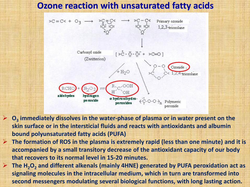
Ozone reaction with unsaturated fatty acids
O3 immediately dissolves in the water-phase of plasma or in water present on the skin surface or in the intersticial fluids and reacts with antioxidants and albumin bound polyunsaturated fatty acids (PUFA)
The formation of ROS in the plasma is extremely rapid (less than one minute) and it is accompanied by a small transitory decrease of the antioxidant capacity of our body that recovers to its normal level in 15-20 minutes.
The H2O2 and different alkenals (mainly 4HNE) generated by PUFA peroxidation act as signaling molecules in the intracellular medium, which in turn are transformed into second messengers modulating several biological functions, with long lasting action.
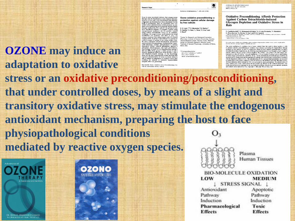
OZONE may induce an
adaptation to oxidative
stress or an oxidative preconditioning/postconditioning,
that under controlled doses, by means of a slight and
transitory oxidative stress, may stimulate the endogenous
antioxidant mechanism, preparing the host to face
physiopathological conditions
mediated by reactive oxygen species.
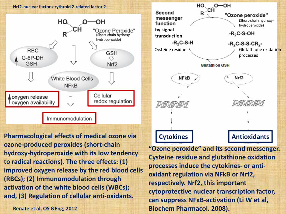
Pharmacological effects of medical ozone via ozone-produced peroxides (short-chain hydroxy-hydroperoxide with its low tendency to radical reactions). The three effects: (1) improved oxygen release by the red blood cells (RBCs); (2) Immunomodulation through activation of the white blood cells (WBCs); and, (3) Regulation of cellular anti-oxidants.
“Ozone peroxide” and its second messenger. Cysteine residue and glutathione oxidation processes induce the cytokines- or anti-oxidant regulation via NFkB or Nrf2, respectively. Nrf2, this important cytoprotective nuclear transcription factor, can suppress NFκB-activation (Li W et al, Biochem Pharmacol. 2008). Renate et al, OS &Eng, 2012
Cytokines Antioxidants
(Short-chain hydroxy-hydroperoxide)
(Short-chain hydroxy-hydroperoxide)
Cysteine residue Glutathione oxidation processes
by signal transduction
Nrf2-nuclear factor-erythroid 2-related factor 2
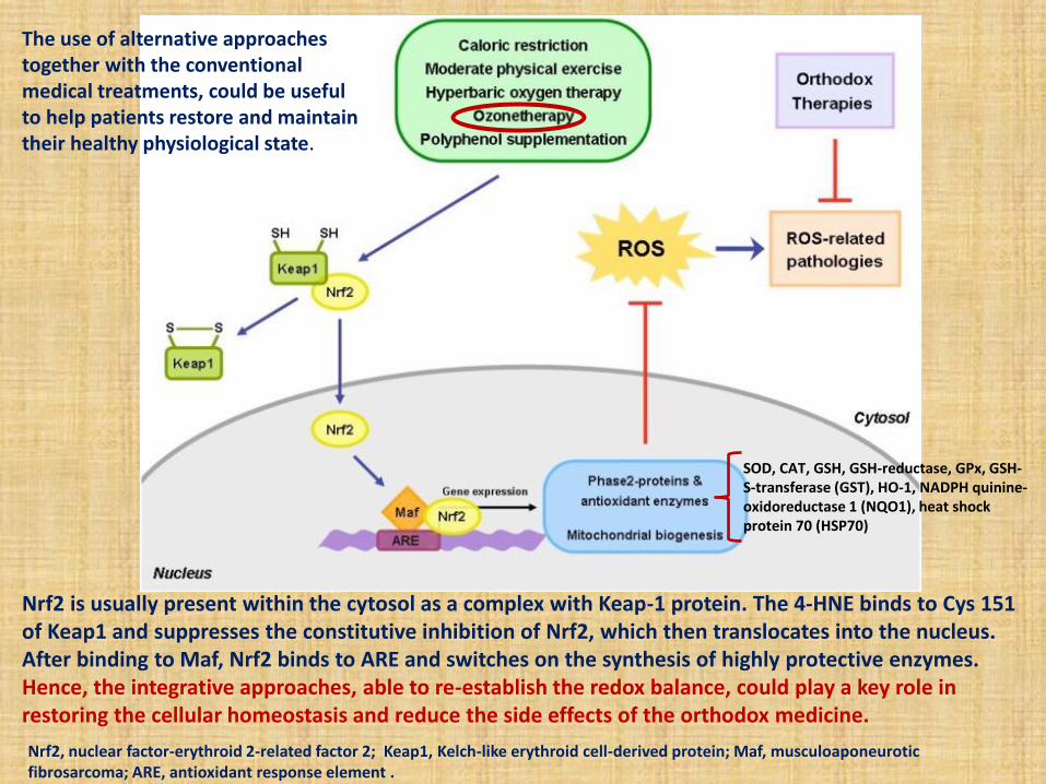
Nrf2 is usually present within the cytosol as a complex with Keap-1 protein. The 4-HNE binds to Cys 151 of Keap1 and suppresses the constitutive inhibition of Nrf2, which then translocates into the nucleus. After binding to Maf, Nrf2 binds to ARE and switches on the synthesis of highly protective enzymes. Hence, the integrative approaches, able to re-establish the redox balance, could play a key role in restoring the cellular homeostasis and reduce the side effects of the orthodox medicine.
The use of alternative approaches together with the conventional medical treatments, could be useful to help patients restore and maintain their healthy physiological state.
Nrf2, nuclear factor-erythroid 2-related factor 2; Keap1, Kelch-like erythroid cell-derived protein; Maf, musculoaponeurotic fibrosarcoma; ARE, antioxidant response element .
SOD, CAT, GSH, GSH-reductase, GPx, GSH-S-transferase (GST), HO-1, NADPH quinine-oxidoreductase 1 (NQO1), heat shock protein 70 (HSP70)
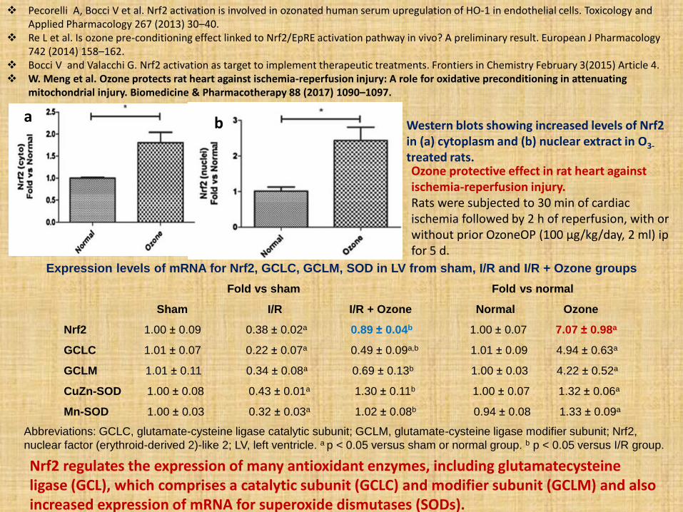
Expression levels of mRNA for Nrf2, GCLC, GCLM, SOD in LV from sham, I/R and I/R + Ozone groups
Abbreviations: GCLC, glutamate-cysteine ligase catalytic subunit; GCLM, glutamate-cysteine ligase modifier subunit; Nrf2,
nuclear factor (erythroid-derived 2)-like 2; LV, left ventricle. a p < 0.05 versus sham or normal group. b p < 0.05 versus I/R group.
Fold vs sham Fold vs normal
Sham I/R I/R + Ozone Normal Ozone
Nrf2 1.00 ± 0.09 0.38 ± 0.02a 0.89 ± 0.04b 1.00 ± 0.07 7.07 ± 0.98a
GCLC 1.01 ± 0.07 0.22 ± 0.07a 0.49 ± 0.09a,b 1.01 ± 0.09 4.94 ± 0.63a
GCLM 1.01 ± 0.11 0.34 ± 0.08a 0.69 ± 0.13b 1.00 ± 0.03 4.22 ± 0.52a
CuZn-SOD 1.00 ± 0.08 0.43 ± 0.01a 1.30 ± 0.11b 1.00 ± 0.07 1.32 ± 0.06a
Mn-SOD 1.00 ± 0.03 0.32 ± 0.03a 1.02 ± 0.08b 0.94 ± 0.08 1.33 ± 0.09a
Nrf2 regulates the expression of many antioxidant enzymes, including glutamatecysteine ligase (GCL), which comprises a catalytic subunit (GCLC) and modifier subunit (GCLM) and also increased expression of mRNA for superoxide dismutases (SODs).
Ozone protective effect in rat heart against ischemia-reperfusion injury. Rats were subjected to 30 min of cardiac ischemia followed by 2 h of reperfusion, with or without prior OzoneOP (100 µg/kg/day, 2 ml) ip for 5 d.
Western blots showing increased levels of Nrf2 in (a) cytoplasm and (b) nuclear extract in O3-
treated rats.
Pecorelli A, Bocci V et al. Nrf2 activation is involved in ozonated human serum upregulation of HO-1 in endothelial cells. Toxicology and Applied Pharmacology 267 (2013) 30–40.
Re L et al. Is ozone pre-conditioning effect linked to Nrf2/EpRE activation pathway in vivo? A preliminary result. European J Pharmacology 742 (2014) 158–162.
Bocci V and Valacchi G. Nrf2 activation as target to implement therapeutic treatments. Frontiers in Chemistry February 3(2015) Article 4. W. Meng et al. Ozone protects rat heart against ischemia-reperfusion injury: A role for oxidative preconditioning in attenuating
mitochondrial injury. Biomedicine & Pharmacotherapy 88 (2017) 1090–1097.
a b
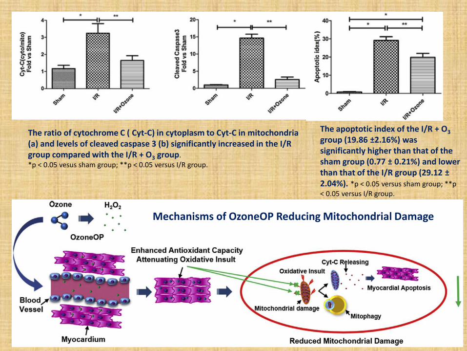
The apoptotic index of the I/R + O3 group (19.86 ±2.16%) was significantly higher than that of the sham group (0.77 ± 0.21%) and lower than that of the I/R group (29.12 ± 2.04%). *p < 0.05 versus sham group; **p < 0.05 versus I/R group.
The ratio of cytochrome C ( Cyt-C) in cytoplasm to Cyt-C in mitochondria (a) and levels of cleaved caspase 3 (b) significantly increased in the I/R group compared with the I/R + O3 group. *p < 0.05 vesus sham group; **p < 0.05 versus I/R group.
Mechanisms of OzoneOP Reducing Mitochondrial Damage
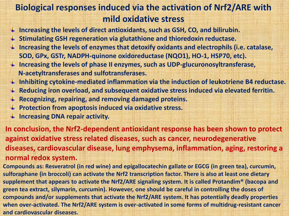
Increasing the levels of direct antioxidants, such as GSH, CO, and bilirubin. Stimulating GSH regeneration via glutathione and thioredoxin reductase. Increasing the levels of enzymes that detoxify oxidants and electrophils (i.e. catalase, SOD, GPx, GSTr, NADPH-quinone oxidoreductase (NQO1), HO-1, HSP70, etc). Increasing the levels of phase II enzymes, such as UDP-glucuronosyltransferase,
N-acetyltransferases and sulfotransferases. Inhibiting cytokine-mediated inflammation via the induction of leukotriene B4 reductase. Reducing iron overload, and subsequent oxidative stress induced via elevated ferritin. Recognizing, repairing, and removing damaged proteins. Protection from apoptosis induced via oxidative stress. Increasing DNA repair activity.
In conclusion, the Nrf2-dependent antioxidant response has been shown to protect against oxidative stress related diseases, such as cancer, neurodegenerative diseases, cardiovascular disease, lung emphysema, inflammation, aging, restoring a normal redox system.
Biological responses induced via the activation of Nrf2/ARE with mild oxidative stress
Compounds as: Resveratrol (in red wine) and epigallocatechin gallate or EGCG (in green tea), curcumin, sulforaphane (in broccoli) can activate the Nrf2 transcription factor. There is also at least one dietary supplement that appears to activate the Nrf2/ARE signaling system. It is called Protandim® (bacopa and green tea extract, silymarin, curcumin). However, one should be careful in controlling the doses of compounds and/or supplements that activate the Nrf2/ARE system. It has potentially deadly properties when over-activated. The Nrf2/ARE system is over-activated in some forms of multidrug-resistant cancer and cardiovascular diseases.
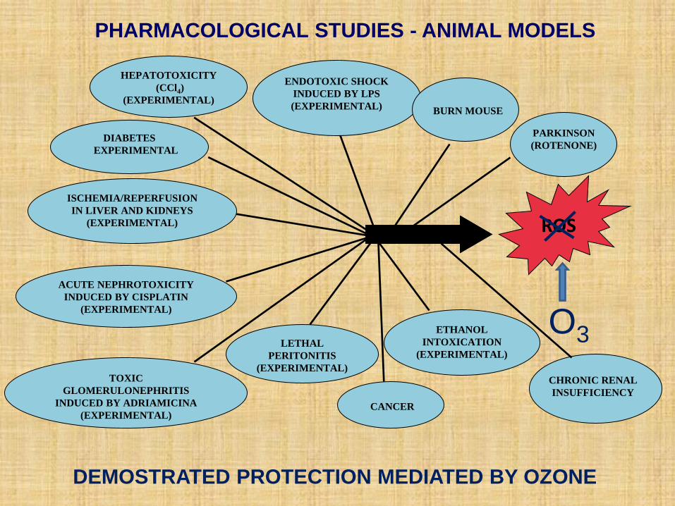
HEPATOTOXICITY
(CCl4)
(EXPERIMENTAL)
DIABETES
EXPERIMENTAL
ISCHEMIA/REPERFUSION
IN LIVER AND KIDNEYS
(EXPERIMENTAL)
ROS
ACUTE NEPHROTOXICITY
INDUCED BY CISPLATIN
(EXPERIMENTAL)
TOXIC
GLOMERULONEPHRITIS
INDUCED BY ADRIAMICINA
(EXPERIMENTAL)
ENDOTOXIC SHOCK
INDUCED BY LPS
(EXPERIMENTAL)
LETHAL
PERITONITIS
(EXPERIMENTAL)
BURN MOUSE
CHRONIC RENAL
INSUFFICIENCY
PHARMACOLOGICAL STUDIES - ANIMAL MODELS
PARKINSON
(ROTENONE)
ETHANOL
INTOXICATION
(EXPERIMENTAL)
O3
DEMOSTRATED PROTECTION MEDIATED BY OZONE
CANCER
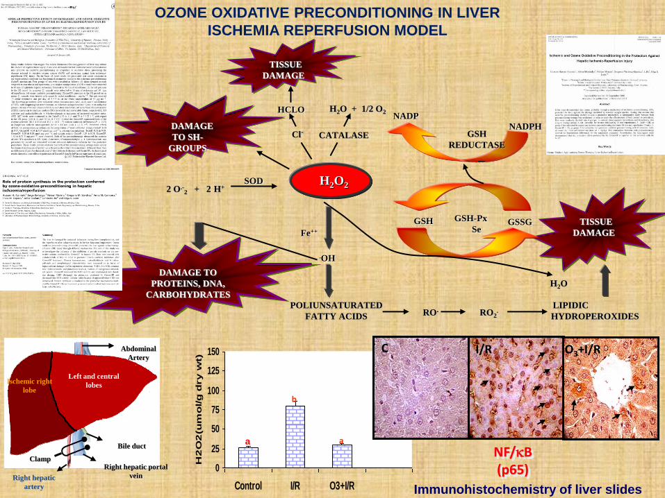
TISSUE
DAMAGE
HCLO H2O + 1/2 O2 NADP
NADPH
H2O2
.OH
POLIUNSATURATED
FATTY ACIDS
RO. RO2.
LIPIDIC
HYDROPEROXIDES
DAMAGE TO
PROTEINS, DNA,
CARBOHYDRATES
DAMAGE
TO SH-
GROUPS
Cl- CATALASE GSH
REDUCTASE
SOD 2 O.-
2 + 2 H+
Fe++
GSH GSSG GSH-Px
Se
TISSUE
DAMAGE
H2O
0
25
50
75
100
125
150
Control I/R O3+I/R
H2
O2
(u
mo
l/g
dry
wt)
b
a
Ischemic right
lobe
Left and central
lobes
Right hepatic
artery
Right hepatic portal
vein
Bile duct
Clamp
Abdominal
Artery
a
OZONE OXIDATIVE PRECONDITIONING IN LIVER
ISCHEMIA REPERFUSION MODEL
NF/B (p65)
Immunohistochemistry of liver slides
C I/R O3+I/R
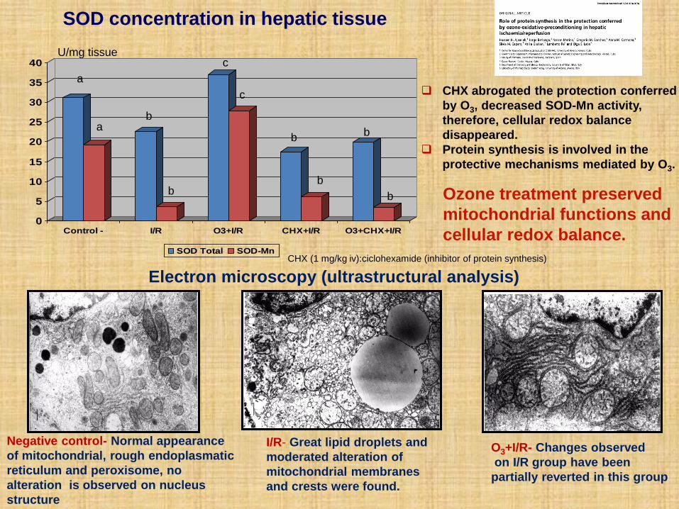
Negative control- Normal appearance
of mitochondrial, rough endoplasmatic
reticulum and peroxisome, no
alteration is observed on nucleus
structure
I/R- Great lipid droplets and
moderated alteration of
mitochondrial membranes
and crests were found.
O3+I/R- Changes observed
on I/R group have been
partially reverted in this group
U/mg tissue
0
5
10
15
20
25
30
35
40
Control - I/R O3+I/R CHX+I/R O3+CHX+I/R
SOD Total SOD-Mn
a
b
c
b b
a
b
c
b
b
SOD concentration in hepatic tissue
CHX (1 mg/kg iv):ciclohexamide (inhibitor of protein synthesis)
Electron microscopy (ultrastructural analysis)
CHX abrogated the protection conferred
by O3, decreased SOD-Mn activity,
therefore, cellular redox balance
disappeared.
Protein synthesis is involved in the
protective mechanisms mediated by O3.
Ozone treatment preserved
mitochondrial functions and
cellular redox balance.
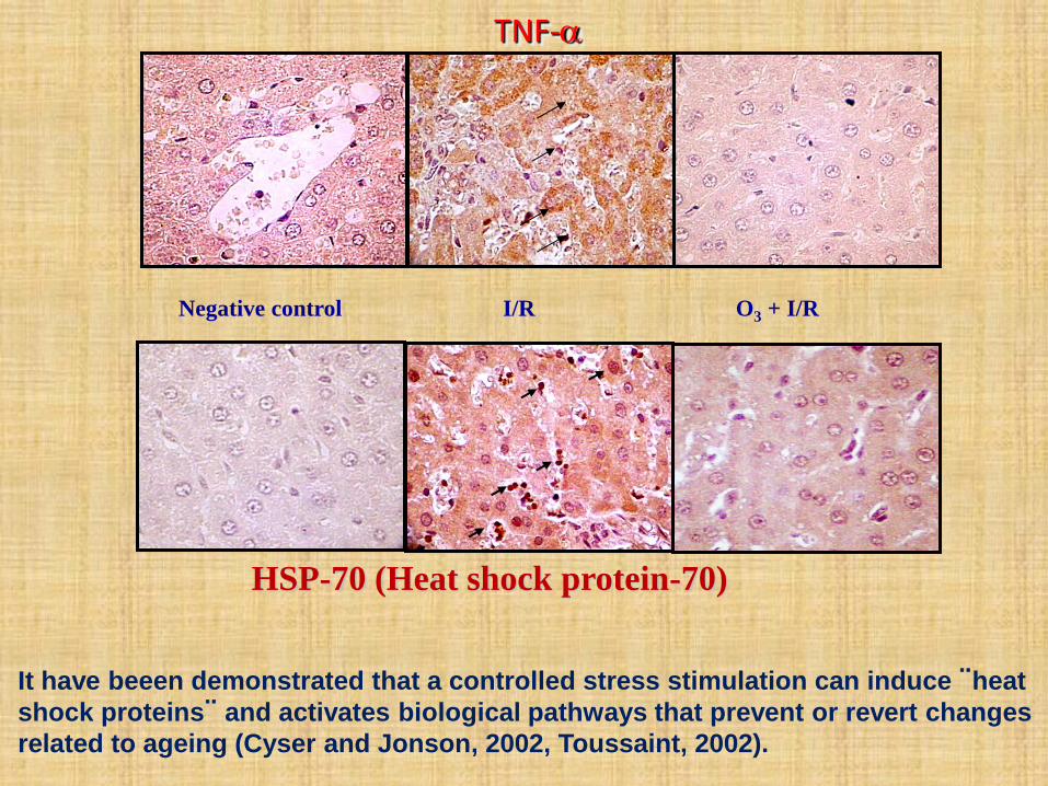
Negative control I/R O3 + I/R
TNF-
HSP-70 (Heat shock protein-70)
It have beeen demonstrated that a controlled stress stimulation can induce ¨heat
shock proteins¨ and activates biological pathways that prevent or revert changes
related to ageing (Cyser and Jonson, 2002, Toussaint, 2002).
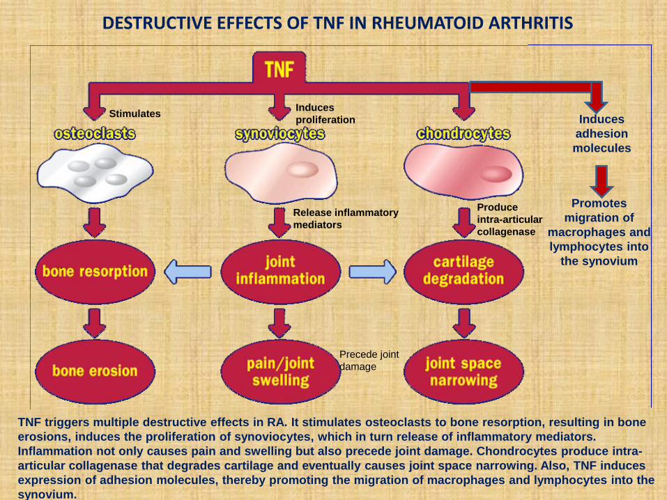
DESTRUCTIVE EFFECTS OF TNF IN RHEUMATOID ARTHRITIS
TNF triggers multiple destructive effects in RA. It stimulates osteoclasts to bone resorption, resulting in bone
erosions, induces the proliferation of synoviocytes, which in turn release of inflammatory mediators.
Inflammation not only causes pain and swelling but also precede joint damage. Chondrocytes produce intra-
articular collagenase that degrades cartilage and eventually causes joint space narrowing. Also, TNF induces
expression of adhesion molecules, thereby promoting the migration of macrophages and lymphocytes into the
synovium.
Induces
adhesion
molecules
Promotes
migration of
macrophages and
lymphocytes into
the synovium
Stimulates Induces
proliferation
Release inflammatory
mediators
Produce
intra-articular
collagenase
Precede joint
damage
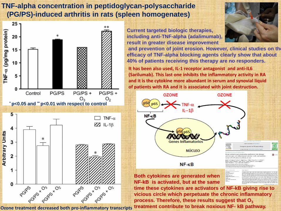
TNF-alpha concentration in peptidoglycan-polysaccharide
(PG/PS)-induced arthritis in rats (spleen homogenates)
* p<0.05 and ** p<0.01 with respect to control
Current targeted biologic therapies,
including anti-TNF-alpha (adalimumab),
result in greater disease improvement
and prevention of joint erosion. However, clinical studies on the
efficacy of TNF-alpha blocking agents clearly show that about
40% of patients receiving this therapy are no responders.
It has been also used, IL-1 receptor antagonist and anti-IL6 (Sarilumab). This last one inhibits the inflammatory activity in RA and it is the cytokine more abundant in serum and synovial liquid of patients with RA and it is associated with joint destruction.
Both cytokines are generated when
NF-kB is activated, but at the same
time these cytokines are activators of NF-kB giving rise to
vicious circle which perpetuate the chronic inflammatory
process. Therefore, these results suggest that O3
treatment contribute to break noxious NF- kB pathway. Ozone treatment decreased both pro-inflammatory transcripts
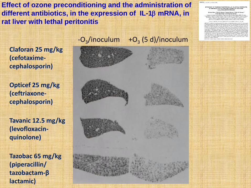
Effect of ozone preconditioning and the administration of
different antibiotics, in the expression of IL-1 mRNA, in
rat liver with lethal peritonitis
Claforan 25 mg/kg (cefotaxime-cephalosporin)
Opticef 25 mg/kg (ceftriaxone-cephalosporin)
Tavanic 12.5 mg/kg (levofloxacin-quinolone)
Tazobac 65 mg/kg (piperacillin/ tazobactam-β lactamic)
-O3/inoculum +O3 (5 d)/inoculum
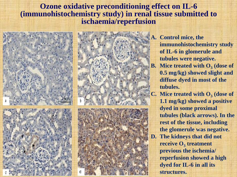
A. Control mice, the
immunohistochemistry study
of IL-6 in glomerule and
tubules were negative.
B. Mice treated with O3 (dose of
0.5 mg/kg) showed slight and
diffuse dyed in most of the
tubules.
C. Mice treated with O3 (dose of
1.1 mg/kg) showed a positive
dyed in some proximal
tubules (black arrows). In the
rest of the tissue, including
the glomerule was negative.
D. The kidneys that did not
receive O3 treatment
previous the ischemia/
reperfusion showed a high
dyed for IL-6 in all its
structures.
Ozone oxidative preconditioning effect on IL-6 (immunohistochemistry study) in renal tissue submitted to
ischaemia/reperfusion
0
0,2
0,4
0,6
0,8
1
1,2
NO
(n
mo
l/m
g t
ejid
o)
a
b
c
a
d
e
f
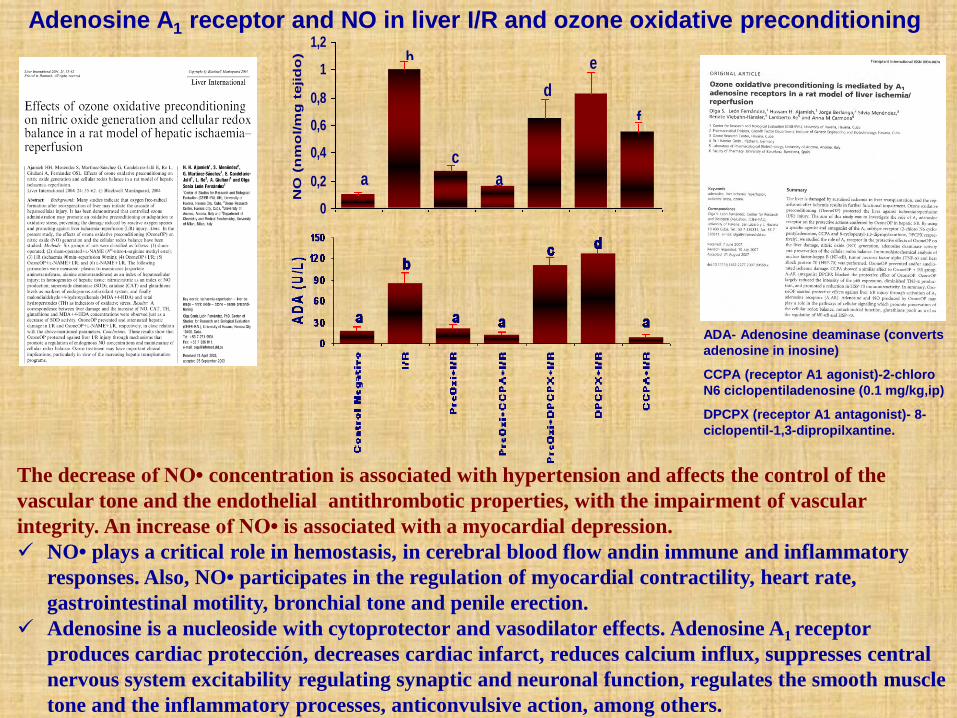
ADA- Adenosine deaminase (converts
adenosine in inosine)
CCPA (receptor A1 agonist)-2-chloro
N6 ciclopentiladenosine (0.1 mg/kg,ip)
DPCPX (receptor A1 antagonist)- 8-
ciclopentil-1,3-dipropilxantine.
The decrease of NO• concentration is associated with hypertension and affects the control of the
vascular tone and the endothelial antithrombotic properties, with the impairment of vascular
integrity. An increase of NO• is associated with a myocardial depression.
NO• plays a critical role in hemostasis, in cerebral blood flow andin immune and inflammatory
responses. Also, NO• participates in the regulation of myocardial contractility, heart rate,
gastrointestinal motility, bronchial tone and penile erection.
Adenosine is a nucleoside with cytoprotector and vasodilator effects. Adenosine A1 receptor
produces cardiac protección, decreases cardiac infarct, reduces calcium influx, suppresses central
nervous system excitability regulating synaptic and neuronal function, regulates the smooth muscle
tone and the inflammatory processes, anticonvulsive action, among others.
Adenosine A1 receptor and NO in liver I/R and ozone oxidative preconditioning
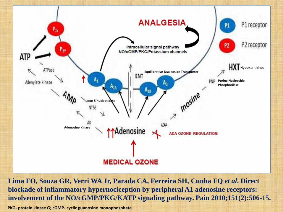
Lima FO, Souza GR, Verri WA Jr, Parada CA, Ferreira SH, Cunha FQ et al. Direct
blockade of inflammatory hypernociception by peripheral A1 adenosine receptors:
involvement of the NO/cGMP/PKG/KATP signaling pathway. Pain 2010;151(2):506-15.
Equilibrative Nucleoside Transporter
Adenosine Kinase
ecto-5’nucleotidase
Purine Nucleoside Phosphorilase
Hypoxanthines
PKG- protein kinase G; cGMP- cyclic guanosine monophosphate.
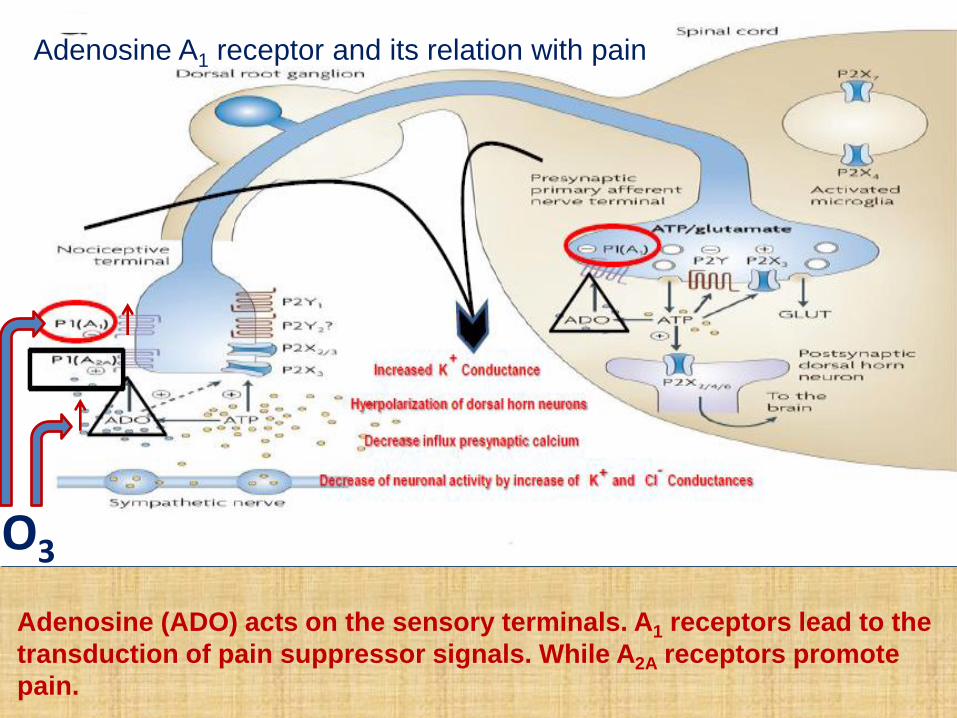
Adenosine (ADO) acts on the sensory terminals. A1 receptors lead to the
transduction of pain suppressor signals. While A2A receptors promote
pain.
Adenosine A1 receptor and its relation with pain
O3
0
2
4
6
8
10
initial 5 sessions 10 sessions 15 sessions
VAS
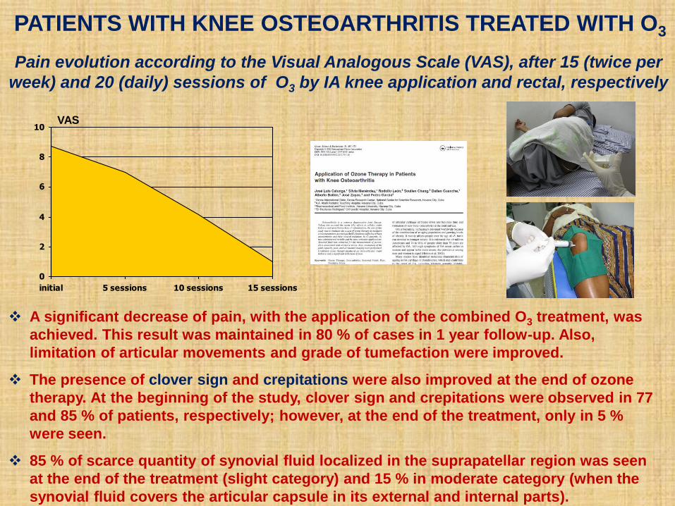
Pain evolution according to the Visual Analogous Scale (VAS), after 15 (twice per
week) and 20 (daily) sessions of O3 by IA knee application and rectal, respectively
A significant decrease of pain, with the application of the combined O3 treatment, was
achieved. This result was maintained in 80 % of cases in 1 year follow-up. Also,
limitation of articular movements and grade of tumefaction were improved.
The presence of clover sign and crepitations were also improved at the end of ozone
therapy. At the beginning of the study, clover sign and crepitations were observed in 77
and 85 % of patients, respectively; however, at the end of the treatment, only in 5 %
were seen.
85 % of scarce quantity of synovial fluid localized in the suprapatellar region was seen
at the end of the treatment (slight category) and 15 % in moderate category (when the
synovial fluid covers the articular capsule in its external and internal parts).
PATIENTS WITH KNEE OSTEOARTHRITIS TREATED WITH O3

Presynaptic adenosine A1 receptors is able to inhibit the release of
glutamate at the excitatory synapse diminishing neuronal
hyperexcitability .
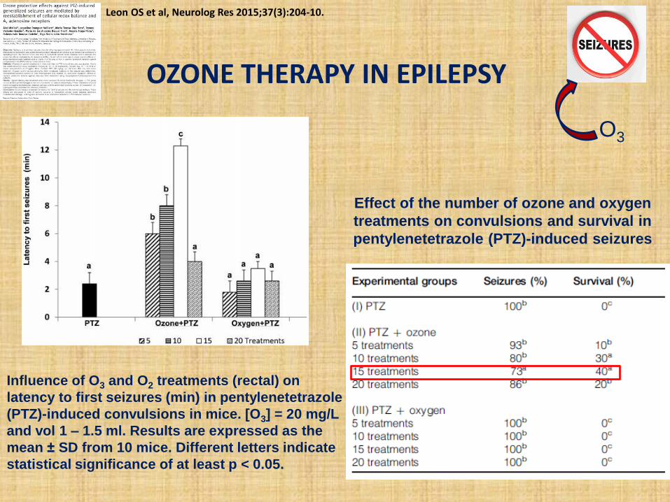
Influence of O3 and O2 treatments (rectal) on
latency to first seizures (min) in pentylenetetrazole
(PTZ)-induced convulsions in mice. [O3] = 20 mg/L
and vol 1 – 1.5 ml. Results are expressed as the
mean ± SD from 10 mice. Different letters indicate
statistical significance of at least p < 0.05.
OZONE THERAPY IN EPILEPSY
O3
Leon OS et al, Neurolog Res 2015;37(3):204-10.
Effect of the number of ozone and oxygen
treatments on convulsions and survival in
pentylenetetrazole (PTZ)-induced seizures
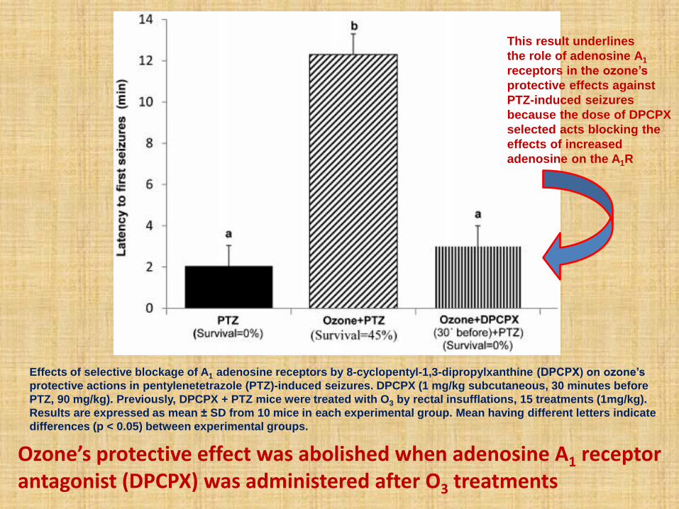
Effects of selective blockage of A1 adenosine receptors by 8-cyclopentyl-1,3-dipropylxanthine (DPCPX) on ozone’s
protective actions in pentylenetetrazole (PTZ)-induced seizures. DPCPX (1 mg/kg subcutaneous, 30 minutes before
PTZ, 90 mg/kg). Previously, DPCPX + PTZ mice were treated with O3 by rectal insufflations, 15 treatments (1mg/kg).
Results are expressed as mean ± SD from 10 mice in each experimental group. Mean having different letters indicate
differences (p < 0.05) between experimental groups.
This result underlines
the role of adenosine A1
receptors in the ozone’s
protective effects against
PTZ-induced seizures
because the dose of DPCPX
selected acts blocking the
effects of increased
adenosine on the A1R
Ozone’s protective effect was abolished when adenosine A1 receptor antagonist (DPCPX) was administered after O3 treatments
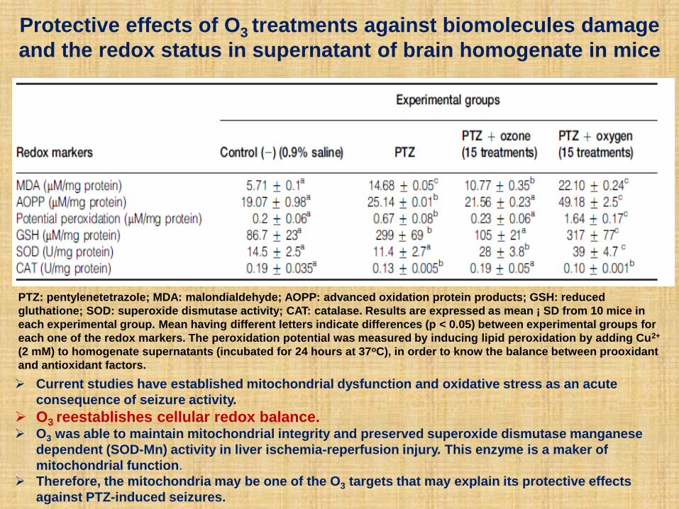
Protective effects of O3 treatments against biomolecules damage
and the redox status in supernatant of brain homogenate in mice
PTZ: pentylenetetrazole; MDA: malondialdehyde; AOPP: advanced oxidation protein products; GSH: reduced
gluthatione; SOD: superoxide dismutase activity; CAT: catalase. Results are expressed as mean ¡ SD from 10 mice in
each experimental group. Mean having different letters indicate differences (p < 0.05) between experimental groups for
each one of the redox markers. The peroxidation potential was measured by inducing lipid peroxidation by adding Cu2+
(2 mM) to homogenate supernatants (incubated for 24 hours at 37oC), in order to know the balance between prooxidant
and antioxidant factors.
Current studies have established mitochondrial dysfunction and oxidative stress as an acute
consequence of seizure activity.
O3 reestablishes cellular redox balance. O3 was able to maintain mitochondrial integrity and preserved superoxide dismutase manganese
dependent (SOD-Mn) activity in liver ischemia-reperfusion injury. This enzyme is a maker of
mitochondrial function.
Therefore, the mitochondria may be one of the O3 targets that may explain its protective effects
against PTZ-induced seizures.
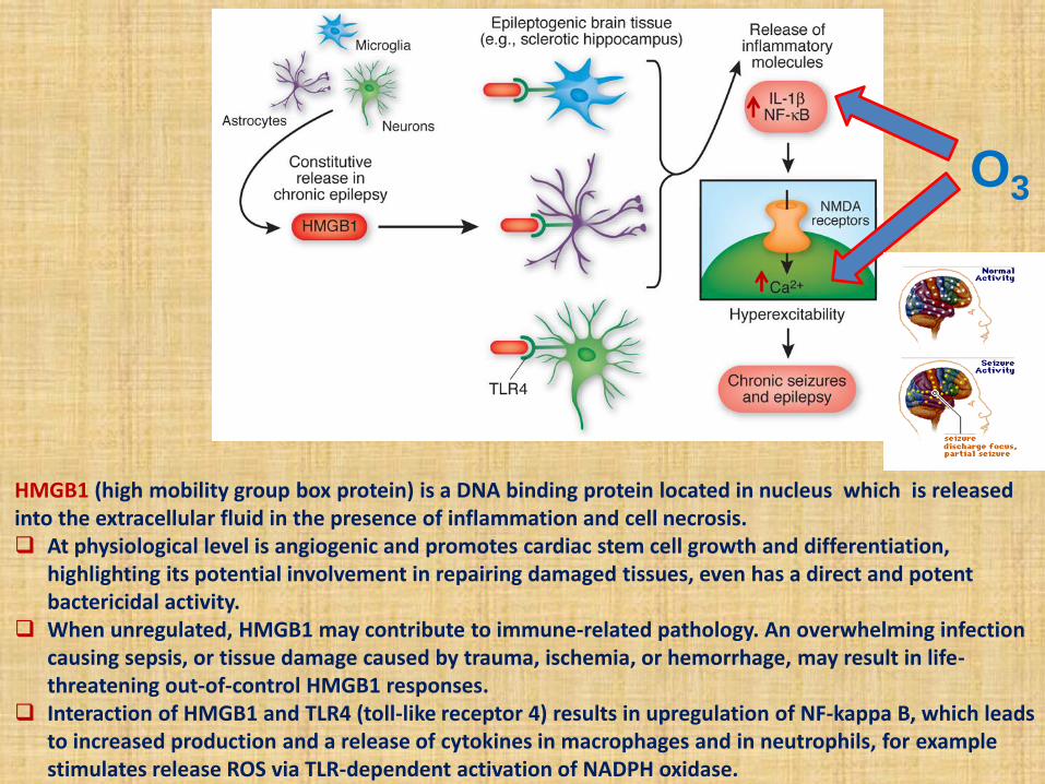
HMGB1 (high mobility group box protein) is a DNA binding protein located in nucleus which is released into the extracellular fluid in the presence of inflammation and cell necrosis. At physiological level is angiogenic and promotes cardiac stem cell growth and differentiation,
highlighting its potential involvement in repairing damaged tissues, even has a direct and potent bactericidal activity.
When unregulated, HMGB1 may contribute to immune-related pathology. An overwhelming infection causing sepsis, or tissue damage caused by trauma, ischemia, or hemorrhage, may result in life-threatening out-of-control HMGB1 responses.
Interaction of HMGB1 and TLR4 (toll-like receptor 4) results in upregulation of NF-kappa B, which leads to increased production and a release of cytokines in macrophages and in neutrophils, for example stimulates release ROS via TLR-dependent activation of NADPH oxidase.
O3
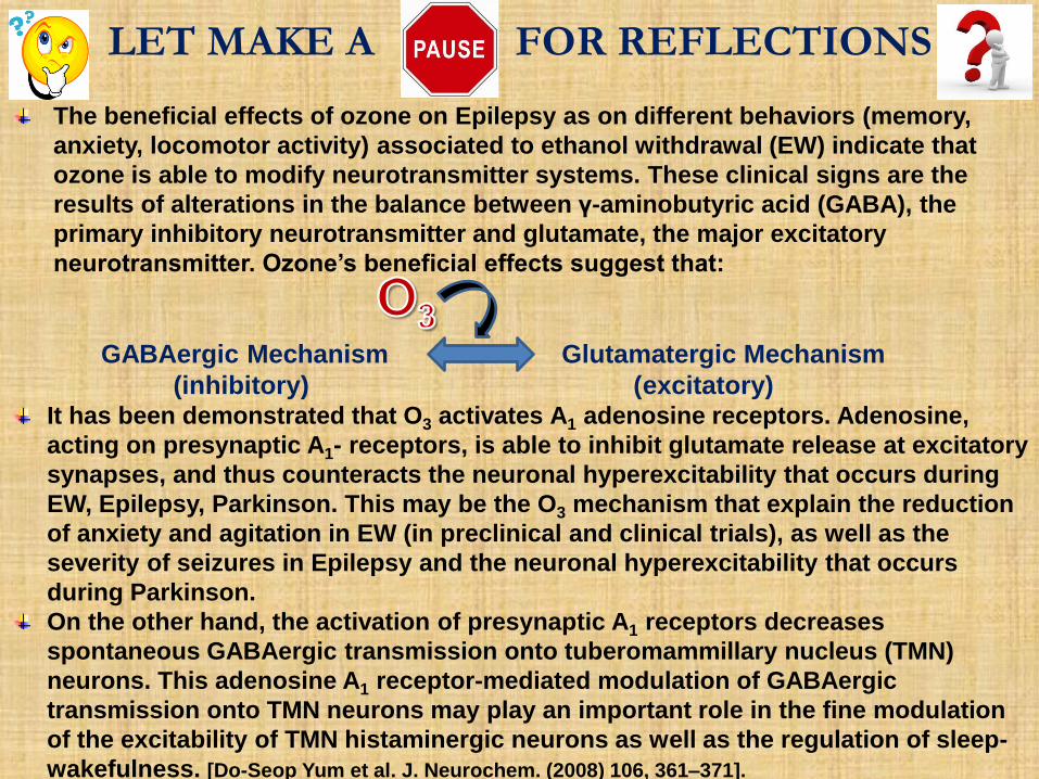
The beneficial effects of ozone on Epilepsy as on different behaviors (memory,
anxiety, locomotor activity) associated to ethanol withdrawal (EW) indicate that
ozone is able to modify neurotransmitter systems. These clinical signs are the
results of alterations in the balance between γ-aminobutyric acid (GABA), the
primary inhibitory neurotransmitter and glutamate, the major excitatory
neurotransmitter. Ozone’s beneficial effects suggest that:
GABAergic Mechanism Glutamatergic Mechanism
(inhibitory) (excitatory) It has been demonstrated that O3 activates A1 adenosine receptors. Adenosine,
acting on presynaptic A1- receptors, is able to inhibit glutamate release at excitatory
synapses, and thus counteracts the neuronal hyperexcitability that occurs during
EW, Epilepsy, Parkinson. This may be the O3 mechanism that explain the reduction
of anxiety and agitation in EW (in preclinical and clinical trials), as well as the
severity of seizures in Epilepsy and the neuronal hyperexcitability that occurs
during Parkinson.
On the other hand, the activation of presynaptic A1 receptors decreases
spontaneous GABAergic transmission onto tuberomammillary nucleus (TMN)
neurons. This adenosine A1 receptor-mediated modulation of GABAergic
transmission onto TMN neurons may play an important role in the fine modulation
of the excitability of TMN histaminergic neurons as well as the regulation of sleep-
wakefulness. [Do-Seop Yum et al. J. Neurochem. (2008) 106, 361–371].
LET MAKE A FOR REFLECTIONS
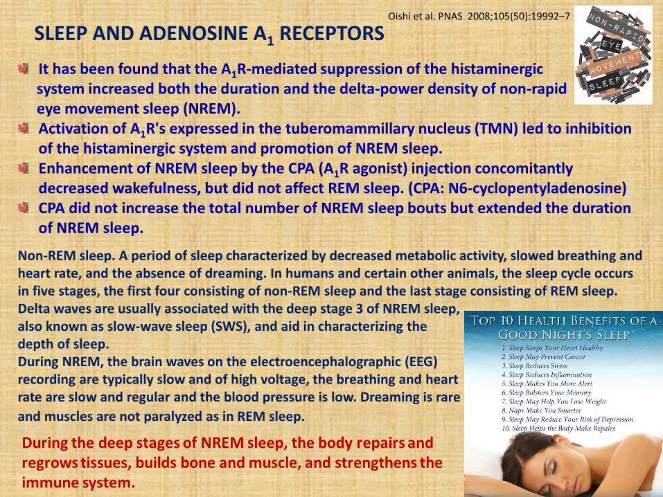
It has been found that the A1R-mediated suppression of the histaminergic system increased both the duration and the delta-power density of non-rapid eye movement sleep (NREM).
Activation of A1R's expressed in the tuberomammillary nucleus (TMN) led to inhibition of the histaminergic system and promotion of NREM sleep. Enhancement of NREM sleep by the CPA (A1R agonist) injection concomitantly decreased wakefulness, but did not affect REM sleep. (CPA: N6-cyclopentyladenosine) CPA did not increase the total number of NREM sleep bouts but extended the duration of NREM sleep.
Non-REM sleep. A period of sleep characterized by decreased metabolic activity, slowed breathing and heart rate, and the absence of dreaming. In humans and certain other animals, the sleep cycle occurs in five stages, the first four consisting of non-REM sleep and the last stage consisting of REM sleep. Delta waves are usually associated with the deep stage 3 of NREM sleep, also known as slow-wave sleep (SWS), and aid in characterizing the depth of sleep. During NREM, the brain waves on the electroencephalographic (EEG) recording are typically slow and of high voltage, the breathing and heart rate are slow and regular and the blood pressure is low. Dreaming is rare
and muscles are not paralyzed as in REM sleep.
Oishi et al. PNAS 2008;105(50):19992–7
SLEEP AND ADENOSINE A1 RECEPTORS
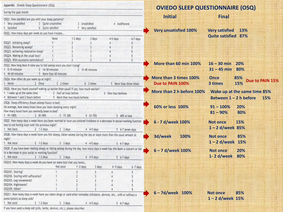
Initial Final
Very unsatisfied 100% Very satisfied 13% Quite satisfied 87%
More than 60 min 100% 16 – 30 min 20% 31 – 45 min 80%
More than 3 times 100% Once 85% Due to PAIN 100% 3 times 15%
More than 2 h before 100% Wake up at the same time 85%
Between 1 – 2 h before 15%
60% or less 100% 91 – 100% 20% 81 – 90% 80%
6 - 7 d/week 100% Not once 15% 1 – 2 d/week 85%
3d/week 100% Not once 85% 1 – 2 d/week 15%
6 – 7 d/week 100% Not once 20% 1- 2 d/week 80%
6 – 7d/week 100% Not once 85% 1 – 2 d/week 15%
Due to PAIN 15%
OVIEDO SLEEP QUESTIONNAIRE (OSQ)
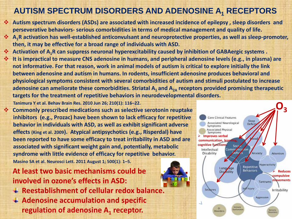
Autism spectrum disorders (ASDs) are associated with increased incidence of epilepsy , sleep disorders and perseverative behaviors- serious comorbidities in terms of medical management and quality of life.
A1R activation has well-established anticonvulsant and neuroprotective properties, as well as sleep-promoter, then, it may be effective for a broad range of individuals with ASD.
Activation of A1R can suppress neuronal hyperexcitability caused by inhibition of GABAergic systems . It is impractical to measure CNS adenosine in humans, and peripheral adenosine levels (e.g., in plasma) are
not informative. For that reason, work in animal models of autism is critical to explore initially the link between adenosine and autism in humans. In rodents, insufficient adenosine produces behavioral and physiological symptoms consistent with several comorbidities of autism and stimuli postulated to increase adenosine can ameliorate these comorbidities. Striatal A1 and A2A receptors provided promising therapeutic targets for the treatment of repetitive behaviors in neurodevelopmental disorders.
Tanimura Y et al. Behav Brain Res. 2010 Jun 26; 210(1): 116–22.
Commonly prescribed medications such as selective serotonin reuptake inhibitors (e.g., Prozac) have been shown to lack efficacy for repetitive behavior in individuals with ASD, as well as exhibit significant adverse effects (King et al. 2009). Atypical antipsychotics (e.g., Risperdal) have been reported to have some efficacy to treat irritability in ASD and are associated with significant weight gain and, potentially, metabolic syndrome with little evidence of efficacy for repetitive behavior. Masino SA et al. Neurosci Lett. 2011 August 1; 500(1): 1–5.
AUTISM SPECTRUM DISORDERS AND ADENOSINE A1 RECEPTORS
At least two basic mechanisms could be involved in ozone’s effects in ASD:
Reestablishment of cellular redox balance. Adenosine accumulation and specific regulation of adenosine A1 receptor.
O3
Improves verbal communication, learning, cognitive functions.
Reduces compulsive movements
0
100
200
300
400
500
0
500
1000
1500
0
5000
10000
15000
20000
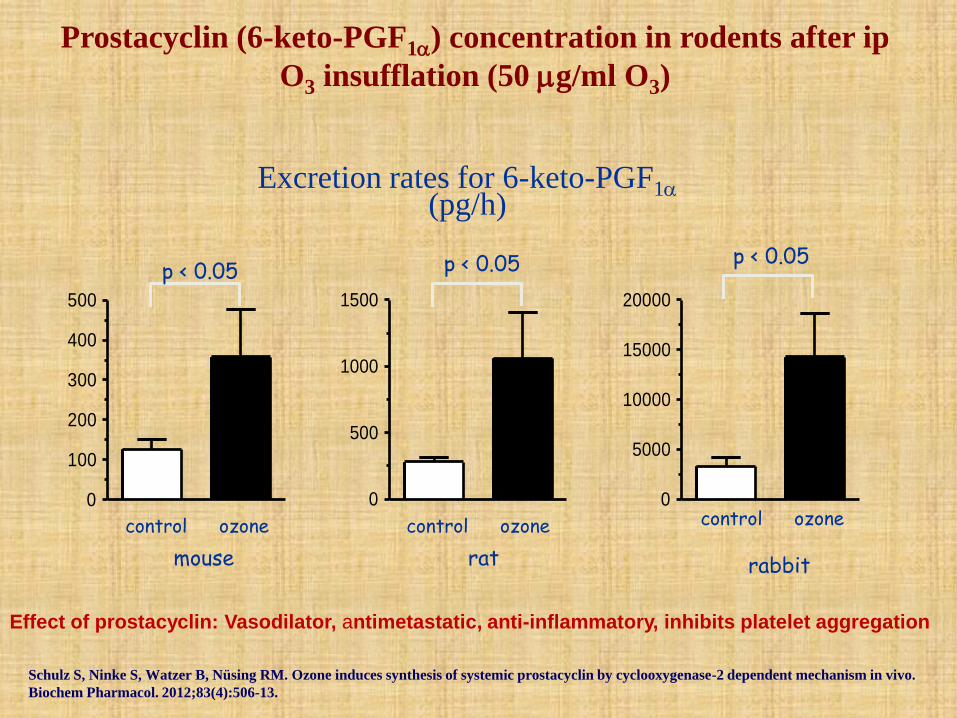
Excretion rates for 6-keto-PGF1 (pg/h)
control ozone control ozone control ozone
mouse rat rabbit
Prostacyclin (6-keto-PGF1) concentration in rodents after ip
O3 insufflation (50 g/ml O3)
p < 0.05 p < 0.05 p < 0.05
Schulz S, Ninke S, Watzer B, Nüsing RM. Ozone induces synthesis of systemic prostacyclin by cyclooxygenase-2 dependent mechanism in vivo.
Biochem Pharmacol. 2012;83(4):506-13.
Effect of prostacyclin: Vasodilator, antimetastatic, anti-inflammatory, inhibits platelet aggregation
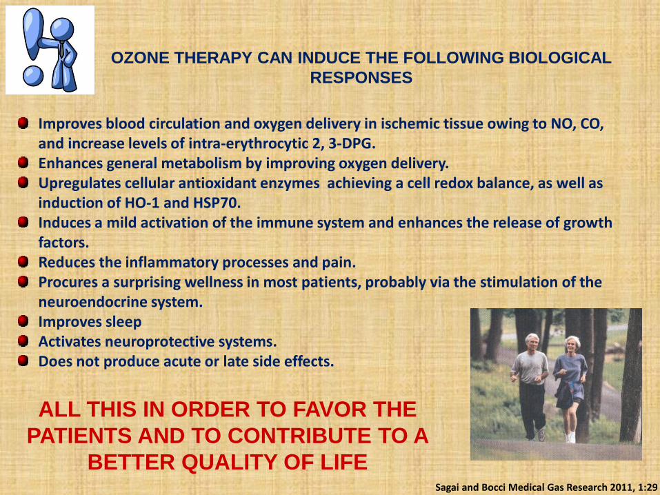
Improves blood circulation and oxygen delivery in ischemic tissue owing to NO, CO, and increase levels of intra-erythrocytic 2, 3-DPG. Enhances general metabolism by improving oxygen delivery. Upregulates cellular antioxidant enzymes achieving a cell redox balance, as well as induction of HO-1 and HSP70. Induces a mild activation of the immune system and enhances the release of growth factors. Reduces the inflammatory processes and pain. Procures a surprising wellness in most patients, probably via the stimulation of the neuroendocrine system. Improves sleep Activates neuroprotective systems. Does not produce acute or late side effects.
Sagai and Bocci Medical Gas Research 2011, 1:29
OZONE THERAPY CAN INDUCE THE FOLLOWING BIOLOGICAL
RESPONSES
ALL THIS IN ORDER TO FAVOR THE
PATIENTS AND TO CONTRIBUTE TO A
BETTER QUALITY OF LIFE
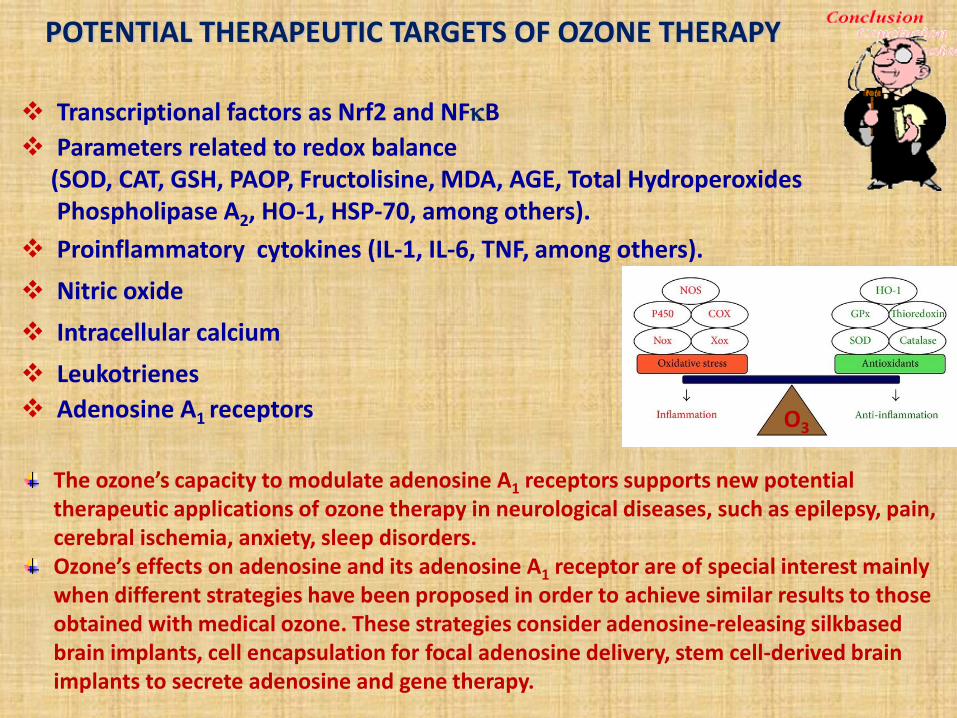
Transcriptional factors as Nrf2 and NFB
Parameters related to redox balance (SOD, CAT, GSH, PAOP, Fructolisine, MDA, AGE, Total Hydroperoxides Phospholipase A2, HO-1, HSP-70, among others).
Proinflammatory cytokines (IL-1, IL-6, TNF, among others).
Nitric oxide
Intracellular calcium
Leukotrienes
Adenosine A1 receptors
POTENTIAL THERAPEUTIC TARGETS OF OZONE THERAPY
The ozone’s capacity to modulate adenosine A1 receptors supports new potential therapeutic applications of ozone therapy in neurological diseases, such as epilepsy, pain, cerebral ischemia, anxiety, sleep disorders. Ozone’s effects on adenosine and its adenosine A1 receptor are of special interest mainly when different strategies have been proposed in order to achieve similar results to those obtained with medical ozone. These strategies consider adenosine-releasing silkbased brain implants, cell encapsulation for focal adenosine delivery, stem cell-derived brain implants to secrete adenosine and gene therapy.
O3