nervous system 2 cerebrovascular disease prof john simpson
TRANSCRIPT

Nervous System 2Cerebrovascular Disease
Prof John Simpson

Cerebrovascular disease (CVD)
• “strokes”• brain disease due to vascular pathology
– thrombosis, embolism or hypotension causing ischaemia/hypoxia
– haemorrhage causing disruption
• major cause of death and disability, especially in more developed countries
• commonly associated with atheroma, diabetes and hypertension

Two major pathologies
• infarction– thrombotic (overall 80%+ of all strokes)– embolic– hypotensive– (venous)
• haemorrhage– intracerebral– subarachnoid
• but, one can lead to the other!

Hypoxia and the brain
• brain highly oxygen (and glucose) dependent
• blood flow normally autoregulated
• problems arise from 1) major fall in BP or systemic hypoxia causing
diffuse damage or
2) vessel blockage, causing focal damage

Diffuse hypoxic damage
• depends on severity and duration of hypoxia• most susceptible neurons in hippocampus,
Purkinje cells, cerebral cortex• affected brain oedematous, raising ICP• causes anything from mild confusion to PVS to
immediate brain death• in acute hypotension, may also be focal damage
– “watershed” (border zone) infarcts – most often between anterior cerebral and middle cerebral artery supplies

Focal hypoxic damage
• results depend on presence of collaterals– some exist on surface, e.g. Circle of Willis – but not within brain
• focal vascular abnormality due to– thrombosis or embolism
• clinical effects ~ site, extent and speed of onset of vascular block
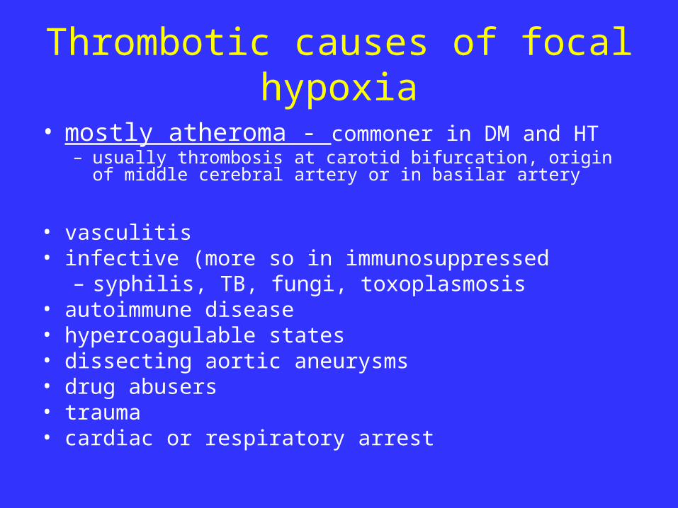
Thrombotic causes of focal hypoxia
• mostly atheroma - commoner in DM and HT– usually thrombosis at carotid bifurcation, origin of middle cerebral
artery or in basilar artery
• vasculitis• infective (more so in immunosuppressed
– syphilis, TB, fungi, toxoplasmosis• autoimmune disease• hypercoagulable states• dissecting aortic aneurysms• drug abusers• trauma• cardiac or respiratory arrest

Embolic causes of focal hypoxia
• commonest are cardiac mural thrombi– MI, valvular disease, atrial fibrillation
• arterial thromboemboli - especially from carotid plaques (sometimes include plaque material)
• paradoxical emboli - children with cardiac anomalies• emboli of other material (tumour, fat, marrow, air)

Cerebral embolism
• middle cerebral territory most often affected• emboli lodge at branches or stenoses• often, occlusion cannot be identified PM
– ?thromboemboli already lysed
• “shower” embolism of fat may occur after fractures– capillary blockages – disturb higher cortical function and
consciousness, often with no localizing signs
• widespread haemorrhagic lesions of white matter characteristic of bone marrow embolism after trauma
• tumour emboli more important as source for metastases, then cause of hypoxia

Cerebral infarcts
• sometimes classified as red or pale• depends on presence of haemorrhage from
infarcted vessels• (any infarct may show surrounding zone of
lesser hypoxic damage and hyperaemic reaction, which may be oedematous)
• venous infarcts – usually beside sinuses – associated with infection, dehydration and drugs (oral contraceptives)

Natural history of infarcts
• effects depend on site, size and speed of onset– in some effect complete from the start, in others clinical picture
evolves
• thrombotic infarcts most commonly internal capsule (corticospinal paths), hence hemiplegias etc
• reperfusion (micro)haemorrhages may occur• if patient survives, infarcted tissue phagocytosed by
microglia and monocytes from blood, then gliosis– macrophages persist at site for years as lipid-containing
“compound granular corpuscles”– in red infarct, macrophages also contain iron– end result of repair often a cystic cavity with gliotic wall

Microscopic changes in infarct
• increased eosinophilia of neurons
• then neuronal death and cell infiltrate
• eventual gliosis

Atheroma of Circle of Willis

Haemorrhagic infarct

Infarct with reperfusion haemorrhages
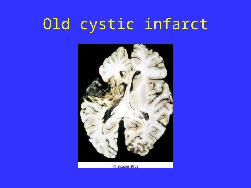
Old cystic infarct

Cerebral infarct – cystic change

Petechial haemorrhages in bone marrow embolism

Intracranial haemorrhage
• secondary– following infarction
• primary– extradural and subdural
• usually traumatic in origin
– subarachnoid and intraparenchymal (aka intracerebral)
• usually due to vascular disease

Subarachnoid haemorrhage
• most often due to cerebral artery berry (saccular) aneurysms
• but also by extension from intracerebral haemorrhages or due to bleeding diseases, trauma, tumour, vasculitis etc

Berry (saccular) aneurysms
• incidental finding in ~ 2% of post-mortem examinations, multiple in maybe a third
• occur near major branch points on Circle of Willis or just beyond
• more common on anterior part of Circle or its branches

Berry aneurysms


Aetiology of berry aneurysms
• genetic factors may be important in some cases– e.g. increased risk in ADPKD, Ehlers-Danlos
syndrome, Marfan’s syndrome) etc
• cigarette smoking and hypertension also predisposing factors
• “congenital”, but not present at birth, though underlying defect in media may be

Berry aneurysms
• thin-walled out-pouching
• usually < 1 cm diameter
• wall consists only of intima
• rupture at apex, usually into subarachnoid space, but sometimes into brain or both

Berry aneurysms

Berry aneurysm

Berry aneurysms
• rupture most often in 40- 50s • may be precipitated by sudden ICP rise
– also by hypertension
• typically sudden severe headache and rapid loss consciousness
• ~ 10-15% die, but most recover consciousness in minutes
• may show meningism• rebleeding common and makes prognosis worse

Subarachnoid haemorrhage
• early effects include– increased risk of vasospasm of other vessels– can lead to additional ischemic injury, espec.
if spasm involves Circle of Willis– presumably due to vascular mediator
• late sequelae– meningeal fibrosis and scarring– possible obstruction of CSF flow/reabsorption.

CSF in subarachnoid haemorrhage
• initially bright red blood
• later, xanthochromia as red cells degenerate

Intraparenchymal (intracerebral or cerebral) haemorrhage
• 80 % death rate
• sudden onset, causing rapid rise in ICP
• 50%+ associated with hypertension– ? microaneurysms (of Charcot-Bouchard)– ? just arteriosclerotic branch points
• remainder due to vascular malformations, bleeding disease, vasculitis etc

Intracerebral haemorrhage
• usually affects basal ganglia, brainstem, cerebellum or cerebral cortex
• major tissue disruption and destruction
• may extend into ventricles and/or subarachnoid space
• in survivors, haematoma surrounded – like infarcts - by zone of reaction, then repair with gliosis

Intracerebral haemorrhage rupturing into ventricle

Intracerebral haemorrhage with intraventricular extension

Pontine haemorrhage rupturing into 4th ventricle

Other causes of haemorrhage
• angiomas, AV malformations etc

Hypertension and CVD
• common cause of CVD• frequently associated with atheroma and
diabetes• responsible for -
– intracerebral haemorrhage• and rupture of berry aneurysms, so subarachnoid
haemorrhage
– lacunar infarcts– hypertensive encephalopathy
• acute or chronic

Hypertension and lacunar infarcts
• arteriosclerosis +/- occlusion of vessels supplying basal ganglia, hemispheres and brainstem
• causes single/multiple small cavitated infarcts (“lacunes”)– tissue loss with scattered compound granular
corpuscles surrounded by gliosis
• clinical effects depend on location - may be “silent”

Lacunar infarcts in caudate & putamen

Acute hypertensive encephalopathy
• syndrome of diffuse cerebral dysfunction– headaches, confusion, vomiting and convulsions,
sometimes leading to coma
• usually part of “malignant” phase hypertension• rapid treatment needed to reduce raised ICP• at PM, oedematous brain +/- tentorial or tonsillar
herniation • arteriolar fibrinoid necrosis and petechiae
throughout brain

Chronic hypertensive encephalopathy
• one cause of vascular (multi-infarct) dementia– dementia often with focal neurological defects
• caused by multifocal vascular disease over long time– cerebral atheroma– thrombosis or embolism from carotids or heart– cerebral hypertensive arteriolosclerosis
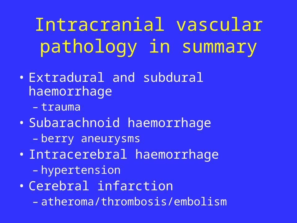
Intracranial vascular pathology in summary
• Extradural and subdural haemorrhage– trauma
• Subarachnoid haemorrhage– berry aneurysms
• Intracerebral haemorrhage – hypertension
• Cerebral infarction– atheroma/thrombosis/embolism
