nervous system 3 infections prof john simpson. this lecture is now on the intranet issue of lumbar...
TRANSCRIPT

Nervous System 3Infections
Prof John Simpson

• this lecture is now on the intranet
• issue of lumbar puncture in raised ICP
• review your microbiology!

Most organisms can target the brain
• bacteria
• viruses
• protozoa
• metazoa
• fungi
• prions

Transmission of infection to NS
• blood borne– septicaemia, viraemia, infected blood cells, septic
embolism (e.g. endocarditis, bronchiectasis, IV drug use)
• direct spread– adjacent infection, head injury etc
• trauma (including iatrogenic - LP, ventriculo-peritoneal shunts)
• vertical transmission in pregnancy • (important role for immunosuppression)

Bacterial infections
• problems arise because of inflammatory reaction or tissue destruction
• result in meningitis or abscess

Meningitis
• inflammation in subarachnoid space (arachnoid and pia mater) strictly speaking = leptomeningitis
• pachymeningitis = predominantly dural disease– usually direct spread of infection from skull
(otitis media, mastoiditis or fracture)– Gram-neg bacilli from middle ear, haemolytic
strep from sinuses or mixed organisms, often with Staph aureus, from skull fractures.
– can cause dural abscess]

Pachymeningitis
• epidural infection– suppuration between dura and skull or
vertebral column - abscess (SOL)
• subdural infection– abscess unusual– pus spreads in subdural space over
hemispheres causing subdural empyema. – involvement of subdural vessels may cause
thrombophlebitis and venous infarction of brain

Meningitis(i.e. leptomeningitis)
• usually blood-borne infection, but can be direct spread from the skull bones
• most common bacteria – – neonates: coliforms, streptococci– 2-5 years: haemophilus – older children - adults: meningoccus, pneumococcus– old age: pneumococcus
• in immunocompromised– pneumococcus, meningococcus, listeria
• (TB and syphilis also important causes)
Meningitis
• incubation period ~ 4 days
• once in SA space, bacteria multiply
• pathogenetic effects follow release of agents inducing fever and acute inflammation (hyperaemia, exudation etc)
• inflammatory exudate can raise ICP and can reduce cerebral blood flow

Meningococcal meningitis
• commonest variety worldwide, but less so in Malawi
• sporadic (URT) or epidemics in small communities (droplet spread from asymptomatic nasal carriers)
• petechial rash can herald DIC accompanied by potentially lethal adrenal haemorrhage (Waterhouse-Friederichsen syndrome)
• (immunisation programmes in colleges etc)
Pneumococcal meningitis
• commonest meningitis in Malawi
• usually sporadic cases
• at all ages
• not just in AIDS

Pathology of bacterial meningitis
• meningeal and superficial cortical vessels congested, often haemorrhagic: cord can be involved too
• infiltrate of neutrophils, so often obvious suppuration – basal cisterns and sulci
• CSF often turbid - reduced glucose, increased cells (neutrophils) and increased protein
• complications (~ 25% die) include – – cerebral abscess, subdural empyema, cerebral
infarction, obstructive hydrocephalus, epilepsy, cranial nerve palsies (VI & VIII)
– and DIC if meningococcal

Downloaded from: Robbins & Cotran Pathologic Basis of Disease (on 2 February 2007 04:53 PM)
© 2007 Elsevier
Suppurative (purulent ) meningitis

Suppurative meningitis

Suppurative meningitis
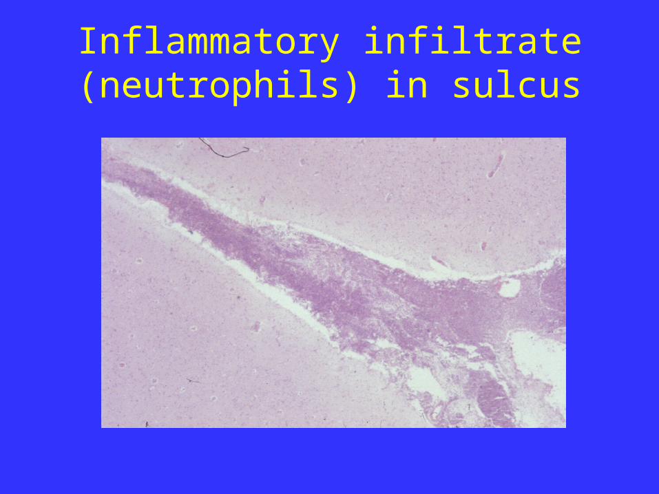
Inflammatory infiltrate (neutrophils) in sulcus

Cerebral abscess
• usually from – direct spread - sinuses or middle ear – septic sinus thrombosis - spread of infection
from mastoid or middle ear via sigmoid sinus – blood spread, e.g. infective endocarditis,
bronchiectasis etc - often multiple abscesses in parietal lobes
• adjacent brain markedly oedematous • abscesses frequently enlarge and become
multiloculate

Cerebral abscess
• presentation can be similar to meningitis, but often with focal signs, epilepsy and fever
• but also act/present as space-occupying lesions• complications include –
– meningitis– focal neurological deficit– epilepsy– herniation of the brain

Cerebral abscess

Cerebral Abscess

Downloaded from: Robbins & Cotran Pathologic Basis of Disease (on 2 February 2007 04:53 PM)
© 2007 Elsevier
Cerebral abscesses

NS tuberculosis
• secondary to infection (75% primary) elsewhere• meningitis (espec. in young) and/or abscesses
(tuberculomas)• meningitis from rupture of subependymal tubercles
– (rarely from direct spread from vertebral body) – thick gelatinous exudate in basal cisterns and sulci– causes subacute meningitis with occasional isolated
cranial nerve palsies– but can be non-specific and diagnosed only after LP
• tuberculomas present like other cerebral abscesses

NS syphilis
• blood spread• effects include –
– silent meningitis during 1ry and secondary stages– meningeal thickening in tertiary stage, causing cranial
nerve palsies – gummas, causing cerebral or spinal compression – tabes dorsalis due to degeneration of dorsal columns – “general paralysis of the insane” due to cerebral
atrophy in chronic infection

Viral infections of NS
• usually haematogenous spread during viraemia– usually cause meningitis or encephalitis
• neural spread along peripheral sensory nerves by retrograde axonal transport, e.g. rabies
• some viruses are neurotropic - tend to spread specifically to CNS from initial site of infection, e.g. polio virus from the gut
• pathogenetic effects because of multiplication inside NS cells or immune response (with lymphoid infiltration) to virus

Effects of viral infections
• usual effect is meningitis• less commonly encephalitis• also
– reactivation of latent viral infection (e.g. zoster)
– ‘slow' virus infections responsible for subacute spongiform encephalopathy, rare cause of dementia
– acute disseminated encephalomyelitis, demyelinating disorder, resulting from virus-induced immune reaction

Viral meningitis
• common• acute onset, but usually less severe than
bacterial meningitis• usually haematogenous spread• common organisms –
– mumps – echovirus – coxsackie– HSV

Viral meningitis
• meninges infiltrated by mononuclear cells (lymphocytes, plasma cells and macrophages) with typical perivascular lymphocytic cuffing in meninges and superficial brain
• characteristic CSF – normal glucose, increased cells (lymphocytes) and slight increase protein

Viral encephalitis• most commonly HSV, EBV, zoster and arboviruses
– mode of spread varies with virus– viral type may also determine part of brain affected
• pathology– mononuclear infiltration - as perivascular cuffing
– +/- cell lysis and phagocytosis of cell debris by macrophages - when neurones involved, process is known as neuronophagia
– reactive astrocytes and microglia, often in cell clusters – vasogenic oedema– viral inclusions may be diagnostic, e.g. 'owl-eyes‘ CMV
and Negri bodies in rabies

Viral encephalitis
• most cases mild, self-limiting conditions, but may result in death or severe
• most common effects – fever, personality change and seizures
• focal neurological signs very unusual
• (some viruses can also damage brain not by invasion, but secondary to an immune mediated demyelination)

Perivascular cuffing in viral encephalitis

Congenital and childhood viral disease
• CMV and rubella commonest NS viruses – cause necrotising encephalomyelitis resulting
in developmental malformations and microcephaly, particularly if infected during first trimester
• persistent viral infections - rare diseases in which infection occurs in early life, with NS disease occurring years later– e.g. subacute sclerosing panencephalitis
caused measles virus

Herpes Simplex Encephalitis

Downloaded from: Robbins & Cotran Pathologic Basis of Disease (on 2 February 2007 04:53 PM)
© 2007 Elsevier
Herpes simplex encephalitis

Parasitic infections - neurocystocercosis
• (all parasitic infections uncommon unless human parasites endemic)
• most important here is taenia solium, causing neurocystocercosis– predeliction for NS, causing cysts in brain
parenchyma and/or subarachnoid space– either SOLs in brain (typically seizures) or
cord - or meningitis, often more important in chronic repair phase (obstr. hydrocephalus, cranial nerve palsies)

Parasitic infections - toxoplasmosis
• most frequent cause of focal NS disease in AIDS
• ~ 50% patients in Africa and Europe
• often constitutional symptoms/signs at first, but then more obviously neurological ones, sometimes with localising signs
• ICP may be raised with coma/death if untreated

Cerebral toxoplasmosis

Cerebral malaria
• usually only seen in children under 10 or newcomers to falciparum malarial areas
• acute diffuse parenchymal disease accompanied by fever +/- meningitis
• rapidly fatal in ~ 25-50%• histological hallmark is sequestration of
microcirculation by parasitised/non-parasitised red cells
• causes ring-like lesions in brain

Cerebral malaria – parasites in vessels

Malaria
• decreased flexibility and increased sticking/rosetting of red cells occlude microcirculation– probably cause hypoxia, often with petechial
haemorrhages and tiny granulomas
• curiously, raised ICP not due to oedema, but ?increased blood volume
• mechanism of coma is not clear– ? increased cerebral glycolysis producing lactate
and/or interference with neurotransmission

Other parasitic infections
• trypanosomaiasis– chronic meningoencephalitis
• entamoeba histolytica– amoebic abscess
• echinococcus granulosus– hydatid cyst
• toxocara canis– eosinophilic meningitis with granulomas

Fungal infections of NS
• more common in immunosuppression• usually blood spread from lungs, but also direct• cryptococcus
– usually causes meningitis
• candida and aspergillus– usually cause abscesses
• mucormycosis– usually uncontrolled diabetics – granulomatous nasal
infection spreading to brain

Prion disease
• CJD (Creuzfeldt-Jakob disease
• variant CJD

CJD (Creuzfeldt-Jakob disease)
• presents in adults as rapidly progressive dementia often with focal signs – always fatal
• sporadic disorder in 1:1 000 000 per year worldwide • transmissible to primates by modified host protein, prion
protein• human-human transmission recorded from electrode
implantation, grafts and human growth hormone• cortical atrophy, neuron loss and reactive proliferation of
astrocytes, but no inflammation• numerous small vacuoles present in neuron and glial
processes, so known as spongiform encephalopathy• akin to kuru in New Guinea

Prion plaques in variant CJD
Variant CJD
• new variant form of CJD identified in young patients in UK
• probably from transmission of BSE (bovine spongiform encephalopathy - 'mad cow' disease) to humans by contaminated beef
• several hundred cases of variant CJD so far - ? in future?

Fetal NS infections
• rubella (deafness, blindness, microcephaly)
• CMV (microcephaly)
• toxoplasma (microcephaly)
• syphilis (tertiary forms include GPI, tabes dorsalis and meningovascular syphilis)
• (HIV)

We’ll cover NS infections in immunosuppression and HIV in the next lecture, along with tumours of the NS
