new and revisited species in aspergillus section nigri
TRANSCRIPT

1
Copyright 2011 CBS-KNAW Fungal Biodiversity Centre, P.O. Box 85167, 3508 AD Utrecht, The Netherlands.
You are free to share - to copy, distribute and transmit the work, under the following conditions:Attribution: You must attribute the work in the manner specified by the author or licensor (but not in any way that suggests that they endorse you or your use of the work).Non-commercial: You may not use this work for commercial purposes. Noderivativeworks:You may not alter, transform, or build upon this work. For any reuse or distribution, you must make clear to others the license terms of this work, which can be found at http://creativecommons.org/licenses/by-nc-nd/3.0/legalcode. Any of the above conditions can be waived if you get permission from the copyright holder. Nothing in this license impairs or restricts the author’s moral rights.
available online at www.studiesinmycology.org StudieS in Mycology 69: 1–17. 2011.doi:10.3114/sim.2011.69.01
INTRODUCTION
The black aspergilli (Aspergillus section Nigri; Gams et al. 1985) are an important group of species in food mycology, medical mycology and biotechnology. Many species cause food spoilage, but on the other hand are also used in the fermentation industry to produce hydrolytic enzymes, such as amylases or lipases, and organic acids, such as citric acid and gluconic acid (Varga et al. 2000). They are also candidates for genetic manipulation in the biotechnology industries since A. niger used under certain industrial conditions has been granted the GRAS (Generally Regarded As Safe) status by the Food and Drug Administration of the US government. Although the main source of black aspergilli is soil, members of this section have been isolated from various other sources (Kozakiewicz 1989, Abarca et al. 2004, Samson et al. 2004b, Ferracin et al. 2009). Black aspergilli are one of the more difficult groups concerning classification and identification, and several taxonomic schemes have been proposed. New molecular approaches have shown that there is a high biodiversity, but that species are occasionally difficult to recognise based solely on their phenotypic characters (Samson et al. 2007).
During a study of the genetic relationships among black aspergilli collected worldwide, four isolates have been identified which did not fit to any of the currently accepted 19 species of Aspergillus section Nigri (Samson et al. 2007, Noonim et al. 2008,
Perrone et al. 2008). We used a polyphasic approach including sequence analysis of parts of the β-tubulin and calmodulin genes and the ITS region, macro- and micromorpholocigal analyses and examination of extrolite profiles of the isolates to describe four new species in this section. Besides, the applicability of various approaches for distinguishing the two closely related species A. niger and A. awamori has also been examined. The methods tested include morphological, physiological, ecological and molecular approaches.
MATERIALSANDMETHODS
Isolates
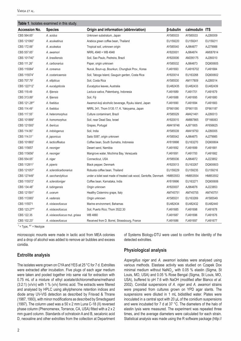
The strains used in this study are listed in Table 1.
Morphologicalanalysis
For macromorphological observations, Czapek Yeast Autolysate (CYA) agar, Malt Extract Autolysate (MEA) agar, Yeast Extract Sucrose Agar (YES), Creatine Agar (CREA), and Oatmeal Agar (OA) were used (Samson et al. 2004a). The isolates were inoculated at three points on each plate of each medium and incubated at 25 °C and 37 °C in the dark for 7 d. For micromorphological observations,
NewandrevisitedspeciesinAspergillussectionNigri
J. Varga1, 2*, J.C. Frisvad3, S. Kocsubé2, B. Brankovics2, B. Tóth4, G. Szigeti2 and R.A. Samson1
1CBS-KNAW Fungal Biodiversity Centre, Uppsalalaan 8, 3584 CT Utrecht, The Netherlands; 2Department of Microbiology, Faculty of Science and Informatics, University of Szeged, H-6726 Szeged, Közép fasor 52, Hungary; 3Center for Microbial Biotechnology, Department of Systems Biology-DTU, Søltofts Plads, Building 221, Technical University of Denmark, DK-2800 Kgs. Lyngby, Denmark; 4Cereal Research Non-Profit Ltd., H-6726 Szeged, Alsókikötő sor 9, Hungary.
*Correspondence: János Varga, [email protected]
Abstract: Four new species, Aspergillus eucalypticola, A. neoniger, A. fijiensis and A. indologenus are described and illustrated. Aspergillus eucalypticola was isolated from Eucalyptus leaf from Australia, and is related to A. tubingensis and A. costaricaensis, but could clearly be distinguished from them based on either β-tubulin or calmodulin sequence data. Aspergillus eucalypticola produced pyranonigrin A, funalenone, aurasperone B and other naphtho-γ-pyrones. Aspergillus neoniger is also a biseriate species isolated from desert sand in Namibia, and mangrove water in Venezuela, which produces aurasperone B and pyranonigrin A. Aspergillus fijiensis is a uniseriate species related to A. aculeatinus, and was isolated from soil in Fiji, and from Lactuca sativa in Indonesia. This species is able to grow at 37 °C, and produces asperparalines and okaramins. Aspergillus indologenus was isolated from soil, India. This species also belongs to the uniseriate group of black aspergilli, and was found to be related to, but clearly distinguishable from A. uvarum based on β-tubulin, calmodulin and ITS sequence data. Aspergillus indologenus produced the insecticidal compounds okaramins A, B, H, and two types of indol-alkaloids which have not been structure elucidated. Two other species, A. violaceofuscus and A. acidus, are revalidated based on molecular and extrolite data. Aspergillus violaceofuscus was found to be related to A. japonicus, and produced some of the same interesting indol-alkaloids as A. indologenus, and also produced several families of partially characterised extrolites that were also found in A. heteromorphus. Aspergillus acidus (previously known as A. foetidus var. pallidus and A. foetidus var. acidus) is also a valid species, while A. foetidus is a synonym of A. niger based on molecular and physiological data. Two other species described previously, A. coreanus and A. lacticoffeatus, were found to be colour mutants of A. acidus and A. niger, respectively. Methods which could be used to distinguish the two closely related and economically important species A. niger and A. awamori are also detailed. Although these species differ in their occurrence and several physiological means (elastase activities, abilities to utilise 2-deoxy-D-glucose as sole carbon source), our data indicate that only molecular approaches including sequence analysis of calmodulin or β-tubulin genes, AFLP analysis, UP-PCR analysis or mtDNA RFLP analysis can be used reliably to distinguish these sibling species. Aspergillus section Nigri now includes 26 taxa.
Keywords: Aspergillus section Nigri, phylogeny, polyphasic taxonomy, extrolites.Taxonomicnovelties: Aspergillus eucalypticola Varga, Frisvad & Samson sp. nov., Aspergillus fijiensis Varga, Frisvad & Samson sp. nov., Aspergillus indologenus Frisvad, Varga & Samson sp. nov., Aspergillus neoniger Varga, Frisvad & Samson sp. nov.
2
Varga et al.
microscopic mounts were made in lactic acid from MEA colonies and a drop of alcohol was added to remove air bubbles and excess conidia.
Extroliteanalysis
The isolates were grown on CYA and YES at 25 °C for 7 d. Extrolites were extracted after incubation. Five plugs of each agar medium were taken and pooled together into same vial for extraction with 0.75 mL of a mixture of ethyl acetate/dichloromethane/methanol (3:2:1) (v/v/v) with 1 % (v/v) formic acid. The extracts were filtered and analysed by HPLC using alkylphenone retention indices and diode array UV-VIS detection as described by Frisvad & Thrane (1987, 1993), with minor modifications as described by Smedsgaard (1997). The column used was a 50 x 2 mm Luna C-18 (II) reversed phase column (Phenomenex, Torrance, CA, USA) fitted with a 2 x 2 mm guard column. Standards of ochratoxin A and B, secalonic acid D, neoxaline and other extrolites from the collection at Department
of Systems Biology-DTU were used to confirm the identity of the detected extrolites.
Physiologicalanalysis
Aspergillus niger and A. awamori isolates were analysed using various methods. Elastase activity was studied on Czapek Dox minimal medium without NaNO3, with 0.05 % elastin (Sigma, St Louis, MO, USA) and 0.05 % Rose Bengal (Sigma, St Louis, MO, USA), buffered to pH 7.6 with NaOH (modified after Blanco et al. 2002). Conidial suspensions of A. niger and A. awamori strains were prepared from cultures grown on YPD agar slants. The suspensions were diluted in 1 mL bidistilled water. Plates were inoculated in a central spot with 20 μL of the conidium suspensions and were incubated for 7 d at 37 °C. The diameters of the halo of elastin lysis were measured. The experiment was repeated three times, and the average diameters were calculated for each strain. Statistical analysis was made using the R software package (http://
Table1. Isolates examined in this study.AccessionNo. Species Originandinformation(abbreviation) β-tubulin calmodulin ITSCBS 564.65T A. acidus Unknown substratum, Japan AY585533 AY585533 AJ280009CBS 121060T A. aculeatinus Arabica green coffee bean, Thailand EU159220 EU159241 EU159211CBS 172.66T A. aculeatus Tropical soil, unknown origin AY585540 AJ964877 AJ279988CBS 557.65T A. awamori NRRL 4948 = WB 4948 AY820001 AJ964874 AM087614CBS 101740T A. brasiliensis Soil, Sao Paulo, Pedreira, Brazil AY820006 AM295175 AJ280010CBS 111.26T A. carbonarius Paper, origin unknown AY585532 AJ964873 DQ900605CBS 119384T A. coreanus Nuruk, Boun-up, Bounkun, Chungbuk Prov., Korea FJ491693 FJ4916702 FJ491684
CBS 115574T A. costaricaensis Soil, Taboga Island, Gauguin garden, Costa Rica AY820014 EU163268 DQ900602CBS 707.79T A. ellipticus Soil, Costa Rica AY585530 AM117809 AJ280014CBS 122712T A. eucalypticola Eucalyptus leaves, Australia EU482435 EU482433 EU482439CBS 119.49 A. fijiensis Lactuca sativa, Palembang, Indonesia FJ491689 FJ491701 FJ491679CBS 313.89T A. fijiensis Soil, Fiji FJ491688 FJ491695 FJ491680CBS 121.28NT A. foetidus Awamori-koji alcoholic beverage, Ryuku island, Japan FJ491690 FJ491694 FJ491683CBS 114.49T A. foetidus NRRL 341, Thom 5135.17; K. Yakoyama, Japan EF661090 EF661155 EF661187CBS 117.55T A. heteromorphus Culture contaminant, Brazil AY585529 AM421461 AJ280013CBS 101889T A. homomorphus Soil, near Dead Sea, Israel AY820015 AM887865 EF166063CBS 121593T A. ibericus Grapes, Portugal AM419748 AJ971805 AY656625CBS 114.80T A. indologenus Soil, India AY585539 AM419750 AJ280005CBS 114.51T A. japonicus Saito 5087, origin unknown AY585542 AJ964875 AJ279985CBS 101883T A. lacticoffeatus Coffee bean, South Sumatra, Indonesia AY819998 EU163270 DQ900604CBS 115657 A. neoniger Desert sand, Namibia FJ491692 FJ491699 FJ491681CBS 115656T A. neoniger Mangrove water, Mochima Bay, Venezuela FJ491691 FJ491700 FJ491682CBS 554.65T A. niger Connecticut, USA AY585536 AJ964872 AJ223852CBS 112811T A. piperis Black pepper, Denmark AY820013 EU163267 DQ900603CBS 121057T A. sclerotiicarbonarius Robusta coffee bean, Thailand EU159229 EU159235 EU159216CBS 127449T A. saccharolyticus under a toilet seat made of treated oak wood, Gentofte, Denmark HM853553 HM853554 HM853552CBS 115572T A. sclerotioniger Coffee bean, Karnataka, India AY819996 EU163271 DQ900606CBS 134.48T A. tubingensis Origin unknown AY820007 AJ964876 AJ223853CBS 121591T A. uvarum Healthy Cisternino grape, Italy AM745751 AM745755 AM745751CBS 113365T A. vadensis Origin unknown AY585531 EU163269 AY585549CBS 115571 A. violaceofuscus Marine environment, Bahamas EU482434 EU482432 EU482440CBS 123.27NT A. violaceofuscus Soil, Puerto Rico; Thom 3522.30 FJ491685 FJ491698 FJ491678CBS 122.35 A. violaceofuscus mut. grisea WB 4880 FJ491687 FJ491696 FJ491676CBS 102.23T A. violaceofuscus Received from D. Borrel, Strassbourg, France FJ491686 FJ491697 FJ491677T = Type, NT = Neotype
3www.studiesinmycology.org
new and reViSited SpecieS in aspergillus Section Nigri
www.r-project.org/). The assumptions of ANOVA were tested using the diagnostic plots in R. According to Quantile-Quantile (QQ) Plot the data were not normally distributed, thus the Kruskal-Wallis test was applied to compare the average diameters between the two species.
Carbon source assimilation tests were performed on minimal medium (MM: 0.5 % (NH4)2SO4, 0.1 % KH2PO4, 0.05 % MgSO4, 2 % agar) with 0.2 % single carbon source. Conidial suspensions of eight A. niger and eight A. awamori strains were prepared from 5-d-old cultures grown on YPD agar slants. The suspensions were diluted in bidistilled water and conidia were filtered. An YPD plate was inoculated in 16 points with 15 μL of the conidium suspensions and was incubated for 3 d at 25 °C. Strains were replicated to the MM plates, which contained single carbon sources using a 16-pronged replicator. Plates were incubated for 7 d at 25 °C. The experiment was repeated twice and control series was made on MM plates without carbon source and YPD plates.
Thirty carbon sources were tested, which were selected based on previous carbon source utilisation experiments (Varga et al. 2000): glucose, D-xylose, galactose, D-lyxose, L-sorbose, L-rhamnose, lactose, eritrit, galactit, L-valine, L-β-phenilalanine, L-triptophane, L-treonine, L-serine, L-cysteine, L-asparagine acid, L-tyrosine, L-lysine, L-histidine, L-citrulline, cis-aconitic acid, vanillin, vanillin acid, L-ascorbic acid, D-glucoseamine, glycylglycine, salicin, pectin, melezitose, α-ketoglutaric acid. Different growth patterns of the strains belonging to the two species were observed simply in the case of L-sorbose, so the test was extended to 2-deoxy-D-glucose because of the structural similarity of these two compounds.
Genotypicanalysis
The cultures used for the molecular studies were grown on malt peptone (MP) broth using 10 % (v/v) of malt extract (Oxoid) and 0.1 % (w/v) bacto peptone (Difco), 2 mL of medium in 15 mL tubes. The cultures were incubated at 25 °C for 7 d. DNA was extracted from the cells using the Masterpure™ yeast DNA purification kit (Epicentre Biotechnol.) according to the instructions of the manufacturer. The ITS region and parts of the β-tubulin and calmodulin genes were amplified and sequenced as described previously (Varga et al. 2007a–c).
Part of the FUM8 gene was amplified using primers vnF1 and vnR3 as described by Susca et al. (2010). Primer sets were also designed to target part of the chloroperoxidase gene of black aspergilli presumably taking part in ochratoxin biosynthesis. Construction of the primers was carried out by using the homologous sequences identified in the genomic sequences of Aspegillus niger CBS 513.88 and Aspergillus carbonarius ITEM 5010 isolates. The designed chloroperoxidase specific PCR primers were BCPOF (5’- CTGGGCGACTGCATCCAC – 3’) and BCPOR (5’- TTCATCGTACGGCAGACGCT - 3’) which generated specific amplicons of about 250 base pairs. Amplifications were performed on a PTC-0148 Mini48 thermocycler (BioRad, USA), using the following amplification steps: 4 min of initial denaturation at 94 °C followed by 35 amplification cycles of 20 s at 94 °C, 15 s at 62 °C and 30 s at 2 °C and a final extension step for 1 min at 72 °C.
DNA sequences were edited with the DNASTAR computer package and an alignment of the sequences and neighbour joining analyses were performed using the MEGA v. 4 software (Tamura et al. 2007). To determine the support for each clade, a bootstrap analysis was performed with 1 000 replications. Aspergillus flavus CBS 100927T was used as outgroup in these analyses.
Phylogenetic analysis of sequence data was also performed using PAUP* v. 4.0b10 (Swofford 2000). Alignment gaps were treated as fifth character state, parsimony uninformative characters were excluded and all characters were unordered and equal weight. Maximum parsimony analysis was performed for all data sets using the heuristic search option. To assess the robustness of the topology, 1 000 bootstrap replicates were run by maximum parsimony (Hillis & Bull 1993). Other measures including tree length, consistency index and retention index were also calculated. Sequences were deposited at GenBank under accession numbers listed in Table 1.
UP-PCR analyses were carried out according to Bulat et al. (2000). DNA was isolated as described in the literature (Leach et al. 1986). The primers used were L45, AS15inv, L15/AS19, AA2M2, L21, 3-2, AS4, AS15 (Lübeck et al. 1998, Bulat et al. 2000). The amplification process consisted of a predenaturation step for 1 min at 94 °C, followed by 35 cycles (30 s at 94 °C, 45 s at 55 °C, and 1 min at 72 °C), plus a final extension of 2 min at 72 °C. The amplification products were separated by electrophoresis in 1 % agarose gels, stained with ethidium bromide, and visualised under UV light. All amplifications were repeated at least two times. The faint bands which did not appear in all repeated experiments were not counted during cluster analysis.
Altogether 88 fragments were noted and a binomial matrix was created so that presence and absence of DNA fragments were scored as 1 or 0, respectively. Cluster analysis was carried out by using PHYLIP v. 3.67 software package (Felsenstein 2007). Phylogenetical tree was created by using neighbor-joining method (Saitou et al. 1987) with the program NEIGHBOR from the PHYLIP program package.
RESULTSANDDISCUSSION
Phylogeneticanalysisofsequencedata
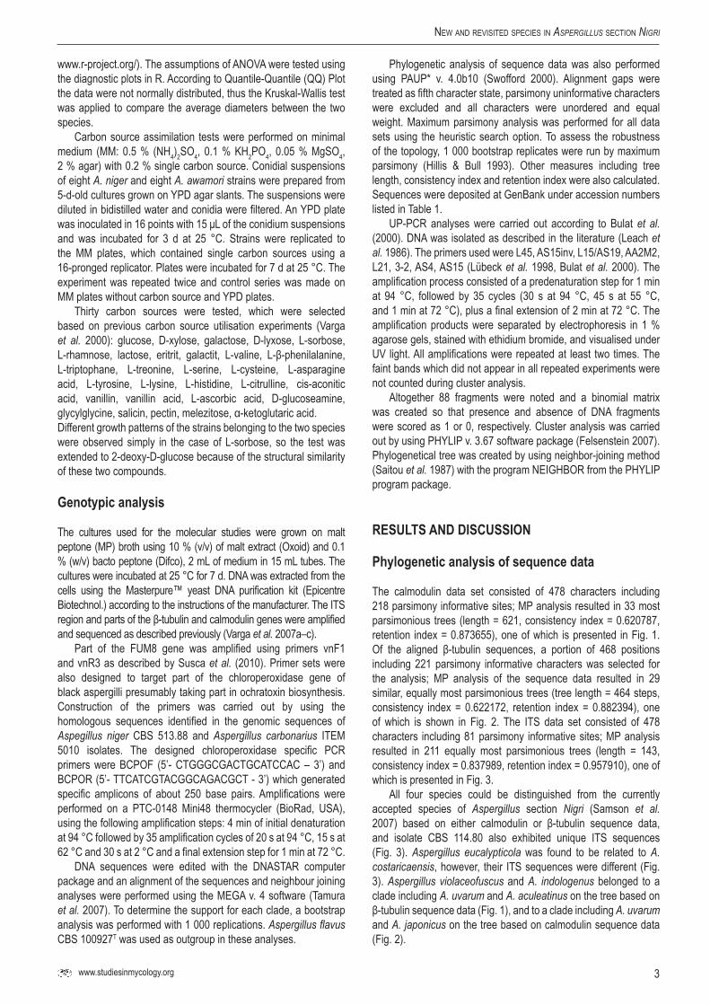
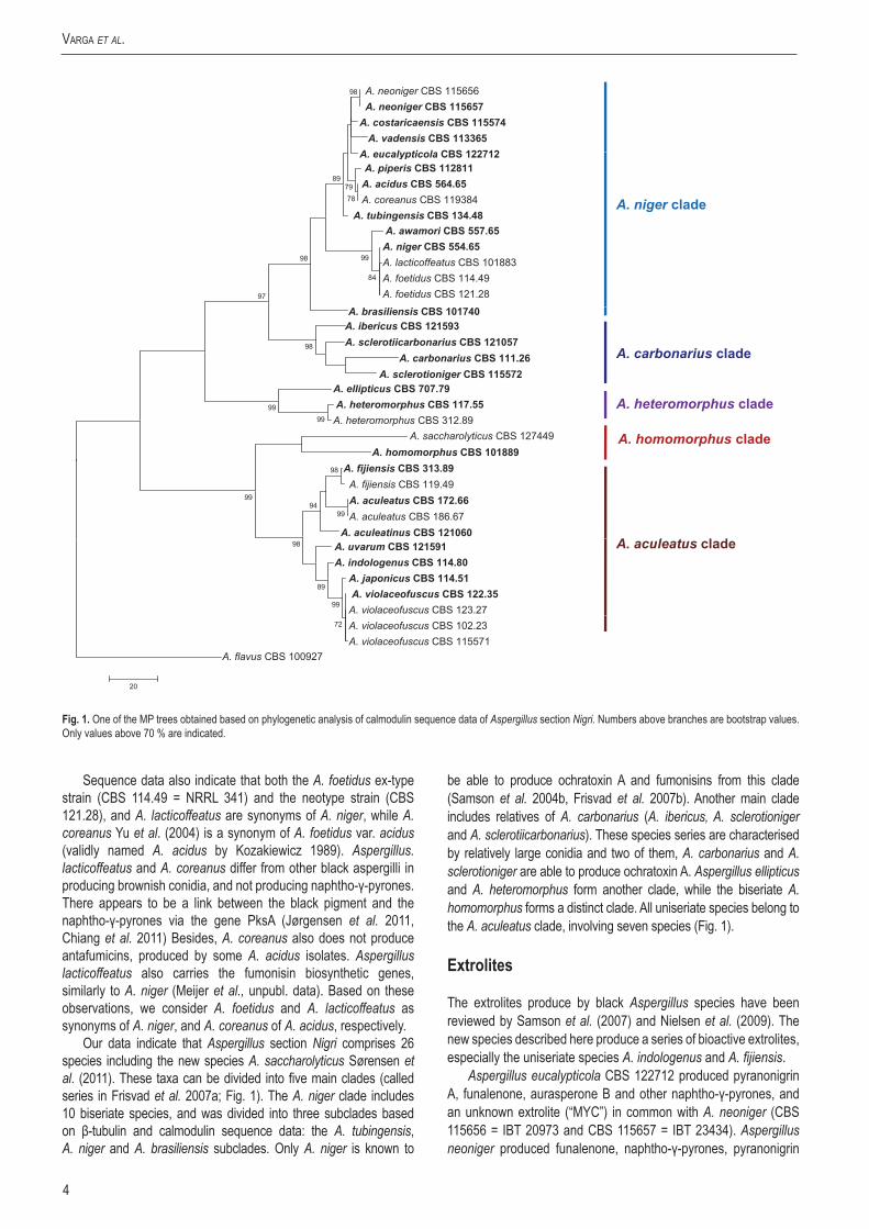
The calmodulin data set consisted of 478 characters including 218 parsimony informative sites; MP analysis resulted in 33 most parsimonious trees (length = 621, consistency index = 0.620787, retention index = 0.873655), one of which is presented in Fig. 1. Of the aligned β-tubulin sequences, a portion of 468 positions including 221 parsimony informative characters was selected for the analysis; MP analysis of the sequence data resulted in 29 similar, equally most parsimonious trees (tree length = 464 steps, consistency index = 0.622172, retention index = 0.882394), one of which is shown in Fig. 2. The ITS data set consisted of 478 characters including 81 parsimony informative sites; MP analysis resulted in 211 equally most parsimonious trees (length = 143, consistency index = 0.837989, retention index = 0.957910), one of which is presented in Fig. 3.
All four species could be distinguished from the currently accepted species of Aspergillus section Nigri (Samson et al. 2007) based on either calmodulin or β-tubulin sequence data, and isolate CBS 114.80 also exhibited unique ITS sequences (Fig. 3). Aspergillus eucalypticola was found to be related to A. costaricaensis, however, their ITS sequences were different (Fig. 3). Aspergillus violaceofuscus and A. indologenus belonged to a clade including A. uvarum and A. aculeatinus on the tree based on β-tubulin sequence data (Fig. 1), and to a clade including A. uvarum and A. japonicus on the tree based on calmodulin sequence data (Fig. 2).
4
Varga et al.
Sequence data also indicate that both the A. foetidus ex-type strain (CBS 114.49 = NRRL 341) and the neotype strain (CBS 121.28), and A. lacticoffeatus are synonyms of A. niger, while A. coreanus Yu et al. (2004) is a synonym of A. foetidus var. acidus (validly named A. acidus by Kozakiewicz 1989). Aspergillus. lacticoffeatus and A. coreanus differ from other black aspergilli in producing brownish conidia, and not producing naphtho-γ-pyrones. There appears to be a link between the black pigment and the naphtho-γ-pyrones via the gene PksA (Jørgensen et al. 2011, Chiang et al. 2011) Besides, A. coreanus also does not produce antafumicins, produced by some A. acidus isolates. Aspergillus lacticoffeatus also carries the fumonisin biosynthetic genes, similarly to A. niger (Meijer et al., unpubl. data). Based on these observations, we consider A. foetidus and A. lacticoffeatus as synonyms of A. niger, and A. coreanus of A. acidus, respectively.
Our data indicate that Aspergillus section Nigri comprises 26 species including the new species A. saccharolyticus Sørensen et al. (2011). These taxa can be divided into five main clades (called series in Frisvad et al. 2007a; Fig. 1). The A. niger clade includes 10 biseriate species, and was divided into three subclades based on β-tubulin and calmodulin sequence data: the A. tubingensis, A. niger and A. brasiliensis subclades. Only A. niger is known to
be able to produce ochratoxin A and fumonisins from this clade (Samson et al. 2004b, Frisvad et al. 2007b). Another main clade includes relatives of A. carbonarius (A. ibericus, A. sclerotioniger and A. sclerotiicarbonarius). These species series are characterised by relatively large conidia and two of them, A. carbonarius and A. sclerotioniger are able to produce ochratoxin A. Aspergillus ellipticus and A. heteromorphus form another clade, while the biseriate A. homomorphus forms a distinct clade. All uniseriate species belong to the A. aculeatus clade, involving seven species (Fig. 1).
Extrolites
The extrolites produce by black Aspergillus species have been reviewed by Samson et al. (2007) and Nielsen et al. (2009). The new species described here produce a series of bioactive extrolites, especially the uniseriate species A. indologenus and A. fijiensis.
Aspergillus eucalypticola CBS 122712 produced pyranonigrin A, funalenone, aurasperone B and other naphtho-γ-pyrones, and an unknown extrolite (“MYC”) in common with A. neoniger (CBS 115656 = IBT 20973 and CBS 115657 = IBT 23434). Aspergillus neoniger produced funalenone, naphtho-γ-pyrones, pyranonigrin
98 A. neoniger CBS 115656A. neoniger CBS 115657
A. costaricaensis CBS 115574 A. vadensis CBS 113365
A eucalypticola CBS 122712
7879
89A. piperis CBS 112811
A. acidus CBS 564.65A. coreanus CBS 119384
A. tubingensis CBS 134.48A awamori CBS 557 65
A. eucalypticola CBS 122712
A. niger clade
84
9998
97
A. awamori CBS 557.65 A. niger CBS 554.65A. lacticoffeatus CBS 101883A. foetidus CBS 114.49A. foetidus CBS 121.28
98
A. brasiliensis CBS 101740A. ibericus CBS 121593A. sclerotiicarbonarius CBS 121057
A. carbonarius CBS 111.26A. sclerotioniger CBS 115572
A. carbonarius clade
9999
A. ellipticus CBS 707.79A. heteromorphus CBS 117.55A. heteromorphus CBS 312.89
A. homomorphus CBS 101889A. homomorphus clade
A. heteromorphus clade
A. saccharolyticus CBS 127449
99
98
9499
A. fijiensis CBS 313.89A. fijiensis CBS 119.49A. aculeatus CBS 172.66A. aculeatus CBS 186.67
A. aculeatinus CBS 121060A l l d
99
89
98 A. uvarum CBS 121591A. indologenus CBS 114.80
A. japonicus CBS 114.51A. violaceofuscus CBS 122.35
A. violaceofuscus CBS 123.27
A. aculeatus clade
72
20
A. violaceofuscus CBS 102.23A. violaceofuscus CBS 115571
A. flavus CBS 100927
Fig.1. One of the MP trees obtained based on phylogenetic analysis of calmodulin sequence data of Aspergillus section Nigri. Numbers above branches are bootstrap values. Only values above 70 % are indicated.

5www.studiesinmycology.org
new and reViSited SpecieS in aspergillus Section Nigri
A and “MYC”, and it is chemically very closely related to A. eucalypticola.
Aspergillus indologenus CBS 114.80 = IBT 3679 produced the insecticidal compounds okaramins A, B, H earlier also reported from an A. aculeatus isolate (Hayashi et al. 1999), partially characterised polar alkaloids, a series of very apolar sclerotial indol-alkaloids (related to aflavinins) and unique indol-alkaloids with similar UV spectra as the fumitremorgins. Aspergillus violaceofuscus produced some indol-alkaloids also found in A. indologenus, but it also produced several families of partially characterised extrolites that have also been found in A. heteromorphus [“SMIF”, “PON”, “SENGLAB” (a pyranonigrin-related compound), and yellow compounds with characteristic UV spectra]. Aspergillus fijiensis CBS 313.89 and CBS 119.49 produced asperparalins, secalonic acid D, F and the partially characterised “BAM”, “PON” = “FIB1” & “FIB2”, “GLABRINOL”, “SENGLAB”, and “YE1”. CBS 313.89 in addition produced “DERH” and “YE2” and CBS 119.49 additionally produced neoxaline, and “TRU”. Asperparaline A (= aspergillimide = VM55598), asperparaline B and C have earlier been reported
from Aspergillus japonicus ATCC 204480 (Hayashi et al. 1997, 2000) and asperparaline A, 16-keto aspergillimide, VM54159, SB203105 and SB 200437 have been isolated from “a black Aspergillus with pink sclerotia” IMI 337664 (Banks et al. 1997) and neoxaline has been isolated from A. japonicus (Hirano et al. 1979). Based on the extrolite data, ATCC 204480 and IMI 337664 may indeed belong to A. fijiensis, but we have not examined these cultures yet.
Species related to A. niger, such as A. eucalypticola and A. neoniger and the well known species A. carbonarius and A. tubingensis, produce different combinations of pyranonigrins, tensidols, kotanins, fumonisins, funalenones, naphtho-γ-pyrones, ochratoxins, asperazines, and pyrophen (Samson et al. 2004b, 2007), while species related to A. aculeatus, A. aculeatinus, A. japonicus, A. uvarum, and the new species described here, A. indologenus, A. fijiensis and the revived A. violaceofuscus produce different combinations of asperparalins, okaramins, neoxaline, sclerotial indol-alkaloids, and secalonic acids (Parenicova et al. 2001, Samson et al. 2004b, Samson et al. 2007, Noonim et al. 2008).
70
72
96
97
91
84
90
75
84
100
100
83
100
20
A. brasiliensis CBS 101740
A. ibericus CBS 121593 A. carbonarius CBS 111.26 A. sclerotiicarbonarius CBS 121057 A. sclerotioniger CBS 115572
A. acidus CBS 564.65
A. coreanus CBS 119384 A. piperis CBS 112811 A. eucalypticola CBS 122712 A. costaricaensis CBS 115574
A. niger CBS 554.65 A. awamori CBS 557.65 A. foetidus CBS 114.49 A. foetidus CBS 121.28 A. lacticoffeatus CBS 101883
A. heteromorphus CBS 117.55 A. ellipticus CBS 707.79
A. aculeatus CBS 172.66
A. violaceofuscus mut. grisea CBS 122.35 A. violaceofuscus CBS 123.27 A. violaceofuscus CBS 102.23 A. violaceofuscus CBS 115571 A. japonicus CBS 114.51
A. flavus CBS 100927
A. tubingensis CBS 134.48 A. neoniger CBS 115656 A. neoniger CBS 115657 A. vadensis CBS 113365
A. homomorphus CBS 101889 A. uvarum CBS 121591 A. indologenus CBS 114.80
A. aculeatinus CBS 1212060 A. fijiensis CBS 313.89 A. fijiensis CBS 119.49
A. saccharolyticus CBS 127449
Fig.2. The single MP tree obtained based on phylogenetic analysis of β-tubulin sequence data of Aspergillus section Nigri. Numbers above branches are bootstrap values. Only values above 70 % are indicated.
6
Varga et al.
ApproachestodistinguishbetweenisolatesofthesiblingspeciesA. nigerandA. awamori
Aspergillus awamori has recently been revalidated as a cryptic species within the A. niger species (Perrone et al. 2011). These species cannot be reliably separated from each other using either morphological or extrolite data. However, molecular data including sequence-based approaches using either β-tubulin, calmodulin or translation elongation factor a sequences and AFLP analysis were found to be useful for distinguishing these species (Perrone et al. 2011). Aspergillus niger and A. awamori are economically important as isolates of both species are able to produce fumonisins and/or ochratoxins (Varga et al. 2010, Perrone et al. 2011). In view of the importance of these species in mycotoxin contamination of various agricultural products (see below), we examined other possibilities which could be used for the easy identification of these species.
82
90
82
86
82
99
99
5
A. brasiliensis CBS 101740
A. carbonarius CBS 111.26 A. sclerotioniger CBS 115572 A. sclerotiicarbonarius CBS 121057 A. ibericus CBS 121593
A. awamori CBS 557.65 A. niger CBS 554.65 A. foetidus CBS 114.49 A. foetidus CBS 121.28 A. lacticoffeatus CBS 101883
A. ellipticus CBS 707.79 A. heteromorphus CBS 117.55
A. homomorphus CBS 101889
A. aculeatinus CBS 1212060 A. aculeatus CBS 172.66 A. indologenus CBS 114.80 A. uvarum CBS 121591 A. japonicus CBS 114.51 A. violaceofuscus mut. grisea CBS 122.35
A. violaceofuscus CBS 123.27 A. violaceofuscus CBS 102.23 A. violaceofuscus CBS 115571 A. flavus CBS 100927
A. tubingensis CBS 134.48 A. piperis CBS 112811 A. costaricaensis CBS 115574 A. coreanus CBS 119384 A. eucalypticola CBS 122712 A. acidus CBS 564.65
A. neoniger CBS 115656 A. neoniger CBS 115657 A. vadensis CBS 113365
A. fijiensis CBS 313.89 A. fijiensis CBS 119.49
A. saccharolyticus CBS 127449
Fig.3. One of the MP trees obtained based on phylogenetic analysis of ITS sequence data of Aspergillus section Nigri. Numbers above branches are bootstrap values. Only values above 70 % are indicated.
0.02
A. tubingensis 355
A. niger 9655 A. niger 2321 A. niger 1284 A. niger 3478 A. niger 8651 A. niger 8165 A. niger 9143
A. awamori 6270 A. awamori 6552 A. awamori 2380 A. awamori 2750
A. niger 1181 A. niger 8663 A. niger 7255 A. niger 8720 A. niger 8940 A. niger 5765
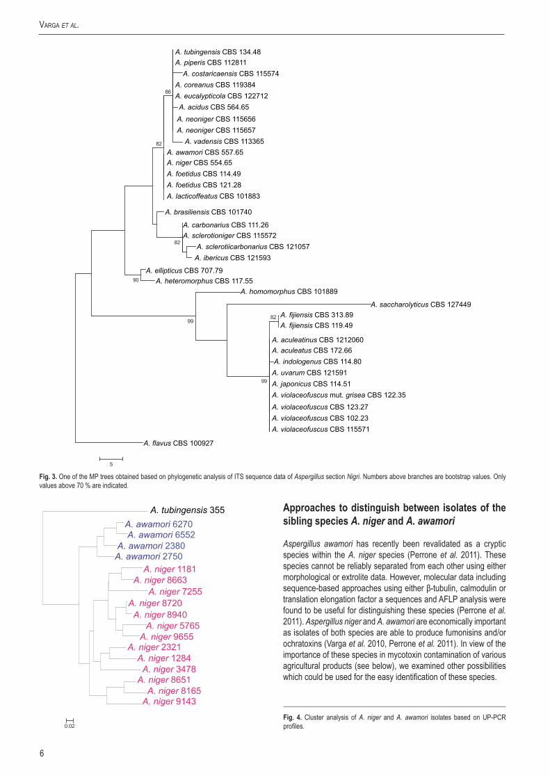
Fig. 4. Cluster analysis of A. niger and A. awamori isolates based on UP-PCR profiles.
7www.studiesinmycology.org
new and reViSited SpecieS in aspergillus Section Nigri
100
97
100
100
86100
74
74
92
83
50
A. awamori Sultha7 A. awamori Rozijn4 A. awamori ITEM 5266 A. awamori ITEM 7097 A. awamori ITEM 5277
F. verticillioides FigF1 F. verticillioides Fus46 F. verticillioides Fus60
A. awamori Torok8 A. awamori ITEM 4502
A. niger Magn2 A. niger ITEM 4547 A. niger ITEM 4501 A. niger Sultha2 A. niger Sultha4 A. niger California5
A. niger Magn3 A. niger ITEM 5419
A. awamori ITEM 4541
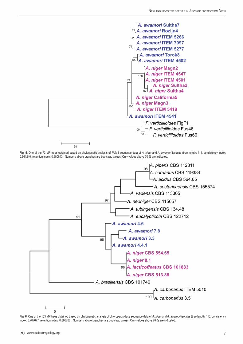
Fig.5. One of the 73 MP trees obtained based on phylogenetic analysis of FUM8 sequence data of A. niger and A. awamori isolates (tree length: 411, consistency index: 0.961240, retention index: 0.980843). Numbers above branches are bootstrap values. Only values above 70 % are indicated.
100
98
96
97
91
95
5
A. piperis CBS 112811
A. acidus CBS 564.65 A. costaricaensis CBS 155574
A. tubingensis CBS 134.48
A. vadensis CBS 113365
A. neoniger CBS 115657
A. eucalypticola CBS 122712
A. awamori 3.3
A. niger CBS 554.65
A. awamori 4.6 A. awamori 7.8
A. awamori 4.4.1
A. niger 8.1
A. niger CBS 513.88 A. lacticoffeatus CBS 101883
A. brasiliensis CBS 101740 A. carbonarius ITEM 5010
A. carbonarius 3.5
A. coreanus CBS 119384
Fig.6. One of the 153 MP trees obtained based on phylogenetic analysis of chloroperoxidase sequence data of A. niger and A. awamori isolates (tree length: 113, consistency index: 0.767677, retention index: 0.886700). Numbers above branches are bootstrap values. Only values above 70 % are indicated.
8
Varga et al.
Molecularapproaches
UP-PCR analysis (Bulat et al. 2000) was found to be also useful for species delineation (Fig. 4). This technique is similar to RAPD, but is more reliable as it uses higher annealing temperatures and longer primers (Bulat et al. 2000). Besides, UP-PCR analysis is easier to perform than AFLP analysis. Similarly to AFLP analysis (Perrone et al. 2011), this technique could also be used successfully to separate the examined A. niger and A. awamori isolates into two clusters (Fig. 4).
Another possibility is the application of mitochondrial DNA RFLP analyses. This technique was previously used to assign isolates of the A. niger species aggregate to different haplotypes (Varga et al. 1993, 1994). Our study revealed that one of these types previously called mtDNA type 1c actually corresponds to A. awamori (data not shown).
Attempts have also been made to use sequences of mycotoxin biosynthetic genes for distinguishing A. niger from A. awamori. Susca et al. (2010) examined the presence of FUM8 encoding an a-oxoamine synthase in black aspergilli came from grapes. They found no strict correlation between the phylogenetic trees based on sequences of partial calmodulin gene and FUM8 (Fig. 5). Similar results were found in our laboratory using sequences of either FUM8, or another fumonisin biosynthetic gene, FUM1, encoding for a polyketide synthase taking part in fumonisin biosynthesis (Varga et al., unpubl. data). It was suggested that, similarly to that observed in the trichothecene biosynthesis gene cluster of the Fusarium graminearum species complex, balancing selection could be responsible for maintaining sequence polymorphisms within the fumonisin gene cluster (Ward et al. 2002, Susca et al. 2010).
The applicability of another mycotoxin biosynthetic gene, a chloroperoxidase gene presumably taking part in ochratoxin biosynthesis was also examined for distinguishing A. niger and A. awamori. This gene has been found to take part in ochratoxin biosynthesis in Penicillium verrucosum and P. nordicum (Geisen 2007). Homologues of these genes were identified in the full genome sequences of A. niger and A. carbonarius, and primers were designed to amplify orthologues in species assigned to the A. niger species complex. Phylogenetic analysis of the sequence data indicate that sequences of a chloroperoxidase gene are useful for species delineation in the A. niger species aggregate (Fig. 6).
Aspergillus niger and A. awamori could also be distinguished based on their chloroperoxidase sequences.
Morphologicalandphysiologicalapproaches
Molecular methods are commonly used today for species identification among fungi. However, in accordance with the polyphasic species concept, other criteria have also been searched for. Aspergillus niger and A. awamori cannot be distinguished based on morphology alone. Regarding extrolite production, isolates of both species produce several metabolites in common including the mycotoxins ochratoxin A and fumonisin B2, and they also share the production of pyranonigrin A, tensidol B, funalenone, malformins and naphtho-γ-pyrones. The growth rates of the isolates of these species are also similar at different temperatures (Varga et al., unpubl. data).
Carbon source utilisation tests revealed that A. niger and A. awamori has very similar utilisation spectra (data not shown).Different growth of the strains belonging to the two species was observed only in the case of L-sorbose: A. awamori strains grew less intensively on this sugar than A. niger strains. Consequently, the test was extended to 2-deoxy-D-glucose because of the structural similarity of these two compounds. Of the 30 isolates examined, 13 of the examined A. niger isolates grew well, while 13 of the 15 examined A. awamori isolates failed to grow on 2-deoxy-D-glucose as sole carbon source (data not shown). Microscopical analysis of the colonies indicated that conidial germination was inhibited in the case of A. awamori isolates (data not shown). Furthermore 2-deoxy-D-glucose was earlier found to inhibit conidium germination in Penicillium expansum (Kazi et al. 1997).
Antifungal susceptibilities of the isolates has also been examined using five antifungal drugs including amphotericin B, fluconazole, itraconazole, ketoconazole and terbinafine (Szigeti et al. 2011). Species-specific differences were not observed between A. niger and A. awamori isolates. All isolates were highly susceptible to terbinafine, while exhibited moderate susceptibilities against amphotericin B, fluconazole and ketoconazole. However, in general, A. niger and A. awamori were found to have higher MICs for azoles than A. tubingensis (Szigeti et al. 2011).
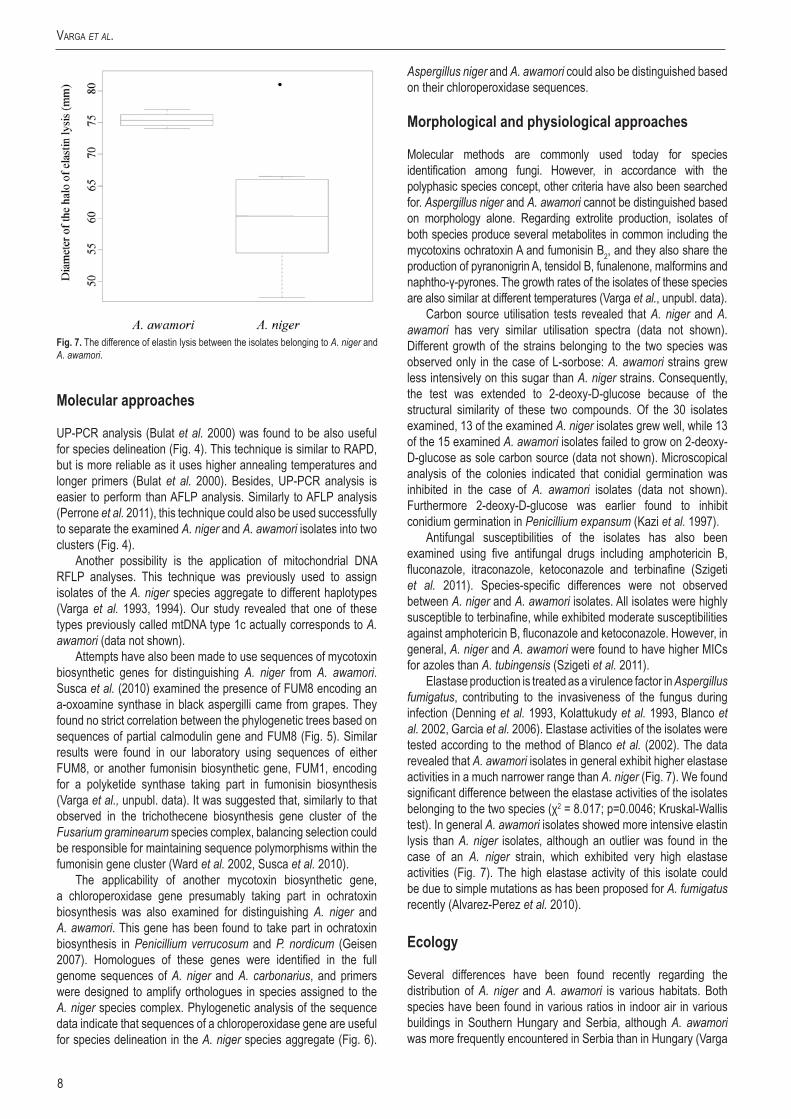
Elastase production is treated as a virulence factor in Aspergillus fumigatus, contributing to the invasiveness of the fungus during infection (Denning et al. 1993, Kolattukudy et al. 1993, Blanco et al. 2002, Garcia et al. 2006). Elastase activities of the isolates were tested according to the method of Blanco et al. (2002). The data revealed that A. awamori isolates in general exhibit higher elastase activities in a much narrower range than A. niger (Fig. 7). We found significant difference between the elastase activities of the isolates belonging to the two species (χ2 = 8.017; p=0.0046; Kruskal-Wallis test). In general A. awamori isolates showed more intensive elastin lysis than A. niger isolates, although an outlier was found in the case of an A. niger strain, which exhibited very high elastase activities (Fig. 7). The high elastase activity of this isolate could be due to simple mutations as has been proposed for A. fumigatus recently (Alvarez-Perez et al. 2010).
Ecology
Several differences have been found recently regarding the distribution of A. niger and A. awamori is various habitats. Both species have been found in various ratios in indoor air in various buildings in Southern Hungary and Serbia, although A. awamori was more frequently encountered in Serbia than in Hungary (Varga
Fig.7. The difference of elastin lysis between the isolates belonging to A. niger and A. awamori.
9www.studiesinmycology.org
new and reViSited SpecieS in aspergillus Section Nigri
J., unpubl. data). Similarly, both species were present on dried vine fruits collected in various countries (Varga et al. 2010). However, neither A. niger nor A. awamori could be isolated from pistachio nuts from Iran (Sedaghati et al. 2011), nor from dates from Iran and Tunesia (Varga J., unpubl. data). Aspergillus awamori was found to be the predominant black Aspergillus species on onions cultivated in Hungary, and is presumably the causative agent of black mold rot in this country (Varga et al., unpubl. data), and was also found to be the causative agent of seed rot of Welwitschia mirabilis in Namibia (Varga et al., unpubl. data). However, A. awamori was not detected on figs from Tunesia, Turkey and Iran (Varga J., unpubl. data). Instead, A. tubingensis and A. niger were found to contaminate these fig samples.
Regarding clinical significance, both species have been identified as causative agents of otomycosis in Iran and in Hungary, although at different frequencies, with A. niger being the dominant species in Iran, while A. awamori was most frequently identified in Hungary (Szigeti et al. 2011, unpubl. data).
In conclusion, A. niger and A. awamori are two very closely related species which seem to be in the course of speciation, similarly to the recently described species of Fusarium graminearum sensu lato (Ward et al. 2002, Starkey et al. 2007). Although these species differ in their occurrence on various substrates and several physiological characteristics (elastase activities, abilities to utilise 2-deoxy-D-glucose as sole carbon source), our data indicate that only molecular approaches including sequence analysis of calmodulin or β-tubulin genes, AFLP analysis, UP-PCR analysis or mtDNA RFLP analysis can be used reliably to distinguish these sibling species.
Speciesdescriptions
Aspergillus acidusKozak. Mycol. Pap. 161: 110 (1989) Fig. 8.
Culture ex-type: IMI 104688 = CBS 564.65, Japan, Nakazawa, 1936.
CYA, 7 d, 25 °C: 37–80 mm; MEA, 7 d, 25 °C: 43–68 mm; YES, 7 d, 25 °C: 38–80 mm; OA, 7 d, 25 °C: 38–55 mm; CYA, 7 d, 37 °C: 30–67 mm; CREA: poor growth but good acid production; CYAS: 16–69 mm (strong sporulation on all media, except CREA). Colony reverse colour on CYA: cream yellow, reverse colour on YES: yellow to cream yellow.
Conidiophores biseriate with globose vesicles 55–80 µm, stipe smooth–walled to finely roughened, hyaline, 17–22 µm. Conidia globose, 3–4 µm, brown, smooth-walled to roughened. Sclerotia not observed.
Kozakiewics (1989) proposed the name A. acidus for the variety acidus of A. foetidus on the basis on the verrucose conidium ornamentation as seen by scanning electron microscopy. This variety was recognised by Raper & Fennell (1965) as a variety of A. citricus. Al-Musallan (1980) however, could not distinguish this variety from the A. niger aggregate.
Aspergillus acidus seems to be the dominant black Aspergillus species on tea leaves (Mogensen et al. 2009), and has also been identified in human aspergillosis cases (Alcazar-Fuoli et al. 2009). Aspergillus coreanus, isolated from Korean fermented nuruk, was invalidly described because a Latin diagnosis was lacking. The ex-type strain of this species is morphologically different of A. acidus, because it was described with yellow green colonies. We observed that the colonies were light yellow brown.
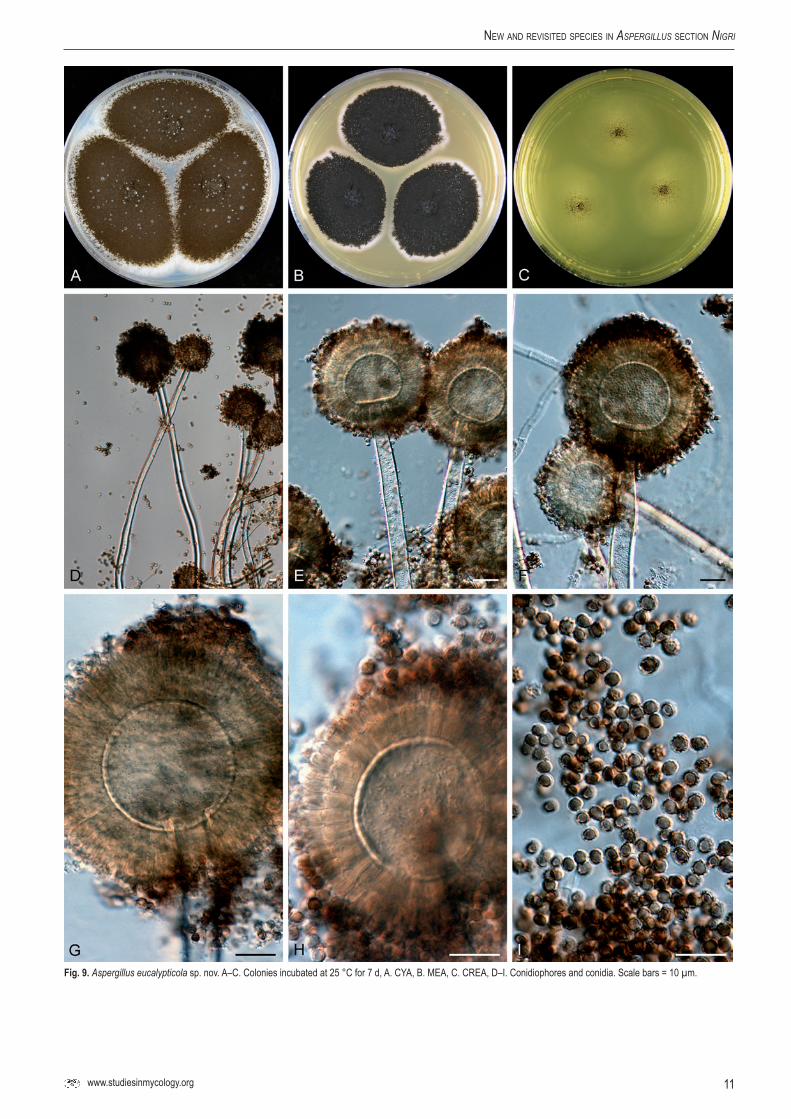
Aspergillus eucalypticolaVarga, Frisvad & Samson, sp.nov.MycoBank MB560387. Fig. 9.
Conidiophoris biseriatis, vesiculis globosis, 30–55 µm diam, stipitibus levibus vel subtiliter exasperatis. hyalinis, 8–14 µm latis. Conidiis globosis, 2.5–3.5 µm, brunneis, levibus vel grosse exasperatis. Sclerotia nulla.
Typus: ex leaves of Eucalyptus sp., New South Wales, Australia, isolated by P.W. Crous, 2007. (CBS H-20627 -- holotypus, culture ex-type CBS 122712 = IBT 29274).
CYA, 7 d, 25 °C: 68–72 mm; MEA, 7 d, 25 °C: 46–51 mm; YES, 7 d, 25 °C: 70–80 mm; OA, 7 d, 25 °C: 45–50 mm; CYA, 7 d, 37 °C: 30–50 mm; CREA: poor growth but good acid production; CYAS: 50–54 mm (strong sporulation on all media, except CREA). Colony reverse colour on CYA: beige to cream yellow, reverse colour on YES: yellow.
Conidiophores biseriate with globose vesicles 30–55 µm, stipe smooth-walled to finely roughened, hyaline, 8–14 µm. Conidia globose, 2.5–3.5 µm, brown, smooth-walled to coarsely roughened. Sclerotia not observed.
Aspergillus eucalypticola was isolated from an Eucalyptus leaf from Australia, and resembles morphologically A. tubingensis and A. costaricaensis. It can be distinguished from these two taxa by the β-tubulin or calmodulin sequence data. Aspergillus eucalypticola produces pyranonigrin A, funalenone, aurasperone B and other naphtho-γ-pyrones.
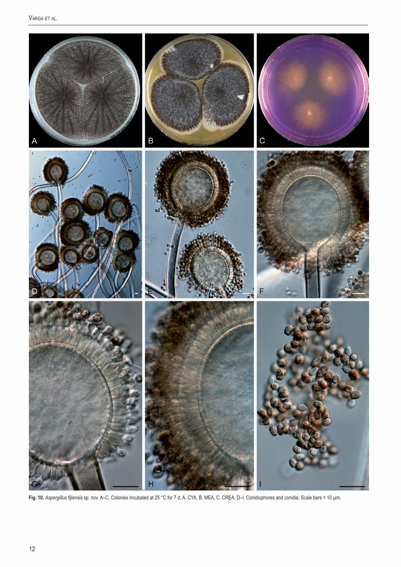
Aspergillus fijiensisVarga, Frisvad & Samson, sp. nov.MycoBank MB560388. Fig. 10.
Conidiophoris uniseriatis, vesiculis globosis vel ellipsoideis, 35–70 µm diam, stipitibus levibus vel subtiliter exasperatis, hyalinis, 8–12 µm latis. Conidiis ellipsoideis vel leniter fusiformibus, 3–3.5 × 3.4–4 µm, brunneis, grosse exasperatis vel echinulatis. Sclerotia nulla.
Typus: ex soil, Fiji Islands, K. Bundgaard. (CBS H-20628 -- holotypus, culture ex-type CBS 313.89 = IBT 13989).
Additional isolate: CBS 119.49 = IBT 4580, ex Lactuca sativa, Palembang, Indonesia).
CYA, 7 d, 25 °C: 71–78 mm; MEA, 7 d, 25 °C: 46–57 mm; YES, 7 d, 25 °C: 74–80 mm; OA, 7 d, 25 °C: 46–56 mm; CYA, 7 d, 37 °C: 12–25 mm; CREA: poor growth but moderate acid production; CYAS: 52–57 mm (strong sporulation on all media, except CREA). Colony reverse colour on CYA: beige to yellow, reverse colour on YES: yellow.
Conidiophores uniseriate with globose to ellipsoidal vesicles 35–70 µm wide, stipe smooth-walled to finely roughened, hyaline, 8–12 µm. Conidia ellipsoidal to slightly fusiform, 3–3.5 × 3.4–4 µm, brown, coarsely roughened to echinulate. Sclerotia not observed.
Aspergillus fijiensis is characterised by uniseriate conidial heads and is related to A. aculeatinus. It was isolated from soil in Fiji, and from Lactuca sativa in Indonesia. This species is able to grow at 37 °C, and produces asperparalines and okaramins.
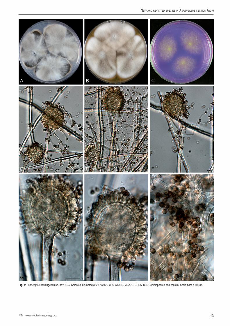
Aspergillus indologenus Frisvad, Varga & Samson, sp.nov.MycoBank MB560389. Fig. 11.
Conidiophoris uniseriatis, vesiculis ellipsoideis, 20–45 µm diam, stipitibus levibus vel subtiliter exasperatis, hyalinis, 5–11 µm latis. Conidiis globosis, 3–4 µm diam, brunneis, grosse exasperatis vel echinulatis. Sclerotia nulla.
Typus: ex soil India (CBS H-20629 -- holotypus, culture ex-type CBS 114.80 = IBT 3679).
10
Varga et al.
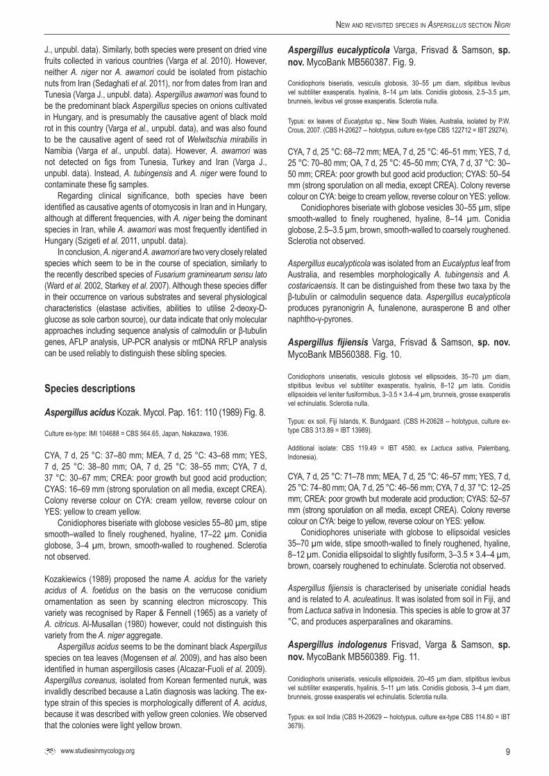
Fig.8. Aspergillus acidus A–C. Colonies incubated at 25 °C for 7 d, A. CYA, B. MEA, C. CREA, D–I. Conidiophores and conidia. Scale bars = 10 µm.
11www.studiesinmycology.org
new and reViSited SpecieS in aspergillus Section Nigri
Fig.9. Aspergillus eucalypticola sp. nov. A–C. Colonies incubated at 25 °C for 7 d, A. CYA, B. MEA, C. CREA, D–I. Conidiophores and conidia. Scale bars = 10 µm.
12
Varga et al.
Fig.10. Aspergillus fijiensis sp. nov. A–C. Colonies incubated at 25 °C for 7 d, A. CYA, B. MEA, C. CREA, D–I. Conidiophores and conidia. Scale bars = 10 µm.
13www.studiesinmycology.org
new and reViSited SpecieS in aspergillus Section Nigri
Fig.11. Aspergillus indologenus sp. nov. A–C. Colonies incubated at 25 °C for 7 d, A. CYA, B. MEA, C. CREA, D–I. Conidiophores and conidia. Scale bars = 10 µm.

14
Varga et al.
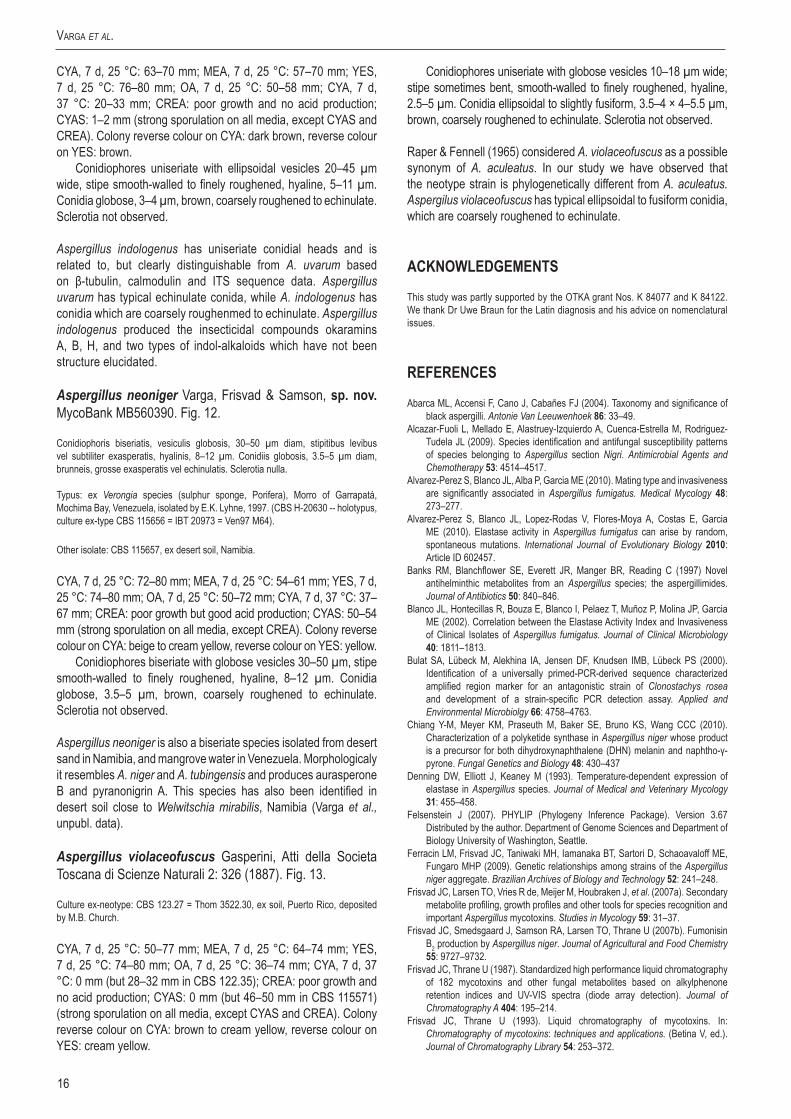
Fig.12. Aspergillus neoniger sp. nov. A–C. Colonies incubated at 25 °C for 7 d, A. CYA, B. MEA, C. CREA, D–I. Conidiophores and conidia. Scale bars = 10 µm.
15www.studiesinmycology.org
new and reViSited SpecieS in aspergillus Section Nigri
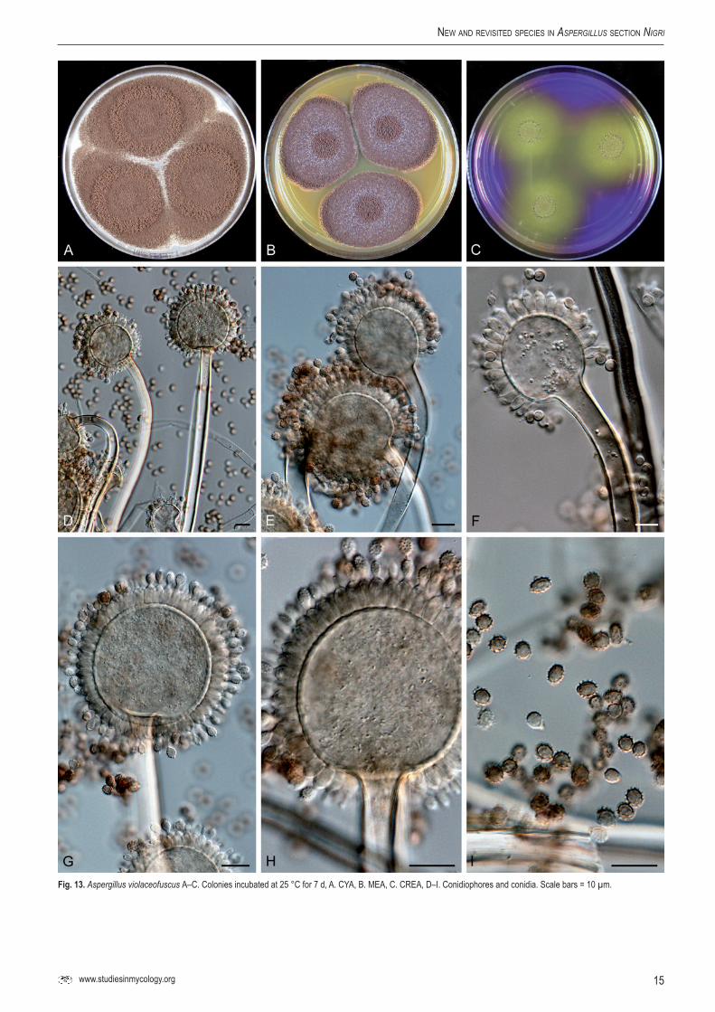
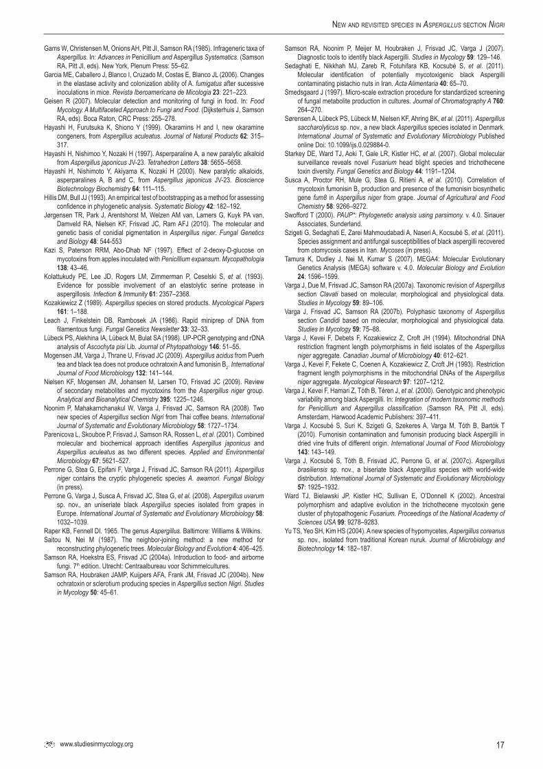
Fig.13.Aspergillus violaceofuscus A–C. Colonies incubated at 25 °C for 7 d, A. CYA, B. MEA, C. CREA, D–I. Conidiophores and conidia. Scale bars = 10 µm.
16
Varga et al.
CYA, 7 d, 25 °C: 63–70 mm; MEA, 7 d, 25 °C: 57–70 mm; YES, 7 d, 25 °C: 76–80 mm; OA, 7 d, 25 °C: 50–58 mm; CYA, 7 d, 37 °C: 20–33 mm; CREA: poor growth and no acid production; CYAS: 1–2 mm (strong sporulation on all media, except CYAS and CREA). Colony reverse colour on CYA: dark brown, reverse colour on YES: brown.
Conidiophores uniseriate with ellipsoidal vesicles 20–45 µm wide, stipe smooth-walled to finely roughened, hyaline, 5–11 µm. Conidia globose, 3–4 µm, brown, coarsely roughened to echinulate. Sclerotia not observed.
Aspergillus indologenus has uniseriate conidial heads and is related to, but clearly distinguishable from A. uvarum based on β-tubulin, calmodulin and ITS sequence data. Aspergillus uvarum has typical echinulate conida, while A. indologenus has conidia which are coarsely roughenmed to echinulate. Aspergillus indologenus produced the insecticidal compounds okaramins A, B, H, and two types of indol-alkaloids which have not been structure elucidated.
Aspergillus neonigerVarga, Frisvad & Samson, sp.nov.MycoBank MB560390. Fig. 12.
Conidiophoris biseriatis, vesiculis globosis, 30–50 µm diam, stipitibus levibus vel subtiliter exasperatis, hyalinis, 8–12 µm. Conidiis globosis, 3.5–5 µm diam, brunneis, grosse exasperatis vel echinulatis. Sclerotia nulla.
Typus: ex Verongia species (sulphur sponge, Porifera), Morro of Garrapatá, Mochima Bay, Venezuela, isolated by E.K. Lyhne, 1997. (CBS H-20630 -- holotypus, culture ex-type CBS 115656 = IBT 20973 = Ven97 M64).
Other isolate: CBS 115657, ex desert soil, Namibia.
CYA, 7 d, 25 °C: 72–80 mm; MEA, 7 d, 25 °C: 54–61 mm; YES, 7 d, 25 °C: 74–80 mm; OA, 7 d, 25 °C: 50–72 mm; CYA, 7 d, 37 °C: 37–67 mm; CREA: poor growth but good acid production; CYAS: 50–54 mm (strong sporulation on all media, except CREA). Colony reverse colour on CYA: beige to cream yellow, reverse colour on YES: yellow.
Conidiophores biseriate with globose vesicles 30–50 µm, stipe smooth-walled to finely roughened, hyaline, 8–12 µm. Conidia globose, 3.5–5 µm, brown, coarsely roughened to echinulate. Sclerotia not observed.
Aspergillus neoniger is also a biseriate species isolated from desert sand in Namibia, and mangrove water in Venezuela. Morphologicaly it resembles A. niger and A. tubingensis and produces aurasperone B and pyranonigrin A. This species has also been identified in desert soil close to Welwitschia mirabilis, Namibia (Varga et al., unpubl. data).
Aspergillus violaceofuscus Gasperini, Atti della Societa Toscana di Scienze Naturali 2: 326 (1887). Fig. 13.
Culture ex-neotype: CBS 123.27 = Thom 3522.30, ex soil, Puerto Rico, deposited by M.B. Church.
CYA, 7 d, 25 °C: 50–77 mm; MEA, 7 d, 25 °C: 64–74 mm; YES, 7 d, 25 °C: 74–80 mm; OA, 7 d, 25 °C: 36–74 mm; CYA, 7 d, 37 °C: 0 mm (but 28–32 mm in CBS 122.35); CREA: poor growth and no acid production; CYAS: 0 mm (but 46–50 mm in CBS 115571) (strong sporulation on all media, except CYAS and CREA). Colony reverse colour on CYA: brown to cream yellow, reverse colour on YES: cream yellow.
Conidiophores uniseriate with globose vesicles 10–18 µm wide; stipe sometimes bent, smooth-walled to finely roughened, hyaline, 2.5–5 µm. Conidia ellipsoidal to slightly fusiform, 3.5–4 × 4–5.5 µm, brown, coarsely roughened to echinulate. Sclerotia not observed.
Raper & Fennell (1965) considered A. violaceofuscus as a possible synonym of A. aculeatus. In our study we have observed that the neotype strain is phylogenetically different from A. aculeatus. Aspergilus violaceofuscus has typical ellipsoidal to fusiform conidia, which are coarsely roughened to echinulate.
ACKNOWLEDGEMENTS
This study was partly supported by the OTKA grant Nos. K 84077 and K 84122. We thank Dr Uwe Braun for the Latin diagnosis and his advice on nomenclatural issues.
REFERENCES
Abarca ML, Accensi F, Cano J, Cabañes FJ (2004). Taxonomy and significance of black aspergilli. Antonie Van Leeuwenhoek 86: 33–49.
Alcazar-Fuoli L, Mellado E, Alastruey-Izquierdo A, Cuenca-Estrella M, Rodriguez-Tudela JL (2009). Species identification and antifungal susceptibility patterns of species belonging to Aspergillus section Nigri. Antimicrobial Agents and Chemotherapy 53: 4514–4517.
Alvarez-Perez S, Blanco JL, Alba P, Garcia ME (2010). Mating type and invasiveness are significantly associated in Aspergillus fumigatus. Medical Mycology 48: 273–277.
Alvarez-Perez S, Blanco JL, Lopez-Rodas V, Flores-Moya A, Costas E, Garcia ME (2010). Elastase activity in Aspergillus fumigatus can arise by random, spontaneous mutations. International Journal of Evolutionary Biology 2010: Article ID 602457.
Banks RM, Blanchflower SE, Everett JR, Manger BR, Reading C (1997) Novel antihelminthic metabolites from an Aspergillus species; the aspergillimides. Journal of Antibiotics 50: 840–846.
Blanco JL, Hontecillas R, Bouza E, Blanco I, Pelaez T, Muñoz P, Molina JP, Garcia ME (2002). Correlation between the Elastase Activity Index and Invasiveness of Clinical Isolates of Aspergillus fumigatus. Journal of Clinical Microbiology 40: 1811–1813.
Bulat SA, Lübeck M, Alekhina IA, Jensen DF, Knudsen IMB, Lübeck PS (2000). Identification of a universally primed-PCR-derived sequence characterized amplified region marker for an antagonistic strain of Clonostachys rosea and development of a strain-specific PCR detection assay. Applied and Environmental Microbiolgy 66: 4758–4763.
Chiang Y-M, Meyer KM, Praseuth M, Baker SE, Bruno KS, Wang CCC (2010). Characterization of a polyketide synthase in Aspergillus niger whose product is a precursor for both dihydroxynaphthalene (DHN) melanin and naphtho-γ-pyrone. Fungal Genetics and Biology 48: 430–437
Denning DW, Elliott J, Keaney M (1993). Temperature-dependent expression of elastase in Aspergillus species. Journal of Medical and Veterinary Mycology 31: 455–458.
Felsenstein J (2007). PHYLIP (Phylogeny Inference Package). Version 3.67 Distributed by the author. Department of Genome Sciences and Department of Biology University of Washington, Seattle.
Ferracin LM, Frisvad JC, Taniwaki MH, Iamanaka BT, Sartori D, Schaoavaloff ME, Fungaro MHP (2009). Genetic relationships among strains of the Aspergillus niger aggregate. Brazilian Archives of Biology and Technology 52: 241–248.
Frisvad JC, Larsen TO, Vries R de, Meijer M, Houbraken J, et al. (2007a). Secondary metabolite profiling, growth profiles and other tools for species recognition and important Aspergillus mycotoxins. Studies in Mycology 59: 31–37.
Frisvad JC, Smedsgaard J, Samson RA, Larsen TO, Thrane U (2007b). Fumonisin B2 production by Aspergillus niger. Journal of Agricultural and Food Chemistry 55: 9727–9732.
Frisvad JC, Thrane U (1987). Standardized high performance liquid chromatography of 182 mycotoxins and other fungal metabolites based on alkylphenone retention indices and UV-VIS spectra (diode array detection). Journal of Chromatography A 404: 195–214.
Frisvad JC, Thrane U (1993). Liquid chromatography of mycotoxins. In: Chromatography of mycotoxins: techniques and applications. (Betina V, ed.). Journal of Chromatography Library 54: 253–372.
17www.studiesinmycology.org
new and reViSited SpecieS in aspergillus Section Nigri
Gams W, Christensen M, Onions AH, Pitt JI, Samson RA (1985). Infrageneric taxa of Aspergillus. In: Advances in Penicillium and Aspergillus Systematics. (Samson RA, Pitt JI, eds). New York, Plenum Press: 55–62.
Garcia ME, Caballero J, Blanco I, Cruzado M, Costas E, Blanco JL (2006). Changes in the elastase activity and colonization ability of A. fumigatus after sucessive inoculations in mice. Revista Iberoamericana de Micologia 23: 221–223.
Geisen R (2007). Molecular detection and monitoring of fungi in food. In: Food Mycology. A Multifaceted Approach to Fungi and Food. (Dijksterhuis J, Samson RA, eds). Boca Raton, CRC Press: 255–278.
Hayashi H, Furutsuka K, Shiono Y (1999). Okaramins H and I, new okaramine congeners, from Aspergillus aculeatus. Journal of Natural Products 62: 315–317.
Hayashi H, Nishimoo Y, Nozaki H (1997). Asperparaline A, a new paralytic alkaloid from Aspergillus japonicus JV-23. Tetrahedron Letters 38: 5655–5658.
Hayashi H, Nishimoto Y, Akiyama K, Nozaki H (2000). New paralytic alkaloids, asperparalines A, B and C, from Aspergillus japonicus JV-23. Bioscience Biotechnology Biochemistry 64: 111–115.
Hillis DM, Bull JJ (1993). An empirical test of bootstrapping as a method for assessing confidence in phylogenetic analysis. Systematic Biology 42: 182–192.
Jørgensen TR, Park J, Arentshorst M, Welzen AM van, Lamers G, Kuyk PA van, Damveld RA, Nielsen KF, Frisvad JC, Ram AFJ (2010). The molecular and genetic basis of conidial pigmentation in Aspergillus niger. Fungal Genetics and Biology 48: 544-553
Kazi S, Paterson RRM, Abo-Dhab NF (1997). Effect of 2-deoxy-D-glucose on mycotoxins from apples inoculated with Penicillium expansum. Mycopathologia 138: 43–46.
Kolattukudy PE, Lee JD, Rogers LM, Zimmerman P, Ceselski S, et al. (1993). Evidence for possible involvement of an elastolytic serine protease in aspergillosis. Infection & Immunity 61: 2357–2368.
Kozakiewicz Z (1989). Aspergillus species on stored products. Mycological Papers 161: 1–188.
Leach J, Finkelstein DB, Rambosek JA (1986). Rapid miniprep of DNA from filamentous fungi. Fungal Genetics Newsletter 33: 32–33.
Lübeck PS, Alekhina IA, Lübeck M, Bulat SA (1998). UP-PCR genotyping and rDNA analysis of Ascochyta pisi Lib. Journal of Phytopathology 146: 51–55.
Mogensen JM, Varga J, Thrane U, Frisvad JC (2009). Aspergillus acidus from Puerh tea and black tea does not produce ochratoxin A and fumonisin B2. International Journal of Food Microbiology 132: 141–144.
Nielsen KF, Mogensen JM, Johansen M, Larsen TO, Frisvad JC (2009). Review of secondary metabolites and mycotoxins from the Aspergillus niger group. Analytical and Bioanalytical Chemistry 395: 1225–1246.
Noonim P, Mahakarnchanakul W, Varga J, Frisvad JC, Samson RA (2008). Two new species of Aspergillus section Nigri from Thai coffee beans. International Journal of Systematic and Evolutionary Microbiology 58: 1727–1734.
Parenicova L, Skouboe P, Frisvad J, Samson RA, Rossen L, et al. (2001). Combined molecular and biochemical approach identifies Aspergillus japonicus and Aspergillus aculeatus as two different species. Applied and Environmental Microbiology 67: 5621–527.
Perrone G, Stea G, Epifani F, Varga J, Frisvad JC, Samson RA (2011). Aspergillus niger contains the cryptic phylogenetic species A. awamori. Fungal Biology (in press).
Perrone G, Varga J, Susca A, Frisvad JC, Stea G, et al. (2008). Aspergillus uvarum sp. nov., an uniseriate black Aspergillus species isolated from grapes in Europe. International Journal of Systematic and Evolutionary Microbiology 58: 1032–1039.
Raper KB, Fennell DI. 1965. The genus Aspergillus. Baltimore: Williams & Wilkins.Saitou N, Nei M (1987). The neighbor-joining method: a new method for
reconstructing phylogenetic trees. Molecular Biology and Evolution 4:406–425.Samson RA, Hoekstra ES, Frisvad JC (2004a). Introduction to food- and airborne
fungi. 7th edition. Utrecht: Centraalbureau voor Schimmelcultures. Samson RA, Houbraken JAMP, Kuijpers AFA, Frank JM, Frisvad JC (2004b). New
ochratoxin or sclerotium producing species in Aspergillus section Nigri. Studies in Mycology 50: 45–61.
Samson RA, Noonim P, Meijer M, Houbraken J, Frisvad JC, Varga J (2007). Diagnostic tools to identify black Aspergilli. Studies in Mycology 59: 129–146.
Sedaghati E, Nikkhah MJ, Zareb R, Fotuhifara KB, Kocsubé S, et al. (2011). Molecular identification of potentially mycotoxigenic black Aspergilli contaminating pistachio nuts in Iran. Acta Alimentaria 40: 65–70.
Smedsgaard J (1997). Micro-scale extraction procedure for standardized screening of fungal metabolite production in cultures. Journal of Chromatography A 760: 264–270.
Sørensen A, Lübeck PS, Lübeck M, Nielsen KF, Ahring BK, et al. (2011). Aspergillus saccharolyticus sp. nov., a new black Aspergillus species isolated in Denmark. International Journal of Systematic and Evolutionary Microbiology Published online Doi: 10.1099/ijs.0.029884-0.
Starkey DE, Ward TJ, Aoki T, Gale LR, Kistler HC, et al. (2007). Global molecular surveillance reveals novel Fusarium head blight species and trichothecene toxin diversity. Fungal Genetics and Biology 44: 1191–1204.
Susca A, Proctor RH, Mule G, Stea G, Ritieni A, et al. (2010). Correlation of mycotoxin fumonisin B2 production and presence of the fumonisin biosynthetic gene fum8 in Aspergillus niger from grape. Journal of Agricultural and Food Chemistry 58: 9266–9272.
Swofford T (2000). PAUP*: Phylogenetic analysis using parsimony. v. 4.0. Sinauer Associates, Sunderland.
Szigeti G, Sedaghati E, Zarei Mahmoudabadi A, Naseri A, Kocsubé S, et al. (2011). Species assignment and antifungal susceptibilities of black aspergilli recovered from otomycosis cases in Iran. Mycoses (in press).
Tamura K, Dudley J, Nei M, Kumar S (2007). MEGA4: Molecular Evolutionary Genetics Analysis (MEGA) software v. 4.0. Molecular Biology and Evolution 24: 1596–1599.
Varga J, Due M, Frisvad JC, Samson RA (2007a). Taxonomic revision of Aspergillus section Clavati based on molecular, morphological and physiological data. Studies in Mycology 59: 89–106.
Varga J, Frisvad JC, Samson RA (2007b). Polyphasic taxonomy of Aspergillus section Candidi based on molecular, morphological and physiological data. Studies in Mycology 59: 75–88.
Varga J, Kevei F, Debets F, Kozakiewicz Z, Croft JH (1994). Mitochondrial DNA restriction fragment length polymorphisms in field isolates of the Aspergillus niger aggregate. Canadian Journal of Microbiology 40: 612–621.
Varga J, Kevei F, Fekete C, Coenen A, Kozakiewicz Z, Croft JH (1993). Restriction fragment length polymorphisms in the mitochondrial DNAs of the Aspergillus niger aggregate. Mycological Research 97: 1207–1212.
Varga J, Kevei F, Hamari Z, Tóth B, Téren J, et al. (2000). Genotypic and phenotypic variability among black Aspergilli. In: Integration of modern taxonomic methods for Penicillium and Aspergillus classification. (Samson RA, Pitt JI, eds). Amsterdam, Harwood Academic Publishers: 397–411.
Varga J, Kocsubé S, Suri K, Szigeti G, Szekeres A, Varga M, Tóth B, Bartók T (2010). Fumonisin contamination and fumonisin producing black Aspergilli in dried vine fruits of different origin. International Journal of Food Microbiology 143: 143–149.
Varga J, Kocsubé S, Tóth B, Frisvad JC, Perrone G, et al. (2007c). Aspergillus brasiliensis sp. nov., a biseriate black Aspergillus species with world-wide distribution. International Journal of Systematic and Evolutionary Microbiology 57: 1925–1932.
Ward TJ, Bielawski JP, Kistler HC, Sullivan E, O’Donnell K (2002). Ancestral polymorphism and adaptive evolution in the trichothecene mycotoxin gene cluster of phytopathogenic Fusarium. Proceedings of the National Academy of Sciences USA 99: 9278–9283.
Yu TS, Yeo SH, Kim HS (2004). A new species of hypomycetes, Aspergillus coreanus sp. nov., isolated from traditional Korean nuruk. Journal of Microbiology and Biotechnology 14: 182–187.