partial thrombosed parasagittal avm, complete resection ... · pdf fileromanian neurosurgery...
TRANSCRIPT
Romanian Neurosurgery (2015) XXIX 3
Partial thrombosed parasagittal AVM, complete resection,
case report
Aurelia Mihaela Sandu1, M.R. Gorgan2
1PhD Student in Neurosurgery, University of Medicine and Pharmacy “Carol Davila” Bucharest,
Faculty of Medicine, Department of Neurosurgery; Clinic of Neurosurgery, Fourth Department
of Neurosurgery, Emergency Clinical Hospital Bagdasar-Arseni, Bucharest 2Professor in Neurosurgery, University of Medicine and Pharmacy “Carol Davila” Bucharest,
Faculty of Medicine, Department of Neurosurgery; Clinic of Neurosurgery, Fourth Department
of Neurosurgery, Emergency Clinical Hospital Bagdasar-Arseni, Bucharest
Abstract: INTRODUCTION: Arteriovenous malformations (AVMs) are congenital
lesions formed by a network of dysplastic vessels. CASE REPORT: We report a case of a
63 years old man, admitted with seizures and headache. Imaging findings, angio-CT,
angio-MR and angiography revealed a partially thombosed right parasagittal frontal
AVM, with fully thrombosed associated flow-related aneurysm on the main arterial
feeder. The patient underwent surgery and we performed total resection of the AVM.
The particularity of this case is the rare possibility of outcome with regression of the
vascular malformation. CONCLUSIONS: Brain AVMs are evolutive lesions. Regression,
through progressive thrombosis of the nidus is a rare possible outcome in brain AVMs.
In thrombosed AVMs angiography is not reliable, and angio-CT and/or angio-MR are
mandatory, in order to correctly evaluate nidus size and associated lesions. Symptomatic
AVMs require surgery. Partial thombosed AVMs can be safely resected.
Key words: arteriovenous malformation, AVM surgery, thrombosed nidus
Introduction
Vascular malformations of the brain are a
heterogeneous group of non-neoplastic
anomalies of cerebral blood vessels, arteries,
veins, capillaries, with modified flow. (14;44)
Vascular malformations of the brain occur as
a consequence of persistence of a primitive
model secondary to a defect in embryological
development. (44) McCormick divided
vascular malformations of the brain in:
arteriovenous malformations (AVMs),
cavernomas, venous angiomas, capillary
telangiectasia and arteriovenous fistulas. (23)
Brain AVMs are congenital non-neoplastic
lesions, containing a high complexity network
of dysplastic vessels. The network of vessels,
called nidus, is formed by complex direct
connections between arteries and veins
Sandu, Gorgan Partial thrombosed parasagittal AVM, complete resection
through vascular shunts, thus oxygenated
blood is carried from the arterial system
directly into the venous system without
passing through capillary bed. The nidus is fed
through dilated arteries and blood is drained
through arterialized veins. (14;21;22;38;44)
Brain AVMs are rare lesions. The incidence
of symptomatic AVMs is 0.89-1.34
cases/100,000 inhabitants/year (4;5;19) and
prevalence is 0.02-0.2%(2;3;19;37;42).
Although they are no common pathology,
brain AVMs represent a continuous and
prolific field of research (10-12;33-35),
because social impact of this disease is high.
AVMs are commonly found in young people,
mean age at diagnosis being 33.7-35
years.(3;4;15;19) Natural history reveals a o
mortality rate of 0.7-2.9%/year.(4;19;28)
Case report
We report a case of a right parasagittal
frontal, partial trombosed AVM who was
operated by the senior neurosurgeon into the
Fourth Department of Neurosurgery,
Emergency Clinical Hospital Bagdasar-Arseni
from Bucharest. We reviewed medical records,
imaging, treatment and follow-up. A man, 63
years old was admitted in department with
grand mal seizures for four years and
headache. The patient presented history of
ischemic coronary disease, old myocardial
infarction, coronary stent and diabetes
mellitus type II. The neurological exam
revealed no neurological deficits.
Brain CT-scan showed a right frontal
lesion, inhomogeneous, contrast enhancing,
with calcifications. Angio-CT showed an right
parasagittal frontal AVM, with nidus sizing
3/2 cm, containing partial calcified vessels,
with feeding arteries coming from right
anterior cerebral artery (ACA), which is
enlarged (3 mm in dimeter). A saccular
aneurysm was found on the A2 segment of
right ACA (4 mm dome, 2 mm neck).
Brain MRI showed a right parasagittal
frontal AVM, located in girus cinguli and
superior frontal lobe, with 3 cm maximal
diameter, with feeding arteries from right
ACA and venous drainage into a dilated
anterior frontal vein and finally into the
superior sagittal sinus (SSS). Right ACA is
enlarged, 3 mm in diameter and had high flow.
The saccular aneurysm from A2 segment of
right ACA had no vascular flow void inside. In
the surrounding brain there are hemosiderin
deposits, areas of calcifications and gliosis. A
porencephalic cavity is a sign of previous
bleeding.
Four vessels angiography showed a low
flow right frontal AVM, fed from the right
callosomarginal artery. Right callosomarginal
artery presented areas of stenosis and irregular
caliber. The nidus was 1 cm in size. Venous
drainage is not detected and is probably done
into the SSS. The angiography also revealed
marked atherosclerosis.
The patient underwent surgery. We
entered the interhemispheric fissure and we
found an AVM corticalized on the medial
surface of the frontal lobe. The nidus was
composed of patial thrombosed vessels. We
identified and coagulated two feeding arteries
coming from the right callosomarginal artery.
The nidus was circumferentially dissected and
mobilized into the interhemispheric fissure to
facilitate deep dissection. The deep part of the

Romanian Neurosurgery (2015) XXIX 3
nidus reached the ependymal surface, and
frontal horn of the lateral ventricle was
opened. Finally, two draining veins, into the
SSS and into the inferior sagittal sinus were
occluded with vascular clips. An external
ventricular drainage was left in the frontal
horn of the lateral ventricle. The wound was
closed in anatomical layers.
The outcome was favorable, the patient
presented no postoperative neurological
deficits. Following surgery, the patient
presented no seizures under 600 mg
Carbamazepin/day. The external ventricular
drainage was kept for 3 days.
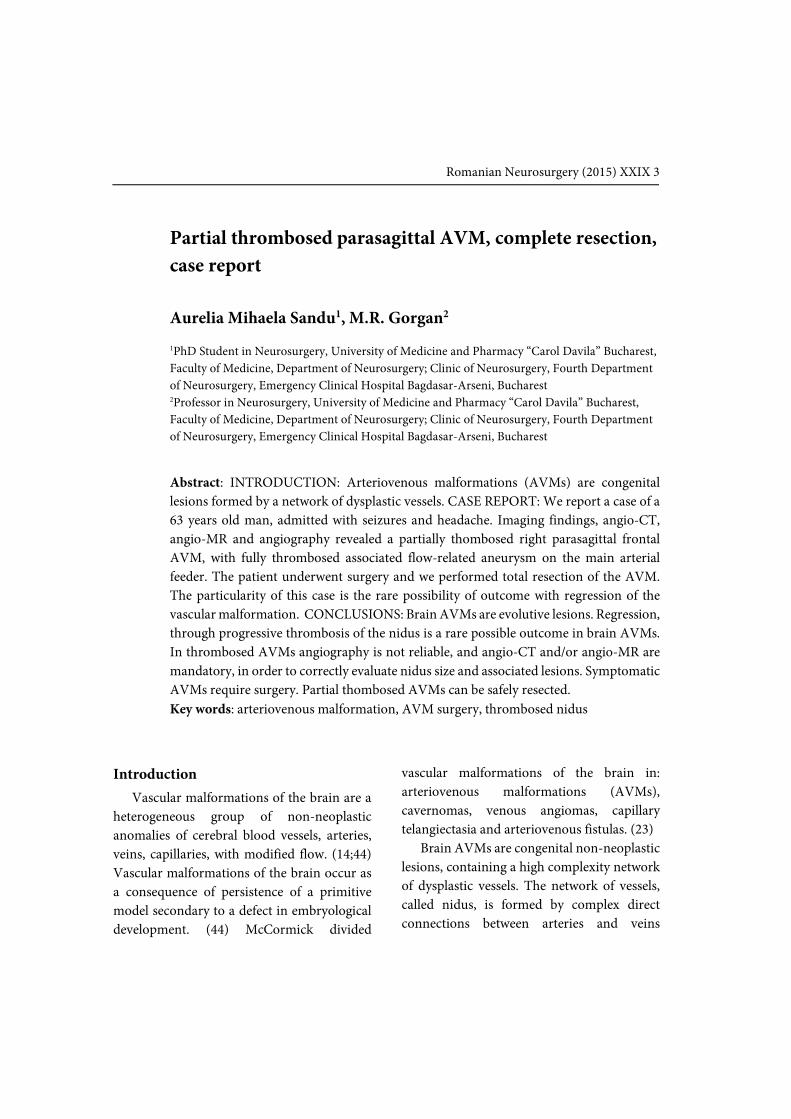
Figure 1 - Angio-CT scan. Right parasagittal frontal
AVM, with calcifications, feeding arteries from right
ACA. Saccular aneurysm, A2 segment of right ACA

Sandu, Gorgan Partial thrombosed parasagittal AVM, complete resection
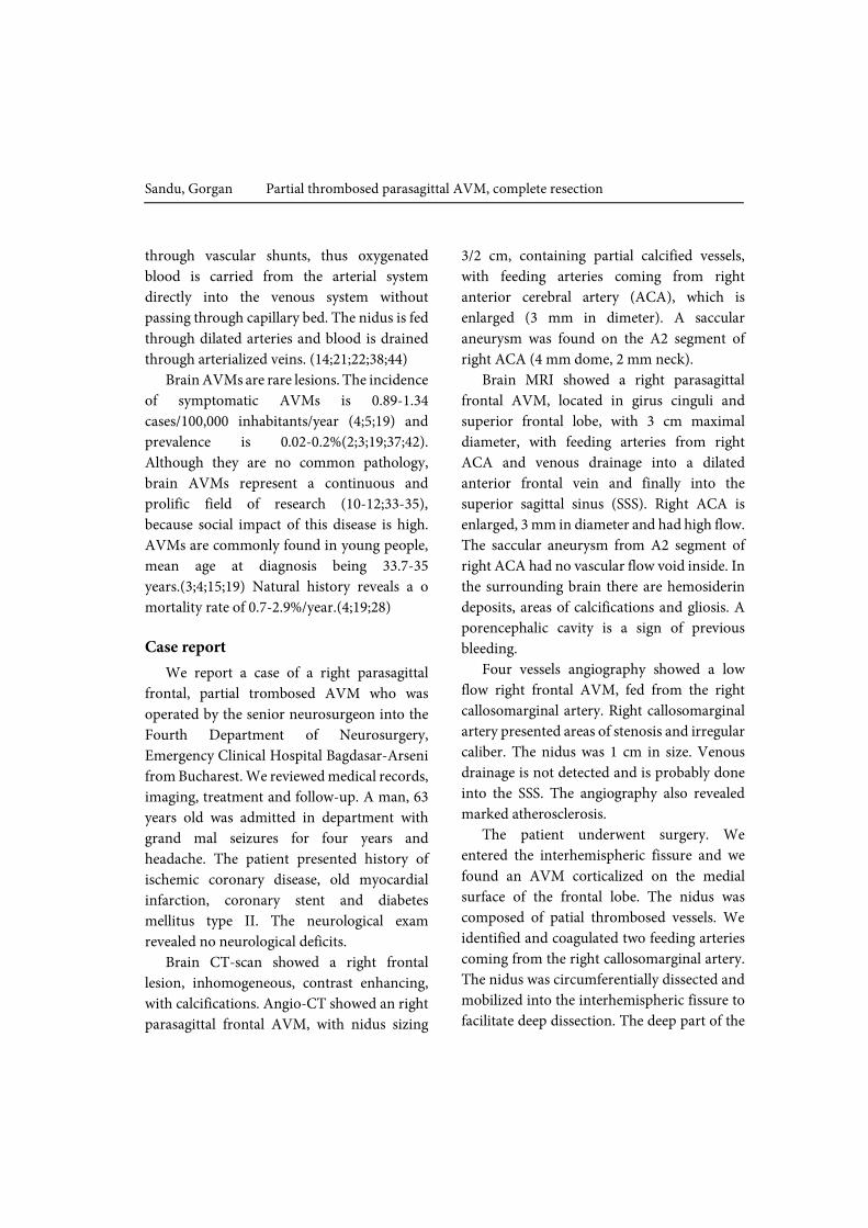
Figure 2 - Brain MRI. Right parasagittal frontal
AVM, with feeding arteries from right ACA and
venous drainage into a dilated anterior frontal vein.
Hemosiderin deposits, areas of calcifications and
gliosis in the surrounding brain
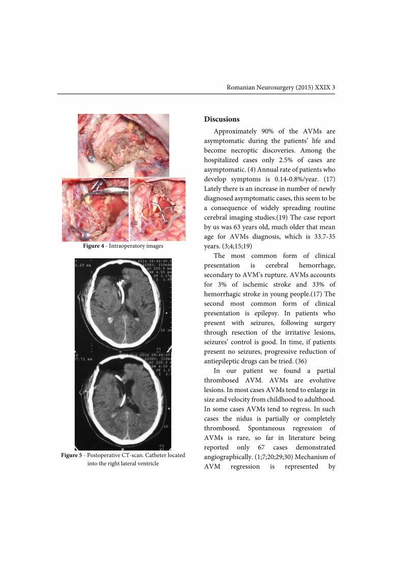
Figure 3 - Right ICA angiography. Low flow right
frontal AVM, with feeding artery from right
callosomarginal artery. Nidus is 1 cm in diameter,
injected in the venous phase. Atherosclerosis
Romanian Neurosurgery (2015) XXIX 3
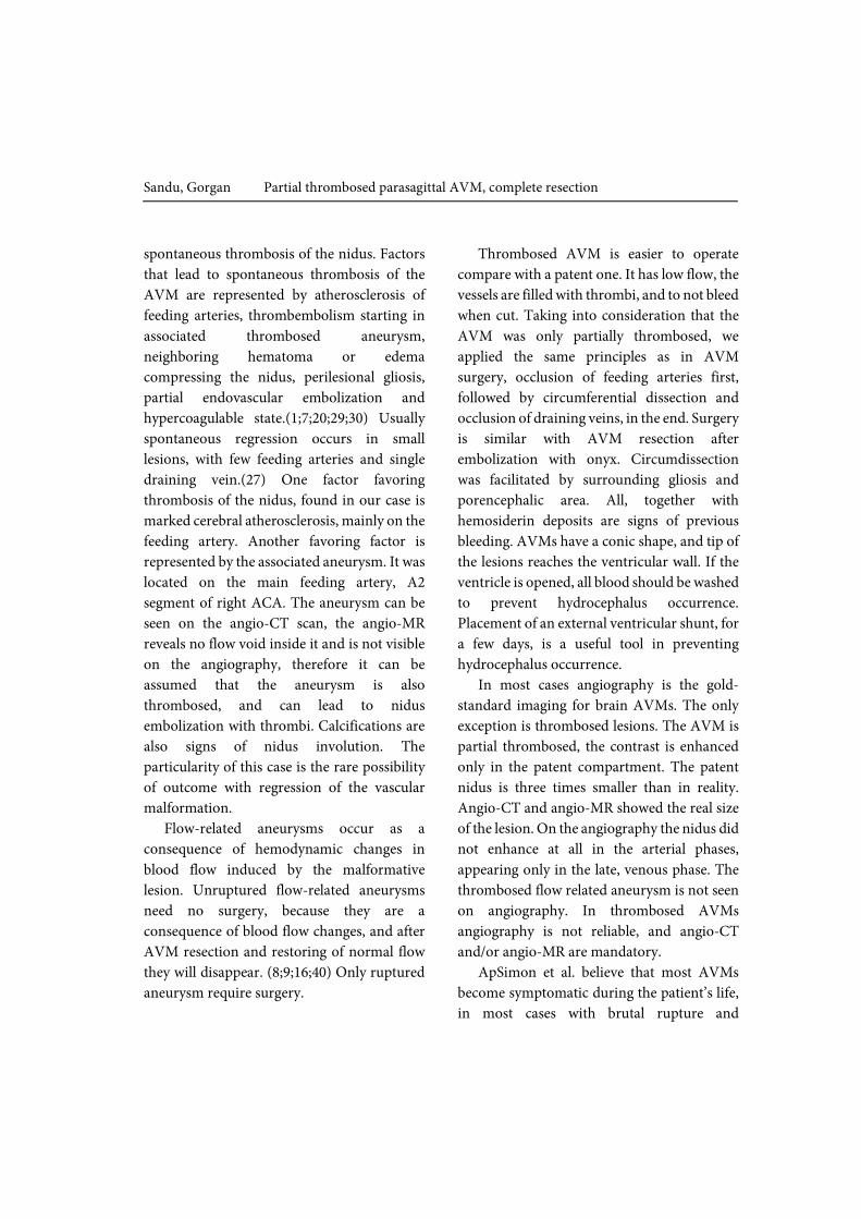
Figure 4 - Intraoperatory images
Figure 5 - Postoperative CT-scan. Catheter located
into the right lateral ventricle
Discusions
Approximately 90% of the AVMs are
asymptomatic during the patients’ life and
become necroptic discoveries. Among the
hospitalized cases only 2.5% of cases are
asymptomatic. (4) Annual rate of patients who
develop symptoms is 0.14-0.8%/year. (17)
Lately there is an increase in number of newly
diagnosed asymptomatic cases, this seem to be
a consequence of widely spreading routine
cerebral imaging studies.(19) The case report
by us was 63 years old, much older that mean
age for AVMs diagnosis, which is 33.7-35
years. (3;4;15;19)
The most common form of clinical
presentation is cerebral hemorrhage,
secondary to AVM’s rupture. AVMs accounts
for 3% of ischemic stroke and 33% of
hemorrhagic stroke in young people.(17) The
second most common form of clinical
presentation is epilepsy. In patients who
present with seizures, following surgery
through resection of the irritative lesions,
seizures’ control is good. In time, if patients
present no seizures, progressive reduction of
antiepileptic drugs can be tried. (36)
In our patient we found a partial
thrombosed AVM. AVMs are evolutive
lesions. In most cases AVMs tend to enlarge in
size and velocity from childhood to adulthood.
In some cases AVMs tend to regress. In such
cases the nidus is partially or completely
thrombosed. Spontaneous regression of
AVMs is rare, so far in literature being
reported only 67 cases demonstrated
angiographically. (1;7;20;29;30) Mechanism of
AVM regression is represented by
Sandu, Gorgan Partial thrombosed parasagittal AVM, complete resection
spontaneous thrombosis of the nidus. Factors
that lead to spontaneous thrombosis of the
AVM are represented by atherosclerosis of
feeding arteries, thrombembolism starting in
associated thrombosed aneurysm,
neighboring hematoma or edema
compressing the nidus, perilesional gliosis,
partial endovascular embolization and
hypercoagulable state.(1;7;20;29;30) Usually
spontaneous regression occurs in small
lesions, with few feeding arteries and single
draining vein.(27) One factor favoring
thrombosis of the nidus, found in our case is
marked cerebral atherosclerosis, mainly on the
feeding artery. Another favoring factor is
represented by the associated aneurysm. It was
located on the main feeding artery, A2
segment of right ACA. The aneurysm can be
seen on the angio-CT scan, the angio-MR
reveals no flow void inside it and is not visible
on the angiography, therefore it can be
assumed that the aneurysm is also
thrombosed, and can lead to nidus
embolization with thrombi. Calcifications are
also signs of nidus involution. The
particularity of this case is the rare possibility
of outcome with regression of the vascular
malformation.
Flow-related aneurysms occur as a
consequence of hemodynamic changes in
blood flow induced by the malformative
lesion. Unruptured flow-related aneurysms
need no surgery, because they are a
consequence of blood flow changes, and after
AVM resection and restoring of normal flow
they will disappear. (8;9;16;40) Only ruptured
aneurysm require surgery.
Thrombosed AVM is easier to operate
compare with a patent one. It has low flow, the
vessels are filled with thrombi, and to not bleed
when cut. Taking into consideration that the
AVM was only partially thrombosed, we
applied the same principles as in AVM
surgery, occlusion of feeding arteries first,
followed by circumferential dissection and
occlusion of draining veins, in the end. Surgery
is similar with AVM resection after
embolization with onyx. Circumdissection
was facilitated by surrounding gliosis and
porencephalic area. All, together with
hemosiderin deposits are signs of previous
bleeding. AVMs have a conic shape, and tip of
the lesions reaches the ventricular wall. If the
ventricle is opened, all blood should be washed
to prevent hydrocephalus occurrence.
Placement of an external ventricular shunt, for
a few days, is a useful tool in preventing
hydrocephalus occurrence.
In most cases angiography is the gold-
standard imaging for brain AVMs. The only
exception is thrombosed lesions. The AVM is
partial thrombosed, the contrast is enhanced
only in the patent compartment. The patent
nidus is three times smaller than in reality.
Angio-CT and angio-MR showed the real size
of the lesion. On the angiography the nidus did
not enhance at all in the arterial phases,
appearing only in the late, venous phase. The
thrombosed flow related aneurysm is not seen
on angiography. In thrombosed AVMs
angiography is not reliable, and angio-CT
and/or angio-MR are mandatory.
ApSimon et al. believe that most AVMs
become symptomatic during the patient’s life,
in most cases with brutal rupture and
Romanian Neurosurgery (2015) XXIX 3
intracranial hemorrhage. (4) ARUBA
phenomenon radically changed the way of
thinking in unruptured brain AVMs.(25;26)
ARUBA was a prospective, controlled,
randomized, multicentre, international trial,
which counted the risk of developing cerebral
symptomatic stroke or death in patients with
unruptured AVMs, who underwent either
surgery or conservative treatment. ARUBA
phenomenon completely changes the vision
regarding this pathology, the interventionist
therapeutical attitude from the pre-ARUBA
era, being replaced by a conservative one.
Relevant literature study reveals that similar
results can be found, such as reports of old,
asymptomatic patients, with incidental AVM.
This study had sparked various reactions to
the medical world(13;18;24;31;32;39;41;43),
and so far there is no consensus regarding an
optimal therapy algorithm in unruptured
AVMs. Choosing between surgical and
conservative attitude depends on a variety of
factors related to the characteristics of the
lesion, medical status of the patient, patient
and family desire and preference of
neurosurgeon. Other authors proved the
superiority of surgery for certain patients with
unruptured AVMs. (6)
Conclusions
Brain AVMs are evolutive lesions.
Regression, through progressive thrombosis of
the nidus is a rare possible outcome in brain
AVMs. In thrombosed AVMs angiography is
not reliable, and angio-CT and/or angio-MR
are mandatory, in order to correctly evaluate
nidus size and associated lesions. Symptomatic
AVM require surgery. Partial thombosed
AVMs can be safely resected.
Correspondence
Aurelia Mihaela Sandu
Address: Emergency Clinical Hospital Bagdasar-
Arseni, No. 10-12, Berceni Street, Sector 4,
Bucharest; e-mail: [email protected]; tel.
0724.263.023
Abreviations
ACA – anterior cerebral artery
AVM - arteriovenous malformation
SSS – superior sagittal sinus
Acknowledgement
This paper was co-financed from the European
Social Fund, through the Sectorial Operational
Programme Human Resources Development 2007-
2013, project number POSDRU/159/1.5/S/138907
“Excellence in scientific interdisciplinary research,
doctoral and postdoctoral, in the economic, social
and medical fields - EXCELIS”, coordinator The
Bucharest University of Economic Studies.
References
1. Abdulrauf SI, Malik GM, & Awad IA. (1999).
Spontaneous angiographic obliteration of cerebral
arteriovenous malformations. Neurosurgery 44, 280-287.
2. Al-Shahi R, Bhattacharya JJ, Currie DG,
Papanastassiou V, Ritchie V, Roberts RC, Sellar RJ, &
Warlow CP. (2003). Prospective, population-based
detection of intracranial vascular malformations in
adults: the Scottish Intracranial Vascular Malformation
Study (SIVMS). Stroke 34, 1163-1169.
3. Al-Shahi R & Warlow C. (2001). A systematic review of
the frequency and prognosis of arteriovenous
malformations of the brain in adults. Brain 124, 1900-
1926.
4. ApSimon HT, Reef H, Phadke RV, & Popovic EA.
(2002). A population-based study of brain arteriovenous
malformation: long-term treatment outcomes. Stroke 33,
2794-2800.

Sandu, Gorgan Partial thrombosed parasagittal AVM, complete resection
5. Berman MF, Sciacca RR, Pile-Spellman J, Stapf C,
Connolly ES Jr, Mohr JP, & Young WL. (2000). The
epidemiology of brain arteriovenous malformations.
Neurosurgery 47, 389-396.
6. Bervini D, Morgan MK, Ritson EA, & Heller G. (2014).
Surgery for unruptured arteriovenous malformations of
the brain is better than conservative management for
selected cases: a prospective cohort study. J Neurosurg
121, 878-890.
7. Bruis DR, van den Berg R, Lycklama G, van der Worp
HB, Driven CM, & Vanterhop WP. (2004). Spontaneous
regression of brain arteriovenous malformations--a
clinical study and a systematic review of the literature. J
Neurosurg 251, 1375-1382.
8. Cunha MJ, Stein BM, Solomon RA, & McCormick PC.
(1992). The treatment of associated intracranial
aneurysms and arteriovenous malformations. J
Neurosurg 77, 853-859, 1992.
9. Flores BC, Klinger DR, Rickert KL, Barnett SL, Welch
BG, White JA, Batjer HH, & Samson DS. (2014).
Management of intracranial aneurysms associated with
arteriovenous malformations. Neurosurg Focus 37, E11.
10. Giovani A, Sandu A, Neacsu A, and Gorgan RM:
Surgical treatment and outcome of cerebral cavernomas
– a 10 years' experience. Rom Neurosurg 21:394-404,
2014.
11. Gorgan MR, Bucur N, Neacșu A, Sandu AM, Brehar
FM, Prună VM, & Giovani A. (2014). Aspecte clinice și
microchirurgicale în cavernoamele cerebrale (Clinical
aspects and microsurgery in brain cavernomas. A XVII-a
Conferință Naționala de Stroke (AVC) cu participare
internațională, 15-17 octombrie 2014, Brașov, România;
abstractul lucrării a fost publicat în Revista Română de
Stroke (AVC) 17, 62-64.
12. Gorgan MR, Ciubotaru VG, Tătăranu LG, Tașcu A,
Iliescu A, Bucur N, Neacșu A, Brehar FM, & Sandu AM.
(2014). Our experience in a series of 277 brain AVMs,
microsurgical treatment and outcome. The 40th Congress
of the Romanian Society of Neurosurgery with
International Participation, 25-27 septembrie 2014,
București, România; abstractul lucrării a fost publicat în
Rom Neurosurg 21, 525-526.
13. Grasso G. (2014). The ARUBA study: what is the
evidence? World Neurosurg 82, e576.
14. Greenberg MS. (2010). Vascular malformations, in
Greenberg MS (ed): Handbook of neurosurgery. New
York, Thieme Medical Publisher, pp 1098-1142.
15. Gross BA & Du R. (2013). Natural history of cerebral
arteriovenous malformations: a meta-analysis. J
Neurosurg 118, 437-443.
16. Halim AX, Singh V, Johnston SC, Higashida RT,
Down CF, Halbach W, Lawton MT, Gress DR,
McCulloch CE, Young WL, & UCSF BAVM Study
Project Brain Arteriovenous Malformation. (2002).
Characteristics of brain arteriovenous malformations
with coexisting aneurysms: a comparison of two referral
centers. Stroke 33, 675-679.
17. Kale SS & Agarwal D. (2013). CNS arteriovenous
malformations: classifications and diagnosis. .docstoc
www.docstoc.com/docs/81960364/CNS-Arteriovenous-
Malformations-Classifications-and-
Diagnosis[11.05.2013].
18. Knopman J & Stieg PE. (2014). Management of
unruptured brain arteriovenous malformations. Lancet
383, 581-583.
19. Laakso A & Hernesniemi J. (2012). Arteriovenous
malformations: epidemiology and clinical presentation.
Neurosurg Clin N Am 23, 1-6.
20. Lee SK, Vilela P, Willinsky R, & ter Brugge KG.
(2002). Spontaneous regression of cerebral arteriovenous
malformations: clinical and angiographic analysis with
review of the literature. Neuroradiology 44, 11-16.
21. Marciano FF, Vishteh AG, Apostolides PJ, & Spetzler
RF. (2000). Arteriovenous malformations -
supratentorial, in Kaye AH, Black P (eds): Operative
neurosurgery. London, Harcourt Publishers Limited, pp
1079-1091.
22. McCormick WF. (1966). The pathology of vascular
(“arteriovenous”) malformations. J Neurosurg 24, 807-
816.
23. McCormick WF. (1978). Classification, pathology
and natural history of angiomas of the central nervous
system. Wkly Update Neurol Neurosurg 1, 2-7.
24. Meling TR, Proust F, Gruber A, Niemela M, Regli L,
Roche PH, & Vajkoczy P. (2014). On apples, oranges, and
ARUBA. Acta Neurochir (Wien) 156, 1775-1779, 2014.
25. Mohr JP, Moskowitz AF, Stapf C, Hartmann A, Lord
A, Marshall SM, Mast H, Moquete E, Moy CS, Parides M,
Pile-Spellman J, Al-Shahi Salman R, Young WL, Gobin
YP, Kureshi I, & Brisman JL. (2010). The ARUBA trial:
current status, future hopes. Stroke 41, e537-540.
26. Mohr JP, Parides MK, Stapf C, Moquete E, Moy CS,
Overbey JR, Al-Shahi Salman R, Vicaut E, Young WL,
Houdart E, Cordonnier C, Stefani MA, Hartmann A, von

Romanian Neurosurgery (2015) XXIX 3
Kummer R, Brondi A, Berkefeld J, Klijn C, Harkness K,
Libman R, Barreau X, Moskowitz AJ, & international
ARUBA investigators. (2014). Medical management with
or without interventional therapy for unruptured brain
arteriovenous malformations (ARUBA): a multicentre,
non-blinded, randomised trial. Lancet 383, 614-621.
27. Omojola MF, Fox AJ, Vinuela F, & Debrun G. (1985).
Stenosis of afferent vessels of intracranial arteriovenous
malformations. ANJR Am J Neuroradiol 6, 791-793.
28. Ondra SL, Troupp H, George ED. (1990). Scawab K.
The natural history of symptomatic arteriovenous
malformations of the brain: a 24-year follow-up
assessment. J Neurosurg 73, 387-391.
29. Panciani PP, Fontanella M, Carlino C, Bergiu M, &
Ducati A. (2008). Progressive spontaneous occlusion of a
cerebellar AVM: pathogenetic hypothesis and review of
literature. Clin Neurol Neurosurg 110, 502-510.
30. Patel MC, Hodgson TJ, Kemeny AA, & Forster DM.
(2001). Spontaneous obliteration of pial arteriovenous
malformations: a review of 27 cases. AJNR Am J
Neuroradiol 22, 531-536.
31. Proust F, Roche PH, & Meling TR. (2014). Does
ARUBA study improve our knowledge as regards the
management of unruptured brain arteriovenous
malformations? Neurochirurgie 60, 2-4.
32. Russin J & Spetzler R. (2014). Commentary: the
ARUBA trial. Neurosurgery 75, E96-E97.
33. Sandu A & Gorgan M. (2015). Microsurgery and
outcome in 277 brain AVMs, a common cause of
hemorrhagic stroke in young people. The 9th World
Congress on Controversies in Neurology (CONy), 26-28
March 2015, Budapest, Hungary.
34. Sandu AM. (2015). Electronic national registry of
brain arteriovenous malformations - a useful tool in
monitoring patients with cerebral vascular pathology.
Congresul Universității de Medicină și Farmacie Carol
Davila 28-30 mai 2015, București, România.
35. Sandu AM, Ciubotaru VG, Tătăranu LG, Tașcu A,
Bucur N, Neacșu A, & Gorgan MR. (2014). Clinical
aspects, management and oucome of brain arteriovenous
malformations - result with microsurgery first policy.
Rom Neurosurg 21, 369-383.
36. Sandu AM & Gorgan MR. (2013). Total resection in a
giant left frontal arteriovenous malformation, grade V
Spetzler-Martin - case report. Rom Neurosurg 20, 46-56.
37. Soderman M, Andersson T, Karlsson B, Wallace MC,
& Edner G. (2003). Management of patients with brain
arteriovenous malformations. Eur J Radiol 46, 195-205.
38. Spagmolo E. (2012). Surgical management of cerebral
arteriovenous malformations, in Quinones-Hinojosa A
(ed): Schmidek & Sweet operative neurosurgical
techniques: indications, methods, and results.
Philadelphia, Elsevier-Saunders, pp 1003-1018.
39. Starke RM, Sheehan JP, Ding D, Liu KC, Kondziolka
D, Crowley RW, Lunsford LD, & Kassell NF. (2014).
Conservative management or intervention for
unruptured brain arteriovenous malformations. World
Neurosurg 82, e668-e669.
40. Thompson RC, Steinberg GK, Levy RP, & Marks MP.
(1998). The management of patients with arteriovenous
malformations and associated intracranial aneurysms.
Neurosurgery 43, 202-211.
41. Warlow C, Al-Shahi Salman R, Lawton MT, Abla AA,
Gross BA, Scott RM, Smith ER, Solomon RA, & Connolly
ES Jr. (2014). Management of brain arteriovenous
malformations - Comments on Medical management
with or without interventional therapy for unruptured
brain arteriovenous malformations (ARUBA): a
multicentre, non-blinded, randomised trial. Lancet 383,
1632-1635.
42. Weber F & Knopf H. (2006). Incidental findings in
magnetic resonance imaging of the brains of healthy
young men. J Neurol Sci 240, 81-84.
43. Weber MA. (2014). Cerebral arteriovenous
malformations: Results of the ARUBA study. Radiologe
54, 422-424.
44. Yasargil MG. (1987). Microneurosurgery. IIIA. AVM
of the brain, history, embryology, pathological
considerations, hemodynamics, diagnostic studies,
microsurgical anatomy. Georg Thieme Verlag, Stuttgart;
Thieme Medical Publishers, Inc., New York.