pellionisz a, llinás r. neuroscience. 1979;4(3):323 … · brain modeling by tensor network theory...
TRANSCRIPT

Brain modeling by tensor network theory and computer simulation. The cerebellum: distributed processor for predictive coordination. Pellionisz A, Llinás R. Neuroscience. 1979;4(3):323-48. PMID: 431817 See Pubmed Reference at the link http://www.ncbi.nlm.nih.gov/pubmed/431817 and searchable full .pdf file below


Nc,,r,,wmc<‘ Vol. 4. pp. 323 to 348 Pergamon Press Ltd. 1979. Prrnted in Great Briinin
BRAIN MODELING BY TENSOR NETWORK THEORY AND COMPUTER SIMULATION. THE CEREBELLUM:
DISTRIBUTED PROCESSOR FOR PREDICTIVE COORDINATION
A. PELLIONKSZ and R. LY_IN,~S
Department of Physiology & Biophysics, New York University Medical Center, 550 First Avenue, NY 10016, U.S.A.
Abstract-A fu~amental problem regarding the functional jnterpretation of neural networks in the central nervous system is that of establishing the principles of their parallel, distribute organization. Available morphological and physiological knowledge concerning the cerebellum suggests that the central nervous system may use organizational principles other than the traditionally assumed ‘random connectivity’, ‘reflex loops’ or ‘redundancy’.
We propose formally, and demonstrate by computer modeling, that the firing frequencies of individual cells over a cerebellar cortical area may be interpreted as a spatially distributed, finite, series expansion of a time function, which is reconstructed, by summation, in the nucleus where the cortical cells project. Thus, for example, the firing of Purkinje cells, when considered as representing a Taylor expansion, yield a prediction in the cerebellar nuclei of the frequency-time function of the input arriving at the cortex. This ‘lookahead’ (A) is an emergent property of the inherently parallel, distributed network.
In order to analyze how a Taylor expansion-like process is used by the cerebellum on a system fevet, the linear algebraic matrix- and v~tor-representation of a distribute network was generalized in such a way as to regard the neuronal networks as tensors. Thus, the brain is envisioned as a set of tensorial systems which communicate with each other through vectorial channels (the pathways). These pathways carry multidimensional frequency vectors which are transformed by the tensors. In these terms the function of a particular subsystem of the central nervous system, in the present case the cerebellum, is represented in a multidimensional space. The frequency-hyperspace is characterized by the matrix of the cerebellar tensor which specifies a curved set of trajectories: a cerebellar vector field. Dynamic posture and balance are interpreted as displacement or stabilization of the functional status vector of the motor system along these curved trajectories. Cerebellar coordination of ballistic movements can be described as guiding the movement onto the ‘wired in’ trajectories of the vector field, by virtue of the coordination and inhibition vectors provided by the cerebellum. A proposal is also introduced that the climbing fiber system operates by momentary perturbations of the vector field, leading to deformations of the trajectories of the cerebellar frequency hyperspace.
To bridge the gap between an attempt to treat parallel, distributed, neuronal networks, such as the cerebellum, as geometrical objects (leading, at the first approximation, to a linear mathematical formulation) and the simultaneous task of incorporating and interpreting experimental data, computer simulation methods are required. This combined approach of mathematical treatment (which provides an abstract language) and computer simulation (which, by accommodating data into the model, explores the significance of deviations from linear character) appears at present to be the most adequate technique for dealing with central nervous system function.
A FUNDAMENTAL task of any general theory for central nervous system (CNS) function is that of explaining the organizational principles of the spatially distri- buted and intrinsically parallel character of its neur- onal networks (cf. EDELMAN & MOU~CASTLE, 1978).
The term ‘parallel’ is used here to indicate that infor- mation transfer and processing is performed by the simultaneous action of large arrays of in>erconnected neurons, rather than serially by chains of individual nerve cells.
Termin&gy: Throughout the paper vectors are desig- nated by barred capitals, matrices by double-barred capi- tals. All (e.g. either one- or two-dimensional, etc.) arrays which identify fensors, will be designated by Greek capitals.
There are profound and intriguing differences between the parallel mode of operation of the brain and the serial organization of most man-made infor- mation processors (VON NEUMANN, 1956; 1959).
While the latter offer great economy in the number
of elements, they require high reliability of their units, since a breakdown of a single link in the chain would disrupt the function of the whole system. In addition, serial systems are characterized by lengthy functional chains and, hence, the total time of the processing along the chain is long.
In the brain, by contrast, due to the parallel organ- ization of neuronal systems, the breakdown of large numbers of elements may remain largely unnoticed, VON NEUMANN (1959) also pointed out that the re-
323

324 A. PELLIONISZ and R. LLINAS
markably shallow logical depth of neuronal systems permits the use of relatively slow-acting individual elements, The price of their reliability and resistance to lesions is paid by the large number of units required in their function.
Hitherto little has been achieved in the analysis of known neuronal networks as distributed and parallel systems. Instead, in order to deal with particular neuronal circuits before the nature of their parallel organization has been understood, three convenient fallacies have long been adopted:
(1) One is the concept of ‘loops’. Here the complex- ities of parallel neuronal networks are reduced to a chain of a few serially connected individual neurons, and it is assumed that such a ‘reflex arc’ is concep- tually equivalent to the total neuronal assembly.
(2) The other extreme view is that of ‘mass action’ where the concept of diffuse ‘random’ interconnec- tions of the neuronal elements is seen as the principle of organ~ation. This view is seldom held in a pure form, because of its theoretical extremity and because it is in serious contradiction to some obvious struc- tural features of neuronal networks (i.e. a considerable degree of non-random structural and functional speci- ficity is always present). While this view does not ignore the complex connections within the network, by rendering them functionally irrelevant it leads to their easy dismissal from theoretical accounts.
(3) A third view reflects more carefully upon the inevitability of reckoning functionally with the parallel structural features of networks. This concept of ‘redundancy’ combines the loop-view with the assertion that the large network is basically many such loops together. The functional usefulness (as sug- gested by VON NEUMANN, 1959) of the multiple exist- ence of a functional unit would be to provide a high degree of redundancy that would increase the reliab- ility of a system composed of unreliable elements. However, the above concept of redundancy is in con- tradiction with experimental findings regarding indi- vidual electrophysiological properties of cells.
It is apparent that a proper interpretation and suit- able abstract handling of the parallel organization of neuronal systems is crucial for the understanding of their functional principles. Although this is true at all levels in the CNS, this problem seems p~t~cularly important in the case of the cerebellum.
PRESENT VIEWS OF CEREBELLAR ORGANIZATION
in the special case of the cerebellum, the large var- iety of hypotheses regarding its functional organiz- ation have also been a means of avoiding confron- tation with the central issue of parallel organization, of which the cerebellum is probably one of the best- known examples. Yet familiarity with the eminently regular, almost c~stalline-lye microstructure of cere- bellar circuitry (see RAM&N Y CAJAL, 1911; ECCLQ IT0 & SZENTAGOTHAI, 1967; LLIN& 1969a; PALAY & CHAN-PALAY. 1974) makes it difficult to avoid the
issue. Here, the system of connections is compara- tively well known, even in detailed quantitative terms (BRAITENBERG, 1961; WILLMAN, 1969a,b; 1977; SOTELO, 1969; 1976; LLIN.&, 1971; PALKOVITS,
MAGYAR & SZENT.&NHAI, 1971; PALKOVITS, MEZEY.
HAMORI & SZENT~~MAI, 1977). These quantitative studies have enabled the construction of realistic com- puter models of the cellular machinery (PELLIONISZ, 1970; MORTIMER, 1970: MENO, 1971; CALVERT & MENO, 1972; FELLJONISZ & SZENTAGOTHAI, f973; 1974; MITTENTHAL, 1974; RUMORE, 1975; MITTEN- THAL & LEAS, 1978; PELLIONISZ, LLINAS 8~ PERKEL, 1977). For this system even a single numerical example (e.g. that a ‘beam’ of as many as 4~,0~ parallel fibers crosses the dendritic trees of a stack of Purkinje cells [in the cat: PALKOVITS et al., 1971]), makes it evident that cerebellar function must not be interpreted in terms of single loops. Instead, acti- uation patterns of large sets of mossy jibers, parallel fibers and P~~k~~ie cells must he t~u~g~t of as j&w- tiona! entities.
During the past two decades, the wealth of data gained by single unit electrophysiological recordings led to the implicit adoption of the serial organiz- ational view. This gradually resulted in a certain ovet- emphasis on the loop-concept (see, e.g. ECCLES, 1969). Still more impetus was generated in this direction by the frequent applications of linear control system analysis (cf. ROBINSON, 1975) to cerebellar systems (although that powerful tool was originally developed for man-made spatially concentrated serially organized systems). Accordingly, the cerebellum is often depicted as a set of control-Ioops (reflex-loopsl such as for example the vestibufo-ocular reflex), con- ceptually representing serially connected chains of neurons. Nevertheless, electrophysiologists have been aware that such a one-to-one mentality, injected by their meth~ology, may lead to conceptual oversim- plification (cf. LLINAS, 1974~). More specifically, it has been realized that, particularly for the interpretation of the large diffuse body of data on cerebellar networks, a global representation should be devel- oped which allows for the limited structural specificity and, at the same time, is able to explain the emerging functional specificity (LLIN& & WALTON, 1978).
In order to begin to unravel the intricacies of any neuronal subsystem with some degree of scientific rigor, it is necessary that (I) a quantitative map of the connections be established; (2) the physiological properties of the neuronal elements be known in detail; and (3) the above-mentions morphological and physiological data be quantitatively handled by suitable means; in addition (4) beyond (or theoreti- cally more precisely, before) considering the above re- quirements, the nature of the information processing within the investigated part of the subsystem must be defined in itself; and (5) this de~nitjon must be the basis for an explanation of the overall function of the given nervous subsystem.
Since for the cerebellum the first three of these pre-

Tensorial brain modeling: cerebellum 325
requisites have been amply fulfilled (see above), in this paper we will introduce a dual set of premises by which the remaining requirements may be reached, not in sequential order but by simultaneously tackling these fundamental problems. A preliminary account of the concepts introduced here has been given by PELLIONISZ & LLIN~S (1978).
PRINCIPLE OF PREDICTION BY CORTICONUCLEAR NETWORKS: NUCLEAR
RECONSTRUCTION OF A FINITE SERIES EXPANSION SPATIALLY DISTRIBUTED
OVER THE CORTICAL NEURONS
Present experimental evidence permits the intro- duction of the first tenet of the theory: The activity of individual cells ocer a cortical area maq’ be repre- sented by a spatial/~ distributed, finite, series expansion of a time function. This function is reconstructed on a set of nuclear neurons.
Let us consider, for example, a set of Purkinje cells along a parallel fiber beam, which project onto a single nuclear neuron. (Such a microprojection system was shown to exist by SCHWARZ & WOOD, 1977.) It has been demonstrated by LLIN& PRECHT 8z CLARKE (1971, Fig. 12) that even closely neighboring Purkinje cells show remarkably wide differences in the dyna- mics of their response to identical vestibular stimu- lation. In that paper it was concluded that ‘although it is difficult to demonstrate the reason for this differ- ence, it is most probably related to electrophysiologi- cal and geometrical characteristics of the Purkinje cells rather than to the functional properties of the vestibular fibers’ (LLINAS et (I!., 1971). This view was rendered plausible by computer simulation (PEL- LIONISZ, 1979) since it was shown that the individual character of the activity of Purkinje cells originates from their particular electroresponsive properties rather than from variances in their parallel fiber in- puts.
If we assume that these individual firing properties represent 0. 1,. . . k order time-derivatives of the time function of the summed input frequencies arriving at the Purkinje cells along the common parallel fiber stream, then each Purkinje cell contributes to one term in a finite Taylor expansion. Such dynamisms of Purkinje cell firing are shown in Fig. 12 of LLIN~S et a/. (1971). It is evident, in retrospect, that these recordings resemble rather closely the zero-to-second order derivatives of the input ramp function. It fol- lows from the first tenet that the nuclear cells may reconstruct a time function with a prediction by summing the finite individual Purkinje cell activities. [Taylor series have been mentioned in a different gen- eral context of control systems (GREENE, 1972), with no relation, however, to any detailed structural and functional characteristics of the cerebellum.] Beyond proposing the principle that a time-prediction may be gained from corticonuclear arrangements (if they are interpreted as representing a spatially distributed
series expansion, reconstructed in the nucleus), a further step is also needed. It is important to embed this principle into a functional model of the entire cerebellar system. This latter task will be attempted in the second part of this paper.
Purkinje cell spiking frequencies us u Taylor series expansion
Suppose that J number of Purkinje cells are con- nected by I parallel fibers; let the firing frequency of the i-th parallel fiber be M,(t), a time function. Let the total parallel fiber input to the Purkinje cells be denoted by:
M(t) = x M&l. (1) i=l
If the number of Purkinje cells taking k-order time derivatives of M is pk, then:
Cm = J. k
(2)
From the theory of power series it is known that M(t + A) can be produced by its Taylor series from the known value of M(t) in the conventional form of infinite series expansion:
I
M(t+A)= xckAp; where ct= k=O
d$. k+ (3)
provided that M(t) and its derivatives are existing functions on the (r, t + A) interval. This mathematical restriction means in physiological terms that ‘abruptly’ changing values of the time function (at which points the function may not have time deriva- tives) cannot be predicted; i.e. the function has to be ‘smooth’ enough to have derivatives. It is a reason- able assumption that coordinated movements, in which the cerebellum plays a primary role, involve ‘smooth’ functions. Putting (3) in the form of
hf(t + A) = i rk dT Ak
where rk = -, k! (4)
k=O
we may note that the rk coefficients are independent of either M(t) or t. Therefore, if the requirement
Ak’ I)k = ‘f = k! (3
is fulfilled, then this algorithm for corticonuclear neuronal networks provides a running tally which predicts (by extrapolation from the trends of past neuronal activities) a future value of the function at a A ‘lookahead’ time.
The above principle of prediction by corticonuclear networks is shown in more detail in Fig. 1. In order to enable the mathematical treatment to be accom- panied by an easily visualized computer-modeled numerical example, an arbitrary frequency-time func- tion is given as an input (A). We made sure that the input function used fulfilled the mathematical require-
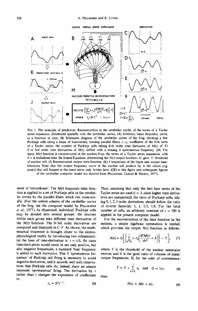
326 A. @3_LIONISZ and R. LLINiS
K PREDICTIONS Nh)=Mll+J)
CORTEX SPATIAL SENES RRPANSION MRIVAT~VES
~?..~~~~~~.~ -.:,, I
NUCtf%JS:PREDICTIVE RECONSTRUCTION
FIG. 1. The principle of prediction. Reconstruction in the cerebellar nuclei, of the terms of a Taylor series expansion, distributed spatially over the cerebellar cortex. (A) Arbitrary input frequency curve, as a function of time. (8) Schematic diagram of the cerebellar cortex of the frog showing a few Purkinje cells along a beam of transversely running parallel fibers. c. re: coe&ient of the 0-th term of a Taylor series: the number of Purkinje cells taking O-th order time derivative of M(t). (C-F) 0 to 3rd order time derivatives of M(t), shifted with a biasing S spontaneous frequency. (H) The input M(r) function is reconstructed in the nucleus from the terms of a Tayior series expansion, with 6 = A looka~ead-time. (In frame) Equation, determining the N(t) output function; G: gain, T: threshold of nuclear cell. (J) Reconstructed output time-function. (K) Comparison of the input and output time- functions. Note that the output frequency curve of the nuclear ceU predicts by A the values (e.g. peaks) that will happen in the input curve only A-time later. {[S’j in this figure and subsequent figures
of the cerebellar computer model are derived from PELLIONISZ, LLINAS & PERKEL. 1977).
ment of ‘smoothness’. The M(r) frequency-time func- tion is applied to a set of Purkinje cells in the eerebel- tar cortex by the parallel fibers which run transvers- ally. (For the central scheme of the cerebeflar cortex of the frog, see the computer model by PELLIONISZ
et af., 1977.) As iI~ustrated, individual Purkinje cells may be divided into severai groups: the neurons within each group take different time derivatives of the M(t) fun~ion. The O-3rd order derivatives are computed and displayed in C-F. As shown, the math- ematical treatment is brought closer to the electro- physiological reality by introducing two re~nements: (a) the base of time-derivation is z = r/A, (b) since time-derivatives would result in not only positive, but also negative frequencies, a standard ‘bias’ frequency is added to each derivative. This S *spontaneous fre- quency’ of Purkinje cell firing is necessary to avoid negative derivatives, and it accords well with observa- tions that Purkinje cells do, indeed, show an almost incessant ‘spontaneous’ firing. The derivation by r rather than t changes the expression of coeffejents to:
r, = (k!)_‘. (6)
Thus, assuming that only the first four terms of the Taylor series are used (i = 3, since higher time deriva- tives are unexpected), the ratio of Purkinje ceils, tak- ing 0, 1, t,3 order derivatives, should follow the ratio of inverse factorial: 1, 1, t/2, l/6. For the total number of cells, au arbitrary constant of c = 100 is applied in the present computer model.
For the reconstruction of the time function in the nucteus, a simple algebraic summation is needed, which provides the output N(t) function as follows:
where T is the threshold of the nuclear summator neuron, and G is the ‘gain’ ratio of volumes of input/ output frequencies. If, for the sake of convenience:
then
rk and G = l/c, k=O
(8)
(9)

Tensorial brain modeling: cerebellum 327
In J of Fig, 1 it is shown that the output function N(t) provides a prediction by A of the input M(t) function (compare the two functions in K, Fig. 1).
Several assumptions, serving only the simplification of this demonstration, should be commented on: We may note that the convenient selection of G and T values in equation (8) and for each derivative an iden- tical S is only necessary for arriving at identical ampli- tude of the input and output functions. It must be strongly emphasised however since we are describing functional properties of neurons that (1) the prediction does not change if these simpIify~ng assumptions do not hold (only the amplitude and bias of the output wili change) and (2) it is not necessary that given neurons take exact lst, 2nd, etc., order derivatives, and not even that given Purkinje celis take time derivatives of the same time-base. The principle of prediction will still hold (of course, with varying degrees of preci- sion). Also, the simplifying assumption of high T (which takes away the ‘bias’ added to each individual Purkinje cell) is only for the convenience of the graphic demonstration.
The central concept of the first part of this paper is, therefore, that a distributed system can provide prediction by means of spatial series expansion. A key ~sumption is, of course, the ability of Purkinje cells to produce an output frequency response that is the time derivative of the input Frequency-time function. Indeed, as mentioned above, such results have already been observed experimentally (Fig. 12; LLIN~ et al., 1971). Still, it remains to be rigorously demonstrated to what extent the present predictive property does arise in the cerebellar nuclei.
Feasibility study of the principle of prediction
Given that the core of this concept is a mathemati- cal idea, and that one cannot expect neuronal networks to behave with mathematical rigor, it is not surprising that the mathematically precise infinite series expansion is rendered into a finite series by the finite number of Purkinje cells. Similarly, the re- petitive firing properties of neurons preclude the extractions of very high order time derivatives. For- tunately, From the rapid convergence of the inverse factorial function it is evident that no more than i = 0,1,2,3 (i.e. no more than third order time de- rivatives) is necessary to provide satisfactory ‘biologi- cal precision’. On the basis of experimentai findings (LLIN~S ef al., 1971) the system seems indeed to be limited perhaps even to second order derivatives. As regards the values of coefficients (i.e. the number of Purkinje cells taking different derivatives), from equa- tion (4) it can be seen that a different distribution of values provides different ‘lookahead’ values, and an erroneous configuration of coefficients results in an erroneous shape of the predicted frequency-profile.
Since the above principle of prediction is based on a linear mathematical treatment, it is fundamental to explore to what extent the firing properties of the Purkinje cell can be approximated with such hnear-
ization. Thus, the linear mathematical treatment, which is always the first approximation, must be com- bined with detailed computer simulation studies, that are known to be capable of handling noniinear fea- tures of complex systems. Such computer modeling techniques of Purkinje cell firings are already avail- able (PELLIONISZ & LLIN.&, 1977) and have answered these questions unambiguously.
Thus, such a computer model demonstrates that subtle changes of the exponent of n in the HODGKIN
& HUXLEY (1952) equations (changing the exponent of n from being a constant into a function of the history of previous firings of the membrane) can in- deed tune the membrane to produce a more phasic. derivative type of firing (&LLION1SZ, 1976). We used this model for a feasibility study of the prediction principle, as shown in Fig. 2. Here an arbitrary time- function of the strength of mossy fiber input (D) was assumed (in the form of a current injection function). This results in the numerically calculated spike-train in C (the asymptote of the floating exponent of n is taken to be 2). For three different Purkinje cells receiving an identical input from this mossy fiber spike train, three different asymptote values are taken: 2, 6 and 10 in E, F and G, respectively. Differ- ent asymptotes (cf. PELLIONISZ, 1976) result in a tonic or more and more phasic type of firing of the cell. (Since a transient response is required for stabilization of the floating exponent of n. about 0.1 s activity is omitted in Fig. 2.) The initial burst responses show clearly the different tonic or phasic characters of fir- ing. In E. F and G of Fig. 2, it is apparent that the maximal frequency occurs at an earlier point of time if the response is more phasic. Accordingly, assuming an arbitrary number of type E, F and G neurons (1. 5 and 9 respectively in the case shown). the summed frequency-time function shown in J indicates a clear prediction of the peak.
The study shown in Fig. 2 is a simplification repre- sented by a few neurons: indeed, it represents only single membrane compartments, not entire neurons. Also, the working assumption that membrane proper- ties provide tonic and phasic responses does not in- volve the use of rigorous mathematical derivatives. The fact that the prediction is demonstrable etlen with these nonlinear conditions supports the biological feasibility of the mathematical idea and suggests that prediction does not require biologically unrealistic firing properties.
Potential sign~cance of the principle of prediction
(1) At the Iecef of single Purkinje cells, the tenet of prediction attributes a well-determined function to single neurons. Their role is to take individual/y dt$Jer- ent time derivatives of the input fiequencJ-time func- tion. Beyond this, if on the basis of the previous history of firing the membrane may be modified so as to produce more phasic responses (i.e. more deriva- tive-type firing or firing modified in the opposite direction) as proposed earlier (~ELLIONISZ, 1976) the

328 A. PELLIONISZ and R. LLIN,~S
C
prediction E~
O.lsec
FIG. 2. Computer simulation study of the principle of prediction. (A) Schematic diagram of the cerebel- lum of the frog in situ, the cerebellum marked by an asterisk. (B) Computer model of the cerebellar cortex of the frog, showing a sample of mossy fiber input which projects (via mossy fiber-granule ceil-parallel fiber system) to several Purkinje cells. A few Purkinje axons are shown to leave the cerebellar cortex to the cerebeltar nucleus. (C) Numerical solution of the Hodgkin-Huxley membrane equations by computer simulation (PELLIONlSZ & LLINAS, 1975; 1977; PELHONISZ, 1976). (D) Strength- time function of an arbitrary current injection applied to the membrane model. After an initial burst response about 0.1 s of activity is omitted from the Figure, to show only the initial transient activity and the stabilized steady state of firing. (E-G) Numerical solution of the Hodgkin-Huxley equations, assuming different firing dynamics (PELUO~ISZ, 1976): leading to tonic (E), and to more and more phasic (F and G) firing. Initial transient bursts show the tonic or phasic character of a single membrane response. In a steady state, note that the maximum frequency occurs earlier as the phasic character of the firing is increased. (H) Summation on a cerebellar nuclear cell of the three above-mentioned spike trains. (J) The frequency response along the time of the summed Purkinje activities. Note that
even with such a simple arrangement a prediction of the input peaks occurs at the nuclei.
above description of the functional role of individual neurons may be generalized. It has been proposed that the basic functioning of neurons may be based on a plasticity of their spike generating properties (PELLIONISZ, 1976; TRAUB & LLIN,g,S, 1977). It is fully realized that in the context of the above interpre- tation, if Purkinje cells can be tuned to take different derivatives (e.g. by climbing fiber activation as pro- posed by PELLIONISZ [1976]; or by altering the entry of calcium: LLIN/~S [1979]), such dynamic change would translate to a ' tailored Taylor series expansion' where Pk and therefore rk, and thus A are variables ra ther than constants.
(2) On a funct ional level, the tenet of prediction states that the spatial series expansion, reconstructed in the nuclei, leads to a forecast of an on-going time- function. The functional advantages offered by such a property are obvious, especially in regard to ballis- tic (fast, goal-oriented) movements where prediction is essential. However, the predictive feature provided by this corticonuclear neuronal network in i tsel f does
not describe motor coordination. It only provides an insight into some of the emergent properties derived from the circuit organization. Such emergent proper- ties must be put in the context of global cerebellar function.
The above principle of prediction accomplishes the prerequisite of stage 4 (providing an idea of the nature of information processing in a network) and may even give a hint for stage 5 (putting the idea into the con- text of overall function). However, for a proper embedding of the above idea into a general interpre- ta t ion of cerebellar coordination, a further step is needed. The remainder of this paper is an attempt at such a synthesis.
TENSOR NETWORK THEORY OF PARALLEL AND DISTRIBUTED BIOLOGICAL SYSTEMS
SUCH AS NEURONAL NETWORKS
As was said at the outset, the analysis of parallel neuronal networks is difficult since the methodology

Tensorial brain modeling: cerebellum 329
to be applied must be both conceptually appropriate to be desired. This should encompass both the identi-
and technically feasible. Two separate areas of scien- fication of the role of experimentally established struc-
tific endeavor, neurobiology and artificial intelligence tural and functional data and also the interpretation
research, have confronted this very problem from dif- of the overall function of a biological system with
ferent sides. parallel organization.
Hitherto, several mathematical and computer simu- lation techniques in both fields have involved the use of matrix and vector operations. From a neurobiolo- gical point of view, especially with regard to the cere- bellum, computer simulation methods have provided the first steps toward an interdisciplinary synthesis of a vast array of increasingly unwieldy experimental results. Computer models handling large matrices representing patterns of activities in a layer-by-layer analysis of realistic neuronal networks (PELLIONISZ, 1970) were initiated to bridge the gap between under- standing of the detailed components and the emergent properties of the neuronal machinery. (For an over- view of such computer modeling, see PELLIONISZ, 1979.)
DISTRIBUTED BIOSYSTEMS AS TENSORIAL ENTITIES
In an attempt to achieve the above goal, we pro- pose the following central assumption: Regard and treat parallel distributed biological systems as tensors. While implications of a general tensorial theory go beyond the scope of this paper, we introduce below the conceptual outlines of its special case: the Tensor Network Theory of neuronal systems. Our basic tenet is that the brain is a tensoriul entity.
In artificial intelligence research, which approaches the neurosciences from the direction of analysis of overall functional properties (without special regard for their actual detailed implementation), mathemati- cal tools have frequently been used for handling unde- tailed, arbitrary, neuronal assemblies. For example, linear matrix algebra was used for working out a scheme for associative memory (KOHONEN, LEHTIB, ROVAMO, HYVARINEN, BYR & VAINIO, 1977). Similarly, WIGSTR~M (1977) modeled a cortex-like neuronal network in order to analyze the spatial propagation of associations. Since associative memory was modeled as a spatially distributed feature, vector notations were used along with probability notations. Non-associative features (similarly activated groups of neurons in cortical columns) were treated by vector and matrix methods by LEGBNDY (1978). In another recent model of feature detectors in a distributed, cor- tex-like network (ANDERSON, SILVERSTEIN, RITZ & RANDALL, 1977), linear algebra was extensively used, providing one of the best examples of the power of these methods.
Neuronal networks in the central nervous system are prominent examples of distributed biosystems. Thus, consider neuronal networks as tensors over fre- quency space, each tensor identifying a scalar-valued function of r number of vector variables, the function being linear in each variable. For example, if the par- ticular neuronal circuit has one kind of n input fibers with n variables (usually frequencies. but graded potential-values in nonspiking CNS subsystems) and one sort of output of nl frequency variables, then the in- put-output relation is described by a tensor of rank 2 which assigns for every input vector of n com- ponents an output vector of 111 components. (Of course, the number of r vectors need not be limited to 2; e.g. the cerebellar cortex, which establishes a relation of mossy fiber and climbing fiber inputs and Purkinje cell output, is regarded as a tensor of rank 3.)
As regards the cerebellum, starting with a general scheme of organization of motor control, GREENE (1972) suggested that ‘low-level generators may be represented by mapping of various methematical spaces like the space of all mdimensional linear con- trol systems having a particular k-dimensional sub- space’. He concluded: ‘this method will not be derived from a general model or an abstract study of these spaces . . . because the problems that determine the types of applicable mathematics can be defined only in special types of situations, namely the common practical situations actually encountered by engineers and brains’ (ibid.).
Suppose, for the sake of simplicity, that a neuropile is endowed with one input and one output fiber bundle and that each of these consists of n individual fibers. Then the firing frequencies of the input fibers are interpreted as an n-dimensional vector. the output frequencies as another n-dimensional vector, and their relation (the network tensor) is identified by a matrix of n x n elements. (To be precise, the input and out- put are vector-curves, since they are a function of time.) We may note that in this particular case the network is technically described by two n-dimensional vectors and one n x n dimensional matrix. Further- more, in transforming the input vector into the output vector by the network-matrix the laws of linear algebra can be applied.
In the above examples, matrices and vectors were used. These tools for handling overall ‘parallel’ fea- tures of total systems and of arbitrary neuronal networks have evidently been technically quite ade- quate. However, a generalized abstract concept is still
However, as KRON (1939) showed in his epoch making book, Tensor Analysis of Networks, an abstract universal treatment of particular networks is
possible by employing the tensorial concept on geo- metrical objects. Tensors, used as the universal geo- metrical language of physical reality, relate vectors to each other through quantities whose actual values are relative to the particular frame of reference. The bases of coordinates may be arbitrarily changed, in which case the tensorial quantities will change, by
330 A. PELLIONISZ and R. LLINI~S
definition, in such a manner that the trun~ormat~on of the phasing vectors info one another remains in- variant. Now let us note that in the case of neuronal networks the frame of reference of the multidimen- sional space (denoted as the hyperspace of the firing frequencies) is fixed, since the coordinates are given by the existence of the neurons themselves, and thus it is impossible to modify the network’s frame of reference. This does not mean, however, that the vector-vector relationship (the environment-behavior response, in a general sense) which is expressed by different particular networks of the same class does not obey universal laws which apply to ail members of the class. It is known that fundamcnt~ properties of universals can be characterized by such reference- invariant tensorial laws (cf. MCCONNELL, 1957). Our hypothesis is that brains are isomorphic, or at least homomorphic, with the universe they reflect; thus, for the CNS, universal tensorial laws apply and thus the internal lanyuaye of the brain is vectorial.
Let us be more specific, in a quantitative sense. The vector-vector relation in a particular network is determined by its specific connectivity matrix. Such an individual matrix can be completely described by determining alf its components, which is no small task. For example, in the case of par? of the feline cerebellar cortex there are 1.2 x IO” Purkinje cells and 1792 times as many granule cells (PALKOVITS, MAGYAR & SZENT~GOTNAI, 1972). Thus, the granule cell-Purkinje cell connectivity matrix is described by determining all its 1.2* x 1012 x 1792 = 2.4 x lOi elements. Obviously, such an astronomical number of elements cannot be individually specified geneti- cally. Hence, the question arises as to how matrices of a given class come about in such a way that an amazingly invariant vector-vector function can be determined without actually pr~pecifying the value for each matrix element. While several ‘local’ mechan- isms for ‘matrix construction’ have been postulated (e.g. cell to cell recognition via surface mucopoly- saccharides), none answers the above fundamental question. Our hypothesis is that the networks are endowed with tensorial properties. Thus, since all
matrices of a given class incorporate a tensor, then the particular connectivity matrices (which are clearly not identical and which are overcomplete) are ex- pressions in different frames of reference of the same tensor. Thus, the vector-vector function determined by them is invariant to the reference frame!
From this point of view, the genetic code which determines the structure of the system should be regarded as a code for creating tensors which do
determine the overall vector-vector function of the system but need not spell out the total set of ‘numeri- cal values’ in the matrix for a particular coordinate- system. Thus, the genetic code would provide ‘only’ the ontogenetic guidelines for building a tensor in a genera1 sense, leaving the particular selection of the frame of reference and the establishment of the corre- sponding numerical values of connections to the indi-
vidual epigenetic development, i.e. determined by ‘local’ therm~yn~ic processes. As a result, hy encoding reference-invariant tensors, not particular matrices, genetic specification is relieved from the awesome task of determining each and every neural connection in the brain.
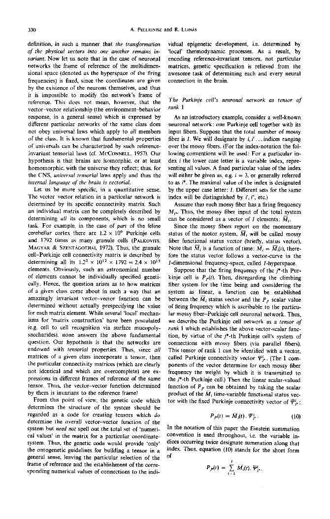
The Purkinje cell’s neuronal network as tensor of
rank 1
As an introductory example, consider a well-known neuronal network: one Purkinje cell together with its input fibers. Suppose that the total number of mossy fiber is 1. We will designate by i, i . . . indices ranging over the mossy fibers. (For the index-notation the fol- lowing conventions will be used: For a particular in- dex i the lower case letter is a variable index, repre- senting all values. A fixed particular value of the index will either be given as, e.g. i = 3, or generally referred to as i*. The maximal value of the index is designated by the upper case letter: I. Different sets for the same index will be distinguished by i’, i”, etc.)
Assume that each mossy fiber has a firing frequency Mi,. Thus, the mossy fiber input of the total system can be considered as a vector of f elements: Mi.
Since the mossy fibers report on the momentary status of the motor system, Mi will be called mossy fiber functional status vector (briefly, status vector). Note that ai is a function of time: ai = at(t), there- fore the status vector follows a vector-curve in the I-dimensional frequency-space, called I-hyperspace.
Suppose that the firing frequency of the j*-th Pur- kinje cell is P,(t). Then, disregarding the climbing fiber system for the time being and considering the system as linear, a function can be established between the @, status vector and the Pp scalar value of firing frequency which is ascribable to the particu- lar mossy fi~r-Purkinje cell neuronal network. Thus, we describe the Purkinje cell network as a tensor qf rank 1 which establishes the above vector-scalar func- tion, by virtue of the j*-th Purkinje cell’s system of connections with mossy fibers (via parallel fibers). This tensor of rank 1 can be identified with a vector, called Purkinje connectivity vector \rf.. (The I com- ponents of the vector determine for each mossy fiber frequency the weight by which it is transmitted to the j*-th Purkinje cell.) Then the linear scalar-valued function of Pp can be obtained by taking the scalar product of the ai time-variable functional status vec- tor with the fixed Purkinje connectivity vector of pi.:
P,*(r) = M&). F-j,. (10)
In the notation of this paper the Einstein summation convention is used throughout, i.e. the variable in- dices occurring twice designate summation along that index. Thus, equation (IO) stands for the short form of

Tensorial brain modeling: cerebellum 331
At this point it may be useful to illustrate the above notat ions since they provide a link between the pre- vious section and the following more abstract hand- ling of the circuitry. Figure 3 serves this purpose (the schematic representat ion of the cerebellar cortex of the frog in Fig. 3(A) and later in this paper is from the computer model by PELLIONISZ et at. (1977). Sup- pose that the number of mossy fibers entering the system is I = 8. (This is chosen only for ease of illus- t ra t ion since in the case of the cerebellum of the frog, I is of the order of 10,000.) In Fig. 3(B) a simple numerical example is given: the Mi status vector is presented by the row vector of eight (arbitrarily chosen) frequency values. As seen in Fig. 3(A), the connections from input mossy fibers to the j*- th Purkinje cell are determined by an intricate and rather complex circuitry. However, ultimately simpli-
fled, a column vector of I = 8 scalars may be assigned to one Purkinje cell, determining the 'weights' by which each mossy fiber frequency is contr ibut ing to the firing of the Purkinje cell. These weights are sym- bolized in Fig. 3(B) by the connecting lines (e.g. mossy fiber i = 1 to the j*- th Purkinje cell by two lines and mossy fiber i = 4 by three lines). Thus, the ~}~ Pur- kinje connectivity vector is a column vector of I = 8 elements and Pj is the scalar product of the status vector M~ and the Purkinje connectivity vector ~/'j, (in the given numerical example Pj~ = 41 units). Figure 3(C) shows a vectorial representat ion of the Mi input / -dimensional t ime-variable status vector
- - i
and ~ j , / - d i m e n s i o n a l connectivity vector. (To avoid difficulties in visualization, the I-hyperspace is limited to I = 3.) Since the status vector is a function of time, it represents a vector-curve in /-space while the Pur-
m
Mi
I
- - i
0
0
0
0
O
0
0
o
B I Mi " 'I'i'= = , , c IL _
| Mi(t)~'i~ 2\ *~.
o 3 e 2 o 0 o 1 o 0 o 1
FIG. 3. The network of a Purkinje cell as a tensor of rank one. (A) / ~ : mossy fiber input functional status vector represented by the eight firing frequencies of eight mossy fibers shown to enter the cerebellar peduncle. As shown, the mossy fiber input reaches the Purkinje cells by a system of granule cells and parallel fibers. The firing frequency of the j*-th Purkinje cell is a scalar value: Pj,. The weights of the mossy fibers in determining the activation of the /*-th Purkinje cell identify the t~, column vector of/-elements. This column vector of/-elements (a tensor) establishes the relation between the 2~i state vector and the Pj, scalar value of Purkinje activity. (B) A simplified numerical example and pictorial representation of the Purkinje tensor, assigning a scalar to an input vector by virtue of a connectivity vector. M~ is symbolized by eight dots, each being a mossy fiber firing with a given arbitrary frequency. The connections from the i-th mossy fiber to the single j*-th Purkinje cell are shown by ~connectivity lines', and the Purkinje cell network tensor is shown numerically by the ~ , column vector. The inner product of ;~i and Vg}, vectors provides the scalar of P2. (C) Vectorial depiction of the transformation of the 2~r(r) functional status vector to the P;, scalar by the tensor of a Purkinje cell. el el: /-number of unit vectors (I is limited to three to ease visualization). ~},: the tensor of the j*-th Purkinje cell: identified by a vector of /-dimensions. The scalar value of Pj is proportional to the projection of the /~ functional status vector in t.he direction of the Purkinje cell's tensor. According to the principle of prediction, the Pj(t + A) is provided by M(O, and not
by 3Tf(t + A).

332 A. PELLIONISZ and R. LL1NAS
kinje connectivity vector determined by the wiring of the network is fixed in /-space. Since P j, = Mi.~'~, the physical interpretation of the Purkinje cell's firing frequency is that it is actually a scalar measure of the projection of the status vector in the fixed direc- tion of the Purkinje cell connectivity vector. In other words, the Purkinje cell measures, by its firing fre- quency, the effect of the status vector in a predeter- mined 'wired in' direction of the /-space. It should be noted that the Purkinje cell connectivity vectors are likely to determine a direction mostly in particu- lar subspaces of the I-hyperspace, since one Purkinje cell is not connected to all input mossy fibers. That
is, the Purkinje connectivity column vectors are likely to contain many zeros (no afferents from particular input systems): their 'dimensionali ty ' is usually less than I. The biological interpretat ion of such subspace
vectors may be that they determine a special 'mix' of mossy fibers from different sources (e.g. head pos- ition, head acceleration, the status of neck and fore- l imb muscles, etc.). The co-linearity of the status vec- tor with any given t/ '-direction in the /-space is then measured by the firing frequency of the Purkinje cell.
At this point it is important to note that the predic- tive features of an assembly of many Purkinje cells in a 'stack' along a Parallel fiber beam may enable us not only to take the scalar product of M(t) and
at all times (e.g. at t and at t + A), but actually to predict the P(t + A) value already at the t ime t. This implies that instead of the j*- th Purkinje cell the j*-th set of Purkinje cells is meant, where the set of Purkinje cells, together with the summator nuclear cell, performs the ' lookahead ' by A as assumed above.
NETWORK TENSOR A
VECTOR INPUT
J VECTOR OUTPUT
B - - o
- - I D
[29 41 31] 9 4 0 7 70 921 ~--- I=1 2 3 4 56 78 ] F-I "_ 1 2
l - o 1 1 0 0
2 0 o 0 0
"divergence":3 ~---D'-- k21 ; J I ~ "convergence':7
FIG. 4. The network of the cerebellar cortex as a tensor of rank two. (A) A computer simulation of the activated granule cells and parallel fibers as a result of a mossy fiber input scattered over one-fourth of the cerebellar peduncle. Showing only three representative Purkinje cells which receive the parallel fiber input, i t is stressed that the system is presented with a vectorial input (in the form of a mossy fiber functional status vector: M~), and accordingly its output is a vectorial variable of the firing of Purkinje cells: Pj. The relation as a vector-vector function is determined by the tensor of the network of the cerebellar cortex: Fp~. (B) Simplified numerical example and pictorial represen- tation of the input-output vectors and the matrix of ~ tensor of rank two (cf. Fig. 3). Unlike, as shown in Fig. 3, here not one, but three, Purkinje cells are represented by three output elements (dots). A system of connectivity is shown by 'lines' (for j = 2 Purkinje cell the ~ is identical to that in Fig. 3), The tensor matrix, which produces the Pj vector product from Mi.tP~ is known if all the matrix elements are known. The overall numbers of input and output elements (I and J) determine the size of the matrix, and the numbers of 'connectivity lines' originating from any particular input element or 'lines' arriving at a particular output element may be established with relative ease. However, these values, which correspond to the morphological 'divergence' and 'convergence', provide
only a sum of row or column elements, having little mathematical significance.
Tensorial brain modeling: cerebellum 333
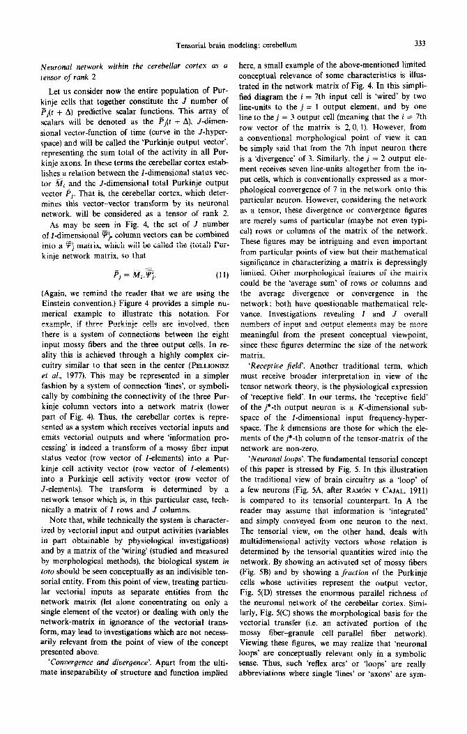
Neuronal network within the cerebellar cortex as a
tensor of rank 2
Let us consider now the entire population of Pur- kinje cells that together constitute the J number of pj(t + A) predictive scalar functions. This array of scalars will be denoted as the p,(t + A), J-dimen- sional vector-function of time (curve in the J-hyper- space) and will be called the ‘Purkinje output vector’, representing the sum total of the activity in all Pur- kinje axons. In these terms the cerebellar cortex estab- lishes a relation between the I-dimensional status vec- tor Mi and the J-dimensional total Purkinje output vector pj. That is, the cerebellar cortex, which deter- mines this vector-vector transform by its neuronal network, will be considered as a tensor of rank 2.
As may be seen in Fig. 4, the set of J number of I-dimensional F$ column vectors can be combined into a Fj matrix, which will be called the (total) Pur- kinje network matrix, so that
(Again, we remind the reader that we are using the Einstein convention.) Figure 4 provides a simple nu- merical example to illustrate this notation. For example, if three Purkinje cells are involved, then there is a system of connections between the eight input mossy fibers and the three output cells. In re- ality this is achieved through a highly complex cir- cuitry similar to that seen in the center (PELLIONISZ et a/., 1977). This may be represented in a simpler fashion by a system of connection ‘lines’, or symboli- cally by combining the connectivity of the three Pur- kinje column vectors into a network matrix (lower part of Fig. 4). Thus, the cerebellar cortex is repre- sented as a system which receives vectorial inputs and emits vectorial outputs and where ‘information pro- cessing’ is indeed a transform of a mossy fiber input status vector (row vector of I-elements) into a Pur- kinje cell activity vector (row vector of I-elements) into a Purkinje cell activity vector (row vector of J-elements). The transform is determined by a network tensor which is, in this particular case, tech- nically a matrix of I rows and J columns.
Note that, while technically the system is character- ized by vectorial input and output activities (variables in part obtainable by physiological investigations) and by a matrix of the ‘wiring’ (studied and measured by morphological methods), the biological system in toto should be seen conceptually as an indivisible ten- sorial entity. From this point of view, treating particu- lar vectorial inputs as separate entities from the network matrix (let alone concentrating on only a single element of the vector) or dealing with only the network-matrix in ignorance of the vectorial trans- form, may lead to investigations which are not necess- arily relevant from the point of view of the concept presented above.
‘Convergence and divergence’. Apart from the ulti- mate inseparability of structure and function implied
here, a small example of the above-mentioned limited conceptual relevance of some characteristics is illus- trated in the network matrix of Fig. 4. In this simpli- fied diagram the i = 7th input cell is ‘wired’ by two line-units to the j = 1 output element, and by one line to the j = 3 output cell (meaning that the i = 7th row vector of the matrix is 2,0, 1). However, from a conventional morphological point of view it can be simply said that from the 7th input neuron there is a ‘divergence’ of 3. Similarly, the j = 2 output ele- ment receives seven line-units altogether from the in- put cells, which is conventionally expressed as a mor- phological convergence of 7 in the network onto this particular neuron. However, considering the network as a tensor, these divergence or convergence figures are merely sums of particular (maybe not even typi- cal) rows or columns of the matrix of the network. These figures may be intriguing and even important from particular points of view but their mathematical significance in characterizing a matrix is depressingly limited. Other morphological features of the matrix could be the ‘average sum’ of rows or columns and the average divergence or convergence in the network; both have questionable mathematical rele- vance. Investigations revealing I and J overall numbers of input and output elements may be more meaningful from the present conceptual viewpoint, since these figures determine the size of the network matrix.
‘Receptive jield’. Another traditional term, which must receive broader interpretation in view of the tensor network theory, is the physiological expression of ‘receptive field’. In our terms, the ‘receptive field’ of the j*-th output neuron is a Kdimensional sub- space of the Z-dimensional input frequency-hyper- space. The k dimensions are those for which the ele- ments of the j*-th column of the tensor-matrix of the network are non-zero.
‘Neuronal loops’. The fundamental tensorial concept of this paper is stressed by Fig. 5. In this illustration the traditional view of brain circuitry as a ‘loop’ of a few neurons (Fig. 5A, after RAM~N Y CAJAL, 1911) is compared to its tensorial counterpart. In A the reader may assume that information is ‘integrated’ and simply conveyed from one neuron to the next. The tensorial view, on the other hand, deals with multidimensional activity vectors whose relation is determined by the tensorial quantities wired into the network. By showing an activated set of mossy fibers (Fig. 5B) and by showing a fraction of the Purkinje cells whose activities represent the output vector, Fig. 5(D) stresses the enormous parallel richness of the neuronal network of the cerebellar cortex. Simi- larly, Fig. 5(C) shows the morphological basis for the vectorial transfer (i.e. an activated portion of the mossy fiber-granule cell-parallel fiber network). Viewing these figures, we may realize that ‘neuronal kp’ are conceptually relevant only in a symbolic sense. Thus, such ‘reflex arcs’ or ‘loops’ are really abbreviations where single ‘lines’ or ‘axons’ are sym-
334 A. PELLIONISZ and R. LLINJ, S
TENSOR >,OOP • , - i
MF ACTIVITY U SOR STATE VECTOR curve in I -space
A - F- ~ p ~ - P RK
C
FIG. 5. A network is a tensor, not a loop. A comparison is given between (A) and (B D) in order to stress the conceptual difference between considering neuronal networks as loops of a handful of neurons or as tensors which determine vector vector functions. (A) (from RAMdN Y CAJAL, 1911). Simplified diagram of the neuronal network in the cerebellar cortex: MF: mossy fiber, GC: granule cells, PF: parallel fibers, PC: Purkinje cells. As shown by diagrams (B-D) from the computer model of the cerebellum, even in a case when only about 1~ of the mossy fibers are activated, the fired mossy fiber terminals (B) and granule cells and parallel fibers (C) show an immense parallelism in the excitation pattern which results. (D) Shows the Purkinje cell population (thinned gradually from the distal peduncle towards the proximal one), which receives the parallel fiber activation, shown in (C). The central inset stresses that mossy fiber activity (MF) should be considered as a vector of I-hyperspace, the resulting activities of Purkinje cell firings also as a multi-dimensional vector curve, and the network itself as the kuj tensor, which determines their relation. Thus, the loop-representation may be symbolic, at best, the 'lines' carrying vectors, where 'single cells' are really network tensors.
bols for bundles carrying activity vectors of multi- dimensional spaces, and the depicted single neurons represent network tensors which establish a relation between the vectors.
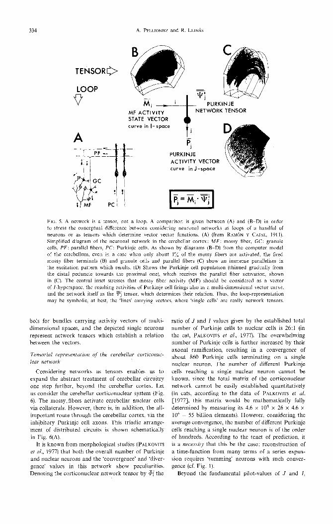
Tensorial representation o f the cerebellar corticonuc- lear network
Considering networks as tensors enables us to expand the abstract t reatment of cerebellar circuitry one step further, beyond the cerebellar cortex. Let us consider the cerebellar cort iconuclear system (Fig. 6). The mossy fibers activate cerebellar nuclear cells via collaterals. However, there is, in addition, the all- important route th rough the cerebellar cortex, via the inh!bitory Purkinje cell axons. This triadic arrange- ment of distributed circuits is shown schematically in Fig. 6(A).
It is known from morphological studies (PALKOVITS et al., 1977) that bo th the overall number of Purkinje and nuclear neurons and the 'convergence' and 'diver- gence' values in this network show peculiarities. Denoting the corticonuclear network tensor by ~i the
ratio of J and I values given by the established total number of Purkinje cells to nuclear cells is 26:1 (in the cat, PALKOVITS et aI., 1977). The overwhelming number of Purkinje cells is further increased by their axonal ramification, resulting in a convergence of about 860 Purkinje cells terminat ing on a single nuclear neuron. The number of different Purkinje cells reaching a single nuclear neuron cannot be known, since the total matr ix of the corticonuclear network cannot be easily established quantitatively (in cats, according to the data of PALKOVITS et al. [1977], this matr ix would be mathematical ly fully determined by measuring its 4.6 x 104 x 26 x 4.6 x 104 = 55 billion elements). However, considering the average convergence, the number of different Purkinje cells reaching a single nuclear neuron is of the order of hundreds. According to the tenet of prediction, it is a necessity that this be the case: reconstruction of a t ime-function from many terms of a series expan- sion requires ' summing' neurons with such conver- gence (cf. Fig. 1).
Beyond the fundamental pilot-values of J and I,

Tensorial brain modeling: cerebellum 335
%i *
CB
Cf”, %_
C
Gii
=i .- -?fj
FIG. 6. Tensorial representation of cerebellar corticonuclear network. (A) Scheme of cerebellar cortex (CB) and cerebellar nucleus (CN), showing that mossy fiber input (M) reaches the nucleus both directly. and through the cerebellar cortex, contributing to C output in two ways. (B) Applying the concept that networks are tensors, relating vectorial inputs and outputs, the network of the cerebellar cortex and the cerebellar nucleus are represented as single entities (CB and CN, respectively). For example CB may be represented by a Purkinje-like cell. Input and outpEt ‘lines’ are carriers of vectors. ai: mossy fiber functional status vector; row vector of i elements. YJ: network tensor of Purkinje cells: matrix of i rows and J columns. pj: row vector of Purkinje activities. 3: network tensor of corticonuc- lear connectivity. ji: inhibitor vector: acting in the cerebellar nucleus, resulting from the Purkinje activity vector transformed by the corticonuctear tensor. I?~: coordination vector: the difference of the mossy fiber status vector and the nuclear inhibitor vector. (C) Vectorial equations summarized: the matrix of the network tensor of Purkinje cells and the matrix of the cotticonuclear connectivity tensor combined to the I$ tensor of the cerebellum. (D) Expression of the c, coordination vector, as the output of the cerebellum, from the ai input status vector, 4’ cerebellar tensor and 8’ Kronecker- delta. The pictorial symbol represents the combined cerebellar cortex and nucleus, characterized by
4’ cerebellar tensor, transforming the Ji? input vector to the C output vector.
there is very little known about the matrix of the 5; corticonuclear network tensor. This is in contrast to the case of the Purkinje connectivity matrix, P. In that case at least two significant facts are relatively well known: one is that the number of parallel fiber inputs to every Purkinje cell is about the same magni- tude (80,000 in the cat, PALKOVITS Ed al., 19X), mean- ing that Purkinje connectivity vectors operate in ap- proximately equally small subspaces of the I-hyper- space. Another feature of that connectivity has long been known: that is, when the cortical surface of the human cerebellum is unfolded, certain macroprojec- tion systems (the so-called ‘homunculi’) can be revealed (for the cerebellum, see e.g. SNILIER, 1952). Note that these ‘global’ observations apply to all cor- tical structures. Such homuncuti, when interpreted in the tensorial concept of networks, translate into the ~ma~rostr~ctu~e’ of fhe co~~ectiuit~ matrix, meaning
that certain areas of the matrix contain relatively large numbers, while the numbers for other areas may be small. Unfortunately, no such numerical ‘mapping’ is presently available for the matrix of the i corti- conuclear tensor.
Also, on the basis of morphology one may assume that the mossy fiber collaterals which reach the cere- behar nucleus through a system of interconnections form another network tensor. However, in order to ’ simplify the system (in the absence of numerical details), let us introduce the following considerations. Assume that (1) the mossy collaterals reach directly into the cerebellar nucleus (eliminating the above tensor) and that (2) the dimensionality of the cortico- nuclear tensor matches that of ai (i.e. for 3, K = I).
Then the ~j Purkinje activity vector communi~tes to the nucleus by an inhibitor vector (since Purkinje
336 A. PELLIONISZ and R. LLIN~S
cells are inhibitory; ITO & YOSHIDA, 1964), denoted by ii (scalar I and vector i should not be confused), for which
ii = Fj.&, (12)
and this inhibitor vector and the mossy fiber status vector (provided by the collaterals) produce directly the cerebellar output vector, denoted by ci, in the form of
ci = Mi - ii (see Fig. 6B). (13)
A further consideration which is not, in a strict sense, required by the theory but simplifies its abstract handling is the assumption that afferent pro- jections with coeval ontogenetical development are likely to establish morphological connections with each other.
Accordingly, assume that the matrices are such that a given Purkinje cell receives a particular combina- tion of inputs from a set of mossy fibers and projects to nuclear cells which, in turn, receive the correspond- ing set of mossy fibers. Ultimately this would imply that the network tensor and the corticonuclear tensor are transposes of each other with respect to the metric 6i.i (where 6 is the Kronecker-delta).
gd = ,$j’Fj: si.i. (14)
This assumption allows the combination of the Pur- kinje network tensor and the cortlconuclear tensor into a symmetrical square tensor of
@’ =F;.@, (15)
where 3’ is called the matrix of the cerebellar tensor (see Fig. 6D). Ci can be expressed as
Ci = ai - fi = Mi - Mi, .$. (16)
Or, with the Kronecker-delta, where
;(f = ’ i
1 if i = i’
Oifi # i (17)
Ti = &. (8’ - 2’)
Vector-hyperspace of the cerebellar tensor
According to the above equations, the function of the cerebellum may be best analyzed in the Idimen- sional hyperspace. For the sake of visualization, in Fig. 7 I is limited to two. Here the $ Purkinje con- nectivity vectors are given arbitrary values. Altogether six Purkinje connectivity vectors are represented in this system (J = 6), each being an I = 2 dimensional column vector. These vectors combined determine the
A &(&ii) VECTOR-HYPERSPACE OF THE TENSOR OF CEREBELLUM
POSTURE 8 BALANCE
COORDINATION VECTOR
,I’ COORDINATION VECTOR : Ci= Mi -1;
C _ - -. Ci= /&;((il’-@) Qq: ( qyq
- - _. Ii- Mi; 01
3:CEREBELLAR TENSOR
FIG. 7. Vector-field of the cerebellum, shown by curved field-lines as determined by the cerebeilar tensor. (A) ii vector-field illustrated for I = 2, with six arbitrary Purkinje connectivity vectors. ai: status vector of the system, 1,: inhibitory vector, generated from Ri by the 81’ cerebellar tensor. (Ii vectors lie in the tensor-ellipsoid, whose positive quarter is illustrated only.) C, is the difference of M, and ii vectors: it is the coordination vector. (B) Symbolic pictorial representation of the cerebel- lum, its input being the Mi mossy fiber status vector and its output the c, coordination vector. (C)
. - Equations describing the relation of I&,), via 0 matrix of the cerebellar tensor. (d is a symmetrical square matrix since it is a product of the pj matrix and its transpose matrix.) Therefore, the C,
coordination vector can be expressed using the 8:’ Kronecker-delta.

Tensorial brain modeling: cerebellum 337
5uj matrix (a 2-row and 6-column array) of the Pur- kinje network tensor. According to the above connec- tivity principle, the matrix of the corticonuclear network tensor is assumed to be the transposed matrix of ~, and therefore the i~ inhibitor vector is a function of the input mossy fiber functional status vector M through the 6 ) i '= ~}..(~})r.q (I-dimen- sional) symmetrical square matrix (see Fig. 7C), where q is a constant.
Note, therefore, that since the/ -dimensional vector space of [i(Mi) is endowed with a symmetrical tensor of rank 2, the hyperspace is Riemannian; however, in the first approximation of this linear theory it is fiat. When, in the next obvious step, frequency-depen- dent phenomena like saturation are treated, the space becomes nonlinear. The O1' is called the cerebellar tensor. Thus, to any arbitrary M~ status vector an i~ inhibitor vector belongs, determining a vector field, shown by curved field-lines (trajectories) in Fig. 7(A). This will be called the posture and balance (P & B) field for reasons to be explained later. As shown in Fig. 7, the O1' cerebellar tensor carries the M~ vector
into the i~ inhibitor vector via the tensor, represented by the network of the cerebellum. The i vectors all lie in the (positive quarter of the) tensor ellipsoid of 63. However, the actual output of the cerebellum is the vector which leaves the cerebellar nuclei:
d~ = M i - ii. (18)
Since the function of the cerebellum has long been described as related to motor coordination, this total cerebellar output will be denoted the coordination vector.
The functional interpretation of these vectors can be followed in Fig. 8. Initially, the cerebellar neuronal network is put into the context of the rest of the motor control system. In Fig. 8(A) (cf. PELLIONISZ, 1979) the two blocks (outlined with broken lines) rep- resent the motor 'generator' unit (assumed here to be, for instance, the motor cortex) and the cerebellum, respectively. The movement 'order' leaves the 'genera- tor' in the form of a vector (taken here to be also /-dimensional) which is met in the brain stem nuclei (BSN) by the cerebellar input, the excitatory coor-
A ,~NM ~r~ c E ~ I. i ~ - , I i n " - I
i i i~p I i l l CN] J [ I
: 1 . J ~..__..~--.)--,..4-.~ . . . . . . . . . . . . . . . . . . . . . . . . ,~
I - - " L - ~ 1 . / i
,~NM
B NC-NC NO CERE~LLUM
el
NO CURVED FIELD- LINES IN I-SPACE
"~S'.
C NM-NM D ""'::': . . . . . . . . . .
NO MOVEMENT ORDER BALLISTIC MOVEMENT T
."~ C ~ C ' a e; C ° J ' " - o . . . . 7 1 ] ,,I"U_., ~I_Ro H'G =~ i _i.~,,~RO_Oa.e).o4.0 "-.I,¢~ - ~ r
RETURN OR BALANCE COORDINATION VECTOR GUIDES ALONG P&B TRAJECTORY MOVEMENT TO P&B TRAJECTORY
~ B
FIG. 8. Dynamic posture, balance and coordination of ballistic movements as described by the cerebellar tensorial system. (A) Scheme of. the cerebellar system. Lines are symbols of vectorial channels. CN: cerebellar nuclei; BSN: brain stem nuclei; C.GEN: motor command generator; G~: goal vector; ~rz:
• 0 1 . mossy fiber status vector: Pj: Purkinje activity vector; C~: coordination vector; -~" cerebellar tensor; ~j.=~' Purkinje network tensor. Dotted lines separate the system: NC with no cerebellum, NM with no movement. (B) Without cerebellum no i(M) vector field exists. With no movement generated, 3A decays to the origin. (C) Displacement of the 2~ status vector with no movement order, but using the cerebellum. M slips to the origin along the trajectory, determined by the i(2kr) vector. Decay can be stabilized by the H0 holding vector. P and B: dynamic posture and balance trajectory. (D) Displacement of the M status vector in case of ballistic movement, coordinated by the cerebellum. (E) Curved vector field (P & B) of the cerebellar /-space, as determined by six arbitrary Purkinje connectivity vectors, ~. The /~r status vector is driven from the origin or from a different starting point S' to the target point T; however, in all cases it follows curved paths since the coordination
vector C guides the movement of the M vector onto P & B trajectories as it homes in at T.

338 A. PELLION~ and R. L~ttdS
diuation vector, ci. The sum of these vectors Wiii then affect the mossy fiber status vector, the output of the system. AS indicated in the Figure, this status vector returns to the motor generator through a vectorial channel called, in the old terminolo~, the ‘outer feed- back loop’. The cerebellum receives its input from the executed mossy fiber status vector plus the move- ment order leaving the generator. The cerebeliar out- put is also returned to the generator through the cere- bello-cerebrai vectorial channel.
As shown above, the generator unit is supposed to be driven by a Gi goal vector, whose role is self- explanatory: it drives the status vector into the des- tination point in the I-hyperspace. In this context, the contribution of these vectors to movement per- formance can be analyzed as follows. First, let us remove the cerebellum in its totality. Then, if the sys- tem is brought into a particular Mi status, the mossy fiber status vector in time is expected to ‘decay’ into the origin of the I-space with some velocity. This is shown in Fig. 8(B): that is, the displacement of the status vector is determined by an eventual G goal -.. vector. and the -Mn ‘decay’ vector. Suppose now, that the cerebellum is reinstated into the system. Let us investigate the behavior of the system first without issuing movement orders (i.e. let Gi be a null vector), As shown in Fig. 8(C), in this case the @ status vector generates by the 3 cerebellar tensor the i inhibitor vector, and the e = &i - f coordinator vector. Assuming that a Ca fraction of this coordinator vec- tor is added to the --&?a decay vector in the brain stem nuclei, then
Ca-%?a= -ia. WI
Thus, the d status vector will now decay to the origin not along a straight path, but ~1~0~~~ the curved trajectories of the cerebellar hyperspace. Translating this into physiological terms, the motor system, by being equipped with the cerebellum, will have a ‘dynamic posture’ even during a collapse (spon- taneous decay) of the status vector. At this point, we emphasize that the above usage of ‘posture’ does plot imply the physiologic~ly conventional ‘static posture’ which in this theory is only a static status vector. Therefore, the term ‘posture’ throughout this paper should be understood as ‘dynamic posture’.
From Fig. 8(C) it can also be seen that in order to avoid a ‘postured collapse’ (i.e. ta maintain a bulatfce of a given a status vector), the only require- ment is the -( -ia) = ia = -(a - C)a vector, which is readily available in the motor generator through the cerebelb-cerebral vectorial channels. Since this vector holds the balance, it will be called PO, the holding vector.
Thus, we may conclude that the cerebellar tensor, by determining a curved trajectory of the cerebellar hyperspace, provides the ‘natural rails’ of the displace- ment of the status vector, along which the system spontaneously returns to resting status, or on which orbits a particular status vector may be balanced.
Therefore, the name ‘posture and balance trajectory’ of the cerebellar vector hyperspace seems justified (of course, in a dyn~ic sense).
Since the role of the cerebella-cerebral vectorial channel is to supply the e vector to the command generator (which combined with the li? status vector will result in the 8’ holding vector), it is implied in this theory that severing the cerebella-cerebral tract will result in an inability to maintain a given motor status: a loss of balance.
As a general point of the theory, it may be stressed here that the use of such traditional terms as ‘positive feedback reflex loop’ for the cerebella-cerebral tract is not applicable in the framework of the presented concept. First of all, channels (bundles of nerve fibers, conceptually oversimplified as loops) may be better described as vector carriers. (‘Loops’ in the everyday usage of linear control systems suggest that the car- ried variable is scalar.) Therefore, whether the vector- ial channel which returns will increase the absolute value of the emitted vector (‘positive’ feedback) or will decrease it (‘negative’ feedback) depends on rhe relative direction qfthe two t’ector variables. The diagram of the vectorial channel does nor specify this relation at all since the relation of vectors is determined only by the tensor of the network. Secondly, this impties the need for reconsideration of the age-old term ‘reflex’ also. This term generally imphes a scalar- scalar function (e.g. a relation between the amplitude of an input signal and the ampl~ude of an output signal).
As shown in Fig. 7, for every a vector presented to the tensorial system, there is a resulting i vector. This vector-vector relationship does not specify, in itself, whether a scalar input (a one-dimensional vec- tor) will be tra~form~ into another scalar or, as is usually the case, into a vector. It is understood, of course, that the narrow interpretation of ‘reflexes’ as a scalar-scalar function is largely due to the wide- spread application of Iinear control systems theory. This deals most often with concentrated (not distri- buted) systems where ‘feedback and feedforward loops’ usually carry scalar quantities (such as the amplitude of a voltage).
We should note that as a special case the tensorial response of the system may yield a scalar-scalar input-output relation (a conventional reflex). This would occur if the system is tested in a one-dimen- sional subspace of the I-hyperspace and the one- dimensional vector happens to be such a vector of the network-matrix, whose direction does not change
in the vector-vector transformation. (Mathematically this is called=an eigenvector.) Looking at the tensor- ellipsoid of 0, we may notice that even in this ulti- mately simplified case (I = 2) the above special case does not apply. It is true that the a status vector, pointing in the direction of the axis of the ehipsoid (which is an eigenvector) would be carried into an f wctm of the same direction talthough its ~plitude may possibly be changed). However, these eigenvec-

Tensorial brain modeling: cerebellum 339
tors, the axes, even in this simple case do not happen to lie in the direction of either axes of the coordinate system and thus the simple scalar-scalar reflex does not occur.
Stimulating one input element, and analyzing the response of one output element (e.g. a single neuron), the largest response would be yielded if the input and output are eigenvectors. Therefore, the investigator’s search for single units with maximal response can be considered as the effort of searching out such lucky one-dimensional eigenvectors of the system. This is an unyielding enterprise. However, in natural cases even multidimensional vectors may fall into the direc- tion of eigenvectors of the system. For example, in the described cerebellar system, if for the a status vector those vectors are applied which are eigenvec- tors of the 5, the effect of the cerebellum would be minimal and would only affect the amplitude of the motor command. In this case, ablation of the cerebel- lum would not result in deficiency in the coordination of the motor performance. Such natural cases of oper- ating with eigenvectors may be the motor activities concerned, for example, with ‘looming’ which are known to be practicaliy unaffected by cerebellar lesions (LLINAS & WALTON, 1978).
Coordination of mouements by the cerebellar tensorial system
While it has been demonstrated that the cerebellar vectors can be effectively used for determining (dyna- mic) posrural trajectories (by the i inhibition vector), and may keep the system balanced along these trajec- tories (by virtue of t? coordination vector, used with a to supply the R holding vector), the role of these vectors in ballistic movements is perhaps even more characteristic.
Suppose (as seen in Fig. 8D) that the system is characterized by a M mossy fiber status vector, and the G goal vector is driving the system to a distant orbital point of the trajectory. Here, the displacement of the &? status vector would be determined by the previous vectors (as shown in C in Fig. 8) plus the goal vector of the system. Let us assume that the holding vector and the coordination vector are each used by the command generator depending on their angles from the intended movement, as determined by the goal vector. In assuming a relation of the C’ actually used fraction of co coordination vector, and similarly the 8’ operating part of R” holding vector, two points of view should be taken into account. First, it is required that if the goal vector is a nullvec- tor, case D should be reduced to C. Second, only such vectorial operations should be assumed, which can reasonably be expected from neuronal networks found in the so-called command generators such as motor cortex. Such an operation would take an Ai fraction of x0, where the ratio of the absolute values of Al’ and 2’ is proportional to the cosine function of the angle between A and a B pilot vector.
(Physically speaking, to take only that fraction of
the A vector whose length is equal to the projection of A to B.)
A’ = AOCOS(U) = A0 &. (20)
As seen in the above equation, the required operation implies a multiplication of the A vector by a scalar, where the scalar is the inner product of the A and B vectors, each normalized in amplitude to one.
Thus, suppose that
HOG Hi = R0 po, ,(;,’ (21)
( COG c’ = co ltYOl pITI
PH --- IC’OllRI > .
(22)
Note, that if the angle between 6 and c is denoted by u, the one between c and i? by /3 and that of R and G by y, then
and
A’ = HOBOS, (23)
ct = CO[cos(a) - cos(/I)J. (24)
Suppose, that if c is a nullvector, then (since the above angles from c are undefined) the above cosine values vanish. This would result in the required reduction of case D in Fig. 8 to case C. Also note, that since c1 is physically interpreted as a fraction of C, the term in parentheses must not be smaller than zero; if it is, e1 vanishes.
With these assumptions, the bailistic movement of the @ status vector shows the features illustrated in Fig. 8(E). Here the $? status vector of the system is driven from the origin to a final destination by a G goal vector. (In the case illustrated the curved cerebel- lar hyperspace is set up by six arbitrarily chosen Pur- kinje connectivity vectors.) Then, as the status vector moves along in the direction of the destination point, its velocity and direction changes, since both the G goal vector, and the cerebellar (?’ and 8’ vectors are variables. (c is taken here to be always l/l0 of the difference vector between the actual status vector and the d~tination point; while e* and &ii are deter- mined by the equations above.)
As can be seen in Fig. 8(E), the moving pattern of the @ status vector is characterized by the follow- ing feature: The ballistic movement (in the I-space) does not follow a straight line (‘shortest distance’ between starting and destination points) but is guided to the trajectory of the dynamic posture and balance so that it arrives at the destination precisely along the trajectory. This means that the movement path of the status vector-curve, as it ‘homes in’, gradually fits to the orbit which is predetermined by the tensor of the cerebellar hyperspace. This guidance is per- formed by the ‘additive’ of the c coordination vector,
340 A. PELLIONW and R. LuNh
supplied by the cerebellum. As we see, the coordina- tion vector puts the intended movements in the con- text of the movement-patterns which are predeter- mined by the cerebellar tensor. Also, it can be fol- lowed in Fig. 8(E), that the use of the coordination vector is maximat at the stage of the movement befoe the status vector reaches the crucial proximity of the destination point. At that stage, since the vector already moves along the trajectory, the vector ‘homes in’ by relying only on the holding vector.
The computer simulation of the movement-path of the status vector (as shown in Fig. SE) brings the time-dimension into the analysis of the system. For example, it is known that all vectorial channels from the motor ‘generator’ to brain stem nuclei and down to the motor executors involve time-delays, and so do the outer returning vectorial channels and those carrying the I@, c, P vectors. Proper timing of these vectorial channels ensures (as shown in Fig. 8E) that there is no ‘overshoot’ in guiding the status vector curve into the dynamic posture and balance trajec- tories, but the C? coordination vector component diminishes smoothly as the status vector homes in. It should be stressed that Fig. 8(E) represents a highfy simplified and abstract demonstration of the prin- ciples of this theory, since the dimensionality of the cerebellar hyperspace is I = 2. Also, the ‘movement paths’ represent displacement in the multidimensional frequency-space, and nor physical movements of the executor system in the existing three-dimensional physical space, as the v~uali~tion may suggest. For a less simplified, less abstract, usage of the principles of this theory, a computer modeling project of the coordination of locomotory movements is being de- veloped (using the simulation techniques outlined in PELLIONISZ, 1979). In that paper the timing features will be discussed in detail. Obviously the predictive feature of the corticonuclear neuronal network plays a major role in eliminating the deficiencies caused by time delays inherent in the vector&I channels.
Role of the cerebellum in the execution of ballistic movement. As a preview to a more complex model, a computer simulation result is shown in Fig. 9. This demonstrates the role of the cerebellum in the execu- tion of a ballistic movement. In this case the cerebel- lum generates the inhibition and coordination vec- tors, and through them, it guides the ballistic move- ments onto the ‘pre-wired’ tracks of the dynamic pos- ture and balance trajectories.
A two-dimensional graph in Fig. 9 is taken as an intended movement. The (A) graph represents the path of the G goal vector moving in time with a con- stant speed in the frequency-space. The resulting path of the a status vector is shown in Fig. 9(B). Although this model is crude, the emerging ‘writing pattern’ of the computer model shows several characteristics of what we may call ‘coordinated movements’. Thus, since the mode1 is in a different initial state than is required by the departing goal vector, the ‘writing’ by the computer starts with a ‘hook’ before it takes
off for its ballistic course. Even in this very simple case, where the initial Y-offset is being corrected by the system, it is clear that both X and Y displace- ments of the @ vector are affected by the ‘coordina- tive’ activity which is exerted by the c and i vectors. Another important feature is that sections of the in- tended movements (which are straight lines in the Euclidean I-space if there is no cerebellar tensor) become distorter in the cerebellar hyperspace of the actual execution. This is, of course, due to several factors: a more simple one is that, since the model involves delays, the c goal vector leads in time and the @ status vector lags behind. Therefore, before the @ status vector reaches a ‘corner’, the goal vector has already ‘turned around’ and thus the status vector will ‘round’ the corner. However, it is very clearly not the case in the falling section of letter K. Here the straight section is distorted into a ‘more natural curved one much before the rounded cornering effect takes place. This is due to the ballistic coordination: the cerebellum blends the coordination vector to the executed movement in order to guide the displace- ment of a onto the trajectories of the hyperspace. Thus, as can be seen at once, the whole ‘writing’ of the computer has a distinct ‘style’ of tilted letters, characteristically distorted O’s, etc., which reflect the curved trajectories of the vector field as determined by the cerebehar tensor,
Interpretation of the climbing fiber subsystem in the framework of the te~orial system of the cerebella
Up to this point the climbing fiber system of the cerebellum has been disregarded. Now a preliminary attempt will be made to reconsider con~ptually our understanding of this enigmatic second input of the cerebellum, looking at it in the light of the vectorial- tensorial representation. Such a new point of view has disadvantages, but is in part quite helpful.
There is a marked difference between the climbing fiber and mossy fiber input to the cerebellum for, while the mossy fiber related activities may be con- sidered as continuous time functions (at least at first appro~mation), the firing of any one climbing fiber is discontinuous: characterized by abrupt bursts, or compounds of several very high frequency spikes (ECCLES, LLIN.& & SASAKI, 1966b). These so-called complex spikes are widely scattered in time, providing an ‘average’ frequency usually below l/s. In contrast, the parallel fiber evoked, so-called simple, spike frequency may be as high as 100/s, even in the ‘spontaneous’ state. Therefore, we must interpret the activity of the climbing fiber system in the f-dimen- sional frequency-space as discontinuous, discrete per- turbations of the time-continuous vectorial variables.
Yet, the interpretation of the cerebellum as a sys- tem which deals with vectors appears to be quite ade- quate when approaching the problem of explaining the function of the climbing fiber system. This over- comes the limitation (pointed out in LLIN&, 1974a) that most conventional analyses of this system have
Tensorial brain modeling: cerebellum 341
FIG. 9. Computer modeling of the coordinated ballistic movement of the a functional status vector in the I-space. (A) Intended movement in the Euclidean frequency space. (B) Execution of commands in the style of ‘writing’ guided by the cerebellar coordination vector to the curved trajectories in
the cerebellar hyperspace.
concentrated on the individual effect of the climbing fiber activation of a single Purkinje cell. (This is un- de~tandable in view of single-unit electrophysiologi- cal methods, and considering the uniquely dramatic character of the climbing fiber-evoked response of Purkinje cells.) This attitude, however, may be so in- grained that even total system stimulations are corre- lated with the effect of single climbing fibers on single Purkinje cells. On the other hand, in recent years several approaches have been initiated which have stressed that configurations of many simultaneously acting climbing fibers should be regarded as a unitary action of the climbing fiber system. These efforts have been based partly on micro-morphological and mi~o~lectrophysiological observations (electrotonic coupling of olivary neurons: LLIN.& er at., 1974) and the micro-projection system of climbing fibers in the cerebellar cortex (the so-called Oscarsson-strips: Osc~~sso~, 1969). A computer model by B~YLLS
(1975) was also designed basically to bring out the spatially determined features of the effect of grouped climbing fiber activity.
However, the results of the physiological experi- mental paradigm designed by LLINA~ & WALTON
(1978) to investigate the functional effects of the
climbing fiber system at a truly system level, render it quite imperative that the interpretations at different levels of the action of climbing fibers be somehow unified. We feel that the vectorial-tensorial represen- tation may have the potential to fulfill this role. Some preliminary considerations on this basis are intro- duced below; a more detailed analysis of this problem will follow later.
As for the temporal aspect of the climbing fiber intervention, we treat the effect of the climbing-fiber- evoked response as a discrete ~err~rbotion of the cere- be&r vector-field. Let us examine, in vectorial terms, how climbing fibers would perturb the cerebellar action.
As is known, climbing fibers, ascending from the inferior olive, reach the Purkinje cells in the cerebellar cortex. However, climbing fibers are also known to have collaterals, which provide an excitatory input to the cerebellar nuclei (cf. CHAN-PALAY, 1977). Thus,
activation of a set of climbing fibers would represent an excitatory vector-increment of r in the cerebellar nuclei and, after a notable (several ms) delay, would also produce an increment of the i vector by exci- tation of the Purkinje cells. Suppose, for the sake of simplicity, that such vectorial increments fall precisely
342 A. PELLIONISZ and R. LLINAS
into the direction of C and I, respectively. (This does not necessarily occur in reality. However, as long as the increments increase the absolute value of the vec- tors, the consideration will hold, even if the incre- ments deviate by some angle from the vectors which are being incremented. And this requirement is, by definition, fulfilled, since the climbing fibers are exci- tatory.)
According to the above, an increment of the c coordination vector would result in a deformation of trajectories, as shown in Fig. lo(B). It is easy to see that the increase of c changes momentarily the taut- ness of the trajectory in the neighborhood of the status-vector. This change means a temporary slack- ening of the trajectories. On the other hand, as seen in Fig. lo(C), an increment of the i vector would make a deformation of the trajectory momentarily in- creasing its local tautness (it ‘pulls’ the trajectories tighter). As we noted above, the second type of pertur- bation occurs with some delay following the first type of perturbation. Thus, the time-course of the climbing
fiber perturbations of the posture and balance (P and B) trajectory could he visualized as throwing a leash: a quick slackening of the field-lines is followed by a quick tightening action.
Before discussing the potential functional signifi- cance of such transient deformations of .trajectories, let us point out a circumstance that was neglected in Fig. 8(D) and (E). We noted there that proper tim- ing of the cerebellar vectorial channels is required to avoid an ‘overshoot’ of the status vector. (That is, by excessive usage of the r? coordination vector, the status vector may be ‘pushed’ outside the field-line which contains the destination point.) As seen in Fig. 10(D) in the case of such overshoot (MJ the r? and &i vectors will both lie on the same side of G. However, before such an overshoot (MU), r? and A both lie on the opposite sides of c. In terms of the equations in Fig. 8(D) this means that the quantity in parentheses becomes negative (an impossible physi- cal condition). In this case, therefore, the actually used r?” fraction of the co coordination vector would
FIG. 10. Interpretation of the climbing fiber action in the cerebellar vector space. (A) Scheme of vectorial channels of the cerebellar system, including the climbing fibers (cf. Fig. 8A). CG: command generator; IO: inferior olive. Climbing fiber action increments the f inhibition vector (by the excitation of Purkinje cells): incr i, and increments the C coordination vector (by excitation of cerebellar nuclei). (B) Vectorial scheme of the momentary perturbation of the posture and balance (P & B) trajectory by incrementing the C coordination vector by climbing fiber action in the nuclei. Note that the deformation of the trajectory amounts to its ‘slackening’: S. (C) Perturbation of the P & B trajectory in incrementing the i inhibition vector by climbing fiber action on Purkinje cells. Note that the climbing-fiber-induced momentary deformation is a ‘tightening’ of the trajectory: T. (D) Scheme of the 2 status vector, G goal vector, C coordination vector, R holding vector in cases of ‘undershoot’: fiM and ‘overshoot’: li_i.. D: destination point of status vector. Note that in undershoot e and R are on the opposite sides of G, while in overshoot C and If lie both on the same side of G. (E) ‘Catching’ the overshooting a, state vector by momentary Perturbations of the P & B trajectories. Note that the first phase of climbing fiber action (‘slackening of field-lines: S catches M, and the second phase (‘tightening’
of field-lines: T) pulls it to the original field-line that holds the D destination point.
Tens&al brain modehng: cerebellum 343
vanish. Thus, the status vector, by going beyond the trajectory lines, would also go beyond the coordinat- ing power of the cerebellum: it would no longer be able to guide the state vector onto the posture and balance trajectories.
Let us assume, however, that the occurrence of this condition is monitored by the inferior olive, and that the climbing fiber-evoked increment vectors are issued from the olive if and when this condition occurs. (For this to happen, the inferior olive is - - required to receive G, C and H vectors as indicated in Fig. lOA.)
Note that the time-course of perturbation of the posture and balance trajectories by the climbing fiber system is precisely such, that the first phase of ‘slack- ening’ the trajectory would ‘catch’ such an ‘escaping’ status vector (as slackening of a net would secure once more into the inside of a net a fish which is just breaking through) and the slightly delayed ‘tightening’ of trajectories would make up for the pre- vious slackening, guiding the state vector back to the original trajectories (cf. Fig. 1OE).
The above preliminary assumptions regarding the action of the climbing fiber system are, indeed, specu- lative, and-since they are so heavily time-related- will be worked out in a computer model which enables us quantitatively to handle the whole, rather complex vectorial system. However, such vectorial representation already puts the existing wealth of data on the climbing fiber system into new perspective, and opens up fresh possibiliti~ of breaking some deadlocks in conceptual interpretation. Also, we may view this approach optimistically, since some charac- teristic features of the climbing fiber system, such as the intermittent abrupt character of the firing of grouped climbing fibers, emerge naturally from the above considerations. (Such a view has been expressed as the ‘phasic’ character of the climbing fiber system, LLINAS. 1969b.) This phasic firing occurs whenever a discrepancy, i.e. an ‘overshoot’, takes place in the I-space during the course of the ballistic movement.
An ultimate theoretical possibility in the above vectorial inte~retation is worth mentioning namely that if any part of the transient perturbation is made permanent (e.g. by such mechanism as proposed by F%LLIONISZ, 1976), this would correspond to subtle ~rmanent ‘deformations’ (or, as we would rather say, formations).of the curvature of the vector-field in the cerebellar I-space.
DISCUSSION
In its broadest sense the present paper attempts to offer a new conceptual framework for the interpre- tation of the garatlel and distribute nature of nerve nets in the brain. We chose the cerebellum as our immediate subject of study because of OUT direct acquaintance with this CNS subsystem and because it offers the necessary background knowledge in both
morphology and function which are prerequisites for the development of the type of model described here. However, we feel that the overall approach (i.e. the understanding of brain function through the use of tensor network theory) is applicable to other aspects of brain function as well. Therefore, we are tacitly assuming that approaches such as those described in this paper would be used in a variety of network analyses. White the present paper represents a gener- alized abstract approach to the problem, it must be considered only as a partial description of a continu- ing study of cerebellar modeling. Two other main projects are in preparation at the moment. Thus, we are exploring in the light of this theory (I) the role of the climbing fiber system in cerebeltar function, and (2) the use of the tensorial interpretation for a less abstract, more detailed and explicit computer model of the cerebeflar coordination of locomotion.
At this point we would like to (A) highlight the central concepts of tensorial interpretation of networks as an approach to the understanding of the parallel and distributed nature of brain functions, (B) compare this interpretation with the traditional con- cept of ‘reflexes’, (C) describe our views of cerebellar function in the light of the tensorial concept, and (D) describe the way in which the above approach may be used to study other functions of the brain.
(A) Tensorial concept of networks as an approach to the understanding of parallel and distributed, brain functions
As stated in the Introduction, the single most saiient character of the organization of nerve nets in the brain is their parallel nature. With only few excep- tions (probably those present in the giant system of invertebrates) information transfer between different parts of the brain is accomplished by a multiplicity of channels. However, even in those cases where such giant neuronal systems occur, final goals may be achieved by the system utilizing other pathways.
As far as vertebrate CNS is concerned, the cerebel- lum may be considered the epitome of such distri- buted organization, It is an anatomical fact that large numbers of parallel fibers converge onto a single Pur- kinje cell and, in turn, also a high number of Purkinje axons project onto neurons in the cerebellar nuclei (cf. PMXOVITS et al., 1977). This implies not only that this system is organized in a parallel and distributed manner but also that it is unlikely that all connections in such a complex neural network as the cerebellum are genetically pr~etermined. Thus, there must be other principles of distributed organization. In the past it has been assumed that the parallel and distri- buted nature of nerve nets represents redundancy, whose purpose is to ensure a high safety factor for the function of the network. Our interpretation of the parallel distributed organization is, however, differ- ent: The paraliel system allows the expressions of the funct~na~ particuiarities of i~iu~uai neurons. The sum of such particularities generates the overall func
344 A. PELLICJNISZ and R. LLINAS
tional properties which compose the functional reper- toire of the system, An example of this is the feature of prediction, which is a distributed property of the cerebellar co&on&ear system. Such a system, in which a given common input produces particular in- dividual responses {diff~ent time~~ivativ~), may be considered a biological analog of the mathematical reconstruction of a function from its Taylor series expansion. In the case presented here, the cerebeilum integrates the different responses which the Purkinje cells produce following a common mossy fibe-paral- Iel fiber input and the overall product is the emer- gence, in the cerebellar nuclei, of a pr~ictive pro- perty. Thus, within a biologically reasonable fre- quency range, events may be predicted in the time- domain. This result could well be produced by the different Purkinje cells responding in either a phasic (derivative-type firing) or a tonic manner. While such observations are not new lL~1N.b et al,, %‘I), their functional interpretation, namely that distinct predic- tive properties may arise from them, has only now been realized.
The above view, then, puts the concept of sheer redundancy in direct contrast with a we&defined scheme iu which every neuron plays au individual role. Thus, this ‘neuronal society’ seems to be organized not as a regjmeoted mass unanimo~ly echoing a message, but rather with the subtlety of an orchestral performan~ where the overall theme emerges from the sum of the actions of distributed elements, each making a small individual contribu- tion. Such parallel representation of a function by its d~tribut~ series expansion is advantageo~ in both a passive and an active sense. In a passive sense a loss (or dysfunction) of one element does not pose a threat to the total performance (therefore, in a simi- lar way to redundancy but in a more sophisticate manner, this kind of distributed function also in- creases reliability). In an active sense, it efficiently in- duces subtle or gradual changes in the overall func- tion. I? should be mentioned that this organizational principle of prediction by spatial series expansion, while suiting the cerebellar ~o~iconu~lear circuit most readily, probably reflects a general principle of organization of other CNS subsystems (e.g. the retina or the auditory system). Moreover, the principle could as well be applied not only to supracellular systems (such as the cerebellar network) but also to subcellu- Iar parallel-organized microsystems (e.g. a single neuron with its synapses).
A more general and abstract conciusion to be reached from our studies on the cerebellar system can be summarized as follows: If we consider that the firing of an axon in any pathway is a onedimension~ vector, ail pathways in the CNS should be regarded as vector-carrier channels. In turn, all j~tegrative sites jneuropiie) should be treated as tensorial systems which determine the vectorial relations. Therefore, a generaiized concept can be derived from the techni- calIy linear-aigebra~c vector and matrix represen-
tation of neuronal networks: existing circuits, ‘wired in a given particular manner, are concrete numerical incorporations of tensors of a general. kind.
Thus, the genetic code would specify the general teusorial properties of networks which determine the overall vector-vector functions of the system, but need not specie the total set of ‘numerical values’ of con- nections among network elements in any particular coordinate system. Thus, the genetic code provides the ontogenetical guidelines for building a tensoriaI system, leaving the particular selection of the bases of the frame of reference and the establ~hment of the ~~~pond~ng ‘numerical’ values of network- matrix to the epigenetic factors.
Such a generalized tensorial theory of distribute biological systems goes, however, much beyond the scope of this paper. Here we have aimed only to cover the conceptual outlines of a special case: the tensor network theory of neuronai systems. This itseIf will doubtless have to be further defined and formulated. Here, for example, we have only to mention the vexed problem of interpreting, by computer simulations, the nonline~iti~ of the system or the homogeneous treatment of space-time by the tensorial theory of neuronal networks, or the dimensionaiity and ortho- gonality of, for example, the cerebellar multidimen- sional space. However, if the conceptual fr~ework we have outlined proves to be as fruitful as expected on the basis of its use in exploring cerebellar function, these efforts will be justified.
The rn~an~~ a~~rrn in ~~ranal circuits. The mean- ing of morphology in the light of the tensorial inter- pretation of neuronal networks requires some etabor- ation. From the concept outlined above, it follows that when considering the CNS as a tensor&l system its morphology and physiology describe two sides of one inseparable entity: the tensorial properties of the system. While structure (a particular ‘wiring’ of a matrix of a tensor) and function (vectorial inputs and outputs) should not be considered separately, never- theless, the fundamental properties lie in the structure of a tensorial system. Equally important, we may assume that the organization of the CNS in general mimics the properties of the outside world; more pre- cisely, we propose that the brain is a geometric object (in the sense of KRON), capable of being isomorphic or at least homomorphic with external reality.
Several important implications can be derived from the above statements. First, as indicated previously (cf. LLINAS, 19745), genetic information generate mainly form and connectivity, This is corroborated by the well-known fact that neuronal populations (e.g, the cerebellar or the cerebral cortex) contain neurons with shapes and sizes quite similar from brain to brain. This is true in a given species and, even more important, it is also true hong different species. Moreover, as shown by CRAIN, BORNSTEIN 62 PETER- SON (1968) and MODEL, BORNSTEIN, CRAIN & PAPPAS ffQ71) in tissue culture of rat cerebral cortex incu- bated in the presence of local anesthetics so as to

Tensorial brain modeling: cerebellum 345
prevent electrical activity, normal synaptic and neur- onal organization were found at both the morpho- logical and electrophysiological levels. Thus, growth and connectivity may be accomplished without the influence of electroresponsive information or, as stated by the above authors, ‘the potentiality of complex bioelectric activity can be developed in the absence of its expression’ (CRAIN et al., 1968).
One may extrapolate that, just as the structure of our limbs determines their function, the function of our brain is similarly given in its structure which spe- cifies its tensorial properties. To us the function of limbs is intuitively obvious since they form part of our percept. In principle, the function of the brain would probably be equally evident if we could observe its function with the same ease with which we can see a limb move. While this is not possible, the detailed morphology of brain structures can, per- haps, be as indicative of their function as the morpho- logy of the most apparent regions of our body, pro- vided that we are looking at that morphology from an appropriate conceptual viewpoint.
Our emphasis on structure is derived from our view that, in order to describe the vector-vector functions of a tensorial neuronal network, the matrix of the system must be known. However, as pointed out earlier, arbitrarily selected, detailed, features of a given connectivity matrix (such as ‘convergences’ and ‘divergences’ to particular cell groups) have limited value in defining the tensorial properties of the network.
On the other hand, the morphological concept of a ‘module’ (e.g. of the cerebellar neuronal network [SZENT~GOTHAI, 19631) represents a special case. In a generalized sense, the modular concept attempts to define ‘the minimum amount of neural tissue that would be able to perform the essential part of infor- mation processing for which that piece of tissue is built’ (SZENTAGOTHAI, 1975, p. 112). Although the above module-concept is logically controversial, for it attempts to define minimum modular requirements before knowing the nature of information processing, such concepts are useful. For instance, in the particu- lar case of the cerebellar cortex, we may indeed find (if the cerebellar cortex is considered together with the nuclei on the basis of a parallel and distributed organizational principle) that the Purkinje cells con- nected by a common strip of parallel fibers can be regarded as belonging to a common group, and in- deed we do arrive at a meaningful functional interpre- tation of this morphological fact: i.e. prediction.
(B) Relationship between the tensorial interpretation of networks and the traditional concept of reflexes
The tensorial interpretation of CNS functions is probably a more adequate organizational principle than, for instance, the three-cehtury-old reflex-con- cept. All that we know about reflexes can be derived, as a special case, from the more general description of tensorial response. In addition, tensorial response
not only specifies that the input and output are vectorial variables but also relates them in an un- ambiguous way to the system itself.
With this interpretation, scalar-scalar responses of a CNS subsystem (if such a one-dimensional vectorial input-output response can be obtained at all) are rather special cases of one-dimensional eigenvectors. While such vectors (the so-called simple reflexes) may exist, one should not rely overly upon them in experi- mental investigations and conceptual interpretations of a complex neuronal network. This view has been expressed by SHERRINCTON (1906) as ‘a simple reflex is probably a purely abstract conception’. In contrast, a proper tensorial definition of the system would not only yield all vectorial responses to any given vector- ial input, but in addition would provide the descrip- tion of the system itself.
(C) Cerebellar function as a tensorial interpretation of neuronal networks capable of distributed series expan- sion
As stated in the Introduction our motivation for developing tensor network theory in general, and for using such theory in relation to the cerebellum in particular, was to attempt to develop a formal theory of cerebellar functions. A final description of cerebel- lar function cannot be offered at this time. Neverthe- less we do propose a theory capable, in principle, of explaining some of the basic features which are at present known about this system and which can further define the function of the cerebellum. This theory is demonstrated as both feasible and adequate by computer modeling techniques.
In the proposed conceptual framework it is stressed that the cerebellum should be treated as a parallel and distributed processor, which handles vectorial variables by tensorial means. In this sense this paper represents the outlines of a novel approach, which, of necessity, leaves many aspects of cerebellar func- tions untreated. An example of such is the intentional omission of any treatment of the granular layer of the cerebellar cortex. A similar preliminary simplifica- tion was the disregard of the inhibitory interneuron systems of the cerebellar cortex. This is, in part, due to the attention these parts of the subsystem have previously attracted both in experimental and model- ing approaches (ECCLES et al., 1966a,b; SZENT~GOTHAI 1963; PELLIONISZ & SZENT~GOTHAI, 1973; MITTEN- THAL, 1974). Just as we did not intend to treat all subsystems without selection, our review of published theoretical approaches is limited to those deemed of direct interest to the present model. For a review see PELLIONISZ, 1978.
From a theoretical, rather than a practical, point of view, we emphasize that this paper defines coor- dination as an emergent property of the distributed cerebellar system. Two aspects of this statement may be emphasized. Since according to this hypothesis the cerebellum carries out conversion of vectorial vari- ables into other vectorial variables (and the dimen-

346 A. PELLIONISZ and R. LLIN~S
sionality of these vectors is high) the derebellum cannot be divided into different portions dealing in limited-eg. one-dimensional-subspaces of the total cerebellar multidimensional space. The other aspect is, that all such vectorial attributes are inherently related not only to the actual input (the state of the motor system) but also to the set of all preferred forms of motor activities which are expressed by the trajectory of cerebellar space, being “wired’ into the tensorial network of the ~re~ll~. The cerebellum, by generating the coordination vector, translates multidimensiona1 pre-motor vectorial signals into the context of on-going motor activity within the realm of the prespecified properties of the system.
(D) General application of tensor network theory to properties of the central nervous system
Our main point throughout this paper has been that tensor network theory may develop into a useful tool to treat central nervous structures where the necessary morphological and physiological data are available. We also feel that the concepts and methods of this paper are not limited to the conventional CNS ‘networks’ but apply equally well to the so-called local circuit systems such as the ‘synaptic triads’
which evidently constitute a distributed system within the universe of a single neuron. Theoretical consider-
ations have already been given to these subcellular
‘triadic’ arrangements; e.g. by applying a McCulloch- Pitts approach (LABos, 1978). It is felt, however, that by considering such arrangements as tensorial distri- buted systems at a subcellular level, the present approach may fruitfully be expanded.
In conclusion, it is clear that tensor network theory is a possible treatment of parallel distributed systems. In fact, it represents due to the power of tensors, one of the few formal methodologies available to treat the global functional properties of the CNS. We would be satisfied, however, if all we ac~ompl~h here is an explanation-sketch which convinces the reader that the brain cannot be conceptually treated as a set of serially organized neuronal networks and, thus, that tools and concepts developed to study serial sys- tems are inadequate in this case. We feel that the brain must be regarded as a distributed system which has evolved with, and is characterized by, the same relentlessness as the rest of the body.
Acknowledgements-This work was supported by USPHS grant NS-13742 from NINCDS. One of us (A.P.) was until 16 September, 1978, on leave from the ist Department of Anatomy, Semmelweis University Medical School, Budapest. We thank C. NICHOLSON, J. I. SIMPSON and K. WALTON for reading the manuscript and for their valuable criticism. The criticisms of D. FINKELSTEIN are acknow- ledged with special thanks; he brought our attention to the fundamental works of G. KRON.
REFERENCES
ANDERSON .I. A., SILVERSTEIN J. W., RITZ S. A. & RANDALL S. J. (1977) Distinctive features, categorical perception, and probability learning: Some applications of a nuerai model. Psychos. Rev. II4, 413-451.
Bovtts C. C. (1975) A theory of cerebellar function with application to Locomotion-I. The physiologi~l role of climbing fiber inputs in anterior lobe operation. COTNS Tech. Rept. 75C-6, Univ. of M~sachussetts, Amherst.
BRAITEN~ERG V. (1961) Functional interpretation of cerebellar histology. Nature, Land. 190, 539-540. CALVERT T. W. & MENO F. (1972) Neural systems modeling applied to the cerebellum. IEEE Trans. Systems, Man,
Cybern. SMC-2 3, 36>374. CHAN-PALAY V. (1977) Cerebeilar Dentate Nucleus: Organization, Cytology and Transmitters. Springer, Heidelberg. GRAIN S. M., BORNSTEIN M. B. & PETERSON E. R. (1968) Maturation of cultured embryonic CNS tissues during chronic
exposure to agents which prevent bioelectric activity. Brain Rks. 8, 363372, ECCLES J. C. (1969) The dynamic loop hypothesis of movement control. In Information Processing in the Nervous
System (ed. LE~OVIC K. N.), pp. 245-269. Springer, New York, ECCLE~ J. C., ITO M. & SZENT~GOTHAI J. (1967) The Cerebellum as a Neuronal Machine. Springer, Berlin. ECCLF~ J. C., LLINA~ R. & SA~AK~ K. (1966~) Parallel fibre stimuiation and the responses induced thereby in the
Purkinje ceils of the cerebehum. Expt Brain Res. 1, 17-39. EC~LES J. C., LLIN~~S R. & SASAKI K. (1966b) The excitatory synaptic action of climbing fibres on the Purkinje cells
of the cerebellum. 1. ~~~Js~o~., Loti. 182, 268-296. EDELMAN G. M. & MOUNTCA~XE V. B. (1978) The mindful Bruin: Control Orga~izatjon and the Grou~selective Theory
of Higher Brain Function. MIT. Press, Cambridge, Mass. GREENE P. H. (1972) Problems of organization of motor systems. In Progress in Theoretical &o&y (eds ROSEN R.
& SNELL F. M.). Vol. 2, pp. 303-338. Academic Press, New York. HILLMAN D. E. (1969a) Morphological organization of frog cerebellar cortex: A light and electron microscopic study.
J. Neurophysiol. 32, 818-846. HILLMAN D. E. (1969b) Light and electron microscopical study of the relationships between the cerebellum and the
vestibular organ of the frog. Expl Brain Res. 9, l-15. HILLMAN D. E. (1977) Morphology of peripheral and central vestibular systems. In Frog Neurobiology: A Handbook
(eds LLIN~ R. & PRECHT W.), pp. 452480. Springer, Heidelberg. HODGKIN A. L. & HUXLEY A. F. (1952) A quantitative description of membrane current and its application to conduction
and excitation in nerve. J. PhysioL, Land. 117, 5t?@-544. ITO M. 8c YOSH~DA M. (1964) The cerebeliar evoked monosynaptic inhibition of Deiters’ neurons. Ex~r~e~t~ 20, 515-516.

Tensorial brain modeling: cerebellum 347
KOHONEN T., L~mlij P., ROVAMO J., HYWXRINEN J., fMu K. & VAINIO L. (1977) A principk Of neural assOCk&C
memory. Neuroscience 2, 1065-1076. KRON G. (1939) Tensor Analysis of Networks. Wiley, New York. L.&OS E. (1978) Theoretical considerations of local neuron circuits and their triadic synaptic arrangements (TSA) in
subcortical sensory nuclei J. Neurosci. Res. 3, l-10. LE&NDY C. (1978) Cortical columns and the tendency of neighboring neurons to act similarly. Brain Res. 1% 89-107. LLlN,&s R. (]96Ya) Functional aspects of interneurona] evolution in the cerebellar Cortex. In The Interneuron (Cd. BRAZtER
M. A. B.), UCLA Forum in Medicine No. 11, pp. 329-347. Univ. Calif. Press, LOS Angeles. LLlN.& R. (ed.) (19696) Neurobiology of Cerehellar Euolution and Development. American Med. Assn., Chicago. LLIN.& R. (1971) Frog cerebellum: Biological basis for a computer model. Math. Biosci. 11. 137-151. LLIN.& R. (lY74a) Eighteenth Bowditch Lecture: Motor aspects of cerebellar Control. Physiologist 17. 19-46.
LLIN& R. (19746) Form and function of nerve cells. La Recherche NO. 43, 232-240. LLINAS R. (1979) The role of calcium in neuronal function. In The Neurosciences: Fourth Study Program (eds SCHMK~
F. 0. & WORDEN F. G.). M.I.T. Press, Cambridge. LL&.s R., BAKER R. & SOTELO C. (1974) Electrotonic coupling between inferior olive neurons in the cat. Electroenceph.
clin. Neurophysio]. 37, 198. LLINAS R., PRECHT W. & CLARKE C. (1971) Cerebellar Purkinje cell responses to physiological stimulation of the
vestibular system in the frog. Expl Brain Res. 13, 408431. LLINAS R. & WALTON K. (1978) The role of the olivo-cerebellar system in motor learning. In Brain Mechanisms in
Memory and Learning (ed. BRAZIER M. A. B.). Raven Press, New York. MCCONNELL A. J. (1957) Applications of Tensor Analysis. Dover, New York. MENO F. (1971) Neural systems modeling. Technical Report, Carnegie-Mellon Univ., Pittsburg, Pa. MITTENTHAL J. E. (1974) Reliability of pattern separation by the cerebellar mossy fiber granule cell system. Kybernetik
16, 93-101. MIT~ENTHAL J. E. & LEAS D. (1978) A probabilistic model for the granular layer of frog cerebellar cortex. Neuroscience
3, 181-198. MODEL P. G., BORNSTEIN M. B., CRAIN S. M. & PAPPAS G. D. (1971) An electron microscopic study of the development
of synapses in continued fetal mouse cerebrum continuously exposed to Xylocaine. J. Cell Biol. 49, 362-371. MORT~MER J. A. (1970) A cellular model for mammalian cerebellar cortex. Technical Report, Univ. of Michigan. OSCARSSON 0. (1969) The sagittal organization of the cerebellar anterior lobe as revealed by the projection patterns
of the climbing fiber system. In Neurobiology of Cerebellar Evolution and Development (Cd. LLIN.&S R.), pp. 525-537. American Med. Assn., Chicago.
PALAY S. L. & CHAN-PALAY V. (1974) Cerebellar Cortex: Cytology and Organization. Springer, Berlin. PALKOVITS M.. MAGYAR P. 8c SZENT~GOTHAI J. (1971) Quantitative histological analysis of the cerebellar cortex in
the cat--I. Number and arrangement in space of the Purkinje cells. Brain Res. 32, l-13. PALKOVITS M., MAGYAR P. & SZENTAG~~HAI J. (1972) Quantitative histological analysis of the cerebellar cortex in
the cat-IV. Mossy fiber-Purkinje cell numerical transfer. Brain Res. 45, 15-29. PALKOVITS M., MEZEY E.. H.&MORI J. & SZENT~GOTHAI J. (1977) Quantitative histological analysis of the cerebellar
huclei in the cat--I. Numerical data on cells and on synapses. Expl Brain Res. 28, 189-209. PELLIONISZ A. (1970) Computer simulation of the pattern transfer of large cerebellar neuronal fields. Actu biochim.
biophys. Acad. Sci. Hung. 5, 71-79. PELLIONISZ A. (1976) Proposal for shaping the dynamism of Purkinje cells by climbing fiber activation. Brain Theor!
Newsl. 2, 2-6.
PELLIONISZ A. (1978) Cerebellar control theory. In Recent Developments of Neurobiology in Hungary (ed. LISSAK K.),
Vol. 8. Akadtmiai Kiad6, Budapest. PELLIONISZ A. (1979) Modeling of neurons and neuronal networks. In The Neurosciences: Fourth Study Program (eds
SCHMITT F. 0. & WORDEN F. G.). pp. 525-546. M.I.T. Press, Cambridge. PELLIONISZ A. & LLINP;S R. (1975) Simple and complex spike generation in a computer mode1 of cerebellar Purkinjc
cells. Sot. Neurosci. Abst. 1, 203. PELLIONISZ A. & LLIN~S R. (1977) A computer model of cerebellar Purkinje cells. Neuroscience 2. 3748. PELLIONISZ A. & LLIN& R. (1978) A forma1 theory for cerebellar function: The predictive distributed property of
the cortico-nuclear cercbellar system as described by tensor network theory and computer simulation. Neuroscience Abs. 4, 68.
PELLloNIsz A., LLINAS R. & PERKEL D. H. (1977) A computer model of the cerebellar cortex of the frog. Neuroscience 2, 19-35.
PELLIoNIsz A. & SZENTAGOTHAI J. (1973) Dynamic single unit simulation of a realistic cerebellar network mode]. Brain Res. 49, 83-99.
PELLIONlsz A. & SZEN~~CO~HAI J. (1974) Dynamic single unit simulation of a realistic cerebellar network mode]. II. Purkinje cell activity within the basic circuit and modified in inhibitory systems. Brain Res. 68, 19-40.
RAM& v CAJAL S. (1911) Histologic du Systkme Nerueux de I’Homme et des VertPbrks, Vol. 1-2. Maloine, Paris. RoBINsoN D. A. (1975) Ocu]omotor control signals. In Basic Mechanisms of Ocular Motility and Their C/in&/ Imp[ica-
ti0n.s (4s LENNERSTRAND G. & BACH-Y-RITA P.), pp. 337-374. Pergamon Press, Oxford. RUMoRE A. C. (1975) Fourier and Laplace transform methods for linear spatial-temporal neural net modeling applied
to the cerebellum. Ph.D. Thesis, Univ. of Illinois, Urbana/Champaign. SCHwAaz D. W. F. & WOOD S. (1977) Microgeometry of cerebellar corticonuclear projection in the pigeon. Neuroscience
Abs. 3, 61.

348 A. PELLIONISZ and R. LLIN~S
SHERRINGTON C. (1906) The Integrative Action of the Nervous System. Scribner, New York. SNIDER R. S. (1952) Interrelations of cerebellum and brain stem. Res. Pub/. Assn. Res. Nerv. Ment. Dis. 30, 267-281. SOTELO C. (1969) Ultrastructural aspects of the cerebellar cortex of the frog. In Neurobiology of CerebeUur Evolution
and Development (ed. LLINAS R.), pp. 329-371. American Med. Assn., Chicago. SOTELO C. (1976) Morphology of the cerebellar cortex. In Frog Neurobiology: A Handbook (eds LLINAS R. & PRECHT
W.), pp. 864-891. Springer, Heidelberg. SZENTAC~THAI J. (1963) Ujabb adatok a synapsis funkcionilis anat6miajahoz. MTA Biol. Tud. 0s.~. K&l. 6, 217-227. SZENT~GOTHAI J. (1975) From the last skirmishes around the neuron theory to the functional anatomy of neuron
networks. In The Neurosciences: Paths of Discovery (eds WORDEN F. G., SWAZEY J. P. & ADELMAN G.). M.I.T. Press, Cambridge.
TRAUB R. D. & LLINAS R. (1977) The spatial distribution of ionic conductances in normal and axotomized motoneurons. Neuroscience 2, 829-849.
VON NEUMANN J. (1956) Probabilistic logics and the synthesis of reliable organisms from unreliable components. In Automata Studies (eds SHANNON C. E. & MCCARTHY J.), pp. 4398. Princeton Univ. Press, Princeton.
VON NEUMANN J. (1959) The Computer and the Brain. Yale Univ. Press, New Haven. WIGSTROM H. (1977) Spatial propagation of associations in a cortex-like neuronal network model. J. Neurosci. Res.
3, 301-319.
(Accepted 5 November 1978)