peri-operative changes of immune sensitivity in patients...
TRANSCRIPT

Peri-Operative Changes of Immune Sensitivity in Patients Undergoing
Cardiac Surgery with Cardiopulmonary Bypass or Interventional
Cardiology Procedures.
PhD thesis
Gabor Erdős, M.D.
Doctoral School of Clinical Medicine, Semmelweis University
PhD Program Director: Prof. Barna Vásárhelyi, Ph.D.
Supervisor: Dr. István Kocsis, Ph.D.
Official reviewers: Dr. Andrea Székely, Ph.D.
Dr. Rita Padányi, Ph.D.
Head of the Final
Examination Committee: Dr. Szabolcs Várbíró, Ph.D.
Members of the Final
Examination Committee: Prof. Dr. Katalin Darvas, Ph.D, D.Sc.
Dr. Petronella Hupuczi, Ph.D.
Budapest,
2015

2
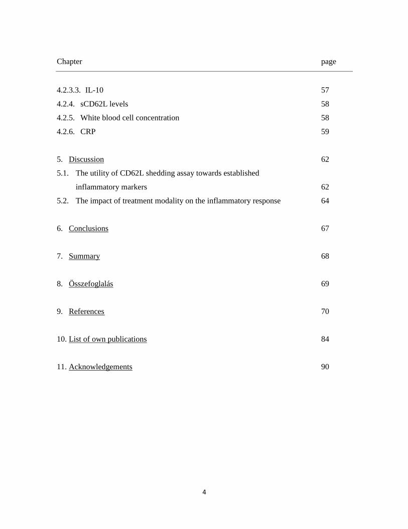
TABLE OF CONTENTS
Chapter page
List of abbreviations 5
1. Introduction 9
1.1. The systemic inflammatory response syndrome (SIRS) 9
1.1.1. General aspects 9
1.1.2. Pathophysiology and characteristics 10
1.1.3. Definition and the role of inflammatory markers 11
1.2. SIRS in cardiac surgery 12
1.3. SIRS in interventional cardiology procedures 14
2. Objectives 15
3. Methods 18
3.1. Patients` selection 18
3.2. Cardiac surgery and extracorporeal perfusion techniques 21
3.3. Interventional cardiology procedures 26
3.4. Blood sampling for inflammatory markers 29
3.4.1. CD62L shedding assay 30
3.4.2. HLA-DR expression 30
3.4.3. ELISA 33
3.4.3.1. IL-6 33
3.4.3.2. IL-8 34
3.4.3.3. IL-10 35
3.4.3.4. sCD62L 35
3.4.3.5. sTLR-2 36

3
Chapter page
3.4.3.6. ADAM 17 (TACE) 36
3.4.3.7. CRP 36
3.4.4. Determination of myocardial injury 37
3.5. Statistical analyses 38
3.5.1. Study 1 38
3.5.2. Study 2 and Study 3 38
3.5.3. General considerations 38
4. Results 39
4.1. Assessment of immune sensitivity in cardiopulmonary bypass
procedures using CD62L shedding assay. 39
4.1.1. Patients 39
4.1.2. CD62L shedding assay 39
4.1.2.1. CD62L shedding between the different perfusion techniques
and cardiac surgery procedures 44
4.1.3. HLA-DR expression 46
4.1.4. Association of HLA-DR expression data with CD62L
shedding data 48
4.1.5. IL-8 plasma level 48
4.1.6. Plasma levels of the soluble factors 48
4.2. Investigation of inflammatory response to treatment of AS
and investigation of inflammatory response to surgical treatment of AS. 52
4.2.1. Patients 52
4.2.2. HLA-DR expression 56
4.2.3. Cytokine release 56
4.2.3.1. IL-6 56
4.2.3.2. IL-8 57
4
Chapter page
4.2.3.3. IL-10 57
4.2.4. sCD62L levels 58
4.2.5. White blood cell concentration 58
4.2.6. CRP 59
5. Discussion 62
5.1. The utility of CD62L shedding assay towards established
inflammatory markers 62
5.2. The impact of treatment modality on the inflammatory response 64
6. Conclusions 67
7. Summary 68
8. Összefoglalás 69
9. References 70
10. List of own publications 84
11. Acknowledgements 90

5
LIST OF ABBREVIATIONS
ACCP - American College of Chest Physicians
ACT - Activated clotting time
ADAM 17 - ADAM metalloproteinase domain 17
AKI - Acute kidney injury
ALI - Acute lung injury
ANOVA - Analysis of variance
AS - Aortic valve stenosis
BIS - Bispectral index
BSA - Body surface area
°C - Celsius
CABG - Coronary artery bypass grafting
CAD - Coronary artery disease
CA - Carbohydrate antigen
CD - Cluster of differentiation
CE - Conformité européenne
CECC - Conventional extracorporeal circulation
cm2
- Square centimeter
COPD - Chronic obstructive pulmonary disease
CRP - C-reactive protein

6
ECC - Extracorporeal circulation
ECG - Electrocardiogram
EDTA - Ethylene diamine tetra acetic acid
EEG - Electroencephalography
ELISA - Enzyme-linked immunosorbent assay
ES - EuroSCORE
FACS - Fluorescence-activated cell sorting
g - Gram
h - Hour
ICU - Intensive care unit
HLA-DR - Human leukocyte antigen
hrs - Hours
IBW - Ideal body weight
IMC - Intermediate care unit
IL - Interleukin
IU - International unit
Kg - Kilogram
L - Liter
LTA - Lipoteichoic acid
m2
- Square meter

7
mg - Milligram
mL - Milliliter
MECC - Minimized extracorporeal circulation
min - Minutes
µL - Microliter
mmHg - Millimeters of mercury
nm - Nanometer
NO - Nitric oxide
NOS - Nitric oxide synthase
ns - Non-significant
NT-proBNP - Amino terminal B-type natriuretic peptide
OD - Optical density
OPCAB - Off-pump coronary artery bypass grafting
pg. - Picogram
RVP - Rapid ventricular pacing
s - Seconds
SAVR - Surgical aortic valve replacement
sCD62L - Soluble L-selectin
SIRS - Systemic inflammatory response syndrome
SCCM - Society of Critical Care Medicine

8
STS - Society of Thoracic Surgery
TACE - Tumor necrosis factor-α-converting enzyme
TA-TAVI - Transapical transcatheter aortic valve implantation
TA-TAVR - Transapical transcatheter aortic valve replacement (synonymously used for
TA-TAVI)
TAVI - Transcatheter aortic valve implantation
TF-TAVI - Transfemoral transcatheter aortic valve implantation
TF-TAVR - Transfemoral transcatheter aortic valve replacement (synonymously used
for TF-TAVI)
TLR - Toll-like receptor
sTLR - Soluble toll-like receptor
TNF-α - Tumor necrosis factor alpha
VS - Valve surgery
WBC - White blood cell

9
1. INTRODUCTION
1.1. The systemic inflammatory response syndrome
1.1.1. General aspects
The systemic inflammatory response syndrome (SIRS) is a serious, potentially life-
threatening clinical condition that can be defined as a pro-inflammatory state that involves
the entire human organism.[1] The prevalence of SIRS is high, and it affects one-third of all
hospitalized patients and 50 - 80 % of critically ill patients treated in the intensive care unit
(ICU).[2]
In contrast to sepsis, which is the result of a confirmed infectious process, SIRS occurs
without a proven source of infection and usually occurs in association with a non-infectious
insult or a tissue injury (e.g., autoimmune disorder, pancreatitis, vasculitis, thrombo-
embolism, burns). Although differing in origin,[3] SIRS and sepsis involve common patho-
physiologic pathways and therapeutic strategies. Moreover, there is a continuum between
the entities: SIRS may evolve to sepsis, which has a high risk of further developing to
severe sepsis and septic shock. Accordingly, prognosis and patient outcome are poorer with
the increasing severity of the inflammatory processes and associated organ dysfunction or
failure.[2, 4, 5]
Despite of the advances in intensive care therapy and the use of new-generation
antimicrobial-therapeutics, lethality due to SIRS and sepsis remained unchanged between
20 - 30 %, in septic shock even between 70 - 80 %, in the last two decades.[6, 7]
Thus, the occurrence of SIRS still represents a relevant socio-economic and financial factor
that should be addressed early in patient selection and allocation to a specific treatment
strategy, especially in elderly patients.[8]

10
1.1.2. Pathophysiology and characteristics
An injury to the human body triggers an almost uniform response of the innate and
humoral immune system.[9, 10] Within minutes following tissue damage, the production of
pro-inflammatory cytokines increases, thus forming a complex signaling system together
with molecules involved in neuroendocrine, hemostatic and metabolic processes. The net
effect of the inflammatory response is the restoration of fluid and cardiovascular homeo-
stasis and the promotion of wound healing.[11] After uncomplicated operations or once
healing is established, the pro-inflammatory state becomes attenuated, and anti-
inflammatory components predominate until the non-inflammatory condition is restored in
the normal physiologic state.
SIRS and sepsis can be viewed as pro-inflammatory responses that include an exaggerated
production of pro-inflammatory mediators. The associated cellular and microvascular
injury leads to devastating consequences for cell integrity, energy supply and oxygen
delivery, which results in the alteration of enzyme function, increased apoptosis and
mitochondrial dysfunction. The concomitant activation of the coagulation cascade also
leads to diffuse microvascular thrombosis and associated ischemic (multi)organ damage,
which determine patient outcome.
Paradoxically, the human organism rapidly suffers from an increased susceptibility to
infection in SIRS, although a heightened non-specific immunity is present.[10] As a con-
sequence, a parallel state of inflammation and immunoparesis results. At this stage,
invading microorganisms cannot be combated effectively due to the consumption and
subsequent imbalance of immune components.[10] In elderly patients in particular, the
ability of the immune system to address an injury is markedly diminished, which puts this
patient population at a high risk for SIRS and sepsis.[12, 13] Due to factors known as
inflammageing and immunosenescence, both the baseline inflammatory state and the
inflammatory response through non-specific (innate) and specific (adaptive) pathways are
suppressed and thereby contribute to an early immunoparesis with a more severe
inflammatory course.[14-20]

11
1.1.3. Definition and the role of inflammatory markers
As endorsed by the American College of Chest Physicians (ACCP) and the Society
of Critical Care Medicine (SCCM),[21, 22] the diagnosis of SIRS is confirmed when at
least two of the following conditions are recognized:
Body temperature less than 36 °C or greater than 38 °C
Heart rate greater than 90 beats / minute
Respiratory rate greater than 20 breaths / minute or an arterial PaCO2 less than
32 mmHg
Abnormal white blood cell count less than 4 cells per µL or greater than 12
cells per µL or 10 % bands.
Considering these conditions, clinicians are able to identify with high sensitivity patients
who have SIRS both early and in real time to initiate further diagnostic measures and
accurate therapy.[3]
In contrast, this classification is less appropriate for detecting the impact of different
therapeutic strategies (e.g., gold standard surgery vs. minimally invasive intervention) on
the extent of the inflammatory cascade activated. Because the response to the infection,
rather than the infection itself, seems to be the major determinant of patient outcome,[2]
investigating the inflammatory markers, especially those that reflect critical nodes on the
inflammatory pathways, is advisable.[2, 10]
The modern diagnostic and laboratory methods (e.g., enzyme-linked immunosorbent assay,
fluorescence-activated cell sorting) allow for the determination of nearly all of the factors
involved in inflammatory processes. Because different components of the pro- and anti-
inflammatory cascade peak heterogeneously after a procedure, an objective assessment of
the injury on the immune system is possible when multiple inflammatory markers are
sampled at different peri-procedural time points.[3, 9, 23, 24]

12
1.2. SIRS in cardiac surgery
In cardiac surgery, both preexisting disorders (e.g., impaired left ventricular function,
diabetes) and surgical factors such as sternotomy, pericardectomy or cardiac ischemia can
cause a pathological inflammatory response and concomitant organ dysfunction, which
manifests as coagulopathy, cardiac or respiratory insufficiency, or cognitive
dysfunction.[25-28]. In addition to the surgical trauma, the usage of extracorporeal
circulation (ECC) technique is considered to be a further cause of abnormalities in the
inflammatory response and related SIRS development.[29-33] The contact of blood with air
and the foreign surfaces that are included in the ECC circuit (e.g., tubings, oxygenator)
triggers a complex cascade of reactions that involve the inflammatory, coagulation and
complement systems.[34-36] Leukocytes, platelets, and endothelial cells are indirectly
activated by humoral mechanisms or receive a direct stimulation via lipopolysaccharides,
ischemic-reperfusion injury or mechanic stress during ECC runs. The complex interaction
ultimately results in a pro-coagulatory and vasodilatatory state. Together with the pro-
inflammatory state that occurs at this time, this interaction leads to severe disturbances of
the microcirculation and a concomitant organ dysfunction, bleeding (due to
hyperfibrinolysis) and to a vasoplegic shock with high lethality as a further manifestation of
SIRS. In addition to the above-mentioned, different organs are directly affected during and
after periods of ECC. Even after an uncomplicated ECC course, post-operative pulmonary
complications such as acute lung injury (ALI) can occur because the lungs are not
ventilated during ECC and risk being exposed to an ischemic-reperfusion injury with
consecutive increased permeability, interstitial edema and atelectasis with an increased
shunt fraction and pulmonary vascular resistance. The risk and severity of pulmonary injury
has been directly linked to ECC and mortality.[37-39]
Additionally, during this procedure, the heart suffers from ischemia, a complication that
cannot be entirely avoided despite the application of cardioplegia. After cardioplegic
cardiac arrest, leukocytes migrate into the myocardium and produce endothelial lesions and
cell edema in the process. The cardial reperfusion injury aggravates the local inflammatory
response, which often manifests as myocardial stunning, ischemia, dysfunction and β-

13
adrenergic desensitization and leads to difficulties in weaning the patient from the cardio-
pulmonary bypass.[40, 41]
The ECC-induced inflammatory response plays a pivotal role in the pathogenesis of
neurologic injury.[42] Focal cerebral deficits, seizures and, in up to 69 % of the patients,
cognitive dysfunction and disability can occur, which contribute to increased mortality.[43]
A main factor in the emergence of neurologic complications following ECC seems to be
related to the alteration of nitric oxide (NO) homeostasis. NO is formed via the up-
regulation of neuronal NO synthase (nNOS) and is a potent neurotoxin that causes cerebral
vasodilatation and the associated cerebral perfusion deficit. [44, 45]
Post-operative renal and hepatic ischemia-reperfusion injury, which is induced by the
inflammatory response during ECC, further contributes to organ dysfunction after cardiac
surgery and increases both the length of ICU stays and patient mortality.[46, 47] Finally,
ECC-associated immunosuppression, which results from anti-inflammatory cytokine
production and the impairment of cell-mediated immunity, contributes to an altered
inflammatory response and adverse outcome after cardiac surgery.[48, 49]
In conclusion, the incidence of SIRS remains a serious complication after cardiac surgery
with ECC. Once activated, a complex cascade of humoral and cell-mediated reactions is
initiated, which is self-limited in the majority of cases. Nevertheless, the coincidence of
potential confounders (e.g., age, pre-operative health condition, intra-operative
complications, transfusion, anesthetic technique, perfusion technique) may act unfavorably
(“multiple hit scenario”), which can lead to a dysbalance of pro- and anti-inflammatory
pathways and the subsequent development of SIRS and multiple organ dysfunction
syndrome (MODS), with the associated poor outcomes. Future therapeutic strategies,
including technical innovations, which allow for cardiac surgery using modified variants of
ECC, or even without the use of ECC, may represent a promising alternative to modulate
the peri-operative inflammatory response, especially considering those patients who might
be expected to have an increased risk for SIRS.

14
1.3. SIRS in interventional cardiology procedures
Interventional and invasive cardiology procedures include treatments such as trans-
catheter aortic valve implantation (TAVI) via the transfemoral (TF-TAVI) or the trans-
apical route (TA-TAVI). Since its introduction into clinical practice in 2007, more than
9000 high-risk patients with severe aortic valve stenosis (AS) who are not suitable for
surgical aortic valve replacement (SAVR) undergo TAVI in Europe every year.[50]
Although in TAVI extracorporeal circulation is entirely avoided, SIRS remains a common
phenomenon.[51] According to previous reports, SIRS occurred in TAVI in 40 - 60 % of
affected individuals and was independently associated with acute kidney injury (AKI),
worse short-term outcome and mortality.[52-56]
In contrast to the mechanisms that are known to contribute to SIRS in cardiac surgery,
SIRS pathogenesis and triggering factors have not been completely identified in TAVI.
Currently, intra-procedural low cardiac output states, such as periods with rapid ventricular
pacing (RVP) for aortic valve implantation in the native aortic annulus, are considered
partially accountable for the induction of ischemic injury in the organs and, consequently,
for inducing SIRS.[56-58]
As expected, the inflammatory response is present in TAVI procedure, though to a lesser
extent than is observed in cardiac surgery with ECC.[53] In TF-TAVI procedures, an even
more significant reduction of SIRS can be anticipated.[59] In TF-TAVI, in contrast to TA-
TAVI, no thoracotomy and no myocardial incision are performed. Instead, the aortic bio-
prosthesis is delivered by protrusion of the delivery system via the descending aorta in a
retrograde fashion following the puncture of the femoral artery. Thus, the myocardium, as a
potential source of pro-inflammatory cytokines, is less injured when this technique is
implemented.[60]
At present, several new (less invasive) TAVI devices, aortic bioprostheses and prosthesis
deployment techniques are on the horizon. Therefore, it is likely that SIRS as currently
detected in TAVI procedures will be diminished to an even greater extent in the near future.
15
2. OBJECTIVES
In preceding studies to the PhD thesis a) patients undergoing TF-TAVI and TA-
TAVI were examined for procedural complications and predictors for adverse events in a
prospective observational manner and b) the clinical outcomes of TAVI patients were
evaluated in comparison with medical treatment and SAVR by using data from the
institutional prospective registry.[61, 62] In both studies and despite the high procedural
success rates, the all-cause mortality rates were high in the selected high-risk patient
population with severe AS allocated to TAVI or SAVR (22.6 and 22.4 %, respectively).
Because the contribution of treatment modality (surgical vs. interventional) on the patients’
peri-procedural course, especially in terms of inflammatory response and immuno-
modulation, was not evident at that time, subsequent studies were performed to investigate
these issues in detail. It was hoped that these results may contribute to finding a perennial
pattern of immune response that is characteristic for each procedure. Such information
would help predict more precisely the extent of the immunomodulation that occurs during
procedures, which would allow cardiac surgeons and interventional cardiologists to select
the most appropriate treatment strategy for an individual patient.
The aim of the PhD thesis was to provide a detailed insight into the inflammatory response
and the changes in immune sensitivity that occur in the peri-operative period during
different cardiac surgery and interventional cardiology procedures. In particular, this
project intends to investigate the influence of two different extracorporeal circulation
techniques and non-pharmacological treatment strategies on the peri-procedural immune
response in patients with coronary or valvular heart disease to better understand the impact
of these strategies on the patients’ peri-operative course and procedural outcome.
Data from two prospective observational studies (Study 1 and Study 2) and from one
prospective randomized controlled trial (Study 3) were then obtained. All studies were
conducted as single-center investigations in a tertiary referral hospital in which cardiac
surgery and interventional cardiology procedures are performed on a daily basis.
16
The aim of Study 1 (Assessment of the immune sensitivity in cardiopulmonary bypass
procedures using CD62L shedding assay) was to investigate changes in the perioperative
immune sensitivity in patients who were scheduled to undergo coronary artery bypass graft
surgery (CABG), cardiac valve surgery (VS) or a combination of both by using the
conventional extracorporeal circulation (CECC) technique. The main focus was on CECC-
induced immunomodulation, which was determined by the quantification of a broad
network of molecules that are involved in peri-operative immune reactions (i.e.,
interleukins, human leukocyte antigen expression, plasma concentration of soluble factors
as soluble L-selectin or soluble toll-like receptor). In addition and compared with the
established methods that are commonly used to describe immune response, the functional
status of the immune system was determined by using a novel laboratory method to assess
the responsiveness of inflammatory effector cells directly. This method, referred to as the
CD62L shedding assay, quantifies the cleavage of the adhesion molecule L-selectin
(CD62L) from the surface of neutrophils (membrane-bound part of CD62L molecules) after
ex-vivo microbial stimulation with toll-like receptor (TLR) ligands, thereby providing
information on granulocyte and monocyte sensitivity.
The functional changes in immunity have not been quantified using the CD62L
shedding assay under these clinical circumstances. The CD62L shedding assay may provide
a novel and more detailed view of the immunological processes that are induced by the use
of extracorporeal circulation.
The aim of Study 2 (Investigation of inflammatory response to treatment of AS) was to
investigate patients with severe symptomatic AS for an inflammatory response during the
peri-operative course. The main focus lay in the comparison of cardiac surgical and
interventional cardiology modalities in the treatment of aortic stenosis. Whereas SAVR
using CECC represents the current gold standard, over the last decade, TAVI has become
widely available for selected high-risk patients who have multiple co-morbidities and
allows for the treatment of AS without the use of any extracorporeal circulation support.
Few previous studies have investigated the difference in the extent of the
inflammatory response in SAVR and TAVI. Our results may contribute to an improved
17
understanding of immunological processes and the inflammatory response in patients with
AS, which, due to the increased age and frailty of affected individuals, can be completely
different compared with other cardiac surgical or interventional cardiology patients.
Study 3 (Investigation of inflammatory response to surgical treatment of AS) investigated,
in a randomized controlled fashion, the inflammatory response occurred in patients who
were treated solely surgically for AS. The main focus lay on the comparison of two
different modalities of extracorporeal circulation: the CECC and its less invasive variant,
the minimized extracorporeal circulation (MECC) technique. This issue has also been the
subject of previous studies investigating patients undergoing coronary artery bypass graft
surgery. The technical composition of the MECC system differs, however, for CABG and
SAVR. Moreover, the pathological mechanisms that are involved in coronary artery disease
(CAD) and AS also differ.[63-65] Whereas inflammation predominates in CAD, the
progression of AS is driven primarily by osteoblast processes that lead to calcification. It is
therefore likely that patients who are treated for AS exhibit a milder immune response
despite being treated with a similar extracorporeal circulation technique.
The general study hypothesis stated that significant peri-operative immune changes are
detectable during different cardiac surgery and interventional cardiology procedures and
that both the established measurement of molecules involved in the inflammatory cascade
and the novel CD62L shedding assay would produce complementary results.
Further, the most severe impairment of the immune system was expected in patients
undergoing cardiac surgery using CECC, followed by patients treated with MECC. In
contrast, only minimal changes in peri-operative immune sensitivity were anticipated in
patients treated for AS with the TAVI technique, primarily due to the avoidance of
extracorporeal circulation and related surgical trauma. Altogether, a correlation was
expected between the extent of inflammatory response and the peri-operative course.
18
3. METHODS
3.1. Patients` selection
We studied the peri-operative changes of immune sensitivity in different patient
populations. In the population planned to undergo cardiac surgery, patients selected for
CABG, VS or for a combined cardiac surgical procedure (CABG + VS) treated on CECC
or MECC were eligible to be included in the studies. In the population planned to undergo
interventional cardiology procedure, patients with severe AS, defined as an aortic valve
area <1 cm2 and an aortic valve mean pressure gradient >40 mmHg, allocated either to
SAVR using CECC or TAVI using the transapical or the transfemoral access route were
eligible.
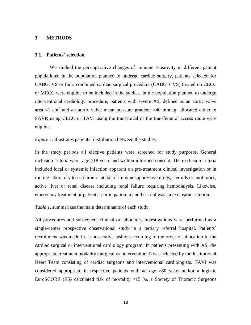
Figure 1. illustrates patients` distribution between the studies.
In the study periods all elective patients were screened for study purposes. General
inclusion criteria were: age ≥18 years and written informed consent. The exclusion criteria
included local or systemic infection apparent on pre-treatment clinical investigation or in
routine laboratory tests, chronic intake of immunosuppressive drugs, steroids or antibiotics,
active liver or renal disease including renal failure requiring hemodialysis. Likewise,
emergency treatment or patients’ participation in another trial was an exclusion criterion.
Table 1. summarizes the main determinants of each study.
All procedures and subsequent clinical or laboratory investigations were performed as a
single-center prospective observational study in a tertiary referral hospital. Patients`
recruitment was made in a consecutive fashion according to the order of allocation to the
cardiac surgical or interventional cardiology program. In patients presenting with AS, the
appropriate treatment modality (surgical vs. interventional) was selected by the Institutional
Heart Team consisting of cardiac surgeons and interventional cardiologists. TAVI was
considered appropriate in respective patients with an age >80 years and/or a logistic
EuroSCORE (ES) calculated risk of mortality ≥15 %, a Society of Thoracic Surgeons
19
(STS) score risk of mortality ≥10 %, or in those with an age >70 years with a predicted or
prohibitive risk of morbidity or/and mortality for the surgical aortic valve replacement.
Source: own design
Figure 1. Patients` distribution between the studies. The figure shows patients` allocation
to different cardiac surgical and interventional cardiology therapies (blue square), the
overall number of individuals in each treatment group (purple shapes) and in each study
(orange circles).
All patients gave written informed consent for cardiac surgery with extracorporeal
circulation, interventional cardiology procedure, the related anesthesia technique and to the
use of anonymized data for research purposes. Ethical approval for all study-relevant
investigations was provided by the local Ethics Committee.
20
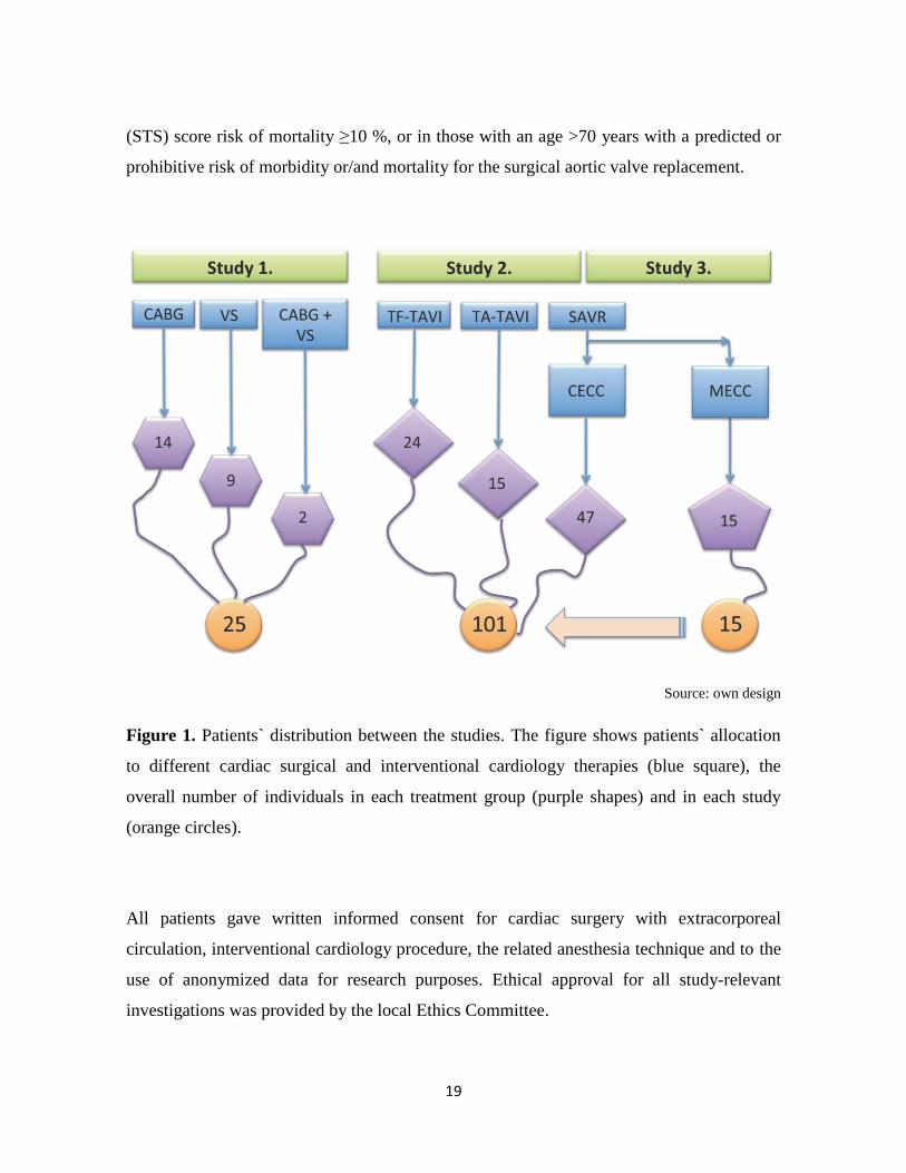
Table 1. Main determinants of the studies.
Study 1 Study 2 Study 3
Modality prospective
observational trial
randomized
controlled trial
Patient population CABG, VS,
CABG + VS
VS (AS)
and TAVI
VS (AS)
Inclusion criteria ≥18 yrs., IC
Exclusion criteria infection, intake of immunosuppressant or antibiotics, liver disease,
renal disease, emergency treatment, participation in another study
Primary aims
(comparators)
CECC-induced
immunomodulation
(established markers
vs. CD62L assay)
Inflammatory
response in patients
with AS
(SAVR vs. TAVI)
Inflammatory
response in patients
with AS
(CECC vs. MECC)
Determined
inflammatory
markers
CD62L shedding
and
IL-8, HLA-DR,
sCD62L,
sTLR-2,
ADAM17
IL-6, Il-8, IL-10
HLA-DR
sCD62L
CRP, WBC
Sampling time
points
Pre-operatively
End of surgery
48 hrs postop.
Pre-operatively
4, 24 and 48 hrs postop.
Legend: CABG indicates coronary artery bypass graft surgery; VS, cardiac valve surgery;
AS, aortic valve stenosis; TAVI, transcatheter aortic valve implantation; yrs., years; IC,
informed consent; CECC, conventional extracorporeal circulation; MECC, minimized
extracorporeal circulation; SAVR, surgical aortic valve replacement; CD62L, L-selectin;
IL, interleukin; HLA, human leukocyte antigen; s, soluble; TLR, toll-like receptor; ADAM,
ADAM metalloproteinase domain 17; CRP, C-reactive protein; WBC, white blood cell.

21
3.2. Cardiac surgery and extracorporeal perfusion techniques1
All single cardiac valve surgery (i.e., aortic valve replacement, mitral valve
replacement or reconstruction) and combined cardiac surgical procedures were performed
using CECC. Coronary artery bypass graft surgery was solely performed using MECC.
Figures 2-4. illustrate the schematic composition and the real life setup of the different
extracorporeal systems as used in the studies.
The MECC system (Jostra AG, Hirrlingen, Germany) used in this study consisted of a
centrifugal pump (Rotaflow, Maquet, Gossau, Switzerland), a microporous capillary
membrane oxygenator (Quadrox; Maquet, Gossau, Switzerland, Capiox FX25; Terumo,
Tokyo, Japan or Affinity Fusion, Medtronic, MN, USA), an anesthesia vapor (isoflurane), a
pulmonary artery vent line and a suction device, which controlled blood aspiration by an
optoelectrical sensor (SmartSuction, Cardiosmart, Muri, Switzerland). In contrast to
MECC, CECC included a roller pump (Maquet, Rastatt, Germany), a hardshell venous
reservoir, conventional cardiotomy suction, and a left ventricular vent line inserted via the
right upper pulmonary vein. MECC was primed with 600 mL of Ringer’s solution and
5.000 IU of heparin, whereas CECC was primed with 500 mL of hydroxyethyl starch
(130/0.4), 1,000 mL of Ringer’s solution, 100 mL of 20 % mannitol and 10.000 IU of
heparin. None of the extracorporeal circuits used a heparin coating or neutrophil filter.
Figure 2. illustrates the different composition of the CECC and the MECC system.
Continuous anesthesia monitoring consisted of ECG, pulse oximetry, capnography,
invasive arterial and central venous pressure, transesophageal echocardiography and
processed EEG (BISTM
, Covidien, Medtronic, MN, USA).
1 except for the figures, the text is quoted from Ref. 85

22
General anesthesia induction was performed with midazolam, etomidate and sufentanil,
followed by isoflurane and sufentanil maintenance. Cardiac surgery was performed through
a median sternotomy. After heparinization (MECC, 400 IU/kg ideal body weight (IBW);
CECC, 500 IU/kg IBW), extracorporeal circulation was established, with arterial inflow
through the ascending aorta and venous drainage through a two-stage cannula secured in
the right atrium. An activated clotting time (ACTkaolin) of >480 s (ACT Plus, Medtronic,
MN, USA) was targeted in both groups and monitored every 20 min.
All patients received a bolus of tranexamic acid (30 mg/kg IBW) followed by a continuous
infusion (15 mg/kg IBW/h) until sternal closure. Extracorporeal circuit flow rates were set
to 2.0 L/min/m2 body surface area (BSA) in MECC, and 2.4 L/min/m
2 BSA in CECC. Core
temperature was allowed to drift to a nadir of 34 °C. Myocardial protection was applied
with a single dose of 100 mL of crystalloid cardioplegia (Cardioplexol, Bichsel Laboratory,
Interlaken, Switzerland) followed by high-potassium cold blood cardioplegia, with
repetition if any electrical or mechanical activity was observed. Following separation from
extracorporeal bypass, heparin was neutralized with protamine sulfate in a ratio of 1:1 with
regard to the initial bolus.

23
Source: Department of Cardiothoracic Surgery, University Hospital Bern, Switzerland
Figure 2. Schematic configuration of the MECC (left picture) and the CECC (right picture)
system. Note that in the MECC system consists of a different pump (centrifugal pump vs.
roller pump) and does not have a venous reservoir. Likewise, suctioning of blood from the
surgical field (optoelectrical device vs. conventional suction device) and the blood venting
technique (pulmonary artery vent vs. pulmonary vein vent) are divergent.

24
Source: Department of Cardiothoracic Surgery, University Hospital Bern, Switzerland
Figure 3. MECC setup as used in the study.

25
Source: Department of Cardiothoracic Surgery, University Hospital Bern, Switzerland
Figure 4. CECC setup as used in the study.

26
3.3. Interventional cardiology procedures
TA-TAVI and TF-TAVI were performed according to the strategy described
previously in detail.[66, 67] There was no pre-specified recommendation regarding access
route selection (transfemoral vs. transapical), which was chosen according to the decision
of the interventionist and the local expertise.
In all TA-TAVI cases the balloon-expandable Edwards Sapien XT aortic bioprosthesis
(Edwards Lifesciences, CA, USA) was selected. In TF-AVI procedures depending on the
aortic valve and annular calcific load and distribution, either the Edwards Sapien XT aortic
bioprosthesis or the self-expanding CoreValve Revalving System (Medtronic, MN, USA)
was chosen.
Figure 5. displays the two bioprostheses used in the studies.
TA-TAVI included echocardiography-guided anterolateral mini-thoracotomy, access site
preparation, insertion of the apical introducer and balloon aortic valvuloplasty of the
calcified native aortic valve during rapid ventricular pacing followed by angiography-
guided delivery of the TA-TAVI device. The self-expanding CoreValve was deployed after
puncture of the femoral artery and introduction of the bioprosthesis in the aortic annulus
through the descending aorta.
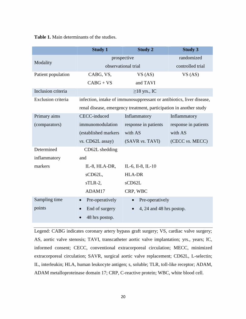
Figure 6. displays the different access routes indicating bioprosthesis positioning into the
aortic annulus.
All TA-TAVI procedures were performed in general endotracheal anesthesia using total
intravenous anesthesia (TIVA) with propofol 2 % (Disporivan™, AstraZeneca AG, Zug,
Switzerland) combined with remifentanil (Ultiva™, GalaxoSmithKline AG, München-
buchsee, Switzerland). Continuous anesthesia monitoring consisted of ECG, pulse
oximetry, capnography, invasive arterial and central venous pressure, transesophageal
echocardiography and processed EEG (BISTM
, Covidien, Medtronic, MN, USA).

27
Source: Edwards LifeSciences. www.edwards.com
Figure 5. Transcatheter aortic valve implantation using the Edwards Sapien XT aortic
bioprosthesis (transfemoral and transapical cases, left picture) and the CoreValve
Revalving System (only transfemoral cases, right picture). Note the divergent form and
implantation procedure of the bioprostheses.
28
Source: Edwards LifeSciences. www.edwards.com
Figure 6. Transcatheter aortic valve implantation using the transfemoral (left picture) and
the transapical (right picture) access route. Note that in TF-TAVI no myocardial puncture
and incision occurs.
Post-operatively, patients were either extubated in the angiography suite, or remained
intubated until circulatory and respiratory stabilization. All TA-TAVI and all intubated TF-
TAVI patients were transferred to the intensive care unit, whereas extubated TF-TAVI
patients transferred to the intermediate care unit (IMC).

29
3.4. Blood sampling and determination of inflammatory response markers1
In all cases, blood was collected in three different tubes (2x EDTA tubes with a
filling volume of 2.7 mL and 1x heparinized tube with a filling volume of 4.7 mL), which
were immediately transferred to the in-house laboratory. The first sample (baseline value)
was drawn in all studies from the patient`s arterial line prior to the application of anesthetic
drugs. The subsequent samples were drawn from the central venous line after 4 (Study 2
and Study 3), 24 (Study 2 and Study 3) and 48 hrs (Studies 1, 2 and 3) post-operatively.
Figure 7. displays the blood sampling strategy in the studies.
Source: own design
Figure 7. Frequency of blood sampling in each study. The figure illustrates the blood
sampling time points for inflammatory markers in relation to the peri-procedural processes.
30
3.4.1. CD62L shedding assay
Twenty-five ml citrate whole blood was stimulated with 25 ml of six 10-fold dose-
titrations starting at 10 mg/ml (LTA, Invivogen, San Diego, USA), 20 ng/ml (TNF, R&D).
After 45 minutes incubation at 37 °C / 5 % CO2, cells were washed in PBS/BSA 1% and
stained with 25 ml PBS/BSA 1% containing APC-anti-human CD33 (allowing
identification of both granulocytes and monocytes in combination with side-scatter
measurement, and remaining stable during stimulation) and FITC-anti-human CD62L
antibodies (Biolegend, San Diego, USA) for 15 minutes. Red blood cells were lysed in 200
ml FACS Lysis solution (BD Biosciences, San Diego, USA) and acquired on a FACSArray
SORP (BD Biosciences, San Diego, USA). Blood monocytes and granulocytes were
distinguished on the basis of CD33 expression and side scatter using FlowJo Software
(Tree Star Inc., Ashland, USA). Median FITC fluorescence intensity is computed for each
sample’s granulocytes and monocytes and plotted against dilution factor to enable parallel
analysis of multiple agonists. Four-parameter curves are fitted using non-linear regression
and LogEC50 values were extracted, corresponding to the dilution factor giving 50%
granulocyte or monocyte CD62L-shedding. The corresponding ligand concentration was
calculated and plotted for each ligand.
Figure 8. – 11. displays the operational steps of CD62L assay.
3.4.2. HLA-DR expression
Fifty microliters of heparinized blood were stained with 20 µL of Quantibrite™
anti-human-HLA-DR PE (phycoerythrin)/anti-monocyte PerCP (peridinin chlorophyll
protein)-Cy5.5 (anti-human-CD14 and anti-human-CD64) (BD Biosciences) at room
temperature in the dark for 25 min. Red blood cells were subsequently lysed with FACS
lysis solution (BD Biosciences) for 5-10 min and washed twice with phosphate-buffered
saline solution (Sigma-Aldrich) and fixed with 400 µL of 4 % paraformaldehyde. One
unstained sample was treated in the same manner. The fluorescence intensity of the samples

31
was measured on an LSR II (BD Biosciences) using the software FACSDiva v6.1.3 (BD
Biosciences) as duplicates. A total of 500-1,000 monocyte events were recorded. For
quantification of the signal, Quantibrite™ PE beads (BD Biosciences) were acquired with
each measurement. The FACS data were analyzed using FlowJo v7.5 (Treestar) with gating
for CD14- and CD64-positive cells (monocytes). The HLA-DR PE channel was calibrated
using the data from the PE beads, which allows fluorescence intensity to be correlated with
the mean number of PE molecules per cell. The results were recorded as the median of the
calibrated PE channel fluorescence intensity of each sample. The mean and standard
deviation were calculated for the duplicate samples, and an analysis of variance (ANOVA)
was performed using GraphPad Prism v5.04 (GraphPad Software).
Source: M. Book / M. Schiff
Figure 8. General CD62L assay setup.

32
Source: M. Book / M. Schiff
Figure 9. CD62L assay principals.
Source: M. Book / M. Schiff
Figure 10. Identification of granulocytes and monocytes.

33
Source: M. Book / M. Schiff
Figure 11. Data processing to determine logEC50.
3.4.3. ELISA
Plasma from each time point was separated from 5 mL of EDTA-whole blood by
centrifugation at 3,000 g for 5 min and stored at -80 °C.
3.4.3.1. IL-6
Interleukin-6 levels were measured by a sandwich ELISA kit (eBioScience), and all
samples were diluted 1:2 using the reagents provided by the kit. The standards ranged from

34
1.56 to 100 pg/mL. The provided 96-well plate was treated according to the manufacturer’s
instructions, and all standards, controls and samples were loaded in duplicate. Optical
density was measured using an eL800 microplate reader (Biotek Instruments) set to record
at 450 and 630 nm. Blank values and OD values at 630 nm were subtracted from all OD
450 nm values, and the IL-6 concentration of all samples was determined by a 4-parameter
curve fit of the standards (Gen5 v.1.09 software, Biotek Instruments). Samples that fell out
of the standard curve range were repeated at a higher dilution. According to the
manufacturer, the sensitivity and intra- and inter-assay variations were 0.92 pg/mL, 3.4 %
and 5.2 %, respectively. A coefficient of variation of the sample duplicates of below 20 %
was considered acceptable.
3.4.3.2. IL-8
Interleukin-8 levels were measured by a sandwich ELISA kit (eBioScience), and all
samples were diluted 1:2 using the reagents provided by the kit. The standards ranged from
15.6 pg/ml to 1000 pg/ml. The provided 96-well plate was treated according to the
manufacturer’s instructions and all standards, controls and samples were loaded in
duplicate. Optical density was measured using an eL800 microplate reader (Biotek
Instruments) set to record at 450 and 630 nm. Blank values and OD values at 630 nm were
subtracted from all OD 450 nm values and the IL-8 concentration of all samples was
determined by a 4-parameter curve fit of the standards (Gen5 v.1.09 software, Biotek
Instruments). Samples that fell out of the standard curve range were repeated at a higher
dilution. According to the manufacturer, the sensitivity and the intra- and inter-assay
variations were 2 pg/ml, 6.3 % and 8.7 %, respectively. A coefficient of variation of the
sample duplicates of below 20 % was considered acceptable.

35
3.4.3.3. IL-10
Interleukin-10 levels were measured by a sandwich ELISA kit (eBioScience), and
all samples were diluted 1:2 using the reagents provided by the kit. The standards ranged
from 3.1 to 200 pg/mL. The provided 96-well plate was treated according to the
manufacturer’s instructions, and all standards, controls and samples were loaded in
duplicate. Optical density was measured using an eL800 microplate reader (Biotek
Instruments) set to record at 450 and 630 nm. Blank values and OD values at 630 nm were
subtracted from all OD 450 nm values, and the IL-10 concentration of all samples was
determined by a 4-parameter curve fit of the standards (Gen5 v.1.09 software, Biotek
Instruments). Samples that fell out of the standard curve range were repeated at a higher
dilution. According to the manufacturer, the sensitivity and intra- and inter-assay variations
were 1.0 pg/mL, 3.2 % and 5.6 %, respectively. A coefficient of variation of the sample
duplicates of below 20 % was considered acceptable.
3.4.3.4. sCD62L
Soluble L-selectin (sCD62L) levels were measured by a sandwich ELISA kit
(eBioScience), and all samples were diluted 1:200 using the reagents provided by the kit.
The standards ranged from 0.4 to 25 ng/mL. The provided 96-well plate was treated
according to the manufacturer’s instructions, and all standards, controls and samples were
loaded in duplicate. Optical density was measured using an eL800 microplate reader
(Biotek Instruments) set to record at 450 and 630 nm. Blank values and OD values at 630
nm were subtracted from all OD 450 nm values, and the sCD62L-selectin concentration of
all samples was determined by a 4-parameter curve fit of the standards (Gen5 v.1.09
software, Biotek Instruments). Samples that fell out of the standard curve range were
repeated at a higher dilution. According to the manufacturer, the sensitivity and intra- and
inter-assay variations were 0.198 pg/mL, 3.7 % and 4.2 %, respectively. A coefficient of
variation of the sample duplicates of below 20 % was considered acceptable.

36
3.4.3.5. sTLR-2
Soluble toll-like receptor (sTLR)-2 plasma concentrations were detected using a
human TLR-2 ELISA kit (Cusabio, Wuhan, China). Samples were diluted 1:3 and
measured together with standards and blanks in duplicate. The detection range (standard
curve) was 0.312–20 ng/ml, intra- and interassay variation was given as 8 and 10 % CV by
the manufacturer.
3.4.3.6. ADAM 17 (TACE)
All samples were measured undiluted and in duplicates using a TACE human
ELISA kit (Abcam, Cambridge, UK) according to the manufacturer’s instructions. The
detection range (standard curve) was 78.15 – 5000 pg/ml, intra- and interassay variation
was given as 10 and 12 % CV by the manufacturer.
3.4.3.7. CRP
C-reactive protein (CRP) levels were measured by an ELISA kit (eBioScience), and
all samples were diluted 1:8,000, 1:16,000, 1:32,000, 1:40,000 or 1:64,000 using the
reagents provided by the kit. The dilution was determined by several pre-tests. The
standards ranged from 0.15 to 10 ng/mL. The provided 96-well plate was treated according
to the manufacturer’s instructions, and all standards, controls and samples were loaded in
duplicate. Optical density was measured using an eL800 microplate reader (Biotek
Instruments) set to record at 450 and 560 nm. Blank values and OD values at 560 nm were
subtracted from all OD 450 nm values, and the IL-6 concentration of all samples was
determined by a 4-parameter curve fit of the standards (Gen5 v.1.09 software, Biotek
Instruments). Samples that fell out of the standard curve range were repeated at a higher
dilution. A coefficient of variation of the sample duplicates of below 20 % was considered
acceptable.

37
3.4.4. Determination of myocardial injury
Intra-procedural cardiac tissue injury was determinated by the analysis of IL-6
levels and their course during the observation period. As reported by previous investigators,
Il-6 levels are the highest in the coronary sinus blood under cardiopulmonary bypass
conditions, indicating that cardiac myocytes represent the major source in the release of this
pro-inflammatory cytokine when myocardial ischemia occurs.[33, 60, 68]

38
3.5. Statistical analyses
3.5.1. Study 1: Assessment of the immune sensitivity in cardiopulmonary bypass
procedures using CD62L shedding assay.
Comparisons between the groups and repeated measurements were tested for
normality by the Shapiro-Wilk test. In cases of non-normal distribution, non-parametric
testing was performed by Friedman Repeated Measures ANOVA on ranks with the Tukey
post-hoc test for all pairwise multiple comparisons. Comparisons between the different tests
for immune sensitivity were performed using the Pearson product moment correlation.
3.5.2. Study 2 and Study 3: Investigation of inflammatory response to treatment of
AS and investigation of inflammatory response to surgical treatment of AS.
Different treatment strategies for aortic valve stenosis were compared using a
general linear model for repeated measures (as in Study 1) followed by Bonferroni
correction. The Greenhouse-Geisser correction was applied to detect inter-subject effects.
3.5.3. General considerations
Data are presented as integral number (percentage), mean ± standard deviation or
median with interquartile ranges as appropriate. A p-value <0.05 was considered
significant. In case of multiple comparisons and where Bonferroni adjustment of the
Wilcoxon tests were performed, p values <0.0125 (p =0.05/4) were considered significant.
Statistical analyses were performed with SigmaPlot software version 12.0 (Systat Software
Inc., San Jose, CA, USA) in Study 1, and with SPSS v22 (IBM Statistics; USA) in the
Studies 2 and 3.

39
4. RESULTS
4.1. Assessment of the immune sensitivity in cardiopulmonary bypass procedures
using CD62L shedding assay.
4.1.1. Patients
Overall, 25 consecutive patients completed Study 1. Among these, 14 patients
underwent CABG, 9 patients VS (aortic valve replacement (5 patients) and mitral valve
replacement (1 patient) or reconstruction (3 patients)) and 2 patients a combined cardiac
surgery procedure (CABG with aortic valve replacement or CABG with mitral valve
reconstruction). Eleven patients were treated using CECC and fourteen patients using
MECC.
Table 2. summarizes the basic clinical and procedural characteristics in the peri-operative
period.
4.1.2. CD62L shedding assay
Granulocytes and monocytes were stimulated either with lipoteichoic acid (LTA)
from the gram positive bacterium Staphylococcus aureus or with tumor necrosis factor
alpha (TNF-α) to assess changes in the sensitivity of inflammatory effector cells.
Granulocyte and monocyte sensitivity to LTA decreased at the end of cardiac surgery but
recovered to the baseline after 48 hrs. Decreased immune sensitivity was apparent as a
roughly 10-fold increase of the initial LTA concentration was necessary to cause shedding
of 50 % of CD62L from the cell surface of granulocytes or monocytes.

40
Table 2. Basic clinical and procedural characteristics in the peri-operative period displayed
according to the type of surgery and type of extracorporeal circulation.
Type of surgery All CABG VS CABG + VS p
Number of patients, n 25 14 9 2
Age, yrs. 67 ± 12 71 ± 10 63 ± 12 54 ± 19 ns
Female sex, n 12 (48) 6 (43) 5 (55) 1 (50) ns
Diabetes, n 9 (36) 7 (50) 2 (22) 0 (0) ns
EuroScore (additive) 6 ± 3 5 ± 2 6 ± 4 6 ± 7 ns
Surgery time, min. 230 ± 58 228 ± 41 232 ± 77 235 ± 106 ns
Time on ECC, min 83 ± 42 60 ± 14 112 ± 51 105 ± 49 .003
LOS, days 9.7 ± 2.7 9.4 ± 2 10.3 ± 3.8 9 ± 0 ns
In-hospital mortality 0 (0) 0 (0) 0 (0) 0 (0) ns
Type of ECC CECC MECC p
Number of patients, n 11 14
Age, yrs. 61 ± 12 71 ± 10 ns
Female sex, n 6 (54) 6 (43) ns
Diabetes, n 2 (18) 7 (50) ns
EuroScore (additive) 7 ± 4 5 ± 2 ns
Surgery time, min. 233 ± 77 228 ± 41 ns
Time on ECC, min 211 ± 48 60 ± 14 < .001
LOS, days 10 ± 3 9 ± 2 ns
In-hospital mortality 0 (0) 0 (0) ns
Legend: CABG, coronary artery bypass graft surgery; VS, valve surgery; ECC,
extracorporeal circulation; CECC conventional extracorporeal circulation; MECC,
minimized extracorporeal circulation; LOS, length of post-operative hospital stay. Values
are presented as number (percentage) and as mean ± standard deviation.

41
Figure 12. and Table 3. display the relevant results.
Source: Reference 69.
Figure 12. Granulocyte and monocyte sensitivity to LTA stimulation
Legend. LC50 represents the concentration of LTA which was necessary to cause shedding
of 50 % of the membrane-bound CD62L molecules of granulocytes or monocytes. Thus,
high LTA concentration values are associated with decreased sensitivity of the affected
cells. Black lines = data from an individual patient; orange lines = median values of all
patients at a certain blood sampling point with interquartile ranges (= purple lines).
*indicates p <0.05 compared with the previous sampling time point.
42
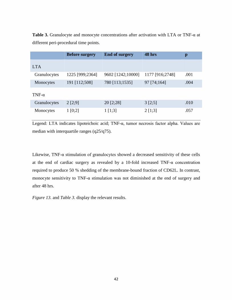
Table 3. Granulocyte and monocyte concentrations after activation with LTA or TNF-α at
different peri-procedural time points.
Before surgery End of surgery 48 hrs p
LTA
Granulocytes 1225 [999;2364] 9602 [1242;10000] 1177 [916;2748] .001
Monocytes 191 [112;508] 780 [113;1535] 97 [74;164] .004
TNF-α
Granulocytes 2 [2;9] 20 [2;28] 3 [2;5] .010
Monocytes 1 [0;2] 1 [1;3] 2 [1;3] .057
Legend: LTA indicates lipoteichoic acid; TNF-α, tumor necrosis factor alpha. Values are
median with interquartile ranges (q25/q75).
Likewise, TNF-α stimulation of granulocytes showed a decreased sensitivity of these cells
at the end of cardiac surgery as revealed by a 10-fold increased TNF-α concentration
required to produce 50 % shedding of the membrane-bound fraction of CD62L. In contrast,
monocyte sensitivity to TNF-α stimulation was not diminished at the end of surgery and
after 48 hrs.
Figure 13. and Table 3. display the relevant results.

43
Source: Reference 69.
Figure 13. Granulocyte and monocyte sensitivity to TNF-α stimulation.
Legend. LC50 represents the concentration of LTA which was necessary to cause shedding
of 50 % of the membrane-bound CD62L molecules of granulocytes or monocytes. Thus,
high LTA concentration values are associated with decreased sensitivity of the affected
cells. Black lines = data from an individual patient; orange lines = median values of all
patients at a certain blood sampling point with interquartile ranges (= purple lines).
*indicates p <0.05 compared with the previous sampling time point.

44
4.1.2.1. CD62L shedding between the different perfusion techniques and cardiac
surgery procedures
Granulocyte and monocyte stimulation with LTA and TNF-α and associated CD62L
shedding did not reveal differences between CECC and MECC or CABG, VS and
combined cardiac procedures, respectively.
Table 4. summarizes the changes in LTA and TNF-α concentrations according to the ECC
types.
Table 5. summarizes the changes in LTA and TNF-α concentrations according to cardiac
surgery procedures.
Table 4. Impact of ECC type on LTA / TNF-α values in the peri-operative period.
CECC MECC p
LTA
Before
surgery
granulocytes 1770 [966;2564] 1112 [946;2342] ns
monocytes 264 [119;494] 174 [90;575] ns
After
surgery
granulocytes 10000 [1242;12951] 6209 [1211;10000] ns
monocytes 804 [103;2000] 769 [185;1429] ns
48 hrs granulocytes 1291 [986;2748] 1086 [830;3758] ns
monocytes 111 [74;175] 96 [59;109] ns
TNF-α
Before
surgery
granulocytes 2 [2;8] 3 [2;12] ns
monocytes 1 [1;2] 1 [0;2] ns
After
surgery
granulocytes 20 [2;36] 12 [2;25] ns
monocytes 2 [1;4] 1 [0;4] ns
48 hrs granulocytes 3 [2;5] 4 [1;6] ns
monocytes 2 [1;2] 2 [1;7] ns
Legend: LTA indicates lipoteichoic acid; TNF-α, tumor necrosis factor alpha; CECC,
conventional extracorporeal circulation; MECC, minimized extracorporeal circulation.
Values are median with interquartile ranges (q25/q75).

45
Table 5. Impact of different cardiac surgery procedures on LTA / TNF-α values in the peri-
operative period.
CABG VS p
LTA
Before
surgery
granulocytes 1112 [859;2133] 1770 [1087;7319] ns
monocytes 319 [93;634] 191 [117;379] ns
After
surgery
granulocytes 10000 [1526;10000] 9205 [1195;22629] ns
monocytes 870 [355;1918] 290 [98;1397] ns
48 hrs granulocytes 1086 [850;2973] 1618 [1013;5220] ns
monocytes 96 [63;110] 116 [74;192] ns
TNF-α
Before
surgery
granulocytes 3 [2;10] 2 [2;9] ns
monocytes 1 [0;2] 1 [0;3] ns
After
surgery
granulocytes 4 [2;22] 28 [2;50] ns
monocytes 1 [0;3] 2 [2;4] ns
48 hrs granulocytes 3 [1;5] 3 [2;4] ns
monocytes 2 [1;6] 2 [1;2] ns
Legend: LTA indicates lipoteichoic acid; TNF-α, tumor necrosis factor alpha; CABG
coronary artery bypass graft surgery; VS, valve surgery. Values are median with
interquartile ranges (q25/q75).
46
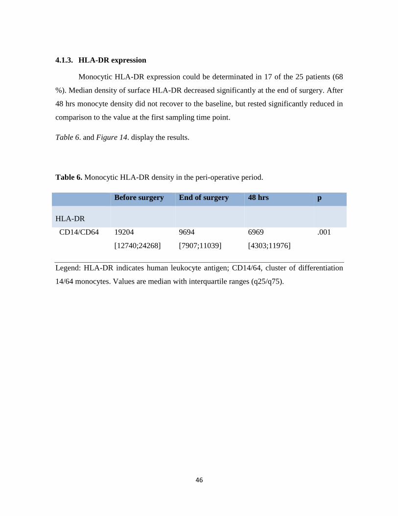
4.1.3. HLA-DR expression
Monocytic HLA-DR expression could be determinated in 17 of the 25 patients (68
%). Median density of surface HLA-DR decreased significantly at the end of surgery. After
48 hrs monocyte density did not recover to the baseline, but rested significantly reduced in
comparison to the value at the first sampling time point.
Table 6. and Figure 14. display the results.
Table 6. Monocytic HLA-DR density in the peri-operative period.
Before surgery End of surgery 48 hrs p
HLA-DR
CD14/CD64 19204
[12740;24268]
9694
[7907;11039]
6969
[4303;11976]
.001
Legend: HLA-DR indicates human leukocyte antigen; CD14/64, cluster of differentiation
14/64 monocytes. Values are median with interquartile ranges (q25/q75).

47
Source: Reference 69.
Figure 14. Monocytic HLA-DR expression
Legend. HLA-DR/cell means HLA-DR molecules per CD14 and CD64 positive
monocytes. Black lines = data from an individual patient; orange lines = median values of
all patients at a certain blood sampling point with interquartile ranges (= purple lines).
*indicates p <0.05 compared with the first sampling time point.
48
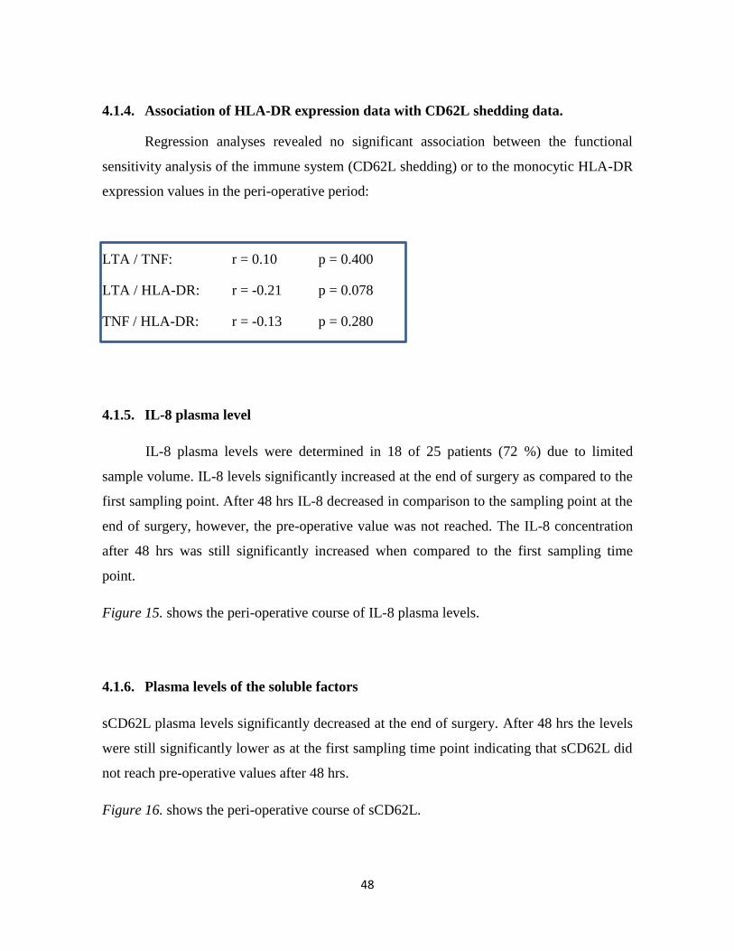
4.1.4. Association of HLA-DR expression data with CD62L shedding data.
Regression analyses revealed no significant association between the functional
sensitivity analysis of the immune system (CD62L shedding) or to the monocytic HLA-DR
expression values in the peri-operative period:
LTA / TNF: r = 0.10 p = 0.400
LTA / HLA-DR: r = -0.21 p = 0.078
TNF / HLA-DR: r = -0.13 p = 0.280
4.1.5. IL-8 plasma level
IL-8 plasma levels were determined in 18 of 25 patients (72 %) due to limited
sample volume. IL-8 levels significantly increased at the end of surgery as compared to the
first sampling point. After 48 hrs IL-8 decreased in comparison to the sampling point at the
end of surgery, however, the pre-operative value was not reached. The IL-8 concentration
after 48 hrs was still significantly increased when compared to the first sampling time
point.
Figure 15. shows the peri-operative course of IL-8 plasma levels.
4.1.6. Plasma levels of the soluble factors
sCD62L plasma levels significantly decreased at the end of surgery. After 48 hrs the levels
were still significantly lower as at the first sampling time point indicating that sCD62L did
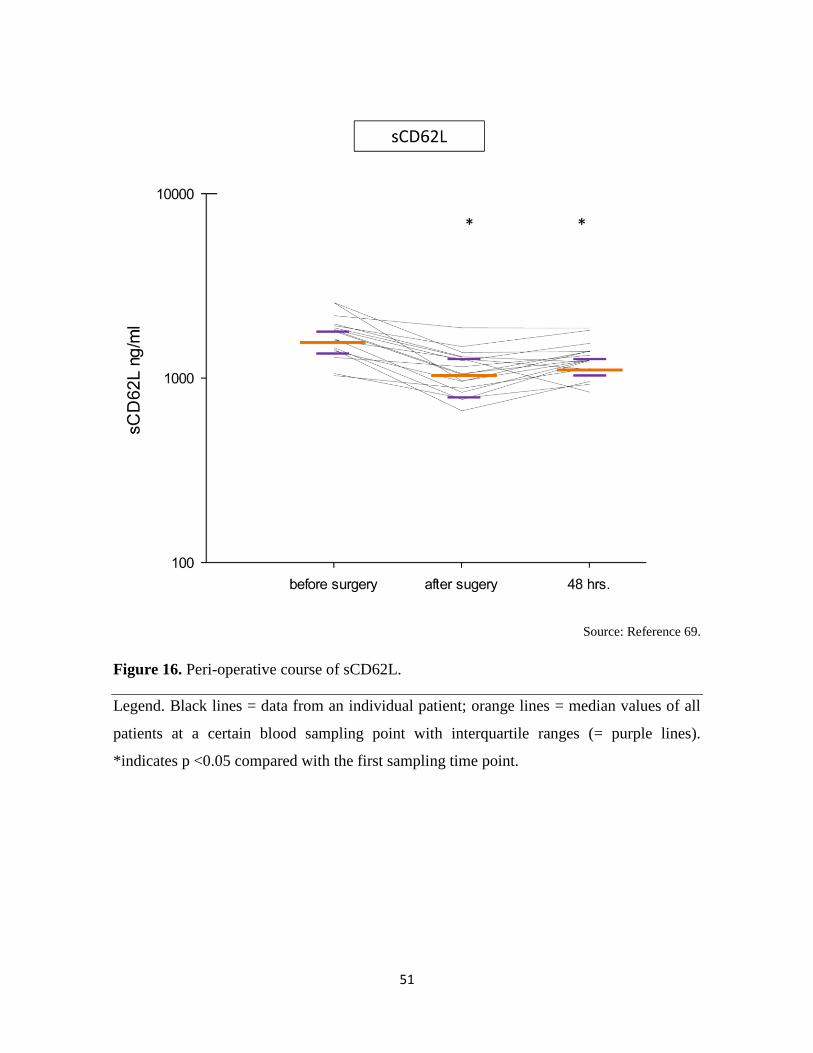
not reach pre-operative values after 48 hrs.
Figure 16. shows the peri-operative course of sCD62L.

49
Source: Reference 69.
Figure 15. Peri-operative concentration of IL-8.
Legend. Black lines = data from an individual patient; orange lines = median values of all
patients at a certain blood sampling point with interquartile ranges (= purple lines).
*indicates p <0.05 compared with the first sampling time point.

50
sTLR-2 showed a significant increase at the end of surgery. After this peak sTLR-2 levels
decreased to a minimum at 48 hrs showing a significant difference to the sampling point at
the end of surgery.
ADAM17 levels increased constantly during the peri-operative course, however, a
significant difference between two sampling points cannot be detected.
Table 7. displays the peri-operative values of sTLR-2 and ADAM17.
Table 7. sTLR-2 and ADAM17 concentrations in the peri-operative course.
Before surgery End of surgery 48 hrs p
sTLR-2 1.4 [1.2;2.4] 2.5 [1.9;3.6] 0.8 [0.6;1.2] .004
ADAM17 436 [227;849] 574 [274;769] 761 [402;1145] .401
Legend: sTLR indicates soluble toll-like receptor; ADAM 17, ADAM metalloproteinase
domain 17. Values are median with interquartile ranges (q25/q75).
51
Source: Reference 69.
Figure 16. Peri-operative course of sCD62L.
Legend. Black lines = data from an individual patient; orange lines = median values of all
patients at a certain blood sampling point with interquartile ranges (= purple lines).
*indicates p <0.05 compared with the first sampling time point.

52
4.2. Investigation of inflammatory response to treatment of AS and investigation of
inflammatory response to surgical treatment of AS.
Study 2 and Study 3 were performed independently with either a prospective observational
(Study 2) or a randomized controlled design (Study 3). Nevertheless, both studies
investigate the same topic (AS) in the same patient population: in Study 2 cardiac surgical
patients are investigated in comparison to interventionally treated patients (SAVR vs.
TAVI) and in Study 3 cardiac surgical patients are investigated relating to different extra-
corporeal perfusion techniques (SAVR with CECC vs. SAVR with MECC), respectively.
Moreover, in both studies the same inflammatory markers and the same sampling points
were used.
Thus, for a better readability and related understanding of the scientific message, the results
of both studies and their interpretation are presented together in the following chapters.
4.2.1. Patients
Overall, 101 patients treated with a different modality (surgical or interventional)
for severe symptomatic aortic stenosis were compared. Patients allocation to the different
treatment modalities were as follows: TF-TAVI: 24 patients; TA-TAVI: 15 patients; SAVR
with CECC: 47 patients and SAVR with MECC: 15 patients (Figure 1.).
Patients` characteristics were significantly different in the four groups. Patients in the
MECC group were the youngest, whereas patients in the TF-TAVI group were the oldest
(mean age MECC: 65 ± 8 yrs.; TF-TAVI 82 ± 4 yrs., p <0.001). The extent of heart failure,
as a result of severe aortic stenosis over years, represented by the NYHA classification, was
in the majority of surgical patients mild (percentage of NYHA class II in MECC: 60 %;
CECC: 49 % vs. others, p =0.02). In contrast, in the interventional groups the extent of
heart failure was severe (percentage of NYHA class III in TF-TAVI: 62 % and TA-TAVI
66 % vs. others, p =0.01). Co-morbidities, as coronary artery disease (CAD), chronic
obstructive pulmonary disease (COPD) or a higher procedural risk (redo procedure) were
more frequently present in patients selected for an interventional treatment (e.g., percentage

53
of patients with CAD: TF-TAVI: 58 % vs. others, p <0.001). The longest procedural time
was present in the CECC group, whereas TF-TAVI was performed the fastest (median
procedural time: CECC: 165 min. vs. TF-TAVI 75 min., p <0.001). Intermediate care unit
(IMC) or intensive care unit (ICU) stay was roughly double in patients selected for TF-
TAVI as in the CECC group (median stay: TF-TAVI 48 hrs vs. CECC: 22 hrs, p <0.001).
Systemic inflammatory syndrome (SIRS), defined according to the current guidelines, was
present in 27 % of TA-TAVI patients, whereas other groups were only minimally affected
(percent of patients with SIRS: TA-TAVI: 27 % vs. others, p = 0.02).
Table 8. summarizes patients` pre-operative and procedural characteristics.

54
Table 8. Patients` pre-operative and procedural characteristics in Study 2 and 3.
Legend: TF-TAVI indicates transcatheter transfemoral aortic valve replacement; TA-TAVI,
transcatheter transapical aortic valve replacement; MECC, minimized extracorporeal
circulation; CECC, conventional extracorporeal circulation; BMI, body mass index;
NYHA, New York Heath Association class; CAD, coronary artery disease; COPD, chronic
obstructive pulmonary disease; Δp mean, mean transaortic gradient; ECC, extracorporeal
circulation; IMC, intermediate care unit; ICU, intensive care unit; SIRS, systemic
inflammatory response syndrome. aDefinition according to current guidelines.
TF-TAVI
n = 24
TA-TAVI
n = 15
MECC
n = 15
CECC
n = 47
p
Baseline characteristics
Age, yrs. 82 ± 4 79 ± 5 65 ± 8 68 ± 9 <0.001*
Female sex 9 (37) 5 (33) 3 (20) 15 (32) 0.13
BMI, kg/m2 28 ± 4 24 ± 4 28 ± 5 28 ± 5 0.14
NYHA, class
I 0 (0) 1 (7) 1 (7) 5 (11) 0.43
II 5 (21) 3 (20) 9 (60) 23 (49) 0.02*
III 15 (62) 10 (66) 4 (26) 15 (32) 0.01*
IV 4 (17) 1 (7) 1 (7) 4 (8) 0.72
CAD 14 (58) 8 (53) 1 (7) 7 (15) <0.001*
Diabetes 7 (29) 6 (40) 3 (20) 9 (19) 0.31
Statins 6 (25) 8 (53) 7 (47) 17 (36) 0.29
COPD 6 (25) 4 (27) 0 (0) 0 (0) <0.001*
Dialysis 0 (0) 1 (6) 1 (7) 1 (2) 0.52
Redo surgery 10 (42) 4 (27) 1 (7) 2 (4) <0.001*
Ejection fraction, % 60 [45;65] 55 [40;65] 60 [55;70] 60 [59;65] 0.14
Aortic valve area, cm2 0.7 [0.6;0.8] 0.7 [0.4;0.4] 0.7 [0.5;0.8] 0.7 [0.5;0.8] 0.77
Δp mean, mmHg 41 [29;57] 34 [27;59] 47 [40;57] 42 [34;59] 0.59
Procedural characteristics
Procedural time, min 75 [58;89] 100 [84;114] 152 [130;173] 165 [145;185] <0.001*
ECC time, min --- --- 65 [51;74] 66 [56;86] 0.28
Crossclamp time, min --- --- 49 [42;55] 47 [40;64] 0.87
Outcome
IMC / ICU time, h 48 [33;75] 20 [17;23] 21 [18;22] 22 [21;23] <0.001*
New infection 1 (4) 1 (7) 1 (7) 1 (2) 0.80
SIRSa 0 (0) 4 (27) 1 (7) 3 (6) 0.02*
Stroke 1 (4) 0 (0) 1 (7) 1 (2) 0.71
30-d Mortality 0 (0) 1 (7) 0 (0) 0 (0) 0.13

55
Values are number (percent), mean ± standard deviation or median with interquartile range.
P <0.05 is considered significant. (p-values refer to the comparison among all groups).

56
4.2.2. HLA-DR expression
The plasma level of HLA-DR showed a significantly different course within the
four treatment modalities (group interaction: p <0.001). The highest baseline value of
HLA-DR was detected in MECC patients. In this population HLA-DR decreased
continuously having significantly different values from the baseline at all sampling points
(4 hrs p =0.002, 24 hrs p =0.003, 48 hrs p =0.005). CECC, TF-TAVI and TA-TAVI patient
exhibited much lower HLA-DR values. In CECC and TA-TAVI patients HLA-DR levels
were significantly different from the baseline at all sampling points (CECC: all p <0.001;
TA-TAVI all p =0.002). In TF-TAVI patients HLA-DR values decreased significantly from
the baseline at 4 hrs and 24 hrs (both p <0.001). After 48 hrs, however, HLA-DR levels in
TF-TAVI were not significantly different from the baseline.
Figure 17. displays the HLA-DR course in the observation period.
4.2.3. Cytokine release
4.2.3.1. IL-6
The plasma level of IL-6 showed a significantly different course within the four
treatment modalities (group interaction: p =0.017). The highest increase was detected in the
TA-TAVI group peaking at 4 hrs post-operatively. TA-TAVI, MECC and CECC groups
peaked also within 24 hrs post-operatively, however at lower levels. The most attenuated
IL-6 release was measured in the TF-TAVI group having only a slight increase in the
observation period.
In all groups IL-6 plasma levels were significantly different from the baseline at the time
points measured (4, 24, and 48 hrs). At the end of the observation period no group returned
to the pre-operative baseline (all p-values ≤0.001).
Figure 17. displays the IL-6 course in the observation period.

57
4.2.3.2. IL-8
The plasma level of IL-8 showed a significantly different course within the four
treatment modalities (group interaction: p =0.017). The highest increase was noted at 4 hrs
post-operatively in the CECC group, followed by MECC group. TF-TAVI and TA-TAVI
peaked at 4 hrs and 24 hrs, respectively.
In the patient population undergoing interventional treatment, no significant differences
were noted in the observation period. In contrast, the changes in the IL-8 plasma level in
the CECC and MECC group were significantly different during the peri-operative course.
In the MECC group IL-8 plasma level at 4 hrs and at 48 hrs was significantly different from
the baseline (both p =0.008). In the CECC group IL-8 was significantly different from the
baseline at 4, 24 and 48 hrs (p <0.001). In contrast to the interventional treatment, in
surgically treated patients IL-8 values did not return to the baseline at the end of the
observation period.
Figure 17. displays the IL-8 course in the observation period.
4.2.3.3. IL-10
The plasma level of IL-10 did not show a significantly different course within the
four treatment modalities (group interaction: p =0.814). Highest IL-10 values were detected
in the CECC group, followed by MECC treatment. In the interventional groups IL-10
peaked at 4 hrs. with TA-TAVI and at 24 hrs with TF-TAVI, however with lower values as
in the surgical groups.
With interventional treatment IL-10 plasma levels were significantly different from the
baseline at 4 hrs (TA-TAVI, p =0.005) and at 24 hrs (TF-TAVI, p =0.005 and TA-TAVI,
p =0.008). In both groups IL-10 levels returned to the baseline at the end of the observation
period. Likewise, IL-10 levels in MECC patients, although having the second highest
values, were not significantly different at the different sampling points and at the end of the
observation period (p not significant). In the CECC population, IL-10 was different from

58
the baseline at 4 hrs (p =0.004) and at 24 hrs (p <0.001). At the sampling point at the end of
the observation period, however, the IL-10 levels of CECC patients did not differ from the
baseline.
Figure 17. displays the IL-10 course in the observation period.
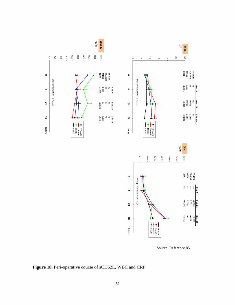
4.2.4. sCD62L levels
The plasma level of sCD62L showed a significantly different course within the four
treatment modalities (group interaction: p <0.001). Highest values were detected in the
MECC group, followed by the CECC group. In the MECC group sCD62L values only at 4
hrs and 48 hrs (both p =0.003) were significantly different from the baseline, whereas in the
CECC group sCD62L values at all sampling points were significantly different from the
baseline (all p <0.001). Thus surgically treated patients did not did not return with sCD62L
to the baseline at the end of the observation period.
Interventionally treated patients did not show peaks in sCD62L levels in the observation
period (all p not significant).
Figure 18. displays the sCD62L course in the observation period.
4.2.5. White blood cell concentration
White blood cell (WBC) concentration showed a significantly different course within the
four treatment modalities (group interaction: p <0.001). WBC levels continuously increased
until the end of the observation period with the highest values in TA-TAVI patients
(significant at 24 hrs (p =0.03) and at 48 hrs (p =0.006). MECC and CECC patients
exhibited significantly different WBC levels from the baseline at all sampling points (all p
values ≤0.002).

59
TF-TAVI patients did not show significant peaks in WBC levels in the observation period
(all p not significant).
Figure 18. displays the WBC course in the observation period.
4.2.6 CRP
The CRP plasma level showed a significantly different course within the four
treatment modalities (group interaction: p =0.007). Similarly to the WBC levels, the CRP
level continuously increased in the peri-operative period. Highest values were detected with
CECC and TA-TAVI, whereas MECC and TF-TAVI were associated with lower values.
In all groups values at 4 hrs were not significantly different from the baseline. CRP levels
increased to a significantly different value from the baseline at 24 hrs in all groups (all p
≤0.009) and at 48 hrs in TF-TAVI (p<0.001), TA-TAVI (p =0.001) and CECC (p <0.001).
In MECC patients CRP at 48 hrs returned to the baseline (p not significant).
Figure 18. displays the CRP course in the observation period.

60
Figure 17. Peri-operative course of interleukins and HLA-DR.
Source: Reference 85.
61
Figure 18. Peri-operative course of sCD62L, WBC and CRP
Source: Reference 85.

62
5. DISCUSSION
5.1. The utility of CD62L shedding assay towards established inflammatory markers
Pro- and anti-inflammatory markers allow clinicians to determine the extent of the
peri-procedural inflammatory response following a treatment. Similarly, inflammatory
markers can be used alongside clinical symptoms to diagnose SIRS and initiate appropriate
therapy, thus reducing the risk for consecutive organ dysfunction or failure at an early
stage. However, it is important to note that established inflammatory markers peak at
various time points in the post-operative course. Therefore, studies investigating the
absolute level of inflammatory markers alone are difficult to interpret because the time
points and frequency for blood sampling can often be heterogeneous.
The CD62L shedding assay, which was developed in our research group, differs
significantly in technique from this approach. Instead of determining a certain level of an
inflammatory marker, the sensitivity of the inflammatory effector cells (blood granulocytes
and monocytes) is assessed on a functional level. As a surrogate parameter of inflammatory
response, the cleavage of the membrane-bound portion of CD62L molecules is
determined.[69] Because shedding occurs minutes after cell activation [70-72] and because
the absolute number of CD62L-expression molecules is not investigated,[24, 73] this
method provides new and different information on immune state and early cell functionality
in the peri-procedural period.[69] Moreover, increases in the neutrophil sub-sets involved
in immune changes following endotoxin challenge or severe injury (e.g., ECC usage)[74]
are not assessable by conventional inflammatory markers but are detectable with the
CD62L shedding assay because it is altered by changes in all CD33-intermediate, side-
scatter high cells.
Interestingly, significant changes in neutrophil sensitivity by the CD62L assay were only
detected when LTA or TNF was used as an ex-vivo stimulant. Two explanations are
possible. First, contamination of ECC with gram positive bacteria, as occurs in up to 5.6 %
of all ECC blood cultures,[75] may have been present and may have led to neutrophil

63
receptor modulation and signaling component expression in pre-existing immune cells.
However, this would imply that the rate of recovery of live bacteria from blood culture
would have to significantly underestimate the actual exposure because almost all of the
patients examined in the study showed a loss of sensitivity to TLR2/6 ligands compared
with the rates suggested by Hamers et al.[75] A second explanation could be the activation
of damage-sensing pathways by the surgical procedure, with this activation releasing a high
number of fresh granulocytes and monocytes in the patients’ circulation, which, at the time
of investigation, did not have the full repertoire of signaling receptors and transducers that
is otherwise present in fully mature immune cells.
Overall, the changes in the immune sensitivity that were detected using the CD62L
shedding assay correspond well to the information provided by the established methods of
inflammatory response, as investigated by the peri-operative course of IL-8, HLA-DR,
sCD62L, sTLR-2 and ADAM 17.
The observed peri-operative levels of IL-8 and HLA-DR are in accordance with other
groups [76-79] and confirm the pro-inflammatory response and immunomodulation that
was detected with the CD62 shedding assay. The lack of association between HLA-DR and
the CD62L shedding data with LTA and TNF is not a contradiction but an indication that
the three responses represent independent aspects of post-surgical immune function.
Similarly, the peri-operative course of sCD62L as a marker of in vivo neutrophil activation
was in line with previous reports.[80, 81] The reduced levels at the time points “after
surgery” and at “48 hrs” reflect reduced CD62L shedding and are suggestive of a decrease
in circulating neutrophil activity.[82] This was also suggested by a reduced neutrophil
sensitivity to LTA and TNF in the CD62L shedding assay.
The sTLR-2 plasma levels showed a largely parallel movement with the LC50 values in the
CD62L shedding assay. After an initial increase at the time point “after surgery” that
reflected decreased granulocyte and monocyte reactivity, both assets decreased at 48 hrs,
which is indicative of normalized immune reactivity. This observation suggests a major
contribution of sTLR-2 to an altered responsiveness to LTA in the post-surgical period, as

64
it was previously assumed that sTLR-2 is a negative regulator of TLR-2 mediated signaling
pathways.[83]
Finally, an analysis of ADAM17 confirmed immunomodulation in this patient cohort,
although significant changes and a relation to CD62L shedding values were not detected.
This finding is, however, in line with the findings of other reports, which confirm the role
of ADAM17 in regulating the surface density, not the cleavage of CD64L, which is also the
major result of our study.[84]
5.2. The impact of treatment modality on the inflammatory response
Cardiac surgery and interventional cardiology procedures differ significantly in their
ability to trigger an inflammatory response and in the extent of immunomodulation that
occurs during their peri-operative course. Our data suggest that the inflammatory response
is most attenuated in TF-TAVI and is most pronounced in cardiac surgery using CECC.[85]
Nevertheless, not all of the changes detected in studies 2. and 3. can be interpreted by the
presence or absence of an extracorporeal circuit and the associated degree of activation of
inflammatory pathways.
The use of CECC released the highest levels of IL-8 and IL-10, in accordance with
previous investigations on this subject.[53, 86-89] The detected modulation of these
inflammatory markers is attributable to the specific features of CECC. As mentioned
previously, both the presence of a large artificial surface area, which is an open hardshell
venous reservoir that allows blood-air contact, and the use of cardiotomy suctioning
represent factors that contribute to a considerable extent of circulating leukocyte activation
and the associated initiation of pro- and anti-inflammatory cytokine pathways.[90-94] In
contrast, the MECC circuit, which is a modification of CECC in terms of biocompatibility,
led to a diminished pro- and a compensatory anti-inflammatory response. This is in line
with previous publications and is easily understood considering the radically reduced
dimensions in tubing length, the reduced priming volume, and the omission of components
triggering leukocyte activation in the MECC system. Thus, a portion of this study clearly

65
demonstrated the impact of extracorporeal circulation on the release of cytokines and the
possibility of influencing cytokine release by modifying the CECC composition.[87, 95-97]
Interestingly, the IL-6 levels were the highest in TA-TAVI. IL-6 is a pro-inflammatory
cytokine that peaks in situations in which ischemia-reperfusion injury is present. Thus, the
highest levels, which likely occur at levels similar to IL-8, were expected under
cardiopulmonary bypass conditions.[31, 95, 98] In contrast to IL-8, however, IL-6 is
closely related to tissue trauma, especially in cases of myocardial involvement. [60, 68, 99,
100] It is thus understandable that TA-TAVI, which combines surgical and minimally
invasive techniques together with phases of (induced) low-perfusion (e.g., RVP) to implant
aortic bioprosthesis, was associated with an even higher IL-6 release compared with CECC
or MECC, where direct myocardial trauma and low-perfusion states were rare. Likewise,
the high incidence of SIRS, as observed in the TA-TAVI population, was a contributing
factor to the increased concentrations of IL-6,[54] and was the cause of the conflicting
results in a similar investigation.[53]
SIRS, as a non-pathogenic activator of the inflammatory response, is often reflected by the
suppression of HLA-DR expression on monocytes, which indicates their impaired ability to
present foreign antigen to immune cells.[101] In the cohort investigated in this study,
MECC patients were the youngest compared with the other patients in the other treatment
groups. For this reason, the corresponding HLA-DR levels were the highest in the MECC
patients, which indicated a negligible baseline immunosuppression. In contrast, TF-TAVI
patients, who were the oldest in this study population, were already immunosuppressed and
showed the most co-morbidities. The TF-TAVI procedure did not further alter the HLA-DR
expression in these patients; this was a consistent result with other studies that also con-
firmed a reduced inflammatory stimulus with the transfemoral access route in TAVI.[59,
102] However, CECC and TA-TAVI affected the HLA-DR expression in a similar manner.
In view of the lack of comparable studies, it can be only speculated that the extracorporeal
circuit, as composed in CECC, and the myocardial trauma, as occurred in the highest
degree in TA-TAVI, are responsible for this effect. Nevertheless, the HLA-DR expression
changes were only minimal in CECC and TA-TAVI, in contrast to MECC, where the

66
decline over the observation period was the strongest. A higher susceptibility for infections
in the MECC group accordingly with the changes in the HLA-DR expression is, however,
unlikely. Instead, it reflects a less compromised immune state in MECC patients before
surgery, as suggested by the baseline HLA-DR values.
Finally, the sCD62L levels revealed that the use of extracorporeal circulation affects the
sCD62L levels more negatively than does using the transcatheter techniques. Low levels of
sCD62L are linked to reduced CD62L shedding, which indicates reduced neutrophil
activity and the associated low sensitivity to microbial stimulation.[69, 81, 82] However,
neutrophil activity is also influenced by the patients’ pre-operative health condition. As
such, the results in the sCD62L course should be interpreted cautiously, knowing that
patients undergoing transcatheter treatments were frailer and presented with a preexisting
impaired neutrophil activity at the baseline and with no further possibility to change in the
observation period. In this regard, ECC-induced changes in the sCD62L levels and the
associated extent of endothelial damage may be less different compared with transcatheter
techniques and may even be less pronounced than initially indicated by a sole interpretation
of the cytokine levels.[86, 103]
67
6. CONCLUSIONS
The results of this work indicate that SIRS and the associated decreased immunity are
present in all cardiac surgery and interventional cardiology procedures. The extent of peri-
operative immunomodulation is detectable with both conventional inflammatory markers
and the CD62L shedding assay. Moreover, the application of this assay may provide
additional information concerning the extent of peri-operative immunomodulation, which is
not assessable by using the sole determination of conventional markers. As a novel test
investigating the functional status of the immune system and the sensitivity of
inflammatory effector cells to ex-vivo microbial stimulation, the use of the CD62L assay
may contribute to a better understanding of peri-procedural inflammatory modulations and
will likely help elucidate whether the detected changes in the established parameters relate
to a surgical procedure or to an inter-individual variability.
The impact of extracorporeal circulation on the inflammatory response is evident. Patients
who are treated with CECC show the most pronounced inflammatory response, whereas in
patients undergoing TF-TAVI, only a diminished inflammatory response is detectable.
However, the use of an extracorporeal circuit is not the sole factor that influences the extent
of inflammatory molecule release. Likewise, patient pre-treatment condition and the extent
of myocardial trauma affect the inflammatory course, as indicated by the peri-operative
course of inflammatory markers in MECC and TA-TAVI patients.
Nevertheless, the time-course and the extent of inflammatory response after a specific
treatment modality do not predict the post-operative course, and there is no direct link to
patient outcome. According to our results, the investigated variables (e.g., CECC, MECC,
TAVI) represent only one factor that may influence the post-operative patient course. In
this regard, it is likely that supposed inflammatory-mediating or anti-inflammatory
properties of new treatment modalities in cardiac surgery and interventional cardiology
procedures become clinically more effective when they are combined with further anti-
inflammatory measures in the post-operative course.

68
7. SUMMARY
The aim of this work was to evaluate the peri-procedural inflammatory response, which
occurs in patients who are undergoing cardiac surgery or interventional cardiology
procedures. For this purpose, three prospective studies were performed. Study 1 aimed to
investigate CECC-induced immunomodulation by the CD62L shedding assay, which assess
sensitivity of inflammatory cells on a functional level. In Study 2 and 3, the extent of the
inflammatory response was investigated in different treatment modalities in patients who
were undergoing cardiac surgical or interventional cardiology treatment for aortic stenosis.
The key findings can be summarized as follows:
First, cardiac surgery and interventional cardiology procedures are associated with marked
immunomodulation in the peri-procedural course. Second, the extent of peri-procedural
inflammatory response is detectable with conventional markers but also with the CD62L
shedding assay, which provides complementary information to the established methods in
the understanding of inflammatory processes and its disturbances in the peri-operative
course. Third, the peri-procedural inflammatory response differs significantly between
different treatment modalities. Overall, CECC usage is associated with the most
pronounced pro- and anti-inflammatory response. MECC, as a modification of CECC in
terms of improved biocompability, activates the immune system in a reduced extent as
compared with CECC. TA-TAVI, which combines minimally invasive interventional
cardiology strategies and open surgical methods, is associated with the greatest extent of
myocardial injury. In TF-TAVI, only an attenuated pro- and anti-inflammatory activation is
observed. Fourth, the course and extent of different inflammatory markers are only partially
characteristic for a certain treatment modality. Likewise, the incidence of SIRS cannot be
explained or predicted based solely on the procedure performed. Patients` pre-operative
health condition and the occurrence of myocardial injury appear to contribute significantly
to altering the inflammatory process. Finally, according to our data, no link exists between
the extent of inflammatory markers and the clinical outcome. It can be expected that only
by using a number of anti-inflammatory measures to target multiple inflammatory nodes in
the peri-procedural course can we have an impact on clinical outcome.

69
8. ÖSSZEFOGLALÁS
A jelen munka célja a szívműtéten vagy kardiológiai intervención áteső betegekben a
beavatkozás körüli időszakban kialakuló gyulladásos válasz értékelése volt. Három pros-
pektív vizsgálatot végeztünk ennek meghatározására. Az 1. vizsgálat célja a CECC által
kiváltott immunmoduláció vizsgálata volt a CD62L leválásának vizsgálatával. Ez a
vizsgálat funkcionális szinten értékeli a gyulladásos sejtek szenzitivitását. A 2. és a 3.
vizsgálatban a gyulladásos válasz mértékét vizsgáltuk különböző kezelési modalitások
esetén olyan betegeknél, akik szívsebészeti beavatkozáson vagy kardiológiai intervención
estek át aorta stenosis miatt. A legfontosabb megállapítások a következők:
Először is a szívműtétetek és a kardiológiai intervenciók jelentős immunmodulációval
járnak a beavatkozás körüli időszakban. Másodszor, a beavatkozás körüli időszakban
kialakuló gyulladás mértéke hagyományos markerekkel, illetve a CD62L leválásának
vizsgálatával is kimutatható, ez utóbbi kiegészítő információt nyújt a gyulladásos
folyamatokról és azoknak a beavatkozás körüli időszakban történő megváltozásáról.
Harmadszor, a beavatkozás körüli időszakban kialakuló gyulladásos válasz jelentősen eltér
a különböző kezelési modalitások esetén. Összességében a CECC alkalmazása jár a leg-
kifejezettebb pro- és antiinflammatorikus válasszal. A MECC, a CECC módosításaként
kisebb mértékben aktiválja az immunrendszert. A TA-TAVI a minimálisan invazív
kardiológiai intervenciós stratégiákat ötvözi a nyitott szívsebészeti módszerekkel, ebben az
esetben a legmagasabb a myocardium károsodásának mértéke. TF-TAVI esetén csupán
kisebb pro- és antiinflammatorikus aktivációt figyeltünk meg. Negyedszer, a különböző
gyulladásos markerek felszabadulásának időbeli változása és mértéke csupán részben
jellemző egy adott kezelési modalitásra. Ugyanígy a SIRS előfordulása sem magyarázható
vagy jósolható meg csupán az elvégzett eljárás alapján. A beteg preoperatív egészségi
állapota és a myocardium károsodásának megjelenése úgy tűnik, jelentős mértékben
befolyásolhatja a gyulladásos folyamatot. Végül az adataink alapján nincs összefüggés a
gyulladásos markerek mennyisége és a klinikai kimenetel között. A beavatkozás körüli
időszakban csupán számos antiinflammatorikus marker mérésével, és ezáltal több
gyulladásos folyamat vizsgálatával várhatóan befolyásolni tudjuk a klinikai kimenetelt.

70
9. REFERENCES
1. Landis RC, Arrowsmith JE, Baker RA, de Somer F, Dobkowski WB, Fisher G, Jonas
RA, Likosky DS, Murkin JM, Poullis M, Stump DA, Verrier ED. (2008) Consensus
statement: Defining minimal criteria for reporting the systemic inflammatory response
to cardiopulmonary bypass. Heart Surg Forum, 11: 316–322.
2. Brun-Buisson C. (2000) The epidemiology of the systemic inflammatory response.
Intensive Care Med, 26 Suppl 1: S64–74.
3. Balk RA. (2014) Systemic inflammatory response syndrome (SIRS): where did it come
from and is it still relevant today? Virulence, 5: 20–26.
4. Pittet D, Thiévent B, Wenzel RP, Li N, Auckenthaler R, Suter PM. (1996) Bedside
prediction of mortality from bacteremic sepsis. A dynamic analysis of ICU patients.
Am J Respir Crit Care Med, 153: 684–693.
5. Brun-Buisson C, Doyon F, Carlet J, Dellamonica P, Gouin F, Lepoutre A, Mercier JC,
Offenstadt G, Régnier B. (1995) Incidence, risk factors, and outcome of severe sepsis
and septic shock in adults. A multicenter prospective study in intensive care units.
French ICU Group for Severe Sepsis. JAMA, 274: 968–974.
6. Martin GS, Mannino DM, Eaton S, Moss M. (2003) The epidemiology of sepsis in the
United States from 1979 through 2000. N Engl J Med, 348: 1546–1554.
7. Angus DC, Linde-Zwirble WT, Lidicker J, Clermont G, Carcillo J, Pinsky MR. (2001)
Epidemiology of severe sepsis in the United States: analysis of incidence, outcome, and
associated costs of care. Crit Care Med, 29: 1303–1310.
8. Lee L, Heckman G, Molnar FJ. (2015) Frailty: Identifying elderly patients at high risk
of poor outcomes. Can Fam Physician, 61: 227–231.

71
9. Watt DG, Horgan PG, McMillan DC. (2015) Routine clinical markers of the magnitude
of the systemic inflammatory response after elective operation: a systematic review.
Surgery, 157: 362–380.
10. Lord JM, Midwinter MJ, Chen Y-F, Belli A, Brohi K, Kovacs EJ, Koenderman L,
Kubes P, Lilford RJ. (2014) The systemic immune response to trauma: an overview of
pathophysiology and treatment. Lancet, 384: 1455–1465.
11. Desborough JP. (2000) The stress response to trauma and surgery. Br J Anaesth,
85:109–117.
12. Bruijns SR, Guly HR, Bouamra O, Lecky F, Lee WA. (2013) The value of traditional
vital signs, shock index, and age-based markers in predicting trauma mortality. J
Trauma Acute Care Surg, 74: 1432–1437.
13. Jones JM, Skaga NO, Søvik S, Lossius HM, Eken T. (2014) Norwegian survival
prediction model in trauma: modelling effects of anatomic injury, acute physiology,
age, and co-morbidity. Acta Anaesthesiol Scand, 58: 303–315.
14. Duggal NA, Upton J, Phillips AC, Sapey E, Lord JM. (2013) An age-related numerical
and functional deficit in CD19(+) CD24(hi) CD38(hi) B cells is associated with an
increase in systemic autoimmunity. Aging Cell, 12: 873–881.
15. Shaw AC, Goldstein DR, Montgomery RR. (2013) Age-dependent dysregulation of
innate immunity. Nat Rev Immunol, 13: 875–887.
16. Shaw AC, Joshi S, Greenwood H, Panda A, Lord JM. (2010) Aging of the innate
immune system. Curr Opin Immunol, 22: 507–513.
17. Franceschi C, Capri M, Monti D, Giunta S, Olivieri F, Sevini F, Panourgia MP, Invidia
L, Celani L, Scurti M, Cevenini E, Castellani GC, Salvioli S. (2007) Inflammaging and
anti-inflammaging: a systemic perspective on aging and longevity emerged from
studies in humans. Mech Ageing Dev, 128: 92–105.

72
18. Franceschi C, Campisi J. (2014) Chronic inflammation (inflammaging) and its
potential contribution to age-associated diseases. J Gerontol A Biol Sci Med Sci, 69
Suppl 1: 4–9.
19. Frasca D, Blomberg BB. (2015) Inflammaging decreases adaptive and innate immune
responses in mice and humans. Biogerontology, 2015 April 29. [Epub ahead of print]
20. Giefing-Kröll C, Berger P, Lepperdinger G, Grubeck-Loebenstein B. (2015) How sex
and age affect immune responses, susceptibility to infections, and response to
vaccination. Aging Cell, 14: 309–321.
21. Bone RC, Balk RA, Cerra FB, Dellinger RP, Fein AM, Knaus WA, Schein RM,
Sibbald WJ; ACCP/SCCM Consensus Conference Committee. (2009) Definitions for
sepsis and organ failure and guidelines for the use of innovative therapies in sepsis. The
ACCP/SCCM Consensus Conference Committee. American College of Chest
Physicians/Society of Critical Care Medicine. Chest, 136 5 Suppl: e28.
22. Levy MM, Fink MP, Marshall JC, Abraham E, Angus D, Cook D, Cohen J, Opal SM,
Vincent JL, Ramsay G; SCCM/ESICM/ACCP/ATS/SIS. (2003) 2001 SCCM/ESICM/
ACCP/ATS/SIS International Sepsis Definitions Conference. Crit Care Med, 31: 1250-
1256.
23. de Werra I, Jaccard C, Corradin SB, Chioléro R, Yersin B, Gallati H, Assicot M,
Bohuon C, Baumgartner JD, Glauser MP, Heumann D. (1997) Cytokines,
nitrite/nitrate, soluble tumor necrosis factor receptors, and procalcitonin concentrations:
comparisons in patients with septic shock, cardiogenic shock, and bacterial pneumonia.
Crit Care Med, 25: 607–613.
24. Pillay J, Hietbrink F, Koenderman L, Leenen LPH. (2007) The systemic inflammatory
response induced by trauma is reflected by multiple phenotypes of blood neutrophils.
Injury, 38: 1365–1372.

73
25. Gasz B, Lenard L, Racz B, Benko L, Borsiczky B, Cserepes B, Gal J, Jancso G, Lantos
J, Ghosh S, Szabados S, Papp L, Alotti N, Roth E. (2006) Effect of cardiopulmonary
bypass on cytokine network and myocardial cytokine production. Clin Cardiol, 29:
311–315.
26. Matata BM, Galiñanes M. (2000) Cardiopulmonary bypass exacerbates oxidative stress
but does not increase proinflammatory cytokine release in patients with diabetes
compared with patients without diabetes: regulatory effects of exogenous nitric oxide. J
Thorac Cardiovasc Surg, 120: 1–11.
27. Reinhold D, Ansorge S, Schleicher ED. (1996) Elevated glucose levels stimulate
transforming growth factor-beta 1 (TGF-beta 1), suppress interleukin IL-2, IL-6 and
IL-10 production and DNA synthesis in peripheral blood mononuclear cells. Horm
Metab Res, 28: 267–270.
28. Nilsson J, Jovinge S, Niemann A, Reneland R, Lithell H. (1998) Relation between
plasma tumor necrosis factor-alpha and insulin sensitivity in elderly men with non-
insulin-dependent diabetes mellitus. Arteriosclerosis, Thrombosis, and Vascular
Biology, 18: 1199–1202.
29. Warren OJ, Smith AJ, Alexiou C, Rogers PL, Jawad N, Vincent C, Darzi AW,
Athanasiou T. (2009) The inflammatory response to cardiopulmonary bypass: part 1--
mechanisms of pathogenesis. J Cardiothorac Vasc Anesth, 23: 223–231.
30. Wan S, LeClerc JL, Vincent JL. (1997) Inflammatory response to cardiopulmonary
bypass: mechanisms involved and possible therapeutic strategies. Chest, 112: 676–692.
31. Levy JH, Tanaka KA. (2003) Inflammatory response to cardiopulmonary bypass. Ann
Thorac Surg, 75: 715–720.
32. Paparella D, Yau TM, Young E. (2002) Cardiopulmonary bypass induced
inflammation: pathophysiology and treatment. An update. Eur J Cardiothorac Surg, 21:
232–244.

74
33. Wan S, Marchant A, DeSmet JM, Antoine M, Zhang H, Vachiery JL, Goldman M,
Vincent JL, LeClerc JL. (1996) Human cytokine responses to cardiac transplantation
and coronary artery bypass grafting. J Thorac Cardiovasc Surg, 111: 469–477.
34. Levy JH. (2013) The effects of cardiopulmonary bypass on coagulation. Clin Adv
Hematol Oncol, 11: 589–591.
35. Chai PJ, Nassar R, Oakeley AE, Craig DM, Quick G Jr, Jaggers J, Sanders SP,
Ungerleider RM, Anderson PA. (2000) Soluble complement receptor-1 protects heart,
lung, and cardiac myofilament function from cardiopulmonary bypass damage.
Circulation, 101: 541–546.
36. Cavarocchi NC, England MD, Schaff HV, Russo P, Orszulak TA, Schnell WA Jr,
O'Brien JF, Pluth JR. (1986) Oxygen free radical generation during cardiopulmonary
bypass: correlation with complement activation. Circulation, 74: 130–133.
37. Rady MY, Ryan T, Starr NJ. (1997) Early onset of acute pulmonary dysfunction after
cardiovascular surgery: risk factors and clinical outcome. Crit Care Med, 25: 1831–
1839.
38. Messent M, Sullivan K, Keogh BF, Morgan CJ, Evans TW. (1992) Adult respiratory
distress syndrome following cardiopulmonary bypass: incidence and prediction.
Anaesthesia, 47: 267–268.
39. Sinclair DG, Haslam PL, Quinlan GJ, Pepper JR, Evans TW. (1995) The effect of
cardiopulmonary bypass on intestinal and pulmonary endothelial permeability. Chest,
108: 718–724.
40. Hennein HA, Ebba H, Rodriguez JL, Merrick SH, Keith FM, Bronstein MH, Leung JM,
Mangano DT, Greenfield LJ, Rankin JS. (1994) Relationship of the proinflammatory
cytokines to myocardial ischemia and dysfunction after uncomplicated coronary
revascularization. J Thorac Cardiovasc Surg, 108: 626–635.

75
41. Oddis CV, Finkel MS. (1997) Cytokines and nitric oxide synthase inhibitor as
mediators of adrenergic refractoriness in cardiac myocytes. Eur J Pharmacol, 320: 167–
174.
42. Roach GW, Kanchuger M, Mangano CM, Newman M, Nussmeier N, Wolman R,
Aggarwal A, Marschall K, Graham SH, Ley C. (1996) Adverse cerebral outcomes after
coronary bypass surgery. Multicenter Study of Perioperative Ischemia Research Group
and the Ischemia Research and Education Foundation Investigators. N Engl J Med,
335: 1857–1863.
43. Engelman RM, Pleet AB, Rousou JA, Flack JE 3rd, Deaton DW, Pekow PS, Gregory
CA. (1999) Influence of cardiopulmonary bypass perfusion temperature on neurologic
and hematologic function after coronary artery bypass grafting. Ann Thorac Surg, 67:
1547–1555.
44. Baumgartner WA, Walinsky PL, Salazar JD, Tseng EE, Brock MV, Doty JR, Redmond
JM, Blue ME, Goldsborough MA, Troncoso JC, Johnston MV. (1999) Assessing the
impact of cerebral injury after cardiac surgery: will determining the mechanism reduce
this injury? Ann Thorac Surg, 67: 1871–1873.
45. Tseng EE, Brock MV, Kwon CC, Annanata M, Lange MS, Troncoso JC, Johnston MV,
Baumgartner WA. (1999) Increased intracerebral excitatory amino acids and nitric
oxide after hypothermic circulatory arrest. Ann Thorac Surg, 67: 371–376.
46. Mangano CM, Diamondstone LS, Ramsay JG, Aggarwal A, Herskowitz A, Mangano
DT. (1998) Renal dysfunction after myocardial revascularization: risk factors, adverse
outcomes, and hospital resource utilization. The Multicenter Study of Perioperative
Ischemia Research Group. Annals of Internal Medicine, 128: 194–203.
47. Yamashita C, Wakiyama H, Okada M, Nakao K. (1998) Hepatic microcirculation
during transient hepatic venous occlusion--intravital microscopic observation using
hepatic vein clamp model in the mouse. Kobe J Med Sci, 44: 199–203.

76
48. McBride WT, McBride SJ. (1998) The balance of pro- and anti-inflammatory
cytokines in cardiac surgery. Curr Opin Anaesthesiol, 11: 15–22.
49. Klava A, Windsor AC, Farmery SM, Woodhouse LF, Reynolds JV, Ramsden CW,
Boylston AW, Guillou PJ. (1997) Interleukin-10. A role in the development of
postoperative immunosuppression. Arch Surg, 132: 425–429.
50. Mylotte D, Osnabrugge RLJ, Windecker S, Lefèvre T, de Jaegere P, Jeger R,
Wenaweser P, Maisano F, Moat N, Søndergaard L, Bosmans J, Teles RC, Martucci G,
Manoharan G, Garcia E, Van Mieghem NM, Kappetein AP, Serruys PW, Lange R,
Piazza N. (2013) Transcatheter aortic valve replacement in Europe: adoption trends and
factors influencing device utilization. J Am Coll Cardiol, 62: 210–219.
51. Schwietz T, Behjati S, Gafoor S, Seeger F, Doss M, Sievert H, Zeiher AM,
Fichtlscherer S, Lehmann R. (2015) Occurrence and prognostic impact of systemic
inflammatory response syndrome in transfemoral and transapical aortic valve
implantation with balloon- and self-expandable valves. EuroIntervention, 10: 1468–
1473.
52. Lindman BR, Goldstein JS, Nassif ME, Zajarias A, Novak E, Tibrewala A, Vatterott
AM, Lawler C, Damiano RJ, Moon MR, Lawton JS, Lasala JM, Maniar HS. (2015)
Systemic inflammatory response syndrome after transcatheter or surgical aortic valve
replacement. Heart, 101: 537–545.
53. Goetzenich A, Roehl A, Spillner J, Haushofer M, Dohmen G, Tewarie L, Moza A.
(2011) Inflammatory response in transapical transaortic valve replacement. Thorac
Cardiovasc Surg, 59: 465–469.
54. Sinning J-M, Scheer A-C, Adenauer V, Ghanem A, Hammerstingl C, Schueler R,
Müller C, Vasa-Nicotera M, Grube E, Nickenig G, Werner N. (2012) Systemic
inflammatory response syndrome predicts increased mortality in patients after
transcatheter aortic valve implantation. Eur Heart J, 33: 1459–1468.

77
55. Sinning J-M, Ghanem A, Steinhäuser H, Adenauer V, Hammerstingl C, Nickenig G,
Werner N. (2010) Renal function as predictor of mortality in patients after
percutaneous transcatheter aortic valve implantation. JACC Cardiovasc Interv, 3:
1141–1149.
56. Rettig TCD, Rigter S, Nijenhuis VJ, van Kuijk JP, ten Berg JM, Heijmen RH, van de
Garde EM, Noordzij PG. (2015) The systemic inflammatory response syndrome
predicts short-term outcome after transapical transcatheter aortic valve implantation. J
Cardiothorac Vasc Anesth, 29: 283–287.
57. Brunkhorst FM, Clark AL, Forycki ZF, Anker SD. (1999) Pyrexia, procalcitonin,
immune activation and survival in cardiogenic shock: the potential importance of
bacterial translocation. Int J Cardiol, 72: 3–10.
58. Asimakopoulos G. (1999) Mechanisms of the systemic inflammatory response.
Perfusion, 14: 269–277.
59. Stähli BE, Grünenfelder J, Jacobs S, Falk V, Landmesser U, Wischnewsky MB,
Lüscher TF, Corti R, Maier W, Altwegg LA. (2012) Assessment of inflammatory
response to transfemoral transcatheter aortic valve implantation compared to
transapical and surgical procedures: a pilot study. J Invasive Cardiol, 24: 407–411.
60. Wan S, DeSmet JM, Barvais L, Goldstein M, Vincent JL, LeClerc JL. (1996)
Myocardium is a major source of proinflammatory cytokines in patients undergoing
cardiopulmonary bypass. J Thorac Cardiovasc Surg, 112: 806–811.
61. Wenaweser P, Pilgrim T, Roth N, Kadner A, Stortecky S, Kalesan B, Meuli F,
Büllesfeld L, Khattab AA, Huber C, Eberle B, Erdös G, Meier B, Jüni P, Carrel T,
Windecker S. (2011) Clinical outcome and predictors for adverse events after
transcatheter aortic valve implantation with the use of different devices and access
routes. Am Heart J, 161: 1114–1124.

78
62. Wenaweser P, Pilgrim T, Kadner A, Huber C, Stortecky S, Buellesfeld L, Khattab AA,
Meuli F, Roth N, Eberle B, Erdös G, Brinks H, Kalesan B, Meier B, Jüni P, Carrel T,
Windecker S. (2011) Clinical outcomes of patients with severe aortic stenosis at
increased surgical risk according to treatment modality. J Am Coll Cardiol, 58: 2151–
2162.
63. Yutzey KE, Demer LL, Body SC, Huggins GS, Towler DA, Giachelli CM, Hofmann-
Bowman MA, Mortlock DP, Rogers MB, Sadeghi MM, Aikawa E. (2014) Calcific
aortic valve disease: a consensus summary from the Alliance of Investigators on
Calcific Aortic Valve Disease. Arterioscler Thromb Vasc Biol, 34: 2387–2393
64. Abdelbaky A, Corsini E, Figueroa AL, Subramanian S, Fontanez S, Emami H,
Hoffmann U, Narula J, Tawakol A. (2015) Early aortic valve inflammation precedes
calcification: a longitudinal FDG-PET/CT study. Atherosclerosis, 238: 165–172.
65. Dweck MR, Khaw HJ, Sng GKZ, Luo EL, Baird A, Williams MC, Makiello P,
Mirsadraee S, Joshi NV, van Beek EJ, Boon NA, Rudd JH, Newby DE. (2013) Aortic
stenosis, atherosclerosis, and skeletal bone: is there a common link with calcification
and inflammation? Eur Heart J, 34: 1567–1574.
66. Walther T, Möllmann H, van Linden A, Kempfert J. (2011) Transcatheter aortic valve
implantation transapical: step by step. Semin Thorac Cardiovasc Surg, 23: 55–61.
67. Buellesfeld L, Wenaweser P, Gerckens U, Mueller R, Sauren B, Latsios G, Zickmann
B, Hellige G, Windecker S, Grube E. (2010) Transcatheter aortic valve implantation:
predictors of procedural success--the Siegburg-Bern experience. Eur Heart J, 31: 984–
991.
68. Kukielka GL, Smith CW, Manning AM, Youker KA, Michael LH, Entman ML. (1995)
Induction of interleukin-6 synthesis in the myocardium. Potential role in
postreperfusion inflammatory injury. Circulation, 92: 1866–1875.

79
69. Erdoes G, Balmer ML, Slack E, Kocsis I, Lehmann LE, Eberle B, Stüber F, Book M.
(2013) CD62L (L-selectin) shedding for assessment of perioperative immune
sensitivity in patients undergoing cardiac surgery with cardiopulmonary bypass. PLoS
One 8: e53045.
70. Griffin JD, Spertini O, Ernst TJ, Belvin MP, Levine HB, Kanakura Y, Tedder TF.
(1990) Granulocyte-macrophage colony-stimulating factor and other cytokines regulate
surface expression of the leukocyte adhesion molecule-1 on human neutrophils,
monocytes, and their precursors. J Immunol, 145: 576–584.
71. Spertini O, Freedman AS, Belvin MP, Penta AC, Griffin JD, Tedder TF. (1991)
Regulation of leukocyte adhesion molecule-1 (TQ1, Leu-8) expression and shedding by
normal and malignant cells. Leukemia, 5: 300–308.
72. Zhao LC, Edgar JB, Dailey MO. (2001) Characterization of the rapid proteolytic
shedding of murine L-selectin. Dev Immunol, 8: 267–277.
73. Videm V, Strand E. (2004) Changes in neutrophil surface-receptor expression after
stimulation with FMLP, endotoxin, interleukin-8 and activated complement compared
to degranulation. Scand J Immunol, 59: 25–33.
74. Pillay J, Kamp VM, van Hoffen E, Visser T, Tak T, Lammers JW, Ulfman LH, Leenen
LP, Pickkers P, Koenderman L. (2012) A subset of neutrophils in human systemic
inflammation inhibits T cell responses through Mac-1. J Clin Invest, 122: 327–336.
75. Hamers LAC, Linssen CFM, Lance MD, Maessen JG, Weerwind P, Winkens B,
Bergmans DC, van Mook WN. (2011) Positive cultures from cardiopulmonary bypass:
prevalence and relevance regarding postoperative infection. Eur J Cardiothorac Surg,
40: 372–378.
76. Ascione R, Lloyd CT, Underwood MJ, Lotto AA, Pitsis AA, Angelini GD. (2000)
Inflammatory response after coronary revascularization with or without
cardiopulmonary bypass. Ann Thorac Surg, 69: 1198–1204.

80
77. Kawamura T, Wakusawa R, Okada K, Inada S. (1993) Elevation of cytokines during
open heart surgery with cardiopulmonary bypass: participation of interleukin 8 and 6 in
reperfusion injury. Can J Anaesth, 40: 1016–1021.
78. Hambsch J, Osmancik P, Bocsi J, Schneider P, Tárnok A. (2002) Neutrophil adhesion
molecule expression and serum concentration of soluble adhesion molecules during and
after pediatric cardiovascular surgery with or without cardiopulmonary bypass.
Anesthesiology, 96: 1078–1085.
79. Döcke W-D, Höflich C, Davis KA, Röttgers K, Meisel C, Kiefer P, Weber SU,
Hedwig-Geissing M, Kreuzfelder E, Tschentscher P, Nebe T, Engel A, Monneret G,
Spittler A, Schmolke K, Reinke P, Volk HD, Kunz D. (2005) Monitoring temporary
immunodepression by flow cytometric measurement of monocytic HLA-DR
expression: a multicenter standardized study. Clin Chem, 51: 2341–2347.
80. Williams HJ, Rebuck N, Elliott MJ, Finn A. (1998) Changes in leucocyte counts and
soluble intercellular adhesion molecule-1 and E-selectin during cardiopulmonary
bypass in children. Perfusion, 13: 322–327.
81. Blume ED, Nelson DP, Gauvreau K, Walsh AZ, Plumb C, Neufeld EJ, Hickey PR,
Mayer JE, Newburger JW. (1997) Soluble adhesion molecules in infants and children
undergoing cardiopulmonary bypass. Circulation, 96: 352–357.
82. Spertini O, Schleiffenbaum B, White-Owen C, Ruiz P Jr, Tedder TF. (1992) ELISA for
quantitation of L-selectin shed from leukocytes in vivo. J Immunol Methods, 156: 115–
123.
83. Raby A-C, Le Bouder E, Colmont C, Davies J, Richards P, Coles B, George CH, Jones
SA, Brennan P, Topley N, Labéta MO. (2009) Soluble TLR2 reduces inflammation
without compromising bacterial clearance by disrupting TLR2 triggering. J Immunol,
183: 506–517.

81
84. Li Y, Brazzell J, Herrera A, Walcheck B. (2006) ADAM17 deficiency by mature
neutrophils has differential effects on L-selectin shedding. Blood, 108: 2275–2279.
85. Erdoes G, Lippuner C, Kocsis I, Schiff M, Stucki M, Carrel T, Windecker S, Eberle B,
Stueber F, Book M. (2015) Technical Approach Determines Inflammatory Response
after Surgical and Transcatheter Aortic Valve Replacement. PLoS One 10: e0143089.
86. Jongman RM, Zijlstra JG, Kok WF, van Harten AE, Mariani MA, Moser J, Struys
MM, Absalom AR, Molema G, Scheeren TW, van Meurs M. (2014) Off-pump CABG
surgery reduces systemic inflammation compared with on-pump surgery but does not
change systemic endothelial responses: a prospective randomized study. Shock, 42:
121–128.
87. Bical OM, Fromes Y, Gaillard D, Fischer M, Ponzio O, Deleuze P, Gerhardt MF,
Trivin F. (2006) Comparison of the inflammatory response between miniaturized and
standard CPB circuits in aortic valve surgery. Eur J Cardiothorac Surg, 29: 699–702.
88. Onorati F, Rubino AS, Nucera S, Foti D, Sica V, Santini F, Gulletta E, Renzulli A.
(2010) Off-pump coronary artery bypass surgery versus standard linear or pulsatile
cardiopulmonary bypass: endothelial activation and inflammatory response. Eur J
Cardiothorac Surg, 37: 897–904.
89. Eppinger MJ, Ward PA, Bolling SF, Deeb GM. (1996) Regulatory effects of
interleukin-10 on lung ischemia-reperfusion injury. J Thorac Cardiovasc Surg, 112:
1301–1305.
90. Gessler P, Pfenninger J, Pfammatter J-P, Carrel T, Baenziger O, Dahinden C. (2003)
Plasma levels of interleukin-8 and expression of interleukin-8 receptors on circulating
neutrophils and monocytes after cardiopulmonary bypass in children. J Thorac
Cardiovasc Surg, 126: 718–725.

82
91. Dybdahl B, Wahba A, Haaverstad R, Kirkeby-Garstad I, Kierulf P, Espevik T, Sundan
A. (2004) On-pump versus off-pump coronary artery bypass grafting: more heat-shock
protein 70 is released after on-pump surgery. Eur J Cardiothorac Surg, 25: 985–992.
92. Biglioli P, Cannata A, Alamanni F, Naliato M, Porqueddu M, Zanobini M, Tremoli E,
Parolari A. (2003) Biological effects of off-pump vs. on-pump coronary artery surgery:
focus on inflammation, hemostasis and oxidative stress. Eur J Cardiothorac Surg, 24:
260–269.
93. Kiaii B, Fox S, Swinamer SA, Rayman R, Higgins J, Cleland A, Fernandes P,
MacDonald J, Dobkowski WB, Stitt LW, Novick RJ, Singh B, Bureau Y, Summers K.
(2012) The early inflammatory response in a mini-cardiopulmonary bypass system: a
prospective randomized study. Innovations (Phila), 7: 23–32.
94. Brown MC, Donnelly JE. (2000) Impact of closed versus open venous reservoirs on
patient outcomes in isolated coronary artery bypass graft surgery. Perfusion, 15: 467–
472.
95. Kobayashi Y, Mitsuno M, Yamamura M, Tanaka H, Ryomoto M, Fukui S, Tsujiya N,
Kajiyama T, Miyamoto Y. (2010) Evaluation of closed cardiopulmonary bypass circuit
for aortic valve replacement. ASAIO J, 56: 309–312.
96. Remadi JP, Rakotoarivello Z, Marticho P, Trojette F, Benamar A, Poulain H,
Tribouilloy C. (2004) Aortic valve replacement with the minimal extracorporeal
circulation (Jostra MECC System) versus standard cardiopulmonary bypass: a
randomized prospective trial. J Thorac Cardiovasc Surg, 128: 436–441.
97. Issitt RW, Mulholland JW, Oliver MD, Yarham GJ, Borra PJ, Morrison P, Dimarakis I,
Anderson JR. (2008) Aortic surgery using total miniaturized cardiopulmonary bypass.
Ann Thorac Surg, 86: 627–631.

83
98. Fitzgerald DC, Holmes SD, St Onge JR, Ioanou C, Martin LM, Ad N. (2015) Systemic
Inflammatory Response During Cardiac Surgery: A Pilot Study. Innovations (Phila),
10: 125-132
99. Wan IYP, Arifi AA, Wan S, Yip JH, Sihoe AD, Thung KH, Wong EM, Yim AP.
(2004) Beating heart revascularization with or without cardiopulmonary bypass:
evaluation of inflammatory response in a prospective randomized study. J Thorac
Cardiovasc Surg, 127: 1624–1631.
100. Cruickshank AM, Fraser WD, Burns HJ, Van Damme J, Shenkin A. (1990) Response
of serum interleukin-6 in patients undergoing elective surgery of varying severity. Clin
Sci, 79: 161–165.
101. Faist E, Kupper TS, Baker CC, Chaudry IH, Dwyer J, Baue AE. (1986) Depression of
cellular immunity after major injury. Its association with posttraumatic complications
and its reversal with immunomodulation. Arch Surg, 121: 1000–1005.
102. Lindman BR, Pibarot P, Arnold SV, Suri RM, McAndrew TC, Maniar HS, Zajarias
A, Kodali S, Kirtane AJ, Thourani VH, Tuzcu EM, Svensson LG, Waksman R, Smith
CR, Leon MB. (2014) Transcatheter versus surgical aortic valve replacement in
patients with diabetes and severe aortic stenosis at high risk for surgery: an analysis of
the PARTNER Trial (Placement of Aortic Transcatheter Valve). J Am Coll Cardiol, 63:
1090–1099.
103. Boldt J, Osmer C, Linke LC, Dapper F, Hempelmann G. (1995) Circulating adhesion
molecules in pediatric cardiac surgery. Anesth Analg, 81: 1129–1135.

84
10. LIST OF OWN PUBCLICATIONS
Related to PhD thesis
Gabor Erdoes, Christoph Lippuner, Istvan Kocsis, Marcel Schiff, Monika Stucki,
Thierry Carrel, Stephan Windecker, Balthasar Eberle, Frank Steuer, Malte Book. (2015)
Technical Approach Determines Inflammatory Response After Surgical and
Transcatheter Aortic Valve Replacement.
PLoS One, DOI: 10.1371/journal.pone.0143089 (in press). IF 3.234
Erdoes G, Balmer ML, Slack E, Kocsis I, Lehmann LE, Eberle B, Stuber F, Book M.
(2013) CD62L (L-selectin) shedding for assessment of perioperative immune sensitivity
in patients undergoing cardiac surgery with cardiopulmonary bypass.
PLoS One, 8: e53045. IF 3.354
Wenaweser P, Pilgrim T, Kadner A, Huber C, Stortecky S, Buellesfeld L, Khattab AA,
Meuli F, Roth N, Eberle B, Erdos G, Brinks H, Kalesan B, Meier B, Juni P, Carrel T,
Windecker S. (2011) Clinical outcomes of patients with severe aortic stenosis at
increased surgical risk according to treatment modality.
J Am Coll Cardiol, 58: 2151-2162. IF 14.156
Wenaweser P, Pilgrim T, Roth N, Kadner A, Stortecky S, Kalesan B, Meuli, F,
Bullesfeld L, Khattab AA, Huber C, Eberle B, Erdos G, Meier B, Juni P, Carrel T,
Windecker S. (2011) Clinical outcome and predictors for adverse events after
transcatheter aortic implantation with the use of different devices and access routes.
Am Heart J, 161: 1114-1124. IF 4.651

85
Not related to the PhD thesis
Reineke D, Winkler B, Konig T, Meszaros K, Sodeck G, Schonhoff F, Erdoes G,
Czerny M, Carrel T. (2015) Minimized extracorporeal circulation does not impair
cognitive brain function after coronary artery bypass grafting.
Interact Cardiovasc Thorac Surg, 20: 68-73. IF 1.155
Boehme S, Bentley AH, Hartmann EK, Chang S, Erdoes G, Prinzing A, Hagmann M,
Baumgardner JE, Ullrich R, Markstaller K, David M. (2015) Influence of inspiration to
expiration ratio on cyclic recruitment and derecruitment of atelectasis in a saline lavage
model of acute respiratory distress syndrome.
Crit Care Med, 43: e65-74. IF 6.312
Erdoes G, Kadner A, Hutter D, Eberle B. (2015) Transcranial Doppler-guided deairing
of a pediatric ventricular assist device: experience with twins.
A A Case Rep, 4: 5-7. IF 3.472
Erdoes G, Gerster G, Colucci G, Kaiser H, Alberio L, Eberle B. (2015) Prediction of
Post-Weaning Fibrinogen Status during Cardiopulmonary Bypass: An Observational
Study in 110 patients.
PloS One, 10: e0126692. IF 3.234
Schiefer J, Lebherz-Eichinger D, Erdoes G, Berlakovich G, Bacher A, Krenn CG,
Faybik P. (2015) Alterations of Endothelial Glycocalyx During Orthotopic Liver
Transplantation in Patients with End-Stage Liver Disease.
Transplantation, 2015 Mar 9. [Epub ahead of print] IF 3.828

86
Stanger O, Bleuel I, Reineke S, Banz Y, Erdoes G, Tevaearai H, Gober V, Carrel T,
Englberger L. (2014) Pitfalls and premature failure of the Freedom SOLO stentless
valve.
Eur J Cardiothoracic Surgery, 2014 Dec 27 [Epub ahead of print] IF 3.304
Erdoes G, Basciani RM, Eberle B. (2014) Etomidate: a review of robust evidence for
its use in various clinical scenarios.
Acta Anaesthesiol Scand, 58: 380-389.
Erdoes G, Huber C, Basciani R, Stortecky S, Windecker S, Wenaweser P, Carrel T,
Eberle B. (2014) The self-expanding Symetis Acurate does not increase cerebral
microembolic load when compared to the balloon-expandable Edwards Sapien
prosthesis: a transcranial Doppler study in patients undergoing transapical aortic valve
implantation.
PLoS One, 9: e108191. IF 3.234
Englberger L, Dietrich W, Eberle B, Erdoes G, Keller D, Carrel TP. (2014) A Novel
Blood-Sparing Agent in Cardiac Surgery? First-In-Patient Experience with the
Synthetic Serine Protease Inhibitor MDCO-2010: A Phase II, Randomized, Double-
Blind, Placebo-Controlled Study in Patients Undergoing Coronary Artery Bypass
Grafting with Cardiopulmonary Bypass.
Anesth Analg, 119: 16-25. IF 3.472
Dick F, Erdoes G (equal contribution), Opfermann P, Eberle B, Schmidli J, von
Allmen RS. (2013) Delayed volume resuscitation during initial management of ruptured
abdominal aortic aneurysm.
J Vasc Surg, 57: 943-950. IF 2.980

87
Erdoes G, Basciani R, Huber C, Stortecky S, Wenaweser P, Windecker S, Carrel T,
Eberle B. (2012) Transcranial Doppler-decteced cerebral embolic load during
transcatheter aortic valve implantation.
Eur J Cardiothorac Surg, 41: 778-783. IF 2.674
Erdoes G, Wenaweser P, Kadner A, Basciani R, Carrel T, Eberle B. (2011) Ventricular
prosthesis embolization during transapical aortic valve implantation: the role of
transesophageal echocardiography in diagnosis and management.
J Am Soc Echocardiogr, 24: 227.e1-4. IF 3.707
Carrel TP, Erdoes G, Eberle B, Englberger L, Pfammatter JP, Schmidli J, Stalder M.
(2011) Surgical treatment of cardiac tumors : an overview and presentation of
interesting cases.
Journal of Cardiovascular medicine, 14: 242-257
Czerny M, Krahenbuhl E, Reineke D, Sodeck G, Englberger L, Weber A, Schmidli J,
Kadner A, Erdoes G, Schoenhoff F, Jenni H, Stalder M, Carrel T. (2011) Mortality
and neurologic injury after surgical repair with hypothermic circulatory arrest in acute
and chronic proximal thoracic aortic pathology: effect of age on outcome.
Circulation, 124: 1407-1413. IF 14.739
Erdoes G, Reineke D, Basciani R, Carrel T, Eberle B. (2010) Left atrial myxoma
attached to the anterior mitral leaflet with symptoms suggestive of infective
endocarditis.
Eur J Echocard, 11: E8. IF 2.117
Erdoes G, Dick F, Schmidli J. (2010) Giant aneurysm formation after aortic
coarctation: repair without circulatory arrest.
J Card Surg, 25: 560-562. IF 0.557
88
Erdoes G, Stalder M, Basciani R, Gugger M, Carrel T, Eberle B. (2010) An uncommon
cause of coronary artery ostial obstruction: papillary fibroelastoma.
Echocardiography, 27: 337-340. IF 1.415
Erdos G, Tzanova I. (2009) Perioperative anaesthetic management of mediastinal mass
in adults.
Eur J Anaesthesiol, 26: 627-632.
Erdos G, Tzanova I, Schirmer U, Ender J. (2009) Neuromonitoring und
Neuroprotection in der Kardioanästhesie: Budesweite Umfrage des Arbeitskreises
Kardioanästhesie der Deutschen Gesellschaft für Anasthesiologie und Intensivmedizin
e.V.
Anaesthesist, 58: 247-258. IF 0.891
Ender J, Erdoes G, Krohmer E, Olthoff D, Mukherjee C. (2009) Transesophageal
echocardiography for verification of the position of the electrocardiographically-placed
central venous catheter.
J Cardiothorac Vasc Anesth, 23: 457-461. IF 1.062
Depta AL, Erdös G, Werner C. (2006) Anästhesie bei Patienten mit Glucose-6-
Phosphat-Dehydrogenase-Mangel.
Anaesthesist, 55: 550-554. IF 0.849
Gast KK, Schreiber WG, Herweling A, Lehmann F, Erdos G, Schmiedeskamp J,
Kauczor HU, Eberle B. (2005) Two-dimensional and three-dimensional oxygen
mapping by 3He-MRI validation in a lung phantom.
Eur Radiol, 15: 1915-1922. IF 2.437

89
Erdos G, Lobmann R, Wolcke B, Werner C. (2005) Hypoglykämie trotz
Hyperglykämie. Ist eine zerebrale Minderversorgung mit Glucose auch bei erhöhtem
Blutzuckerspiegel möglich?
Anaesthesist, 54: 673-678. IF 0.849
Erdos G, Kunde M, Tzanova I, Werner C. (2005) Anasthesiologisches Management
bei mediastinaler Raumforderung. Aktuelle Konzepte der perioperativen Versorgung.
Anaesthesist, 54: 1215-1228. IF 0.849
90
11. ACKNOWLEDGEMENTS
I would like to thank Dr. Kocsis Istvan, PhD for his continuous professional and
friendly support during all phases of this work. I am deeply grateful to him for that he
strongly believed in me and the completion of this work.
I would thank my beloved wife Judit and my son Barna for their love and the possibility to
gave me the necessary spaces to finalize this work.
Last but not least I would like to thank my parents, Judit and Peter Erdős, for their love
which I receive since the beginning of my life.