phd thesis in science of crop production - corephd thesis in science of crop production trichoderma...
TRANSCRIPT

Department of Agriculture, Food and Environment, University of Pisa, Italy
PhD thesis in Science of Crop Production
Trichoderma spp. in innovative substrates for ornamentals plants
SSD AGR/12
Tutor Coordinator Prof. Giovanni Vannacci Prof. Alberto Pardossi
Dr. Gianluca Burchi
Candidate Domenico Prisa
Examining Board: Prof. Alberto Pardossi, University of Pisa, Italy Prof. Aniello Scala, University of Florence, Italy Dr. Mariarosaria Vergara, Scuola Normale Superiore of Pisa, Italy
Academic years: 2008//2011

2
ABSTRACT Trichoderma spp. are free-living fungi commonly widespread in soil and root
ecosystems. Recent discoveries show them as opportunistic, avirulent plant
symbionts as well as parasites of other fungi. Some strains establish robust and
long-lasting colonization of roots by entering into the first epidermal layers. Root
colonization frequently results in enhancing of growth and development, crop
productivity or induction of resistance to abiotic and biotic factors.
Peat, mainly imported from the northern and eastern European regions, is the basic
constituent of organic substrates commonly utilized in the cultivation of
ornamental plants in pots or in benches. During the past few years, the supply of
the peat is hampered by environmental and economical constraints. Recently, the
European Commission decided to exclude all substrates containing peat from the
release of the Community Eco-Label Mark. In this optic the need to reduce peat in
ornamental substrates devised great attention and resulted in pressing and
increasing research activity to set up new and innovative substrates for ornamental
market.
The aim of the present PhD thesis is to select beneficial fungi belonging to
Trichoderma genus, to be add as soil inoculants, in order to develop an innovative,
economical and suitable substrate alternative to peat for cultivation of seed plants
(Limonium sinuatum and Cupressus sempervirens) and of acidophilus species
(Camellia japonica) of ornamental interest. The activity involved the selection of
Trichoderma spp. isolates for their ability to grow in the roots, as endophytes, or in
the rhizosphere, to protect plants against plant pathogens or to act as plant growth
promoters. The preliminary screening for endophytism resulted in 10 interesting
isolates (out of 162) for Limonium sinuatum, 9 (out of 162) for Cupressus
sempervirens and 8 (out of 202) for Camellia japonica. From following rounds of
screening, three Trichoderma isolates, among which T. asperellum 2046 in

3
common for all the species, confirmed the best endophytic performance and
improved growth.
The antagonistic activity of these selected strains, against fungal plant pathogens as
Sclerotinia sclerotiorum, S. minor, Colletotrichum gleospoiroides and Rhizoctonia
solani, has been evaluated in order to analyse if these isolates could be considered
good beneficial fungi. In addition. T2046 was evaluated in biocontrol experiments
on Limonium, against S. sclerotiorum and S. minor with mycoparasistim
investigated as principal mechanisms.
Encouraging results herewith obtained, suggest that T. asperellum 2046 could be
taken into account as bioactive ingredient of new biopesticide and/or biofertilizer to
be used as inoculant for innovative substrates for ornamental plants.

Table of contents
4
Table of contents
ABSTRACT 2
TABLE OF CONTENTS 4
1. INTRODUCTION 6
1.1. Endophytism and ecological role of endophytic fungi 6
1.2. Biological control 9
1.3. Trichoderma spp. 11
1.4. Trichoderma: endophytism and plant growth promotion 13
2. AIM OF THE WORK 16
3. MATERIALS AND METHODS 17
3.1 Fungal isolates 17
3.2 Fungal inoculum 19
3.3 Screening of Trichoderma isolates as endophytes and growth promoters of Limonium, Cupressus and Camellia 20
3.4 Evaluation of selected Trichoderma as inoculants and growth promoters for Cupressus sempervirens and Camellia japonica 25
3.5 Identification of Trichoderma spp. isolates 27
3.6 Antagonistic and mycoparasitic activity of selected Trichoderma isolates by in vitro test. 28
3.7 Biocontrol of Sclerotinia sclerotiorum, Sclerotinia minor and Rhizoctonia solani by T. asperellum 2046 on Limonium by in vivo test 30
4. RESULTS 32

Table of contents
5
4.1 Screening of Trichoderma isolates as endophytes and growth promoters of Limonium, Cupressus and Camellia 32
4.2 Evaluation of selected Trichoderma as inoculants of innovative substrates for Cupressus sempervirens and Camellia japonica 36
4.3 Identification of Trichoderma spp. isolates 40
4.4 Antagonistic and mycoparasitic activity of selected Trichoderma isolates by in vitro tests 42
4.5 Effects of T. asperellum 2046 on Sclerotinia sclerotiorum, Sclerotinia minor and Rhizoctonia solani by in vivo test 47
5. DISCUSSION 50
INDEX OF PICTURES 56
INDEX OF TABLES 59
REFERENCES 60

Introduction
6
1. INTRODUCTION
1.1. Endophytism and ecological role of endophytic fungi
The environment consists of a complex set of ecological niches and relationships
between different organisms. Symbiotic associations are widespread in the
biosphere and take on an important ecological role when involving autotrophic and
heterotrophic organisms. These associations can be carried out by three kinds of
symbiosis: harmonics, as the mutualism (i.e. mycorrhiza or bacterial root nodules)
in which both organisms have a benefit; neutrals, as commensalism where any
benefit for an organism doesn’t involve any damage to the other; inharmonious
(antagonism), when an organism takes benefit from the other. In any case, this
symbiosis is configured always a relationship of parasitism, where parasitic
organism takes a benefit, sometimes without causing significant damage to the host
(Graniti, 2002).
The association (interaction) microorganism/plant represents, especially for the
first ones, an optimal system to satisfy its needs in terms of nutrition, water,
protection to adverse environmental factors (water stress, radiation, extreme
temperatures) and to competitors. Plants, therefore, represent a valuable habitat for
many heterotrophic organisms and a source of nutrients. Endophytism is an
important example of microorganism/plants interaction. The meaning of the term
endophyte has undergone various transformations in the last decade and there is
still considerable disagreement about what constitutes an endophyte.
The term endophyte was introduced by De Bary (1866) to describe microorganisms
that colonize internal tissues of stems and leaves (cited by Wilson, 1995). De
Bary’s definition has since modified many times. Two widely accepted definitions
follow:

Introduction
7
“Endophytes colonized symptomlessly the living, internal tissues of the host, even
though the endophyte may, after incubation or latency period, cause disease”
(Petrini, 1991) and “Endophytes are fungi or bacteria which, for all part of their
life cycle….cause unapparent and asymptomatic infections entirely within plant
tissue” (Wilson, 1995).
The results of a century of research on endophytic fungi suggest that these
microorganisms establish relationships with plants (Siegel et al., 1987; Smith et al.,
1996; Germinda et al., 1998, Rodrigues and Samuels, 1999; Gutierrezzmora and
Martinez-Romero, 2001, Schena et al., 2003, Hoff et al., 2004; Raviraja, 2005,
Rodriguez et al., 2009). The best-known endophytic fungi are mutualists as
Neotyphodium species that infect cool-season grasses. They are of great economic
importance and a superb model system to study the biology of interactions.
Because Neotyphodium is an example of mutualism, there is widespread
misconception (in the case of those who do not work with endophytes) or fantasy
(in the case of those who do it) that most endophytes are mutualists as well.
However, in most case a mutualistic relationship has not been demonstrates
(Bayman, 2007).
Endophytic fungi have been examined in conifers (Petrini et al.,1993) including
Pinus spp (Sieber et. al., 1999) Taxus spp. (Fisher and Petrini, 1987) and Juniperus
spp. (Petrini and Muller, 1979; Petrini and Carroll, 1981) and they are presumed to
be ubiquitous. Endophytic fungi have been described as playing a protective role
against insect herbivory not only in grasses (Clay, 1990) but also in conifers
(Carroll, 1991).
Fungal endophytes live internally, either intercellularly or intracellularly, and
asymptomatically (i.e. without causing overt signs of tissue damage) within plant
tissues. Endophytes usually occur in above-ground plant tissues, but also
occasionally in roots, and are distinguished from mycorrhizae by lacking external
hyphae or mantels (Saikkonen, 1998).

Introduction
8
The endophytes can colonize the plant tissue in a systemic and localized manner
(Stone et al., 2000). Furthermore, they can manifest a specifically preferred organ
and tissue as a result of their adaptation to different physiological conditions in
plants (Rodrigues and Samuels, 1999) and, therefore, only colonize the leaves or
needles (Stone, 1986; Deckert et al. 2001), roots (Bacon and Hinton 1996), or adapt
to grow in the cortex (Fisher and Petrini, 1990). The endophytic colonization of
epigeal tissues (in particular leaves and buds) is different from that of the roots.
Many studies showed that colonization of the shoots may be intracellular, and
confined to individual cells or localized intercellular, while the endophytic
colonization of the roots generally occurs extensively both intra and intercellular.
The infection can occur through appressoria and haustoria, or thorugh the naturals
opening such as stomata, lenticels and idatodi (Stone, 1987; Cabral et al., 1993;
Stone et al., 1994).
Colonization can have a major impact on plants that is manifested by an increased
tolerance to abiotic and biotic stresses, an increase of vigor, or with an alteration on
physiology. Research in the last twenty years have demonstrated the extreme
specialization of endophytic colonization in plants. It is evident that endophytes
colonize all taxa of plants, from those less evolved like mosses and ferns to
evoluted plants (gimnosperms and angiosperms) that grow in tropical, temperate
and boreal forests; they also colonize poliannual and annual herbs that grow in
extreme environments such as arctic, alpine and xeric (Zhang et al, 2006).
From a single plant is possible to isolate hundreds endophytic species, some with a
specificity for host (Tan and Zou, 2001). This specificity may be influenced by
environmental conditions such as changes seasonal climate, and the physiological
conditions of the plant.

Introduction
9
1.2. Biological control
Plant diseases need to be controlled to maintain the quality and abundance of food,
feed and fibre produced by growers around the world. A number of different
strategies may be used to manage and control plant pathogens.
The broad definition of biological control proposed by Cook and Baker (1983) is:
“the reduction of the amount of inoculum or disease-production activity of a
pathogen accomplished by or through one or more organisms than man”. This
broad definition includes the use of less virulent pathogen, more resistant cultivars
of the host, and microbial antagonists “that interfere with the survival or disease-
production activity of the pathogen”.
Since biological control is a result of many different types of interactions among
microorganisms, scientists have concentrated on characterization of mechanisms
occurring in different experimental situations. In all cases, pathogens are
antagonized by the presence and activities of other microorganisms that they
encounter. Different modes of actions of biocontrol-active microorganisms in
controlling fungal plant disease include mycoparasitism, antibiosis, competition for
site and nutrient and induced resistance. The most effective biocontrol active
microorganisms appear to antagonize plant pathogens employing several models of
action (Cook, 1993).
Mycoparasitism: Mycoparasitism, the direct attack of one fungus to another one,
is a very complex process that involves sequential events, including recognition,
attack and subsequent penetration and killing of the host. The various mechanisms
used by fungi to antagonize or parasitize their competitors include antibiotic
production, secretion of lytic enzymes, hyphal interference and direct penetration
of the host. Any particular fungus-fungus interaction may encompass more than
one of these mechanisms either individually or simultaneously (Jeffries, 1997).
Mycoparasitism involves morphological changes, such as coiling and formation of
appressorium-like structures, which serve to penetrate the host and contain high

Introduction
10
concentrations of osmotic solutes such as glycerol (McIntyre et al., 2004). Lysis of
the host cell wall of the plant pathogenic fungi has been demonstrated to be an
important step in the mycoparasitic attack (Kubicek et al., 2001; Howell, 2003).
Antibiosis: In a general definition, antibiotics are microbial toxins that can, at low
concentrations, poison or kill other microorganisms. It has been shown that some
antibiotics produced by microorganisms are particularly effective against plant
pathogens and the disease they cause (Homma et al., 1989; Howell and Stipanovic,
1980; Islam et al., 2005; Shanahan et al., 1992). In all cases, the antibiotics have
been shown to be particularly effective at suppressing growth of the target
pathogen in vitro and/or in situ conditions. Fungi have been demonstrated to
produce a wide variety of toxic substances that have activity against a range of
prokaryotic and eukaryotic organisms. The ability of a fungus to produce antibiotic
may thus be very important in determining its ability to colonize or maintain its
presence on a substrate (Faull, 1988).
Competition: Competition occurs when two (or more) organisms require the same
resource and the use of this by one reduces the amount available to the other. The
nutrient sources in the soil and rhizosphere are frequently not sufficient for
microorganisms and starvation is the most common cause of death for
microorganisms. For a successful colonization of phyllosphere and rhizosphere a
microbe must effectively compete for the available nutrients. There is a general
believe that competition between pathogens and non-pathogens for nutrient
resources is an important issue in biocontrol. It is also believed that competition is
more critical for soil borne pathogens, including Fusarium and Pythium species
that infect through mycelial contact than foliar pathogens that germinate directly on
plant surfaces and infect through appressoria and infection pegs (Elad and Baker,
1985; Keel et al., 1989; Loper and Buyer 1991). Competition for rare but essential
micronutrients, such as iron, has also been shown to be important in biological
Introduction
11
disease control. Competition is also possible for oxygen, space and, in the case of
autotrophs, light.
Induction of resistance: Plants actively respond to a variety of environmental
stimulating factors, including gravity, light, temperature, physical stress, water and
nutrient availability and chemicals produced by soil and plant associated
microorganisms. Such stimuli can either induce or condition plant host defences
through biochemical changes that enhance resistance against subsequent infection
by a variety of pathogens. Induction of host defences can be local and/or systemic
in nature, depending on the type, source and amount of stimulating agents
(Audenaert et al., 2002; De Meyer and Hofte, 1997; Kloepper et al, 1980; Leeman
et al., 1995).
1.3. Trichoderma spp.
The genus Trichoderma consists of anamorphic fungi isolated primarily from soil
and decomposing organic matter, with teleomorphs, when known, belonging to the
ascomycete genus Hypocrea (order Hypocreales).
Fungal species belonging to this genus are worldwide in occurrence and easily
isolated from soil, decaying wood and other plant organic matter. Trichoderma
isolates are characterized by a rapid growth rate in culture and by the production of
numerous spores (conidia) with varying shades of green. Their lifestyle is generally
saprotrophic with minimal nutritional requirements; they are able to grow rapidly
on many substrates, can produce metabolites with demonstrable antibiotic activity
and may be mycoparasitic against a wide range of pathogens (Grondona et al.,
1997). The abundance of Trichoderma spp. in various soils, coupled with a wide
metabolic versatility, a dynamic colonization of plant rhizosphere and the ability to
antagonize and repress a great number of plant pathogens are direct evidence of the
role that these fungal species may play in biological control (Papavizas, 1985;
Chet, 1987).

Introduction
12
A number of isolates of Trichoderma have been found to be effective biocontrol
agents of various soil-borne plant pathogenic fungi under greenhouse and field
conditions. The knowledge of mechanisms of interaction of Trichoderma spp. with
plant pathogenic fungi and the plant host is of importance to enhance the practical
application of these beneficial microorganisms. They can work against fungal
phytopathogens either directly through mechanisms such as mycoparasitism,
competing for nutrients and space, modifying environmental conditions and
antibiosis or indirectly promoting plant growth and plant defensive mechanisms.
In the direct interactions between Trichoderma spp. and the plant pathogenic fungi,
mycoparasitism is one of the mechanisms observed with the antagonist that coils
around the hyphae of the pathogen, develops hook like structures known as
appressoria coupled with production of lytic enzymes and then penetrates the
pathogen hyphae (Chet 1987; Kubicek et al., 2001, Rocha-Ramirez et al. 2002;
Howell 2003).
Trichoderma spp. have also been reported to produce a plethora of secondary
metabolites showing antimicrobial activity (Vinale et al. 2008). The chemical
composition of these compounds depends on the strains and they may be classified
as volatile, water-soluble or water-insoluble compounds (Ghisalberti and
Sivasithamparam, 1991).
The competition for space, infestation sites and nutrients has also been shown to be
possible mechanisms involved in the biocontrol activities of Trichoderma spp.
(Dennis and Webster 1971a, b; Chet 1987; Tronsmo and Hjeljord 1998).
The first demonstration of induced resistance was reported in 1997 (Bigirimana)
who described the acquisition of resistance of bean plants towards Botrytis cinerea
and Colletotrichum lindemuthianum after inoculation of the root with the strain T-
39 of Trichoderma harzianum (Yedidia et al. 1999). Certain Trichoderma isolates
invade the vascular tissue or epidermal cells of plant root, giving rise to
accumulation of signal molecules, salicylic acid (SA) and jasmonic acid (JA).

Introduction
13
These compounds induce the PR genes function coding pathogenesis-related
proteins (PR protein), expressed by plant to defence pathogen infection (Hurtado,
2004; Wasternack et al., 2006). The PR proteins were classified into 14 families:
among them the degrading enzymes chitinase and β-1,3-glucanases that are capable
to lyses the fungal plant pathogen cell wall. Different reports revealed species
diversity of Trichoderma spp. in tomato seed production fields and its effectiveness
against Fusarium wilt (Saksirirat et al., 2005; Saepaisan, 2006).
1.4. Trichoderma: endophytism and plant growth promotion
In recent years, Trichoderma spp. have been widely used in agriculture as
biocontrol agents and inoculants to provide plant growth promotion. They are
involved in fundamental activities that ensure the stability and productivity of both
agricultural and natural ecosystems.
Some Trichoderma strains, described as rhizosphere competent and selectively
used for commercial development, can cause an asymptomatic infection of roots,
where the fungus colonization is limited to the outer cortical regions. These fungi
behave as endophytes, colonizing the root epidermis and outer cortical layers and
release bioactive molecules. At the same time, the transcriptome and proteome of
plants are substantially altered. This intimate interaction with the plant provides a
number of benefits only recently recognized for their variety and importance,
including increased resistance of the plant to various biotic stresses through
induced or acquired systemic resistance and to abiotic stresses such as water
deficit/excess, high salinity and extreme temperature; enhanced nitrogen use
efficiency by improved mechanisms of nitrogen reduction and assimilation and
reduced overexpression of stress genes or accumulation of toxic compounds during
plant response to pathogen (Shoresh et al, 2010).
An additional benefit to consumer comes from an increased content of antioxidants
in the fruit from plants treated by selected Trichoderma strains (Lorito et al., 2010).

Introduction
14
Moreover, it was also observed that the fertility of soils treated with some
Trichoderma strains could be significantly improved beyond disease control, which
increased the attractiveness of these fungi for a general use in crop production. The
effect could be particularly strong in terms of root growth promotion, even though
it has been not unusual to detect an increase in stem length and thickness, leaf area,
chlorophyll content and yield (size and/or number of flowers or fruits) (Harman et
al., 2004).
The molecular mechanisms supporting this highly desirable beneficial effect of
plant growth promotion are not fully clarified and include improvement of nutrient
availability and uptake for the plant (Altomare et al., 1999, Lorito et al., 2010). As
example, maize plants grown from seeds treated with T. harzianum T-22, grown
using 40% less of nitrogen in the fertilizer, have obtained a maximum of efficiency
equal to that of untreated plants but with a supply of nitrogen optimal (Harman,
2000). Further analysis show a general increase in the absorption of many elements
such as Pb, Mn, Zn, Al and the ability to solubilize some nutrients in the soil, such
as phosphates, ions Fe3+, Cu2+, Mn4+, many times not easily available from the
plant. (Altomare et al., 1999).
Moreover, the involvement of growth phytormones from both plant and fungal
origin could be involved in the phenomenon of plant growth promotion (Vinale et
al. 2008).
In combination with the direct effects on plant pathogens and with the ability of
promote plant growth, Trichoderma spp. have also been found to stimulate plant
defence mechanisms. The presence of Trichoderma in plants involves an induction
of resistance, often localized or systemic (Harman et al., 2004). This phenomenon,
also observed in field, has been attributed to a fungus-root biochemical cross talk
involving many bioactive metabolites produced by the biocontrol agents (Harman
et al., 2004; Shoresh et al, 2010; Woo et al., 2006). The first demonstration of
induced resistance was reported in 1997 (Bigirimana) who described the

Introduction
15
acquisition of resistance of bean plants towards Botrytis cinerea and
Colletotrichum lindemuthianum after inoculation of the root with the strain
Trichoderma harzianum T-39 (Yedidia et al. 1999). Many Trichoderma strains
colonize plant roots of dicots and monocots. During this process Trichoderma
hyphae coil around the roots, form appressoria-like structures, and finally penetrate
the root cortex. During the intercellular Trichoderma growth in the root epidermis
and cortex the surrounding plant cells have been induced to deposit cell wall
material and to produce phenolics compounds. This plant reaction limits the
Trichoderma growth inside the root (Vinale et al., 2008). Effective Trichoderma
srains are able to induce a stronger response in the plant compared to pathogen-
triggered immunity by producing a variety of microbe-associated molecular
patterns (MAMP) as hydrophobins, expansin-like proteins, secondary metabolites,
and enzymes having direct antimicrobial activity such as peroxidase, chitinase and
glucanase. In addition, there is an accumulation of antimicrobial compounds and
phytoalexins (Lorito et al., 2010).
For all these reasons, the use of Trichoderma spp. strains as inoculants of substrates
to be employed in nursery could confer an additional value both in order to control
soilborne pathogens, to induce resistance or to promote growth of plants.

Aim of the Work
16
2. Aim of the Work
Due to the recent European Commission decision to exclude all substrates
containing peat from the release of the Community Eco-Label Mark, the aim of the
present PhD’s thesis was to develop an innovative, economical and suitable
substrate alternative to peat for production of seed plants (Limonium sinuatum and
Cupressus sempervirens) and for growing acidophilic plants (Camellia japonica).
This activity was performed within the project “SUBARTIFLOR: Messa a punto di
substrati artificiali innovativi per il florovivaismo” funded by the Italian Ministry
of Agricultural, Food and Forestry Policies (MIPAAF). The activity involved the use of a fungal collection maintained by the Mycology
Lab of the Department of Agriculture, Food and Environment (University of Pisa),
consisting of about thousand Trichoderma spp. isolates, in order to select beneficial
isolates to be used as inoculants of selected substrates. Such fungal isolates should
be able to grow in the rhizosphere or as endophytes in the roots and/or to be able to
act as antagonists or to induce resistance to pathogens and/or to have effect as plant
growth promoters.
The optimization of the recipe of new plant growth substrates for the reduction of
the peat and the selection of beneficial fungi, an added value to the new substrates
focussed on:
- the definition of the fungal collection on which operate the selection;
- the definition of the screening procedure;
- the formulation of the fungi under selection to be added to the substrate..

Materials and Methods
17
3. Materials and Methods
3.1 Fungal isolates
A screening of 162 Trichoderma spp. isolates for Limonium sinuatum (herbaceous
plant, reproduced by seed, with a short “cycle”) and Cupressus sempervirens
(reproduced by seed, with a medium “cycle”) and 202 for Camellia japonica cv
Margherita and cv Woronzoff (an acidiphilous plant, reproduced by cuttings, with a
long “cycle”), all included within a larger fungal collection maintained by the
Department of Agriculture, Environment and Food (University of Pisa) was
performed, in order to select endophytes and growth promoters of the three chosen
species (Fig. 1)
Fig. 1. Flowers of Limonium sinuatum (a), Flower of Camellia japonica (b), greenhouse cultivation of Cupressus sempervirens (c).
The fungal isolates, belonging to more than 20 different species of Trichoderma
Fig. 2) are shown in Tab. 1.

Materials and Methods
18
Tab. 1. Trichoderma spp. isolates used for the screening on Limonium, Cupressus and Camellia.
Species Limonium/Cupressus CamelliaTrichoderma spp. 108 151 T. aggressivum 1 1 T. asperellum 3 3 T. atroviride 3 3 T. aureoviride 1 1 T. crassum 0 1 T. effusum 3 3 T. erinaceum 1 1 T. fasciculatum 1 1 T. flavofuscum 1 1 T. gamsii 1 1 T. hamatum 2 2 T. harzianum 13 9 T. helicum 1 1 T. koningii 4 4 T. minutisporum 1 1 T. oblongisporum 1 1 T. polysporum 1 1 T. saturnisporum 1 1 T. sinensis 1 1 T. stromaticum 1 1 T. velutinum 1 1 T. virens 2 2 T. viride 10 10 TOTAL 162 202

Materials and Methods
19
Fig. 2. Morphological aspect of Trichoderma spp. in the environment (a) and on agar plate (b).
These fungi were isolated from different matrices such us agricultural soil, natural
parks soil, desert sand, peat, compost, plant parts, seeds, decaying organic matter,
animal pellets, tree bark or unusual substrates such as Chernobyl Nuclear Power
Plant sarcophagus, ant nest or mummy skin. The isolates are of different
geographic origins such as Europe (largest part), North Africa, North and South
America, Middle and Far East, Australia and New Zealand, mostly from temperate
regions.
3.2 Fungal inoculum
In order to reduce the percentage of peat in new substrates for ornamental plants
and set up a fermentation procedure to prepare Trichoderma inoculum, a
preliminary survey was performed to assess the effect of the addition of an organic
residue of processed barley, the Biomax, at different concentrations in the peat-
based substrate usually used, on the germination of Limonium and Cupressus.
Among all different tested combinations, substrate containing 90% of peat added
by 10% of Biomax showed the highest percentage and a lower time of germination
for both species and was used for all the fungal screening procedures.

Materials and Methods
20
Biomax resulted also a suitable growth substrate for the fermentation of
Trichoderma spp. to prepare the fungal inoculum for peat/Biomax substrate.
Fungal inoculum was prepared in glass jars containing 40 g of Biomax, 5 mL of
water and 1 mL of conidial suspension (approximately 106 spore mL-1). Inoculation
of the fungus occurred 10 days before its addition to peat and jars were incubated at
24°C, photoperiod 12h/12h light/dark. After incubation, fungal inoculum was
added to the peat at the final concentration of 10% inoculated Biomax, 90% peat
(Fig. 3).
Fig. 3. Fungal inoculum (a), fermentation of inoculated Biomax (b) and mix of peat (90%) and Biomax (10%).
3.3 Screening of Trichoderma isolates as endophytes and growth promoters of Limonium, Cupressus and Camellia
- Limonium sinuatum and Cupressus sempervirens
The screening procedure provided for 4 rounds of tests for Limonium sinuatum and
3 rounds for Cupressus sempervirens in order to select the best Trichoderma
isolates.
In the first round, 162 Trichoderma isolates were tested. Limonium and Cupressus
were sown in 160 holes plateau, with 2 replicates for each thesis, 24 seeds (holes)
for replicate. Peat inoculated with Biomax (10%) previously fermented with each

Materials and Methods
21
Trichoderma isolate as described before, was used for inoculated thesis; 100% peat
and peat added with 10% not fermented. Biomax were used as uninoculated
controls. Plateaus were kept in a growth chamber at 15°C until at least a single
plateau reached a pre-established percentage of germination (30% for Limonium
and 20% for Cupressus), then all the plateaus were moved to the greenhouse.
Percentages of germination were periodically registered until the number of
germinated seeds didn’t change with time. Percentages of germination and time of
germination was submitted to statistical analysis assuming P<0.05 as significant
level (ANOVA). All thesis with values equal or higher than control (90% peat +
not fermented Biomax 10%) were selected (Fig. 4).
Fig. 4. Plateaus in a growth chamber (a) and Cupressus seeds germination (b). Six plants from each replicates of every selected thesis were collected (after 1
month for Limonium and 3 months for Cupressus); roots were excised, washed,
superficially sterilized in an aqueous solution of NaClO (1% active Chlorine) and
Ethanol (50%) for 5 minutes and rinsed three times in distilled sterilized water.
Three small radical portions for each seedling were plated on P190, a Trichoderma
semi-selective agar medium (PDA added with Plantvax 190 ppm, Streptomycin 50
ppm, Bacitracin 7500 u.i. l-1 and Hymexazol 0.3 g l-1). Plates were incubated at
24°C, 12h/12h darkness/light and percentages of endophytism were assessed on the
basis of radical portions colonized by each Trichoderma spp. isolate. Colonies
grown up from sterilized roots were isolated on PDA and submitted to

Materials and Methods
22
monoconidial cultures. Resulting colonies were submitted to a phenotypic analysis
to compare their morphology with those of the inoculated strains (Fig. 5).
Fig. 5. Wash of roots (a), sterilization (b) and development of Trichoderma spp. Isolates from plated radical portions (c). Trichoderma isolates showing 100% of endophytism were submitted to a second
round of screening that was performed by a farmer, with an automatic seeder, in
order to exactly simulate the usual commercial procedure of Limonium and
Cupressus cultivation in nursery. 240 seed x 4 replicates were used for each thesis
(inoculated thesis and uninoculated controls); endophytism and different growth
parameters (leaves number, leaf area and dry weight for Limonium and height for
Cupressus) were recorded, in order to confirm the endophytic fitness and the
biostimulation effects of the selected strains (Fig. 6).

Materials and Methods
23
Fig. 6. Plateaus of Cupressus (a) and automatic seeder (b). According to results deriving from this experiment, the best Trichoderma isolates,
for each plant species, were chosen for a third round of screening, following the
same experimental procedure as previously described. From this third round two
isolates for Limonium and two isolates for Cupressus, confirming the 100% of
endophytism and showing promising activity in terms of growth stimulation, were
finally selected. A fourth test using the two selected isolates was performed for
Limonium.
- Camellia japonica
The Camellia screening procedure provided for only 2 rounds of experiments, due
to the long production cycle of this ornamental species. In the first round, 202
Trichoderma isolates were evaluated for their growth stimulating effects. The
screening was performed on two different commercial cultivars, Margherita and
Woronzoff. Ten days before transplanting in pots, 2 ml of a conidial suspension
(approximately 106 spore mL-1) of each Trichoderma was inoculated into the rolls
of turf of each plant. In addition, every Trichoderma isolates was fermented on
Biomax for ten days, according to the protocol described for Limonium and
Cupressus. At the transplant, fermented Biomax was added to peat at the final
concentration of 10%. One cutting x isolate x cultivar was transplanted.
Uninoculated cuttings transplanted in 100% peat and in 90% peat + 10% Biomax
were used as uninoculated controls. During the cultivation cycle, the height of the
stem and the number of leaves were measured as plant growth parameters. After 1
year, fungal isolates inoculated in plants showing height and number of leaves
higher than in controls for both cultivars were chosen for a second round, to
confirm the biostimulation effect and to evaluate the eventual endophytic activity
(Fig. 7).

Materials and Methods
24
Fig. 7. Inoculation of conidial suspension into the roll of turf (a) and transplanting of Camellia into peat + inoculated Biomax (b). The second round of screening was performed only on cv Margherita using 20
cuttings for 3 replicates for each thesis according to the same protocol as described
for the first screening. The height of the stem and the number of leaves were
measured at the beginning and at the end of the trial (one year) and data were
submitted to statistical analysis (ANOVA) assuming P<0.05 as significant level. In
order to evaluate the endophytic ability of the isolates, roots were excised, washed,
superficially sterilized in an aqueous solution of NaClO (1% active Chlorine) and
Ethanol (50%) for 5 minutes and rinsed three times in distilled sterilized water. and
50 small radical portions from each root were plated on P190. Plates were
incubated at 24°C, 12h/12h darkness/light and percentages of endophytism were
assessed on the basis of radical portions colonized by each Trichoderma isolate.
Phenotypic analysis on monoconidial cultures of Trichoderma isolates developed
from Camellia roots was performed as described for Limonium and Cupressus
(Fig. 8).
Materials and Methods
25
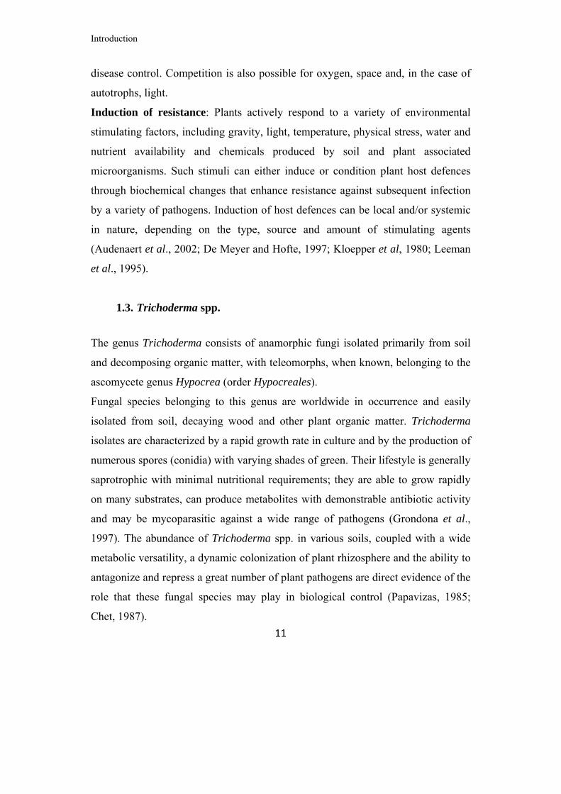
Fig. 8. Camellia cuttings (a) and Camellia plants after 1 year (b) in nursery.
3.4 Evaluation of selected Trichoderma as inoculants and growth promoters for Cupressus sempervirens and Camellia japonica
At the end of all the concurrent screenings, T. asperellum 2046 resulted to be the
most interesting isolate in terms of endophytism and plant growth promotion for
Limonium sinuatum and Cupressus sempervirens and only plant growth promotion
for Camellia japonica. In addition, T. viride 8238 showed interesting results in
Cupressus. In order to follow the effects of T. asperellum 2046 and T. viride 8238
on Cupressus plants growth and of T. asperellum 2046 on Camellia plants growth,
a further experiment was performed.
The experiment was performed by using peat added with Biomax, uninoculated or
inoculated with Trichoderma isolates.
Fungal inoculum for Cupressus, was prepared in bags containing 800 g of Biomax,
100 mL of water and 20 mL of conidial suspension (approximately 106 spore mL-
1). Inoculation of the fungus occurred 10 days before addition to peat and bags
were incubated at 24°C, photoperiod 12h/12h light/dark. After incubation, fungal
inoculum was added to the peat at the final concentration of 10% fermented
Biomax, 90% peat.
After three months from sowing, plants of Cupressus were transplanted in pots
(7x7x10 cm) with peat + Biomax inoculated or uninoculated, 15 plants for
replicate, 4 replicates for thesis. Plant height was recorded once per month.

Materials and Methods
26
In Camellia, ten days before rooted cuttings transplanting in pots, 2 ml of a
conidial suspension (approximately 106 spore mL-1) of each Trichoderma was
inoculated into the rolls of turf of each cutting. In addition, every Trichoderma
isolates was fermented on Biomax for ten days, according to the protocol described
for Cupressus. At the transplant, fermented Biomax was added to peat at the final
concentration of 10%.
Five months old Camellia cuttings (cv Margherita and cv Sea Foam) were
transplanted in 8 pots (12cm diameter) with peat + Biomax inoculated or
uninoculated, with 3 replicates for the first cv and 6 replicates for the second cv for
each thesis. Plant height and the number of leaves per plant was recorded once per
month.
At the fourth month, stomatal conductance (Gs), net photosyntesis (Pn),
transpiration (Tr), internal CO2 concentrations (Ci) and Water Use Efficiency
(WUE) were evaluated by a CIRAS equipment (Fig. 9) in both species of Camellia.
All data have been subjected to analysis of variance (Anova) to evaluate the
beneficial action of Trichoderma isolates on Cupressus and Camellia plants.
Fig. 9. Greenhouse experiments: Cupressus (a) and Camellia (b), equipment (CIRAS) for net photosynthesis measurement (c).

Materials and Methods
27
3.5 Identification of Trichoderma spp. isolates
Molecular identification of Trichoderma isolates considered interesting after the
first part of screening procedures was based on DNA sequencing of the ribosomal
ITS region. Mycelia were harvested after 2–4 days growth on PDA at 24°C and
genomic DNA was isolated using the DNeasy Plant DNA extraction kit (Qiagen
Inc., Valencia, CA, USA) following the manufacturer’s protocol.
Amplification of nuclear rDNA, containing the ITS1 and 2 and the 5.8S rRNA
gene was done using primers ITS1 (5’- TCCGTAGGTGAACCTGCGG-3’) and
ITS4 (5’- TCCTCCGCTTATTGATATGC-3’) (White et al., 1990), in a final
volume of 50 µl by mixing 2 µl of DNA with 0.5 µM of each of the primers and 25
µl of 2x PCR Master Mix (Promega, Madison, WI, USA). Amplifications were
conducted with an initial denaturation of 1 min at 94° C followed by 30 cycles of
30 sec denaturation at 94° C, 1 min primer annealing at 54°C, 1 min extension at
72° C and a final extension of 4 min at 72°C. Template DNA for sequencing was
prepared directly from PCR products with the QIAquick PCR purification kit
(Qiagen Inc.,Valencia, CA, USA). Both strands were sequenced for each isolate
using both the forward and reverse primers.
Sequence identities were determined using both the different tools available online
from the International Subcommission on Trichoderma and Hypocrea (ISTH,
www.isth.info): TrichOKEY v.2.0 based on an oligonucleotide barcode within the
ITS1 and ITS2 sequences (Druzhinina et al. 2005; Kopchinskiy et al. 2005). In
some cases, BLAST analyses were also performed from the National Centre for
Biotechnology Information (NCBI) available online.

Materials and Methods
28
3.6 Antagonistic and mycoparasitic activity of selected Trichoderma isolates by in vitro test.
T. asperellum 2046 and T. harzianum 8227, resulting to be the most interesting as
endophyte of Limonium, were submitted to antagonistic tests against Rhizoctonia
solani, Botrytis cinerea and Colletotrichum gleosporioides. Antibiosis and
mycoparasitism were evaluated on PDA (Potato Dextrose Agar, 39 gl-1, Difco) and
WA (Water Agar, agar 20 g l-1, Difco), respectively.
PDA disks of 6 mm diameter, cut from the edge of actively growing colony of each
antagonist and pathogen, were placed at the opposite sides (at 4.5 cm each other)
on PDA plates and on a sterile cellophane membrane laid on WA (Fig. 10).
Fig. 10. Confrontation plates for antagonistic tests.
Plates were incubated at 24 ± 2°C with 12 h/12 h darkness/light cycles. Radii of
each pathogen approaching (Ra) and not approaching (Rc) the colony of
antagonists and the distance between the two fungi were measured on PDA three
times a day until the two colonies came in contact. Values were used to create

Materials and Methods
29
growth curves (Sigmaplot 10) and radial growth data were submitted to analysis of
variance of regression in order to compare the slope and the elevation of curves in
presence/absence of the antagonist, assuming P < 0.05 as a significant level.
Mycoparasitism was evaluated borth on PDA and WA plates. On PDA, after 14
days, overgrowth and sporulation of the antagonists on pathogens’ colonies were
assessed. On WA plates, interaction zones and overlapping regions for each
antagonist/pathogen combination were analysed by microscopic investigations and
coilings and short loops around the host hyphae were recorded.
On the basis of its behaviour in in vivo biocontrol test (described in the following
paragraph), T. asperellum T2046 was also used in a further in vitro test, aimed to
evaluate mycoparasitic activity against sclerotia of Sclerotinia sclerotiorum and S.
minor in a 24 wells microplate. Microplates containing PDA were inoculated with
T2046 and incubated for one week at 24°C, 12h/12H light/darkness. Four sclerotia
of each pathogen were sown in each well, a row of six wells was considerate as a
replicates (24 sclerotia for replicate, 4 replicates). After 7 days (S. minor) and 14
days (S. sclerotiorum) of incubation in presence of the antagonist, firmness of
sclerotia was evaluated by pressure. Hard sclerotia were surface sterilized in an
acqueous solution of NaClO (1% active chlorine), in 50% ethanol for 5 minutes,
washed in distilled sterilized water for three times, blotter-dried and plated on PDa
in order to evaluate the ability of T2046 to internally colonize the resting stuctures
(Fig. 11).

Materials and Methods
30
Fig. 11. Sclerotia of S. sclerotiorum (a), sclerotia of S. minor (b) and microplate test (c).
3.7 Biocontrol of Sclerotinia sclerotiorum, Sclerotinia minor and Rhizoctonia solani by T. asperellum 2046 on Limonium by in vivo test
The ability of T. asperellum 2046 to reduce the attacks of Sclerotinia sclerotiorum,
Sclerotinia minor and Rhizoctonia solani to Limonium plants was evaluated.
Biomax was inoculated with a conidial suspension of T2046 spp. as previously
described and incubated for one week at 24°C, photoperiod 12h/12h light/dark. In
parallel, 450g of peat were inoculated with 0.2g of sclerotia. Fermented Biomax
was added to the peat at the final concentration of 10% and after two days
Limonium seeds were sown. Four thesis were evaluated: i) Peat + Biomax (PB) as
uninoculated control; ii) Peat + Biomax inoculated with T2046 (PB2046); iii) Peat
inoculated with Sclerotinia spp. + Biomax inoculated with T2046 (PBS2046) and
iv) Peat + Biomax, inoculated with Sclerotinia spp. (PBS). 6 replicates for each
thesis, 16 seeds for replicate were arranged (Fig. 12).
Rhizoctonia solani was inoculated on millet seed and incubated for fourteen days at
24°C, while Biomax was inoculated with a conidial suspension of T2046 spp. as
previously described. After ten days six millet seeds inoculated with R.solani were
added to the peat with fermented Biomax. Four thesis were evaluated: i) Peat +
Biomax (PB), as uninoculated control; ii) Peat + Biomax inoculated with T2046
(PB2046); iii) Peat inoculated with R. solani + Biomax inoculated with T2046

Materials and Methods
31
(PBR2046) and iv) Peat + Biomax, inoculated with R. solani (PBR). 6 replicates
for each thesis, 16 seeds for replicate were arranged.
Percentages of emergence were periodically registered until the number of
seedlings ceased in increasing (after 1 month) and submitted to statistical analysis
(ANOVA).
Fig. 12. Inoculated boxes in growth chamber (a and b).

Results
32
4. RESULTS
4.1 Screening of Trichoderma isolates as endophytes and growth promoters of Limonium, Cupressus and Camellia
- Limonium sinuatum and Cupressus sempervirens
At the end of the preliminary screening aimed to find Trichoderma spp. isolates
able to colonize, as endophytes, root apparatus and possibly stimulate plant growth,
10 out of 162 strains for Limonium and 9 out of 162 strains for Cupressus were
chosen and submitted to successive round of experiments. These isolates were
selected because of their ability to colonize 100% of internal root tissues of
inoculated plants. In the second round of screening performed on Limonium, 5
isolates (T3148, T5961, T8227, T8233 e T8245) shown percentages of
endophytism up to 95%.with T. asperellum T2046 and T. harzianum T8227 as the
most interesting. These last two isolates were chosen for a further analysis and they
resulted able to confirm 100% of root colonization after a third and a fourth
experiment, also showing interesting plant growth promotion ability (Tab. 2).
T2046 caused a statistically significant improvement in leaves number and leaf
area whereas T8227 resulted in significant improvement of leaf area (Fig. 13).
At this growth stage no significant differences in dry weight could be detected.
Tab. 2. Effects of T. asperellum T2046 and T. viride T8227 on Limonium after 1 month of growth.
Thesis Leaves number
Leaf area (cm2)
Dry weight (g)
Control 7.9b* 6.2c 4.1a T2046 9.6a 7.9a 3.7a T8227 7.5b 7.3b 3.6a *At different letters, within the same column, correspond values statistically different (ANOVA, P<0.05).

Results
33
Fig. 13. Effects of selected endophytic Trichoderma isolates on Limonium sinuatum plants. T. asperellum T2046 (a), T. harzianum T8227 (b) and uninoculated control (c). On Cupressus, among the nine selected Trichoderma isolates, six resulted to be very
interesting during the second round of selection (T8139, T8144, T8234, T8235,
T8238 and T2046) with T. asperellum T2046 and T. viride T8238 as the most
interesting for further analysis. These two Trichoderma isolates showed 100% of
endophytism in a third test. In the same experiment they resulted also able to increase
plant growth after 3 months (Tab. 3). In details, T2046 was able to significantly
improve the height of Cupressus plants whereas a small improvement, but not
statistically significant, was registered for T8238 (Fig. 14).
Tab. 3. Effect of T. asperellum T2046 and T. viride T8238 on Cupressus after 3 months of growth.
Thesis Height (cm) Control 2.2b* T2046 3.1a T8238 2.7ab *At different letters, within the same column, correspond values statistically different (ANOVA, P<0.05).

Results
34
Fig. 14. Effects of T. asperellum T2046 on Cupressus sempervirens (A). In (B) uninoculated control. Plants growth after 5 months in trays, just after transplanting in 7x7x10 pots. - Camellia japonica
On Camellia, after the first round of selection, lasted for one year, 8 isolates (T4762,
T7630, T7660, T7664, T7666, T7677, T7785 and T8111) were chosen on the basis
of their ability to improve height and number of leaves in both cultivars and
employed for a second round of screening. In this test also T. asperellum T2046 was
included, on the basis of the positive effects registered both on Limonium and
Cupressus.
After one year of cultivation, T2046 resulted to be the most interesting isolate among
those used for the second screening, causing a statistically significant improvement of
plant growth, as shown in Tab. (Fig. 15). An increase, but not statistically significant,
was registered also for leaves number.

Results
35
Tab. 4. Effect of Trichoderma on Camellia plant growth after 1 year.
Thesis Height (cm)
Leaves (number)
Control 17.7b 10a T2046 28.9a 16a *At different letters, within the same column, correspond values statistically different (ANOVA, P<0.05).
Fig. 15. Effect of T. asperellum T2046 (A and D) on stem height (a) and root development (b) of Camellia. Uninoculated control plants (B and C).
Concerning endophytic activities, any Trichoderma isolates, including T2046,
developed on the semiselective medium P190, showing no ability to persist into the
radical tissues of Camellia after one year.
A B C D
a b
A B C D
a b

Results
36
4.2 Evaluation of selected Trichoderma as inoculants of innovative substrates for Cupressus sempervirens and Camellia japonica
- Cupressus sempervirens
Data registered during the first 5 months of growth, showed that T2046 had a
positive effect on stems growth of Cupressus (Fig. 16a).
From the analysis of variance of regression on growth values, T2046 was able to
significantly improve the height of plants compared to control (Pslope 0.006). In
(Fig. 16b) the effects of T. asperellum T2046 are shown.
Different situation occurred for plants grown in soil inoculated with T8238. The
growth rate was not significantly higher than control as show by analysis of
variance of regression (Pslope=0.928 and Pelevation =0.151).
Fig. 16. Growth rate of plants grown in soil inoculated with T2046 and T8238, compared with control (a). Effect of T2046 (b) on Cupressus (A, treated; B, control).

Results
37
- Camellia japonica
The presence of T2046 in soil resulted in beneficial effects on growth of both
cultivars of Camellia, as shown in (Fig. 17). T. asperellum T2046 was able to
increase growth of cv Margherita and cv Sea Foam (Fig. 18 and Fig.19).
Fig. 17 Effect of T2046 on growth of Camellia cv Margherita (A-B) and cv. Sea Foam (C-D). Untreated controls (A-C).

Results
38
Fig. 18. Growth rate, expressed as stem height of Camellia cv Margherita (a) and cv Sea Foam (b).
Fig. 19. Increasing of leaves number of Camellia cv Margherita (a) and cv Sea Foam (b).

Results
39
Physiological parameters evaluated by CIRAS confirmed the beneficial effect of
T2046 on both Camellia cultivars. When inoculated in soil used for cv Margherita,
the antagonist was able to increase net photosynthesis (Pn) and the Water Use
Efficiency (W.U.E.) as showed in Tab. 5.
Tab. 5. Effect of T2046 on physiological parameters (cv Margherita)
Thesis Pn CI Gs Tr W.U.E.
Control 3.57 b 255.00 a 85.73 a 1.68 a 2.18 b T2046 4.52 a 257.60 a 92.60 a 1.69 a 2.90 a *At different letters, within the same column, correspond values statistically different (ANOVA, P<0.05). Pn = net photosynthesis (µmol CO2 m
-2s-1); Gs = stomatal conductance (mmol CO2 m
-2s-1); Tr = transpiration (mmol H2O m-2s-1); CI = internal co2 concentrations (ppm); W.U.E = water use efficiency (Pn/Tr, (Pn/Tr, µmolCO2/mmol H2O).
On cv Sea Foam all analyzed parameters but transpiration, showed value
significantly higher in plants grown in presence of T2046, as shown in Tab. 6.
Tab. 6. Effect of T2046 on physiological parameters (cv Sea Foam)
Thesis Pn CI Gs Tr W.U.E.
Control 2.39 b 240.53 b 48.77 b 1.07 a 2.22 b T2046 3.44 a 262.87 a 56.70 a 1.15 a 3.05 a *At different letters, within the same column, correspond values statistically different (ANOVA, P<0.05). Pn = net photosynthesis (µmol CO2 m-2s-1); Gs = stomatal conductance (mmol m-2s-1); Tr = transpiration (mmol H2O m-2s-1); CI= internal co2 concentrations (ppm); W.U.E = water use efficiency (Pn/Tr, (Pn/Tr, µmolCO2/mmol H2O).

Results
40
4.3 Identification of Trichoderma spp. isolates
With the aim of checking the taxonomic position of the Trichoderma spp. isolates
preliminary selected on the basis of the endophytic activity on Limonium,
Cupressus and Camellia, DNA sequencing of the ribosomal ITS region was
performed. Amplified fragments of almost 600 bp (Fig. 20), obtained by PCR,
were purified and submitted to sequencing and resulting sequences were compared
with those deposited in databases (Genebank and Trichokey). In Tab. 7 results
from molecular identification are reported.
Fig. 20. Amplified ITS fragments of Trichoderma spp. obtained by PCR.
- 1000 bp- 750 bp- 500 bp
- 1000 bp- 750 bp- 500 bp

Results
41
Tab. 7. Molecular identification, based on ITS sequences, of Trichoderma isolates preliminary selected for endophytic abilities on Limonium, Camellia and Cupressus.
Isolate Morphological
Identification
Molecular
Identification
T8233 T. atroviride H. atroviridis/ T. atroviride
T8227 T. harzianum H. lixii/ T. harzianum
T3148 T. harzianum H. lixii/ T. harzianum
T8245 NI Clade XII
T5961 T. harzianum H. lixii/ T. harzianum
T8139 T. atroviride H. atroviridis/ T. atroviride
T8235 T. atroviride H. atroviridis/ T. atroviride
T8234 T. harzianum H. lixii/ T. harzianum
T8144 T. viride H. rufa/ T. viride
T8238 T. viride H. rufa/ T. viride
T8111 T. viride H. rufa/ T. viride
T7785 NI T. asperellum
T7677 T. viride H. rufa/ T. viride
T7666 T. viride H. rufa/ T. viride
T7664 T. viride H. rufa/ T. viride
T7660 NI T. asperellum
T7630 T. viride H. rufa/ T. viride
T4762 T. crassum H. crassa/ T. crassum
T2046 NI T. asperellum

Results
42
4.4 Antagonistic and mycoparasitic activity of selected Trichoderma isolates by in vitro tests
Antagonistic tests were performed in order to investigate the ability of T2046 and
T8227 isolates to in vitro inhibit growth of R. solani, B. cinerea and C.
gleospoiroides, potential pathogens of Limonium. Growth rates of each
pathogen/antagonist combination were submitted to regression analysis in order to
compare slope and elevation of pathogen’s growth curves in presence/absence of
the antagonist. All growth slopes resulted to be highly significant (R2>0.96,
P<0.0001).
Comparison of slopes shown that T2046 was not able to statistically reduce growth
rate of any pathogens (Pslope >0.069), whereas significant P elevation resulted from
all combinations, suggesting a delay in starting the exponential growth phase by the
pathogens in presence of T2046 (Fig. 21).
When growth curves of the pathogens grown in presence of T8227 were submitted
to regression analysis, a similar behaviour emerged. The isolate was not able to
inhibit any pathogen, as shown by P slope values (Pslope >0.118). A highly
significant P elevation resulted only for Colletotrichum (P=0.000), underlining a
delay in starting the exponential growth phase for this pathogen (Fig. 22).
The lack of significance in slope differences suggests the exclusion of the
involvement of antibiotic diffusible compounds as potential biocontrol mechanism
by T. asperellum T2046 and T. harzianum T8227 towards the three pathogens.

Results
43
Fig. 21. Growth curves of R. solani (a), B. cinerea (b) and C. gleospoiroides (c) in presence/absence of T. asperellum T2046.
Time (h)
0 10 20 30 40 50 60 70
Ra
dial
gro
wth
(m
m)
0
5
10
15
20
25
30
R. solaniR. solani vs T2046
Time (h)
0 10 20 30 40 50 60 70
Rad
ial g
row
th (
mm
)
0
5
10
15
20
25
30
B. cinereaB. cinerea vs T2046
Time (h)
0 10 20 30 40 50 60 70
Rad
ial g
row
th (
mm
)
0
5
10
15
20
25
30
C. gleospoiroides C. gleosporioides vs T2046
Pslope=0.322Pelev =0.046
Pslope=0.069Pelev =0.000
Pslope=0.195Pelev =0.004
a
b
c
Time (h)
0 10 20 30 40 50 60 70
Ra
dial
gro
wth
(m
m)
0
5
10
15
20
25
30
R. solaniR. solani vs T2046
Time (h)
0 10 20 30 40 50 60 70
Rad
ial g
row
th (
mm
)
0
5
10
15
20
25
30
B. cinereaB. cinerea vs T2046
Time (h)
0 10 20 30 40 50 60 70
Rad
ial g
row
th (
mm
)
0
5
10
15
20
25
30
C. gleospoiroides C. gleosporioides vs T2046
Pslope=0.322Pelev =0.046
Pslope=0.069Pelev =0.000
Pslope=0.195Pelev =0.004
a
b
c

Results
44
Fig. 22. Growth curves of R. solani (a), B. cinerea (b) and C. gleospoiroides (c) in presence/absence of T. harzianum T8227.
Time (h)
0 10 20 30 40 50 60 70
Rad
ial g
row
th (
mm
)
0
5
10
15
20
25
30
R. solaniR. solani vs T8227
Time (h)
0 10 20 30 40 50 60 70
Rad
ial g
row
th (
mm
)
0
5
10
15
20
25
30
B. cinereaB. cinerea vs T8227
Time (h)
0 10 20 30 40 50 60 70
Rad
ial g
row
th (
mm
)
0
5
10
15
20
25
30
C. gleosporioidesC. gleosporioides vs T8227 Pslope=0.118
Pelev =0.000
Pslope=0.124Pelev =0.112
Pslope=0.554Pelev =0.162
a
b
c
Time (h)
0 10 20 30 40 50 60 70
Rad
ial g
row
th (
mm
)
0
5
10
15
20
25
30
R. solaniR. solani vs T8227
Time (h)
0 10 20 30 40 50 60 70
Rad
ial g
row
th (
mm
)
0
5
10
15
20
25
30
B. cinereaB. cinerea vs T8227
Time (h)
0 10 20 30 40 50 60 70
Rad
ial g
row
th (
mm
)
0
5
10
15
20
25
30
C. gleosporioidesC. gleosporioides vs T8227 Pslope=0.118
Pelev =0.000
Pslope=0.124Pelev =0.112
Pslope=0.554Pelev =0.162
a
b
c

Results
45
Mycoparasitic ability of T2046 and T8227 was expressed as the ability to produce
coilings around pathogens hyphae. Both Trichoderma isolates succeeded in coiling
R. solani hyphae, whereas no coilings were detected against the other two
pathogens (Fig. 23).
Fig. 23. Coilings of T. asperellum T8227 around R. solani hyphae.
Overgrowth and sporulation, on PDA, of the antagonists on pathogens’ colonies
were evaluated as further signs of mycoparasitism: T2046 was able to grow and
sporulate over R. solani, B. cinerea and C. gleospoiroides colonies whereas T8227
was able to overgrow and sporulate profusely on R. solani and B. cinerea and
scarcely on C. gloeosporioides colony (Fig.24).

Results
46
Fig. 24. Overgrowth and sporulation of T. asperellum T2046 and T. harzianum T8227 on R. solani, B. cinerea and C. gloeosporioides.
When evaluated for its mycoparasitic activity on sclerotia of S. sclerotiorum and S.
minor, T2046 has was able to degrade 100% of resting structures of both the
pathogen (Fig. 25), providing evidence that the mycoparassitism of these quiescent
structures could be one of the mechanisms for the control of disease incidence (as
shown in the next paragraph) on Limonium.
T8227
R. solani B. cinerea C. gleospoiroides
T2046

Results
47
Fig. 25. Microplates test on S. minor sclerotia (a) and valutation of sclerotia after 1 week of incubation (b).
4.5 Effects of T. asperellum 2046 on Sclerotinia sclerotiorum, Sclerotinia minor and Rhizoctonia solani by in vivo test
Results obtained by in vivo biocontrol test on Limonium, showed that T.
asperellum T2046 had a significant effect on Sclerotinia sclerotiorum and
Sclerotinia minor (Tab. 8). When in presence of Sclerotinia sclerotiorum in soil
T2046 was able to increase the percentage of emergence of Limonium of almost
60% (Fig. 26). When alone S. sclerotiorum caused the death of all seedlings. The
same trend occurred in presence of Sclerotinia minor where T2046 was able to
increase the percentage of emerged seedlings of more than 30%. In presence of this
pathogen alone, only 20% of emergence occurred. The effect of T2046 on
Limonium was also confirmed in soil inoculated only with the beneficial fungus
that allowed to obtain significant higher emergence of plants.
T. asperellum T2046 had a significant effect also on Rhizoctonia solani (Tab. 9).
When in presence of R. solani in soil T2046 was able to increase the percentage of
emergence of Limonium of almost 40% (Fig. 27). When alone R. solani showed
ba

Results
48
only 21% of emergence of Limonium. Also in this test, the effect of T2046 on
Limonium was confirmed, in soil inoculated only with the beneficial fungus.
Tab. 8. Biocontrol of S. sclerotiorum and S. minor by T2046
Thesis % emergence
S. sclerotiorum S. minor
control
PB2046 PBS2046 PBS
63.47a
68.67a 61.57a 0.00b
62.52 a
62.63 a 54.33 a 19.24 b
*At different letters, within the same column, correspond values statistically different (ANOVA, P<0.05). PB2046 = soil inoculated with T2046; PBS2046 = soil inoculated with T2046 and Sclerotinia spp.; PBS = soil inoculated with Sclerotinia spp.
Fig. 26. Effect of T2046 on S. sclerotiorum on Limonium. Uninoculated soil used as control (a), soil inoculated with T2026 (b), soil inoculated with T2046 and Sclerotinia minor (c) and soil inoculated with S. minor (d).
a b c d

Results
49
Tab. 9. Biocontrol of R.solani by T2046
Thesis % emergence
R.solani
control PB2046 PBR2046 PBR
72.64a
73.47a
38.92b
21.32c
*At different letters, within the same column, correspond values statistically different (ANOVA, P<0.05). PB2046 = soil inoculated with T2046; PBR2046 = soil inoculated with T2046 and R.solani; PBR = soil inoculated with R.solani
Fig. 27. Effect of T2046 on R. solani on Limonium. Uninoculated soil used as control (a), soil inoculated with T2026 (b), soil inoculated with T2046 and R. solani (c) and soil inoculated with R. solani (d).
a b c d

Discussion
50
5. DISCUSSION
Peat is actually the most basic component of substrates used in nursery, expecially
for ornamental plants. For some crops peat is used as it is while, in most cases, it is
employed in mixtures with other components. Peats are identified as those
materials containing more or less decomposed plant residues, with an ash content
below 10%. It is worldwide collected from natural deposits called peat lands. The
deeper layers of such deposits are ten thousand years old and can be assigned to the
late glacial or post-glacial period, while commonly used peat lands are almost one
thousand years old. Due to its characteristics (homogeneity, high water absorption
capacity, good aeration, structural stability, a limited nutrient content, pH around 3)
sphagnum peat represents the starting material more frequently utilised for the
production of substrates (D’Angelo et al., 1992).
In recent years, the price has increased as a consequence of the higher costs of
energy for extraction and transport from the producing Countries in Northern
Europe or Canada (Rea, 2005). In addition, there is an increasing demand for “Peat
free” substrates as a result of the environmentalist campaign against the
exploitation of peat lands because of the natural and archaeological value of these
areas and the fact that peat is not a renewable resource (Armstrong, 2004).
It should be also noted that, in 2001, the European Commission excluded all
substrates containing peat from the release of the Community Eco-Label Mark.
In many Countries such as Holland (Armstrong, 2004) and also in our country
(Project PROBIORN of Arsia, 2004-06; Project FLORPRO of M.i.P.a.a.f., 2007-
10; Project SUBARTIFLOR of M.i.P.a.a.f., 2009-12) projects to find materials
alternatives to peat, that combine low cost with optimal physical, chemical and
biological properties, have been proposed.

Discussion
51
In this optic, marketing investigation underlined the increasing demand of
alternative substrates that could solve all these questions, mostly the reduction of
peat amount, making new substrates available for ornamental field.
The research activity of the present PhD thesis has been focussed within the wider
project “SUBARTIFLOR: Messa a punto di substrati artificiali innovativi per il
florovivaismo” project funded by the Italian Ministry of Agricultural, Food and
Forestry Policies (MIPAAF). The main aim was to develop an innovative,
economical and suitable substrate alternative to peat for cultivation of seed plants
(Limonium sinuatum and Cupressus sempervirens) and of acidophilus species
(Camellia japonica) of ornamental interest.
The first step for reaching the prefixed scope was to select beneficial fungi
belonging to Trichoderma genus to be add as soil inoculants, in order to give an
added value to the new substrates but, at the same time, maintaining an ecofriendly
approach. Our screening started from a large collection of Trichoderma spp.
isolates, belonging to at least 20 different species and isolated from a wide variety
of environment spread over different geographic origins, such as agricultural soil,
natural parks soil, desert sand, peat, compost, plant parts, seeds, decaying organic
matter, animal pellets, tree bark or unusual substrates as Chernobyl sarcophagus,
ant nest or mummy skin, from Europe (largest part), North Africa, North and South
America, Middle and Far East, Australia and New Zealand, mostly from temperate
regions. The choice to start from this large collection derived also by the
knowledge that there is a great diversity of useful characters in this fungal genus,
and efficient biological control agents or endophytic plant symbionts are usually
selected among many, sometimes, hundred or thousands, less active wild strains, as
recognized by studies on rhizosphere competence. In fact, most of the
investigations have been conducted with elite strains extensively tested for their
efficacy in the lab and field. In addition, even selected strains often fully express

Discussion
52
their beneficial effects (e.g., disease control, abiotic stress resistance, etc.) only on
plants under stress conditions (Lorito et al., 2010).
In the present work Trichoderma spp. isolates were tested on three completely
different host plants, two seed species as Limonium sinuatum and Cupressus
sempervirens and an acidophilic plant, Camellia japonica, cultivated starting from
cuttings. Despite of this wide variety of Trichoderma isolates and physiological
diversity of host plants, at the end of the concurrent selection, started from 162
isolates for Limonium and Cupressus and 202 for Camellia, only one, T.
asperellum T2046 was able to positively affect growth of all the tested plants.
Trichoderma spp. are known for mutualistic relationships with plants (Harman et al.,
2004) and many biocontrol strains can interact intimately with roots, colonizing the
outer epidermis layers and acting as opportunistic, avirulent plant symbionts (Shoresh
et al, 2010;). In the few cases that have been examined thoroughly, Trichoderma
isolates colonize root surfaces sometimes with morphological features reminiscent
of those seen during mycoparasitism and hyphae invade the root epidermis
(Harman et al., 2004). Some Trichoderma strains can colonize only local sites on
roots, but rhizosphere-competent strains colonize entire root surfaces for several
weeks or months (Harman et. al, 2004).
An association between high Trichoderma populations and plant growth promotion
was indicated for Trichoderma harzianum treated petunias and chrysanthemums,
plants of ornamental interest (Chang et. al., 1986). However, the study did not
specifically examine the rhizosphere and also did not consider the temporal
dynamics of T. harzianum. Other studies related qualitative observations of
Trichoderma root colonisation and penetration to plant growth promotion (Chacòn
et al., 2007) and biocontrol activity (Miranda et al., 2006). Similarly, a significant
correlation between Trichoderma population levels in the rhizosphere and its
ability to antagonise Sclerotium rolfsii and improve plant growth was determined
for axenically grown tomato plants (Tsahouridou and& Thanassoulopoulos, 2002).

Discussion
53
No information about endophytic ability on Camellia, Cupressus and Limonium, to
the best of our knowledge, are available. Several species of Trichoderma (including
T. hamatum) have been shown to elicit promoting effects on different perennial
woody plants (including Pinus radiata and Pinus sylvestris (Hohmann et al., 2011).
However the distribution and the fate of these isolates in the root systems were not
examined and there have been no reports on the association between Trichoderma
root colonization and plant growth performance on Cupressus sempervirens.
When our best Trichoderma isolates, particularly T2046, were analyzed for the ability
to colonize roots, they have been collected from radical portions after a relative long
time from inoculation in soil, i.e. one month for Limonium and three months for
Cupressus but, when inoculated in soil for Camellia cultivation, it was not possible to
collect Trichoderma from roots after one year. This could depend by the difficult to
persist for a long time in this kind of plants or they were present but our isolating
approach, a casual sampling as for Camellia roots, underestimated the endophytic
presence of T2046. In agriculture, where the use of products based on Trichoderma
is now common, best results, in terms of biocontrol, have been obtained when
periodic inoculation of the fungus in soil have been done (Perez- Pivat and Arcia,
2008; Rabeendran et al., 2006). Anyway, also after one year, the positive action of
our T. asperellum T2046 was still evident on Camellia, allowing to suppose a long-
term effect on plant.
It’s known that an effective bio-inoculant should penetrate the roots not only to
directly antagonise root pathogens, but also to stimulate plant growth and vigour
through various mechanisms such as nutrient mobilization, nitrogen use efficiency in
crops, induction of host defence as well as the involvement of growth phytormones
from both plant and fungal origins (Altomare, 1999; Vinale et al., 2008). Some
studies performed on cultivars of corn and tomato showed that the different species
of Trichoderma are able to increase seed germination and development of epigeal
and hypogeal part of the plant (Windham et al., 1986; Harman and

Discussion
54
Bjorkman,1997).These energy-requiring processes, along with improved growth,
stimulate plant respiration and thus enhance photosynthesis or photosynthetic
efficiency (Shoresh et al., 2010). These fungi are considered to act as full symbionts:
they receive nutrients from the plant (root exudates) and a protected niche to colonize,
while providing to the host improved nutrients uptake and stress (biotic and abiotic)
protection. In addition to the well known ability to colonize roots and stimulate
growth and defence of plant, Trichoderma have also been applied to fruit, flowers
and foliage and plant diseases can be controlled by their application to any of these
sites (Lorito et al., 2010).
This beneficial effects were observed in all of the three ornamental species we used.
The Trichoderma isolates that have been selected, in particular T2046, on
Limonium, Cupressus and Camellia showed their effects as higher stems, a
significant increase of number of leaves, a higher percentage of germination.
Moreover, photosynthetic parameters have been evaluated in the two cultivars of
Camellia, and they resulted to be improved in plants grown in soil inoculated with
T2046.
Almost 80 years ago, it was discovered that Trichoderma spp. have the ability to
attack and control plant pathogenic fungi. Studies on the antagonistic mechanisms
of Trichoderma demonstrated the involvement of many hydrolytic enzymes with
chitinases playing an important role (Seidl, 2008; Gruber et al., 2010, Matarese et
al., 2012)., also capable of acting synergically with highly fungitoxic antibiotics,
and a complex system for fungal prey detection coupled with competition for space
and substrates (Howell, 2003; Harman, 2006; Lorito et al., 2010). Recent
comparison of the genome of T. virens and T. atroviride with T. reesei, a
saprotrophic species, suggested that the ancestral state of Hypocrea/Trichoderma
was mycoparasitic (Kubicek et al. 2011), supporting an earlier speculation
(Rossmann et al., 1999) that the ancestors of Trichoderma were mycoparasites on
wood-degrading basidiomycetes and acquired saprotrophic characters to follow

Discussion
55
their prey into their substrate. Parasitism by Trichoderma species has been reported
against pathogens such as Rhizoctonia solani as well as Sclerotinia spp. and
Sclerotium spp. Sclerotium rolfsii, together with Sclerotinia sclerotiorum and
Sclerotinia minor, represents one of the most destructive pathogen of many
economically important crops (Viterbo et al., 2007).
Due to the promising and interesting performance of T2046 as soil inoculants of
potential new substrates for ornamental use, we decided to test this isolate against
some important pathogens of Limonium, both by in vitro and in vivo test. Results
obtained from in vitro tests suggested that this isolate is able to parasitize hyphae of
Rhizoctonia solani and to colonize sclerotia of Sclerotinia sclerotiorum and S.
minor, causing decay. That’s not a surprise as mycoparasitism against S.
sclerotiorum and S. rolfsii is widespread within the genus Trichoderma
(Tsahouridou and Thanassoupolus, 2001; Sarrocco et al., 2004). Some species of
Trichoderma, after penetrating the rind, can colonize the inner layers of sclerotia,
often completely destroying them or rendering them not viable. The ability to
degrade melanin, the main constituent of the external rind, may be an important
trait for these mycoparasitic fungi (Goméz and Nosanchuk 2003; Sarrocco et al.,
2006) and laccase would be considered the most likely candidates for melanin
degrading enzymes (Giardina et al., 2010; Catalano et al., 2011). Moreover, the
extraordinary ability of Trichoderma to attack these resistant, resting structures is
related to the synergistic action of lytic enzymes, including proteases, lipases,
glucanases and chitinases (Benhamou and Chet, 1996).
The interesting antagonistic performance of T. asperellum T2046, coupled with the
ability to endophytically colonize roots and to stimulate growth of plant host, allow
us to consider this Trichoderma isolate as a good candidate for bioactive
ingredients for innovative substrate to be employed for germination and growing of
ornamental plants.

Index
56
Index of pictures Fig. 1. Flowers of Limonium sinuatum (a), Flower of Camellia Japonica (b), greenhouse cultivation of Cupressus sempervirens (c). 17 Fig. 2. Morphological aspect of Trichoderma spp. in the environment (a) and on agar plate (b). 19 Fig. 3. Fungal inoculum (a), fermentation of inoculated Biomax (b) and mix of peat (90%) and Biomax (10%). 20 Fig. 4. Plateaus in a growth chamber (a) and Cupressus seeds germination (b). 21 Fig. 5. Wash of roots (a), sterilization (b) and development of Trichoderma spp. Isolates from plated radical portions (c). 22 Fig. 6. Plateaus of Cupressus (a) and automatic seeder (b). 22 Fig. 7. Inoculation of conidial suspension into the roll of turf (a) and transplanting of Camellia into peat + inoculated Biomax (b). 24 Fig. 8. Camellia cuttings (a) and Camellia plants after 1 year (b) in nursery. 25 Fig. 9. Greenhouse experiments: Cupressus (a) and Camellia (b), equipment (CIRAS) for net photosynthesis measurement (c). 26 Fig. 10. Confrontation plates for antagonistic tests. 28 Fig. 11. Sclerotia of S. sclerotiorum (a), sclerotia of S. minor (b) and microplate test (c). 30 Fig. 12. Inoculated boxes in growth chamber (a and b). 31
Fig. 13. Effects of selected endophytic Trichoderma isolates on Limonium sinuatum plants. T. asperellum T2046 (a), T. harzianum T8227 (b) and uninoculated control (c). 33

Index
57
Fig. 14. Effects of T. asperellum T2046 on Cupressus sempervirens (A). In (B) uninoculated control. Plants growth after 5 months in trays, just after transplanting in 7x7x10 pots. 34
Fig. 15. Effect of T. asperellum T2046 (A and D) on stem height (a) and root development (b) of Camellia. Uninoculated control plants (B and C). 35 Fig. 16. Growth rate of plants grown in soil inoculated with T2046 and T8238, compared with control (a). Effect of T2046 (b) on Cupressus 36 Fig. 17. Effect of T2046 on growth of Camellia cv Margherita (a-B) and cv. Sea Foam (b-B). Untreated controls (A-C). 37 Fig. 18. Growth rate, expressed as stem height of Camellia cv Margherita (a) and cv Sea Foam (b). 38 Fig. 19. Increasing of leaves number of Camellia cv Margherita (a) and cv Sea Foam (b). 38 Fig. 20. Amplified ITS fragments of Trichoderma spp. obtained by PCR. 40 Fig. 21. Growth curves of R. solani (a), B. cinerea (b) and C. gleospoiroides (c) in presence/absence of T. asperellum T2046. 43 Fig. 22. Growth curves of R. solani (a), B. cinerea (b) and C. gleospoiroides (c) in presence/absence of T. harzianum T8227. 44 Fig. 23. Coilings of T. asperellum T8227 around R. solani hyphae. 45 Fig. 24. Overgrowth and sporulation of T. asperellum T2046 and T. harzianum T8227 on R. solani, B. cinerea and C. gloeosporioides. 46 Fig. 25. Microplates test on S. minor sclerotia (a) and valutation of sclerotia after 1 week of incubation (b). 47 Fig. 26. Effect of T2046 on S. sclerotiorum on Limonium. Uninoculated soil used as control (a), soil inoculated with T2026 (b), soil inoculated with T2046 and Sclerotinia minor (c) and soil inoculated with S. minor (d). 48

Index
58
Fig. 27. Effect of T2046 on R. solani on Limonium. Uninoculated soil used as control (a), soil inoculated with T2026 (b), soil inoculated with T2046 and R. solani (c) and soil inoculated with R. solani (d). 49
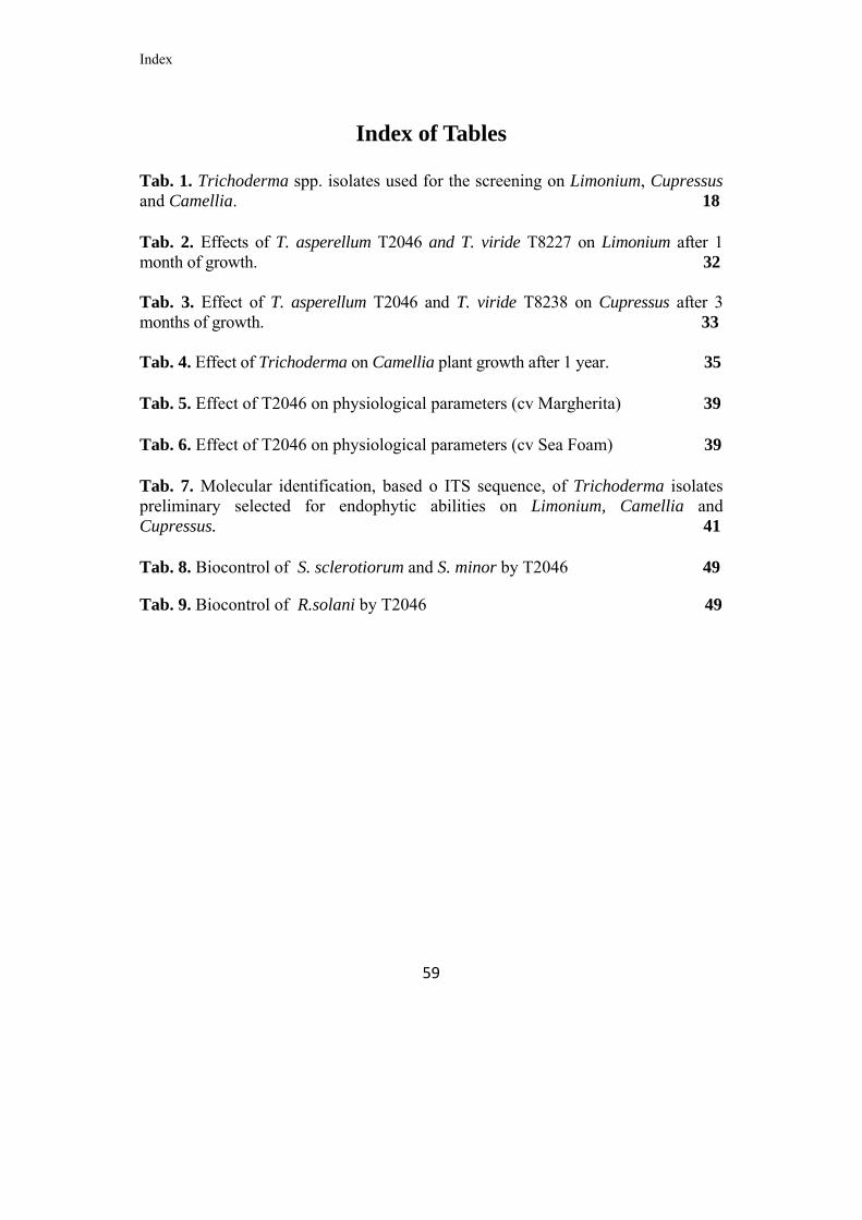
Index
59
Index of Tables Tab. 1. Trichoderma spp. isolates used for the screening on Limonium, Cupressus and Camellia. 18 Tab. 2. Effects of T. asperellum T2046 and T. viride T8227 on Limonium after 1 month of growth. 32 Tab. 3. Effect of T. asperellum T2046 and T. viride T8238 on Cupressus after 3 months of growth. 33 Tab. 4. Effect of Trichoderma on Camellia plant growth after 1 year. 35 Tab. 5. Effect of T2046 on physiological parameters (cv Margherita) 39 Tab. 6. Effect of T2046 on physiological parameters (cv Sea Foam) 39 Tab. 7. Molecular identification, based o ITS sequence, of Trichoderma isolates preliminary selected for endophytic abilities on Limonium, Camellia and Cupressus. 41 Tab. 8. Biocontrol of S. sclerotiorum and S. minor by T2046 49
Tab. 9. Biocontrol of R.solani by T2046 49

References
60
References
Altomare C., Norvell W.A., Biork.man T., Harman G.E., (1999). Solubilization of
phosphates and micronutrients by the plant-growth-promoting and biocontrol
fungus Trichoderma harzianum. Rifai 1295-22. Appl. Environ. Microbiol. 65:
2926-2933.
Armstrong H., (2004). No response tbio-alternative for peat. FlowerTech, 7:24-25
Audenaert K., Pattery T., Cornelis P., Höfte M., (2002). Induction of systemic
resistance to Botrytis cinerea in tomato by Pseudomonas aeruginosa 7NSK2: role
of salicylic acid, pyochelin and pyocyanin. Molecular Plant- Microbe Interactions
15: 1147-1156.
Bacon C.W., Hinton, D.M., (1996). Symptomless endophytic colonization of
maize by Fusarium moniliforme. Canadian Journal of Botany, 74: 1195-1202.
Bayman P., (2007). Fungal Endophytes In:. Environmental and Microbial
Relationships, The Mycota, Vol.4, Part 3, 213-227.
Bijirimana J., (1997). Induction of systemic resistance on bean (Phaseolus
vulgaris) by Trichoderma harzianum. Med. Fac. Landbouww. Univ. Gent. 62:
1001-1007.
Cabral D., Stone J.K., Carroll G.C., (1993). The internal mycobiota of Juncus
spp.: microscopic and cultural observations of infection patterns. Mycological
Research, 97: 367-376.

References
61
Carbone I., Kohn L.M., (1999). A method for designing primer sets for speciation
studies in filamentous ascomycetes. Mycologia 91: 553-556.
Carroll, G.C., (1991). Fungal associates of woody plants as insect antagonist in
leaves and stems. In P. Barbosa, V.A. Krischik, and C.G. Jones (eds.), Microbial
Mediation of Plant-herbivore Interactions. Wiley & Sons, New York, pp. 253-271.
Carsolio C., Benhamou N., Haran S., Cortés C., Gutiérrez A., Chet I., Herrera-
Estrella A., (1999). Role of Trichoderma harzianum Endochitinase Gene ech 42 in
Mycoparasitism. Applied andEnvironmental Microbiology 65: 929-935.
Catalano V., Vergara M., Hauzenberger J.R., Seiboth B., Sarrocco S., Vannacci
G., Kubicek C.P., Seidl-Seiboth V., (2011). Use of a nonhomologous end joining
deficient strain (delta-ku70) of the biocontrol fungus Trichoderma virens to
investigate the function of the laccase gene lcc1 in sclerotia degradation. Current
Genetics 57:13-23.
Chacòn MR, Rodriguez-Galan O., Benitez T., Sousa S., Rey M., Llobell A.,
Delgado-jarana J., (2007). Microscopic and transcriptome analyses of early
colonization of tomato roots by Trichoderma harzianum. International
Microbiology 10:19-27.
Chang YC, Baker R., Kleifeld O., Chet I., (1986). increased growth of plants in
the presence of the biological control agent Trichoderma harzianum. Plant
Disease 70:145-148.

References
62
Chet I., (1987). Trichoderma application, mode of action and potential as
biocontrol agent of soilborne plant pathogenic fungi. In: I. Chet (ed.). Innovative
Approaches to Plant Disease Control. Wiley, New York, 137-160.
Clay. K., (1990). Fungal endophytes of grasses. Ann. Rev. Ecol. Syst. 21: 275-
295.
Cook R.J., Baker K.F., (1983) The nature and practice of biological control of
plant pathogens. American Phytopathological Society, St Paul, MN, 539
Cook R.J., (1993) Making greater use of microbial inoculants in agriculture.
Annual Review of Phytopathology 31: 53-80.
D’Angelo G., Castelnuovo M., Galli A., Valagussa M., (1992). Relations between
physical and chemical properties of the substrate and growth of some pot
ormanentals, Acta Horticulturae (ISHS) 342: 313-324
Deckert R.J., Melville L.H., Peterson R.L., (2001). Structural features of a
Lophodermium endophyte during the cryptic life-cycle phase in the foliage of
Pinus strobus. Mycological Research, 105(8): 991-997.
De Meyer G., Hofte M., (1997). Salicylic acid produced by the rhizobacterium
Pseudomonas aeruginosa 7NSK2 induces resistance to leaf infection by Botrytis
cinera on bean. Phytopathology 87: 588-593.
Dennis C., Webster J., (1971a). Antagonistic properties of species groups of
Trichoderma. I. Production of non-volatile antibiotics. Transactions of the British
Mycological Society 57: 25-39.

References
63
Dennis C., Webster J., (1971b). Antagonistic properties of species groups
ofTrichoderma. II. Production of volatile antibiotics. Transactions of the British
Mycological Society 57: 41- 48.
Druzhinina I.S., Kopchinskiy A.G., Komon M., Bissett J., Szakacs G., Kubicek
C.P., (2005). An oligonucleotide barcode for species identification in Trichoderma
and Hypocrea. Fungal Genetics and Biology 42: 813-828.
Elad Y., Baker R., (1985). Influence of trace amounts of cations and
siderophoreproducing pseudomonads on chlamydiospore germination of Fusarium
oxysporum. Phytopathology 75: 1047-1052.
Faull J.L., (1988). Competitive antagonism of soilborne plant pathogens. In: Fungi
in Biological control systems. Edited by M. N. Burge. Manchester University .
Fisher PJ, Petrini O., (1987). Location of fungal endophytes in tissues of Suaeda
fruticosa: a preliminary study. Trans Br Mycol Soc 89:246–249.
doi:10.1016/S0007-1536(87) 80161-4.
Fisher P.J., Petrini O., (1990). A comparative study of fungal endophytes in xylem
and bark of Alnus species in England and Switzerland. Mycological Research, 94:
313-319.
Germida J.J., Siciliano S.D., de Freitas J.R., Seib A.M., (1998). Diversity of root
associated bacteria associated with field-grown canola (Brassica napus L.) and
wheat (Triticum aestivum L.). FEMS Microbiology Ecology, 26:43-50.

References
64
Ghisalberti E.L., Sivasithamparam K., (1991) Antifungal antibiotics produced by
Trichoderma spp. Soil Biology & Biochemistry 23: 1011-1020.
Giardina P., Faraco V., Pezzella C., Piscitelli A., Vanhulle S., Sannia G., (2010).
Laccases: a never-ending story. Cell Molecular Life Science 67:369-385.
Gomez, BL., Nosanchuk J.D., (2003). Melanin and fungi. Current Opinion in
Infectious Diseases 16(2): 91-96.
Graniti A., (2002). L’endofitismo nei funghi: un adattamento ecologico o un modo
di vita?. Atti del Convegno “L’endofitismo di funghi e batteri patogeni in piante
arboree e arbustive”, A. Franceschini e F. Marras (Ed.), Sassari- Tempio Pausania,
19-21 maggio 2002: 147-156.
Grondona I., Hermosa R.,Tejada M., Gomis M.D.,Mateos P.F., Bridge P.D.,Monte
E., Garcia-Acha I., (1997). Physiological and biochemical characterisation of
Trichoderma harzianum, abiological control agent against soilborne fungal plant
pathogens. Applied and Environmental Microbiology 63: 3189-3198.
Gruber S., Vaaje-Kolstad G., Matarese F., López-Mondéjar R., Kubicek C.P. and
Seidl-Seiboth V., (2010). Analysis of subgroup C of fungal chitinases containing
chitin-binding and LysM modules in the mycoparasite Trichoderma atroviride.
Glycobiology 21, 122-133.
Gutierrez-Zamora M.L., Martinez-Romero E., (2001). Natural endophytic
associazion between Rhizobium etli and maize (Zea mays L.). J. Biotechnol., 91:
117-126.

References
65
Harman G.E. and Bjorkman T., (1997). Potential and existing uses of
Trichodermaand Gliocladium for plant disease control and plant growth
enhancement. InTrichoderma and Gliocladium Vol 2, Harman, G.E. and Kubicek,
C.P. eds.,London: Taylor & Francis, pp 229-261.
Harman G.E. (2000). Myths and dogmas of biocontrol. Changes in perceptions
derived from research on Trichoderma Harzianum T22. Plant Dis. 84: 377-393.
Harman G.E., Petzoldt R., Comis A., Chen J., (2004). Interaction between
Trichoderma harzianum strain T22 and maize inbred line Mo 17 and effect of this
interaction on disease caused by Phitium ultimum and Colletotrichum
graminicola. Phytopathology. 94:147-153.
Hoff J.A., Klopfenstein N.B., McDonald G.I., Tonn J.R., Kim M.S., Zambino P.J.,
Hessburg P.F., Rogers J.D., Peever T.L., Carris L.M. 2004. Fungal endophytes in
woody roots of Douglas-fir (Pseudotsuga menziesii) and ponderosa pine (Pinus
ponderosa). Forest Pathology, 34: 255-271.
Homma Y., Sato Z., Hirayama F., Konno K., Shirahama H., Suzui T., (1989).
Production of antibiotic by Pseudomonas cepacia as an agent for biological
control of soil borne pathogens. Soil Biology and Biochemistry 21: 723-728.
Howell C.R., Stipanovic R.D., (1980). Suppression of Pythium ultimum induced
damping-off of cotton seedlings by Pseudomonas fluorescens and its antibiotic
pyoluteorin. Phytopathology 70: 712-715.

References
66
Howell C.R., (2003). Mechanisms employed by Trichoderma species in the
biological control of plant disease: the history and evolution of current concepts.
Plant Disease 87: 4-10.
Hurtado, O., (2004). Study and manipulation of the salicylic acid-dependent
defense pathway inplants parasitized by Orobanche aegyptiaca Pers. Master of
Sciences Thesis, Plant Physiology, Virginia Polytechnic Institute and State
University, USA.
Islam M.T., Yasuyuki H., Abhinandan D., Toshiaki I., Satoshi T., (2005).
Suppression of damping-off-disease in host plants by the rhizoplane bacterium
Lysobacter sp. strain SB-K88 is linked to plant colonization and antibiosis against
soilborne peronosporomycetes. Applied and Environmental Microbiology 71:
3786-3796.
Jeffries P., (1997). Mycoparasitism. In: The mycota IV, Environmental and
Microbial Relationships Wicklow/Soderstrom (Eds.) Springer-Verlag Berlin
Heidelberg. pp. 149-163
Kubicek C.P., Herrera-Hestrella A., Seidl-Seiboth V., Martinez D.A., Druzhinina
I.S., Thon M., Zeilinger S., et al., (2011). Comparative genome sequence analysis
underscores mycoparasitism as ancestral life style of Trichoderma. Genome and
Biology 12:R40.
Leeman M., van Pelt J.A., den Ouden F.M., Heinsbroek M., Bakker P.A.H.M.,
Schippers B., (1995). Induction of systemic resistance by Pseudomonas
fluorescens in radish cultivars differing in susceptibility to Fusarium wilt, using
novel bioassay. European Journal of Plant Pathology 101: 655-664.

References
67
Lo CT, Nelson EB, Hayes CK, Harman GE, (1998). Ecological studies of
transformed Trichoderma harzianum strain 1295-22 in the rhizosphere and on the
phylloplane of creeping bentgrass. Phytopathology 88:129-136.
Loper J.E., Buyer J.S., (1991). Siderophores in microbial interactions of plant
surfaces. Molecular Plant-Microbe Interactions 4: 5-13.
McIntyre M., Nielsen J., Arnau J., van der Brink H., Hansen K., Madrid S. (Eds.),
(2004). Proceedings of the 7th European Conference on Fungal Genetics.
Copenhagen, Denmark
Miranda MEA, Estrella AH, Cabriales JJP, (2006). Colonization of the rhizosphere,
rhizoplane and endorhiza of garlic (Allium sativum L.) by strains of Trichoderma
harzianum and their capacity to control allium white-rot under field conditions.
Soil Biology & Biochemestry 38:1823-1830
Mousseaux MR, Dumroese RK, James RL, Wenny DL, Knudsen GR, (1998).
Efficacy of Trichoderma harzianum as a biological control of Fusarium oxysporum
in container-grown Douglas-fir seedlings. New Forests 15:11-21
Mach R.L., Peterbauer C.K., Payer K., Jaksits S., Woo S.L., Zeilinger S., Kullnig
C.M., Lorito M., Kubicek C.P., (1999). Expression of two major chitinase genes of
Trichoderma atroviride (T. harzianum P1) is triggered by different regulatory
signals. Applied and Environmental Microbiology 65: 1858-1863.

References
68
Matarese F., Sarrocco S., Gruber S., Seidl-Seiboth V. and Vannacci G., (2012).
Biocontrol of Fusarium Head Blight: interactions between Trichoderma and
mycotoxigenic Fusarium. Microbiology. 158: 98-106.
Nordbring-Hertz B. and Chet, I., (1986). Fungal lectins and agglutinins. In
Microbiollectins and agglutinins: properties and biological activity. (Mirelman D.
coord),John Wiley & Son, 393-407.
Papavizas G.C., (1985). Trichoderma and Gliocladium biology, ecology, and
potential for biocontrol.Annual Review of Phytopathology 23: 23-54.
Petrini O., (1991). Fungal endophytic of tree leaves. In: Andrews J. and Hirano SS
(eds) Microbial ecology of leaves. Spring Verlag, pp 179-197.
Petrini O., Sieber T., Toti L., Viret O., (1993). Ecology, metabolite production, and
substrate utilization in endophytic fungi. Natural Toxins, 1 (3): 185-196.
Petrini, O. and E. Müller. 1979. Pilzliche Endophyten, am Beispiel von Juniperus
communis L. Sydowia 32: 224-251.
Petrini, O. and G.C. Carroll., (1981). Endophytic fungi in foliage of some
Cupressaceae in Oregon. Can. J. Bot. 59: 629-636.
Keel C., Voisard C., Berling C.H., Kahir G., Defago G., (1989) Iron sufficiency is
a prerequisit for suppression of tobacco black root rot by Pseudomonas fluorescnes
strain CHA0 under gnotobiotic contiditions. Phytopathology, 79: 584-589.

References
69
Kloepper J.W., Leong J., Teintze M., Schroth M.N., (1980). Pseudomonas
siderophores: A mechanism explaining disease suppression in soils. Current
Microbiology 4: 317-320.
Kopchinskiy A.G., Komoj M., Kubicek C.P., Druzhinina I.S., (2005).
TrichoBLAST: a Multiloci database for Trichoderma and Hypocrea identification.
Mycological Research 109: 657–660.
Kubiceck C.P., Mach R.L.,Peterbauer C.K., Lorito M., (2001). Trichoderma: From
genes to biocontrol. Journal of Plant Pathology 83: 11-23.
Kudremukh Range, (2000). Western Ghats of India. Journal of Basic
Microbiology, 45(3): 230-235.
Rabeendran N, Jones EE, Moot DJ, Stewart A, (2006). Biocontrol of Sclerotinia
lettuce drop by Coniothyrium minitans and Trichoderma hamatum. Biological
Control 39: 352-362.
Raviraja N.S., (2005). Fungal endophytes in five medicinal plant species from
Kudremukh Range, Western Ghats of India. Journal of Basic Microbiology, 45(3):
230-235.
Rea E., (2005). Panoramica sui substrati per la coltivazione in “fuori suolo”.
Bollettino della Società Italiana della Scienza del Suolo 54: 510-516.
Rocha-Ramírez V., Omero C.,Chet I., Horwitz B.A., Herrera-Estrella A., (2002).
Trichoderma atroviride Gprotein_-subunit gene tag1 is involved in mycoparasitic
coiling and conidiation. Eukaryotic Cell 1:594-605.

References
70
Rodrigues, K.F., Samuels G., (1999). Fungal endophytes of Spondias mombin
leaves in Brazil. Journal of Basic Microbiology, 39: 131-135.
Rodriguez R.J., White J.F., Arnold A.E., Redman R.S., (2009). Fungal endophytes:
diversity and functional roles. New Phytologist, 182(2): 314-330.
Rossmann A.Y., Samuels G.J., Rogerson C.T., Lowen R., (1999). Genera of
Bionectraceae and Nectriaceae (Hypocreales, Ascomycetes). Studies in Mycology
42:1-83.
Saepaisan, S., (2006). Extracellular degrading enzymes, nucleotide sequence
relationship of ITS1-5.8S-ITS2 of rDNA and chitinase gene cloning of
Trichoderma spp. Master of Science Thesis in Plant Pathology, Graduate School,
Khon Kaen University, Thailand.
Saikkonen, K., S. H. Faeth, M. Helander, and T. J. Sullivan., (1998). Fungal
endophytes: A continuum of interactions with host plants. Ann. Rev. Ecol. System,
29:319-343.
Saksirirat, W., Chuebandit, M., Sirithorn, P. and Sanoamung, N., (2005). Species
diversity of antagonistic fungus, Trichoderma spp. from seed production fields and
its potential for control Fusarium wilt of tomato and cucurbits. The IV Int. Conf. on
Biopesticides. 13-15 Feb 2006, Imperial Maeping, Chiang Mai, Thailand.
Sarrocco S., Forti M., Vannacci, G., (2004). Mycoparasitism against sclerotia of
Sclerotium rolfsii and Sclerotinia sclerotiorum is widespread within the genus

References
71
Trichoderma. In: Management of plant diseases and arthropod pests by BCAs and
their integration in agricultural systems. IOCB/wprs Bullettin 27(8): 375-379.
Sarrocco S., Mikkelsen L., Vergara M., Jensen D.F., Lübeck M., Vannacci, G.,
(2006). Histopathological studies of sclerotia of phytopathogenic fungi parasitized
by a GFP transformed Trichoderma virens antagonistic strain. Mycological
Research 110:179-187.
Schena L., Nigro F., Pentimone I., Ligorio A., Ippolito A., (2003). Control of
postharvest rots of sweet cherries and table grapes with endophytic isolates of
Aureobasidium pullulans. Postharvest Biology and Technology, 30: 209- 220.
Schulz BJE., (2006). Mutualistic interactions with fungal root endophytes. In:
Schulz BJE, Boyle CJC, Sieber TN, eds. Microbial root endophytes. Berlin,
Germany: Springer-Verlag, 261–280.
Seidl V., (2008). Chitinases of filamentous fungi: a large group of diverse proteins
with multiple physiological functions. Fungal Biology Reviews 22: 36-42.
Shanahan P., O’Sullivan D.J., Simpson P., Glennon J.D., O’Gara F., (1992).
Isolation of 2, 4-diacetylphloroglucinol from a fluorescent Pseudomonad and
investigation of physiological parameters influencing its production. Applied and
Environmental Microbiology 58: 353-358.
Shoresh M., Harman G.E., Mastouri F., (2010). Induced systemic resistance and
plant responses to fungal biocontrol agents. Annual Review of Phytopathology
48:21-43.

References
72
Sieber, T.N., Rys, J. and Holdenrieder, O., (1999). Mycobiota in symptomless
needles of Pinus mugo ssp. uncinata. Mycological Research 103: 306-310.
Siegel, M.R., U. Jarlfors, G.C. M. Latch, and M.C. Johnson, (1987). Ultrastructure
and Acremonium coenophialum, Acremonium lolii, and Epichloe tryphina
endophytes in host and nonhost Festuca and Lolium species of grasses. Canadian
journal of Botany 65:2357-2367.
Smith H., Wingfield M.J., Petrini O., (1996). Botryosphaeria dothidea endophytic
in Eucalyptus grandis and Eucalyptus nitens in South Africa. Forest Ecology and
Management, 89 (1-3): 189-195.
Stone, J.K., (1986). Foliar endophytes of Douglas Fir: cytology and physiology of
the host-endophyte relationship. Ph.D. dissertation- University of Oregon, 124.
Stone J.K., (1987). Initiation and development of latent infections by Rhabdocline
parkeri on Douglas-fir. Canadi an Journal of Botany, 65(12): 2614-2621.
Stone J. K., Viret O. Petrini O., Chapela I., (1994). Histological studies of host
petietretion and colonization by endophytic fungi. In: Hoer Wall Alterations
by Peraeme Fungi (O. Petrini e G. B. Ouellette, cds): 115-128. American
Phytopathological Society Press, St Paul, MN.
Stone J.K., Bacon C.W., White J.E., (2000). An overview of endophytic microbes:
endophytism defined. In: Microbial Endophytes (Bacon C.W., White J.F., eds.),
Marcel Dekker, New York: 1-29.

References
73
Tan R.X., Zou W.X., (2001). Endophytes: a Rich source of functional metabolites.
Nat Prod Rep 18: 448–459
Tronsmo A., Hjeljord L.G., (1998). Biological control with Trichoderma spp. In:
Boland GJ, Kuykendall LD (eds), Plante Microbe Interactions and Biological
Control Marcel Decker, Inc, New York, pp. 111 126
Tsahouridou PC, Thanassoulopoulos CC, (2002). Proliferation of Trichoderma
koningii in the tomato rhizosphere and the suppression of damping-off by
Sclerotium rolfsii. Soil Biology & Biochemestry 34: 767-776.
Tsahouridou P.C., Thanassoulopoulos CC., (2001). Trichoderma koningii as a
potential parasite of sclerotia of Sclerotium rolfsii. Cryptogamie Mycologie 22(4):
289-295.
Vinale F., Sivasithamparam K.,Ghisalberti E.L., Marra R.,Woo S.L., Lorito M.,
(2008). Trichoderma plant pathogen interactions. Soil Biology & Biochemistry 40:
1–10.
Viterbo A., Inbar J., Hadar Y., Chet I., (2007). Plant disease biocontrol and induced
tresistance via fungal mycoparasites. In: The Mycota IV: Environmental and
microbial relationships, 2nd edition. Eds. Kubicek C.K. and druzhinin I.S., Springer
–Verlab Berlin Heidelberg, pp. 127-146.
Wasternack, C., Stenzel, I., Hause, B., Hause, G., Kutter, C., Maucher, H.,
Neumerkel, J., Feussner, I. and Miersch, O., (2006). The wound response in
tomato-Role of jasmonic acid. J. Pl. Physiol., Vol. 163, pp. 297-306.

References
74
Wilson D., (1995). Endophyte – the evolution of a term, and clarification of its use
and definition. Oikos, 72: 274-27
Windham M. T., Elad Y., Baker R., (1986). A mechanism for increased plant
growth induced by Trichoderma spp. Phytopathology, 76: 518-521.
Woo S.L., Scala F., Ruocco M, Lorito M., (2006). The molecular biology of the
interactions between Trichoderma spp., phytopathogenic fungi, and plants.
Phytopathology. 96:181-185.
Yedidia I., Benhamou N., Chet I., (1999). Induction of defense responses in
cucumber plants (Cucumis sativus L.) by the biocontrol agent Trichoderma
harzianum. Appl. Environ. Microbiol. 65:1061- 1070.
Zhang H.W., Song Y.C., Tan R.X., (2006). Biology and chemistry of endophytes.
Natural Prodoct Reports 23: 753-771.
Zeilinger S., Galhaup C., PayerK., Woo S.L., Mach R.L.,Fekete C., Lorito M.,
Kubicek C.P., (1999). Chitinase gene expression during mycoparasitic interaction
of Trichoderma harzianum with its host. Fungal Genetics and Biology 26: 131-140

Acknowledgements
Acknowledgements to: This research was funded by the Italian Ministry of Agricultural, Food and Forestry
Policies (MIPAAF) within the project: “SUBARTIFLOR”: Messa a punto di
substrati artificiali innovativi per il florovivaismo

Grazie a tutti
76
Un grazie di cuore!!!!! Ebbene si, siamo alla fine. E quando un bel capitolo della nostra vita si conclude, un attimo di tristezza e di nostalgia ci pervade. Non nascondo che scrivendo i ringraziamenti, tutte le immagini di gioia, di soddisfazione e di allegria mi sono passate davanti agli occhi una alla volta, insieme alle persone che in questi anni mi hanno aiutato e mi sono state vicine. I loro volti , molti dei quali all’inizio erano sconosciuti, adesso sono amici. Grazie Prof. Vannacci, non solo per le cose che mi ha insegnato nei momenti di lavoro e che mi aiuteranno nei prossimi anni, ma soprattutto per la disponibilità e la gentilezza sotto il profilo umano. Gianluca….un grazie di cuore a te! Per aver creduto in me, per avermi insegnato tanto, tutto. Per avermi fatto crescere come uomo e come ricercatore. Per essermi stato amico in questi anni e per avermi dato la possibilità di diventare ciò che sono. Cara Sabrina…..se tutto ciò è stato possibile, gran parte del merito va a te. Per avermi aiutato sempre con dolcezza e disponibilità, anche nei momenti più difficili e impegnativi. Per essermi stata vicina e avermi fatto forza, quando la meta sembrava ancora lontana. Tu ormai fai parte di quei volti amici……. Caro Maurizio, di te ricorderò sempre la tua risata durante i momenti insieme. Ti dico grazie per tutto, per avermi aiutato sul lavoro, per la stima e la tua amicizia. Questa tesi è anche tua……... Un ringraziamento speciale va anche ad Antonio, Rosaria e Susanna. Per l’aiuto e l’amicizia che mi avete dato in questi anni. A Gisella…anche se sei da poco nella mia vita, un grazie anche a te, per la pazienza, per la dolcezza e per l’amore che ci metti quando mi stai accanto. A mamma, papà e Fabio, avete visto ce l’ho fatta anche questa volta!!! Vi dico grazie per tutto l’amore che mi avete sempre dato, per essermi stati vicini e aiutato nelle difficoltà, per avermi fatto forza quando le cose si facevano difficili. Non riesco a dirvi spesso che vi voglio bene….le parole non escono facilmente….per questo voglio scriverlo su un qualcosa che per me è importante…..Vi voglio bene!!!