phd thesisphd thesis - center for hjerneskade · phd thesisphd thesis inge linda wilms the impact...
TRANSCRIPT

F A C U L T Y O F S O C I A L S C I E N C E S
U N I V E R S I T Y O F C O P E N H A G E N
PhD thesisPhD thesisPhD thesisPhD thesis
Inge Linda Wilms
The Impact of Feedback in Rehabilitation using The Impact of Feedback in Rehabilitation using The Impact of Feedback in Rehabilitation using The Impact of Feedback in Rehabilitation using Advanced ComputerAdvanced ComputerAdvanced ComputerAdvanced Computer----based Technologybased Technologybased Technologybased Technology
Academic advisor: Prof. Jesper Mogensen, Department of Psychology, University of Copenhagen
Submitted: 31. August 2011
“As the ball hit the net, the weakest threads gave way with a
sudden snap leaving frayed ends dangling. What previously had
been a beautiful structure was now weakened and fragile, unable
to function as before.” (Inge Wilms, 2011)


1
Preface
This Ph.D. thesis represents a major milestone in my career as a scientist and researcher.
Little did I know that the decision eight years ago to enter college, to improve my knowledge
in human-computer interaction, would result in a career as a scientist in computer-based
cognitive rehabilitation.
During this Ph.D. project, I have had the opportunity to study the effects of intensity,
feedback and the progression of level of difficulty in computer-based rehabilitation training
in the fields of aphasia and neglect. The neglect studies have been included in this final Ph.D.
thesis. The aphasia studies, nevertheless, served to teach me, the hard way, about research
in general and the difficulties of rehabilitation research in particular.
The research has provided me with growing of insight into of how wonderfully strange the
inner workings of the brain are, and how dramatically they differ from the perception we
might have as owners of brains. The harnessing of experience-based plasticity for the benefit
of rehabilitation will most likely require research for many years yet to come and I truly hope
to be able to continue to be a humble part of it.
Here and there, the reader may find references to the world of computers, which may seem
odd at first glance. However, given my 20 years of experience in software engineering and
computing, I tend to use computer and software architecture as a way of comprehending
how intricately and indeed differently, nature has implemented programmable and adaptive
mechanisms. I beg the reader’s forgiveness for slipping into the language of my past, now
and again.
Inge Wilms, August 2011
For my Dad, who instilled in me a love of science. I wish you were here.

2
Table of Content
Preface .................................................................................................................................. 1
Acknowledgements ............................................................................................................... 4
Abstract in English ................................................................................................................. 5
Dansk Resumé (Abstract in Danish) ....................................................................................... 6
Introduction .......................................................................................................................... 7
The structure of the thesis ................................................................................................. 8
The Thematic section ..................................................................................................... 8
The Paper section ........................................................................................................... 9
Chapter 1 – Experience-based brain plasticity ...................................................................... 10
Fundamental learning and adaptation .............................................................................. 10
Brain injury ....................................................................................................................... 13
The role of experience-based plastic in recovery from brain injury ................................... 14
Plasticity and feedback ..................................................................................................... 16
Chapter 2 - Neglect ............................................................................................................... 18
Introduction ...................................................................................................................... 18
Symptoms of neglect ........................................................................................................ 19
Neural correlation of neglect ............................................................................................ 20
Prevalence ........................................................................................................................ 21
Neglect diagnostics ........................................................................................................... 22
Neglect rehabilitation ....................................................................................................... 23
Chapter 3 - Prism Adaptation Therapy .................................................................................. 25
Prism Adaptation .............................................................................................................. 25
Prism Adaptation as Therapy ............................................................................................ 27
Prism adaption mechanisms ............................................................................................. 28
Comments on feedback in prism adaptation ..................................................................... 30

3
Chapter 4 - The Feedback Studies ......................................................................................... 32
PAPER 1 – experiment 1, 2, 3 and 4 ................................................................................... 32
STUDY 4 ............................................................................................................................ 33
Experiment 1– actual fingertip versus image of fingertip ............................................... 33
Experiment 2 –concurrent feedback versus terminal feedback ...................................... 34
Experiment 3 –exposure to skewed feedback as either finger image or “X”................... 35
Discussion ..................................................................................................................... 36
Chapter 5 - Advanced Technology and Cognitive Rehabilitation ........................................... 39
A brief history ................................................................................................................... 39
Computer-based rehabilitation training today .................................................................. 42
The Challenge of Individuality in Injury and Treatment ..................................................... 43
The same is not the same ................................................................................................. 44
Chapter 6 - Artificial Intelligence and Rehabilitation ............................................................. 46
Adjusting level of difficulty in cognitive rehabilitation ....................................................... 46
Chapter 7 – Concluding comments ....................................................................................... 49
List of abbreviations ............................................................................................................. 51
Wordlist ............................................................................................................................... 52
References ........................................................................................................................... 53
Co-authorship statements .................................................................................................... 67
Papers .................................................................................................................................. 69

4
Acknowledgements
This work could not have been done without the help and assistance from many different
people who guided and supported me along the way. Two institutions funded my work and
gave me shelter, Center for Rehabilitation of Brain Injury (CRBI) and the Department of
Psychology.
I would like to thank my excellent student assistants: Anna Blomster, Emilie Assentoft, Brith
Klarborg, Loes Kettel, David Estevez, Vibeke Dam, Katrine Barington, Martin Wilms, Marie
Bohlbro and Michelle Olsen who helped me conduct the numerous experiments.
Also many thanks to the people who volunteered as test subjects, my colleagues and
patients at Center for Rehabilitation of Brain Injury, colleagues at the Center for Visual
Cognition, students, family and friends.
Special thanks goes to Lise Randrup Jensen, who supervised my activities in first year of
research in the field of language deficits; Hana Malá Rytter, my fellow research colleague
and friend for sharing knowledge, providing good advice and support along the way; Randi
Starrfelt for advise and tough feedback on the content and language of my written material;
Signe Vangkilde for assistance with the design and tests in the Neglect studies as well as
feedback on papers; Lisbeth Harms for good advice on teaching ; Tom Teasdale for
assistance with the statistics and Carla Caetano for supporting my activities.
Thanks also to Skip Rizzo and his team at the Medical Virtual Reality Unit at the Institute for
Creative Technologies, University of California who shared knowledge and technology during
my stay, in the summer 2010.
To my Ph.D. supervisor, Professor Dr. Jesper Mogensen, I would like to extend my deepest
gratitude for the support, advice, encouragement, and inspiring discussions throughout the
duration of the Ph.D. project. It has been a pleasure to work together and I am forever
grateful.
Last but not least, my venture into the world of science and research would never have been
possible without the loving support and constant encouragement from the two people who
means the most to me, Carsten and Kevin, who never stopped believing in the project. I love
you both.
Inge Wilms,
Allerød, August 2011

5
Abstract in English
The overall theme for the studies performed as part of this Ph.D. project was to study
aspects of the use of advanced technology in rehabilitation after brain injury. In particular,
adaptation mechanisms related to feedback during training.
This Ph.D. thesis consists of three papers, one in press, and two published. In addition to the
papers, the thesis includes a thematic presentation of fields in which the studies are
positioned. Furthermore, the theoretical part includes results from a study yet to be
published.
The thesis investigates two aspects of feedback in relation to experience-based plasticity.
The first aspect is how the format of feedback influences the visuomotor adaptation to a
visual distortion induced by prism goggles. The two studies included in the thesis indicate
that the format of feedback influences the size of the adaptive effect measured by the so-
called after-effect. The largest after-effect is achieved when subjects are allowed to see the
tip of their finger as direct feedback on pointing precision during prism exposure. Indirect
feedback, such as an “X” on a computer-screen and even images of fingertips, results in
smaller after-effects.
The second aspect being investigated is how a training program for rehabilitation may adapt
level of difficulty and training progression through a feedback loop during training. The study
demonstrates that artificial intelligence algorithms are able to control a set of parameters
each of which represents a potential aspect of difficulty. Reinforcement algorithms are used
in the direct and online processing of feedback per trial and the results are used to select the
properties of the subsequent action. This advanced computerized control of different
elements of difficulty facilitates a more flexible modulation of progression and presentation
of level of difficulty in relation to the abilities and learning progress of a patient.
The feedback studies emphasize that rehabilitation may benefit from the use of technology
but also caution researchers that seemingly insignificant changes in implementation or
execution of computer-based training may elicit quite different results. The third paper in
the thesis focuses on this particular aspect of technology in rehabilitation.

6
Dansk Resumé (Abstract in Danish)
Det gennemgående tema for Ph.D. projektets studier er brugen af avanceret teknologi i
rehabilitering efter hjerneskade med særligt fokus på tilpasningsmekanismer i forbindelse
med genoptræning.
Denne afhandling er en artikelbaseret afhandling bestående af tre artikler, den ene under
udgivelse og de to andre publiceret. Afhandlingen består af en sammenfatning, som indleder
artikel sektionen og positionerer studierne i forskningsmæssig sammenhæng.
Sammenfatningen inkluderer desuden et endnu ikke udgivet studie, som ligger i forlængelse
af de udgivne resultater.
Afhandlingen undersøger to aspekter af feedback i relation til erfaringsbaseret plasticitet.
Det ene aspekt er, hvordan formatet af feedback øver indflydelse på visuomotor tilpasning
til forskydninger i visuel input forårsaget af prisme briller. De to studier i denne del af
afhandlingen indikerer, at formatet af feedback influerer på størrelsen af tilpasningen,
udtrykt ved den såkaldte ”after-effect”. Den største effekt opnås, når forsøgspersoner
modtager feedback på pegepræcision (under påvirkning af prismeforskydning), ved at se
deres egen fingerspids. Indirekte former for feedback, så som ”X” på en computer skærm og
billeder af fingerspidser, giver anledning til en mindre effekt.
Det andet aspekt er, hvordan et træningssystem automatisk kan tilpasse niveauet for
træning direkte baseret på kontinuert feedback fra patienten under træning. Studiet viser,
hvordan kunstig intelligens kan benyttes til at styre tre parametre, der tilsammen
bestemmer træningens sværhedsgrad og konstant tilpasses patientens nuværende evner og
præstationsniveau.
Feedbackstudierne understreger, at rehabilitering med fordel kan drage nytte af avanceret
computerteknologi, men også at dette skal gøres med omtanke. En tilsyneladende
ubetydelig ændring i opbygning eller udførelse af computerbaseret træning kan medføre
ganske betydelige forskelle i effekten af træningen.
Den tredje artikel i afhandlingen fokuserer på dette særlige forhold i anvendelse af
avanceret teknologi i sammenhæng med erfaringsbaseret plasticitet.

7
Introduction
A fundamental element of adaptability is the ability to respond to feedback. Feedback allows
us to detect a discrepancy between the planned activity and the actual outcome, no matter
the cause of the discrepancy. Feedback may be immediate, when we fail to grasp a target, or
delayed, when we fail to grasp the assembly instruction for furniture from Ikea. Feedback
may be provided for conscious reflection and subsequent conscious modification of
behaviour as when a student gets a report back from a teacher, or it may be subconscious as
when the visuomotor system tracks movements towards a target. In experience-based
plasticity, the feedback mechanism may be considered one of the fundamental elements in
the process of change and learning.
The studies in this thesis focus on ways feedback may be used to harness experience-based
plasticity in relation to rehabilitation training after brain injury. The results from three
experimental research studies have been selected for inclusion, two on feedback in relation
to adaptation to distorted visual input (PAPER 1 and STUDY 4) and one on feedback in
relation to training controlled by artificial intelligence (PAPER 2). The results from the first
research study have been published in PAPER 1 “Indirect versus direct feedback in
computer-based Prism Adaptation Therapy” (Wilms & Malá, 2010). The results from the
second study (STUDY 4) have not yet been published, but have been included in the thesis in
chapter 4, as they further expand on the findings in PAPER 1. The results from the third
study have been published in PAPER 2 “Using Artificial Intelligence to Control and Adapt
Level of Difficulty in Computer- Based, Cognitive Therapy – an Explorative Study” (Wilms,
2011).
An underlying theme of the thesis is the use of advanced technology in rehabilitation and
how controlled interaction between human and computer may be a way forward in
harnessing experience-based plasticity. The third paper, PAPER 3 (Wilms & Mogensen,
2012), theorizes on the subject of technology used in the attempt to re-establish functional
integrity after brain injury. Even though the use of technology may provide new and hitherto
unknown data about the relationship between brain injury and functional impairment and
improve access and content of training, it also challenges our interpretation and translation
of data. As PAPER 1 and STUDY 4 demonstrate, what seemed like a mere conversion of prism
8
adaptation therapy to a computer-based setting introduced changes which affected
experience-based plasticity. Essentially, computers do only what they are programmed to do
placing a huge responsibility on the designers and programmers converting research
knowledge into computerized assessment and training systems within the boundaries of
technology. In turn, careful observation of the results from computer-based interaction may
reveal further information on the mechanisms of recovery.
The structure of the thesis
This thesis is comprised of two main parts: 1) a thematic section which positions the studies
of this thesis within cognitive rehabilitation research, identifies the discoveries, discusses the
results, and reflects upon the implications for future research; 2) a series of research papers
published or accepted for publication.
The Thematic section
The thematic section serves to position the studies and the results of the thesis within the
scientific framework of cognitive rehabilitation research. The studies focus on the role of
feedback from two different angles, both of which relates to adaptation in relation to
experience. Chapters 1-4 deal with the study of feedback in relation to the adaptive
mechanisms of experience-based plasticity of the brain positioning PAPER 1(Wilms & Malá,
2010) and unpublished data from STUDY 2. Chapters 5-6 deal with feedback in relation to
adaptive mechanism in a computer-based training program positioning PAPER 2 (Wilms,
2011).
A final chapter, chapter 7, sums up the results emphasizing that cognitive rehabilitation
research is a constant oscillation between clinical applied research aimed at improving
techniques and basic research aimed at understanding the fundamental aspects of the
deficit and ultimately the brain itself.
Throughout the text, the brain is sometimes being referred to as a subject in itself. This is
not to imply that the brain has a life of its own separate from the rest of the body or the
individual. It is merely done to simplify reference to processes happening in this particular
organ.

9
The Paper section
This section consists of three original publications:
1. PAPER 1: “Indirect versus direct feedback in computer-based Prism Adaptation
Therapy”, published 2010 in Neuropsychological Rehabilitation (Wilms & Malá,
2010).
2. PAPER 2: “Using Artificial Intelligence to Control and Adapt Level of Difficulty in
Computer-Based, Cognitive Therapy – an Explorative Study”, published 2011 in
Journal of Cybertherapy and Rehabilitation (Wilms, 2011).
3. PAPER 3: “Dissimilar Outcomes of Apparently Similar Procedures as a Challenge to
Clinical Neurorehabilitation and Basic Research - when the Same is not the Same”,
accepted for publication in Neurorehabilitation, accepted for publication (Wilms &
Mogensen, 2012).
10
Chapter 1 – Experience-based brain plasticity
“When an axon of cell A is near enough to excite a cell B and repeatedly or persistently takes
part in firing it, some growth process or metabolic change takes place in one or both cells
such that A’s efficiency, as one of the cells firing B, is increased” (p. 62) (Hebb, 1949).
Observing children overcoming acquired brain injury, Donald Hebb wondered about the
mechanisms of the brain that would enable such recovery and leave the children with little
or no apparent mental difficulties (From Cooper, 2005). Inspired by the work of
contemporary peers like Lorente do Nò and Karl Lashley as well as his own work within
development and learning, Hebb hypothesized that the pressure of experience somehow
affected the neural substrate of the brain in the manner captured by the famous postulate
above (Cooper, 2005; Hebb, 1949). Over time, this very simplified view of the intricate
mechanisms involved in experience-based plasticity has been expanded to include not only
the synaptic increase in effectiveness but also the synaptic weakening in response to
decreased activity. Today, Hebbian plasticity mechanisms of long-term potentiation and
long-term depreciation form the basic understanding of most cognitive models of learning
and memory (Abbott & Nelson, 2000). Furthermore, it has been firmly established that
experience-based plasticity is the foundation for learning and adaptation throughout life
(e.g. Hensch, 2005; Kleim & Jones, 2008; Mogensen, 2011a, 2011b; Ward, 2005). Experience-
based plasticity may be accomplished through a range of different mechanisms and this first
chapter provides an overview of the mechanisms and how they may be employed in the
recovery of cognitive function after brain injury. Brain injury in this context refers to sudden
non-progressive injury sustained to a previously healthy brain, primarily through trauma,
ischemia, thrombosis or haemorrhage.
Fundamental learning and adaptation
Experience-based plasticity has been defined as the ability of the nervous system to respond
to intrinsic or extrinsic stimuli through a reorganization of its internal structure (Cramer et
al., 2011) primarily believed to be the result of long-term synaptic and axonal changes in the
neural substrate (Abbott & Nelson, 2000). This reorganization may be observed in various
way, e.g. as regional changes in weight and volume of the neural substrate subserving a
function (e.g. Rosenzweig & Bennett, 1996) as well as localized changes in metabolism on

11
fMRI imagery (e.g. Thimm, Fink, Kust, Karbe, & Sturm, 2006). In 1985, Rumelhart &
MCClelland (1985) proposed that the data carrying structures in the brain were organized in
neural networks in which memory was stored across a landscape of interconnected neurons,
each contributing to the storage and retrieval through weighted modulation. Learning was
defined as a basic weight adjustment in the network based on the statistical propensity
between input and output stimuli through feed forward and feedback realignment. By
adjusting individual weights for triggering activity in the neuron, a group of neurons would
be able to express complex behaviour beyond the ability of the individual parts of the
network.
Computer implementations of the theoretic neural network models have substantiated that
these mechanisms may in fact produce implicit structures or “knowledge” capable of
reacting with sensible output given a certain set of input stimuli (McClelland & Rumelhart,
1987). Computer models, however, bear only superficial resemblance to the implementation
of the neural networks of the brain as computer models cannot be considered plastic by
nature. Whereas computers have a distinct separation between software and hardware, the
brain has no such sharp delineation. The neural substrate of the brain, serving as hardware,
software and data storage, is able to learn and adapt to internal and external stimuli through
the mechanisms of experience-based plasticity (Mogensen & Malá, 2009).
Figure 1 illustrates how the brain may organize and strengthen connectivity and activation of
individual elements of the network through feedback mechanisms. Figure 1 is my simplified
model of how repeated training of a skill may induce changes that improves the
effectiveness and fluency of said skill. The model depicts an untrained network (1), and how
it changes in response to focused, repeated activity (2). Increased and constant activity will
produce stronger and faster connections between nodes in the network (3) and reduce the
dependency on lesser pathways (4) which will then be reduced (5). It suggests that what
would normally be considered the hardware layer - and as such fixed - is in itself an
adaptable entity influenced by continued training and stimuli.

12
Figure 1. The taxonomy of skill learning at the neural level. The circles indicate neurons or clusters of neurons connected
through synapses or axons. As training increases, the connectivity between active areas is increased and optimized by
constant stimulation (adapted from (Robertson & Murre, 1999)).
The model above is an overly simplified representation of the current knowledge and
principles of experience-based plasticity, but it hopefully serves to illustrate that the
acquisition of a skill, be it motor based or cognitive, is more than just new or optimized
programming on top of existing available hardware.
The model is a way to summarize the following points relevant to the later chapters and the
studies in this thesis. Firstly, that the internal organization needed for interaction in the
neural substrate is formed or honed in response to activity and experience; secondly, that
the organization of the neural substrate may be accommodated internally in a manner which
may differ from individual to individual although the surface behaviour may seem similar.
Considering that the learning and honing of skills take an individual course for each of us, no
two people are likely to achieve a skill in precisely the same manner. Thirdly, that skill is
imbedded in an intricate structure which is neither hardware nor software as we know it
from the computer world. The three points help to illustrate the challenge and the
complexity facing anyone trying to isolate the effect of an injury and subsequently attempt
to define a path for recovery of an impaired skill.

13
Brain injury
In Denmark, approx. 22.000 people a year are surviving injury to the brain. The most
common causes of injury are ischemic attacks, haemorrhage and head trauma but also
illness and anoxia may cause lasting non-progressive injury to the brain (Sundhedsstyrelsen,
2011). At the neurophysiological level, initial destruction from obstruction of blood flow or
disease may cause damage and loss of neural substrate serving as basis for cognitive as well
as motor functions. This includes destruction of neurons and synaptic connections as well as
axonal pathways between more distant areas of the brain. This in turn may cause further
disruption due to imbalance in signals caused by lack of inhibitory or excitatory signals from
the destroyed areas (Cramer, et al., 2011). Lack of inhibitory signals from extinct or damaged
areas may cause overexcitement of other areas causing erratic firing or response to stimuli.
Destruction or reduction in neural pathways may cause asynchronous data processing
resulting in slow or delayed processing of incoming stimuli. Circuitry unaffected by the
physical injury itself may be affected by the erratic feedback signals, as have been observed
in cases of neglect (Redding & Wallace, 2006).
The brain keeps internal maps of the topography of the body and the surrounding world in
order to determine the spatial coordinates of objects and stimuli (Redding, Rossetti, &
Wallace, 2005). These internal representation may also be affected by injury causing invalid
translation and response to feedback stimuli and consequently invalid learning and
adaptation (Ramachandran & Hirstein, 1998; Ramachandran & Rogers-Ramachandran,
1996). After the injury, the adaptive learning mechanisms of the brain will continue
responding to stimuli even though they may be considered erratic responses to initiated
action. This faulty adaptation may lead to a state called learned non-use, where parts of the
brain and the subsequent motor control become dormant due to initial decrease in motor
feedback during the initial phases of brain injury (Pulvermüller & Berthier, 2008; Taub, 2004;
Taub & Uswatte, 2006).
Figure 2 depicts two types of injury to the neural network from Figure 1. In the first case
(6.a), the network pathways between two areas are severed leaving only small and decrepit
pathways which delays or completely prevents the signals between two components of the
network. In the second case (6. b), the pathways are more or less intact but the foundation

14
of the function has been diminished or destroyed. The point I want to make is that the
intricate neural network established through experience and training, is ripped apart and the
foundation for execution of a skill is impaired with bits and pieces still responding to signals
and stimuli. This foundation for relearning and rehabilitation is dramatically different from
the foundation present when learning a new skill in an uninjured brain.
Figure 2. Graphical example of types of injury to the foundation of the neural network from figure 1 (adapted from
(Robertson & Murre, 1999)).
The role of experience-based plastic in recovery from brain injury
The hypothesis that the plastic mechanisms of the brain induce change as a result of
experience and activity has fuelled extensive research into understanding the nature of
these mechanisms and the conditions for their control and harnessing (Cramer, et al., 2011;
Duffau, 2006; Kleim & Jones, 2008; Robertson & Murre, 1999). In rehabilitation research,
knowledge about experience-based plasticity has slowly but fundamentally changed the
perception that injury to the neural substrate of functions of the brain would result in final
and permanent impairment. Previously, it was believed that full or partial recovery from
injury mostly happened spontaneously in response to the de-swelling of brain tissue
improving blood flow to affected areas. Now, increasing amount of evidence supports that
experience-based plasticity may be a major factor in the recovery from acquired brain injury.
In a study of the impact of training, Kim et al. (2009) compared a group of healthy subjects
to a group of TBI patients with attention problems. The TBI patients were subjected to 4

15
weeks of attention training, and improvements in speed and accuracy were measured using
a modified Posner test after completed training. In the subsequent fMRI comparisons, the
TBI patients had significantly more activation in frontal and temporoparietal lobes and less in
the anterior cingulate gyrus, temporoccipital region and supplementary motor areas
compared to healthy controls. These changes were not present before training indicating
that attention network resources were susceptible to experience-based plasticity and the
mechanism of recovery included activation of alternative resources.
Another mechanism of experience-based plasticity is the reactivation of neural substrate
rendered dormant due to “learned non-use” (Meinzer & Breitenstein, 2008; Meinzer et al.,
2008; Pulvermuller et al., 2001; Pulvermüller & Berthier, 2008; Taub, 2004). Taub et al.
(1999) had observed that a temporary disruption or depression of motor activity due to
injury would reduce the use and function of upper extremities after recovery. He
demonstrated that subsequent brief and intensive training forcing the use of the affected
limb would indeed improve voluntary control and function. Similar effects have been
demonstrated in rehabilitation of aphasia (Pulvermuller, et al., 2001) and further studies
have indicated that maladaptive plasticity may be responsible for learned non-use and that
the effects can be reversed through training (Breier, Maher, Schmadeke, Hasan, &
Papanicolaou, 2007; Maher et al., 2006; Nudo, Plautz, & Frost, 2001; Sterr & Saunders,
2006). Experience-based plasticity has also been demonstrated in the reorganization of the
internal topological maps which models our body in relation to the world around us and
allows a correct interpretation of the origin of sensation or calculation of the position of
objects (Ramachandran & Hirstein, 1998; Fernandez-Ruiz, 2006; Ward, 2005; Wallace and
Redding, 2005; Gauthier, 2008).
Experience-based plasticity, as a mean for recovery after brain injury, offers hope for future
rehabilitation therapy, but the task of determining the training required to alleviate the
effects of injury, based on the relationship between the injury location and the expression of
the functional impairment caused by the injury, is daunting. In addition, recovery from brain
injury may be defined differently depending on perspective. If skills are considered to be
tools needed to solve a task then, on the surface level, recovery from injury is the
reinstatement of the ability to execute the now impaired task. Viewed in this context, vocal
16
speech is a tool for communication. Since, communication can be achieved through other
means that vocal speech e.g. using writing or artificial speech generation, recovery in this
sense might be achieved by training other ways to communicate. If, on the other hand, the
production of speech is considered to be a task, vocal speech recovery would be understood
as the reestablishment of the ability to speak. The training in this case would be aimed at
recovering the sub-skills needed in the production of speech.
Another challenge is the apparent paradox that the destruction of the neural foundation for
a skill or function does not permanently damage the ability to express the skill at the surface
level. The REF (Reorganization of Elementary Functions) model attempts to bridge the
apparent paradox that destruction of the neural substrate in an area known to subserve a
specific function may not result in total inability to express the function (Mogensen, 2011a,
2011b; Mogensen & Malá, 2009). In this model, the observed expression of a function may
be accomplished through activation of different combinations (Algorithmic Strategies or AS)
of elementary subfunctions. Training is required to establish new AS combinations of
elementary subfunctions and in this way, training shapes and develop and to an extent also
limits the skilled ability. At surface level, improvements to a specific skill may be observed,
but internally, the observed results are now mediated through the activation of novel
combinations of subfunctions.
So planning a path for recovery requires not just knowledge of how a healthy brain executes
a task, but also knowledge of the internal or external resources available to training and how
best to shape the training to support the mechanisms of experience-based plasticity.
Plasticity and feedback
Experience-based plasticity is not just explicit learning in the sense that learning is under
cognitive and consciously control. Experience-based plasticity may occur automatically and
implicitly as demonstrated by Pavlov’s (1927) most famous study with classic conditioning of
dogs. The actual feeding of the dogs happened to coincide with the sound of a bell and over
time the sound of the bell alone would elicit drooling response similar to that of actual
feeding. The timing of stimuli from two otherwise unrelated activities may be in advertently
be learned to signify the same activity.

17
In educational research feedback has been defined as information on aspects of one’s
performance or actions, provided by an agent (e.g. a parent, a teacher or a friend), which
may assist in the adjustment of action or activity to improve behaviour or skill (Hattie &
Timperley, 2007). Feedback is recognized as playing an important role in the retention and
long-term consolidation of knowledge (For review see Kulik & Kulik, 1988; Smith & Kimball,
2010). The temporal aspects of feedback in relation to explicit learning has been investigated
in many studies related to formal education, (e.g. Kulhavy & Anderson, 1972; Kulik & Kulik,
1988; Mory, 2004; Rankin & Trepper, 1978).
In experience-based plasticity, feedback may be understood as the result from comparing
the expected outcome of an action with the actual outcome (Magescas, Urquizar, &
Prablanc, 2009). Discrepancy which hampers the execution of a task will result in an attempt
to adjust parameters of the action. In the area of e.g. visuomotor control, sensory feedback
from visual and tactile channels may be used in a constant action-feedback loop to correct
trajectory of limbs during movement. A failure of precision may also be detected at the end
of a movement in which case the internal representations or parameters for similar future
movements will be adjusted before re-initiation of action (Adams, 1987; Cameron, Franks,
Inglis, & Chua, 2010).
PAPER 1 and STUDY 4, in this thesis, demonstrate that the actual presentation of feedback
may also influence the way the visuomotor system adapts to changes in visual input. The
studies indicate that the property of feedback and not just the timing influence the
experience-based plasticity of visuomotor adaptation to visual input distorted by prism
goggles. Chapter 4 will expand further on the details of the findings.
18
Chapter 2 - Neglect
Introduction
It was in the middle of winter when Mrs P. appeared at the research department at Center
for Rehabilitation of Brain Injury for the first time. She was a textbook neglect patient with
the right side of her face and hair neatly made-up and with the left side in complete disarray.
She wore warm winter clothes, but on her left side she was bare-armed and totally unaware
that her skin was exposed to the freezing cold. When asked to describe her problems, she
only mentioned a problem she had navigating her wheelchair. After a period of extensive
training, I met her again this time carrying a painting of the scenery from her garden. “I
thought I’d made a great painting,” she said, “and only when I moved back about 1,5 meters
from the painting did I realize that all the ladybirds and flowers where placed in the right
hand side. I now make a habit of moving back and forth while painting, to make sure that the
objects are evenly distributed on the canvas.”
Neglect is a cognitive attention deficit that is defined as a failure to respond to, attend to,
report, or orient toward stimuli presented in the contralesional side of space, which cannot
be attributed to primary motor or sensory dysfunction (Heilman & Valenstein, 1972;
Heilman, Valenstein, & Watson, 2000). Space, in this context, should be understood in the
broadest sense of the word. It includes occurrences in the physical environment outside an
arm’s reach of the patients (extrapersonal space), the immediate surroundings (peripersonal
space) and even the body (personal space)(Halligan, Fink, Marshall, & Vallar, 2003) and
internal representations of body (the proprioceptive model) (Redding & Wallace, 2006). In a
now famous study from 1978, Bisiach and Luzzatti demonstrated that even thought and
imagination could be affected. When patients with neglect syndrome were asked to imagine
that they were facing one end of a familiar central town square and describe what they saw,
some would mention only landmarks to the imagined right side of the square. When the
same patients were asked to imagine that they were facing the other end of the square, they
would again describe only landmarks to the imagined right side. Neglect is a challenging
syndrome in that it leaves the patient unaware of the consequences and effects of the
impairment (Bisiach, Vallar, Perani, Papagno, & Berti, 1986). Patients, however, will often
complain about the effects of neglect such as bumping into things, not being able to locate

19
objects in their homes or bruising the contralesional side of the body because of the
inattention.
Symptoms of neglect
Neglect is not a single impairment, but a multifaceted syndrome recognized by a collective
of behaviours characterized by a difficulty to attend to lateralized stimuli. The most common
behaviour of neglect patients is extinction, which is the inability to detect stimuli presented
to the contralesional side, if stimuli are presented simultaneously to the ipsileasonal side
(Kinsbourne, 1987). Extinction has been demonstrated in different modalities with visual,
auditive or somatosensory stimuli, either individually or in combination (e.g. Heilman &
Valenstein, 1972; Karnath, Zimmer, & Lewald, 2002; Vallar, Bottini, Rusconi, & Sterzi, 1993).
In addition to a particular spatial domain, neglect may be observed from different midline-
frames of reference (Figure 3), one being viewer-centered in which the neglected area is
positioned relative to a midline projection from the retina, the head or the torso; the other
being an allocentric reference frame where the neglected area is positioned relative to the
stimulus or object (Medina et al., 2009).
Figure 3. Patterns of performance of different types of unilateral spatial neglect. Dotted line refers to midline of the
subject’s body (from (Medina, et al., 2009)).
20
Neural correlation of neglect
The diversity in neglect symptoms reflects the degree to which attention depends on
different neural mechanisms (Szczepanski, Konen, & Kastner, 2010) and as a consequence
different types of lesions may trigger one or more neglect behaviours. Neglect is often
characterized as being a contralesional impairment and neglect is more frequently observed
with right hemisphere damage than left hemisphere damage (Pedersen, Jorgensen,
Nakayama, Raaschou, & Olsen, 1997; Ringman, Saver, Woolson, Clarke, & Adams, 2004;
Stone, Halligan, & Greenwood, 1993). This asymmetry has so far been observed in humans
only, giving rise to at least two attention models of neglect. The first is the representational
model or hemispatial theory which proposes that the right hemisphere handles attention
stimuli coming from both left and right attention space, whereas the left hemisphere only
handles stimuli from the right attention space, partly because the language processes are
thought to have cannibalized the neural capacity of the left hemisphere (Umiltá, Rizzolatti,
Anzola, Luppino, & Porro, 1985). In other words, injury to the right hemisphere impairs the
only place for left hand side stimuli to be processed and as a consequence neglect behaviour
develops. The second model, the attentional bias model or interhemispheric competition
theory, proposes that the left hemisphere is more biased towards right attention space than
the right hemisphere is towards the left. Inhibitory networks balance the system under
healthy conditions, but, when injured, the inhibitory signals from the contralateral
hemisphere are lost or dampened causing overexcitement in the ipsilateral hemisphere
leading to attentional bias (Cazzoli, Wurtz, Muri, Hess, & Nyffeler, 2009; Kinsborne, 1993;
Mattingley, Bradshaw, Bradshaw, & Nettleton, 1994).
The most common cause of neglect are lesions to the right posterior parietal cortex
(Corbetta, Kincade, Lewis, Snyder, & Sapir, 2005; Mishkin, Ungerleider, & Macko, 1983;
Newport & Jackson, 2006) but also damage to the inferior temporal region and the
superior/middle temporal gyri have been found to correlate with neglect (Buxbaum et al.,
2004). In a very recent study of 55 patients with a focal right neglect, Verdon et al. (2010)
found that damage to the right inferior parietal lobe was correlated with perceptive and
visuo-spatial components of neglect. They also found that damage to the right dorsolateral
prefrontal cortex was correlated to impairments in exploratory/visuomotor components
and, finally, that damage to deep temporal lobe regions was a component of

21
allocentric/object-oriented neglect. Shirani et al (2009) tested 137 patients within 24 hours
post onset and found evidence that hypoperfusion of the cingulate gyrus was the only
significant indicator of viewer-centered neglect whereas hypoperfusion of the superior
temporal cortex was strongly correlated with allocentric neglect. The latter is supported also
by an earlier study by Hillis et al (2005); however, they found that hypoperfusion of the right
angular gyrus correlated with viewer-centered neglect. Others (Chechlacz et al., 2010;
Medina, et al., 2009) got different results, emphasizing the complexity in establishing distinct
correlations between focal lesion and the various expressions of neglect.
Prevalence
Neglect is a fairly common, cognitive impairment in patients with brain injury. The estimated
incidence in the acute stages of brain injury varies significantly depending on the methods
and standards for measurements used for screening; the inclusion criteria used; the motor
skills and cognitive ability of the patient, and the timing of the assessment from onset
(Bowen, McKenna, & Tallis, 1999; Edwards et al., 2006). In the UK, researchers found a
prevalence of neglect ranging from 8% (Sunderland, Wade, & Hewer, 1987), in patients
tested 21 days post onset, to 72% (Stone, et al., 1993) in patients tested within 2-3 days post
onset. In the Danish Stroke study (Pedersen, et al., 1997), neglect was registered in 23 % of
the acute population tested within 7 days post onset. In the US, Ringman et al (2004) found
neglect, 24 hours post onset, in about 30 % of patients with evidence of lesions in CT scans
but three months later only about 2 % of the same patients showed severe neglect
behaviour and about 15 % showed moderate neglect behaviour. Across studies, there seem
to be amble agreement that neglect behaviour fades rapidly, and after 3-4 weeks only
approx. 8-10 % of patients will test positive for neglect (Sunderland, et al., 1987).
Long-term chronicity of neglect does not seem to correlate with sex, handedness or lesion
volume but both the severity and persistence of neglect do increase with age (Gottesman et
al., 2008; Ringman, et al., 2004). Right hemisphere lesions have been measured to cause
neglect symptoms that are more persistent and less responsive to spontaneous remission
(Buxbaum, et al., 2004) and therapy (Appelros, Karlsson, Seiger, & Nydevik, 2003). The
severity of the neglect behaviour in the acute stages of injury has been found to be a strong
predictor for the subsequent severity of symptoms a year post onset (Karnath, Rennig,
22
Johannsen, & Rorden, 2011). Finally, the presence of visual field disturbances and defects
has been shown to be more prevalent amongst patients with chronic neglect (Karnath, et al.,
2011).
Neglect diagnostics
It is a challenge to assess a multifaceted syndrome like neglect, as the cause as well as the
expression of neglect may vary from patient to patient. There exist many different diagnostic
tests for neglect, some more sensitive to different types of neglect than others, but in
general, there are no formal screening for neglect and no clearly defined recommendations
as to which tests are best used in the initial diagnosis of the various subtypes. In the early
phases of injury, neglect may go undetected as symptoms may be overshadowed by other
impairments (Edwards, et al., 2006). In later phases, the symptoms of neglect are often less
salient, as most patients have learned some sort of compensatory technique such as
positioning their body or head differently when solving tasks. This may prevent correct
assessment and even delay or prevent subsequent treatment to the distress of the patients.
In research, the diversity and lack of commonality in the assessment of neglect has been
raised as an issue as it complicates the comparison of results across studies (e.g. Bowen, et
al., 1999; Buxbaum, et al., 2004; Cazzoli, et al., 2009; Hillis, 2006; Verdon, et al., 2010).
Another issue is the sensitivity of the most used standard paper-and-pencil assessments
such as line bisection and star cancellation. My own observation is that often patients will
report neglect-like symptoms when engaged in everyday activities like shopping, dressing,
cooking and negotiating traffic but clear the score of commonly used assessments with no
problem. Studies of the performance during assessment, however, have revealed that even
patients, who have clinically recovered from neglect, may use a different approach in solving
tests like the baking tray test (Appelros, Karlsson, Is, Tham, & Nydevik, 2004; Tham & Tegner,
1996), the line bisection test (Tseng, Diedrichsen, Krakauer, Shadmehr, & Bastian, 2007) and
star cancellation test (Broeren, Samuelsson, Stibrant-Sunnerhagen, Blomstrand, & Rydmark,
2007). Computerizing assessment and scoring has been suggested as a way to increase the
sensitivity of neuropsychological tests in neglect (Donnelly et al., 1999) and the recording of
eye and hand movements during testing has been suggested as a way to increase knowledge
and observations of aberrant behaviour which aids in detecting residual neglect impairments

23
(Guest, Fairhurst, & Potter, 2002). Vangkilde & Habekost (2010) recorded eye movements of
neglect patients scanning images from the children’s book “Find Wally” and detected subtle
deviations in attention behaviour, before and after training.
Virtual reality applications of neglect tests are but the latest innovation within diagnostics.
Currently, most solutions are basically translations of existing paper-and-pencil assessments
with the purpose of increasing precision and the recording of data (Baheux, Yoshizawa, Seki,
& Handa, 2006; Fordell, Bodin, Bucht, & Malm, 2011; Kim et al., 2004). However, new tests
are emerging which use the safety of the virtual reality environment to place patients in
simulated real-life situations, like wheelchair (Buxbaum et al., 2008) or traffic navigation
(Kim et al., 2010; Weiss, Naveh, & Katz, 2003), and measure their reaction.
However, it must be pointed out that introducing advanced technology does not just provide
benefits, it also adds to the complexity as it changes the conditions for the execution of test
and therapy. As indicated in PAPER 1, PAPER 3 and STUDY 4 of this thesis, even seemingly
insignificant disparities between standard training and computer-based training may change
significantly the way experience-based plasticity responds to the therapy. Careful testing and
evaluation are therefore required to ensure that a paper-and-pencil version and a computer-
based version of the same assessment both produce similar results and if not, results should
be analysed to detect why not.
As the knowledge about neglect in relation to experienced-based plasticity improves and the
correlations between lesion types and subtypes of neglect are better understood, hopefully
new tests will be established which are better suited for the planning of treatment and
therapy.
Neglect rehabilitation
Rehabilitation has been defined as a process whereby people disabled by injury or disease
work together with professional staff, relatives, and members of the wider community to
achieve their optimum physical, psychological, social, and vocational well-being (Wilson,
2008). This definition covers a range of rehabilitation initiatives including training.
The multitude of underlying causes of neglect and the difficulty in assessment are reflected
in the approach to training and therapy. No single treatment has been demonstrated
24
effective for all types of neglect (Ting et al., 2011), and a recent Cochrane review from 2007
(Bowen & Lincoln) concludes that no rehabilitation approach for neglect are yet supported
by evidence from randomized trials. In the latest report on rehabilitation from brain injury
from the Danish Board of Health (Sundhedsstyrelsen, 2011), an analysis based on 17 papers
concludes that best effect of treatment of neglect is achieved through a combination of
therapies.
There seems to be general consensus to make a distinction between neglect therapy relying
on top-down processes (goal driven under conscious control) and therapy relying on
bottom-up processes (stimulus-driven, mostly relying on implicit learning mechanisms) (e.g.
Adair & Barrett, 2008; Marshall, 2009; Robertson & Murre, 1999). Examples of successful
top-down based therapies include visual scanning therapy, in which the patient is trained in
voluntary direction of the eyes towards the left with or without the use of aids (e.g. Katz et
al., 2005) and limb activation where patients are encouraged to make movements with the
impaired part of the body (Robertson, McMillan, MacLeod, Edgeworth, & Brock, 2002).
Successful bottom-up strategies include neck vibration therapy (Karnath, Christ, & Hartje,
1993; Schindler, Kerkhoff, Karnath, Keller, & Goldenberg, 2002), optokinetic stimulation, in
which patients are asked to attend to stationary targets on a background moving towards
left (Kerkhoff, Keller, Ritter, & Marquardt, 2006; Pizzamiglio et al., 2004; Schroder, Wist, &
Homberg, 2008) and prism adaptation therapy which will be dealt with in detail in chapter 3.
One challenge in all therapy and training is the lack of efficient, precise, ecologically valid
functional recovery measures. This is not unique to area of neglect but a concern across the
field of cognitive rehabilitation (Donovan et al., 2011).
As illustrated by the case of Mrs P at the beginning of the chapter, she was initially unable to
dress and comb her hair properly. Several approaches were chosen to rehabilitate Mrs P,
one of which was to make her aware of the deficit and its consequences. By learning to
perform conscious actions of attention, like regularly checking the canvas at different
distances, she was able to assume her long-time passion of painting. Hair combing success,
however, continued to depend on the kind feedback from her husband. In addition to
physical therapy, Mrs P. was also exposed to prism adaptation therapy.

25
Chapter 3 - Prism Adaptation Therapy
For more than a century, prism adaptation has been used to study experience-based
plasticity and in particular, how the visuomotor system adapts to the visual distortion
created by the prisms. Stratton (1896) was the first to test if the angle of the retinal
projection of the visual image was a determinant for the subsequent perception, by rotating
visual input 180 degrees. He and others after him found that the brain will adapt to the
distortion over time enabling the exposed subject to navigate and perceive the world as
before.
Prism Adaptation
Studies of experiential adaptation to optical transformations, like distorted input from the
visual field induced by prism goggles or other external apparatus, have been conducted ever
since using many different paradigms (e.g. Biocca & Rolland, 1998; Ewert, 1930; Harris,
1965; Redding & Wallace, 2001; Redding & Wallace, 2006; Stratton, 1896). In most prism
adaptation studies, a temporary discrepancy between the internal representation and the
actual position and extension of the body is created by letting visual input pass through
prism goggles. The prism goggles cause a distorted projection depending on the angle and
dioptre of the goggles (Figure 4).
Figure 4. A prismatic right-shift causes targets in actual position A to appear to be at location B (Vangkilde, 2007). The
dioptre of the prism goggles determine the angle of distortion. A prism dioptre of one will shift visual input 1 cm from A to
B measured at the distance of 100 cm from the prism. This equals to 1.75 prism dioptre per degree. A ten degree shift
requires a prism of 17.5 dioptres.

26
The typical research paradigm used for testing adaptation consists of three steps. The first
step is an initial measure of the proprioceptive accuracy of the subject (without goggles),
usually established either by letting the subjects point out the subjective midline repeatedly
(Rossetti et al., 1998; Uhlarik & Canon, 1971) or by letting the subjects point to targets with
the movement of their arm and hand disguised underneath a non-transparent barrier
(blinded) (Frassinetti, Angeli, Meneghello, Avanzi, & Ladavas, 2002; Redding & Wallace,
1988).
The second step is to expose the subjects to a visual distortion induced by the prism goggles.
During exposure, subjects are provided with feedback on pointing precision allowing them to
adjust to the exposure trial by trial. The degree of distortion may vary from study to study,
but usually subjects will adjust to the visual distortion within a few trials, initially through
conscious control (by forcing the hand to move further to the left than what seems natural)
and after a while through more automated control (Redding, et al., 2005). As the motor
control mechanism changes from conscious control to a level of more automated control,
overcompensation can be observed for a brief period of time, causing a pointing deviation to
the left of the target (Redding, et al., 2005; Wilms & Malá, 2010).
The third step is basically similar to the first step. Visual input is restored to normal by
removing the prism goggles and the blinded pointing precision is re-measured. In healthy
subjects, exposure to prism goggles produces an adaptation effect - the after-effect - that
can be observed as a left-ward deviation in pointing accuracy once the prism goggles has
been removed. The size of the after-effect correlates with the degree of distortion induced
by prism goggles, the larger the deviation, the larger the size (Fernández-Ruiz & Díaz, 1999).
Surprisingly, the average after-effect measured in degrees is almost always less than the
distortion adapted to during exposure, residing at 40%-60% of the prism distortion. There is
currently no explanation for this phenomenon. In my studies, I have observed total
adaptation in a few subjects but have so far been unable to find any common factor like age,
gender, physical condition or education amongst subjects that might explain this exception
(Wilms, unpublished).

27
Prism Adaptation as Therapy
In 1998, Rossetti et al. published a seminal study which demonstrated that exposure to
prism adaptation might alleviate some of the symptoms related to egocentric visual neglect
in patients, regardless of the severity of neglect. Internal data used to interpret sensory
feedback from different modalities must be kept in alignment to ensure that action and
attention are directed towards the same location (Bedford, 1993). Rossetti et al.
hypothesized that the visuomotor realignment of the internal representation of the personal
midline observed in standard prism exposure studies might alleviate symptoms of neglect. In
their study, accuracy of blinded straight-ahead pointing was measured before and after 50
trials of target pointing during prism exposure to a 10 degree rightward visual shift. The
results showed a marked improvement in straight-ahead pointing in the patients exposed to
prism adaptation. Secondly, they tested the effect of the same prism exposure versus a
sham procedure, immediately after adaptation and two hours later, using standard
diagnostic neglect tests. Only the prism exposed group showed marked improvements.
Prism Adaptation Therapy (PAT) has since become one of the most promising therapies in
the treatment of egocentric visual neglect (Frassinetti, et al., 2002; Serino, Barbiani,
Rinaldesi, & Ladavas, 2009; Serino, Bonifazi, Pierfederici, & Ladavas, 2007; Vangkilde &
Habekost, 2010). Usually, rehabilitation requires, to some extent, that the patient is aware
of the impairment which least initially, patients with neglect are not. Even worse, the
lateralized inability of the patient to orient towards incoming stimuli includes a more
fundamental inability to detect the discrepancy between a planned action and the
subsequent outcome and therefore affects some the normal adaptive mechanisms involved
in recovery after injury. This would seem to pose a challenge in the use of experience-based
plasticity in therapy. However, a key advantage of PAT is that it does not require the patient
to be aware of his or her neglect condition, nor does it require cognitive control to maintain
voluntary attention to be effective (Shiraishi, Muraki, Itou, & Hirayama, 2010). In standard
PAT, the patient is exposed to prism distortion sessions, similar to the three steps described
in the research paradigm above, twice a day for 2-3 weeks. Typically step one and three
consist of 30-60 trials and step 2 consists of 90 trials of pointing. Most neglect patients are
able to use the visual feedback implicitly to adjust their pointing activity (Redding & Wallace,
28
2006; Rossetti, et al., 1998), and poor adaptation during exposure is a strong indicator for
poor neglect recovery (Serino, et al., 2007).
As in all rehabilitation research, however, clear and unambiguous results are difficult to
achieve. PAT has been demonstrated many times to have immediate effect on the scores of
standard neglect tests such as line bisection, star cancellation etc. (e.g. Dijkerman, Webeling,
ter Wal, Groet, & van Zandvoort, 2004; Frassinetti, et al., 2002; Serino, et al., 2007; Shiraishi,
et al., 2010). Some studies have confirmed long-lasting effect on visual neglect beyond 6
weeks in most patients when repeated for twice a day for two weeks (Frassinetti, et al.,
2002; Serino, et al., 2007), others have found that the effect must be sustained by repeating
PAT over time (Serino, et al., 2007) and others yet have found no long-term effect (Nys, de
Haan, Kunneman, de Kort, & Dijkerman, 2008). It has been demonstrated that the effects
may generalize into everyday life activities such as wheelchair navigation (Frassinetti, et al.,
2002; Jacquin-Courtois, Rode, Pisella, Boisson, & Rossetti, 2008; Shiraishi, et al., 2010).
However, in a semi random-controlled study by Turton et al. (2010), they found no
improvements of daily self-care in 16 patients exposed to PAT compared to 18 patients
exposed to sham treatment. As for the timing of the intervention, PAT has shown
considerable effect as intervention in both the first period after injury and years post onset
(Shiraishi, et al., 2010).
Prism adaption mechanisms
In their model of visuomotor adaptation, Redding and Wallace distinguish between two
types of corrections induced by the prism distortion - calibration and spatial realignment
(Redding & Wallace, 2001; Redding & Wallace, 2002). Calibration is the fast, temporary and
local rearrangement of spatial representations or parameters needed for planning and to
some extent execution of a particular task. Spatial realignment is the slower alignment
between several unique sensorimotor coordinate systems or spatial maps, which cause
more long-term changes in the proprioceptive frames (for a complete review see Redding,
2005). In their model, the after-effect is the measure of the spatial realignment. When the
arm is visible during the entire pointing movement (concurrent exposure or closed-loop
exposure), little or no spatial realignment occurs during prism exposure (Redding & Wallace,
1988; Redding & Wallace, 2002). In contrast, terminal exposure (open-loop), where the
29
finger becomes visible at the very end of the movement or when reaching the target, creates
the largest realignment after-effect (Redding & Wallace, 1992).
Experience-based plasticity does not require conscious control per se (Mogensen, 2011a)
and adaptation to visual feedback has been demonstrated to be independent of visual
awareness (Schenk, Schindler, McIntosh, & Milner, 2005), which benefits neglect patients
who lack insight into their condition. The after-effect emerges even when the subject is not
conscious of the visual distortion, if the distortion is introduced gradually (Michel, Pisella,
Prablanc, Rode, & Rossetti, 2007). In our studies, subjects were always conscious of being
exposed to the prism distortion, but this conscious knowledge did not in itself induce the
visuomotor adaptation or reset it. Otherwise the removal of the goggles should have reset
the after-effect immediately, which it did not.
In terms of experience-based plasticity, the after-effect may be used to measure the
strength and degree of visuomotor adaptation (Fernández-Ruiz & Díaz, 1999). It has been
demonstrated that the after-effect may in fact be the sum of several types of adaptation
depending on the type of feedback provided during practice (Redding & Wallace, 1988;
Simani, McGuire, & Sabes, 2007; Uhlarik & Canon, 1971). Recent studies have demonstrated
adaptation to target errors (unpredictable changes in target location) differ from adaptation
to internal misalignment (Diedrichsen, Hashambhoy, Rane, & Shadmehr, 2005; Newport &
Jackson, 2006). Furthermore, it has been suggested that the perceptual part of the
visuomotor system consists of several subsystems each of which rely on a spatial mapping,
and that prism adaptation demonstrates the ability of the visuomotor system to maintain
realignment between these subsystems involved in the pointing process (Redding &
Wallace, 1992; Redding & Wallace, 1988). This supports that different adaptation
mechanisms may be available depending on error type and feedback.
Pointing at a target is a visuomotor activity which is thought to involve the ventral stream in
the identification of the target and the selection of the appropriate actions needed to fulfil
the goal of pointing (Milner & Goodale, 2008). The dorsal stream is thought to use the
current information about the egocentric coordinates to program and control the skilled
movements needed to carry out the action (Milner & Goodale, 2008; Milner & Harvey,
1995). Studies by Smith et al.(2006) have demonstrated two distinct fast response patterns

30
to error detection, one being strong but short-lived and the other being weak but more
durable suggesting that at least two distinct neural systems are involved in the adaptive
processes. FMRI studies of the process have unveiled involvement of the anterior
intraparietal sulcus in error detection and activation of the parieto occipital sulcus during
error correction (Luaute et al., 2006; Luaute et al., 2009; Smith, et al., 2006) in healthy
subjects.
Comments on feedback in prism adaptation
Implicitly, sensory feedback plays an active role during execution of the movement towards
the target in the online adjustment of the hand and finger as does the internal mapping or
proprioceptive layout of the body and the position of its components in relation to real
world objects. Fundamentally, adjustments to changes in the environment can only occur if
the changes can be detected consciously or unconsciously as a continued discrepancy
between the initiated action and the subsequent result (Bedford, 1993). However, since
feedback is such an integrate part of the adaptation process it is rarely mentioned as a
separate feature of prism adaptation. The properties of feedback presented as terminal
exposure or end-point feedback has been investigated by Magescas et al. (2009)
demonstrating that repeated online correction does not induce adaptation. Timing in
relation to action feedback has been investigated (e.g. Beaubaton & Hay, 1986) and it has
been demonstrated that delayed feedback produce weaker after-effects than immediate
feedback (Shabbott & Sainburg, 2010). Feedback response has also been found to depend
on whether the target is static or moving (Cameron, et al., 2010; Magescas, et al., 2009) and
Redding and Wallace (2001) found that visual feedback from seeing the start position of a
limb even influenced the locus of adaptation to distorted visual input.
So timing and presence of feedback have been studied to some extent but not the format
itself. Many adaptation studies have been done in darkness with small LED light sources
attached to the moving limb. The feedback has thus been restricted to being a light source,
which experimenters could manipulate both in time and space (e.g. Bedford, 1993; Clower &
Boussaoud, 2000; Henriques, Klier, Smith, Lowy, & Crawford, 1998; Rogers, Smith, & Schenk,
2009). However, by preventing subjects from getting direct feedback from seeing their
actual finger, hand or arm, you may potentially engage different adaptation subsystems.
31
Likewise when presenting indirect feedback in the form of cursors on a screen or virtual
limbs, you cannot be sure what visuomotor adaptation systems are activated. One recent
study reported an initial experiment where screen cursor position used as feedback was
changed to terminal exposure, because the learning curves using cursor feedback differed
from those produced with terminal exposure (Tanaka, Homma, & Imamizu, 2011). The study
offered no further explanation as to why this discrepancy occurred. In the next chapter, I will
argue that it may make a difference whether feedback is presented as direct feedback or
indirect feedback in processing and subsequent adaptation.

32
Chapter 4 - The Feedback Studies
In an attempt to create a computer-based version of the equipment used for Prism
Adaptation Therapy, a test was setup to verify that the computer-based version would
produce after-effects similar to those produced by the standard PAT equipment. The results
were not as we had expected and that initiated two studies to try to isolate the reason why.
A total of seven experiments have been conducted to investigate how changes in feedback
affect the visuomotor adaptation to an induced distortion of visual input as measured by the
after-effect. The first four experiments have been reported in detail elsewhere (PAPER 1), so
here follows only a brief summary of these results. The results from the last three
experiments (STUDY 4) have not yet been published and a more detailed account has
therefore been included in this chapter. These experiments further investigate the property
of feedback and how it influences the after-affect.
PAPER 1 – experiment 1, 2, 3 and 4
In the first experiment, a group of 30 healthy subjects were exposed to one session of PAT
under three different conditions. In the first condition, PAT was provided using the standard
PAT equipment. During prism exposure, the subjects received feedback on pointing precision
through terminal exposure i.e. seeing the tip of their own finger as they reached the target.
In the second condition, PAT was provided using a computer-based implementation, where
targets would appear one at a time at three different locations on a touchscreen. A wooden
box in front of the screen would hide the arm movement as well as the fingertip. Subjects
would receive feedback on pointing precision on the touchscreen in the shape of an “X”,
which would be placed next to the target in a distance equal to the distance from target to
actual pointing position underneath the box. In the third condition, the computer-based
version of PAT was used, but this time subjects did not wear prism goggles during exposure
but had to adjust to skewed feedback. Again, feedback was provided in the shape of an “X”
but this time offset by 10 degrees to the actual pointing position. Under condition one and
two, we observed similar learning curves during exposure. Each subject managed to adjust
pointing movements to hit the actual target during prism exposure whether the feedback
was the fingertip or the “X” on the screen. The after-effect, however, was notably different
being considerably lower for the “X” feedback. Two other experiments were conducted to

33
test if it was indeed the difference in feedback and not some other experimental variation
that was responsible for the measured difference in after-effect. They found no influence on
feedback from other changes. A fourth experiment tested 7 patients with right hemisphere
damage under two conditions with prism exposure, the standard wooden box and the
computer-based solution with fingertip feedback. Each patient responded similarly to both
conditions (Wilms & Malá, 2010).
STUDY 4
As the results from the experiments in STUDY 4 have not yet been published, I will start with
a presentation of the experiments and then propose a hypothesis based on the results from
both PAPER 1 and STUDY 4.
Experiment 1– actual fingertip versus image of fingertip
This experiment was conducted to test if it was the indirectness of the feedback rather than
the category of feedback that changed the after-effect. 27 right-handed, healthy subjects (9
males, 18 females) with normal or corrected vision participated in this experiment. Subjects
were recruited amongst the employees at the Center for Rehabilitation of Brain Injury
(CRBI), and students of the Department of Psychology at the University of Copenhagen,
Denmark. Subjects who had previously participated in prism experiments, subjects with
severe visual dysfunction, or left-handed subjects were excluded from the study. All
participants were tested using a computer-based session of PAT with feedback provided
under two different conditions. Half of the group started with the first condition and half
with the second condition based on a randomized sequence to avoid sequencing effect and
both groups were tested under either of the two conditions a week apart. As in the PAPER 1
experiments, each participant was fitted with a plastic nail fixed with band-aid on the
pointing finger to protect the touchscreen. The two conditions varied only with regards to
how feedback on pointing precision was presented during the exposure trials. In the first
condition, the subjects received direct visible feedback from the terminal exposure of their
fingertip. In the second condition, an image of a fingertip was displayed right above the box,
masking the arm movement, at the vertical position of the touching fingertip immediately
after the subject hit the touch screen. The image was a photo of an actual fingertip with an
artificial nail fixed to the finger with band-aid. To match the visual feedback from an actual
fingertip, the fingertip on the image had been captured at three different angles roughly

34
matching the angle of the actual fingertip when pointing to one of the three target positions.
The software selected the image with the best matching angle based on the actual touch
position during the exposure trials.
Mean pointing precision was calculated from each step (pre, exposure and post) for each
participant. The results were measured in pixels. A paired samples t-test showed no
significant difference t(26)=-.691, two-tailed p=.496 between the pre exposure measures of
the direct feedback condition (M=-45.00, SD=40.179) and the image feedback condition (M=-
37.93, SD=51,305). However, the after effect measured in the post exposure step showed a
highly significant difference t(26)=-3.196, two tailed p<.001 between the direct condition
(M=-151.52, SD=34.584) and the image condition (M=-134.00, SD=31.223).
A repeated measures GLM showed no simple effect of method (F(1,26)=2.564, p=.121) nor
was there any interaction effect between method and step (F(1,26)=2.045,p=.140). Since
these values are higher than 0.0167, it can be concluded that there is a no significant
difference in the mean scores from the after-effects of the two conditions overall. The mean
and standard deviation of the after-effect were as follows: In the direct feedback condition
(M=-151.52, SD=34.58) and in the indirect image feedback condition (M=-134.00,
SD=31.223).
The data indicate that direct feedback from seeing the fingertip (terminal exposure) creates
a slightly larger after-effect than seeing the image on the screen, but the observed
difference is not as large as the one observed in Experiment 1 (see PAPER 1), where the
indirect feedback was presented as an “X” on the touchscreen.
Experiment 2 –concurrent feedback versus terminal feedback
This experiment was basically a simplified replication of an experiment reported by Redding
and Wallace (1988), which compares the after-effect produced by the prism paradigm under
two conditions. In the first condition, subjects view the complete arm movement during
exposure trials and in the second condition, subjects receive only terminal feedback. The
primary reason for replicating the experiment was to ensure that the research paradigm
used in PAPER 1 and STUDY 4 would yield comparable results, even though this paradigm
does not control for head movements. A secondary reason was to test if it shielding the arm

35
movement was crucial in also in computer-based PAT as the additional equipment needed
for this would be inconvenient when offering PAT at the patients home location.
In the experiment, 31 right-handed, healthy subjects (7 males, 24 females) were tested
under the two conditions a week apart. The equipment and general setup equalled those
from STUDY 4 - experiment 1. The terminal feedback was provided by allowing subjects to
see the tip of their finger when touching the target on the computer screen.
The results were measured in pixels. The same resolution was used as in STUDY 4 -
Experiment 1. A paired samples t-test showed a significant difference t(30)=4.151, two-tailed
p<.001 between the post exposure measures of the concurrent exposure condition (M=-
125.77, SD=49.103) and the terminal exposure condition (M=-153.48, SD=40.700). The
results indicate that in this particular paradigm concurrent and terminal exposure do in fact
produce after-effects of different size, thus replicating the observations of Redding &
Wallace (1988). A very recent study confirms that the difference also are observed in actual
treatment using PAT on a patient population (Làdavas, Bonifazi, Catena, & Serino, 2011),
affirming that a change in treatment which might seem trivial surface level may have a
rather large impact on the outcome of the intervention.
Experiment 3 –exposure to skewed feedback as either finger image or “X”
In PAPER 1, the first experiment included a condition where subjects were exposed to
skewed feedback rather than prism distortion during the exposure step. The original idea
was to test if this would create an after-effect similar to the after-effect from prism
exposure, thus removing the need for prism goggles during training. In the old study, the
skewed feedback produced only half the after-effect, similar in size to the after-effect from
the indirect feedback condition.
In this third experiment I wanted to revisit the skewed feedback condition. The result from
the first experiment indicated that the after-effect produced by the direct feedback was
almost similar to the after-effect produced by the indirect finger-image feedback (see PAPER
1). I therefore wanted to test if two types of indirect feedback would produce different after-
effects. The expectation was that if the feedback provided by an image of a finger is

36
perceived similarly to feedback from a real finger, then I would also see a difference also in
the skewed feedback paradigm which compares “X” to image feedback.
Thirty right-handed, healthy subjects (one male, 29 females) were tested under two
conditions one week apart. The basic setup was similar to the previous two experiments as
each condition consisted of a couple of test trials followed by 30 trials blinded measure of
precision, 90 trials of exposure, and finally 60 trials measuring the after-effect. In both
condition, the arm movement was completely hidden during all sessions. During the
exposure step, subjects would not wear prism goggles but instead receive feedback on
pointing precision which was shifted 10 degrees to the right of the actual hidden pointing
position. In the first condition, the feedback would be the letter “X” and in the second
condition, feedback would be the finger images used in STUDY 4, Experiment 1. The subjects
would wear the artificial nail fastened with band-aid in both conditions.
The results were measured in pixels. The same resolution was used as in STUDY 4-
Experiment 1 and 2. A paired samples t-test showed a significant difference t(29)=-2.308,
two-tailed p=.028 between the post exposure measures of the “X” feedback exposure
condition (M=-97.67, SD=33.035) and the image feedback exposure condition (M=-112.57,
SD=33.035).
Although adaptation to skewed feedback indeed differ from adaptation to a distortion
similar in size induced by prism goggles, the fact remains that even using skewed feedback, a
difference in reaction to the two types of indirect feedback can be demonstrated. This
indicates that the difference in adaptation response is probably very fundamental.
Discussion
In a study of the effect of feedback in virtual environments, displaying a hand image did not
produce the same effect as seeing the real hand in a terminal exposure (Mason, 2007).
Movements were faster when feedback was provided by seeing ones actual hand. This may
indicate that actual limb feedback is processed faster and perhaps more directly. My
experiments demonstrate a difference in the way the properties of feedback influence
experience-based plasticity as measured by the size of the after-effect. In PAPER 1, we
speculated that the different reaction to feedback might be attributed to a difference in the

37
way the feedback was processed. Since the publication of PAPER 1, STUDY 4 has been
conducted and the analysis of the results points towards a possible explanation and in the
following, I will try to propose a plausible theory.
In 1992, Goodale and Milner (1992) suggested that visual input is processed in two different
streams, the ventral (what) and the dorsal (how) visual pathway, each transforming visual
information depending on the perceptual output requirement. Goodale and Milner were not
the first to point to separate visual pathways for visual input, but they were first to propose
a distinction between “vision for perception” and “vision for labour” related to the
perceptual contribution of visual input in subsequent action (Milner & Goodale, 2008).
According to their model, the perceptual contribution from the ventral stream is “the
identification of possible and actual goal objects and the selection of appropriate action to
deal with these objects” (ibid, p. 775). The perceptual contribution from the dorsal stream is
in the implementation of the action as well as the “detailed specification and online control
of the constituent movements that form the action, making use of metrical visual
information that maps directly onto the action in the ‘here and now’” (ibid, p. 775). In other
words, the dorsal stream plays no role in action selection but is critical for the execution and
online correction of action. To do this, the dorsal stream relies on the subconscious data
from bottom-up processes of visual input suggesting that at least online feedback is
processed in the dorsal stream (ibid, p.780). Only highly practised and automated actions
like pointing and grasping are likely to escape the intrusion from the ventral stream (ibid, p.
780).
I suggest that the direct feedback is processed by the fast dorsal stream which bypass
conscious control and map actual perceived limb position to expected limb position. When
the visual input has been diverted by prism goggles, the ventral stream issues a planned
action and an expected result. The dorsal stream is used to execute and control the result
from incoming feedback. The dorsal stream expects feedback to be a finger and not an
indirect representation that basically requires conscious control and translation. So seeing a
physical limb as feedback provides direct access to the visuomotor adaptation mechanisms
whereas seeing an image, an X or other indirect representations on the screen depends on
the ventral stream mechanisms for further interpretation and cognitive control. Since the

38
dorsal stream, according to Goodale and Milner, relies on the proprioceptive maps to correct
movements online, the discordance between planned and actual result will be detected
here.
More studies into possible delays in the reaction time to different types of feedback might
be a way to further investigate this proposal.
39
Chapter 5 - Advanced Technology and Cognitive Rehabilitation
As human brains can adapt to changes in stimuli, so can advanced computer software. The
previous four chapters have focused on the role of feedback in relation to human learning
and adaptation. The following two chapters deal with the use of advanced technology in
rehabilitation training and therapy and focus on how feedback, in the interaction between
human and computer, may be used to control software adaptation to the needs and deficits
of the individual patient.
Starting with a brief history of the use of computer technology in rehabilitation, chapter 5
outlines the special challenges and pitfalls associated with use of advanced computer
technology in cognitive rehabilitation positioning PAPER 2 and PAPER 3 in the thesis.
Chapter 6 deals specifically with the challenge of planning and controlling the progress in
rehabilitation training to match the ability and progress of the patient. It sets the stage for
PAPER 2, which demonstrates how artificial intelligence may be used to create a training
environment, which is uniquely and interactively shaped to match the state of impairment
and progress of an individual patient at any given time.
A brief history
In 1968, computer-assisted instruction was being promoted at all major universities in the US
with the specific goal of achieving individualized instructional education of students
(Atkinson & Wilson, 1968). The idea was to let the student progress through a particular
subject at an individual pace and route catering to the differences in ability and motivation.
The expectation at the time was that the rapid technological development would take care
of issues like the exorbitant cost of hardware, lack of random access storage devices, and
limited access to computer mainframes. Time proved them right on these accounts, but
Atkinson and Wilson raised another and more profound concern - the lack of theoretical
background and common framework of metrics for the definition of learning goals and the
subsequent evaluation of achievements. They correctly pointed out that an evaluation of a
computer-assisted instructional system is partly an evaluation of the software and
equipment being used but, more importantly, it is an evaluation of the software designer
and how the stated learning goals have been translated and transformed into an
instructional system based on computer technology (Atkinson & Wilson, 1968, p. 76).This

40
point has been raised again with standard computer-based training (Robertson, 1990;
Robertson, 1999; Ting, et al., 2011) and in the use of virtual reality (Myers & Laenger, 1998;
Rizzo, Buckwalter, & Neumann, 1997; Rose, Brooks, & Rizzo, 2005; Tsirlin, Dupierrix,
Chokron, Coquillart, & Ohlmann, 2009). In other words, the development of a successful
computer-based training system requires a system developer that understands how to
combine training and learning strategies with the potential of advanced technology. Without
a full understanding of the basic elements needed in training, the introduction of advanced
technology is just adding further complexity into the equation.
Computer technology found its way into cognitive rehabilitation training in the early 80’ with
the advent of mini- and microcomputers. Even though the cost of equipment was exorbitant
compared to today, it was recognized that technology could play a vital role in both the
assessment of deficits, as a cognitive prosthetics, and as a way to offer cost effective
treatment to more patients by improving the intensity, contents, and delivery of training
(e.g. Dick, Wood, Bradshaw, & Bradshaw, 1987; Katz & Nagy, 1982; Loverso, Prescott,
Selinger, Wheeler, & Smith, 1985; Mills, 1982). The reports from the 80’s and early 90’s
demonstrate huge enthusiasm in the application of computer training within areas like
aphasia (e.g. Katz & Nagy, 1982; Katz & Nagy, 1983; Mills, 1982), neglect (e.g. Bergego et al.,
1997; Robertson, Gray, & McKenzie, 1988; Robertson, Gray, Pentland, & Waite, 1990), and
memory deficits (e.g. Middleton, Lambert, & Seggar, 1991). The reasons for introducing
computer technology in training then were basically the same as today - to increase
therapeutic efficiency by providing easy and inexpensive access to therapy, to improve the
content and quality, and to increase the intensity of therapy (Katz, 2009; Katz & Nagy, 1982;
Rizzo, Schultheis, Kerns, & Mateer, 2004; Tsirlin, et al., 2009).
The benefits of computer usage was fairly quickly realized in the area of assessment, where
the use of technology introduced improved and exact monitoring and recording of response
time, accuracy and behaviour during assessment. Old and favoured paper-and pencil tests
like line-bisection and cancellation tests in neglect have been converted to computer-based
versions which have led to increased sensitive to the symptoms of neglect (Anton, Hershler,
Lloyd, & Murray, 1988; Liang, Guest, Fairhurst, & Potter, 2010; Potter et al., 2000; Rabuffetti
et al., 2002; Rengachary, d'Avossa, Sapir, Shulman, & Corbetta, 2009), as well as expansions

41
of said tests to include observations and assessment of aberrant behaviour (Baheux, et al.,
2006; Baheux, Yoshizawa, Tanaka, Seki, & Handa, 2005; Broeren, et al., 2007; Donnelly, et
al., 1999; Guest, Donnelly, Fairhurst, & Potter, 2004; Guest, et al., 2002; Rabuffetti, et al.,
2002), and the detection and separation of multiple disorders (Beis, Andre, & Saguez, 1994).
New assessment techniques have also been introduced e.g. with the use of virtual reality
that, in addition to the recording and monitoring benefits, provide assessment opportunities
situated in more realistic environments (Fordell, et al., 2011; Rizzo, et al., 2004). Last but not
least, new meta test systems are being introduced, which are able to select the best
assessment tools based on the actual performance of the patient during testing (Gur et al.,
2010).
Assistive technology and compensatory rehabilitation are also areas which have embraced
the use of technology (e.g. de Joode, van Heugten, Verhey, & van Boxtel, 2010). It is beyond
the scope of this thesis to delve into all major accomplishments but memory aids (Wilson,
Emslie, Quirk, Evans, & Watson, 2005), communicative devices (Fink, Bartlett, Lowery,
Linebarger, & Schwartz, 2008; van de Sandt-Koenderman, Wiegers, Wielaert,
Duivenvoorden, & Ribbers, 2007), and vision aids (Jutai, Strong, & Russell-Minda, 2009) are
but a few of the examples.
Not surprisingly however, efficient use of computer technology in treatment has turned out
to be much more difficult than other areas of use. It was established fairly quickly that
computer-based mindless drill-training, in which the patients were made to repeat the same
tasks over and over again, had little or no effect at all (Katz & Nagy, 1983). It was also
observed that although the patients did improve on the task being trained, often no
generalization or cross-over effect to activities of daily living was found (e.g. Katz, et al.,
2005; McCall, Shelton, Weinrich, & Cox, 2000; Ramsberger, 2005). An early review in 1997
concluded that the effect of the training resulted from the language content of computer-
based training and not from the mere use of the computer per se (Katz & Wetz, 1997).
Another early review at the time, covering a wider range of cognitive treatments based on
computer technology, concluded that efficacy of computer-based treatment did not differ
from normal treatment (Chen, Thomas, Glueckauf, & Bracy, 1997). Others became so

42
desolate by the lack of results in computer-based treatment that they strongly cautioned the
use of computer training pending further research (Robertson, 1990).
By the end of the 90’s the initial excitement of using computers in cognitive rehabilitation
treatment was replaced by resignation to the fact that computers in themselves did not
solve the fundamental problems of cognitive treatment. As in 1968, researchers thirty years
later began to realize that using computer technology to facilitate learning required
standardized measurements of efficacy; treatment that was solidly based on learning and
practice; better design and testing of user interfaces, and a deeper understanding of the
mechanisms of the deficit being treated (Loverso, et al., 1985; Robertson, 1999).
Computer-based rehabilitation training today
Today, there seems to be two main approaches to the use of technology in experience-
based treatment. The first is to try to discover and understand every aspect of a deficit
through detailed assessment and then develop specific solutions which train these aspects
of the impairment. Prism Adaptation Therapy is one such example. The other is to accept
that it may be well into the future before all aspects of brain injury are even uncovered let
alone understood. Therefore, a different strategy is to develop therapy systems that model
reality. With the introduction of virtual reality applications, it has become possible to
emulate real world situations in a safe and controlled environment. It has so far been proven
successful in teaching neglect patients how to navigate safely in traffic (Katz, et al., 2005;
Kim et al., 2007), in training children with ADHD (Rizzo et al., 2006), and in desensitizing
soldiers with PTSD (Rizzo, Reger, Gahm, Difede, & Rothbaum, 2009).
Brain-computer interaction is another field of technological development where signals
related to a specific activity are monitored either intrusively with probes located near the
activated brain areas (Coyle, Ward, & Markham, 2003; Friehs, Zerris, Ojakangas, Fellows, &
Donoghue, 2004; Santana, Ramirez, & Ostrosky-Solis, 2004) or non-intrusively using
advanced electroencephalography (EEG) , fMRI or near-infrared spectroscopy (NIRS)
(Curran & Stokes, 2003; Daly & Wolpaw, 2008; Sitaram, Caria, & Birbaumer, 2009; Vaughan
et al., 2003). The feedback provided by the monitoring of brain signals is then used to check
the progress of the experience-based plasticity resulting from changed behaviour.

43
Games technology is a third area that slowly has begun to interest researchers. Action
computer games can keep people occupied with seemingly boring and trivial activities for
hours on end, in the attempt to achieve rewards and skills. Most often, they contain
elements of practice with increased levels of difficulty and they are set in environments that
require activation of different cognitive skills, are motivating, and to some extent even
addictive. More importantly, action games may model challenges, speed, progression and
even general content to the particular preference of the gamer either stated explicitly
through games parameters or implicitly by measuring the ability of the gamer (Charles &
Black, 2004; Charles et al., 2005; Spronck, 2005). Many of these elements in games resemble
to some extent those found to induce experience-based plasticity (Kleim & Jones, 2008) in
computer-based therapy.
The Challenge of Individuality in Injury and Treatment
Even though advanced technology has been available for some time and keeps inspiring to
new and more exiting features of training and therapy, the fundamental issue of what type
of training has an effect at which point in time is still unresolved. Training a new skill,
cognitive or physical, require that the trainee progress through levels of training with
increased difficulty and challenge (e.g. Kolb, 1983; Pulvermuller, et al., 2001; Rasmussen,
1983). In order to progress steadily, the trainee has to practise to achieve ease and precision
in the execution of the skill (Kolb, 1983; Lave & Wenger, 1991). The same is basically true for
many of our cognitive skills like speech, writing, problem solving etc. Although the complex
neural foundation of cognitive skills is probably there from birth, many cognitive skills are
honed during childhood and adolescence. The first challenge in learning a skill using
computer technology is therefore to recognize the logical sequence of steps needed to
acquire competency in general. Learning by doing or implicit learning is not achieved
through cognitive reflection alone, if at all. The ability to execute a skill may require
consistent and frequent use and practice not to deteriorate, but even a rusty skill can
normally be recovered with a little practice.
However, a failing skill due to brain injury is not the same as the lack of a skill never learned.
I believe that reclaiming a skill, impaired or destroyed by brain injury, may be more
challenging for a number of reasons. They all have to be considered when attempting to

44
reconstruct the impaired ability, be it using computer-based or face-to-face therapy
sessions. Firstly, there may still be traces of knowledge left or more precisely areas of
neurons and connections that respond erratically to stimuli causing maladaptive learning. As
mentioned earlier in chapter one, the lacking skill may be due to destruction of tissue both
full and partial, destroyed or diminished network connectivity, asynchronous processing
caused by slowed metabolisms, or even the lack of inhibitory or excitatory signals from other
areas of the brain. The current state of diagnostics and assessment tools rarely, if ever, offer
full details of what may be the underlying cause of the functional deficit. Secondly, no two
injuries are the same and all patients are different. Motivation and goals for the individual
patient may vary greatly dependent on the patient’s ability, attitude, and even awareness of
injury. As a consequence, what may be extremely difficult to one patient to master and
achieve may be easy for another patient even though they have been diagnosed with the
same impairment. Forcing patients to go through a fixed set of steps in training may in fact
be counterproductive as the training may reinforce aberrant behaviour of the neural
substrate. Thirdly, brain injury may cause fluctuation in performance due to fatigue and
enhanced sensitivity to lack of sleep and changes in the environment. What may seem easy
in one session may be very difficult in the next even for the same patient. It is here the
challenge but also one of the benefits of technology in rehabilitation training presents itself.
PAPER 2 and chapter 6 will expand on this particular aspect.
The same is not the same
To create computer-based training systems, you have to define specifically what you think
will have an effect based on rehabilitation models and theory and on observations from
clinical practice. The detailed test and investigation required when building a training system
will often reveal that what seems to be the same behaviour at a surface level may not
always be so. Any implementation of computer technology is in essence a translation of
ideas or practice into an automated environment. The designer of the system depends on
rigorous definitions of goals and predefined targets to be able to implement and test the
functionality of the system. This conversion of ideas into a computer-based, stringent reality
may reveal information on what actually affects the brain and what does not, as observed in
the feedback studies (PAPER 1 and STUDY 4).

45
I suggest that here the computer technology may play a very important role in treatment
research. Each time computer training fails to achieve results, it is important to analyse why
rather than dismiss technology as being the problem. Barring mundane programming errors,
a failure basically indicates that the underlying model guiding the design of the training is
not correct. My claim is also that the use of technology may be a way to challenge the brain
in ways not ordinarily available to researchers in real life. In the studies of feedback (Wilms
& Malá, 2010), the change in the presentation of feedback revealed that this might in fact
have an impact on the visuomotor adaptation. Other studies seem to have observed similar
effects but not attended to them. As mentioned earlier, in the study of the impact of
delayed feedback, Tanaka et all (2011) mention only briefly that their initial experiment was
done with a cursor position as feedback and that they switched to terminal exposure
because the learning curves differed from those in other studies. Hopefully, they will pursue
a reason for this difference in later study.
PAPER 3 in this thesis (Wilms & Mogensen, 2012) is an attempt to capture the dichotomy in
the use of technology. On the one hand, technology is an excellent tool which, if used
correctly, offers opportunities for better, cheaper and more pervasive assessment and
training. On the other hand, the use of technology introduces more complexity and requires
thorough understanding of the experience-based plasticity mechanisms to be activated
through training to achieve long term and generalized effects. Converting standard training
to a computer-based environment does not in itself add to the effectiveness of the solution.
However, by paying close attention to the similarities and differences in response to training
under different implementations, new and hitherto unknown or hidden aspects of a deficit
and the underlying nature of recovery and compensation may be revealed.
The use of advanced technology in cognitive treatment deserves recognition as a research
field of its own right in need of attention to the special challenges of cognitive experience-
based rehabilitation.
46
Chapter 6 - Artificial Intelligence and Rehabilitation
Following the general chapter on technology in rehabilitation treatment, this chapter focus
on one particular technology, artificial intelligence (AI) and how it may be used to control a
particular aspect important to experience-based plasticity - the progress of training. It
positions the study in PAPER 2.
Adjusting level of difficulty in cognitive rehabilitation
When training a patient, a skilled therapist is able to constantly monitor and modify the
training activity to match the patient’s mood, skills and learning rate. If a particular area
seems difficult to master, the therapist can simplify the training, choose another approach
or, postpone it until later. Constraint-induced language therapy is an example of this type of
training (Meinzer et al., 2004; Pulvermuller, et al., 2001), where a small group of patients are
playing a card game, which requires various forms of communication that can be adjusted by
the therapist according to the skills and capability of the individual player. As the patients
become more competent, they are required to use more sophisticated verbal skills in
communication.
Moving up one step, the therapist can pre-plan a certain set of tasks that the patient need to
accomplish in a predefined sequence. Usually progression from one task to the next is
determined by a percentage of correct answers. Basic computer assistance can be
introduced as in Mortley at al. (2001), where the computer is monitoring the patient’s
performance and providing the therapist with data necessary to determine how to progress.
The tasks can also be pre-programmed by the therapist as in the “Afasi-assistant” system
(Pedersen, Vinter, & Olsen, 2001), where the patient is guided by the computer through the
pre-programmed set of computer-based training tasks, aimed at improving anomia step by
step, based on a specific level of patient performance.
More automatic progression is accomplished by pre-programming (scripting) a set of paths
which may be activated when reaching a pre-programmed level of competency as in
computerized aphasia training (Katz & Nagy, 1984, 1985; Katz & Wertz, 1989; Wertz & Katz,
2004). Another example is the CogMed system aimed at improving short-term memory.
Here, the user is progressing through levels of increasingly more difficult tasks, but is
required to master one before advancing to the next. CogMed has the added feature that
47
difficulty may automatically regress, if the user repeatedly fails a task at a higher level
(Klingberg, 2007; Lundqvist, Grundstrom, Samuelsson, & Ronnberg, 2010; Westerberg et al.,
2007). A similar method of progression was administered in perimetry treatment of visual
impairments (Schmielau & Wong, 2007).
Cueing is another way of increasing or reducing difficulty. By letting the patient get access to
more or less assistive information during training, a task can become easier or harder
without breaking the general progress of the patient. Examples are found in word
mobilization, where the display of pictures of objects or written words may cue verbal
pronunciation (Abel, Schultz, Radermacher, Willmes, & Huber, 2005; Breitenstein et al.,
2007; Fink, Brecher, Sobel, & Schwartz, 2005; Katz & Nagy, 1984, 1985; Kim, et al., 2004;
Ramsberger & Marie, 2007) or attention training where attention may be directed towards a
particular item or spot using sound or light after a period of time (e.g. Myers & Bierig, 2000;
Robertson, Mattingley, Rorden, & Driver, 1998).
A recent development within computerized assessment is to let the scores from one test be
the input for selection of further tests in which testing parameters and elements are set to
match the performance and skills of the individual patient (Donovan, et al., 2011; Gur, et al.,
2010). Since computerized tests can be scored instantly, the scores themselves along with
the detected characteristics of behaviour during assessment may be feed directly into the
assessment system and serve as selection criteria for the next test to follow. The potential to
feed these results into training systems which then tailor a plan based on the assessment
scores may be a viable way for future research into technological advanced training.
Although the controls asserted above, at surface level, may be termed advanced control of
the progression and delivery of training, most of them still only partially address the more
fundamental challenges in the delivery of treatment - the diversity and uniqueness in the
impact of brain injury and the fluctuating performance of the patient. The path to achieving
a skill may vary considerably from patient to patient and computerized training needs to be
flexible and modular since even small variations in training may influence difficulty for the
individual patient. One approach to this challenge has been demonstrated in the study in
PAPER 2. In this study, the question of difficulty was approached from a different angle using
user modelling and artificial intelligence to determine and control difficulty and progression.

48
A fundamental set of parameters each controlled a particular aspect of difficulty. In
combination the value of the parameters defined the continuum from easy to difficult based
on the real-time characteristics of an individual patient. Artificial intelligence algorithms
were used to monitor progress and adjust the value of each parameter accordingly to match
the progress and state of the patient.
Furthermore, to verify the potentials of this approach a patient was subjected to three
weeks of visual attention training in which difficulty was controlled by three parameters:
number of items on the screen, length of a word displayed inside each item and finally the
variety of letters used to compose the words. The AI engine controlling the parameters was
fairly simple, but the result was a fairly complex set of combinations only possible to control
using online assessment and adaptation of the parameters. The study demonstrated that the
AI engine was able to construct a level of difficulty that challenged the patient and changed
as the patient improved. In this study, the reaction time of the patient was fed into the
algorithm as feedback, but from the perspective of the AI algorithms, it might as well have
been error rate or galvanic skin response, EEG input or any type of feedback indicating
treatment progress. Quantitative measurement that changes in response to training might
be used as valid input to the AI algorithms as long as they fit within a set range of acceptable
responses. Even this range may initially be calculated using the AI algorithms and in this
manner, the use of AI would accommodate for the fact that learning and ability do not
always follow a straight progressive line but may occur in jumps (Robertson & Murre, 1999)
and even temporarily relapse (Wilms, 2011).
49
Chapter 7 – Concluding comments
The use of computer technology in rehabilitation of patients with brain injury is not a new
trend. Computer technology is used pervasively within all major aspects of cognitive
rehabilitation research: Mapping, analysis, diagnostics and therapy. Data from brain
scanners and EEG equipment are collected, converted, compressed and translated into
graphical representations, making the complex material easier to interpret and act upon by
humans. Similarly, results from training and assessment are being manipulated, compressed
and modelled using advanced analysis and statistics able to convert data into numbers or
graphs, easier to evaluate and interpret by humans. However, the use of technology in
rehabilitation training is in my view still at a very basic level and does not utilize that
computers are able to analyse and react to complex training data faster than the human
therapist.
As the study in PAPER 2 demonstrates, it is possible to create adaptive computer-based
training that constantly monitors the skill level of a patient and modulates the level of
difficulty accordingly, by manipulating multidimensional difficulty parameters. Although a
therapist may do the same after each training session, the computer is able to do this even
from trial to trial during training. In the PAPER 2 study, response time was used as an
indicator of progress, but, from a technological point of view, any indicator may be used as
long as the indicator changes in response to training. By defining level of difficulty in relation
to parameters, each representing an aspect of skill that might be impaired as a result of
brain injury, the same computer-based training system is able to adapt training to the
individual strengths and weaknesses of a patient. The adaptation is not just based in an
initial model of the patient’s ability but on the actual ability at any given time.
But it is not without problems to introduce advanced technology in such a diverse field as
rehabilitation after brain injury. As the theoretical PAPER 3 and the studies in PAPER 1 and
STUDY 4 imply, adding technology into the equation of therapy and training adds to the
complexity of interpreting the experimental results. Seemingly similar training conditions
conducted on computer or in session with a therapist may yield different results due to
slight variations in the way the training is conducted. The use of advanced technology in
itself does not improve skills or knowledge in humans. Only skilled and clever

50
implementations of training founded on theoretical and empirical knowledge stands a
chance to succeed in the promotion of learning and rehabilitation. Even to this day, there
seems to be a lack of appreciation that although the introduction of advanced technology
will provide huge benefits, it also introduces further complexity and a new set of challenges
to be dealt with.
On the other hand, the activity of trying to translate existing assessments and training into
computer-based versions may very well reveal new and important knowledge about the
brain and how circumstances influence experience-based plasticity.
Feedback is an important element in learning and adjustment, and the results from the
studies in PAPER 1 and STUDY 4 indicate that the mechanisms of experience-based plasticity
may react differently to the direct or indirect presentation of feedback. I have ventured into
proposing the hypothesis that direct feedback require no cognitive control as opposed to
indirect feedback and that this may account for the difference in adaptation. The results
from the studies are not yet conclusive and further studies are required to understand how
and why this difference occurs.
The studies presented in this thesis are linked together in the attempt to investigate how
feedback may be used in the experience-based adaptation of both human and computer in
close interaction. The studies emphasize that progress in rehabilitation training research
depends on input from many different research disciplines as well as close interaction
between basic research, applied clinical research and clinical practice.

51
List of abbreviations
AI Artificial Intelligence – the study and design of intelligent systems (agents) able
to achieve goals through intelligent behaviour.
AS Algorithmic Strategy – a combination of elementary functions needed to
express a behaviour.
CRBI Center for Rehabilitation of Brain Injury – a rehabilitation clinic and research
unit for patients in need treatment after acquired brain injury. CRBI offers
comprehensive cognitive and physical training programmes aimed at returning
the patient to a more self-sustained life.
CVC Center for Visual Cognition – a research unit at the Department of Psychology
at the University of Copenhagen working with basic research in areas such as
visual perception and attention.
EEG Electroencephalography – recording of electrical activity in neurons in cortex
through electrodes placed on the scalp.
fMRI Functional Magnetic Resonance Imaging. An imaging technique which
measures the change in blood flow and deoxygenation related to neural activity
in the brain.
LED Light-Emitting Diode – small light emitting electronic device with very low
energy consumption.
NIRS Near-infrared Spectroscopy - measures cerebral activity by function by
monitoring the changes in emission of near-infrared waves from oxidative
metabolism in cerebral tissue.
PAT Prism Adaptation Therapy – a therapy for patients suffering from the
impairment – neglect. The patient is exposed to prism-induced distortion of
visual input during pointing activity.
PA Prism Adaptation – the ability of the brain to adapt to visual input distortion
from prism goggles.

52
REF Reorganization of Elementary Functions – a model of the possible mechanisms
behind recovery of function in rehabilitation.
Wordlist
Brain injury In this document, brain injury refers to injury sustained to a previously
healthy brain through trauma, ischemia, thrombosis or haemorrhage.
Hypoperfusion Restricted or reduced blood flow.
Neglect An attention deficit characterized by an inability to respond to or orient
towards objects in the contralesional space which cannot be attributed to
visual impairments.

53
References
Abbott, L. F., & Nelson, S. B. (2000). Synaptic plasticity: taming the beast. Nature Neuroscience, 3(11),
1178-1183.
Abel, S., Schultz, A., Radermacher, I., Willmes, K., & Huber, W. (2005). Decreasing and increasing cues
in naming therapy for aphasia. Aphasiology, 19(9), 831-848.
Adair, J. C., & Barrett, A. M. (2008). Spatial Neglect: Clinical and Neuroscience Review A Wealth of
Information on the Poverty of Spatial Attention Year in Neurology 2008 (Vol. 1142, pp. 21-
43). Oxford: Blackwell Publishing.
Adams, J. A. (1987). Historical Review and Appraisal of Research on the Learning, Retention, and
Transfer of Human Motor Skills. Psychological Bulletin, 101(1), 41-74.
Anton, H. A., Hershler, C., Lloyd, P., & Murray, D. (1988). Visual neglect and extinction: a new test.
Arch Phys Med Rehabil, 69(12), 1013-1016.
Appelros, P., Karlsson, G. M., Is, A. T., Tham, K., & Nydevik, I. (2004). Unilateral neglect: Further
validation of the baking tray task. Journal of Rehabilitation Medicine, 36(6), 258-261.
Appelros, P., Karlsson, G. M., Seiger, A., & Nydevik, I. (2003). Prognosis for patients with neglect and
anosognosia with special reference to cognitive impairment. Journal of Rehabilitation
Medicine, 35(6), 254-258.
Atkinson, R. C., & Wilson, H. A. (1968). Computer-assisted instruction. Science, 162(3849), 73-77.
Baheux, K., Yoshizawa, M., Seki, K., & Handa, Y. (2006). Virtual reality pencil and paper tests for
neglect: A protocol. CyberPsychology & Behavior, 9(2), 192-195.
Baheux, K., Yoshizawa, M., Tanaka, A., Seki, K., & Handa, Y. (2005). Diagnosis and rehabilitation of
hemispatial neglect patients with virtual reality technology. Technol Health Care, 13(4), 245-
260.
Beaubaton, D., & Hay, L. (1986). Contribution of visual information to feedforward and feedback
processes in rapid pointing movements. Human Movement Science, 5(1), 19-34.
Bedford, F. L. (1993). Perceptual and Cognitive Spatial Learning. Journal of Experimental Psychology:
Human Perception and Performance, 19(3), 517-530.
Beis, J. M., Andre, J. M., & Saguez, A. (1994). Detection of visual field deficits and visual neglect with
computerized light emitting diodes. Arch Phys Med Rehabil, 75(6), 711-714.
Bergego, C., Azouvi, P., Deloche, G., Samuel, C., Louis-Dreyfus, A., Kaschel, R., et al. (1997).
Rehabilitation of unilateral neglect: A controlled multiple-baseline-across-subjects trial using
computerised training procedures. Neuropsychological Rehabilitation, 7(4), 279-293.
Biocca, F. A., & Rolland, J. P. (1998). Virtual eyes can rearrange your body: Adaptation to visual
displacement in see-through, head-mounted displays. Presence, 7(3), 262-277.
Bisiach, E., & Luzzatti, C. (1978). Unilateral Neglect of Representational Space. Cortex, 14(1), 129-133.

54
Bisiach, E., Vallar, G., Perani, D., Papagno, C., & Berti, A. (1986). Unawareness of Disease following
Lesions of the Right-Hemisphere- Anosognosia for Hemplegia and Anosognosia for
Hemianopia. Neuropsychologia, 24(4), 471-482.
Bowen, A., & Lincoln, N. B. (2007). Cognitive rehabilitation for spatial neglect following stroke.
Cochrane Database of Systematic Reviews(2), 1-46.
Bowen, A., McKenna, K., & Tallis, R. C. (1999). Reasons for variability in the reported rate of
occurrence of unilateral spatial neglect after stroke. Stroke, 30(6), 1196-1202.
Breier, J. I., Maher, L. M., Schmadeke, S., Hasan, K. M., & Papanicolaou, A. C. (2007). Changes in
language-specific brain activation after therapy for aphasia using magnetoencephalography:
A case study. Neurocase, 13(3), 169-177.
Breitenstein, C., Zwitserlood, P., de Vries, M. H., Feldhues, C., Knecht, S., & Dobel, C. (2007). Five days
versus a lifetime: Intense associative vocabulary training generates lexically integrated
words. Restorative Neurology and Neuroscience, 25(5-6), 493-500.
Broeren, J., Samuelsson, H., Stibrant-Sunnerhagen, K., Blomstrand, C., & Rydmark, M. (2007). Neglect
assessment as an application of virtual reality. Acta Neurol Scand, 116(3), 157-163.
Buxbaum, L. J., Ferraro, M. K., Veramonti, T., Farne, A., Whyte, J., Ladavas, E., et al. (2004).
Hemispatial neglect - Subtypes, neuroanatomy, and disability. Neurology, 62(5), 749-756.
Buxbaum, L. J., Palermo, M. A., Mastrogiovanni, D., Read, M. S., Rosenberg-Pitonyak, E., Rizzo, A. A.,
et al. (2008). Assessment of spatial attention and neglect with a virtual wheelchair navigation
task. J Clin Exp Neuropsychol, 30(6), 650-660.
Cameron, B. D., Franks, I. M., Inglis, J. T., & Chua, R. (2010). Reach adaptation to explicit vs. implicit
target error. Experimental Brain Research, 203(2), 367-380.
Cazzoli, D., Wurtz, P., Muri, R. M., Hess, C. W., & Nyffeler, T. (2009). Interhemispheric balance of
overt attention: a theta burst stimulation study. European Journal of Neuroscience, 29(6),
1271-1276.
Charles, D., & Black, M. (2004). Dynamic player modeling: A framework for player-centered digital
games.
Charles, D., Kerr, A., McNeill, M., McAlister, M., Black, M., Kcklich, J., et al. (2005). Player-centred
game design: Player modelling and adaptive digital games.
Chechlacz, M., Rotshtein, P., Bickerton, W. L., Hansen, P. C., Deb, S., & Humphreys, G. W. (2010).
Separating neural correlates of allocentric and egocentric neglect: Distinct cortical sites and
common white matter disconnections. Cognitive Neuropsychology, 27(3), 277-303.
Chen, S. H. A., Thomas, J. D., Glueckauf, R. L., & Bracy, O. L. (1997). The effectiveness of computer-
assisted cognitive rehabilitation for persons with traumatic brain injury. Brain Injury, 11(3),
197-209.
Clower, D. M., & Boussaoud, D. (2000). Selective Use of Perceptual Recalibration Versus Visuomotor
Skill Acquisition. J Neurophysiol, 84(5), 2703-2708.

55
Cooper, S. J. (2005). Donald O. Hebb's synapse and learning rule: a history and commentary.
Neuroscience & Biobehavioral Reviews, 28(8), 851-874.
Corbetta, M., Kincade, M. J., Lewis, C., Snyder, A. Z., & Sapir, A. (2005). Neural basis and recovery of
spatial attention deficits in spatial neglect. Nature Neuroscience, 8(11), 1603-1610.
Coyle, S., Ward, T., & Markham, C. (2003). Brain-computer interfaces: a review. Interdisciplinary
Science Reviews, 28(2), 112-118.
Cramer, S. C., Sur, M., Dobkin, B. H., O'Brien, C., Sanger, T. D., Trojanowski, J. Q., et al. (2011).
Harnessing neuroplasticity for clinical applications. Brain, 134, 1591-1609.
Curran, E. A., & Stokes, M. J. (2003). Learning to control brain activity: A review of the production and
control of EEG components for driving brain-computer interface (BCI) systems. Brain and
Cognition, 51(3), 326-336.
Daly, J. J., & Wolpaw, J. R. (2008). Brain-computer interfaces in neurological rehabilitation. The
Lancet Neurology, 7(11), 1032-1043.
de Joode, E., van Heugten, C., Verhey, F., & van Boxtel, M. (2010). Efficacy and usability of assistive
technology for patients with cognitive deficits: a systematic review. Clinical Rehabilitation,
24(8), 701-714.
Dick, R. J., Wood, R. G., Bradshaw, J. L., & Bradshaw, J. A. (1987). Programmable visual display for
diagnosing, assessing and rehabilitating unilateral neglect. Med Biol Eng Comput, 25(1), 109-
111.
Diedrichsen, J., Hashambhoy, Y., Rane, T., & Shadmehr, R. (2005). Neural correlates of reach errors.
Journal of Neuroscience, 25(43), 9919-9931.
Dijkerman, H. C., Webeling, M., ter Wal, J. M., Groet, E., & van Zandvoort, M. J. E. (2004). A long-
lasting improvement of somatosensory function after prism adaptation, a case study.
Neuropsychologia, 42(12), 1697-1702.
Donnelly, N., Guest, R., Fairhurst, M., Potter, J., Deighton, A., & Patel, M. (1999). Developing
algorithms to enhance the sensitivity of cancellation tests of visuospatial neglect. Behav Res
Methods Instrum Comput, 31(4), 668-673.
Donovan, N. J., Heaton, S. C., Kimberg, C. I., Wen, P.-S., Waid-Ebbs, J. K., Coster, W., et al. (2011).
Conceptualizing functional cognition in traumatic brain injury rehabilitation. Brain Injury,
25(4), 348-364.
Duffau, H. (2006). Brain plasticity: From pathophysiological mechanisms to therapeutic applications.
Journal of Clinical Neuroscience, 13(9), 885-897.
Edwards, D. F., Hahn, M. G., Baum, C. M., Perlmutter, M. S., Sheedy, C., & Dromerick, A. W. (2006).
Screening patients with stroke for rehabilitation needs: Validation of the post-stroke
rehabilitation guidelines. Neurorehabilitation and Neural Repair, 20(1), 42-48.
Ewert, P. H. (1930). A study of the effect of inverted retinal stimulation upon spatially coordinated
behavior. Genetic Psychology Monographs.
56
Fernández-Ruiz, J., & Díaz, R. (1999). Prism adaptation and aftereffect: specifying the properties of a
procedural memory system. Learning & Memory, 6(1), 47-53.
Fink, R. B., Bartlett, M. R., Lowery, J. S., Linebarger, M. C., & Schwartz, M. F. (2008). Aphasic speech
with and without SentenceShaper (R): Two methods for assessing informativeness.
Aphasiology, 22(7-8), 679-690.
Fink, R. B., Brecher, A., Sobel, P., & Schwartz, M. F. (2005). Computer-assisted treatment of word
retrieval deficits in aphasia. Aphasiology, 19(10-11), 943-954.
Fordell, H., Bodin, K., Bucht, G., & Malm, J. (2011). A virtual reality test battery for assessment and
screening of spatial neglect. Acta Neurologica Scandinavica, 123(3), 167-174.
Frassinetti, F., Angeli, V., Meneghello, F., Avanzi, S., & Ladavas, E. (2002). Long-lasting amelioration of
visuospatial neglect by prism adaptation. Brain, 125, 608-623.
Friehs, G. M., Zerris, V. A., Ojakangas, C. L., Fellows, M. R., & Donoghue, J. P. (2004). Brain-machine
and brain-computer interfaces. Stroke, 35(11), 2702-2705.
Goodale, M. A., & Milner, A. D. (1992). Separate visual pathways for perception and action. Trends in
Neurosciences, 15(1), 20-25.
Gottesman, R. F., Kleinman, J. T., Davis, C., Heidler-Gary, J., Newhart, M., Kannan, V., et al. (2008).
Unilateral neglect is more severe and common in older patients with right hemispheric
stroke. Neurology, 71(18), 1439-1444.
Guest, R., Donnelly, N., Fairhurst, M., & Potter, J. (2004). Using image analysis techniques to analyze
figurecopying performance of patients with visuospatial neglect and control groups. Behavior
Research Methods Instruments & Computers, 36(2), 347-354.
Guest, R. M., Fairhurst, M. C., & Potter, J. M. (2002). Diagnosis of visuo-spatial neglect using dynamic
sequence features from a cancellation task. Pattern Analysis and Applications, 5(3), 261-270.
Gur, R. C., Richard, J., Hughett, P., Calkins, M. E., Macy, L., Bilker, W. B., et al. (2010). A cognitive
neuroscience-based computerized battery for efficient measurement of individual
differences: Standardization and initial construct validation. Journal of Neuroscience
Methods, 187(2), 254-262.
Halligan, P. W., Fink, G. R., Marshall, J. C., & Vallar, G. (2003). Spatial cognition: evidence from visual
neglect. Trends in Cognitive Sciences, 7(3), 125-133.
Harris, C. S. (1965). Perceptual adaptation to inverted, reversed, and displaced vision. Psychological
Review, 72(6), 419.
Hattie, J., & Timperley, H. (2007). The power of feedback. Review of Educational Research, 77(1), 81-
112.
Hebb, D. O. (1949). The organization of behaviour: A neuropsychological theory. New York: Wiley.
Heilman, K. M., & Valenstein, E. (1972). Auditory Neglect in Man. Archives of Neurology, 26(1), 32-35.
57
Heilman, K. M., Valenstein, E., & Watson, R. T. (2000). Neglect and related disorders. Seminars in
Neurology, 20(4), 463-470.
Henriques, D. Y. P., Klier, E. M., Smith, M. A., Lowy, D., & Crawford, J. D. (1998). Gaze-Centered
Remapping of Remembered Visual Space in an Open-Loop Pointing Task. The Journal of
Neuroscience, 18(4), 1583-1594.
Hensch, T. K. (2005). Critical period plasticity in local cortical circuits. Nature Reviews Neuroscience,
6(11), 877-888.
Hillis, A. E. (2006). Neurobiology of unilateral spatial neglect. Neuroscientist, 12(2), 153-163.
Hillis, A. E., Newhart, M., Heidler, J., Barker, P. B., Herskovits, E. H., & Degaonkar, M. (2005). Anatomy
of spatial attention: Insights from perfusion imaging and hemispatial neglect in acute stroke.
Journal of Neuroscience, 25(12), 3161-3167.
Jacquin-Courtois, S., Rode, G., Pisella, L., Boisson, D., & Rossetti, Y. (2008). Wheel-chair driving
improvement following visuo-manual prism adaptation. Cortex, 44(1), 90-96.
Jutai, J. W., Strong, J. G., & Russell-Minda, E. (2009). Effectiveness of Assistive Technologies for Low
Vision Rehabilitation: A Systematic Review. Journal of Visual Impairment & Blindness, 103(4),
210-222.
Karnath, H., Christ, K., & Hartje, W. (1993). Decrease of contralateral neglect by neck muscle
vibration and spatial orientation of trunk midline. Brain, 116(2), 383-396.
Karnath, H. O., Rennig, J., Johannsen, L., & Rorden, C. (2011). The anatomy underlying acute versus
chronic spatial neglect: a longitudinal study. Brain, 134, 903-912.
Karnath, H. O., Zimmer, U., & Lewald, J. (2002). Impaired perception of temporal order in auditory
extinction. Neuropsychologia, 40(12), 1977-1982.
Katz, N., Ring, H., Naveh, Y., Kizony, R., Feintuch, U., & Weiss, P. L. (2005). Interactive virtual
environment training for safe street crossing of right hemisphere stroke patients with
Unilateral Spatial Neglect. Disability and Rehabilitation, 27(20), 1235-1243.
Katz, R. C. (2009). Application of computers to the treatment of US veterans with aphasia.
Aphasiology, 23(9), 1116-1126.
Katz, R. C., & Nagy, V. T. (1982, June 6-10 1982). A Computerized Treatment System for Chronic
Aphasic Patients. Paper presented at the Clinical Aphasiology Conference, Oshkosh, WI.
Katz, R. C., & Nagy, V. T. (1983, May 29-June 2, 1983). A Computerized Approach for Improving Word
Recognition in Chronic Aphasic Patients. Paper presented at the Clinical Aphasiology
Conference, Phoenix. AZ.
Katz, R. C., & Nagy, V. T. (1984). An Intelligent Computer-based Spelling Task for Chronic Aphasic
Patients Paper presented at the Clinical Aphasiology Conference, Seabrook Island, SC.
Katz, R. C., & Nagy, V. T. (1985, 2-6 June 1985). A Self-modifying Computerized Reading Program for
Severely Impaired Aphasic Adults. Paper presented at the Clinical Aphasiology Conference,
Ashland, OR.

58
Katz, R. C., & Wertz, R. T. (1989). Hierarchical Computerized Language Treatment for Aphasic Adults.
International Journal of Rehabilitation Research, 12(4), 454-455.
Katz, R. C., & Wetz, R. T. (1997). The Efficacy of computer-provided reading treatment for chronic
aphasia adults. Journal of Speech, Language and Hearing Research, 40(3), 493-507.
Kerkhoff, G., Keller, I., Ritter, V., & Marquardt, C. (2006). Repetitive optokinetic stimulation induces
lasting recovery from visual neglect. Restorative Neurology and Neuroscience, 24(4-6), 357-
369.
Kim, D. Y., Ku, J., Chang, W. H., Park, T. H., Lim, J. Y., Han, K., et al. (2010). Assessment of post-stroke
extrapersonal neglect using a three-dimensional immersive virtual street crossing program.
Acta Neurologica Scandinavica, 121(3), 171-177.
Kim, J., Kim, K., Kim, D. Y., Chang, W. H., Park, C. I., Ohn, S. H., et al. (2007). Virtual environment
training system for rehabilitation of stroke patients with unilateral neglect: Crossing the
virtual street. Cyberpsychology & Behavior, 10(1), 7-15.
Kim, K., Kim, J., Ku, J., Kim, D. Y., Chang, W. H., Shin, D. I., et al. (2004). A Virtual Reality Assessment
and Training System for Unilateral Neglect. CyberPsychology & Behavior, 7, 742-749.
Kim, Y. H., Yoo, W. K., Ko, M. H., Park, C. H., Kim, S. T., & Na, D. L. (2009). Plasticity of the Attentional
Network After Brain Injury and Cognitive Rehabilitation. Neurorehabilitation and Neural
Repair, 23(5), 468-477.
Kinsborne, M. (1993). Orientational Bias Model of Unilateral Neglect: Evidence from Attentional
Gradients Within Hemispace. In I. H. Robertsom & J. C. Marshall (Eds.), Unilateral neglect:
clinical and experimental studies (pp. 63-81). Hove: Lawrence erlbaum Associates Ltd. .
Kinsbourne, M. (1987). Mechanisms of unilateral neglect. In M. Jeannerod (Ed.), Neurophysiological
and neuropsychological aspects of spatial neglect (pp. 69–86). Amsterdam: Elsevier Science
Publishers B.V.
Kleim, J. A., & Jones, T. A. (2008). Principles of experience-dependent neural plasticity: Implications
for rehabilitation after brain damage. Journal of Speech, Language and Hearing Research,
51(1), S225-S239.
Klingberg, T. (2007). Computerized training of working memory in children with ADHD. European
Neuropsychopharmacology, 17, S192-S193.
Kolb, D. A. (1983). Experiental Learning: Experience as the Source of Learning and Development (1
ed.): Financial Times/ Prentice Hall.
Kulhavy, R. W., & Anderson, R. C. (1972). Delay-retention Effect with Mupltiple-choice Tests. Journal
of Educational Psychology, 63(5), 505-512.
Kulik, J. A., & Kulik, C. L. C. (1988). Timing of Feedback and Verbal-Learning. Review of Educational
Research, 58(1), 79-97.
Làdavas, E., Bonifazi, S., Catena, L., & Serino, A. (2011). Neglect rehabilitation by prism adaptation:
Different procedures have different impacts. Neuropsychologia, 49(5), 1136-1145.

59
Lave, J., & Wenger, E. (1991). Situated Learning: Legitimate Peripheral Participation (Learning in
Doing: Social, Cognitive and Computational Perspectives). Oxford: Cambridge University
Press.
Liang, Y. Q., Guest, R. M., Fairhurst, M. C., & Potter, J. M. (2010). A computer-based quantitative
assessment of visuo-spatial neglect using regression and data transformation. Pattern
Analysis and Applications, 13(4), 409-422.
Loverso, F. L., Prescott, T. E., Selinger, M., Wheeler, K. M., & Smith, R. D. (1985, June 2-6, 1985). The
Application of Microcomputers for the Treatment of Aphasic Adults. Paper presented at the
Clinical Aphasiology Conference, Ashland, OR.
Luaute, J., Michel, C., Rode, G., Pisella, L., Jacquin-Courtois, S., Costes, N., et al. (2006). Functional
anatomy of the therapeutic effects of prism adaptation on left neglect. Neurology, 66(12),
1859-1867.
Luaute, J., Schwartz, S., Rossetti, Y., Spiridon, M., Rode, G., Boisson, D., et al. (2009). Dynamic
Changes in Brain Activity during Prism Adaptation. Journal of Neuroscience, 29(1), 169-178.
Lundqvist, A., Grundstrom, K., Samuelsson, K., & Ronnberg, J. (2010). Computerized training of
working memory in a group of patients suffering from acquired brain injury. Brain Injury,
24(10), 1173-1183.
Magescas, F., Urquizar, C., & Prablanc, C. (2009). Two modes of error processing in reaching.
Experimental Brain Research, 193(3), 337-350.
Maher, L. M., Kendall, D., Swearengin, J. A., Rodriguez, A., Leon, S. A., Pingel, K., et al. (2006). A pilot
study of use-dependent learning in the context of Constraint Induced Language Therapy.
Journal of the International Neuropsychological Society, 12(6), 843-852.
Marshall, R. S. (2009). Rehabilitation Approaches to Hemineglect. Neurologist, 15(4), 185-192.
Mason, A. H. (2007). An experimental study on the role of graphical information about hand
movement when interacting with objects in virtual reality environments. Interacting with
Computers, 19(3), 370-381.
Mattingley, J. B., Bradshaw, J. L., Bradshaw, J. A., & Nettleton, N. C. (1994). Residual Rightward
Attentional Bias after Apparent Recovey from Right-Hemisphere Damage - Implications for a
Multicomponent Model of Neglect Journal of Neurology Neurosurgery and Psychiatry, 57(5),
597-604.
McCall, D., Shelton, J. R., Weinrich, M., & Cox, D. (2000). The utility of computerized visual
communication for improving natural language in chronic global aphasia: Implications for
approaches to treatment in global aphasia. Aphasiology, 14(8), 795-826.
McClelland, J. L., & Rumelhart, D. E. (1985). Distributed Memory and the Representation of General
and Specific Information. Journal of Experimental Psychology-General, 114(2), 159-188.
McClelland, J. L., & Rumelhart, D. E. (1987). A Distributed Model of Human Learning and Memory.

60
Medina, J., Kannan, V., Pawlak, M. A., Kleinman, J. T., Newhart, M., Davis, C., et al. (2009). Neural
Substrates of Visuospatial Processing in Distinct Reference Frames: Evidence from Unilateral
Spatial Neglect. Journal of Cognitive Neuroscience, 21(11), 2073-2084.
Meinzer, M., & Breitenstein, C. (2008). Functional image studies of treatment-induced recovery in
chronic aphasia. Aphasiology, 22(12), 1251-1268.
Meinzer, M., Elbert, T., Wienbruch, C., Djundja, D., Barthel, G., & Rockstroh, B. (2004). Intensive
language training enhances brain plasticity in chronic aphasia. BMC Biology, 2(20), 1-9.
Meinzer, M., Flaisch, T., Breitenstein, C., Wienbruch, C., Elbert, T., & Rockstroh, B. (2008). Functional
re-recruitment of dysfunctional brain areas predicts language recovery in chronic aphasia.
Neuroimage, 39(4), 2038-2046.
Michel, C., Pisella, L., Prablanc, C., Rode, G., & Rossetti, Y. (2007). Enhancing visuomotor adaptation
by reducing error signals: Single-step (aware) versus multiple-step (unaware) exposure to
wedge prisms. Journal of Cognitive Neuroscience, 19(2), 341-350.
Middleton, D. K., Lambert, M. J., & Seggar, L. B. (1991). Neuropsychological Rehabilitation -
microcomputer-assisted treatment of brain-injured adults. Perceptual and Motor Skills,
72(2), 527-530.
Mills, R. H. (1982, June 6-10, 1982). Microcomputerized Auditory Comprehension Training. Paper
presented at the Clinical Aphasiology Conference (1982 : 12th : Oshkosh, WI : June 6-10,
1982) Oshkosh, WI.
Milner, A. D., & Goodale, M. A. (2008). Two visual systems re-viewed. Neuropsychologia, 46(3), 774-
785.
Milner, A. D., & Harvey, M. (1995). Distortion of size perception in visuospatial neglect. Curr Biol,
5(1), 85-89.
Mishkin, M., Ungerleider, L. G., & Macko, K. A. (1983). Object vision and spatial vision: two cortical
pathways. Trends in Neurosciences, 6, 414-417.
Mogensen, J. (2011a). Almost Unlimited Potentials of a Limited Neural Plasticity. Journal of
Consciousness Studies, 18, 7(8), 13-45.
Mogensen, J. (2011b). Reorganization of the injured brain: Implications for studies of the neural
substrate of cognition. Frontiers in Psychology, 2.
Mogensen, J., & Malá, H. (2009). Post traumatic functional recovery and reorganization in animal
models: A theoretical and methodological challenge. Scandinavian Journal of Psychology,
50(6), 561-573.
Mortley, J., Enderby, P., & Petheram, B. (2001). Using a computer to improve functional writing in a
patient with severe dysgraphia. Aphasiology, 15(5), 443-461.
Mory, E. H. (2004). Feedback research revisited. In D. H. Jonassen (Ed.), Handbook of research on
educational communications and technology (2 ed., pp. 745-783). Mahwah NJ: Erlbaum.

61
Myers, R. L., & Bierig, T. A. (2000). Virtual reality and left hemineglect: A technology for assessment
and therapy. CyberPsychology & Behavior, 3(3), 465-468.
Myers, R. L., & Laenger, C. J. (1998). Virtual reality in rehabilitation. Disability and Rehabilitation,
20(3), 111-112.
Newport, R., & Jackson, S. R. (2006). Posterior parietal cortex and the dissociable components of
prism adaptation. Neuropsychologia, 44(13), 2757-2765.
Nudo, R. J., Plautz, E. J., & Frost, S. B. (2001). Role of adaptive plasticity in recovery of function after
damage to motor cortex. Muscle & nerve, 24(8), 1000-1019.
Nys, G. M. S., de Haan, E. H. F., Kunneman, A., de Kort, P. L. M., & Dijkerman, H. C. (2008). Acute
neglect rehabilitation using repetitive prism adaptation: A randomized placebo-controlled
trial. Restorative Neurology and Neuroscience, 26(1), 1-12.
Pavlov, I. P. (1927). Conditioned reflexes: An investigation of the physiological activity of the cerebral
cortex. from http://psychclassics.yorku.ca/Pavlov/
Pedersen, P. M., Jorgensen, H. S., Nakayama, H., Raaschou, H. O., & Olsen, T. S. (1997). Hemineglect
in acute stroke - Incidence and prognostic implications - The Copenhagen Stroke Study.
American Journal of Physical Medicine & Rehabilitation, 76(2), 122-127.
Pedersen, P. M., Vinter, K., & Olsen, T. S. (2001). Improvement of oral naming by unsupervised
computerised rehabilitation. Aphasiology, 15(2), 151-169.
Pizzamiglio, L., Fasotti, L., Jehkonen, M., Antonucci, G., Magnotti, L., Boelen, D., et al. (2004). The use
of optokinetic stimulation in rehabilitation of the hemineglect disorder. Cortex, 40(3), 441-
450.
Potter, J., Deighton, T., Patel, M., Fairhurst, M., Guest, R., & Donnelly, N. (2000). Computer recording
of standard tests of visual neglect in stroke patients. Clinical Rehabilitation, 14(4), 441-446.
Pulvermuller, F., Neininger, B., Elbert, T., Mohr, B., Rockstroh, B., Koebbel, P., et al. (2001).
Constraint-Induced Therapy of Chronic Aphasia After Stroke. Stroke 2001, 32, 1621-1626.
Pulvermüller, F., & Berthier, M. L. (2008). Aphasia therapy on a neuroscience basis. Aphasiology,
22(6), 563-599.
Rabuffetti, M., Ferrarin, M., Spadone, R., Pellegatta, D., Gentileschi, V., Vallar, G., et al. (2002). Touch-
screen system for assessing visuo-motor exploratory skills in neuropsychological disorders of
spatial cognition. Medical & Biological Engineering & Computing, 40(6), 675-686.
Ramachandran, V. S., & Hirstein, W. (1998). The perception of phantom limbs. The DO Hebb lecture.
Brain, 121(9), 1603-1630.
Ramachandran, V. S., & Rogers-Ramachandran, D. (1996). Synaesthesia in phantom limbs induced
with mirrors. Proceedings: Biological Sciences, 263(1369), 377-386.
Ramsberger, G. (2005). Achieving conversational success in aphasia by focusing on non-linguistic
cognitive skills: A potentially promising new approach. Aphasiology, 19(10/11), 1066-1073.
62
Ramsberger, G., & Marie, B. (2007). Self-administered cued naming therapy: A single-participant
investigation of a computer-based therapy program replicated in four cases. American
Journal of Speech-Language Pathology, 16(4), 343-358.
Rankin, R. J., & Trepper, T. (1978). Retention and Delay of Feedback in a Computer-assisted
Instructional Task. Journal of Experimental Education, 46(4), 67-70.
Rasmussen, J. (1983). Skills, rules, and knowledge; signals, signs, and symbols, and other distinctions
in human performance models. IEEE TRANS. SYS. MAN CYBER., 13(3), 257-266.
Redding, G., & Wallace, B. (1992). Effects of Pointing Rate and Availability of Visual Feedback on
Visual and Proprioceptive Components of Prism Adaptation. Journal of Motor Behavior,
24(3), 226-237.
Redding, G. M., Rossetti, Y., & Wallace, B. (2005). Applications of prism adaptation: a tutorial in
theory and method. Neuroscience & Biobehavioral Reviews, 29(3), 431-444.
Redding, G. M., & Wallace, B. (1988). Components of prism adaptation in terminal and concurrent
exposure: Organization of the eye-hand coordination loop. Perception & Psychophysics,
44(1), 59-68.
Redding, G. M., & Wallace, B. (2001). Calibration and alignment are separable: Evidence from prism
adaptation. Journal of Motor Behavior, 33(4), 401-412.
Redding, G. M., & Wallace, B. (2002). Strategic calibration and spatial alignment: A model from prism
adaptation. Journal of Motor Behavior, 34(2), 126-138.
Redding, G. M., & Wallace, B. (2006). Prism adaptation and unilateral neglect: Review and analysis.
Neuropsychologia, 44(1), 1-20.
Rengachary, J., d'Avossa, G., Sapir, A., Shulman, G. L., & Corbetta, M. (2009). Is the Posner Reaction
Time Test More Accurate Than Clinical Tests in Detecting Left Neglect in Acute and Chronic
Stroke? Archives of Physical Medicine and Rehabilitation, 90(12), 2081-2088.
Ringman, J. M., Saver, J. L., Woolson, R. F., Clarke, W. R., & Adams, H. P. (2004). Frequency, risk
factors, anatomy, and course of unilateral neglect in an acute stroke cohort. Neurology,
63(3), 468-474.
Rizzo, A., Reger, G., Gahm, G., Difede, J., & Rothbaum, B. O. (2009). Virtual Reality Exposure Therapy
for Combat-Related PTSD. In P. J. Shiromani, T. M. Keane & J. E. LeDoux (Eds.), Post-
Traumatic Stress Disorder (pp. 375-399): Humana Press.
Rizzo, A. A., Bowerly, T., Buckwalter, J. G., Klimchuk, D., Mitura, R., & Parsons, T. D. (2006). A virtual
reality scenario for all seasons: The virtual classroom. Cns Spectrums, 11(1), 35-44.
Rizzo, A. A., Buckwalter, J. G., & Neumann, U. (1997). Virtual reality and cognitive rehabilitation: A
brief review of the future. Journal of Head Trauma Rehabilitation, 12(6), 1-15.
Rizzo, A. A., Schultheis, M., Kerns, K. A., & Mateer, C. (2004). Analysis of assets for virtual reality
applications in neuropsychology. Neuropsychological Rehabilitation, 14(1-2), 207-239.
63
Robertson, I. (1990). Does computerized cognitive rehabilitation work? A review. Aphasiology, 4(4),
381-405.
Robertson, I., Gray, J., & McKenzie, S. (1988). Microcomputer-based cognitive rehabilitation of visual
neglect: three multiple-baseline single-case studies. Brain Inj, 2(2), 151-163.
Robertson, I. H. (1999). Setting goals for cognitive rehabilitation. Current Opinion in Neurology, 12(6),
703-708.
Robertson, I. H., Gray, J. M., Pentland, B., & Waite, L. J. (1990). Microcomputer-Based Rehabilitation
for Unilateral Left Visual Neglect - A Randomized Controlled Trial. Archives of Physical
Medicine and Rehabilitation, 71(9), 663-668.
Robertson, I. H., Mattingley, J. B., Rorden, C., & Driver, J. (1998). Phasic alerting of neglect patients
overcomes their spatial deficit in visual awareness. Nature, 395(6698), 169-172.
Robertson, I. H., McMillan, T. M., MacLeod, E., Edgeworth, J., & Brock, D. (2002). Rehabilitation by
limb activation training reduces left-sided motor impairment in unilateral neglect patients: A
single-blind randomised control trial. Neuropsychological Rehabilitation, 12(5), 439-454.
Robertson, I. H., & Murre, J. M. J. (1999). Rehabilitation of Brain Damage: Brain Plasticity and
Principles of Guided Recovery. Psychological Bulletin, 125(5), 544-575.
Rogers, G., Smith, D., & Schenk, T. (2009). Immediate and delayed actions share a common
visuomotor transformation mechanism: A prism adaptation study. Neuropsychologia, 47(6),
1546-1552.
Rose, F. D., Brooks, B. M., & Rizzo, A. A. (2005). Virtual reality in brain damage rehabilitation: review.
Cyberpsychol Behav, 8(3), 241-262; discussion 263-271.
Rosenzweig, M. R., & Bennett, E. L. (1996). Psychobiology of plasticity: effects of training and
experience on brain and behavior. Behavioural Brain Research, 78(1), 57-65.
Rossetti, Y., Rode, G., Pisella, L., Farne, A., Li, L., Boisson, D., et al. (1998). Prism adaptation to a
rightward optical deviation rehabilitates left hemispatial neglect. Nature, 395(6698), 166-
169.
Santana, D., Ramirez, M., & Ostrosky-Solis, F. (2004). Recent advances in rehabilitation technology: A
review of the brain-computer interface. Revista De Neurologia, 39(5), 447-450.
Schenk, T., Schindler, I., McIntosh, R., & Milner, A. (2005). The use of visual feedback is independent
of visual awareness: evidence from visual extinction. Experimental Brain Research, 167(1),
95-102.
Schindler, I., Kerkhoff, G., Karnath, H., Keller, I., & Goldenberg, G. (2002). Neck muscle vibration
induces lasting recovery in spatial neglect. Journal of Neurology, Neurosurgery & Psychiatry,
73(4), 412-419.
Schmielau, F., & Wong, E. K., Jr. (2007). Recovery of visual fields in brain-lesioned patients by reaction
perimetry treatment. J Neuroeng Rehabil, 4, 1-16.

64
Schroder, A., Wist, E. R., & Homberg, V. (2008). TENS and optokinetic stimulation in neglect therapy
after cerebrovascular accident: a randomized controlled study. European Journal of
Neurology, 15(9), 922-927.
Serino, A., Barbiani, M., Rinaldesi, M. L., & Ladavas, E. (2009). Effectiveness of Prism Adaptation in
Neglect Rehabilitation A Controlled Trial Study. Stroke, 40(4), 1392-1398.
Serino, A., Bonifazi, S., Pierfederici, L., & Ladavas, E. (2007). Neglect treatment by prism adaptation:
What recovers and for how long. Neuropsychological Rehabilitation, 17(6), 657-687.
Shabbott, B. A., & Sainburg, R. L. (2010). Learning a visuomotor rotation: simultaneous visual and
proprioceptive information is crucial for visuomotor remapping. Experimental Brain
Research, 203(1), 75-87.
Shiraishi, H., Muraki, T., Itou, Y. S. A., & Hirayama, K. (2010). Prism intervention helped sustainability
of effects and ADL performances in chronic hemispatial neglect: A follow-up study.
Neurorehabilitation, 27(2), 165-172.
Shirani, P., Thorn, J., Davis, C., Heidler-Gary, J., Newhart, M., Gottesman, R. F., et al. (2009). Severity
of Hypoperfusion in Distinct Brain Regions Predicts Severity of Hemispatial Neglect in
Different Reference Frames. Stroke, 40(11), 3563-3566.
Simani, M. C., McGuire, L. M. M., & Sabes, P. N. (2007). Visual-shift adaptation is composed of
separable sensory and task-dependent effects. Journal of Neurophysiology, 98(5), 2827-2841.
Sitaram, R., Caria, A., & Birbaumer, N. (2009). Hemodynamic brain-computer interfaces for
communication and rehabilitation. Neural Networks, 22(9, Sp. Iss. SI), 1320-1328.
Smith, M. A., Ghazizadeh, A., & Shadmehr, R. (2006). Interacting adaptive processes with different
timescales underlie short-term motor learning. Plos Biology, 4(6), 1035-1043.
Smith, T. A., & Kimball, D. R. (2010). Learning From Feedback: Spacing and the Delay-Retention
Effect. Journal of Experimental Psychology-Learning Memory and Cognition, 36(1), 80-95.
Spronck, P. H. M. (2005). Adaptive game AI: UPM, Universitaire Pers Maastricht.
Sterr, A., & Saunders, A. (2006). CI therapy distribution: Theory, evidence and practice.
NeuroRehabilitation, 21(2), 97-105.
Stone, S. P., Halligan, P. W., & Greenwood, R. J. (1993). The Incidence of Neglect Phenomena and
Related Disorders in Patients with an Acute Right or Left-Hemisphere Stroke. Age and
Ageing, 22(1), 46-52.
Stratton, G. M. (1896). Some preliminary experiments on vision without inversion of the retinal
image. Psychological Review, 3(6), 611-617.
Sunderland, A., Wade, D. T., & Hewer, R. L. (1987). The natural history of visual neglect after stroke
Indications from two methods of assessment. Disability & Rehabilitation, 9(2), 55-59.
Sundhedsstyrelsen. (2011). Hjerneskaderehabilitering - en medicinsk teknologivurdering København:
Sundhedstyrelsen.

65
Szczepanski, S. M., Konen, C. S., & Kastner, S. (2010). Mechanisms of Spatial Attention Control in
Frontal and Parietal Cortex. Journal of Neuroscience, 30(1), 148-160.
Tanaka, H., Homma, K., & Imamizu, H. (2011). Physical delay but not subjective delay determines
learning rate in prism adaptation. Experimental Brain Research, 1-12.
Taub, E. (2004). Harnessing Brain Plasticity through Behavioral Techniques to Produce New
Treatments in Neurorehabilitation. American Psychologist, 59(8), 692-704.
Taub, E., & Uswatte, G. (2006). Constraint-Induced Movement therapy: Answers and questions after
two decades of research. NeuroRehabilitation, 21(2), 93-95.
Taub, E., Uswatte, G., & Pidikiti, R. (1999). Constraint-induced movement therapy: a new family of
techniques with broad application to physical rehabilitation-a clinical review. Journal of
Rehabilitation Research and Development, 36(3), 237-251.
Tham, K., & Tegner, R. (1996). The baking tray task: A test of spatial neglect. Neuropsychological
Rehabilitation, 6(1), 19-25.
Thimm, M., Fink, G. R., Kust, J., Karbe, H., & Sturm, W. (2006). Impact of alertness training on spatial
neglect: A behavioural and fMRI study. Neuropsychologia, 44(7), 1230-1246.
Ting, D. S. J., Pollock, A., Dutton, G. N., Doubal, F. N., Ting, D. S. W., Thompson, M., et al. (2011).
Visual Neglect Following Stroke: Current Concepts and Future Focus. Survey of
Ophthalmology, 56(2), 114-134.
Tseng, Y.-w., Diedrichsen, J., Krakauer, J. W., Shadmehr, R., & Bastian, A. J. (2007). Sensory Prediction
Errors Drive Cerebellum-Dependent Adaptation of Reaching. Journal of Neurophysiology,
98(1), 54-62.
Tsirlin, I., Dupierrix, E., Chokron, S., Coquillart, S., & Ohlmann, T. (2009). Uses of Virtual Reality for
Diagnosis, Rehabilitation and Study of Unilateral Spatial Neglect: Review and Analysis.
CyberPsychology & Behavior, 12(2), 175-181.
Turton, A. J., O'Leary, K., Gabb, J., Woodward, R., & Gilchrist, I. D. (2010). A single blinded
randomised controlled pilot trial of prism adaptation for improving self-care in stroke
patients with neglect. Neuropsychological Rehabilitation, 20(2), 180-196.
Uhlarik, J. J., & Canon, L. K. (1971). Influence of concurrent and terminal exposure conditions on the
nature of perceptual adaptation. Journal of Experimental Psychology, 91(2), 233-239.
Umiltá, C., Rizzolatti, G., Anzola, G. P., Luppino, G., & Porro, C. (1985). Evidence of interhemispheric
transmission in laterality effects. Neuropsychologia, 23(2), 203-213.
Vallar, G., Bottini, G., Rusconi, M. L., & Sterzi, R. (1993). Exploring somatosensory hemineglect by
vestibular stimulation. Brain, 116(1), 71-86.
van de Sandt-Koenderman, W., Wiegers, J., Wielaert, S. M., Duivenvoorden, H. J., & Ribbers, G. M.
(2007). A computerised communication aid in severe aphasia: An exploratory study.
Disability and Rehabilitation, 29(22), 1701-1709.

66
Vangkilde, S., & Habekost, T. (2010). Finding Wally: Prism adaptation improves visual search in
chronic neglect. Neuropsychologia, 48(7), 1994-2004.
Vaughan, T. M., Heetderks, W. J., Trejo, L. J., Rymer, W. Z., Weinrich, M., Moore, M. M., et al. (2003).
Brain-computer interface technology: A review of the second international meeting. Ieee
Transactions on Neural Systems and Rehabilitation Engineering, 11(2), 94-109.
Verdon, V., Schwartz, S., Lovblad, K. O., Hauert, C. A., & Vuilleumier, P. (2010). Neuroanatomy of
hemispatial neglect and its functional components: a study using voxel-based lesion-
symptom mapping. Brain, 133, 880-894.
Ward, N. S. (2005). Neural plasticity and recovery of function. In S. Laureys (Ed.), Boundaries of
Consciousness: Neurobiology and Neuropathology (Vol. 150, pp. 527-535). Amsterdam:
Elsevier Science Bv.
Weiss, P. L., Naveh, Y., & Katz, N. (2003). Design and testing of a virtual environment to train stroke
patients with unilateral spatial neglect to cross a street safely. Occup Ther Int, 10(1), 39-55.
Wertz, R. T., & Katz, R. C. (2004). Outcomes of computer-provided treatment for aphasia. Computers
and aphasia: their role in the treatment of aphasia and the lives of people with aphasia,
18(3), 229-244.
Westerberg, H., Jacobaeus, H., Hirvikoski, T., Clevberger, P., Ostensson, M. L., Bartfai, A., et al. (2007).
Computerized working memory training after stroke - A pilot study. Brain Injury, 21(1), 21-29.
Wilms, I. (2011). Using Artificial Intelligence to Control and Adapt Level of Difficulty in Computer-
Based, Cognitive Therapy – an Explorative Study. J Cybertherapy and Rehabilitation, 4(3),
387-396.
Wilms, I., & Malá, H. (2010). Indirect versus direct feedback in computer-based Prism Adaptation
Therapy. Neuropsychological Rehabilitation, 20(6), 830-853.
Wilms, I., & Mogensen, J. (2012). Dissimilar Outcomes of Apparently Similar Procedures as a
Challenge to Clinical Neurorehabilitation and Basic Research - when the Same is not the
Same. Neurorehabilitation, Accepted.
Wilson, B. (2008). Neuropsychological Rehabilitation. Annual Review of clinical psychology, 4, 141-
162.
Wilson, B. A., Emslie, H., Quirk, K., Evans, J., & Watson, P. (2005). A randomized control trial to
evaluate a paging system for people with traumatic brain injury. Brain Injury, 19(11), 891-
894.

67
Co-authorship statements

68

69
Papers
This section contains the three original research publications:
PAPER 1:
Wilms, I., Malá, H. (2010): “Indirect versus direct feedback in computer-based Prism
Adaptation Therapy”. Neuropsychological Rehabilitation, vol. 20(6), 830-853.
PAPER 2:
Wilms, I. (2011): “Using Artificial Intelligence to Control and Adapt Level of Difficulty in
Computer-Based, Cognitive Therapy – an Explorative Study”. Journal of Cybertherapy and
Rehabilitation, vol. 4(3), 387-397.
PAPER 3:
Wilms, I., Mogensen, J. (2012): “Dissimilar Outcomes of Apparently Similar Procedures as a
Challenge to Clinical Neurorehabilitation and Basic Research - when the Same is not the
Same”. Neurorehabilitation, accepted for publication in January 2012.

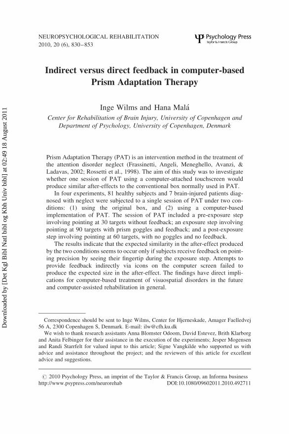
Indirect versus direct feedback in computer-based
Prism Adaptation Therapy
Inge Wilms and Hana Mala
Center for Rehabilitation of Brain Injury, University of Copenhagen and
Department of Psychology, University of Copenhagen, Denmark
Prism Adaptation Therapy (PAT) is an intervention method in the treatment ofthe attention disorder neglect (Frassinetti, Angeli, Meneghello, Avanzi, &Ladavas, 2002; Rossetti et al., 1998). The aim of this study was to investigatewhether one session of PAT using a computer-attached touchscreen wouldproduce similar after-effects to the conventional box normally used in PAT.
In four experiments, 81 healthy subjects and 7 brain-injured patients diag-nosed with neglect were subjected to a single session of PAT under two con-ditions: (1) using the original box, and (2) using a computer-basedimplementation of PAT. The session of PAT included a pre-exposure stepinvolving pointing at 30 targets without feedback; an exposure step involvingpointing at 90 targets with prism goggles and feedback; and a post-exposurestep involving pointing at 60 targets, with no goggles and no feedback.
The results indicate that the expected similarity in the after-effect producedby the two conditions seems to occur only if subjects receive feedback on point-ing precision by seeing their fingertip during the exposure step. Attempts toprovide feedback indirectly via icons on the computer screen failed toproduce the expected size in the after-effect. The findings have direct impli-cations for computer-based treatment of visuospatial disorders in the futureand computer-assisted rehabilitation in general.
Correspondence should be sent to Inge Wilms, Center for Hjerneskade, Amager Faelledvej
56 A, 2300 Copenhagen S, Denmark. E-mail: [email protected]
We wish to thank research assistants Anna Blomster Odoom, David Estevez, Brith Klarborg
and Anita Felbinger for their assistance in the execution of the experiments; Jesper Mogensen
and Randi Starrfelt for valued input to this article; Signe Vangkilde who supported us with
advice and assistance throughout the project; and the reviewers of this article for excellent
advice and suggestions.
NEUROPSYCHOLOGICAL REHABILITATION
2010, 20 (6), 830–853
# 2010 Psychology Press, an imprint of the Taylor & Francis Group, an Informa business
http://www.psypress.com/neurorehab DOI:10.1080/09602011.2010.492711
Dow
nloa
ded
by [
Det
Kgl
Bib
l Nat
l bib
l og
Kbh
Uni
v bi
bl]
at 0
2:49
18
Aug
ust 2
011

Keywords: Neglect; Prism Adaptation Therapy; Feedback; Computer-basedtherapy; Visuomotor plasticity.
INTRODUCTION
Within the field of cognitive rehabilitation, the positive effects of inten-sive, focused training (Kleim & Jones, 2008; Meinzer et al., 2004; Pulver-muller & Berthier, 2008) has generated renewed interest in transferringpaper-and-pencil therapy to a computer-based environment.
Many types of neuropsychological rehabilitation efforts are often con-ducted on a paper-and-pencil basis requiring the constant presence andsupervision of a therapist. The transfer of paper-and-pencil therapy to acomputer environment would provide choice and flexibility in access totraining at rehabilitation clinics and at home. Furthermore, it would facili-tate more detailed and precise recordings of information during training;allow adjustment of training according to individual progress; and poten-tially reduce therapist workload as the demands for intensity and frequencyof therapy increases. Finally, the use of computer-based training in researchwould ensure that the training is delivered in a consistent manner, whichmakes comparisons across subjects more valid.
The usefulness of computers has been demonstrated with standard neuro-psychological tests which, when transferred to computer, have been shown toimprove the quality of observations and the level of detail available totherapists (Chiba, Yamaguchi, & Eto, 2006; Rabuffetti et al., 2002; Tsirlin,Dupierrix, Chokron, Coquillart, & Ohlmann, 2009). Also, experimentaluse of computers in rehabilitation training has been successfully tested invarious research settings (Ansuini, Pierno, Lusher, & Castiello, 2006;Katz et al., 2005; Kim et al., 2007; Smith, Hebert, & Reid, 2007; Turton,O’Leary, Gabb, Woodward, & Gilchrist, 2010; Webster et al., 2001).However, in both test situations and in therapy, it seldom seems to bea point of concern that the transfer of paper-and-pencil training tocomputer may introduce changes to the training, some beneficial and othersdetrimental.
As this study will demonstrate, an important aspect of transferring paper-and-pencil therapy into a computer-based environment is that this requiresnot only technical skills, but also detailed insights into which elements ofthe therapy are actually ameliorating the patient’s symptoms as well asrigorous testing to ensure that the results obtained using one implementationof training are replicable with another, seemingly similar, method ofimplementation.
COMPUTER-BASED FEEDBACK IN THERAPY 831
Dow
nloa
ded
by [
Det
Kgl
Bib
l Nat
l bib
l og
Kbh
Uni
v bi
bl]
at 0
2:49
18
Aug
ust 2
011

Definition of key concepts
Neglect. One of the more common deficits after brain injury to the righthemisphere is hemispatial neglect (Rossetti et al., 1998). Hemispatial neglectis defined as a failure to explore, respond or orient towards stimuli presentedon the contralesional side (Heilman, Valenstein, & Watson, 2000). Increas-ingly, evidence supports that some effects of unilateral neglect can be amelio-rated by Prism Adaptation Therapy (Frassinetti et al., 2002; Rossetti et al.,1998; Serino, Bonifazi, Pierfederici, & Ladavas, 2007; Vangkilde & Habe-kost, in press). Other therapies also exist but are not relevant to this study.
Prism Adaptation Therapy. Prism Adaptation Therapy (PAT) is an inten-sive, bottom-up type therapy thought to affect visuospatial representations aswell as visuomotor abilities (Frassinetti et al., 2002; Serino et al., 2007). APAT session ordinarily consists of three steps; a pre-exposure step measuringthe pointing accuracy of the patient without feedback or intervention; anexposure step where the patient must adapt to a rightward shift of thevisual field induced by prism goggles; and finally a post-exposure step thatmeasures the after-effect resulting from the exposure step. Each session isdelivered twice a day for 2 weeks. During each step of a session of ordinaryPAT, the patient is directed to point to one of several targets at the far end of abox placed between the patient and the therapist. The box is wide enough toallow almost full extension of the arm but constructed to hide the patient’sarm and hand movements. The position of the box is adjusted during trainingto allow or prevent the patient from seeing the fingertip. For more details onPAT, see Serino et al., 2007.
The after-effect. Normally, patients as well as healthy controls are ableto adapt to the rightward shift induced by the prism goggles after a certainnumber of attempts at pointing at targets, when provided with feedbackabout the precision of their pointing in relation to the specific targets (Fras-sinetti et al., 2002; Redding, Rossetti, & Wallace, 2005; Sarri et al., 2008;Serino, Angeli, Frassinetti, & Ladavas, 2006; Serino et al., 2007). Afterremoval of the prism goggles a brief after-effect of off-target pointing tothe left can be observed (Fernandez-Ruiz & Dıaz, 1999; Redding et al.,2005). The size of the after-effect, produced as a result of exposure toprism goggles, has been shown to be affected by whether or not the sub-jects are allowed to see the actual movement of the extremity duringprism exposure (Redding et al., 2005; Redding & Wallace, 1988) on thetask performed (Simani, McGuire, & Sabes, 2007), and may even be theadditive result of the adaptation of different mechanisms (Redding &Wallace, 2002).
832 WILMS AND MALA
Dow
nloa
ded
by [
Det
Kgl
Bib
l Nat
l bib
l og
Kbh
Uni
v bi
bl]
at 0
2:49
18
Aug
ust 2
011

Aim of the study
In the present study, we wanted to investigate if PAT could be successfullytransferred to a computer-based environment. There are several reasons forthis choice. Firstly, PAT is a fairly simple and repetitive type of trainingwith well-defined rules, which lends itself to computer implementation.Secondly, some elements of the therapy, such as the observed adaptationeffect and after-effect, can be measured in a non-injured population(Bedford, 1993; Fernandez-Ruiz & Dıaz, 1999; Redding et al., 2005) thusincreasing the number of tested subjects and the statistical validity of thefindings. Thirdly, using a non-injured population prevents contamination ofthe initial results from unknown effects of the brain injury itself.
Transferring therapy from one setting to another requires detailed study toensure that the elements of therapy that make a difference are conservedacross settings. In order to investigate whether a transfer of Prism AdaptionTherapy to computer affected the effectiveness of the therapy, four exper-iments were carried out with the following aims:
1. To investigate whether the execution of a PAT session in a computer-based environment leads to similar after-effects in healthy subjects as aPAT session conducted in a standard box for each individual tested.
2. To examine whether the use of prism goggles could be replaced by dis-placed feedback on a computer touchscreen in healthy subjects.
3. To study the visuomotor elements characterising PAT.4. Finally, assuming that both conditions would provide similar
responses in after-effect during post-exposure in healthy subjects,we wanted to test if similar results could be obtained with brain-injured patients.
METHOD
In our study, all participants in the four experiments performed a singlesession of PAT on both the box normally used in standard PAT and on a com-puter-based condition of PAT. The after-effect data from the single session onthe box set the standard by which the subjects’ responses in the computer-based conditions were compared. Data on pointing precision were recordedon computer or by a therapist.
A single session of PAT in our experiments consisted of three differentsteps of pointing at targets under different conditions:
1. A pre-exposure step, which served as a baseline for each individualtested. This step consisted of 30 pointing trials.
COMPUTER-BASED FEEDBACK IN THERAPY 833
Dow
nloa
ded
by [
Det
Kgl
Bib
l Nat
l bib
l og
Kbh
Uni
v bi
bl]
at 0
2:49
18
Aug
ust 2
011
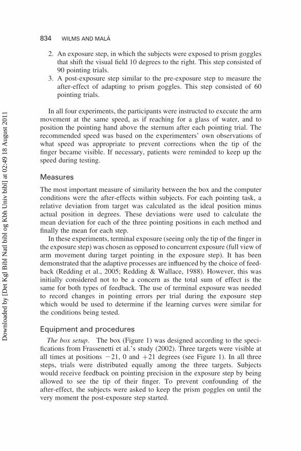
2. An exposure step, in which the subjects were exposed to prism gogglesthat shift the visual field 10 degrees to the right. This step consisted of90 pointing trials.
3. A post-exposure step similar to the pre-exposure step to measure theafter-effect of adapting to prism goggles. This step consisted of 60pointing trials.
In all four experiments, the participants were instructed to execute the armmovement at the same speed, as if reaching for a glass of water, and toposition the pointing hand above the sternum after each pointing trial. Therecommended speed was based on the experimenters’ own observations ofwhat speed was appropriate to prevent corrections when the tip of thefinger became visible. If necessary, patients were reminded to keep up thespeed during testing.
Measures
The most important measure of similarity between the box and the computerconditions were the after-effects within subjects. For each pointing task, arelative deviation from target was calculated as the ideal position minusactual position in degrees. These deviations were used to calculate themean deviation for each of the three pointing positions in each method andfinally the mean for each step.
In these experiments, terminal exposure (seeing only the tip of the finger inthe exposure step) was chosen as opposed to concurrent exposure (full view ofarm movement during target pointing in the exposure step). It has beendemonstrated that the adaptive processes are influenced by the choice of feed-back (Redding et al., 2005; Redding & Wallace, 1988). However, this wasinitially considered not to be a concern as the total sum of effect is thesame for both types of feedback. The use of terminal exposure was neededto record changes in pointing errors per trial during the exposure stepwhich would be used to determine if the learning curves were similar forthe conditions being tested.
Equipment and procedures
The box setup. The box (Figure 1) was designed according to the speci-fications from Frassenetti et al.’s study (2002). Three targets were visible atall times at positions 221, 0 and +21 degrees (see Figure 1). In all threesteps, trials were distributed equally among the three targets. Subjectswould receive feedback on pointing precision in the exposure step by beingallowed to see the tip of their finger. To prevent confounding of theafter-effect, the subjects were asked to keep the prism goggles on until thevery moment the post-exposure step started.
834 WILMS AND MALA
Dow
nloa
ded
by [
Det
Kgl
Bib
l Nat
l bib
l og
Kbh
Uni
v bi
bl]
at 0
2:49
18
Aug
ust 2
011
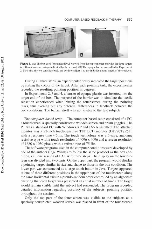
During all three steps, an experimenter orally indicated the target positionsby stating the colour of the target. After each pointing task, the experimenterrecorded the resulting pointing position in degrees.
In Experiments 2, 3 and 4, a barrier of opaque plastic was inserted into thetarget end of the box. The purpose of the barrier was to simulate the tactilesensation experienced when hitting the touchscreen during the pointingtasks, thus evening out any potential differences in feedback between thetwo conditions. The barrier itself was not visible to the test subjects.
The computer-based setup. The computer-based setup consisted of a PC,a touchscreen, a specially constructed wooden screen and prism goggles. ThePC was a standard PC with Windows XP and JAVA installed. The attachedmonitor was a 22-inch touch-sensitive TFT LCD monitor (DT220TSR5U)with a response time ≤5ms. The touch technology was a 5-wire, analogueresistive type with a touch resolution of 4096 x 4096 and a screen resolutionof 1680 x 1050 pixels with a refresh rate of 75 Hz.
The software programs used in the computer conditions were developed byone of the authors (Inge Wilms) to follow the same protocol as the box con-dition, i.e., one session of PAT with three steps. The display on the touchsc-reen was divided into two parts. On the upper part, the program would displaya pointing target similar in size and shape to those in the box condition. Thelower part was constructed as a large touch-button in Java. Targets appearedat one of three different positions in the upper part of the touchscreen alongthe same horizontal axis in a pseudo-random order controlled by an algorithmensuring that each target was presented an equal number of times. The targetwould remain visible until the subject had responded. The program recordeddetailed information regarding accuracy of the subjects’ pointing positionthroughout the session.
Only the top part of the touchscreen was visible to the subjects as aspecially constructed wooden screen was placed in front of the touchscreen
Figure 1. (A) The box used for standard PAT viewed from the experimenter end with the three targets
in different colours on top (indicated by the arrows). (B) The opaque barrier was added in Experiment
2. Note that the top can slide back and forth to adjust it to the individual arm length of the subjects.
COMPUTER-BASED FEEDBACK IN THERAPY 835
Dow
nloa
ded
by [
Det
Kgl
Bib
l Nat
l bib
l og
Kbh
Uni
v bi
bl]
at 0
2:49
18
Aug
ust 2
011

to prevent the subjects from seeing their arm movements (see Figure 2) andthe touch area. The screen had a sliding top that was adjusted to the subjects’arm length. The touchscreen issued a beeping sound when touched, indicatingto the subject that the pointing was recorded. The program ignored any acci-dental repeated touches.
Prism goggles. The prism goggles in this study were constructed using astandard pair of goggles with large viewing area lined with Fresnel prisms of17.5 dioptre, which shifted the visual field 10 degrees to the right. Initially,when wearing the prism goggles, subjects will tend to point too far to theright of the targets because of the deviation of the visual field. Graduallyduring the exposure step, subjects will adapt to the change and the pointingmeasurements settle around the target.
Finger nail. During all trials, the subjects wore a 3mm broad plastic nailon the pointing finger to prevent direct tactile feedback upon touching thescreen or box. The plastic nail was attached with adhesive tape to thefinger to prevent bending and sliding and extended the physical nail byapprox. 5mm.
Statistics. SPSS version 17.0 was used to analyse the data. Kolgorov-Smirnoff tests were used to test normality and MANOVA and ANOVAtests were used to test variance and means. T-tests were employed toisolate group differences where group differences had been demonstratedwith the ANOVA and MANOVA tests.
Figure 2. The equipment used in the computer conditions. (A) Touchscreen and the wooden screen
that hides the arm movements. The wooden screen divides the touchscreen into two parts. The bottom
part is where the subjects point and the top part is where the targets are shown. (B) Note that the top
can slide on the wooden screen to adjust for the arm length of the subjects (indicated by the double
headed arrow).
836 WILMS AND MALA
Dow
nloa
ded
by [
Det
Kgl
Bib
l Nat
l bib
l og
Kbh
Uni
v bi
bl]
at 0
2:49
18
Aug
ust 2
011

EXPERIMENTS
A total of four experiments were carried out to gather data under the computercondition. Before the actual sessions, each participant was allowed five prac-tice trials on the computer and the box to become familiar with the movementrequirements and the touchscreen. The data from these trials were discardedfrom the analysis.
Each experiment is described in detail in the following paragraphs alongwith results. Table 1 provides an overview of the different conditions foreach experiment.
TABLE 1Overview of the experimental conditions for the four experiments.
Type of
Feedback
Artificial
Nail
Pointing
instr.
No. of
targets
visible
Barrier
on box
Distance
between
targets
No. of
test
subjects
Type of
subject
Experiment 1 conditions
Computer A,
goggles
X on
screen
Yes SVT 1 14 cm 33 Normal
Computer B,
no goggles
X on
screen
Yes SVT 1 14 cm 33 Normal
Box Visible
finger
Yes OI 3 no 17.5 cm 33 Normal
Experiment 2 conditions
Computer A,
goggles
Visible
finger
Yes SVT 1 14 cm 28 Normal
Box Visible
finger
Yes OI 3 yes 17.5 cm 28 Normal
Experiment 3 conditions
Computer A,
goggles
Visible
finger
Yes OI 3 14 cm 20 Normal
Box Visible
finger
Yes OI 3 yes 14 cm 20 Normal
Experiment 4 condition
Computer A,
goggles
Visible
finger
Yes SVT 1 14 cm 7 Brain
injured
Box Visible
finger
Yes OI 3 yes 17.5 cm 7 Brain
injured
SVT denotes that a single target was visible at a time on the touchscreen as opposed to the three
permanently visible targets in the box condition. OI (oral instruction) indicates that instruction on
current target was given orally.
COMPUTER-BASED FEEDBACK IN THERAPY 837
Dow
nloa
ded
by [
Det
Kgl
Bib
l Nat
l bib
l og
Kbh
Uni
v bi
bl]
at 0
2:49
18
Aug
ust 2
011

Experiment 1
In the first experiment, we wanted to test if the after-effect observed after asingle session of PAT in the box condition was reproducible with two differ-ent computer conditions.
The computer condition “A” was designed to emulate the three steps of asingle standard session of PAT in the box. Due to the limited width of thetouchscreen, the distance between targets were slightly shorter (by 3.5cm)on the touchscreen as compared to the box. We compensated for this byplacing the subjects 9cm closer to the touchscreen, so the visual angle wasconstant across conditions.
The subjects were instructed to imagine that the displayed target on thetouchscreen extended downwards below the edge of the wooden screenhiding their movement and finger (see Figure 2A) and that their objectivewas to hit the extended target as precisely as possible. A red “X” was dis-played on the touchscreen above the barrier as indirect feedback on the hori-zontal precision of the pointing position in the exposure step. Subjects weretold to try to position the red “X” exactly on top of the target.
In computer condition “B”, the setup was similar to the “A” conditionexcept for the exposure step. In the “B” condition, subjects were not askedto wear prism goggles but instead received displaced feedback on pointingprecision. The displacement equalled a rightward shift of 10 degreessimilar to the effect of wearing prism goggles. The rationale was that by pro-viding displaced feedback the subjects would be forced to adjust eye-to-handcoordination without the use of prism goggles.
A total of 33 healthy subjects completed three sessions of PAT, one oneach of the three conditions: box, computer “A”, and computer “B”. Sinceall three conditions were very similar, each subject was exposed to onlyone condition a week to reduce the effect of repetition inadvertently con-founding the results. Furthermore, the subjects were randomly assigned tosix groups, each trying out the conditions in six different predefinedsequences to avoid any sequencing effect. The six sequence-groups werethe following with “A” and “B” being the computer conditions and “box”being the box: “A, B, box”; “A, box, B”; “B, A, box”; “B, box, A”; “box,A, B” and “box, B, A”.
Participants
Thirty-three subjects participated in this experiment. The age of the sub-jects ranged from 26 to 59 (M ¼ 38.48, SD ¼ 9.38, n ¼ 33), 27 femalesand 6 males. The participants were recruited from the employees at theCenter for Rehabilitation of Brain Injury (CRBI), University of Copenhagen,Denmark.
838 WILMS AND MALA
Dow
nloa
ded
by [
Det
Kgl
Bib
l Nat
l bib
l og
Kbh
Uni
v bi
bl]
at 0
2:49
18
Aug
ust 2
011

Results
Data were tested for normality using the Kolmogorov-Smirnov test and nosignificant deviation from normality was found at the pre-exposure and post-exposure step. A slight deviation was observed in the data from the exposurestep (K-S, p , 16.2) for the box condition. Since the main parameter formeasuring the similarity in effect was the after-effect in the post-exposurestep, parametric statistical models were used to analyse the similarities anddifferences.
To determine if the conditions produced similar after-effects, the generallinear model for repeated measures to analyse variance was used to test theconditions within subjects. It showed a highly significant differencebetween conditions per step, F(4, 128) ¼ 9.223, p , .001. A Mauchly’stest of sphericity on conditions per step was significant (p , .003). As such,the more conservative Greenhouse-Geisser and the Huynh-Feldt correctionswere used as recommended by Field (2009) both of which confirmed thesignificant difference, G–G, F(2.99, 95.91) ¼ 9.223, p , .001; and H–F,F(3.34, 106.92) ¼ 9.223, p , .001.
To isolate the group difference, a paired sample T-test was performed onthe three pairs of conditions (“A”-box, “B”-box, “A”-“B”). The meandiversion for the box condition (M ¼ 4.17, SD ¼ 1.96) and the computer“A” condition (M ¼ 2.01, SD ¼ 1.61) was significantly different (t ¼5.68, df ¼ 32, p , .001); likewise with the paired samples T-testbetween diversion from the box condition and the computer “B” condition(M ¼ 2.25, SD ¼ 1.99). They were also significantly different (t ¼ 3.92, df¼ 32, p , .001). Finally, means from the after-effect measured for the twocomputer conditions were compared. They were not significantly different(t ¼ –0.74, df ¼ 32, p , .47).
In summary, the analysis showed that the after-effect following the stan-dard box setup was different from the one achieved on the two computerisedversions of the experiment. No significant impact on the after-effect wasfound from age, sex and the six different sequences.
Discussion
The results from the single session of PAT conducted on the computer con-ditions “A” and “B” showed the same amplitude in the after-effect. However,both computer conditions differed significantly in the amplitude of the after-effect from the box condition. By far the largest amplitude was measured forthe box with a mean 2 degrees larger than the computer conditions. See-mingly, something about the computer conditions was causing lower ampli-tude in the after-effect. The fact that both computer conditions elicited similarresults suggested further investigation into the major differences between the
COMPUTER-BASED FEEDBACK IN THERAPY 839
Dow
nloa
ded
by [
Det
Kgl
Bib
l Nat
l bib
l og
Kbh
Uni
v bi
bl]
at 0
2:49
18
Aug
ust 2
011

computer conditions and the box condition. The following differences wereidentified:
1. Only a single target was shown on the touchscreen at a time as opposedto three visible targets on top of the box.
2. Feedback was in the shape of an “X” on the touchscreen as opposed tothe fingertip viewed in the box condition.
3. Distance between target position was slightly smaller on the touch-screen than in the box condition.
4. When pointing at the touchscreen, the fingertip would hit a solid surfaceas opposed to the box where subjects would point into open space.
5. The target was indicated by vocal instruction in the box condition versusimplicit positioning of only one target in the computer conditions.
The most prominent difference between the computer conditions and thebox condition was the difference in the presentation of feedback on pointingprecision. We hypothesised that the indirect feedback did not activate the eye-to-hand coordination system adequately even though subjects solved thepointing tasks correctly during all three steps and were explicitly awarethat the “X” on the touchscreen was the feedback on pointing position.Another interesting finding was that goggles and indirect feedback from the“A” condition created the same after-effect size as the displaced feedbackwith goggles.
Experiment 2
Based on the results from Experiment 1, we hypothesised that it may beessential to receive feedback by viewing one’s own fingertip, and discardedthe “B” condition, as any visual feedback would reveal that it was artificiallyskewed by 10 degrees. Therefore in Experiment 2, only a modified versionof the “A” condition was tested against the box condition. The indirect “X”feedback was replaced by direct fingertip feedback by moving the woodenscreen slightly away from the touchscreen in the exposure step.
In addition, the box used in Experiment 1 was modified by inserting anopaque barrier invisible to the subjects at the back end (see Figure 1B).The intention was to mimic the tactile feedback from the touchscreen, thuseliminating any impact this might have on the results. This change alsoeased recording of the pointing position by the experimenter and preventedaccidental exposure of the finger in the post-exposure step (a potential con-founder detected in the first experiment).
As in Experiment 1, only one target was shown at a time on the touch-screen thus implicitly indicating where to point. The pointing regime wassimilar to the one used in Experiment 1.
840 WILMS AND MALA
Dow
nloa
ded
by [
Det
Kgl
Bib
l Nat
l bib
l og
Kbh
Uni
v bi
bl]
at 0
2:49
18
Aug
ust 2
011
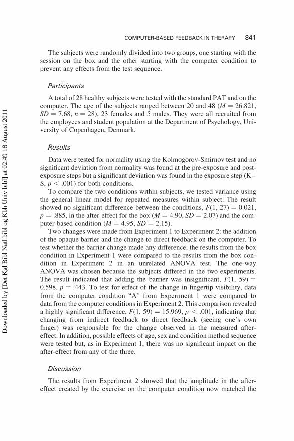
The subjects were randomly divided into two groups, one starting with thesession on the box and the other starting with the computer condition toprevent any effects from the test sequence.
Participants
A total of 28 healthy subjects were tested with the standard PAT and on thecomputer. The age of the subjects ranged between 20 and 48 (M ¼ 26.821,SD ¼ 7.68, n ¼ 28), 23 females and 5 males. They were all recruited fromthe employees and student population at the Department of Psychology, Uni-versity of Copenhagen, Denmark.
Results
Data were tested for normality using the Kolmogorov-Smirnov test and nosignificant deviation from normality was found at the pre-exposure and post-exposure steps but a significant deviation was found in the exposure step (K–S, p , .001) for both conditions.
To compare the two conditions within subjects, we tested variance usingthe general linear model for repeated measures within subject. The resultshowed no significant difference between the conditions, F(1, 27) ¼ 0.021,p ¼ .885, in the after-effect for the box (M ¼ 4.90, SD ¼ 2.07) and the com-puter-based condition (M ¼ 4.95, SD ¼ 2.15).
Two changes were made from Experiment 1 to Experiment 2: the additionof the opaque barrier and the change to direct feedback on the computer. Totest whether the barrier change made any difference, the results from the boxcondition in Experiment 1 were compared to the results from the box con-dition in Experiment 2 in an unrelated ANOVA test. The one-wayANOVA was chosen because the subjects differed in the two experiments.The result indicated that adding the barrier was insignificant, F(1, 59) ¼0.598, p ¼ .443. To test for effect of the change in fingertip visibility, datafrom the computer condition “A” from Experiment 1 were compared todata from the computer conditions in Experiment 2. This comparison revealeda highly significant difference, F(1, 59) ¼ 15.969, p , .001, indicating thatchanging from indirect feedback to direct feedback (seeing one’s ownfinger) was responsible for the change observed in the measured after-effect. In addition, possible effects of age, sex and condition method sequencewere tested but, as in Experiment 1, there was no significant impact on theafter-effect from any of the three.
Discussion
The results from Experiment 2 showed that the amplitude in the after-effect created by the exercise on the computer condition now matched the
COMPUTER-BASED FEEDBACK IN THERAPY 841
Dow
nloa
ded
by [
Det
Kgl
Bib
l Nat
l bib
l og
Kbh
Uni
v bi
bl]
at 0
2:49
18
Aug
ust 2
011

amplitude from the box condition results. As there was no change in the after-effect measured using the box in Experiments 1 and 2, the added barrier wasruled out as being the cause of the change. In other words, it was not the tactilesensation of hitting a barrier or touchscreen that changed the amplitude of theafter-effect. Neither was it the potentially more precise recordings of pointingposition due to the barrier.
The subject population was different for Experiments 1 and 2 and thischanged the average age. If this had been responsible for the differenceobserved, one would have expected data from the box condition to alsochange between Experiments 1 and 2. As this was not observed, we concludedthat the change in population did not influence the results.
The most probable cause of the impact on the after-effect was the changefrom providing indirect feedback about pointing precision using an “X” onthe touchscreen to letting the subject see his/her own fingertip (with the arti-ficial nail) in the computer condition. In conclusion, the results from Exper-iment 2 indicate that wearing prism goggles, doing the arm movements andsolving the task of pointing increasingly precisely during the exposure step,does not in itself produce the desired amplitude of the after-effect. In otherwords being able to relate feedback to the bodily act of pointing by seeingone’s actual fingertip is apparently also required.
Experiment 3
Experiment 2 tested the significance of the barrier and visible feedback. InExperiment 3, the remainder of the differences detected from Experiment 1between the computer condition and the box condition were tested. Thedata were used to analyse the effect of all of the observed differencesbetween the computer and the standard box condition.
The box condition was modified so in addition to the added barrier, the dis-tance between targets was changed to match the distance between targets onthe touchscreen. Due to the limitation in the touchscreen size, i.e., 22 inchesdiagonal, the distance between targets on the touchscreen was 14cm asopposed to 17.5cm in the box condition in the previous experiments.Although subjects were placed closer to the screen, it was a potential con-found and, therefore, in Experiment 3 the distance between the targets inthe box was changed to match those on the touchscreen.
The computer-based condition was changed to match the box condition asclosely as possible. All three targets were made visible at all times andcoloured to match the targets in the box. A recorded voice would statewhich target to point at, simulating the voice of the experimenter. The sub-jects received direct feedback in the exposure step by actually seeing theirown finger.
842 WILMS AND MALA
Dow
nloa
ded
by [
Det
Kgl
Bib
l Nat
l bib
l og
Kbh
Uni
v bi
bl]
at 0
2:49
18
Aug
ust 2
011

The subjects were randomly assigned to two groups, one starting with thebox condition and one starting with the computer condition to avoid effectsfrom the test sequence.
Participants
Twenty normal subjects were tested in both conditions at least one weekapart using one session of PAT. The age of the subjects ranged from 26 to55 (M ¼ 37.9, SD ¼ 10.6, n ¼ 20), 17 females and 3 males. They wererecruited from the student population at the Department of Psychology atthe University of Copenhagen and among the employees at the Center forRehabilitation of Brain Injury (CRBI).
Results
Again data were tested for normality using a Kolmogorov-Smirnov testand no significant deviation from normality was found at any of the steps(K–S, p . .05) for either condition.
To compare the two conditions within subjects, we tested variance usingthe general linear model for repeated measures. The results from theANOVA showed no significant difference between the conditions, F(1, 19)¼ 1.776, p ¼ .198, for the after-effect.
A paired samples t-test of the means showed that the means for the boxcondition in the post step (M ¼ 4.58, SD ¼ 2.176) did not differ significantlyfrom the means from the computer condition (M ¼ 5.32, SD ¼ 2.50) (t ¼1.33, df ¼ 19, p ¼ .20). This supports the hypothesis that the two conditionselicited the same results. In addition, we tested for the effect of age, sex andcondition sequence and, as in Experiment 1, there was no significant impacton the after-effect from any of the three.
To test whether the changes made in Experiment 3 to the box changed theobserved after-effects, we performed a one-way unrelated ANOVA betweenthe results from the box condition in Experiments 1, 2 and 3. None of thechanges seems to have made a significant change to the subjects’ behaviourin relation to the box, F(2, 78) ¼ 0.311, p ¼ .733. The means of the after-effect from the three experiments were 4.457, 4.900 and 4.578 and standardvariation 2.323, 2.071, and 2.176 confirming that they were very much alike.
To check if the changes made to the computer condition in Experiment 3 inany way changed the behaviour of the subjects with regard to the after-effect,we compared the results from Experiments 2 and 3. No significant differencebetween the two versions of the computer condition in Experiments 2 and 3were found, F(1, 46) ¼ 0.304, p ¼ .584. This indicates that the changes madefrom Experiments 2 to 3 did not alter the performance of the subjects in anysignificant way.
COMPUTER-BASED FEEDBACK IN THERAPY 843
Dow
nloa
ded
by [
Det
Kgl
Bib
l Nat
l bib
l og
Kbh
Uni
v bi
bl]
at 0
2:49
18
Aug
ust 2
011

Discussion
In Experiment 3, the distance between targets in the box was changed tomatch the distance on the touchscreen. The computer condition waschanged to show all three targets simultaneously as in the box condition. Arecorded voice instructed the subjects to point to a specific target. The statisti-cal tests support the assumption that none of these changes made any impacton the after-affect observed. Since observations from post-exposure in thebox condition in this experiment matches the findings from the box conditionin Experiment 1, we conclude that it is highly unlikely that any changes madein Experiment 3 had an effect on the after-effect.
This supports the findings from Experiment 2 that receiving direct feed-back regarding pointing precision was the key to the difference in theobserved after-effect.
Experiment 4
A main motivator for this project was to find out if PAT for patients withneglect could be executed effectively on a computer in the hope that thiswould allow more people to train in clinics and at home. Therefore, inExperiment 4, we used the same procedure as for Experiment 2, only thistime the two conditions were tested on seven subjects with acquired braininjury to the right cerebral hemisphere who had previously been diagnosedwith unilateral neglect.
The subjects were randomly divided into two groups, one starting with thebox condition and one starting with the computer condition to avoid effectsfrom the test sequence. The sessions were separated by at least one week todiminish any unwarranted learning effect.
Participants
Seven patients from the CRBI participated in this experiment (see Table 2for details on impairment). All patients were in the chronic phase of recovery(. 6 months post-injury) and all had been referred to CRBI with neglect-likesymptoms in various degrees. In our experience neglect symptoms are muchharder to detect using standard neuropsychological tests when patients aretested later than 6 months post-onset, partly due to interference fromlearned compensatory techniques. However, all subjects were retested forneglect using the Schenkenberg Line Bisection Task (Schenkenberg, Brad-ford, & Ajax, 1980), the Star and Letter Cancellation tasks (Weintraub,2000), the Baking Tray Task (Appelros, Karlsson, Is, Tham, & Nydevik,2004; Tham & Tegner, 1996) and the visual field and neglect test from theTAP (Testbatterie zur Aufmerksamkeitsprufung) battery (Zimmermann &Fimm, 2002). The age of the subjects ranged from 46 to 61 (M ¼ 54.9,
844 WILMS AND MALA
Dow
nloa
ded
by [
Det
Kgl
Bib
l Nat
l bib
l og
Kbh
Uni
v bi
bl]
at 0
2:49
18
Aug
ust 2
011

SD ¼ 9.31, n ¼ 7), two females and five males. All participants were given athorough introduction to the project and care was taken to ensure that eachclearly understood the purpose and the instructions provided. Each participantthen signed a letter of consent.
Results
Data were first tested for normality using a Kolmogorov-Smirnov test andno significant deviation from normality was found at any of the steps (K–S,p . .05) for either condition.
To compare the two conditions within subjects, variance was tested usingthe general linear model for repeated measures. The result from the ANOVAshowed no significant difference between the conditions, F(1, 6) ¼ 0.805,p ¼ .404, for the after-effect.
A paired samples t-test of the means showed that the mean differencein degrees for the box condition in the post-exposure step (M ¼ 4.93,SD ¼ 1.36) did not differ significantly from the means from the computercondition (M ¼ 6.01, SD ¼ 3.27) (t ¼ 0.897, df ¼ 6, p ¼ .404). This
TABLE 2List of subjects and their aetiology
Case Age Sex
Months
post
onset Aetiology Locus Hemianopia Hemiparesis Neglect
CH 59 M 12 Infarct Right
hemisphere
Y N
LD 50 F 6 Haemorrhage Basal ganglia,
occipital/parietal lobe
Y (Y)
LT 56 M 6 Haemorrhage Right temporal/parietal lobe
Y N
NT 61 M 25 Infarct Right temporal
lobe
Y N
SA 46 F 20 Haemorrhage,
hydrocephalus,
meningitis
Right
hemisphere
Y Y (Y)
SH 56 M 27 Fracture
Commotio
cerebrii
Right
hemisphere
Y
SS 56 M 26 Infarct Right
hemisphere
Y
The information on pathology and locus have been copied directly from the original medical jour-
nals and although not as detailed as we would have wished, they are the best available. The “Neglect”
column indicates the results from our tests. Parentheses indicate that results were ambiguous.
COMPUTER-BASED FEEDBACK IN THERAPY 845
Dow
nloa
ded
by [
Det
Kgl
Bib
l Nat
l bib
l og
Kbh
Uni
v bi
bl]
at 0
2:49
18
Aug
ust 2
011

supports the hypothesis that they generate similar effects. Possible effects ofage, sex and condition method sequence were tested using ANOVA and nosignificant impact on the after-effect from any of the three variables wasobserved.
In summary, the patient group showed effects of both standard and compu-terised PAT similar in magnitude to that found with normal subjects in theprevious experiments.
Discussion
The results from Experiment 4 showed no significant difference in themagnitude of after-effect within subjects between the box and the computerconditions in the brain-injured patients. These results confirmed our findingsfrom Experiment 2 with healthy subjects.
The primary reason for trying the conditions on the brain-injured popu-lation was to examine whether brain injury in itself influences the observedafter-effect on either condition. Our data show no indication of this. Eitherone of the two conditions can, therefore, be applied in rehabilitation ofbrain injury with expected similar effects.
SUMMARY OF RESULTS FROM ALL FOUR EXPERIMENTS
In the four experiments, we showed that the amplitude of the after-effect wasdependent on the type of feedback received during the exposure step. Whensubjects saw their own fingertip as feedback (direct feedback) on theirpointing position during the exposure step, the after-effect was twice aslarge as when they received only indirect feedback (in the shape of an “X”on a computer touchscreen). See Table 3, which summarises the means andstandard deviations for the after-effect measured during the four experiments,and Figure 3, which illustrates the observed after-effects from the fourdifferent experiments.
TABLE 3A summary of findings for the after-effect in degrees across experiments
Box mean (degree) Box SD Computer mean (degree) Computer SD n
Exp. 1 4.17 1.96 2.01 1.61 33
Exp. 2 4.90 2.07 4.95 2.15 28
Exp. 3 4.58 2.18 5.32 2.5 20
Exp. 4 4.93 1.36 6.01 3.27 7
846 WILMS AND MALA
Dow
nloa
ded
by [
Det
Kgl
Bib
l Nat
l bib
l og
Kbh
Uni
v bi
bl]
at 0
2:49
18
Aug
ust 2
011
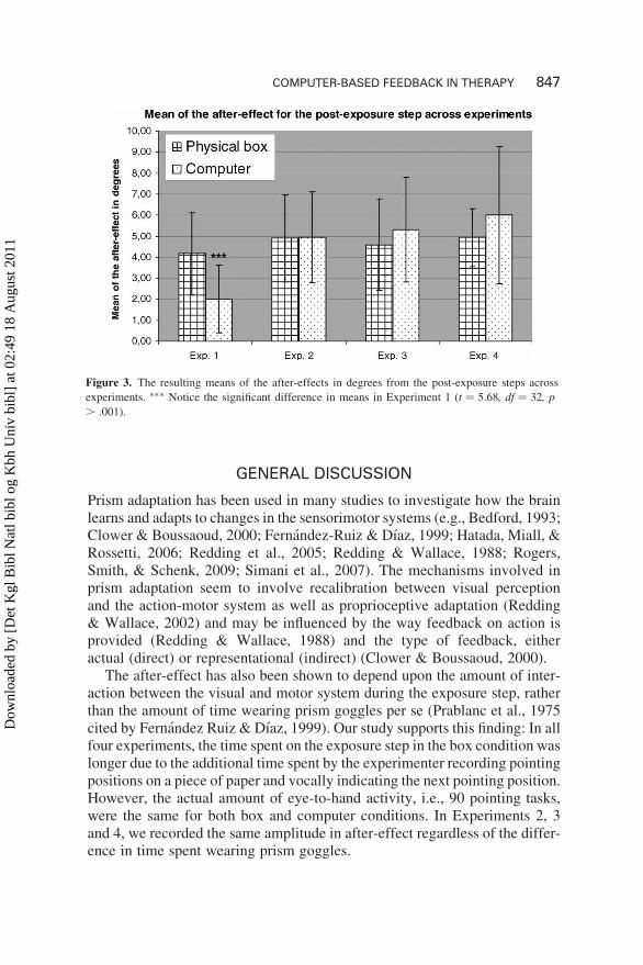
GENERAL DISCUSSION
Prism adaptation has been used in many studies to investigate how the brainlearns and adapts to changes in the sensorimotor systems (e.g., Bedford, 1993;Clower & Boussaoud, 2000; Fernandez-Ruiz & Dıaz, 1999; Hatada, Miall, &Rossetti, 2006; Redding et al., 2005; Redding & Wallace, 1988; Rogers,Smith, & Schenk, 2009; Simani et al., 2007). The mechanisms involved inprism adaptation seem to involve recalibration between visual perceptionand the action-motor system as well as proprioceptive adaptation (Redding& Wallace, 2002) and may be influenced by the way feedback on action isprovided (Redding & Wallace, 1988) and the type of feedback, eitheractual (direct) or representational (indirect) (Clower & Boussaoud, 2000).
The after-effect has also been shown to depend upon the amount of inter-action between the visual and motor system during the exposure step, ratherthan the amount of time wearing prism goggles per se (Prablanc et al., 1975cited by Fernandez Ruiz & Dıaz, 1999). Our study supports this finding: In allfour experiments, the time spent on the exposure step in the box condition waslonger due to the additional time spent by the experimenter recording pointingpositions on a piece of paper and vocally indicating the next pointing position.However, the actual amount of eye-to-hand activity, i.e., 90 pointing tasks,were the same for both box and computer conditions. In Experiments 2, 3and 4, we recorded the same amplitude in after-effect regardless of the differ-ence in time spent wearing prism goggles.
Figure 3. The resulting means of the after-effects in degrees from the post-exposure steps across
experiments. ∗∗∗ Notice the significant difference in means in Experiment 1 (t ¼ 5.68, df ¼ 32, p
. .001).
COMPUTER-BASED FEEDBACK IN THERAPY 847
Dow
nloa
ded
by [
Det
Kgl
Bib
l Nat
l bib
l og
Kbh
Uni
v bi
bl]
at 0
2:49
18
Aug
ust 2
011

Our experiments identify actual visual feedback as an important element inthe amplitude size of the after-effect during visuomotor activity. This con-firms the study by Clower and Boussaoud (2000). However, in our studythe feedback was not provided in a delayed fashion but appeared immediatelyupon touch; all trials were conducted in normal daylight with full body andhead movements allowed, and targets were visible until feedback had beenprovided. The difference in after-effect between direct and indirect feedbackin our study was not as large as in Clower and Boussaoud. Further exper-iments are needed to determine if other types of indirect feedback wouldwork.
Apparently, performing the actual movement and receiving feedback is notin itself enough to produce the after-effect. The manner in which feedback isprovided also plays a crucial role. In our study, seeing the position of thefinger in relation to the appointed target created larger after-effects.
With the pervasive use of advanced tools such as computers, it is surprisingthat, in order to produce changes in eye-to-hand coordination processes, onemust see the finger itself. As our experiments indicate, performing the move-ments and receiving indirect feedback on a touchscreen was not enough. Yet,in many computer-related activities we are able to manipulate objects on thescreen using an extension of our hand, such as the computer mouse, keyboardand game consoles. The activity on the screen in our experiments was verysimple. All the subjects were consciously aware that the indirect feedbackon the touchscreen was indeed feedback on pointing precision and all wereable to adjust their pointing strategy during the exposure step. Furthermore,all subjects had previous computer experience and were accustomed to coor-dinating actions on computer screens using indirect pointing devices such as acomputer mouse. Although the adaptation to the displaced visual input hap-pened regardless of the type of feedback, the after-effect was highly suscep-tible to the type of feedback provided. We therefore conclude that althoughthe executing element of the visuomotor system adapts to the changedinput, allowing subjects to point at targets during the exposure step, otheraspects of adaptation such as the impact depend on the type of feedbackreceived.
Feedback
Neglect can manifest itself in three distinct areas: body space, peripersonalspace and extrapersonal space (Gamberini, Seraglia, & Priftis, 2008). Inother words, neglect can be observed when dealing with objects out ofreach and within reach. Gamberini et al. (2008) carried out a study withthe line bisection test executed under two different conditions in extraperso-nal space. In the first condition, a laser-pointer was used to bisect a line on aremotely placed computer screen; in the second condition, subjects wore a
848 WILMS AND MALA
Dow
nloa
ded
by [
Det
Kgl
Bib
l Nat
l bib
l og
Kbh
Uni
v bi
bl]
at 0
2:49
18
Aug
ust 2
011

glove and pointed with an actual stick into a virtual reality environment,which provided the user with tactile and proprioceptive feedback. Theresults showed that the stick was perceived as part of the peripersonalspace whereas the laser pointer was not. Gamberini et al. suggested thatthe result was due to a remapping of peripersonal space and extrapersonalspace. The results from our study may support another interpretation basedon feedback rather than spaces. Bisecting a line with a laser-pointer (indirect)versus bisecting a line with a simulated extension of the body (direct) corre-lates with our findings where seeing one’s own finger as feedback (direct)influences the visuomotor programming whereas seeing an “X” on ascreen (indirect) does not. Our point is that both the finger and the “X”are within the peripersonal space so it is not as much the distance to thetarget but rather how feedback relates to the proprioceptive sense of body.If feedback is interpreted as coming from an action involving the body oran extension of the body, feedback will strengthen the impact (in our casethe after-effect).
In Experiment 1, we also tested if displaced feedback on the touchscreenwould elicit similar after-effects to those observed in the box condition. Asit turned out, the after-effect produced by displaced feedback was notsimilar to the after-effect from direct feedback. The displaced feedback con-dition produced an after-effect similar to the indirect feedback condition.
Within the field of psychology, any type of training or therapy involvingalmost any type of computer interaction is commonly referred to as virtualreality (VR). The term is usually used more restrictively within the IT com-munity to refer to humans navigating in a virtual 3D-world with interactiveequipment such as helmet, gloves, etc., that creates an illusion of total immer-sion into the virtual world. There are studies that have looked at replacing theprism goggles with displaced or incongruent feedback in a virtual realityenvironment (Castiello, Lusher, Burton, Glover, & Disler, 2004; Glover &Castiello, 2006). The results show that it is possible to improve the codingof visual stimuli in the neglected field using displaced feedback. In ourstudy we found that displaced feedback provided as an “X” on the touchsc-reen did not result in after-effects similar to the training in the box condition.A possible explanation is that in Castiello et al.’s (2004) experiments the levelof immersion was greater than in ours. Subjects wore a haptic glove, whichmade it possible to “feel” the targets, and, on the screen, a virtual handmoved similarly to the actual physical movements. We speculate that thedifference between our results from displaced feedback and those of Castielloet al. may be that in the virtual reality simulation, the visuomotor systems areadapting because feedback is perceived as directly related to a body part (thehand) whereas in our study, the “X” on the screen was not perceived as beingpart of the body even though subjects were never in doubt that it was feedbackon the action of pointing.
COMPUTER-BASED FEEDBACK IN THERAPY 849
Dow
nloa
ded
by [
Det
Kgl
Bib
l Nat
l bib
l og
Kbh
Uni
v bi
bl]
at 0
2:49
18
Aug
ust 2
011

In our view, this raises an important question concerning the use of com-puters in rehabilitation tests and therapy. Our results suggest that it is notenough to be consciously aware of the purpose of a task and even executingit correctly for therapy to have the wanted effect. In the case of visuomotoradaptation, the actions must be perceived as relating to the proprioceptivesense. Further research is needed to establish what exactly is needed for theproprioceptive system to respond to the feedback. Will a picture of a fingerrather than the “X” on the screen be enough? Must the screen depictkinetic action as in the study of Castiello et al. or will a simpler level beenough? In other words, what level of computer-generated simulation isrequired for the adaptive systems to respond as desired?
Computer technology and neglect therapy
Within the research area concerning hemispatial neglect, experimental testingvaries from simple interaction with keyboard, button and mouse that seldomcreates an illusion of immersion, to fully immersed virtual reality systems(Rose, Brooks, & Rizzo, 2005; Tsirlin et al., 2009). However, most of thisresearch is mainly directed towards improving the sensitivity of testing pro-cedures (Anton, Hershler, Lloyd, & Murray, 1988; Baheux, Yoshizawa,Seki, & Handa, 2006; Broeren, Samuelsson, Stibrant-Sunnerhagen, Blom-strand, & Rydmark, 2007; Chiba et al., 2006; List et al., 2008) and hasonly just begun to consider the field of improving training methods(Ansuini et al., 2006; Katz et al., 2005; Kim et al., 2007; Turton et al.,2010; Webster et al., 2001).
Our study emphasises the importance of test and measures when imple-menting computer-based therapy, which works in the real world. Our studywas on the absolute low end scale of immersion but we still managed tocreate a reasonable result after testing various conditions. This holdspromise for the future. Although it is generally agreed that virtual reality tech-nology has a huge potential within research on training and therapy of cogni-tive functions, virtual reality therapy will require much more research anddevelopment before becoming generally available for clinical work. Regard-less of whether therapy is based on a simple PC and a touchscreen or on elab-orate virtual reality technology, careful testing and measurements are neededto ensure that the therapy and tests do in fact target the systems we want totrain. In the process, this research may reveal new knowledge about functionsand dysfunctions of the brain.
CONCLUSIONS
This study was initiated to test the effects of implementing Prism AdaptationTherapy on a computer. We chose the after-effect as a measure of efficacy and
850 WILMS AND MALA
Dow
nloa
ded
by [
Det
Kgl
Bib
l Nat
l bib
l og
Kbh
Uni
v bi
bl]
at 0
2:49
18
Aug
ust 2
011

compared the after-effect of two computer conditions with that from the stan-dard physical box. The study revealed that in visuomotor tasks, it is importantto provide feedback on the action in a manner which targets the systems thatare involved in the adaptive processes. Knowing the task, understanding thetask and even executing the task correctly are not always enough to producethe desired effects.
Translating therapy from paper-and-pencil to computer requires athorough analysis of the individual elements making up the therapy. Thetranslating process may assist in revealing unknown aspects of the workingelements of training but it emphasises the need for careful testing of theresulting conditions.
Last but not least, this study confirms the need for further research in thefield of computer-assisted neurorehabilitation which in turn may providefurther insights both into normal cognitive function and cognitive deficitsfollowing brain injury.
REFERENCES
Ansuini, C., Pierno, A. C., Lusher, D., & Castiello, U. (2006). Virtual reality applications for the
remapping of space in neglect patients. Restorative Neurology and Neuroscience, 24(4–6),
431–441.
Anton, H. A., Hershler, C., Lloyd, P., & Murray, D. (1988). Visual neglect and extinction: A
new test. Archives of Physical Medicine and Rehabilitation, 69(12), 1013–1016.
Appelros, P., Karlsson, G. M., Is, A. T., Tham, K., & Nydevik, I. (2004). Unilateral neglect:
Further validation of the baking tray task. Journal of Rehabilitation Medicine, 36(6),
258–261.
Baheux, K., Yoshizawa, M., Seki, K., & Handa, Y. (2006). Virtual reality pencil and paper tests
for neglect: A protocol. CyberPsychology & Behavior, 9(2), 192–195.
Bedford, F. L. (1993). Perceptual and cognitive spatial learning. Journal of Experimental Psy-
chology: Human Perception and Performance, 19(3), 517–530.
Broeren, J., Samuelsson, H., Stibrant-Sunnerhagen, K., Blomstrand, C., & Rydmark, M. (2007).
Neglect assessment as an application of virtual reality. Acta Neurologica Scandinavica,
116(3), 157–163.
Castiello, U., Lusher, D., Burton, C., Glover, S., & Disler, P. (2004). Improving left hemispatial
neglect using virtual reality. Neurology, 62(11), 1958–1962.
Chiba, Y., Yamaguchi, A., & Eto, F. (2006). Assessment of sensory neglect: A study using
moving images. Neuropsychological Rehabilitation, 16(6), 641–652.
Clower, D. M., & Boussaoud, D. (2000). Selective use of perceptual Recalibration Versus
Visuomotor Skill Acquisition. Journal of Neurophysiology, 84(5), 2703–2708.
Fernandez-Ruiz, J., & Dıaz, R. (1999). Prism adaptation and after-effect: Specifying the
properties of a procedural memory system. Learning & Memory, 6(1), 47–53.
Field, A. (2009). Discovering statistics using SPSS (3rd ed.). London: Sage Publications Inc.
Frassinetti, F., Angeli, V., Meneghello, F., Avanzi, S., & Ladavas, E. (2002). Long-lasting
amelioration of visuospatial neglect by prism adaptation. Brain, 125, 608–623.
Gamberini, L., Seraglia, B., & Priftis, K. (2008). Processing of peripersonal and extrapersonal
space using tools: Evidence from visual line bisection in real and virtual environments.
Neuropsychologia, 46(5), 1298–1304.
COMPUTER-BASED FEEDBACK IN THERAPY 851
Dow
nloa
ded
by [
Det
Kgl
Bib
l Nat
l bib
l og
Kbh
Uni
v bi
bl]
at 0
2:49
18
Aug
ust 2
011

Glover, S., & Castiello, U. (2006). Recovering space in unilateral neglect: A neurological dis-
sociation revealed by virtual reality. Journal of Cognitive Neuroscience, 18(5), 833–843.
Hatada, Y., Miall, R. C., & Rossetti, Y. (2006). Long lasting after-effect of a single prism adap-
tation: Directionally biased shift in proprioception and late onset shift of internal egocentric
reference frame. Experimental Brain Research, 174(1), 189–198.
Heilman, K. M., Valenstein, E., & Watson, R. T. (2000). Neglect and related disorders.
Seminars in Neurology, 20(4), 463–470.
Katz, N., Ring, H., Naveh, Y., Kizony, R., Feintuch, U., & Weiss, P. L. (2005). Interactive
virtual environment training for safe street crossing of right hemisphere stroke patients
with unilateral spatial neglect. Disability and Rehabilitation, 27(20), 1235–1243.
Kim, J., Kim, K., Kim, D. Y., Chang, W. H., Park, C. I., Ohn, S. H., et al. (2007). Virtual
environment training system for rehabilitation of stroke patients with unilateral neglect:
Crossing the virtual street. Cyberpsychology & Behavior, 10(1), 7–15.
Kleim, J. A., & Jones, T. A. (2008). Principles of experience-dependent neural plasticity: Impli-
cations for rehabilitation after brain damage. Journal of Speech, Language and Hearing
Research, 51(1), S225.
List, A., Brooks, J. L., Esterman, M., Flevaris, A. V., Landau, A. N., Bowman, G., et al. (2008).
Visual hemispatial neglect, re-assessed. Journal of the International Neuropsychological
Society, 14(2), 243–256.
Meinzer, M., Elbert, T., Wienbruch, C., Djundja, D., Barthel, G., & Rockstroh, B. (2004).
Intensive language training enhances brain plasticity in chronic aphasia. BMC Biology,
2(20), 1–9.
Pulvermuller, F., & Berthier, M. L. (2008). Aphasia therapy on a neuroscience basis. Aphasiol-
ogy, 22(6), 563–599.
Rabuffetti, M., Ferrarin, M., Spadone, R., Pellegatta, D., Gentileschi, V., Vallar, G., et al.
(2002). Touch-screen system for assessing visuo-motor exploratory skills in neuropsycholo-
gical disorders of spatial cognition. Medical & Biological Engineering & Computing, 40(6),
675–686.
Redding, G. M., Rossetti, Y., & Wallace, B. (2005). Applications of prism adaptation: A tutorial
in theory and method. Neuroscience & Biobehavioral Reviews, 29(3), 431–444.
Redding, G. M., & Wallace, B. (1988). Components of prism adaptation in terminal and con-
current exposure: Organization of the eye–hand coordination loop. Perception & Psycho-
physics, 44(1), 59–68.
Redding, G. M., & Wallace, B. (2002). Strategic calibration and spatial alignment: A model
from prism adaptation. Journal of Motor Behavior, 34(2), 126–138.
Rogers, G., Smith, D., & Schenk, T. (2009). Immediate and delayed actions share a common
visuomotor transformation mechanism: A prism adaptation study. Neuropsychologia,
47(6), 1546–1552.
Rose, F. D., Brooks, B. M., & Rizzo, A. A. (2005). Virtual reality in brain damage rehabilita-
tion: review. Cyberpsychology & Behaviour, 8(3), 241–262; discussion 263–271.
Rossetti, Y., Rode, G., Pisella, L., Farne, A., Li, L., Boisson, D., et al. (1998). Prism adaptation
to a rightward optical deviation rehabilitates left hemispatial neglect. Nature, 395(6698),
166–169.
Sarri, M., Greenwood, R., Kalra, L., Papps, B., Husain, M., & Driver, J. (2008). Prism adap-
tation after-effects in stroke patients with spatial neglect: Pathological effects on subjective
straight ahead but not visual open-loop pointing. Neuropsychologia, 46(4), 1069–1080.
Schenkenberg, T., Bradford, D. C., & Ajax, E. T. (1980). Line bisection and unilateral visual
neglect in patients with neurologic impairment. Neurology, 30(5), 509–517.
Serino, A., Angeli, V., Frassinetti, F., & Ladavas, E. (2006). Mechanisms underlying neglect
recovery after prism adaptation. Neuropsychologia, 44(7), 1068–1078.
852 WILMS AND MALA
Dow
nloa
ded
by [
Det
Kgl
Bib
l Nat
l bib
l og
Kbh
Uni
v bi
bl]
at 0
2:49
18
Aug
ust 2
011

Serino, A., Bonifazi, S., Pierfederici, L., & Ladavas, E. (2007). Neglect treatment by prism
adaptation: What recovers and for how long. Neuropsychological Rehabilitation, 17(6),
657–687.
Simani, M. C., McGuire, L. M. M., & Sabes, P. N. (2007). Visual-shift adaptation is composed
of separable sensory and task-dependent effects. Journal of Neurophysiology, 98(5),
2827–2841.
Smith, J., Hebert, D., & Reid, D. (2007). Exploring the effects of virtual reality on unilateral
neglect caused by stroke: Four case studies. Technology and disability, 19(1), 29–40.
Tham, K., & Tegner, R. (1996). The baking tray task: A test of spatial neglect. Neuropsycho-
logical Rehabilitation, 6(1), 19–25.
Tsirlin, I., Dupierrix, E., Chokron, S., Coquillart, S., & Ohlmann, T. (2009). Uses of virtual
reality for diagnosis, rehabilitation and study of unilateral spatial neglect: Review and analy-
sis. Cyberpsychology & Behavior, 12(2), 175–181.
Turton, A. J., O’Leary, K., Gabb, J., Woodward, R., & Gilchrist, I. D. (2010). A single blinded
randomised controlled pilot trial of prism adaptation for improving self-care in stroke
patients with neglect. Neuropsychological Rehabilitation, 20(2), 180–196.
Vangkilde, S., & Habekost, T. (in press). Finding Wally: Prism adaptation improves visual
search in chronic neglect. Neuropsychologia.
Webster, J. S., McFarland, P. T., Rapport, L. J., Morrill, B., Roades, L. A., & Abadee, P. S.
(2001). Computer-assisted training for improving wheelchair mobility in unilateral
neglect patients. Archives of Physical Medicine and Rehabilitation, 82(6), 769–775.
Weintraub, S. (2000). Neuropsychological assessment of mental state. In M.-M. Mesulam (Ed.),
Principles of behavioural and cognitive neurology (2nd ed., pp. 121–158). New York:
Oxford University Press.
Zimmermann, P., & Fimm, B. (2002). A test battery for attentional performance. In M. Leclercq
& P. Zimmermann (Eds.), Applied neuropsychology of attention: Theory, diagnosis and
rehabilitation (pp. 110–151). Hove, UK: Psychology Press.
Manuscript received February 2010
Revised manuscript received April 2010
First published online September 2010
COMPUTER-BASED FEEDBACK IN THERAPY 853
Dow
nloa
ded
by [
Det
Kgl
Bib
l Nat
l bib
l og
Kbh
Uni
v bi
bl]
at 0
2:49
18
Aug
ust 2
011

J o u r n a l o f C y b e r T h e r a p y & R e h a b i l i t a t i o n
F a l l 2 0 11 , Vo l u m e 4 , I s s u e 3
© Vi r tua l Rea l i ty Med ica l In s t i t u t e
usIng ArtIfIcIAL InteLLIgence to controL And
AdAPt LeveL of dIffIcuLty In comPuter-BAsed
cognItIve therAPy – An exPLorAtIve study
Inge Wilms1, 2
Within the field of cognitive rehabilitation after brain injury, rehabilitation training is constantly ad-
justed to match the skills and progress of the individual patients. As no two patients are alike in func-
tional injury and recovery, it is a challenge to provide the right amount of training at the right level
of difficulty at any given time.
this study investigates whether a modified version of the artificial intelligence (AI) reinforcement
method called the “actor-critic method” is able to detect response time patterns and subsequently con-
trol the level of difficulty in a computer-based, cognitive training program. the efficacy of the AI logic
was tested under the actual training conditions of a brain-injured patient.
the results showed that the AI controlled training system was able to learn and adjust fast enough to
control and adapt the level of difficulty of the training to match the changes in the patient’s abilities
over a three-week period.
Keywords: reinforcement Learning, cognitive rehabilitation,
Adaptive therapy, Actor-critic, Adaptive Progression
IntroductIon
Over the past decade an increasing amount of evidence
supports the notion that cognitive functions injured from
trauma may recover, at least partially, through training
and therapy that target different aspects of brain plastic-
ity (Kleim & Jones, 2008; Friedemann Pulvermüller &
Berthier, 2008). As with healthy brains, certain elements
such as the intensity of the training, the type of feedback
provided and the progression of the level of difficulty
seem to be important, general aspects of the more recent
type of therapy used experimentally in the rehabilitation
of cognitive deficits (Frassinetti, Angeli, Meneghello,
Avanzi, & Ladavas, 2002; Friedemann Pulvermüller &
Berthier, 2008).
In methods like cognitive Constraint Induced Aphasia
Therapy, one of the key elements is the personalized in-
tensive training which challenges the patient gradually
with progressively harder tasks (Friedemann; Pulver-
müller, et al., 2001). However, to advance the training
at the right pace and to the right level of difficulty is per-
haps one of the hardest challenges for the therapists to
do correctly, as performance of patients may fluctuate
from day to day. First of all, no two brain-injured pa-
tients are alike even when diagnosed with similar afflic-
tions. This means that training-progress may vary
substantially from patient to patient. Even slight differ-
ences in impairment may impact the way training will
affect progress and amelioration making it extremely
difficult to determine how fast to advance with the train-
ing (e.g., Wilson, Gracey, Evans, & Bateman, 2009).
Secondly, even with the same category of affliction,
what is considered to be hard or difficult may vary from
patient to patient (Wilson, 1998). Thus, the combination
Corresponding Author:
Inge Wilms, Center for Rehabilitation of Brain Injury, Amagerfaelledvej 56 A, 2300 Copenhagen S, Denmark, Tel: +45 35329036, Fax: +45
35329050, E-mail: [email protected] or [email protected]
1Center for Rehabilitation of Brain Injury, University of Copenhagen2Department of Psychology, University of Copenhagen, Copenhagen, Denmark
387
JCR
388
of parameters that determine the level of difficulty may
very well differ substantially from patient to patient.
It therefore seems advantageous to study systems that
may monitor the activity of the patient and be able to
adjust parameters intelligently to match individual
progress and deficits. However, this poses considerable
challenges to the training system itself. First, it has to
be able to monitor and detect consistent progress during
training under realistic training conditions where
progress may not be linear or constant and where meas-
urements may be influenced by factors not under imme-
diate control, such as the current condition of the
patient. Secondly, the system has to be able to assess the
ability of the patient quickly and consistently in order
to select a correct set of parameters controlling difficulty
level at any given time during training. In essence, the
system must be able to adjust its behavior according to
the individual effect of parameters, some of which may
be known, some of which may not be.
Some computer-based cognitive training systems do
offer automated progression in level of difficulty, most
of them through a “staircase” progression whereby the
program increases the general level of difficulty from
one level to the next when the patient masters a certain
percentage of the tasks at one level (Sturm, Willmes, Or-
gass, & Hartje, 1997; Wertz & Katz, 2004). In other
cases the training progress is administered by the ther-
apist, who is then required to monitor the progress and
offer increasingly harder challenges (Pedersen, Vinter,
& Olsen, 2001).
To some extent, challenges and requirements to intensive
computer-based therapy for cognitive rehabilitation match
those of computer game-play. Artificial intelligence (AI)
has been used successfully in games to solve similar chal-
lenges of adjusting computer-controlled adversaries to
match the skills of the game player at any given time
(Bourg & Seemann, 2004; Spronck, Sprinkhuizen-
Kuyper, & Postma, 2004). The research in this field has
proven that certain machines learning AI algorithms are
fast and efficient enough to learn and subsequently adjust
game-play in real time to the constantly evolving skills of
the player (Ponsen, Spronck, Munoz-Avila, & Aha, 2007;
Spronck, et al., 2004).
PurPose
The purpose of this study was to explore whether an AI
engine, based on the actor-critic machine-learning logic,
would be able to control the multidimensional progression
of level of difficulty (LOD) as defined by three different
parameters in a computer-based, cognitive training pro-
gram for patients with acquired reading difficulty after
brain injury known as pure alexia. In particular, the ques-
tion was whether the engine would be fast and adaptive
enough to compensate for fluctuations in the performance
of the patients over time and be able to detect the subtle
differences in the effect on difficulty provided by each pa-
rameter. This article specifically deals with the technical
aspects and results of the study. The rehabilitation aspects
and results from this study will be presented elsewhere.
consIderAtIons regArdIng the
ArtIfIcIAL InteLLIgence Agent
LeArnIng Agent
The term “learning agent” refers to AI agents or programs
that are able to learn about the environment in which they
operate. This knowledge is in turn used to improve the
agent’s ability to make appropriate choices of action.
Learning can be achieved in several ways through different
types of feedback. Usually, the way AI learning is achieved
is categorized in three groups: supervised learning, unsu-
pervised learning and reinforcement learning (Russell &
Norvig, 2003). The difference being that in supervised
learning the agent is initially taught correct behavior from
samples of input and corresponding output. The outcome
of this supervised teaching is a performance function which
may then act as an expert system capable of solving prob-
lems of a similar kind. In unsupervised learning, the agent
is searching for patterns in input and establishing rules
based on those. Decision trees are examples of results of
unsupervised learning (Russell & Norvig, 2003).
Reinforcement learning (RL) originates from psychology
research, where reinforcement learning is thought to be
one of the fundamental ways living beings learn from in-
teracting with the environment. Similar to living beings,
computer programs can learn through trial and error (Sut-
ton & Barto, 1998). The RL agent learns about the appro-
priateness of selected actions through rewards and
punishments after execution of behavior without prior
knowledge of the environment (Russell & Norvig, 2003;
Sutton & Barto, 1998).
the choIce of AI method
In this study, the principal requirement for the AI algo-
rithm was that it was learning quickly, was flexible and
did not unduly impact the computer as the training pro-
gram needed to respond rapidly and appropriately to the
AI for Cognitive Rehab Therapy

JCR
389Wilms
actions of the test subject. The so-called actor-critic
method from the RL category was chosen as it had previ-
ously been proven able to learn and optimize selection of
actions quickly and easily in the research field of computer
games with similar challenges (Spronck, Ponsen,
Sprinkhuizen-Kuyper, & Postma, 2006; Spronck, et al.,
2004; Spronck & van den Herik, 2005).
In research the term “Game AI” often refers to the appear-
ance of intelligent behavior of computer-controlled game
characters (Spronck & van den Herik, 2005). A large por-
tion of game AI is often either fixed state machines or
scripted logic due to the complexity in using and adapting
the computational logic into usable, programmable entities
(Bourg & Seemann, 2004; Galway, Charles, & Black,
2008). Recent research, however, has tried to establish us-
able solutions to game AI based on reinforcement learning
techniques (Spronck, et al., 2006; Spronck & van den
Herik, 2005).
Spronck et al. (2004) had successfully implemented and
evaluated a solution based on a combination of dynamic
scripting and a slightly modified version of the temporal
difference (TD) learning methods named actor-critic
methods. Using these methods they had optimized the se-
lection of available actions for computer-controlled non-
player characters (NPCs) in a role-playing game to set a
level of difficulty that would challenge the player’s skills
appropriately. By matching NPC strength to the strength
of the player at a given time the player would stay moti-
vated for gaming.
Since Spronck’s team (2005, 2006) had already tested the
actor-critic logic against other RL techniques such as the
Monte Carlo methods and Q-learning, and found it most
suitable with regards to speed and impact, it seemed pru-
dent in this study to build upon their actor-critic findings
given the fundamental similarities in the requirements to
the AI logic.
the Actor-crItIc method
The actor-critic methods differ from other TD-learning
methods in that the policy is independently represented by
a structure known as the “Actor.” The estimated value
function is known as the “Critic.” The Critic must learn
and adjust the value of actions selected by the Actor
through the sensory information from the environment.
Since learning takes place in real time, the critic must learn
and criticize any current action followed by the actor; this
is also know as on-policy learning (Sutton & Barto, 1998).
the AI engIne In thIs study
the mAIn LogIc
The actor-critic method used in this study consisted of
three parts: (i) an actor that selected the appropriate pa-
rameter settings from three databases, (ii) a fitness func-
tion that converted the results from the actions to a
numeric representation which were then passed to the
third part, and (iii) the critic, that adjusted weight values
for each action chosen thus raising or lowering the prob-
ability of the action to be reselected. Figure 1 shows the
architecture of the agent.
The AI engine had no pre-established knowledge of the
environment. The engine learned the appropriateness of
a given set of parameters during actual training by ex-
amining sensory input in the form of response times
achieved at any given time and continuously adjusted
the choice of parameter settings.
Before each trial, the actor of the AI engine selected one
setting from each of the three parameter databases
based on the following algorithm:
Algorithm 1: Action selection (ACTOR)
1: Clear Plist();
2: k = 0;
3: for each parameterDB do
4: sumWeights = 0;
5: for i=0 to parameterDBSize-1 do
6: sumWeights = sumWeights+parmDB[i].weight;
7: end for
8: fraction = Math.random()*sumWeights;
Figure 1. The architecture of the AI engine used in the
VisATT training program.

JCR
390 AI for Cognitive Rehab Therapy
9: sum = 0; j = 0;
10: selected = false;
11: while (not selected) do
12: sum = sum+parameterDB[j].weights;
13: if (sum>fraction) then
14: selected = true;
15: Plist[k] = parameterDB[j];
16: k = k+1;
17: else
18: j = j+1;
19: end if
20: end while
21: end for
____________________________________________
The probability that a specific setting for a parameter
was chosen was influenced by the weight value at-
tached to each setting. The sum of all weights defined
the selection space. The size of each weight made each
setting more or less visible within this selection space.
Using a randomly generated selection criterion, it was
possible to ensure a certain amount of exploration,
since even large weights did not guarantee 100% selec-
tion each time. Initially, all weights were set to the same
value of 100.
The critic of the AI engine receives the response time
from the trial and determines the fate of the selected pa-
rameter settings:
Algorithm 2: Action evaluation (CRITIC)
1: medianRT = dampenInput(responseTime);
2: fitvar = AIFitness(medianRT);
3:
4: for i = 0 to Plist.length-1 do
5: Plist[i].ParameterDB.weight = Plist[i].Parame-
terDB.weight * fitvar;
6:
7: // Ensure exploitation across the range
8: if Plist[i].ParameterDB.weight < atMin then
9: Plist[i].ParameterDB.weight = atMin;
10: end if
11: if Plist[i].ParameterDB.weight > atMax then
12: Plist[i].ParameterDB.weight = atMax;
13: end if
14: i = i+1;
15: end for
––––––––––––––––––––––––––––––––––––––––––––
the AI engIne fItness functIon
The criterion for success for the AI engine was deter-
mined by measuring the time from the target icon was
presented in the middle of the panel until the time the
correct button was pressed (the response time). Two cri-
teria set the lower and upper limit for an acceptable re-
sponse time (see Figure 2). If the subject’s time fell
within these criteria, the LOD was acceptable. If re-
sponse time fell beneath the low threshold (tooEasy),
LOD was deemed too easy and the weights for the pa-
rameters selected would be reduced making those pa-
rameters less eligible for selection. Similarly, if the
response time fell above the upper threshold (tooHard),
LOD was deemed too hard and the weight of the param-
eters involved was reduced.
A bottom limit value of 20 as the lowest and 600 as the
highest weight setting ensured that all parameters would
remain eligible for selection allowing the difficulty to
be reduced if necessary. This logic would ensure that as
response times to a certain parameter setting would im-
prove, the parameter would eventually drop below the
tooEasy/tooHard threshold causing a new setting to be
favored and selected.
Algorithm 3: The fitness function (AIFitness)
1: if responseTime < tooEasy then // Check if this was
too easy
2: return 0.9;
3: else
4: if responseTime >tooHard then // Check if it
was too hard
5: return 0.95;
6: else
7: return 1.05; // Just right
8: end if
9: end if
____________________________________________
The setting of the Easy/Hard range in this study was cal-
culated by testing a range of settings and measuring
Figure 2. The tooEasy/tooHard range which deter-
mines fitness of chosen actions.

JCR
391Wilms
which settings had an effect. A rule of thumb from all
the completed tests seemed to suggest that 35% of all
response times during a random 200-task session must
be within the Easy/Hard settings. Using this rule of
thumb, the subject’s ideal setting proved to be
1000/1800 ms as opposed to normal controls, which es-
timated the setting to be 1000/1500.
method
The AI engine was developed in Java and incorporated
into a computer-based cognitive training program for vi-
sual attention (VisATT). As described above, the purpose
of the engine was to monitor subject performance during
training and constantly try to adapt the LOD of the train-
ing to challenge the subject’s abilities. The adaptation in
this case was done by adjusting the weights of three pa-
rameters according to a fitness criterion. The weights as-
signed to each parameter made the parameters more or
less likely to be selected, resulting in an individualized
increase or decrease of LOD as defined by the combina-
tion of the three parameters.
The testing of the engine was done as part of a real train-
ing program to place a maximum stress on the AI en-
gine. It would have to perform rapidly and flawlessly
under “noisy” conditions, where patient performance
would potentially be influenced by a number of known,
as well as unknown, factors. Quantitative data was
recorded for each trial.
test suBject
The subject was a right-handed male who, in 2005 at the
age of 47, suffered a cerebral hemorrhage following
thrombolysis treatment of a pulmonary embolism. As a
result, the subject was left with an upper right quadran-
tanopia and subsequently diagnosed with pure alexia
(letter-by-letter reading). Reaction times in single word
reading were measured by a voice-key test administered
by a trained neuropsychologist three months post injury.
This showed a word length effect of 380 msec per letter
(r² = .130, F (1, 50) = 7.5, p < .01) for words of three to
nine characters and a mean response time of 1,973 msec.
In winter 2005/2006 the subject attended a rehabilitation
program at the Center for Rehabilitation of Brain Injury
(CRBI), of which the last two months were dedicated to
daily intensive training aimed at improving reading abil-
ities. Neuropsychological tests conducted in March
2006 at CRBI showed that the subject’s performance
was within the normal range compared to Danish norms,
except for three scores on tests that involved psychomo-
tor speed and alphanumerical material. In April 2007,
two months before this study, the subject was retested
on the reading test mentioned above. The results showed
a word length effect of 270 msec per letter (r² = .351, F
(1, 70) = 37.8, p < .001) and a mean response time of
1,717 msec. Further comprehensive investigation and
assessment of this patient’s injury, reading skills and
neuropsychological profile may be found in (Starrfelt,
Habekost, & Gerlach, 2010).
trAInIng scheduLe
The total amount of training consisted of one uninter-
rupted 30-minute session per day, seven days a week,
for three weeks, totalling approximately 13,500 trial
tasks – a trial constituting the task of pressing a button
with a target icon as fast as possible. After each training
session, data on the current status of the parameters was
saved in order to allow training to recommence at that
level the following day. Prior to the initiation of the
training period, the test subject trained for three days to
become acquainted with the functions of the system.
The data from these tests was excluded from the meas-
urements presented here.
All training took place at the subject’s home once a day.
The subject was free to choose the time of day to main-
tain motivation and to avoid interference with other un-
related activities planned during the three weeks of
training.
equIPment
The subject trained on a laptop PC running Windows
XP and Java 1.5. The primary input device was a 17-
inch LG 1730SF TFT finger touch monitor placed hor-
izontally on a table with the touch-screen facing
upwards to reduce fatigue during training. Screen reso-
lution was 1280x1024 pixels.
softWAre
The training software consisted of two components: the
AI engine and the training program for visual attention
(VisATT), which was controlled by the AI engine. Both
components were designed and programmed by the au-
thor especially for this study to ensure proper and timely
interaction between the components and the logging of
relevant data that could be used later on in simulations
of activity.
dAtA coLLectIon
The primary internal data collection was done automati-
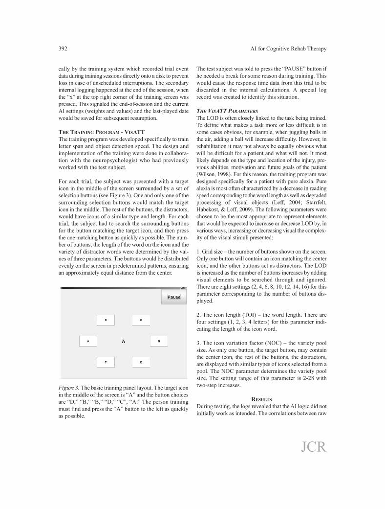
JCR
392 AI for Cognitive Rehab Therapy
cally by the training system which recorded trial event
data during training sessions directly onto a disk to prevent
loss in case of unscheduled interruptions. The secondary
internal logging happened at the end of the session, when
the “x” at the top right corner of the training screen was
pressed. This signaled the end-of-session and the current
AI settings (weights and values) and the last-played date
would be saved for subsequent resumption.
the trAInIng ProgrAm - vIsAtt
The training program was developed specifically to train
letter span and object detection speed. The design and
implementation of the training were done in collabora-
tion with the neuropsychologist who had previously
worked with the test subject.
For each trial, the subject was presented with a target
icon in the middle of the screen surrounded by a set of
selection buttons (see Figure 3). One and only one of the
surrounding selection buttons would match the target
icon in the middle. The rest of the buttons, the distractors,
would have icons of a similar type and length. For each
trial, the subject had to search the surrounding buttons
for the button matching the target icon, and then press
the one matching button as quickly as possible. The num-
ber of buttons, the length of the word on the icon and the
variety of distractor words were determined by the val-
ues of three parameters. The buttons would be distributed
evenly on the screen in predetermined patterns, ensuring
an approximately equal distance from the center.
The test subject was told to press the “PAUSE” button if
he needed a break for some reason during training. This
would cause the response time data from this trial to be
discarded in the internal calculations. A special log
record was created to identify this situation.
The VisATT PArAmeTers
The LOD is often closely linked to the task being trained.
To define what makes a task more or less difficult is in
some cases obvious, for example, when juggling balls in
the air, adding a ball will increase difficulty. However, in
rehabilitation it may not always be equally obvious what
will be difficult for a patient and what will not. It most
likely depends on the type and location of the injury, pre-
vious abilities, motivation and future goals of the patient
(Wilson, 1998). For this reason, the training program was
designed specifically for a patient with pure alexia. Pure
alexia is most often characterized by a decrease in reading
speed corresponding to the word length as well as degraded
processing of visual objects (Leff, 2004; Starrfelt,
Habekost, & Leff, 2009). The following parameters were
chosen to be the most appropriate to represent elements
that would be expected to increase or decrease LOD by, in
various ways, increasing or decreasing visual the complex-
ity of the visual stimuli presented:
1. Grid size – the number of buttons shown on the screen.
Only one button will contain an icon matching the center
icon, and the other buttons act as distractors. The LOD
is increased as the number of buttons increases by adding
visual elements to be searched through and ignored.
There are eight settings (2, 4, 6, 8, 10, 12, 14, 16) for this
parameter corresponding to the number of buttons dis-
played.
2. The icon length (TOI) – the word length. There are
four settings (1, 2, 3, 4 letters) for this parameter indi-
cating the length of the icon word.
3. The icon variation factor (NOC) – the variety pool
size. As only one button, the target button, may contain
the center icon, the rest of the buttons, the distractors,
are displayed with similar types of icons selected from a
pool. The NOC parameter determines the variety pool
size. The setting range of this parameter is 2-28 with
two-step increases.
resuLts
During testing, the logs revealed that the AI logic did not
initially work as intended. The correlations between raw
Figure 3. The basic training panel layout. The target icon
in the middle of the screen is “A” and the button choices
are “D,” “B,” “B,” “D,” “C”, “A.” The person training
must find and press the “A” button to the left as quickly
as possible.

JCR
393Wilms
response time and parameters were not significant
enough for the AI engine to detect changes in the per-
formance of the test subject, due to the fluctuations in
response time. This is a fairly common problem in psy-
chophysical experimental psychology when using raw
response time data to analyze correlations in tasks which
require a high degree of concentration and quick re-
sponses (Howitt & Cramer, 2005). Dampening the influ-
ence of noise in the data was therefore done using a
small, rolling, temporal median table of response times
for each parameter setting. Thus, the median value cal-
culation required a set of raw response times for each
setting of the parameters, but not the combinations of
settings. A fair amount of trial and error showed that the
median response time calculated on the basis of a small
rolling history of the five most recent response times for
each setting was sufficient to dampen noise and at the
same time maintain the responsiveness to changes in the
subject’s ability. This result was passed to the AI engine
instead of the raw response time.
the AI controLLed LeveL of dIffIcuLty
The selection results were compiled from the daily event
log files. Training tasks with wrong buttons activated
were removed from the results as were the records made
due to pressing the PAUSE button. The reason for this
was that these records were considered exceptions and
typically the cause of extreme outliers. Analysis showed
that erroneous button presses constituted less than 1% of
the total number of button presses and they were there-
fore determined by the author to be insignificant indica-
tions of performance in this case study.
Figure 4 shows the changes over time in the preferred
setting of the GS parameter controlling number of but-
tons. Initially, a setting of 2 is weighted highest and se-
lected most often, but already after six days of training
two buttons (GS=2) begins to be too easy and four but-
tons (GS=4) becomes the preferred setting. Towards the
end of the training period six buttons (GS=6) begin to be
chosen more and more often. The subject did not reach
a plateau during the three weeks of training.
The same pattern was seen for the second parameter, the
word length parameter (TOI). Figure 5 shows that to-
wards the end of the training period the settings 1 and 2
were favored above 3 and 4.
The last parameter, the NOC parameter, showed no cor-
relation between median response time and setting caus-
ing the AI engine to select settings totally at random (see
Figure 6).
dIscussIon
the use of AI for AdAPtIve controL In A cognI-
tIve trAInIng system
The research question in this project was whether or not
the chosen AI algorithm was suitable for controlling
and adapting LOD in a cognitive training program
under real-life conditions. A reinforcement learning al-
Figure 4. The weight assignment of the eight settings of the GS parameter (no. of buttons) during the three weeks
of training.

JCR
394 AI for Cognitive Rehab Therapy
gorithm was chosen as the AI method for two reasons,
its ability to learn in an unknown environment and its
fast learning rate. As the weight distribution for the pa-
rameter settings in Figures 4 and 5 showed, the AI en-
gine and logic managed to distinguish a group of
settings as being an appropriate LOD as opposed to oth-
ers that were either too hard or too easy. It also showed
that the selected settings changed as values changed
from day to day depending on the effects measured by
the system through response times. Figure 6 shows the
results from the third parameter, the NOC, which con-
trolled the variety of icons within the remaining dis-
tracters. This parameter never seemed to stabilize at
any preferring setting.
When considering the conditions of the environment in
which the AI engine managed to work, the use of AI
technology in cognitive training looks promising. The
AI engine itself was a simple implementation offering
many opportunities for improvements of the algorithms
Figure 5. The weight assignment of the four settings of the TOI parameter over the three weeks of training.
Figure 6. The weight assignment of the 14 settings of the NOC parameter over the last week of training.

JCR
395
used to control weights and determine fitness. In this
implementation the free assignment of weight values
worked to isolate optimum settings, but there may very
well be further possible improvements in using some
of the culling and clipping techniques that Spronck et
al. (2005) suggest in their research.
the eAsy/hArd settIng – the success crIterIon
The success of the AI engine in this study depends on
the measure of success used to rate the selected actions.
If these measures are not distinct, the agent has no way
of learning or change actions.
In this study the measure of success was determined by
the predefined Easy/Hard range against which the re-
sponse time was compared and selections subsequently
rated. However, the Easy/Hard setting turned out to be
difficult to estimate. If the Easy/Hard range was too
wide, it was too insensitive to changes in performance
related to individual parameter changes causing assign-
ment of the same fitness value to too many adjacent set-
tings of the parameters. This resulted in too much
variation in the selection of parameter settings. If on the
other hand, the Easy/Hard settings were too narrow, all
settings were judged to be either too easy or too hard
causing a leveling out of the influence of the weights
which resulted in random selection rather than selection
by appropriateness. As no established model for deter-
mining the settings existed, a set of guidelines was de-
veloped, which was based on observed results from
testing the programs on volunteering fellow students and
friends.
The heuristic nature of the guidelines, however, does not
ensure optimal settings, so this is definitely an area that
requires further investigation and automation.
Another subject for future research will be to investigate
whether the AI engine in fact needs an independent
measure of success for each parameter controlled. The
TOI parameter did not seem to converge around one or
two settings as the GS parameter did. One explanation
could be that the optimum Easy/Hard range for one pa-
rameter differed from the optimum Easy/Hard range of
another. Another explanation could be that three out of
four settings did in fact provide equal levels of difficulty.
It will require further tests and analysis to determine
whether this is in fact the case.
From another perspective, the Easy/Hard range could
be viewed as the primary parameter of LOD which
needs to be estimated, at least initially, in order to match
a certain level of ability. With further research it might
be possible to automate the estimation and initial set-
tings of the Easy/Hard range using the same AI engine,
but by turning the logic around so the Easy/Hard range
becomes the parameter to be estimated from a fixed task
set of one or more parameters in the parameter database.
Another possibility would be to let the therapist set the
range, but that would require some kind of assistive tool
for the therapist to be able to judge what would be suit-
able.
PArAmeter seLectIon
The decision to let the AI engine control three parameters
was in part based on the fact that in Spronck’s research
(2004, 2005, 2006) the AI solution was able to handle
this, and in part based on the fact that this would reflect
the conditions of challenging cognitive training. How-
ever, the relationship between success criteria and pa-
rameters in Spronck’s game were, in retrospect, much
simpler than those met in this study. In the case of
Spronck, his success was determined by a Boolean vari-
able (win or lose), whereas mine was determined by the
Easy/Hard range of response times which was directly
influenced by the parameters and indirectly by other fac-
tors outside my control. Also, the potential difference in
impact of the several parameters in combination was not
an issue in Spronck’s solution.
Although the use of three parameters did make the
study more demanding and raised new questions, it did
show that the AI engine was able to detect correlations
between response time and two of the parameters. In
an actual training environment there may be other pa-
rameters exerting influence on the response time during
a session. Secondary factors such as fatigue, motiva-
tion, concentration and the visuo-motor movements in-
volved in pressing buttons, may directly or indirectly
influence the performance of the test subject. In theory
however, the influence from these factors presumably
affect the parameters in equal measures like back-
ground noise.
This study faced some of the same difficulties facing
therapists when trying to translate the difficulties intro-
duced by impairment into trainable parameters control-
ling level of difficulty. As opposed to the modified fitness
function of Spronck’s design (Spronck, et al., 2006;
Spronck, et al., 2004; Spronck & van den Herik, 2005),
Wilms

JCR
396
the algorithms in this study had no way of ascertaining
the effect is of the individual parameters. This is defi-
nitely an area for further investigation and improvement
of the AI engine.
Trying to define a programmable logic for the engine has
made me even more convinced that technology can play
an important role in assisting therapists in planning, de-
signing and executing training.
LImItAtIons In the study
Due to the investigative nature of this study, there were
several areas that would require more appropriate control
in future studies. In particular, the noise created by the
physical arm movements of the subject. To create a real-
istic training environment, the buttons on the screen were
not laid out at the same physical distance from the center
of the target icon. The reason for this was that the subject
was allowed free movement of the arm and was not re-
quired to reposition the arm after each pointing trail, so
it was deemed less essential in the initial study. In future
studies, this is an area which could be improved upon.
However, the fact that the AI algorithm managed to ad-
just, despite this, does suggest that the algorithm is fairly
resilient to noise.
concLusIon
The results from this project demonstrate some of the
conditions to be met if training is to be controlled by the
AI actor-critic reinforcement logic (Sutton & Barto,
1998). In terms of controlling level of difficulty it means
that:
• The response time must be influenced by task difficulty.
The harder the task, the longer the response time. This
must be verified with patient data.
• Task difficulty must be expressible through a number
of parameters.
• The criterion for a challenging level of difficulty must
be defined as a response time range with an upper and
lower level within which the patient must operate in
order to progress.
• Each parameter must indicate a range of settings from
low to high LOD.
When considering the conditions of the environment in
which the AI engine managed to work, the use of AI
technology in cognitive training looks promising.
This study was explorative and intended to develop and
test AI controlled training to determine whether this type
of advanced computer technology could, in fact, be used
under the conditions of cognitive training. The conclu-
sions from this study were:
• That it was possible to develop an AI engine able to
measure and adjust LOD using reinforcement learning
methods.
• That it was possible for the AI engine to work under the
very difficult conditions of the real-life cognitive training
of a patient suffering from alexia. By introducing the tem-
poral median filter to dampen the noise in the raw input,
the AI engine did manage to control all three parameters
and show clear indications of being able to adjust LOD
as the patient’s skills improved. The weights controlling
the AI parameter selection converged to an optimum for
the two parameters controlling number of buttons and
length of word that had a clear correlation between meas-
ured median response time and LOD setting. The third
parameter controlling variety of words on the distractor
buttons that had no correlation showed an even distribu-
tion of weights across settings.
The next research step will be to test the AI engine under
different training conditions and with different training
programs. The hope is that this type of intelligent training
tool will assist in the wider study of the effects of inten-
sive, adaptive cognitive training of patients with cogni-
tive impairments after brain injury.
Acknowledgement
The author wishes to express gratitude to Dr. Tom Teas-
dale and Dr. Lisbeth Harms at the University of Copen-
hagen for valuable input during the production of this
article.
AI for Cognitive Rehab Therapy

JCR
397
references
Bourg, D., & Seemann, G. (2004). AI for Game Develop-
ers (1 ed.). Sebatsopol, CA.: O'Reilly Media
Inc.
Frassinetti, F., Angeli, V., Meneghello, F., Avanzi, S., &
Ladavas, E. (2002). Long-lasting amelioration
of visuospatial neglect by prism adaptation.
[Article]. Brain, 125, 608-623.
Galway, L., Charles, D., & Black, M. (2008). Machine
learning in digital games: a survey. [Review].
Artificial Intelligence Review, 29(2), 123-
161.
Howitt, D., & Cramer, D. (2005). Introduction to statistics
in Psychology (3 ed.). Essex, UK: Pearson Ed-
ucation Limited.
Kleim, J. A., & Jones, T. A. (2008). Principles of experi-
ence-dependent neural plasticity: Implications
for rehabilitation after brain damage. Journal of
Speech Language and Hearing Research,
51(1), S225-S239.
Leff, D. (2004). Alexia. Advances in clinical neuroscience
& rehabilitation, 4(3), 18-22.
Pedersen, P. M., Vinter, K., & Olsen, T. S. (2001). Im-
provement of oral naming by unsupervised
computerised rehabilitation. Aphasiology,
15(2), 151-169.
Ponsen, M., Spronck, P., Munoz-Avila, H., & Aha, D. W.
(2007). Knowledge acquisition for adaptive
game AI. Science of Computer Programming,
67(1), 59-75.
Pulvermüller, F., & Berthier, M. L. (2008). Aphasia ther-
apy on a neuroscience basis. Aphasiology,
22(6), 563-599.
Pulvermüller, F., Neininger, B., Elbert, T., Mohr, B., Rock-
stroh, B., Koebbel, P., et al. (2001). Constraint-
Induced Therapy of Chronic Aphasia After
Stroke. Stroke 2001, 32, 1621-1626.
Russell, S., & Norvig, P. (2003). Artificial Intelligence. A
Modern Approach (2 ed.). New Jersey, USA:
Pentice Hall.
Spronck, P., Ponsen, M., Sprinkhuizen-Kuyper, I., &
Postma, E. (2006). Adaptive game AI with dy-
namic scripting. Machine Learning, 63(3), 217-
248.
Spronck, P., Sprinkhuizen-Kuyper, I., & Postma, E.
(2004). Enhancing the performance of dynamic
scripting in computer games. In M. Rauterberg
(Ed.), Entertainment Computing - Icec 2004
(Vol. 3166, pp. 296-307).
Spronck, P., & van den Herik, J. (2005). A tutoring system
for commercial games. In F. Kishino, Y. Kita-
mura, H. Kato & N. Nagata (Eds.), Entertain-
ment Computing - Icec 2005 (Vol. 3711, pp.
389-400).
Starrfelt, R., Habekost, T., & Gerlach, C. (2010). Visual
processing in pure alexia: A case study. [Arti-
cle]. Cortex, 46(2), 242-255.
Starrfelt, R., Habekost, T., & Leff, A. P. (2009). Too Little,
Too Late: Reduced Visual Span and Speed
Characterize Pure Alexia. [Article]. Cerebral
Cortex, 19(12), 2880-2890.
Sturm, W., Willmes, K., Orgass, B., & Hartje, W.
(1997). Do Specific Attention Deficits Need
Specific Training? Neuropsychological Reha-
bilitation: An International Journal, 7(2), 81-
103.
Sutton, R. S., & Barto, A. (1998). Reinforcment Learning:
An Introduction (Adaptive Computation and
Machine Learning). Cambridge, MA.: MIT
Press.
Wertz, R. T., & Katz, R. C. (2004). Outcomes of com-
puter-provided treatment for aphasia. Aphasiol-
ogy, 18(3), 229-244.
Wilson, B. A. (1998). Recovery of cognitive functions fol-
lowing nonprogressive brain injury. Current
Opinion in Neurobiology, 8(2), 281-287.
Wilson, B. A., Gracey, F., Evans, J. J., & Bateman, A.
(2009). Neuropsychological Rehabilitation
Neuropsychological Rehabilitation: Theory,
Models, Therapy and Outcome. (1 ed.). New
York: Cambridge University Press.
Wilms


DISSIMILAR OUTCOMES OF APPARENTLY SIMILAR
PROCEDURES AS A CHALLENGE TO CLINICAL
NEUROREHABILITATION AND BASIC RESEARCH – WHEN
THE SAME IS NOT THE SAME
Inge Wilms1
and Jesper Mogensen2
1Center for Rehabilitation of Brain Injury, University of Copenhagen, and Department of
Psychology, University of Copenhagen 2
The Unit for Cognitive Neuroscience, Department of Psychology,
University of Copenhagen, Denmark
Send correspondence to: Jesper Mogensen
The Unit for Cognitive Neuroscience
Department of Psychology
University of Copenhagen
Oester Farimagsgade 2A
DK-1353 Copenhagen K
Denmark
Phone: (45) 35324873
Fax: (45) 35324802
e-mail: [email protected]

2
ABSTRACT
In the study of the brain and how it adapts to changes or injury, researchers sometimes come across
situations where apparently similar types of tests or training do not achieve similar outcome results.
This is true, in particular, within the field of computer-based rehabilitation where paper-and-pencil
tests and training is converted to computer. This paper raises the attention to the fact that
supposedly similar settings may not, in fact, elicit similar results and caution therapists and
researchers who work with rehabilitation of brain injury. The paper suggests that the underlying
mechanisms behind this may be illuminated by using the REF (Reorganization of Elementary
Functions) model and suggests that further research into the use of advanced technologies such as
computer-generated virtual reality is required.
KEYWORDS: computer-based rehabilitation, cognitive rehabilitation, REF-model, after-effect,
adaptive processes, brain injury, plasticity.

3
Introduction Cognitive rehabilitation training of brain injured patients is often a highly demanding endeavour –
not only with respect to the efforts required by the patient, but also with respect to the time-
demands on the therapist. New developments suggest that at least partial functional recovery may
be achieved through intensive training which challenges the impaired function through increasingly
more difficult tasks [e.g., Kleim & Jones, 2008; Pulvermüller & Marcelo, 2008]. However,
increased training will make even further demands on the scarce time of the therapist. A potential
solution to these increasing demands may be increased utilization of computers in the testing and
especially training of patients during posttraumatic recovery. An added benefit of such a
technological development will not only be precise and continuous collection of data but also carry
the potential for “online” adjustments of training (e.g. regarding the progression of level of
difficulty) and the delivery of consistent and timely feedback during training. Additionally,
computers may provide a safe – but real-life-like – training environment through the use of virtual
reality. Since the advance of the personal computers and minicomputers in the 1980’s there has
been a certain level of experimental introduction of computers in rehabilitation training and a
certain level of success has been achieved [Ansuini et al., 2006; Katz et al., 2005; Kim et al., 2007;
Smith et al., 2007; Webster et al., 2001]. Today, computers are being used extensively within
diverse areas of cognitive rehabilitation such as the modelling of damaged functions; assessment of
cognitive dysfunction; training and therapy; cognitive prosthetics and within assistive technologies.
However, the expanded use of computerized methods challenge our knowledge of how to
“translate” the traditional “paper-and-pencil” versions into computerized methods particularly
within the field of assessment and training of brain injured patients, At first glance, such an
endeavour may appear relatively unproblematic, but the process of translation requires a thorough
understanding and consideration of the underlying cognitive processes, which are involved and
activated during the execution of the traditional version of the test or training. If not, the subsequent
construction of a computer-based solution may inadvertently add or remove features, which may be
relevant or even vital for the therapy to be effective. Using computer-based solutions may change
the way the patient interacts during testing or training – for instance with regards to visuomotor and
visuospatial activity and feedback. This, in turn, may influence the effect of the training and
subsequent adaptation. The steps of the translational process may indeed prove significantly more
challenging as it basically rests on the assumption that a therapist or researcher is fully aware of all
the underlying cognitive processes involved in the outcome of a particular test or training
procedure. This emphasizes the need not only to conduct a thorough testing of the computer-based
solution itself but also to analyse whether the solution has the wanted impact on the cognitive
system.
In the following, we will provide examples, which illustrates that “the same is not always the same”
in terms of cognition and behaviour in what is assumed to be similar environments. Through the use
of the REF (Reorganization of Elementary Functions) model [Mogensen, 2011a, 2011c; Mogensen
& Malá, 2009], we will try to explain why apparently similar behaviours may not be associated with
activation of the same cognitive elements and neural processes in rehabilitation.
Dissimilar performance on apparently similar tests – human studies in neglect
Our understanding of the correlation between the tools used in assessment and the cognitive skills
being assessed is repeatedly being challenged by results from studies both on human subjects and
within the use of animal models.

4
In the rehabilitation of brain injured patients suffering hemispatial neglect, Prism Adaptation
Therapy (PAT) has been shown to be rather successful in ameliorating some of the effects of
neglect [Frassinetti et al., 2002; Rossetti et al., 1998]. In traditional PAT, the patient trains by
executing pointing movements with their hand from sternum to one of three spatially separated
targets. The upper extremities are hidden beneath a screen to prevent the patients from seeing the
actual movement. A PAT session consists of three steps: 1) A pre-exposure step in which the
patient is directed to point to either of the three target in a random fashion. The patient receives no
feedback on precision. 2) An exposure step in which the visual field of the patient is diverted
10 degrees to the right through the use of prism goggles. The patient receives feedback on pointing
precision by being allowed to see the fingertip when pointing at the targets. Most patients will
gradually adapt to the perceptual shift in the visual field during this step. 3) A post-exposure step
where the patient again removes the prism goggles and point to targets. The effect of the prism
adaptation is measured by observing the size of a relatively brief after-effect which manifests itself
as leftward off-target pointing movement after removing the prism goggles. The after-effect is
thought to be a measure how the visuomotor system has adapted to the brief change in the visual
field [Fermandez-Ruiz & Diaz, 1999]. A crucial element of this therapeutic procedure is the
feedback provided to the patient regarding the precision of the target pointing during the training
period [e.g., Frassinetti et al., 2002; Sarri et al., 2008; Serino et al., 2006, 2007].
A recent attempt to convert the traditional version of PAT to a computer-based version [Wilms &
Malá, 2010] clearly demonstrated that apparently similar executions did not result in similar effects.
In a number of experiments, both healthy subjects and patients suffering hemispatial neglect were
subjected to a single session of both a traditional and a computer-based session of PAT. The rather
surprising outcome of the direct comparison between results from the two sessions was that the size
of the after-effect depended on whether or not the subjects received direct feedback regarding the
pointing precision by actually seeing their fingertip. Other types of feedback via icons on the
computer screen failed to produce an after-effect of similar amplitude (ibid).
In an earlier study Luh [1995] wanted to determine if a leftward bias observed in line bisection
tasks for healthy test subjects was related to perceptual asymmetries or a bias in the motor
component. In one experiment 24 persons were given the same line bisection test in two versions,
i.e. one pencil-and-paper and one implemented on computer. The experiment demonstrated that the
observed bias from the manual line bisection task disappeared when the line bisection task was
performed on computer. Luh ascribed this difference in task outcome to the difference in motor
involvement (arm movement) in the two versions.
However, both examples illustrate that a seemingly insignificant difference may have a big
influence on the test subject and may lead to inconclusive results or even wrong assumptions about
the efficacy of computer-based therapy. The benefits of computerizing testing and maybe especially
rehabilitative training procedures are challenged in case such problems occur in the “translation” of
the traditional version into a computerized method. But – as will be discussed below – an even
broader perspective grows from such discrepancies.
Dissimilar performance on apparently similar tests – animal models
The phenomenon of different outcomes of what was believed to be identical procedures in training
and testing of neural and cognitive functions is, however, not restricted to the domain of
computerization of human procedures. Research within animal models of the cognitive

5
consequences of brain injury provides additional examples of situations in which “the same is not
the same”.
Lepore et al. [1985] tested in cats whether information relevant to the performance of a visual
discrimination task could be transferred subcallosally between the hemispheres. Cats subjected to
split-brain operations (lesions of the corpus callosum) trained a visual discrimination task with
visual information presented to only one hemisphere. Subsequently, the performance of the same
task was tested with visual information presented only to the contralateral (previously untrained)
hemisphere. A successful performance of the visual discrimination task under such circumstances
would indicate a subcallosal transfer of information. When the visual discrimination training was
performed in a classic two-choice discrimination box (a maze-type setup) in which food was offered
as reinforcement, no transfer of information between the two hemispheres was found. However,
when the animals were trained and tested in a “Lashley-type” jumping stand which punishes the
wrong choice by letting the animal jump into a locked door, animals subjected to the split-brain
procedure demonstrated an interhemispheric transfer of information. Although a number of aspects
differentiate the two procedures, a priori one would have expected the two to yield the same result.
An impaired performance of spatial delayed alternation tasks is often considered the diagnostic tool
when determining whether or not a group of experimental animals (be it rats, monkeys or other
species) have suffered a lesion or functional disturbance within the prefrontal cortex or associated
structures such as the prefrontal part of the neostriatum [e.g., Mogensen, 2003; Mogensen et al.,
2007, 2008]. In spatial delayed alternation tasks, two spatially separate target positions (e.g., arms
within a maze or cups towards which a monkey can reach) are offered to the animal. Initially, the
animal may freely select one or the other of these positions and will receive a reinforcement. After
this initial choice and a short delay, the animal is required to choose the previously non-chosen
position in order to receive a reinforcement. If the same position is selected, no reinforcement will
be given and a time-out period is imposed. On each subsequent trial, the animal is required to make
alternate selections of position (each time after a short delay). Repetitive visits to the same position
are never reinforced. Mogensen et al. [1987] tested if two variants of spatial delayed alternation
would reflect impairments of the prefrontal system. Rats subjected to lesions of the prefrontal part
of the neostriatum (in addition to undercutting of the prefrontal cortex) were tested in spatial
delayed alternation in both a T-maze and an operant chamber. In the T-maze, the animal chooses
between the two spatially separate arms of the T-shaped maze – and the animal is removed from the
apparatus between trials. In the operant chamber, the animal chooses between two spatially separate
retractable levers situated in a wall of the chamber. The animal remains in the chamber between
trials – but the levers are retracted. While the general requirements of a spatial delayed alternation
task were implemented in both experimental setups, only the T-maze based version was able to
significantly reflect the injury to the prefrontal system (although the same animals were tested in
both setups) [Mogensen et al., 1987].
As should be clear from these examples (further examples may be found in Mogensen [2003;
2011b]) even in animal models two implementations of the same task may reflect a particular type
of brain injury in vastly different manners. However, when subjected to a traditional analysis of the
cognitive demands, the two versions of the test may appear to be similar.
The types of results described above constitute a significant challenge within several fields related
to brain injury and neurorehabilitation. At the conceptual level, a major challenge is that although
presumably similar procedures of tests and training methods have been developed in order to fulfil

6
all currently known cognitive demands – and which falls within the existing cognitive classification
systems – the dissimilar outcomes testify against the validity of the prevailing understanding.
Clearly, basic research into brain injury and posttraumatic recovery is presented with a conceptual
and experimental challenge.
For this reason, a direct translation of established test and training procedures to computerized
methods in neurorehabilitation is likely to create more obstacles than may originally be expected. In
essence, it would require a full understanding of the cognitive mechanisms of traditional tests – as
well as the general process of posttraumatic functional recovery – to achieve a successful translation
of traditional tests into computerized procedures. Obviously, the development of individual test
procedures may not have to depend on a more complete conceptual understanding. For instance,
further research into the elements important with respect to the sight of the pointing finger in the
procedures addressed by Wilms and Malá [2010] may be able to pave the way for the development
of a more successful computerization of the PAT-procedure. Furthermore research aimed at
identifying – in humans as well as in animal models – the crucial aspects differentiating the
cognitive and neural demands of two test manifestations may provide data of importance in
understanding not only the individual tests, but the intricate functional reorganizations of the injured
brain. Consideration must therefore be given to whether the aforementioned conceptual
developments within neurorehabilitation are relevant to the presently discussed issues.
Some suggestions as to why two apparently similar procedures do not result in the same effect may
be found in a recently proposed neural and cognitive model [Mogensen, 2011a, 2011c; Mogensen
& Malá, 2009]. While primarily developed to account for the apparent contradiction between
regional functional specialization within the brain and the ability of the injured brain to
“functionally recover”, this model also carries important implications regarding clinical
neurorehabilitation and for the possibility of translating one manifestation of a test or training
procedure into another potentially computerized manifestation. It should be stressed that although
developed in the context of brain injury and posttraumatic rehabilitation, the REF-model describes
processes which are believed to be occurring in the intact brain as well as posttraumatically
[Mogensen, 2011a, 2011c].
The REF-model The concept of functional localization – the idea that various brain structures are uniquely
specialized in a particular type of information processing – is supported by a wealth of data. The
two major sources of such support are, (a) lesion studies in which brain injured patients or
experimental animals exhibit predictable patterns of behavioural, cognitive and/or emotional
changes after a particular type of brain injury and (b) neuroimaging studies in which particular brain
regions are rather consistently activated in cases of similar types of stimulation or activity. Such
local specialization of brain regions [e.g., Coltheart, 2001; Kringelbach & Rolls, 2004; Monakow,
1914; Selnes, 2001] does, however, appear to be contradicted by the fact that even in the
mechanically lesioned brain – where the lost brain structure is clearly not re-established – a more or
less complete level of posttraumatic functional recovery may occur [e.g., Buller & Hardcastle,
2000; Mogensen et al., 2004, 2007; Panksepp & Panksepp, 2000; Ramachandran & Blakeslee,
1998]. One of the goals of the REF-model [Mogensen, 2011a, 2011c; Mogensen & Malá, 2009] is
to explain how posttraumatic functional recovery is possible in spite of a strict functional
localization. In the REF-model, a distinction is made between two levels of “function”. On the one
hand, the term Elementary Function (EF) is introduced and defined as the information processing
performed by an individual brain structure or substructure. Such EFs are the basic information

7
processing units of the brain and, according to the REF-model, all the EFs mediated by a brain
region are permanently lost in case of irreversible injury to that region. On the other hand, the
“functions” typically characterized in the language of neuropsychology and neurology (e.g.,
episodic memory, expressive language, egocentric spatial orientation) are in the REF-model
considered to be “surface phenomena” of behaviour and/or consciousness. It is at the level of the
surface phenomenon that the functional recovery is observed – since a “function” is considered to
be “recovered” when the individual behaviourally manifests a performance indistinguishable from
the pretraumatic situation. The bridge between the EFs and the surface phenomenon is a kind of
cognitive/neural “program” named an Algorithmic Strategy (AS). An AS consists of a unique
combination of EFs and the interconnections between these. At the neural level, an AS is mediated
by the neural substrates of all its constituent EFs plus the projections and synaptic connections
mediating the information flow between these basic processing modules. At the cognitive level an
AS can be seen as a “program” in which the information processing is achieved by a unique
combination of (/information flow between) the basic information processing by EFs. It is the
activity of an AS that gives rise to a particular surface phenomenon – be it a behavioural
performance or a conscious manifestation. It is, however, also stressed by the REF-model that a
particular surface phenomenon may apparently be achieved by a multitude of ASs. That is:
although, in a strict sense, various ASs will manifest themselves in dissimilar surface phenomena,
the distinction between such surface phenomena may be impossible to realize by the individual
having the experience and/or an observer scoring the task performance of for instance a patient or
an experimental animal, unless a highly sophisticated analysis is performed [Mogensen, 2011a,
2011c; Mogensen & Malá, 2009].
When the brain is injured, all EFs mediated by the brain region affected by the damage are
irreversibly lost. And, consequently, all ASs within which these EFs are represented are lost as well.
Surface phenomena achieved via the activity of the lost ASs will become unavailable and what is
traditionally seen as the symptoms of that type of brain injury is the absence of those surface
phenomena, which are now unachievable. Subsequently, the posttraumatic functional recovery is
achieved by the creation and/or activation of alternative ASs. If these alternative strategies are able
to achieve surface phenomena, which are completely or at least to an extent indistinguishable from
those originally lost, the observed situation is a complete or partial functional recovery. These
alternative ASs must obviously consist of combinations of EFs mediated by the spared parts of the
brain. Neurally, they represent the neural substrate of such remaining EFs and the neural
connections allowing a relevant information flow between these localized circuits.
The posttraumatic process leading to a functional recovery always includes some type of
reorganization. The process may include plastic reorganizations at two separate levels. In all cases
there will be a cognitive and neural reorganization associating a particular
situation/problem/domain with the activation of a particular – alternative – AS. At the neural level
this cognitive association between a situation and the activation of an AS include synaptic
reorganizations within the parts of the brain mediating the processes of selection and evaluation of
cognitive/behavioural strategies. If one of the pretraumatically existing ASs is able to achieve a
satisfactory level of competence by its activation, this may be the only level of reorganization
necessary for that recovery process. If, however, no available AS turns out to be adequate, the
second level or reorganization is also called for. At this level – the “reorganization of elementary
functions”, which has given the REF-model its name – the EFs of the remaining parts of the brain
are reorganized and connected into novel ASs. This is achieved via mechanisms more or less
comparable to the backpropagation algorithm [e.g., Rumelhart & McClelland, 1986; Werbos,

8
1994]. Only when a novel AS has been created and selected will the recovery process be manifested
at the surface level. In these cases of recovery processes, patterns of synaptic connections are
modified even in the connections between the neural substrates of individual EFs.
As mentioned above and by Mogensen [2011a, 2011c], the processes described by the REF-model
are believed to occur in intact as well as injured brains. The mechanisms mediating posttraumatic
functional recovery are likely to have evolved as part of the neural and cognitive mechanisms
allowing flexibility and problem solving in the intact individual. Whenever somebody encounters a
situation demanding problem solving (in the broadest sense of the term) for which there has not
already been established a procedure/strategy leading to successful task solution, the described
mechanisms of search for an adequate AS and potentially even creation of a novel AS are initiated.
What is special about the posttraumatic situation is that even situations, for which solution
mechanisms were available pretraumatically, may now have the appearance of being “novel”
[Mogensen, 2011a, 2011c].
According to the REF-model [Mogensen, 2011a, 2011c; Mogensen & Malá, 2009] both of the
above mentioned levels of reorganization and plasticity are achieved in a constant interaction with
the environment. It is the environmental feedback during attempted task solution (in the broader
sense of the word) that provides the basis for the reorganizational and plastic processes.
Consequently, the plastic reorganization may be relatively specific to a specific environment.
The surface phenomenon – which in the present context means the performance on a particular
variant of a test or training procedure – reflects the selected AS. Viewed in this manner it may be
easier to understand why even apparently insignificant modifications of the procedures can have
drastic consequences with respect to the ability of a test to reflect the consequences of brain injury
and for that matter, the accomplishment of a positive therapeutic outcome through rehabilitative
training.
In the ongoing research aimed towards an improved understanding and characterization of the EFs,
studies addressing closely related test variants, which become differentially affected by a particular
type of brain injury, are a promising avenue. By being at the formal level so similar and still
differentially affected by brain injury, these procedures may reveal important information regarding
the ASs and their constituent elements. This in turn will provide information about how the brain
learns and adapts to changes in the environment and how training needs to be provided in order to
achieve a positive outcome.
The REF-model also carries a clear warning regarding the extent to which one should expect the
results of rehabilitative training to generalize across tasks. As emphasized above, by Mogensen
[2011a, 2011c] and by Mogensen and Malá [2009], novel ASs are constructed via backpropagation
mechanisms, which are the result of situational feedback regarding the success or failure of an
attempted task performance. Additionally, already existing ASs are selected for utilization in a
particular context via the feedback provided in that context. Given the specific nature of ASs, this
may bring into question to what an extent the task solution achieved in one situation can be applied
by the same individual in a different setting. This may be especially true in case of an injured brain
where the total repertoire of EFs and ASs is more limited than what would otherwise be the case.
The latter point emphasizes the final and potentially most problematic way in which
neurorehabilitation has to face situations in which “the same is not the same”. Although

9
rehabilitative cognitive training may be able to achieve a significant level of apparent functional
recovery under institutional or other relatively controlled settings, it may remain in question to what
an extent such a positive outcome is able to generalize to the real-life situations of the patient.
Specific training procedures – be it in a computerized environment or not – may become more and
more efficient. But unless it is constantly tested to what an extent these therapeutic results actually
generalize to the subsequent real-life situations of the patient, many of the results of
neuropsychological training of the brain injured individual may be of little or no benefits in other
contexts of the patient’s life. Therefore, it is important to emphasize two ways in which therapeutic
procedures can be improved.
Firstly, it needs to be consistently validated whether or not a rehabilitative training method
generalizes beyond the actual training situation. This validation process is in many ways a parallel
to what is being conducted within other fields of computerized training aimed at utilization in a
more real-life situation. One such issue is the question whether training performed in computer-
based flight simulators generalize to actual flight – which might be the case in some but clearly not
all instances [e.g., Gopher et al., 1994; Hart & Battiste, 1992]. If, in the brain injured patients,
generalization does not occur, it may be an indication that the perceived correlation between
observed surface phenomena, injury and therapy is not valid. Furthermore, in addition to the current
neurological and neuropsychological tests, it is essential to include assessments that can be
performed in a setting, which closely resembles real-life in order to determine the extent of damage
and subsequent improvement.
Secondly, in cases where the complexity of correlation between injury and observed surface
phenomena is poorly understood, the general rehabilitation program for each and every patient
should include attempts to assure the highest possible degree of “ecological validity” of the training
procedures. The rehabilitation programs will have to include elements, which bridge the
institutional training and setting to the subsequent life at home and at the workplace. Only by
exposure to the complexity of the real-life tasks in training can the brain be induced to form and
select ASs of value to the patient’s particular circumstances of life.
We speculate that the tendency towards computerization may – when properly applied – turn out to
be one of the elements in this bridge between institutionalised training and the subsequent life of the
patient. The use of portable computers and PDAs (potentially including the utilization of GPS-based
localization and navigation) may allow many types of rehabilitative training to continue beyond the
borders of institutions and into the real-life situation of the patient. Within the institution itself,
aspects of the real world life of the patient can be simulated and addressed even without leaving the
institution through the use of computer-simulations and virtual reality settings.
When building virtual realities, however, it has to be emphasized that even taking into consideration
all levels of present day knowledge regarding the essential components of a particular situation,
may be insufficient in achieving the desired rehabilitative goal. As the study of computerized
feedback in Prism Adaptation Training shows, something as simple as whether you see your own
finger or a representational “x” as feedback on a pointing task may in fact dramatically affect the
adaptation effect on the brain [Wilms & Malá, 2010]. Even in this promising development of a
merger between technology and neuropsychological rehabilitative training, clinicians and
researchers alike are challenged to look beyond the surface phenomena and remain open to the
ways in which normal and injured brains reorganize while interacting with the environment.

10
REFERENCES
1. Ansuini, C., Pierno, A. C., Lusher, D., & Castiello, U. (2006). Virtual reality applications for
the remapping of space in neglect patients. Restorative Neurology and Neuroscience, 24, 431-
441.
2. Buller, D. J., & Hardcastle, V. G. (2000). Evolutionary psychology, meet developmental
neurobiology: Against promiscuous modularity. Brain and Mind, 1, 307-325.
3. Coltheart, M. (2001). Assumptions and methods in cognitive neuropsychology. In: Rapp, B.
(Ed.). The Handbook of Cognitive Neuropsychology. Philadelphia PA: Psychology Press;
pp. 3-21.
4. Fernandez-Ruiz, J., & Diaz, R. (1999). Prism adaptation and aftereffect: specifying the
properties of a procedural memory system. Learning & Memory, 6, 47-53.
5. Frassinetti, F., Angeli, V., Meneghello, F., Avanzi, S., & Ladavas, E. (2002). Long-lasting
amelioration of visuospatial neglect by prism adaptation Brain, 125, 608-623.
6. Gopher, D., Weil, M., & Bareket, T. (1994). Transfer of skill from a computer game trainer to
flight. Human Factors, 36, 387-405.
7. Hart, S., & Battiste, V. (1992). Flight test of a video game trainer. Proceedings of Human
Factors Society 36th
Annual Meeting, pp. 1291-1295.
8. Katz, N., Ring, H., Naveh, Y., Kizony, R., Feintuch, U., & Weiss, P.L. (2005). Interactive
virtual environment training for safe street crossing of right hemisphere stroke patients with
Unilateral Spatial –Neglect. Disability and Rehabilitation, 27, 1235-1243.
9. Kim, J., Kim, K., Kim, D. Y., Chang, W.-H., Park, C. I., Ohn, S. H., Han, K., Ku, J.,
Nam, S. W., Kim, I. Y., & Kim, S. I. (2007). Virtual environment training system for
rehabilitation of stroke patients with unilateral neglect: crossing the virtual street.
Cyberpsychology & Behavior, 10, 7-15.
10. Kleim, J. A., & Jones, T. A. (2008). Principles of experience-dependent neural plasticity:
Implications for rehabilitation after brain damage. Journal of Speech, Language and Hearing
Research, 51, 225-239.
11. Kringelbach, M. L., & Rolls, E. T. (2004). The functional neuroanatomy of the human
orbitofrontal cortex: evidence from neuroimaging and neuropsychology. Progress in
Neurobiology, 72, 341-372.
12. Lepore, F., Ptito, M., Provencal, C., Bedard, S., & Guillemot, J-P. (1985). Interhemispheric
transfer of visual training in the split-brain cat: effects of the experimental set-up. Canadian
Journal of Psychology, 37, 527.
13. Luh, K. E. (1995). Line bisection and perceptual asymmetries in normal individuals: What
you see is not what you get. Neuropsychology, 9, 435-448
14. Mogensen, J. (2003). Animal models in neuroscience. In: Hau, J. & van Hoosier, G.L. (Eds.)
Handbook of Laboratory Animal Science, Second Edition, Volume II. Animal Models. Boca
Raton, FL: CRC Press LLC, pp. 95-109.
15. Mogensen, J. (2011a). Almost unlimited potentials of a limited neural plasticity: Levels of
plasticity in development and reorganization of the injured brain. Journal of Consciousness
Studies, in press.
16. Mogensen, J. (2011b). Animal models in neuroscience. In: Hau, J. & Schapiro, S. (Eds.)
Handbook of Laboratory Animal Science, Third Edition, Volume II. Animal Models. Boca
Raton, FL: CRC Press LLC, in press.
17. Mogensen, J. (2011c). Reorganization of the injured brain: Implications for studies of the
neural substrate of cognition. Frontiers in Psychology, 2:7, 1-10.
11
18. Mogensen, J., & Malá, H. (2009). Post-traumatic functional recovery and reorganization in
animal models. A theoretical and methodological challenge. Scandinavian Journal of
Psychology,50, 561-573.
19. Mogensen, J., Iversen, I.H., & Divac, I. (1987). Neostriatal lesions impaired rats’ delayed
alternation performance in a T-maze but not in a two-key operant chamber. Acta
Neurobiologiae Experimentalis, 47, 45-54.
20. Mogensen, J., Lauritsen, K. T., Elvertorp, S., Hasman, A., Moustgaard, A., & Wörtwein, G.
(2004). Place learning and object recognition by rats subjected to transection of the fimbria-
fornix and/or ablation of the prefrontal cortex. Brain Research Bulletin, 63, 217-236.
21. Mogensen, J., Hjortkjær, J., Ibervang, K. L., Stedal, K., & Malá, H. (2007). Prefrontal cortex
and hippocampus in posttraumatic functional recovery: Spatial delayed alternation by rats
subjected to transection of the fimbria-fornix and/or ablation of the prefrontal cortex. Brain
Research Bulletin, 73, 86-95.
22. Mogensen, J., Boyd, M. H., Nielsen, M. D., Kristensen, R. S., & Malá, H. (2008).
Erythropoietin improves spatial delayed alternation in a T-maze in rats subjected to ablation
of the prefrontal cortex. Brain Research Bulletin, 77, 1-7.
23. Monakow, C. V. (1914). Die Lokalisation im Grosshirn und der Abbau der Funktion durch
Kortikale Herde. Wiesbaden: Bergmann.
24. Panksepp, J., & Panksepp, J.B. (2000). The seven sins of evolutionary psychology. Evolution
and Cognition, 6, 108-131.
25. Pulvermüller, F., & Marcelo, L. B. (2008). Aphasia therapy on a neuroscience basis.
Aphasiology, 22, 563-599.
26. Ramachandran, V. S., & Blakeslee, S. (1998). Phantoms in the Brain: Probing the Mysteries
of the Human Mind. New York: William Morrow.
27. Rossetti, Y., Rode, G., Pisella, L., Farne, A., Li, L., Boisson, D., & Perenin, M. T. (1998).
Prism adaptation to a rightward optical deviation rehabilitates left hemispatial neglect. Nature,
395, 166-169.
28. Rumelhart, D., & McClelland, J. (1986). Parallel Distributed Processing. Cambridge, MA:
MIT Press.
29. Sarri, M., Greenwood, R., Kalra, L., Papps, B., Husain, M., & Driver, J. (2008). Prism
adaptation aftereffects in stroke patients with spatial neglect: Pathological effects on
subjective straight ahead but not visual open-loop pointing. Neuropsychologia, 46, 1069-
1080.
30. Selnes, O. A. (2001). A historical overview of contributions from the study of deficits. In:
Rapp, B. (Ed.). The Handbook of Cognitive Neuropsychology. Philadelphia PA: Psychology
Press; pp. 23-41.
31. Serino, A., Angeli, V., Frassinetti, F., & Ladavas, E. (2006). Mechanisms underlying neglect
recovery after prism adaptation. Neuropsychologia, 44, 1068-1078.
32. Serino, A., Bonifazi, S., Pierfederici, L., & Ladavas, E. (2007). Neglect treatment by prism
adaptation: What recovers and for how long. Neuropsychological Rehabilitation, 17, 657-687.
33. Smith, J., Hebert, D., & Reid, D. (2007). Exploring the effects of virtual reality on unilateral
neglect caused by stroke: four case studies. Technology and Disability, 19, 29-40.
34. Webster, J. S., McFarland, P. T., Rapport, L. J., Morrill, B., Roades, L. A., & Abadec, P. S.
(2001). Computer-assisted training for improving wheelchair mobility in unilateral neglect
patients. Archives of Physical Medicine and Rehabilitation, 82, 769-775.
35. Werbos, P. J. (1994). The Roots of Backpropagation: From Ordered Derivatives to Neural
Networks and Political Forecasting. New York: John Wiley & Sons.

12
36. Wilms, I., & Malá, H. (2010). Indirect versus direct feedback in computer-based Prism
Adaptation Therapy. Neuropsychological Rehabilitation, 20, 830-853.