prenatal testing: special tests for your baby during pregnancy
TRANSCRIPT
PRENATALTESTING
Special tests for your baby during pregnancy
CGE booklet cover 15x21_pic.indd 8 7/07/2009 9:31:44 AM
Includes Non-invasive Prenatal Testing (NIPT)
2017

CENTRE FOR GENETICS EDUCATION Level 5, 2c Herbert Street St Leonards NSW 2065 Ph: 02 9462 9599 Fax: 02 9906 7529 Email: [email protected] Website: www.genetics.edu.au
NSW Health’s Centre for Genetics Education aims to raise awareness of the role that genetics plays in family health, both for health professionals and the public.
The Centre can be contacted regarding the availability of a range of genetic services, information and resources. Information about genetics clinics and genetic support groups is also available.
This booklet is subject to copyright law. It may be reproduced in whole or in part for study or training purposes subject to the inclusion of an acknowledgement of the Centre. It may not be reproduced for commercial use or sale. Reproduction for purposes other than those indicated above requires written permission from the Centre.
© NSW Health’s Centre for Genetics Education 2017 ISBN 978-0-7347-3927-8
Further copies of this booklet are available from the Centre or can be downloaded from the Centre’s website www.genetics.edu.au
April 2017
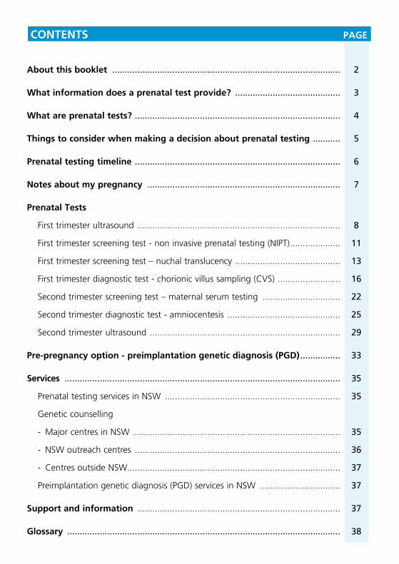
CONTENTS PAGE
About this booklet ........................................................................................... 2
What information does a prenatal test provide? .......................................... 3
What are prenatal tests? .................................................................................. 4
Things to consider when making a decision about prenatal testing ........... 5
Prenatal testing timeline .................................................................................. 6
Notes about my pregnancy ............................................................................. 7
Prenatal Tests
First trimester ultrasound ................................................................................. 8
First trimester screening test - non invasive prenatal testing (NIPT) .................... 11
First trimester screening test – nuchal translucency .......................................... 13
First trimester diagnostic test - chorionic villus sampling (CVS) ......................... 16
Second trimester screening test – maternal serum testing ............................... 22
Second trimester diagnostic test - amniocentesis ............................................. 25
Second trimester ultrasound ............................................................................ 29
Pre-pregnancy option - preimplantation genetic diagnosis (PGD) ................ 33
Services .............................................................................................................. 35
Prenatal testing services in NSW ...................................................................... 35
Genetic counselling
- Major centres in NSW ................................................................................... 35
- NSW outreach centres .................................................................................. 36
- Centres outside NSW..................................................................................... 37
Preimplantation genetic diagnosis (PGD) services in NSW ................................ 37
Support and information ................................................................................. 37
Glossary ............................................................................................................. 38

ABOUT THIS BOOKLET
This booklet is for couples who are planning a pregnancy or who are already pregnant and want further information about the prenatal (during pregnancy) tests available.
Every couple wants to have a healthy baby. However, there are some couples whose baby may have or will develop a serious physical and/or intellectual condition.
There are a number of different tests and procedures available to assess the health and development of a baby before birth. Each has advantages, disadvantages and limitations. The decision to undergo testing during a pregnancy is a very personal one and a decision best made based on all the available information.
It is important to remember that you do not have to have prenatal testing if you do not wish to.
HOW THIS BOOKLET CAN HELP YOU
This booklet provides information about the different types of prenatal tests available during different stages of pregnancy. You might find that there are similar tests that will provide the same information, but they are offered at different stages of pregnancy. For this reason, the booklet is set out to follow a simple time line of pregnancy that you will see on page 6.
You can make this booklet a tool to help you understand what prenatal tests are available during your pregnancy by completing the “Notes about my pregnancy” section on page 7.
A glossary is also provided (see pages 38-44) to explain words used that might not be familiar to you.
Since some prenatal tests are specialised, they might not be available at all medical facilities. If you are planning a pregnancy or are already pregnant, contact your family doctor or one of the prenatal testing services listed on pages 35-36 to find out what is available in your area.
2
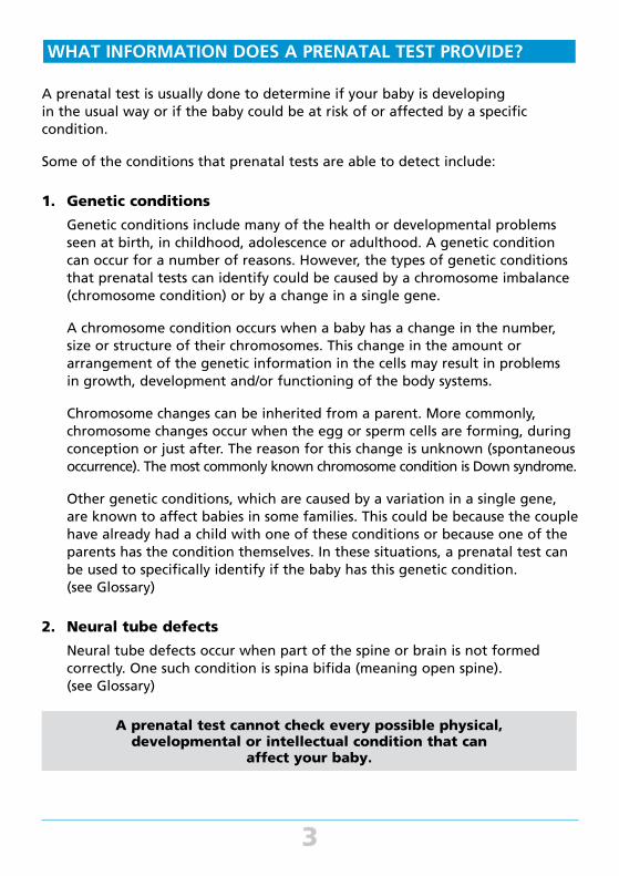
WHAT INFORMATION DOES A PRENATAL TEST PROVIDE?
A prenatal test is usually done to determine if your baby is developing in the usual way or if the baby could be at risk of or affected by a specific condition.
Some of the conditions that prenatal tests are able to detect include:
1. Genetic conditions
Genetic conditions include many of the health or developmental problems seen at birth, in childhood, adolescence or adulthood. A genetic condition can occur for a number of reasons. However, the types of genetic conditions that prenatal tests can identify could be caused by a chromosome imbalance (chromosome condition) or by a change in a single gene.
A chromosome condition occurs when a baby has a change in the number, size or structure of their chromosomes. This change in the amount or arrangement of the genetic information in the cells may result in problems in growth, development and/or functioning of the body systems.
Chromosome changes can be inherited from a parent. More commonly, chromosome changes occur when the egg or sperm cells are forming, during conception or just after. The reason for this change is unknown (spontaneous occurrence). The most commonly known chromosome condition is Down syndrome.
Other genetic conditions, which are caused by a variation in a single gene, are known to affect babies in some families. This could be because the couple have already had a child with one of these conditions or because one of the parents has the condition themselves. In these situations, a prenatal test can be used to specifically identify if the baby has this genetic condition. (see Glossary)
2. Neural tube defects
Neural tube defects occur when part of the spine or brain is not formed correctly. One such condition is spina bifida (meaning open spine). (see Glossary)
A prenatal test cannot check every possible physical, developmental or intellectual condition that can
affect your baby.
3
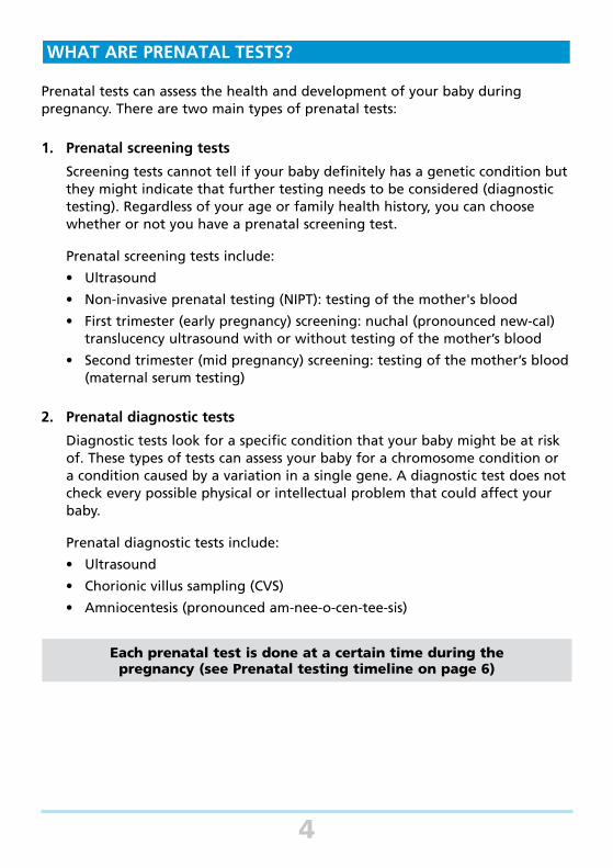
WHAT ARE PRENATAL TESTS?
Prenatal tests can assess the health and development of your baby during pregnancy. There are two main types of prenatal tests:
1. Prenatal screening tests
Screening tests cannot tell if your baby definitely has a genetic condition but they might indicate that further testing needs to be considered (diagnostic testing). Regardless of your age or family health history, you can choose whether or not you have a prenatal screening test.
Prenatal screening tests include:
• Ultrasound
• Non-invasive prenatal testing (NIPT): testing of the mother's blood
• First trimester (early pregnancy) screening: nuchal (pronounced new-cal) translucency ultrasound with or without testing of the mother’s blood
• Second trimester (mid pregnancy) screening: testing of the mother’s blood (maternal serum testing)
2. Prenatal diagnostic tests
Diagnostic tests look for a specific condition that your baby might be at risk of. These types of tests can assess your baby for a chromosome condition or a condition caused by a variation in a single gene. A diagnostic test does not check every possible physical or intellectual problem that could affect your baby.
Prenatal diagnostic tests include:
• Ultrasound
• Chorionic villus sampling (CVS)
• Amniocentesis (pronounced am-nee-o-cen-tee-sis)
Each prenatal test is done at a certain time during the pregnancy (see Prenatal testing timeline on page 6)
4
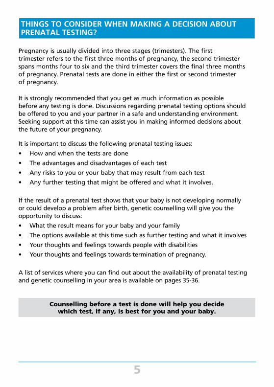
THINGS TO CONSIDER WHEN MAKING A DECISION ABOUT PRENATAL TESTING?
Pregnancy is usually divided into three stages (trimesters). The first trimester refers to the first three months of pregnancy, the second trimester spans months four to six and the third trimester covers the final three months of pregnancy. Prenatal tests are done in either the first or second trimester of pregnancy.
It is strongly recommended that you get as much information as possible before any testing is done. Discussions regarding prenatal testing options should be offered to you and your partner in a safe and understanding environment. Seeking support at this time can assist you in making informed decisions about the future of your pregnancy.
It is important to discuss the following prenatal testing issues:
• How and when the tests are done
• The advantages and disadvantages of each test
• Any risks to you or your baby that may result from each test
• Any further testing that might be offered and what it involves.
If the result of a prenatal test shows that your baby is not developing normally or could develop a problem after birth, genetic counselling will give you the opportunity to discuss:
• What the result means for your baby and your family
• The options available at this time such as further testing and what it involves
• Your thoughts and feelings towards people with disabilities
• Your thoughts and feelings towards termination of pregnancy.
A list of services where you can find out about the availability of prenatal testing and genetic counselling in your area is available on pages 35-36.
Counselling before a test is done will help you decide which test, if any, is best for you and your baby.
5
Birth
6
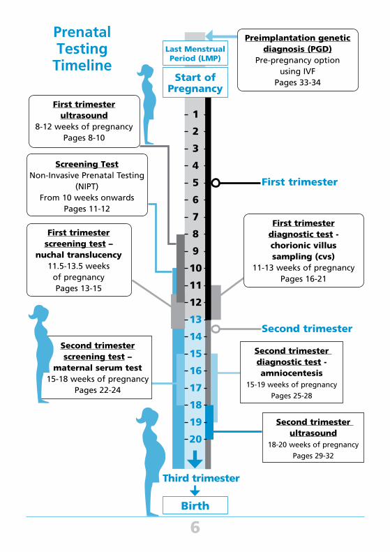
First trimester
ultrasound
8-12 weeks of pregnancy
Pages 8-10
Screening Test
Non-Invasive Prenatal Testing
(NIPT)
From 10 weeks onwards
Pages 11-12
First trimester
screening test –
nuchal translucency
11.5-13.5 weeks
of pregnancy
Pages 13-15
First trimester
diagnostic test -
chorionic villus
sampling (cvs)
11-13 weeks of pregnancy
Pages 16-21
Second trimester
screening test –
maternal serum test
15-18 weeks of pregnancy
Pages 22-24
Second trimester
diagnostic test -
amniocentesis
15-19 weeks of pregnancy
Pages 25-28
Second trimester
ultrasound
18-20 weeks of pregnancy
Pages 29-32
Preimplantation genetic
diagnosis (PGD)
Pre-pregnancy option
using IVF
Pages 33-34
Prenatal Testing
TimelineLast Menstrual Period (LMP)
Start of Pregnancy
1
2
3
4
5
6
7
8
9
10
11
12
13
14
15
16
17
18
19
20
First trimester
Second trimester
Third trimester

7
Date of the first day of my last menstrual period (LMP):
My expected date of delivery (EDD):
Appointments:
Contacts:
Notes:
NOTES ABOUT MY PREGNANCY
First trimester ultrasound8-12 weeks of pregnancy
- An important step if you are considering having testing later in your pregnancy
- A safe way of assessing the growth of your baby in the first trimester of pregnancy
- Does not pose any health risk to you or your baby
- First trimester ultrasounds, like all other prenatal tests, are optional
What is first trimester ultrasound?
Ultrasound uses harmless high frequency sound waves to produce images of your developing baby in the uterus (womb).
During an ultrasound, a transducer (that acts like a microphone) is used to generate sound waves that produce images on a screen. The transducer can do this either through the mother’s abdomen or vagina. Your doctor or sonographer (specially trained ultrasound technician) will discuss which procedure is best for you.
During an abdominal ultrasound, the doctor or sonographer will use a water based jelly-like substance and move the transducer over your abdomen. Sound waves pass from the transducer into the uterus and through the fluid surrounding the baby (called amniotic fluid). The sound waves bounce harmlessly off the baby, creating echoes.
For abdominal ultrasound to be most successful, a full bladder is needed. The uterus and ovaries are often hidden behind the bowel, making it difficult to clearly see inside the uterus. However, when the bladder is full the bowel is pushed out of the way. It is recommended that you have several glasses of non-fizzy fluid one hour before the ultrasound test.
During a vaginal ultrasound, the transducer is passed into the vagina using sterile techniques. The sound waves pass into the uterus to create the same echoes as described above. For vaginal ultrasound it is best if the bladder is empty.
8

Why should I consider having a first trimester ultrasound?
The most common reasons for having a first trimester ultrasound are to:
• Check or confirm how many weeks the pregnancy has progressed
• See if there is more than one baby in the uterus
• View the position of the baby and the placenta
• Check on the wellbeing of the baby when there has been a complication such as bleeding.
A first trimester ultrasound my also be referred to as a dating ultrasound.
When is the first trimester ultrasound done?
An ultrasound scan can be done at any time during the pregnancy. The best time for determining the date and stage of pregnancy, and the number of babies in the uterus is between 8 and 12 weeks.
When will the result of the first trimester ultrasound be available?
In some cases, information about your baby’s ultrasound scan will be made available immediately. However, if the person doing the scan suspects that your baby has a problem, he or she may not be able to discuss the findings until they have been verified by another specialist. The result might be sent to the referring doctor who can discuss the result with you.
It might be useful to discuss how the result will be given, to whom and when, with the sonographer or doctor before the ultrasound.
What will the result of the first trimester ultrasound tell me?
The first trimester ultrasound will provide accurate dating of a pregnancy which is often needed if a woman is considering having a prenatal test later in the pregnancy.
How reliable is the first trimester ultrasound?
A first trimester ultrasound cannot check for every possible physical, developmental or intellectual problem that could affect your baby. First trimester ultrasound is very reliable at determining how advanced the pregnancy is and how many babies there are.
9
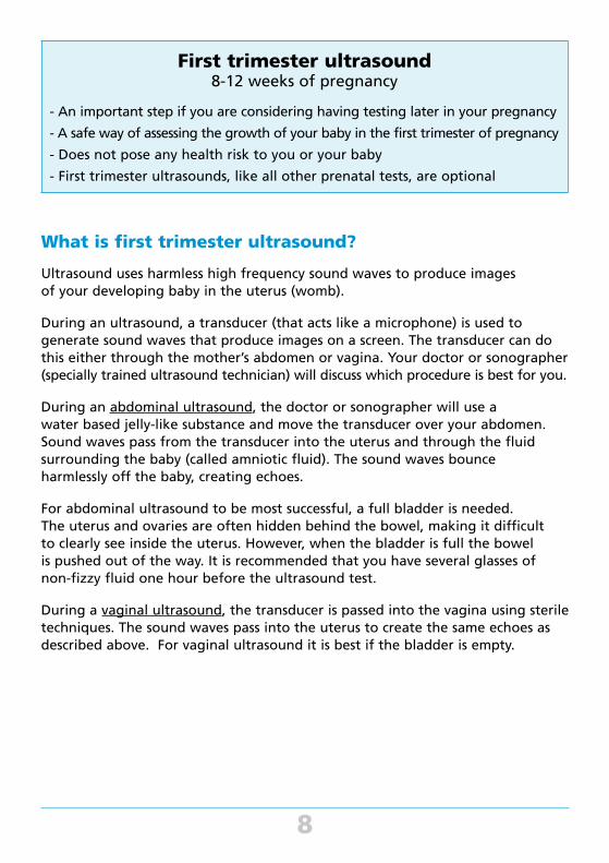
What if the result of the first trimester ultrasound shows my baby has a problem?
In a small number of cases, an ultrasound scan done early in the pregnancy might show that the baby either has or is at risk of having a problem. A more specialised ultrasound scan to determine whether or not the baby actually has a problem might be suggested. Alternatively, depending on how advanced the pregnancy is, the doctor might also discuss the option of having one of the diagnostic prenatal tests (see CVS and amniocentesis, pages 16-28).
Can the first trimester ultrasound harm me or my baby?
No evidence has been found that shows an ultrasound can harm you or your baby.
Discussing your result and options with your doctor, midwife or genetic counsellor will help you to decide if you want
further testing and if so, which test is best for you.
10
ULTRASOUND SCAN AT 11.5 WEEKS
BABY’S HEAD
BABY’S BODY
Screening Test - non-invasive prenatal testing (NIPT) 10 weeks onwards
- Screening test for Down syndrome and some other chromosome conditions
- Involves a special blood test
- Provides an accurate estimate of whether or not your baby has Down syndrome or some other chromosome conditions
- Does not pose any health risk to you or your baby
- NIPT is a new test - currently only offered through some specialist centres
- NIPT, like all other prenatal tests, is optional
What is non-invasive prenatal testing?
Non-invasive prenatal testing (NIPT) is a screening test used to determine the likelihood of your baby having Down syndrome (see Glossary – Page 38) or some other chromosome conditions.
The test can be used from 10 weeks of pregnancy and requires a sample of the mother’s blood. During pregnancy, some of the DNA from the baby (called foetal DNA) crosses into the mother’s bloodstream. This foetal DNA carries the baby’s genetic information. It is this foetal DNA in the mother’s blood that is analysed.
Why should I consider having non-invasive prenatal testing?
This test is highly accurate for detecting Down syndrome. It is also effective at detecting some other chromosome conditions. NIPT is a safe way to screen for these conditions during your pregnancy.
When is non-invasive prenatal testing done?
The test can be used as early as 10 weeks of pregnancy.
When will the result of non-invasive prenatal testing be available?
In most circumstances it will take 2 weeks for you to receive a result. In a small number of cases the lab is not able to provide a result and if this occurs, your doctor, midwife or genetic counsellor can discuss alternative testing options with you.
11
What will the result of non-invasive prenatal testing tell me?
A negative, normal or low risk result indicates that the baby is unlikely to be affected by any of the chromosome conditions included in the screen. A positive, abnormal or high risk result indicates that the baby is likely to be affected by the specified chromosome condition.
The risk result you receive is based on factors in your current pregnancy only. This risk is worked out based on the foetal DNA obtained from the mother’s blood.
Talk to your doctor, midwife or genetic counsellor about exactly what the testing service they use will be looking for. As NIPT becomes more available, there may be new health conditions being included in the screening of the foetal DNA.
How reliable is non-invasive prenatal testing?
As with all screening tests, the results of NIPT will not give a 100% definite answer about whether or not your baby has a chromosome condition. NIPT has been shown to be highly accurate in identifying babies with Down syndrome. The effectiveness may depend on the way in which the testing is done and where. Your doctor, midwife or genetic counsellor can inform you about the reliability of the test for Down syndrome and other chromosome conditions.
What if the result of the NIPT shows my baby might have a problem?
If the NIPT result shows your baby might have a chromosome condition, then you may want to consider having a diagnostic test like chorionic villus sampling (CVS) (see page 16-21) or amniocentesis (see page 25-28) to confirm the result. Your doctor, midwife or genetic counsellor can discuss these options with you.
Can the NIPT harm me or my baby?
The test is conducted using a sample of the mother’s blood and does not pose any health risk to the mother or the baby.
12
First trimester screening test – nuchal translucency11.5-13.5 weeks of pregnancy
- Screening test for Down syndrome and some other chromosome conditions
- Involves a specialised ultrasound with or without a blood test
- Estimates the chance of your baby having a chromosome condition such as Down syndrome
- Identifies 75-90% of babies with Down syndrome
- Does not pose any health risk to you or your baby
- First trimester screening tests, like all other prenatal tests, are optional
What is first trimester screening?
This screening test is used to determine the likelihood of your baby having Down syndrome (see Glossary – Page 38) and some other chromosome conditions.
Between 11.5 and 13.5 weeks a specialised ultrasound will be used to measure your baby’s nuchal translucency (a fluid-filled space at the back of your baby’s neck).
At this early stage of the pregnancy, your baby’s skin is very thin and fluid can be seen in the space between the skin and tissue at the back of the neck. The depth of the fluid in this space can be measured using ultrasound.
An additional blood test might also be done but is not always offered or available. This special blood test is not offered on its own at present, but only combined with the nuchal translucency ultrasound.
First trimester screening is also called nuchal translucency screening (NT screening or NT Plus when it includes the blood test).
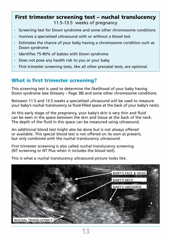
This is what a nuchal translucency ultrasound picture looks like.
13
NUCHAL TRANSLUCENCY
BABY’S FACE & HEAD
BABY’S NECK
BABY’S ABDOMEN
Why should I consider having first trimester screening?
First trimester screening is considered to be a safe way that a woman of any age can screen a pregnancy in the first trimester for certain chromosome conditions.
When is first trimester screening done?
The measurement of nuchal translucency using ultrasound is done between 11.5 and 13.5 weeks of pregnancy. Only doctors or sonographers with special training should take this measurement (see Services: Prenatal testing on page 35).
If you are also having the special blood test, it can be done before or at the same time as the nuchal translucency ultrasound. The blood test will measure the levels of specific proteins in your blood and when used in combination with the nuchal translucency ultrasound result, improves the accuracy of this screening test. It is important that the blood test result is calculated using the correct weeks of pregnancy and accurate maternal weight.
When will the result of first trimester screening be available?
If you are having the nuchal translucency ultrasound without the blood test, you may be given a result immediately.
If first trimester screening includes a blood test, the result will usually be available one week later.
What will the result of first trimester screening tell me?
The result of the first trimester screening test will be given to you in the form of a risk number (such as a chance of 1 in 100 or 1 in 500) that the baby has a chromosome condition.
The risk result you receive will be your own individually calculated risk based on factors in this individual pregnancy only. This number is worked out by a computer program using the thickness of the nuchal translucency and several other factors including maternal age and stage of pregnancy. If you have also had the special blood test, this is taken into account and provides a more accurate estimate.
A risk of 1 in 300 or greater (e.g. 1 in 150) is considered to be in the “increased risk” category.
You can discuss your result with your doctor, midwife or genetic counsellor and decide if you would like to have further testing in this pregnancy.
14
15
How reliable is first trimester screening?
This is a screening test and therefore will not give a definite answer about whether or not your baby has a chromosome condition.
Nuchal translucency alone: If you have the nuchal translucency ultrasound on its own, the test is around 75% accurate at identifying babies that have a chromosome condition like Down syndrome and they will receive a result in the “increased risk” category. Therefore, around 25% of babies with a chromosome condition will not be identified using nuchal translucency.
Nuchal translucency plus the special blood test: The nuchal translucency ultrasound together with the special blood test is around 80 to 90% accurate at detecting babies that have a chromosome condition like Down syndrome and they will receive a result in the “increased risk” category. Therefore, around 10 to 20% of babies with a chromosome condition will not be identified using this test.
It is important to note that an increased risk result does not mean your baby will definitely have a chromosome condition. Likewise, if your result is not in the increased risk category, your baby could still have a chromosome or other health condition.
What if the result of first trimester screening shows my baby has a problem?
About 5% of women (1 in 20) who have this test will receive an increased risk result. However, most of these babies will not have a chromosome condition.
If the result of first trimester screening suggests your baby is at an increased risk of having a chromosome condition or if you would like to have further testing, you may consider having either chorionic villus sampling (CVS) (see page 16-21) or an amniocentesis (see page 25-28). Any concerns you have should be discussed with your doctor, midwife or genetic counsellor.
Can first trimester screening harm me or my baby?
The first trimester screening test does not pose any health risk to you or your baby.

First trimester diagnostic test - chorionic villus sampling (CVS)
11-13 weeks of pregnancy
- Diagnostic test which gives an accurate result
- A sample of the chorion (placenta) is collected and tested to determine if the baby has certain genetic conditions
- Less than 1% of women (1 in 100) will have a miscarriage as a result of having a CVS
- CVS testing, like all other prenatal tests, is optional
What is chorionic villus sampling (CVS)?
Chorionic villus sampling (CVS) is a diagnostic test that uses a sample of the chorion (which develops into the placenta) to determine if your baby has certain genetic conditions. The cells that make up the chorion are mostly the same as the cells of your baby as they both originated from the developing embryo in early pregnancy.
There are two different methods that can be used to test the chorion. The sample can be collected either through the mother’s abdomen (transabdominal) or the vagina (transvaginal). The most appropriate and safest method will depend on the position of the baby and the placenta and will be discussed with you before the procedure.
Transabdominal CVS uses a fine needle passed through your abdomen to obtain a sample of chorion. A local anaesthetic may be used on your skin prior to the procedure to numb the area where the needle is inserted. During the procedure, ultrasound is used to guide the specialist and enable the test to be carried out in the safest way.
16

The picture below shows a side on view of a transabdominal CVS.
Using a tiny sample of the chorion, the test will usually examine your baby’s chromosomes unless your pregnancy is at risk of another type of genetic condition (See Genetic Conditions in the Glossary).
An ultrasound scan is done before and during the test to enable the specialist performing the test to locate the position of the chorion. It also allows them to watch the sampling on the screen and makes sure the procedure is carried out as safely as possible.
The amount of chorion needed for the test is extremely small. Only about 0.001% (1 in 1000) of the total chorion is sampled during this test.
Source: After Vogel, F. & Motulsky, A.G. (1986). Human Genetics, 2nd end. Springer-Verlag, Berlin.
17
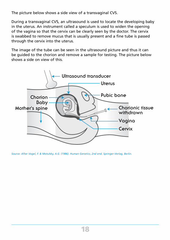
The picture below shows a side view of a transvaginal CVS.
During a transvaginal CVS, an ultrasound is used to locate the developing baby in the uterus. An instrument called a speculum is used to widen the opening of the vagina so that the cervix can be clearly seen by the doctor. The cervix is swabbed to remove mucus that is usually present and a fine tube is passed through the cervix into the uterus.
The image of the tube can be seen in the ultrasound picture and thus it can be guided to the chorion and remove a sample for testing. The picture below shows a side on view of this.
Source: After Vogel, F. & Motulsky, A.G. (1986). Human Genetics, 2nd end. Springer-Verlag, Berlin.
18
Why should I consider having a CVS?
• You received an increased risk result on a first trimester screening test (see pages 13-15)
• You are over 35 years old. The risk of having a baby with a chromosome condition increases with the mother’s age (for further details see “Chromosome condition” in the Glossary section of this booklet).
• You or your partner are known to be at risk of passing on a chromosome condition or other type of genetic condition caused by a gene variation. When the CVS is done to check for a gene variation, the laboratory will need to establish a gene test which can then be used during the CVS. This type of testing is very specialised and usually organised by a genetic counsellor.
When is a CVS done?
A CVS is usually done between 11 and 13 weeks of pregnancy. In special circumstances, the CVS might be carried out later in your pregnancy. However, it should not be done before 10 weeks of pregnancy.
When will the result of a CVS be available?
It might take two to three weeks to receive your CVS result as it can be a lengthy process. After a sample of chorion has been collected from you, it is sent to a specialised laboratory where the testing is done. The process involves growing the cells in a sterile environment and is, therefore, a time consuming method. If the test is looking at the chromosomes, the laboratory may issue an initial result within two to three days. This will provide a result for the common chromosome problems only. The final result will still take to two to three weeks.
19

What will the result of a CVS tell me?
It is important to understand what information the test will identify. Depending on why you are having the test, the laboratory can focus on specific conditions e.g. if you are having the test because you are an older mother, the main conditions that would be of concern are chromosome conditions.
For other women, they may have previously had a child with a specific genetic condition caused by a gene variation or a specific chromosome change. In this case, the laboratory would analyse that particular gene or look carefully at the chromosome in question in the developing baby.
Although doctors and scientists are now able to diagnose a large number of conditions during pregnancy, it is not possible to test for all of the problems that could affect a developing baby. Your doctor or genetic counsellor will discuss with you the range of conditions that can be identified using a CVS test and what each of these might mean for you and your baby.
It is important to remember that a normal test result does not exclude every possible problem with the baby.
How reliable is a CVS?
The CVS test is very accurate at identifying the conditions your baby is being tested for.
However, as the test is done early in pregnancy the chorion, which develops into the placenta, is immature. Occasionally, the doctor might be unable to obtain enough chorion on the first attempt. Samples of the chorion will be examined immediately to make certain there is enough chorion for the laboratory to work with. If not, another sample may need to be taken at this time.
In rare cases, the laboratory might be unable to get a definite answer and a second CVS will have to be done at a later time. In other cases, there will be a need for an amniocentesis to be done to check the CVS result. This means that a very small number of women may need to have both a CVS and an amniocentesis in the same pregnancy. For further information about amniocentesis (see pages 25-28).
20
What if the result of the CVS shows my baby has a problem?
If a CVS result shows that your baby has a problem, you and your partner will be given as much information as possible about the condition. This includes the implications it might have for the future health or development of the baby. You will be given time to make an informed choice about whether or not you would like to continue the pregnancy.
If you and your partner decide to continue the pregnancy following the diagnosis of a health or developmental problem with your baby, you will both be offered support and information before, during and after the birth of the baby.
If you decide to terminate the pregnancy, the procedure might require a general anaesthetic and/or hospitalisation. The method of termination will vary depending on how advanced the pregnancy is and the policy of the particular hospital or clinic. All aspects should be fully discussed with your doctor or genetic counsellor. You will be offered support and information before, during and after your hospital stay.
Please see the services support and information section on page 35-36 of this booklet for more details.
Can a CVS harm me or my baby?
The increase in risk of miscarriage due to having a CVS is < 1% (1 in 100 pregnancies tested with a CVS). This risk is in addition to your “background risk” of miscarriage that all women have in early pregnancy due to natural causes. It is important to discuss your background risk of miscarriage with your doctor, midwife or genetic counsellor.
The specific risk figure for a miscarriage depends on the experience of the doctor doing the test and the difficulty he or she has in obtaining the sample of chorion. It is therefore important that a CVS test is only carried out by a doctor experienced in this technique and should be done after the 11th week of pregnancy (see the Services: Prenatal testing list on page 35).
Some women will experience cramping and occasionally some vaginal bleeding after a CVS. Talk to your doctor about what you can expect after the testing and what symptoms to look out for.
21
Second trimester screening test – maternal serum testing
15-18 weeks of pregnancy
- Screening test for Down syndrome and neural tube defects
- Involves having a blood test
- Provides you with an estimate of whether or not your baby has a chromosome condition such as Down syndrome
- Identifies 60-75% of babies with Down syndrome
- Identifies 95-100% of babies with a neural tube defect
What is second trimester screening?
Second trimester screening (also known as the maternal serum triple test or maternal serum quadruple test, depending on the number of hormones measured) is used to determine the likelihood of your baby having a chromosome condition such as Down syndrome or a neural tube defect such as spina bifida (see Glossary).
It can only estimate the chance of your baby having a chromosome condition or neural tube defect, not provide a definite answer.
Second trimester screening uses a specialised blood test to measure the levels of several proteins in your blood (maternal serum). The levels of these proteins, combined with other factors such as your age, can provide an estimate of the likelihood that your baby has Down syndrome or a neural tube defect such as spina bifida.
Why should I have second trimester screening?
Second trimester screening is considered to be a safe way to screen a pregnancy in the second trimester for certain chromosome conditions and neural tube defects.
When is second trimester screening done?
The second trimester screening test involves having a blood test between 15 and 18 weeks of pregnancy.
22
23
When will the result of second trimester screening be available?
The results will usually be available one week following blood collection. At this time, you will have the opportunity to discuss them with your doctor, midwife or genetic counsellor and decide if you would like to have further testing in your pregnancy.
What will the result of second trimester screening tell me?
The second trimester screening test provides you with an estimated chance of your baby having a chromosome condition such as Down syndrome or a neural tube defect such as spina bifida in this pregnancy. The result will be given to you in the form of a number such as a chance of 1 in 100 or 1 in 500 that the baby has one of these conditions.
A risk of 1 in 250 or greater (e.g. 1 in 100) is considered to be in the “increased risk” category.
This number is worked out by a specialist testing laboratory using the result of the blood test as well as other factors such as your age and stage of pregnancy.
How reliable is second trimester screening?
This is a screening test and therefore will not give a definite answer about whether or not your baby has a chromosome condition.
The second trimester screening test is around 60-75% accurate at identifying babies that have a chromosome condition like Down syndrome, depending on the number of pregnancy hormones measured. They will receive a result in the “increased risk” category. Therefore, around 25-40% of babies with a chromosome condition will not be identified using this test.
It is important to note that an increased risk result does not mean your baby will definitely have a chromosome condition. Likewise, if your result is not in the increased risk category, your baby could still have a chromosome or other health condition.
Most babies who have a neural tube defect will be identified using second trimester screening. If the screening result is used together with a second trimester ultrasound (see pages 29-32), the detection rate for some neural tube defects can be as high as 95-100%.
What if the result of second trimester screening shows my baby has a problem?
About 5% of women (1 in 20) who have this test will receive an increased risk result. It is important to note that most of these babies will not have a chromosome condition.
If the result of second trimester screening suggests your baby is at an increased risk of having a chromosome condition, or if you would like to have further testing to see if your baby has a chromosome condition, you might want to consider having an amniocentesis (see pages 25-28). Any concerns you have should be discussed with your doctor.
If the result suggests that your baby has a neural tube defect, you might want to have a second trimester ultrasound to view the developing baby and in particular the spine. See second trimester ultrasound on pages 29-32.
Can second trimester screening harm me or my baby?
The second trimester screening test does not pose any health risk to you or your baby.
24
Second trimester diagnostic test - amniocentesis15-19 weeks of pregnancy
- Diagnostic test which gives an accurate result
- A sample of the amniotic fluid surrounding the baby is collected and tested to determine if the baby has certain genetic conditions
- Less than 1% of women (1 in 100) will have a miscarriage as a result of having an amniocentesis
- An amniocentesis, like all other prenatal tests, is optional
What is an amniocentesis?
An amniocentesis is a diagnostic test that will use a sample of the amniotic fluid that surrounds the baby to determine if the baby has particular genetic conditions.
Using a sample of the amniotic fluid, which contains cells from the baby, the test will usually examine your baby’s chromosomes unless your pregnancy is at risk of another type of genetic condition (See Genetic Conditions in the Glossary).
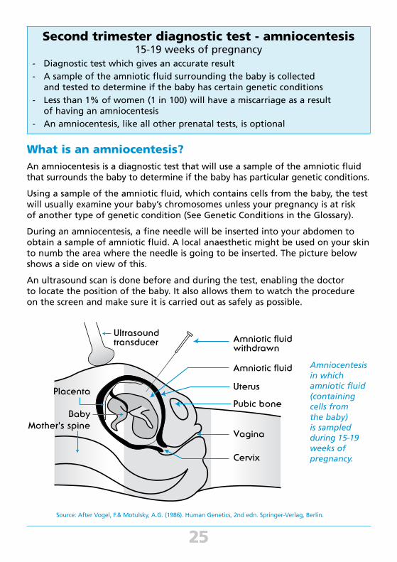
During an amniocentesis, a fine needle will be inserted into your abdomen to obtain a sample of amniotic fluid. A local anaesthetic might be used on your skin to numb the area where the needle is going to be inserted. The picture below shows a side on view of this.
An ultrasound scan is done before and during the test, enabling the doctor to locate the position of the baby. It also allows them to watch the procedure on the screen and make sure it is carried out as safely as possible.
25
Amniocentesis in which amniotic fluid (containing cells from the baby) is sampled during 15-19 weeks of pregnancy.
Source: After Vogel, F.& Motulsky, A.G. (1986). Human Genetics, 2nd edn. Springer-Verlag, Berlin.

26
Why should I consider having an amniocentesis?
An amniocentesis will be offered to you if you are at an increased risk of having
a baby with a genetic condition. This could be due to a number of reasons including:
• You received an increased risk result on a first or second trimester screening
test (see pages 13-15 and 22-24)
• You are over 35 years old. The risk of having a baby with a chromosome
condition increases with the mother’s age. For further details see “chromosome
condition” in the Glossary section of this booklet.
• You or your partner have a family health history of a chromosome
condition or other type of genetic condition caused by a gene variation.
When the amniocentesis is done to check for a gene variation, the laboratory
will need to establish a gene test which can then be used during the
amniocentesis. This type of testing is very specialised and is usually
organised by a genetic counsellor.
When is an amniocentesis done?
An amniocentesis is usually done between 15 and 19 weeks of pregnancy.
When will the result of amniocentesis be available?
It will take two to three weeks to receive the result of an amniocentesis
as it is a lengthy process. After a sample of amniotic fluid has been collected
from you, it is sent to a specialised laboratory where cells from the amniotic fluid
will be grown in a sterile environment. If the test is looking at the chromosomes,
the laboratory may issue an initial result within two to three days. This will
provide a result for the common chromosome problems only. The final result will
still take two to three weeks.
What will the result of amniocentesis tell me?
It is important to discuss with your doctor or genetic counsellor what conditions
the test is able to identify. Depending on the reason why you are having the
test, specific conditions can be focussed on in the laboratory e.g. if you are an
older mother, the main conditions that would be of concern are chromosome
conditions.
For other women, they may have previously had a child with a specific genetic
condition caused by a gene variation or a specific chromosome change. In this
case, the laboratory would analyse that particular gene or look carefully at the
chromosome in question in the developing baby.
Although doctors and scientists are now able to diagnose a large number of
conditions during pregnancy, it is not possible to test for all of the problems
that could affect your developing baby. Your doctor will discuss with you the
range of conditions that can be identified using the amniocentesis test and
what each of these might mean for you and your baby.
It is important to remember that a normal test result cannot exclude every
possible problem with your baby.
How reliable is an amniocentesis?
An amniocentesis is very accurate at identifying the conditions your baby is being
tested for.
Very occasionally, more than one amniocentesis might be needed to make
a diagnosis. This could happen if the doctor cannot get enough fluid on the
first attempt or in rare cases, if the laboratory is unable to get a definite answer.
In this situation, a second amniocentesis might have to be done.
27
What if the result of amniocentesis shows my baby has a problem?
If an amniocentesis result shows that your baby has a problem, you and your partner will be given as much information as possible about the condition. This includes the implications it might have for your baby’s future health and development. You will be given time to make an informed choice about whether or not you wish to continue your pregnancy.
If you and your partner decide to continue the pregnancy, you will be offered support and information before, during and after the birth of your baby.
If you decide to terminate your pregnancy, the procedure requires hospitalisation. The method of termination will vary depending on the policy of the particular hospital or clinic. It might involve an induction of labour and birth. All aspects should be fully discussed with your doctor or genetic counsellor. You will be offered support and information before, during and after your hospital stay.
Please see the services support and information section on pages 35-36 of this booklet for more details.
Can an amniocentesis harm me or my baby?
The increase in risk of miscarriage due to having an amniocentesis is less than 1% (1 in 100 pregnancies tested with amniocentesis). This risk is in addition to your “background risk” of miscarriage that all women have in pregnancy due to natural causes. It is important to discuss your background risk of miscarriage with your doctor, midwife or genetic counsellor.
The specific risk figure for a miscarriage depends on the experience of the doctor doing the test and the difficulty he or she has in getting the sample of fluid. It is therefore important that an amniocentesis is only carried out by a doctor experienced in this technique (see the Services: Prenatal testing list on pages 35-36).
Some women experience cramping and occasionally, vaginal bleeding or leaking of amniotic fluid after an amniocentesis. Talk to your doctor about what you can expect after the testing and what symptoms to look out for.
28
29
Second trimester ultrasound18-20 weeks of pregnancy
- A good way of checking your baby’s development
- A safe way of following up on any concerning results that were obtained from an earlier prenatal test
- Does not pose any health risk to you or your baby
- Second trimester ultrasounds, like all other prenatal tests, are optional
What is a second trimester ultrasound?
A second trimester ultrasound will use high frequency sound waves to produce
images of your developing baby in your uterus (womb).
The sonographer (a specially trained ultrasound technician) or doctor might
spend some time getting the best possible views of the developing baby.
The equipment used to do this type of detailed scan is specialised. It is important
that an experienced, well-trained operator with modern equipment performs the
scan and interprets the ultrasound images. High quality ultrasound is available
from Fetal Medicine Units in public hospitals and from a number of private
practitioners. For details of services in your area, see the Services:
Prenatal testing list on pages 35-36.
During the ultrasound, the doctor or sonographer will use a water based
jelly-like substance and move the transducer (that acts like a microphone)
over your abdomen.
Sound waves will pass from the transducer into the uterus and through the
fluid surrounding your baby (called amniotic fluid). The sound waves bounce
harmlessly off your baby, creating echoes.
A computer will change these echoes into a picture so that the doctor or
sonographer can examine your baby’s physical development. Your baby won’t
be able to hear the sound waves as they are very low.
30
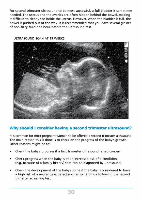
For second trimester ultrasound to be most successful, a full bladder is sometimes needed. The uterus and the ovaries are often hidden behind the bowel, making it difficult to clearly see inside the uterus. However, when the bladder is full, the bowel is pushed out of the way. It is recommended that you have several glasses of non-fizzy fluid one hour before the ultrasound test.
Why should I consider having a second trimester ultrasound?
It is common for most pregnant women to be offered a second trimester ultrasound. The main reason this is done is to check on the progress of the baby’s growth. Other reasons might be to:
• Check the baby’s progress if a first trimester ultrasound raised concern
• Check progress when the baby is at an increased risk of a condition (e.g. because of a family history) that can be diagnosed by ultrasound
• Check the development of the baby’s spine if the baby is considered to have a high risk of a neural tube defect such as spina bifida following the second trimester screening test.
ULTRASOUND SCAN AT 19 WEEKS

31
When is a second trimester ultrasound done?
A second trimester ultrasound to check the baby’s physical development is best done between 18 and 20 weeks of pregnancy.
When will the result of a second trimester ultrasound be available?
In some cases, information about your baby’s ultrasound scan will be made available immediately. However, if the person doing the scan suspects that your baby has a problem, he or she might not be able to discuss the findings until they have been verified by another specialist. The result might be sent to the referring doctor who can discuss the result in this situation. Alternatively, you may be referred to a specialty centre for a second opinion and more detailed assessment.
It might be useful to discuss how the result will be given, to whom and when, with the sonographer or doctor before the ultrasound.
What will the result of second trimester ultrasound tell me?
As the second trimester ultrasound will be done at a later stage of pregnancy, your baby’s major organs and body structures will be visible. Ultrasound can detect any change to the growth, development, structure and position of the baby's organs. It can not necessarily detect how well the organs are functioning.
How reliable is second trimester ultrasound?
A second trimester ultrasound cannot check for every physical or intellectual problem that could possibly affect your baby.
What if the result of a second trimester ultrasound shows my baby has a problem?
Sometimes, there may be a subtle indication that the baby may have a chromosomal problem. These are sometimes called "soft markers". They are seen in up to 15% of pregnancy but more commonly in babies with a chromosomal condition. Depending on how advanced the pregnancy is, the doctor might discuss the option of having an amniocentesis (see amniocentesis on pages 25-28).
In a small number of cases a second trimester ultrasound scan might show that the baby has a genetic condition or some other health or developmental problem. Depending on how advanced the pregnancy is, the doctor might also discuss the option of having a diagnostic prenatal test such as an amniocentesis to give you more information about your baby (see amniocentesis on pages 25-28).
32
You will be given as much information as possible about the condition that might have been identified. This includes the implications it might have for the future health and development of your baby. You and your partner will be given time to make an informed choice about whether or not you wish to continue your pregnancy.
If you decide to continue your pregnancy following the diagnosis of a problem with your baby, you will be offered support and information before, during and after the birth of your baby.
If you decide to terminate your pregnancy, the procedure requires hospitalisation. The method of termination will vary depending on the policy of the particular hospital or clinic. It might involve an induction of labour and birth. All aspects should be fully discussed with your doctor or genetic counsellor. You will be offered support and information before, during and after your hospital stay.
Please see the Services: Support and Information section on pages 35-36 of this booklet for more details.
Can a second trimester ultrasound harm me or my baby?
No evidence has been found that shows an ultrasound can harm you or your baby.
33
Pre-pregnancy optionPreimplantation genetic diagnosis (PGD)
- Involves testing for certain genetic conditions in an embryo created using in vitro fertilisation (IVF)
- Only those embryos that do not have the specific genetic condition that was tested for will be transferred into the woman's uterus
- Currently only offered in the private health setting
What is preimplantation genetic diagnosis (PGD)?
Preimplantation genetic diagnosis (PGD) is a very specialised technique that can help couples who are at risk of passing on a genetic condition to their children avoid doing so. PGD involves testing an embryo that has been created using in-vitro fertilisation (IVF), prior to transferring it to the mother’s uterus and allowing it to develop.
How and when is preimplantation genetic diagnosis (PGD) done?
Hormones are used to stimulate a woman’s ovaries and enable the collection of a number of eggs (oocytes). The eggs are then fertilised with sperm in the laboratory. Those eggs that are successfully fertilised divide and multiply to form a developing embryo. After several days, the embryo has formed into a ball containing many more cells.
Once the embryo reaches this stage, one or two cells are removed and tested for the specific genetic condition in question.
Only those embryos that are found not to be affected by the specific genetic condition will be transplanted into the mother’s uterus. Usually, no more than one or two embryos will be transferred to the uterus at any one time to avoid the possibility of multiple births. In some IVF centres, unaffected embryos that are not used can be frozen for transfer at a later stage.
34
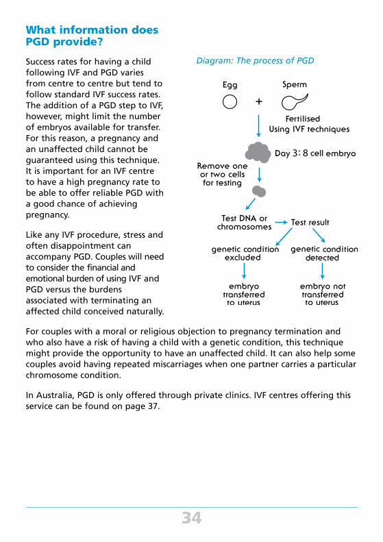
Diagram: The process of PGD
What information does PGD provide?
Success rates for having a child following IVF and PGD varies from centre to centre but tend to follow standard IVF success rates. The addition of a PGD step to IVF, however, might limit the number of embryos available for transfer. For this reason, a pregnancy and an unaffected child cannot be guaranteed using this technique. It is important for an IVF centre to have a high pregnancy rate to be able to offer reliable PGD with a good chance of achieving pregnancy.
Like any IVF procedure, stress and often disappointment can accompany PGD. Couples will need to consider the financial and emotional burden of using IVF and PGD versus the burdens associated with terminating an affected child conceived naturally.
For couples with a moral or religious objection to pregnancy termination and who also have a risk of having a child with a genetic condition, this technique might provide the opportunity to have an unaffected child. It can also help some couples avoid having repeated miscarriages when one partner carries a particular chromosome condition.
In Australia, PGD is only offered through private clinics. IVF centres offering this service can be found on page 37.
35
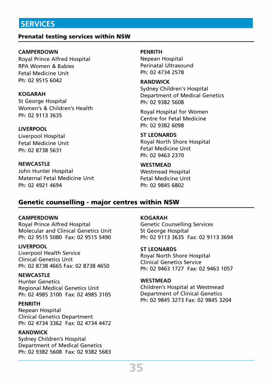
SERVICES
Prenatal testing services within NSW
PENRITH Nepean Hospital Perinatal Ultrasound Ph: 02 4734 2578
RANDWICK Sydney Children's Hospital Department of Medical Genetics Ph: 02 9382 5608
Royal Hospital for Women Centre for Fetal Medicine Ph: 02 9382 6098
ST LEONARDS Royal North Shore Hospital Fetal Medicine Unit Ph: 02 9463 2370
WESTMEAD Westmead Hospital Fetal Medicine Unit Ph: 02 9845 6802
CAMPERDOWN Royal Prince Alfred Hospital RPA Women & Babies Fetal Medicine Unit Ph: 02 9515 6042
KOGARAH St George Hospital Women’s & Children’s Health Ph: 02 9113 3635
LIVERPOOL Liverpool Hospital Fetal Medicine Unit Ph: 02 8738 5631
NEWCASTLE John Hunter Hospital Maternal Fetal Medicine Unit Ph: 02 4921 4694
Genetic counselling - major centres within NSW
CAMPERDOWN Royal Prince Alfred Hospital Molecular and Clinical Genetics Unit Ph: 02 9515 5080 Fax: 02 9515 5490
LIVERPOOL Liverpool Health Service Clinical Genetics Unit Ph: 02 8738 4665 Fax: 02 8738 4650
NEWCASTLE Hunter Genetics Regional Medical Genetics Unit Ph: 02 4985 3100 Fax: 02 4985 3105
PENRITH Nepean Hospital Clinical Genetics Department Ph: 02 4734 3362 Fax: 02 4734 4472
RANDWICK Sydney Children’s Hospital Department of Medical Genetics Ph: 02 9382 5608 Fax: 02 9382 5683
KOGARAH Genetic Counselling Services St George Hospital Ph: 02 9113 3635 Fax: 02 9113 3694
ST LEONARDS Royal North Shore Hospital Clinical Genetics Service Ph: 02 9463 1727 Fax: 02 9463 1057
WESTMEAD Children’s Hospital at Westmead Department of Clinical Genetics Ph: 02 9845 3273 Fax: 02 9845 3204
36
NSW outreach centres
BATHURST Bathurst Health Service Community Health Centre Howick Street, Bathurst NSW 2795 Ph: 02 6330 5677 Fax: 02 6331 0474
COFFS HARBOUR Primary Health Service Locked Mail Bag 812 Coffs Harbour NSW 2450 Ph: 02 6656 7200 Fax: 02 6656 7203
DUBBO Greater Western Area Health Service Dubbo Community Health Centre 2 Palmer Street DUBBO NSW 2830 Ph: 02 6885 8999 Fax: 02 6885 8901
FORSTER Forster Community Health Centre Breeze Pde, Forster NSW 2428 Ph: 02 6539 6300 Fax: 02 6554 8874
GOSFORD Central Coast Health PO Box 361, Gosford NSW 2250 Ph: 02 4328 7994
GOULBURN Child Infant and Family Tertiary Service Locked Bag 15, Goulburn NSW 2580 Ph: 02 4827 3950 Fax: 02 4827 3958
KEMPSEY C/- Port Macquarie Community Health Ctr Ph: 02 6588 2882 Fax: 02 6588 2800
KINGSCLIFF Kingscliff Community Health Turnock Street, Kingscliff NSW 2487 Ph: 02 6674 9500 Fax: 02 6674 9599
ORANGE Community Health Centre 96 Kyte Street, Orange NSW 2800 Ph: 02 6330 5677 Fax: 02 6331 0474
PORT MACQUARIE North Coast Area Health Service Morton Street Port Macquarie NSW 2444 Ph: 02 6588 2882 Fax: 02 6588 2800
TAMWORTH Tamworth Community Health Centre Locked Bag 9783 NEMSC NSW 2348 Ph: 02 6767 8151 Fax: 02 6766 3967
TAREE Mid-North Coast Area Health PO Box 35, Taree NSW 2430 Ph: 02 6592 9703 Fax: 02 6592 9607
WAGGA WAGGA Wagga Wagga Base Hospital PO Box 159, Wagga Wagga NSW 2650 Ph: 02 6938 6411 Fax: 02 6938 6410
WOLLONGONG Northern Illawarra Maternal and Paediatrics Service Wollongong Hospital Crown Street, Wollongong NSW 2500 Ph: 02 4253 4267 Fax: 02 4253 4205
New services are continually being developed. For information on services in other areas and newly developed services, please contact:
The Centre for Genetics Education Level 5, 2c Herbert Street, St Leonards NSW 2065 Ph: 02 9462 9599 Fax: 02 9906 7529 Email: [email protected] Website: www.genetics.edu.au

37
SOUTH AUSTRALIA South Australian Clinical Genetics Services 72 King William Road, North Adelaide SA 5006 Ph: 08 8161 7375 Fax: 08 8161 7754
TASMANIA Tasmanian Clinical Genetics Service C/- Royal Hobart Hospital GPO Box 1061L, Hobart TAS 7001 Ph: 03 6166 8296 Fax: 03 6222 7961
VICTORIA Victorian Clinical Genetics Services Royal Children's Hospital Parkville VIC 3052 Ph: 03 8341 6201 Fax: 03 8341 6390
WESTERN AUSTRALIA King Edward Memorial Hospital for Women 374 Bagot Road, Subiaco WA 6008 Ph: 08 6458 1525 Fax: 08 6458 1678
HUNTER IVF Suite 4, The Heights Private Medical Centre Newcastle Private Hospital 2 Lookout Rd, New Lambton Heights NSW 2305 Ph: 02 4957 8515 Fax: 02 4952 3859Web: www.hunterivf.com.au
WESTMEAD FERTILITY CENTRE Level 2 (Ground Floor), G Block Westmead Hospital, Westmead, NSW 2145Ph: 02 9845 7484Web: www.westmeadivf.com.au
AUSTRALIAN CAPITAL TERRITORY ACT Genetics Services The Canberra Hospital PO Box 11, Woden ACT 2606 Ph: 02 6174 7630 Fax: 02 6244 3021
NORTHERN TERRITORY Clinical Genetics Service Royal Darwin Hospital PO Box 41326, Casuarina NT 0811 Ph: 08 8944 8731 Fax: 08 8922 8463
QUEENSLAND Genetic Health Qld Royal Bribane and Women's Hospital Herston QLD 4029 Ph: 07 3646 1686 Fax 07 3646 1987
Genetic counselling services outside NSW
IVF AUSTRALIA Level 1, 33 York St,Sydney NSW 2000 Freecall 1800 111 (IVF) 483 Ph: 02 8346 6800 Fax: 02 9231 1010 Web: www.ivf.com.au
GENEA 321 Kent St, Sydney, NSW 2000 Ph: 1300 361 795 Web: www.genea.com.au
MONASH IVF Level 2, 1 Fennell St, Parramatta, NSW 2151 Ph: 02 9890 9022 Web: www.monashivf.com
CITY FERTILITY CENTRE Level 1, Suite 101, 1 Moore St, Liverpool, NSW 2170 Ph: 1300 354 354 Web: www.cityfertility.com.au
Preimplantation Genetic Diagnosis (PGD) Services in NSW
Support and Information
The Centre for Genetics Education produces a number of other resources for individuals and families. These include two booklets entitled:
• “Diagnosis of abnormality in an unborn baby – The Impact, Options and Afterwards”
• “When your unborn baby has a problem – How to manage the weeks ahead”
These are available free of charge for residents of NSW.
38
GENETIC ALLIANCE AUSTRALIA (GAA) FORMERLY AGSA provides information and support to families and individuals who are affected by a rare genetic condition. GAA has a client database representing over 500 genetic conditions. In 2005 funding was granted from the NSW Health Department under the NSW Carers Program, to set up face to face counselling on a needs basis and to support families after a prenatal diagnosis of abnormality.
Genetic Alliance Australia C/- Garvan Institute of Medical Research Level 6, 384 Victoria Street, Darlinghurst NSW 2010 Ph: +61 2 9295 8359 Email: info@genetic alliance.org.au Website: www.geneticalliance.org.au
DOWN SYNDROME ASSOCIATION provides a parent support network, information and resources, and peer support for families and people with Down syndrome. Resources are available in many languages.
DOWN SYNDROME NEW SOUTH WALES Level 6, 410 Church St NORTH PARRAMATTA NSW 2151 Ph: 02 9841 4444 Email: [email protected] Website: www.downsyndromensw.org.au
SIDS and KIDS NSW understands the specific issues facing individuals experiencing a prenatal diagnosis of abnormality and its consequent decisions. It also has had experience in offering support to families who are grieving following the sudden loss of a child from the time of conception to six years and maintains an excellent library of grief literature.
SIDS and KIDS NSW Level 1, Bldg 125, Cnr Church & Glover Sts LILYFIELD NSW 2040 Ph: 1300 308 307 Email: [email protected]
THE SPINA BIFIDA GROUP provides support, information and a peer support network for families and people with spina bifida. There are branches of the association in all states of Australia.
Northcott 1 Fennell Street, NORTH PARRAMATTA NSW 2151 Ph: 1800 818 286 Email: [email protected] Website: www.northcott.com.au
THE NATIONAL ASSOCIATION FOR LOSS AND GRIEF (NALAG) promotes loss and grief support groups throughout Australia.
NATIONAL ASSOCIATION FOR LOSS AND GRIEF (NSW) Inc PO Box 379 DUBBO NSW 2830 Ph: 02 6882 9222 Fax: 02 6884 9100 Email: [email protected] Website: www.nalag.org.au
INDUCTION FOR FETAL ABNORMALITY (IFFA) provide support and information to parents who have interrupted a pregnancy for fetal abnormality.
IFFA-INDUCTION FOR FETAL ABNORMALITY PO Box 39 WARATAH NSW 2298 Ph: 02 4967 7413
GLOSSARY
Terms that are underlined appear as separate entries in the Glossary
AMNIOTIC FLUIDTransparent, almost colourless fluid contained within the sac surrounding the developing baby. Cells from the baby are found in this fluid and can be grown in a laboratory and used for testing.
39
1 2 3 4 5
6 7 8 9 10 11 12
13 14 15 16 17 18
19 20 21 22 x y
ANENCEPHALYSee neural tube defects.
CHORIONThe chorion forms part of the placenta and develops from the fertilised egg.
CHROMOSOMESChromosomes are tiny structures present in every cell of the body. Each chromosome contains thousands of genes that are essential to an individual’s health, growth and development. In each normal human cell, except the egg and the sperm, there are 46 chromosomes, each of which is represented twice giving 23 different chromosome pairs. One of the chromosomes in each pair comes from the mother and the other from the father.
The chromosomes vary in size and scientists have given them a number based on that size from 1 to 22. These are called autosomes. The remaining 2 chromosomes are the sex chromosomes, called X and Y. Females have two copies of the X chromosome (XX) and males have one X and one Y chromosome (XY).
In the normal reproductive cycle, the mother’s egg - containing 23 chromosomes is fertilised by the father’s sperm - also containing 23 chromosomes. This union results in an embryo containing 46 chromosomes which will develop into the baby.
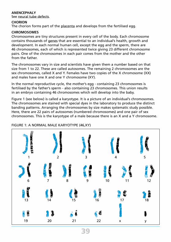
Figure 1 (see below) is called a karyotype. It is a picture of an individual’s chromosomes. The chromosomes are stained with special dyes in the laboratory to produce the distinct banding patterns. Arranging the chromosomes by size makes systematic study possible. Here, there are 22 pairs of autosomes (numbered chromosomes) and one pair of sex chromosomes. This is the karyotype of a male because there is an X and a Y chromosome.
FIGURE 1: A NORMAL MALE KARYOTYPE (46,XY)
40
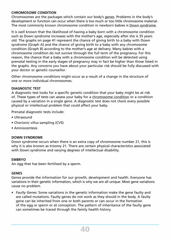
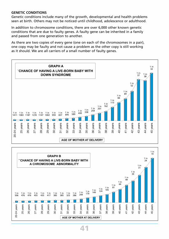
CHROMOSOME CONDITION Chromosomes are the packages which contain our body’s genes. Problems in the body’s development or function can occur when there is too much or too little chromosome material. The most commonly known chromosome condition in newborn babies is Down syndrome.
It is well known that the likelihood of having a baby born with a chromosome condition such as Down syndrome increases with the mother’s age, especially after she is 35 years old. The graphs on page 41 represent the chance of giving birth to a baby with Down syndrome (Graph A) and the chance of giving birth to a baby with any chromosome condition (Graph B) according to the mother’s age at delivery. Many babies with a chromosome condition do not survive through the full term of the pregnancy. For this reason, the chance that a baby with a chromosome condition will be detected using prenatal testing in the early stages of pregnancy may in fact be higher than those listed in the graphs. Any concerns you have about your particular risk should be fully discussed with your doctor or genetic counsellor.
Other chromosome conditions might occur as a result of a change in the structure of one or more individual chromosomes.
DIAGNOSTIC TESTA diagnostic test looks for a specific genetic condition that your baby might be at risk of. These types of tests can assess your baby for a chromosome condition or a condition caused by a variation in a single gene. A diagnostic test does not check every possible physical or intellectual problem that could affect your baby.
Prenatal diagnostic tests include:
• Ultrasound
• Chorionic villus sampling (CVS)
• Amniocentesis
DOWN SYNDROMEDown syndrome occurs when there is an extra copy of chromosome number 21, this is why it is also known as trisomy 21. There are certain physical characteristics associated with Down syndrome and varying degrees of intellectual disability.
EMBRYOAn egg that has been fertilised by a sperm.
GENESGenes provide the information for our growth, development and health. Everyone has variations in their genetic information, which is why we are all unique. Most gene variations cause no problem.
• Faulty Genes: Some variations in the genetic information make the gene faulty and are called mutations. Faulty genes do not work as they should in the body. A faulty gene can be inherited from one or both parents or can occur in the formation of the egg or sperm or at conception. The pattern of inheritance of the faulty gene can sometimes be traced through the family health history.
GENETIC CONDITIONSGenetic conditions include many of the growth, developmental and health problems seen at birth. Others may not be noticed until childhood, adolescence or adulthood.
In addition to chromosome conditions, there are over 6,000 other known genetic conditions that are due to faulty genes. A faulty gene can be inherited in a family and passed from one generation to another.
As there are two copies of every gene (one on each of the chromosomes in a pair), one copy may be faulty and not cause a problem as the other copy is still working as it should. We are all carriers of a small number of faulty genes.
41
20-24 years
25 years26 years27 years28 years29 years30 years31 years32 years33 years34 years35 years36 years37 years38 years39 years40 years41 years42 years43 years44 years45 years
20-2
4 ye
ars
25 y
ears
26 y
ears
27 y
ears
28 y
ears
29 y
ears
30 y
ears
31 y
ears
32 y
ears
33 y
ears
34 y
ears
35 y
ears
36 y
ears
37 y
ears
38 y
ears
39 y
ears
40 y
ears
41 y
ears
42 y
ears
43 y
ears
44 y
ears
45 y
ears
1 in1411
1 in1383
1 in1187
1 in1235
1 in1147
1 in1002
1 in959
1 in837
1 in695
1 in589
1 in430
1 in338
1 in259
1 in201
1 in162
1 in113
1 in84
1 in52
1 in69
1 in37 1 in
38
1 in32
AGE OF MOTHER AT DELIVERY
GRAPH A
CHANCE OF HAVING A LIVE-BORN BABY WITHDOWN SYNDROME
*
20-24 years
25 years26 years27 years28 years29 years30 years31 years32 years33 years34 years35 years36 years37 years38 years39 years40 years41 years42 years43 years44 years45 years
GRAPH B
CHANCE OF HAVING A LIVE-BORN BABY WITHA CHROMOSOME ABNORMALITY
**
AGE OF MOTHER AT DELIVERY20-2
4 ye
ars
25 y
ears
26 y
ears
27 y
ears
28 y
ears
29 y
ears
30 y
ears
31 y
ears
32 y
ears
33 y
ears
34 y
ears
35 y
ears
36 y
ears
37 y
ears
38 y
ears
39 y
ears
40 y
ears
41 y
ears
42 y
ears
43 y
ears
44 y
ears
45 y
ears
1 in506
1 in476
1 in476
1 in455
1 in435
1 in417
1 in385
1 in385
1 in323
1 in286
1 in244
1 in179
1 in149
1 in124
1 in105
1 in81
1 in64
1 in49
1 in39
1 in31
1 in24
1 in19
B
A
42
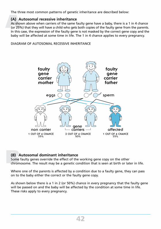
The three most common patterns of genetic inheritance are described below:
(A) Autosomal recessive inheritance As shown above when carriers of the same faulty gene have a baby, there is a 1 in 4 chance (or 25%) that they will have a child who gets both copies of the faulty gene from the parents. In this case, the expression of the faulty gene is not masked by the correct gene copy and the baby will be affected at some time in life. The 1 in 4 chance applies to every pregnancy.
DIAGRAM OF AUTOSOMAL RECESSIVE INHERITANCE
(B) Autosomal dominant inheritance Some faulty genes override the effect of the working gene copy on the other chromosome. The result may be a genetic condition that is seen at birth or later in life.
Where one of the parents is affected by a condition due to a faulty gene, they can pass on to the baby either the correct or the faulty gene copy.
As shown below there is a 1 in 2 (or 50%) chance in every pregnancy that the faulty gene will be passed on and the baby will be affected by the condition at some time in life. These risks apply to every pregnancy.
C
43
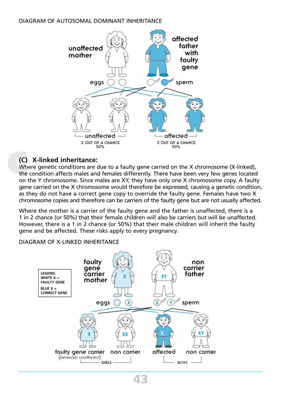
DIAGRAM OF AUTOSOMAL DOMINANT INHERITANCE
(C) X-linked inheritance: Where genetic conditions are due to a faulty gene carried on the X chromosome (X-linked), the condition affects males and females differently. There have been very few genes located on the Y chromosome. Since males are XY, they have only one X chromosome copy. A faulty gene carried on the X chromosome would therefore be expressed, causing a genetic condition, as they do not have a correct gene copy to override the faulty gene. Females have two X chromosome copies and therefore can be carriers of the faulty gene but are not usually affected.
Where the mother is a carrier of the faulty gene and the father is unaffected, there is a 1 in 2 chance (or 50%) that their female children will also be carriers but will be unaffected. However, there is a 1 in 2 chance (or 50%) that their male children will inherit the faulty gene and be affected. These risks apply to every pregnancy.
DIAGRAM OF X-LINKED INHERITANCE
LEGEND: WHITE X = FAULTY GENE
BLUE X = CORRECT GENE
44
IN-VITRO FERTILISATION (IVF)A medical procedure that involves removing eggs from a woman’s ovaries and fertilising them with a man’s sperm in a laboratory. The resulting embryo(s) are then transferred into the woman’s uterus through the cervix with the aim of achieving a pregnancy.
NEURAL TUBE DEFECTSThe most common types of neural tube defects are anencephaly and spina bifida.
• Anencephaly is the most serious as the baby’s brain does not develop properly. Babies with anencephaly do not survive.
• Spina bifida refers to an opening on the baby’s spine which exposes the spinal cord and can cause paralysis and other problems.
The majority of these neural tube defects may be prevented by the supplementation of the mother’s diet with 0.5mg of the vitamin folate for at least one month prior to pregnancy and continuing for the first three months of the pregnancy.
Women at an increased risk for having a baby with a neural tube defect as described below will need to take a higher dose of folate. This should be discussed with your doctor.
In Australia, neural tube defects affect about 1 in 500 babies. The risk for having a baby with a neural tube defect may be increased if:
• There is a close family health history of one or more people affected by a neural tube defect. This can be either on the mother's or father’s side of the family.
• A woman has insulin-dependent diabetes
• A woman is taking specific medicine to control seizures (epilepsy).
Genetic counselling is recommended for these women.
PLACENTAThe placenta, sometimes called afterbirth, is the organ on the wall of the uterus to which the developing baby is attached by the umbilical cord. The placenta is involved in basic nourishment of the developing baby and in hormone production.
SCREENING TEST A screening test cannot tell if your baby definitely has a genetic condition but they might indicate that further testing needs to be considered (See diagnostic test).
Prenatal screening tests include:
• Ultrasound
• NIPT (non-invasive prenatal testing)
• First trimester screening - nuchal translucency
• Second trimester screening - maternal serum testing
SPINA BIFIDASee neural tube defects.
TERMINATION OF PREGNANCYA medical procedure to end a pregnancy prior to its natural completion. Also known as an abortion.
TRISOMY 21See Down syndrome

Copyright NSW Health's Centre for Genetics Education
April 2017 ISBN 978-0-7347-3927-8
NOTES
Further copies of this booklet are available from:
The Centre for Genetics Education
Level 5, 2c Herbert Street
St Leonards NSW 2065
Ph: (02) 9462 9599
Fax: (02) 9906 7529
Email: [email protected]
Website: www.genetics.edu.au
APR17/V6 CATALOGUE NUMBER NS09624-E