prevention and treatment of injuries
DESCRIPTION
Prevention and Treatment of Injuries. Chapter 20 The Knee Dekaney High School Houston, Texas. Anatomy. MCL, Medial Collateral Ligament LCL, Lateral Collateral Ligament PCL, Posterior Cruciate Ligament ACL, Anterior Cruciate Ligament Medial Meniscus Lateral Meniscus. Anatomy. Patella - PowerPoint PPT PresentationTRANSCRIPT

Prevention and Treatment of Injuries
Chapter 20 The KneeDekaney High School
Houston, Texas
Anatomy
• MCL, Medial Collateral Ligament• LCL, Lateral Collateral Ligament• PCL, Posterior Cruciate Ligament• ACL, Anterior Cruciate Ligament• Medial Meniscus• Lateral Meniscus
Anatomy• Patella• Tibia• Fibula• Femur• Patellar Tendon• Hamstrings• Quadriceps• Gastrocnemius

Patella
• Patella, is the largest sesmoid bone in the human body
• Tracking depends on the pull of the quadriceps muscles and the patellar tendon, the depth of the femoral condyles and the shape of the patella

Medial Meniscus
• C-shaped fibrocartilage• Located on the tibia on the medial side
Lateral Meniscus
• Is more O-shaped and located on the the lateral aspect of the tibia
• Both limit lateral movement and serve as a cushion for the knee joint
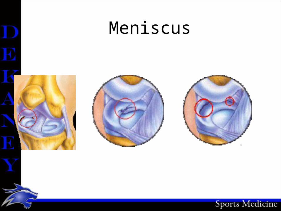
Meniscus
Cruciate Ligaments
• Anterior Cruciate Ligament: comprises three twisted bands: the anteromedial, intermediate, and posterolateral bands.– Prevents the femur from moving posteriorly
during weight bearing. It also stabilizes the tibia against excessive internal rotation and serves as a secondary restraint for valgus or varus stress with collateral ligament damage.
Cruciate Ligaments
• Posterior Ligament: some of the posterior cruciate ligament is taut throughout the full range of motion. It acts as a drag during the gliding phase of motion and resists internal rotation of the tibia. In general, the posterior cruciate ligament prevents hyperextension of the knee, and femur sliding forward during weight bearing.
MCL: Medial Collateral Ligament
• Attaches above the joint line on the medial epicondyle of the femur and below on the tibia.
• The major purpose is to prevent the knee from valgus and external rotating forces.
LCL: Lateral Collateral Ligament
• The LCL is a round, fibrous cord that is shaped like a pencil. It is attached to the lateral epicondyle of the femur and to the head to the fibula.
Knee Musculature• Knee flexion is executed by the biceps
femoris, semitendinosus, semimembranosus, gracilis, gastrocmenius, popliteus, and plantaris muscles.
• Knee extension is executed by the quadriceps muscle of the thigh, consisting of three vasti – vastus medalis, vastus lateralis, and vastus intermedius
Knee Musculature• External rotation of the tibia is controlled by
the biceps femoris.• Internal rotation is accomplished by the
popliteal, semitendinosus, semitmembranosus, sartorius, and gracilis muscles.
• The iliotibial band on the lateral side primarily funcitons as a dynamic lateral stabilizer.
Bursae• A bursa is a flattened sac or enclosed cleft
composed of synovial tissue that is separated by a thin film of fluid. The function of a bursa is to reduce the friction between anatomical structures. Bursae are found between muscle and bone, tendon and bone, tendon and ligament, and so forth. As many as two dozen bursa have been identified around the knee joint.
Bursae
• The Suprapatellar, prepatllar, infrapatellar, pretibial and gastrocnemius bursae are perhaps the most commonly injured about the knee joint.
Fat Pads
• There are several fat pads around the knee. The infrapatellar fat pad is the largest. It serves as a cushion to the front of the knee and separates the patellar tendon from the joint capsule.

Assessing the Knee Joint• History• Current Injury– What did you feel, hear, …. Was there a pop or snap?– Did you get hit by another player? Was your foot
planted? Did this happen without being hit?– Exactly where does you knee hurt, and be specific?– Have you hurt this knee before, when, what was the
injury?

Assessing the Knee Joint
• When did you first notice the condition?• Is there swelling or recurrent swelling?• What activity hurts the most? • Does it ever catch or lock?• Do you fell as if the knee is going to give way,
or has it already done so?• Does it hurt to go up and down stairs?
Observation
• Does the athlete have a limp, or is it easy to walk?
• Cant eh athlete be full weight bearing?• Is the athlete able to perform a half-squat to
extension?• Cant the athlete do up and down stairs?
Testing for Knee Joint Instability
• Through testing of the knee, one can get a better idea of the stability of the joint and an informed decision can be made about playing status. Many tests may point to ligamentous damage, while others will help detect meniscus damage.
• Knowing these test and how to perform them takes practice and time to understand the degrees of damage done to the knee.
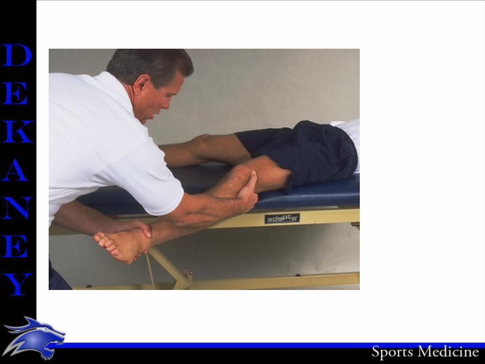
Valgus and Varus Stress Tests
• These are intended to reveal laxity of the medial and lateral collaterals.
• The athlete lie supine with the leg extended.• To test the medial side, the examiner holds the ankle
firmly with one hand while placing the other over the head of the fibula. The examiner then places a force inward in an attempt to open the side of the knee. The valgus force is applied at 0 degrees and then at 30 degree of flexion.
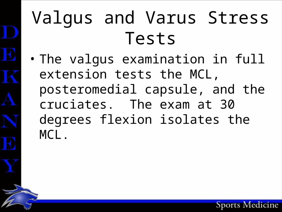
Valgus and Varus Stress Tests
• The valgus examination in full extension tests the MCL, posteromedial capsule, and the cruciates. The exam at 30 degrees flexion isolates the MCL.

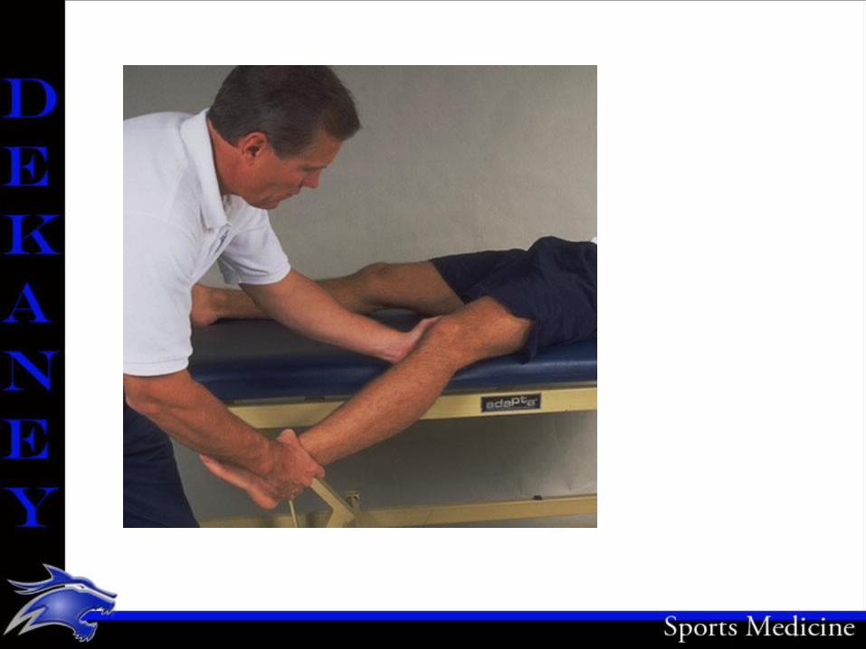
Valgus and Varus Stress Tests
• The examiner reverses hand positions and tests the lateral side with a varus force on the fully extended knee and then with 30 degrees of flexion. With the knee extended, the LCL and posterolater capsule are examined. At 30 degrees of flexion, the LCL is isolated.
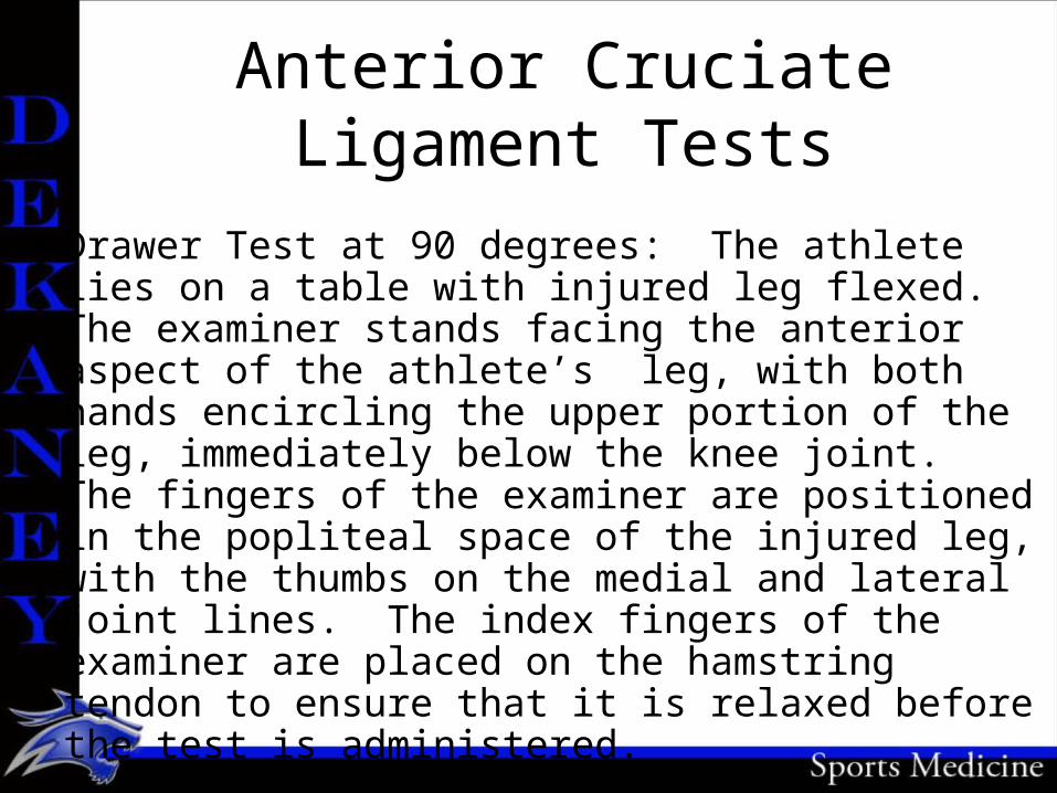
Anterior Cruciate Ligament Tests
• Drawer Test at 90 degrees: The athlete lies on a table with injured leg flexed. The examiner stands facing the anterior aspect of the athlete’s leg, with both hands encircling the upper portion of the leg, immediately below the knee joint. The fingers of the examiner are positioned in the popliteal space of the injured leg, with the thumbs on the medial and lateral joint lines. The index fingers of the examiner are placed on the hamstring tendon to ensure that it is relaxed before the test is administered.
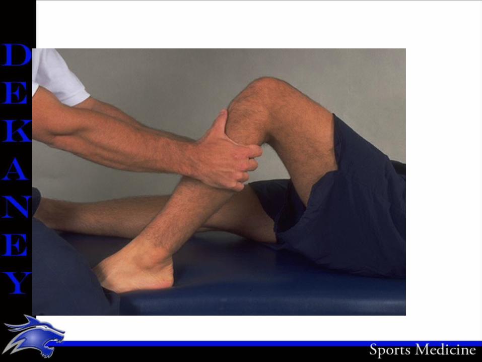
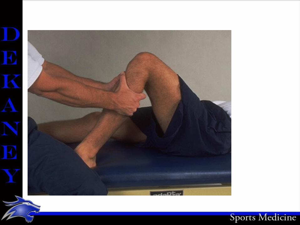
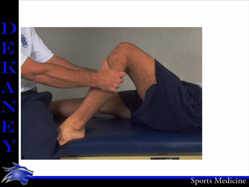
Anterior Cruciate Ligament Tests• The tibia’s sliding forward from under the femur is
considered a positive anterior drawer sign. If a positive anterior drawer sign occurs, the test should be repeated with the athlete’s leg rotated internally 30 degrees and externally 15 degrees. A sliding forward of the tibia when the leg is externally rotated is an indication that the posteromedial aspect of the joint capsule, the ACL, or possibly MCL could be torn. Movement when the leg is internally rotated indicates that the ACL and the posterolateral capsule may be torn.

Anterior Cruciate Ligament Tests
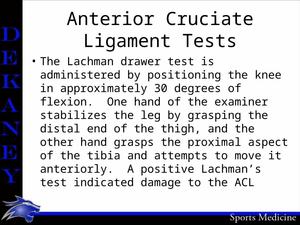
• Lachman’s Drawer Test: is considered to be a better test than the drawer test at 90 degrees of flexion. This is especially true immediately after an injury. One reason for using it immediately after an injury is that it does not force the knee into the painful 90-degree position but tests it at a more comfortable 20 to 30 degrees. It also reduces the contraction of the hamstring muscles. That contraction causes a secondary knee-stabilizing force that tends to mask the real extent of the injury.
Anterior Cruciate Ligament Tests
• The Lachman drawer test is administered by positioning the knee in approximately 30 degrees of flexion. One hand of the examiner stabilizes the leg by grasping the distal end of the thigh, and the other hand grasps the proximal aspect of the tibia and attempts to move it anteriorly. A positive Lachman’s test indicated damage to the ACL

Posterior Cruciate Ligament Tests
• Posterior Drawer Test: is performed with the knee flexed at 90 degrees and the foot in neutral position. Force is exerted in a posterior direction at the proximal tibial plateau. A positive posterior drawer test indicates damage to the posterior cruciate ligament.

Posterior Cruciate Ligament Tests
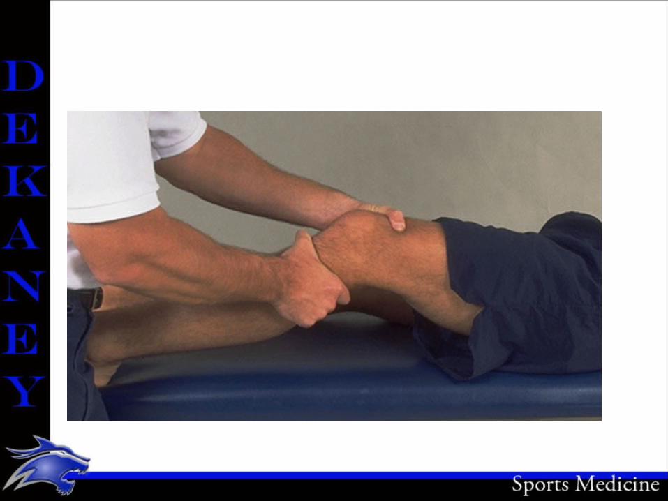
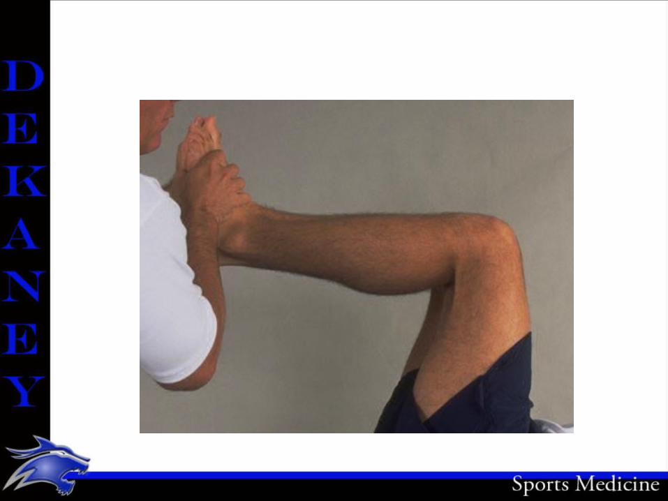
• Posterior Sag Test (Godfrey’s Test): With the athlete supine, both knees are flexed to 9- degrees. Observing laterally on the injured side, the tibia will appear to sag posteriorly when compared to the opposite extremity if the posterior cruciate ligament is damaged.
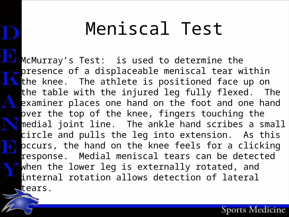
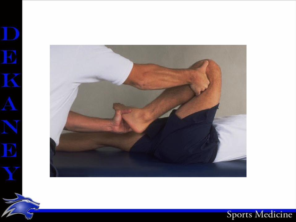
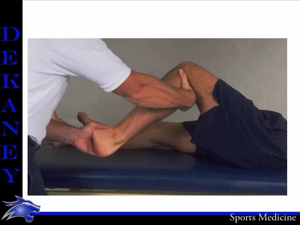
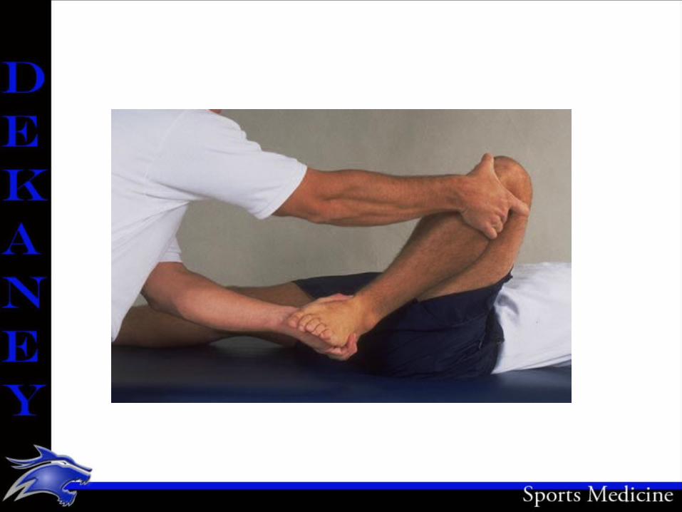
Meniscal Test
• McMurray’s Test: is used to determine the presence of a displaceable meniscal tear within the knee. The athlete is positioned face up on the table with the injured leg fully flexed. The examiner places one hand on the foot and one hand over the top of the knee, fingers touching the medial joint line. The ankle hand scribes a small circle and pulls the leg into extension. As this occurs, the hand on the knee feels for a clicking response. Medial meniscal tears can be detected when the lower leg is externally rotated, and internal rotation allows detection of lateral tears.

Prevention of Knee Injuries
• To avoid injuries to the knee, the athlete must be as highly conditioned as possible, shich means total body conditioning that includes strength, flexibility, cardiovascular and muscle endurance, agility, speed and balance.
• THE MUSCLES around the knee MUST be strong and flexible.
Prevention of Knee Injuries
• Athletes participating in a particular sport should acquire a strength ratio between the quadriceps and hamstring muscle groups. Fro example: the hamstring muscles of football players should have 60 to 70 percent of the strength of the quadriceps muscles. The gastrocnemius muscle should also be strengthened to help stabilize the knee. Although maximizing muscle strength may prevent some injuries, it fails to prevent rotary-type injuries.
Prevention of Knee Injuries
• Shoe Type: – Cleat Length– Atsro Turf shoes: more grip=more injuries– Sneakers are good for artificial surfaces
Functional and Prophylactic Knee Braces
• Functional Knee Braces are used to protect grade 1 and 2 sprains of the ACL and MCL, and reconstructed ACL knees. Most of them are bilateral knee braces, meaning there is a hinge on both sides of the brace. These braces have an important part within the athletic community. They will also give the athlete confidence while playing.

Functional and Prophylactic Knee Braces
• Prophylactic Knee Braces are designed to prevent or reduce the severity of knee injuries. They are worn on the lateral surface of the knee to protect the medial collateral ligament.– The Instructors opinion of Prophylactic Knee
Braces is that they will never replace strength, and should be placed on an athlete with caution.
– Pre-load ligament – – Time for Physics lesson


Prophylactic Knee Braces
• Know what has been presented in the physics lesson.
MCL / LCL Injuries
• MCL injuries are usually caused by a lateral to medial blow to the knee. Also known as a valgus force.
• LCL injuries are usually caused by medial to lateral blow to the knee. Also known as a varus force.
MCL Recognition and Treatment
• GRADE I: Recognition– A few ligamentous fibers are torn and stretched– The joint is stable during valgus stress tests– There is little or no joint effusion– There may be some joint stiffness and point
tenderness just below the medial joint line.– Even with minor stiffness, there is almost full
passive and active ROM.

MCL Recognition and Treatment
• GRADE I: Treatment– Crutches until able to walk without a limp– RICE– Straight leg Raises– Side Leg Raises– Bike– Stair Climber– Functional Progression with pain limiting activity– Return to play with functional bracing or tape
MCL Recognition and Treatment• GRADE II: Recognition– Greater laxity at 30 degrees, as much as 5 to 15
degrees of laxity– Slight or absent of swelling unless the meniscus or ACL
has been torn. – Moderate to severe joint tightness with an inability to
fully, actively extend the knee– Definite loss of ROM– Pain in the medial aspect, with general weakness and
instability
MCL Recognition and Treatment• GRADE II : Treatment– RICE– Crutches– Knee Immobilizer or Don Joy Playmaker Brace– Modalities to control pain and swelling– Ibuprofen, or NSAIDs– SLR– Side LR– Bike , stair climber, Step Ups (2” then 4”)– Functional Progression– Tape and/or Brace to return to activity

MCL Recognition and Treatment
• GRADE III: Recognition– Complete loss of medial stability– Immediate severe pain followed by dull ache– Loss of motion because of effusion and hamstring
guarding– A valgus stress test that reveals some joint opening in
full extension and significant opening at 30 degrees of flexion.
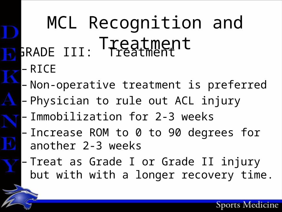
MCL Recognition and Treatment• GRADE III: Treatment– RICE– Non-operative treatment is preferred– Physician to rule out ACL injury– Immobilization for 2-3 weeks– Increase ROM to 0 to 90 degrees for another 2-3
weeks– Treat as Grade I or Grade II injury but with with a
longer recovery time.
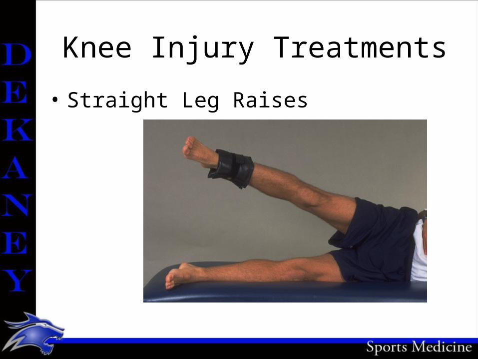
Knee Injury Treatments
• Straight Leg Raises
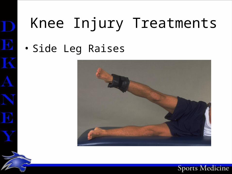
Knee Injury Treatments
• Side Leg Raises
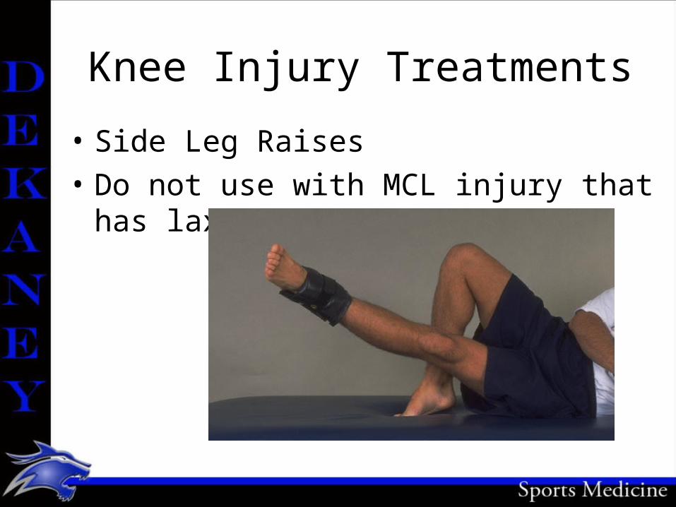
Knee Injury Treatments
• Side Leg Raises• Do not use with MCL injury that has laxity.
Knee Injury Treatments
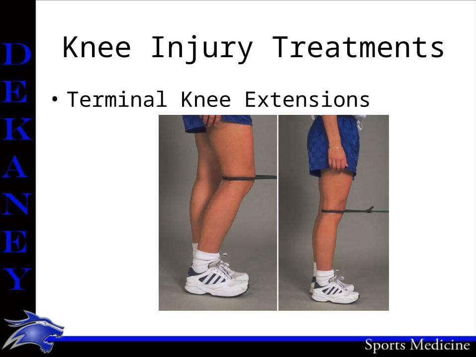
• Terminal Knee Extensions
Knee Injury Treatments
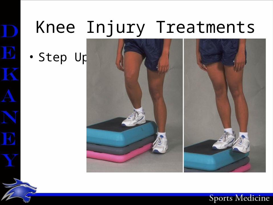
• Step Ups