regulation of alternative macrophage activation by galectin-3 · regulation of alternative...
TRANSCRIPT
of August 20, 2018.This information is current as
Activation by Galectin-3Regulation of Alternative Macrophage
and Tariq SethiLeffler, Ulf J. Nilsson, Christopher Haslett, Stuart J. ForbesHodkinson, Neil C. Henderson, Kirsten M. Atkinson, Hakon Alison C. MacKinnon, Sarah L. Farnworth, Philip S.
http://www.jimmunol.org/content/180/4/2650doi: 10.4049/jimmunol.180.4.2650
2008; 180:2650-2658; ;J Immunol
Referenceshttp://www.jimmunol.org/content/180/4/2650.full#ref-list-1
, 25 of which you can access for free at: cites 62 articlesThis article
average*
4 weeks from acceptance to publicationFast Publication! •
Every submission reviewed by practicing scientistsNo Triage! •
from submission to initial decisionRapid Reviews! 30 days* •
Submit online. ?The JIWhy
Subscriptionhttp://jimmunol.org/subscription
is online at: The Journal of ImmunologyInformation about subscribing to
Permissionshttp://www.aai.org/About/Publications/JI/copyright.htmlSubmit copyright permission requests at:
Email Alertshttp://jimmunol.org/alertsReceive free email-alerts when new articles cite this article. Sign up at:
Print ISSN: 0022-1767 Online ISSN: 1550-6606. Immunologists All rights reserved.Copyright © 2008 by The American Association of1451 Rockville Pike, Suite 650, Rockville, MD 20852The American Association of Immunologists, Inc.,
is published twice each month byThe Journal of Immunology
by guest on August 20, 2018
http://ww
w.jim
munol.org/
Dow
nloaded from
by guest on August 20, 2018
http://ww
w.jim
munol.org/
Dow
nloaded from
Regulation of Alternative Macrophage Activationby Galectin-31
Alison C. MacKinnon,2* Sarah L. Farnworth,2* Philip S. Hodkinson,* Neil C. Henderson,*Kirsten M. Atkinson,* Hakon Leffler,† Ulf J. Nilsson,‡ Christopher Haslett,* Stuart J. Forbes,*and Tariq Sethi3*
Alternative macrophage activation is implicated in diverse disease pathologies such as asthma, organ fibrosis, and granulomatousdiseases, but the mechanisms underlying macrophage programming are not fully understood. Galectin-3 is a carbohydrate-binding lectin present on macrophages. We show that disruption of the galectin-3 gene in 129sv mice specifically restrainsIL-4/IL-13-induced alternative macrophage activation in bone marrow-derived macrophages in vitro and in resident lung andrecruited peritoneal macrophages in vivo without affecting IFN-�/LPS-induced classical activation or IL-10-induced deac-tivation. IL-4-mediated alternative macrophage activation is inhibited by siRNA-targeted deletion of galectin-3 or its mem-brane receptor CD98 and by inhibition of PI3K. Increased galectin-3 expression and secretion is a feature of alternativemacrophage activation. IL-4 stimulates galectin-3 expression and release in parallel with other phenotypic markers ofalternative macrophage activation. By contrast, classical macrophage activation with LPS inhibits galectin-3 expression andrelease. Galectin-3 binds to CD98, and exogenous galectin-3 or cross-linking CD98 with the mAb 4F2 stimulates PI3Kactivation and alternative activation. IL-4-induced alternative activation is blocked by bis-(3-deoxy-3-(3-methoxybenz-amido)-�-D-galactopyranosyl) sulfane, a specific inhibitor of extracellular galectin-3 carbohydrate binding. These resultsdemonstrate that a galectin-3 feedback loop drives alternative macrophage activation. Pharmacological modulation of ga-lectin-3 function represents a novel therapeutic strategy in pathologies associated with alternatively activatedmacrophages. The Journal of Immunology, 2008, 180: 2650 –2658.
M acrophages display broad phenotypic heterogeneitydepending on their microenvironment (1–3). The ini-tial inflammatory response is predominantly mediated
by classically activated (M1-polarized) macrophages, which erad-icate invading organisms and tumor cells (4, 5). The proinflam-matory and cytotoxic activities of classically activated M1 mac-rophages are enhanced in the presence of microbial agents and/orTh1 cytokines such as IFN-� or IL-12 (6, 7). Classical activationby IFN-� in particular is associated with NO synthase 2 (NOS2)4
expression and the production of large amounts of NO and proin-flammatory cytokines, including TNF-� and IL-6 (5, 8, 9). In con-trast, the resolution phase of inflammation is driven by alterna-tively activated (M2-polarized) macrophages. Macrophagesundergo alternative activation when stimulated with the Th2 cy-tokines IL-4 or IL-13 (1, 8, 10). Treatment of murine macrophageswith IL-4 or IL-13 causes up-regulation of mannose receptor (11),arginase (10), Ym1 (chitinase-like lectin) (12, 13), and FIZZ1 (re-sistin-like secreted protein) expression (14, 15). The binding ofIL-4 to the IL-4 receptor �-chain (IL-4R�) induces heterodimer-ization of the �-chain (type I receptor) or the IL-13R�1 (type IIreceptor) (16). This dimerization activates the JAK family of ty-rosine kinases, leading to phosphorylation of the receptor cyto-plasmic tails and exposure of docking sites for STATs, primarilySTAT6 and members of the insulin receptor substrate-1 family(16–19). Phosphorylated STAT6 migrates to the nucleus and ini-tiates transcription of a number of target genes. Phosphorylatedinsulin receptor substrate-1 family binds to the p85 subunit ofPI3K and Grb2. Activation of PI3K and downstream effectors pro-tein kinase B (PKB) and p70S6 kinase are important mediators ofproliferative and survival signaling and regulation of gene expres-sion by IL-4 (16).
Alternative (M2) macrophage activation has been implicated indiverse disease pathologies in the host response to parasitic infec-tion, asthma, wound repair, fibrosis in granulamtous diseases, ath-eromatous plaques, and tumor-associated macrophages (14, 15,20–22). Inhibitors of alternative macrophage activation may re-strict fibrosis in granulomatous diseases (10) and enhance hostimmunity against cancers (22). In disease states where alterna-tively activated macrophages limit tissue injury or promote repair,it might be helpful to augment their activity, for example, by
*Centre for Inflammation Research, The Queen’s Medical Research Institute, Uni-versity of Edinburgh, Edinburgh, United Kingdom; and †Section of Microbiology,Immunology, and Glycobiology, Department of Laboratory Medicine and ‡Depart-ment of Organic Chemistry, University of Lund, Lund, Sweden
Received for publication August 2, 2007. Accepted for publication December10, 2007.
The costs of publication of this article were defrayed in part by the payment of pagecharges. This article must therefore be hereby marked advertisement in accordancewith 18 U.S.C. Section 1734 solely to indicate this fact.1 This work was supported by the Wellcome Trust, U.K. (Clinical Training Fellow-ship to N.C.H. and Senior Research Leave Fellowship to T.S.), the Medical ResearchCouncil, U.K. (Clinical Training Fellowship to P.S.H and Ph.D. studentship toS.L.F.), the Swedish Research Council (Vetenskpasrådet), and by the Swedish Foun-dation for Strategic Research (to H.L. and U.N.).2 A.C.M. and S.L.F. contributed equally to this work.3 Address correspondence and reprint requests to Dr. Tariq Sethi, University of Ed-inburgh, Queen’s Medical Research Institute, 49 Little France Crescent, EdinburghEH16 4TJ, U.K. E-mail address: [email protected] Abbreviations used in this paper: NOS2, NO synthase 2; BMDM, bone marrow-derived macrophage; IL-4R�, IL-4 receptor �-chain; PBM, peripheral bloodmonocyte-derived macrophage; PBS, phosphate-buffered saline; PI(3,4,5)P3,phosphatidylinositol-3,4,5-triphosphate; PI3K, phosphatidylinositol 3-kinase;PKB, protein kinase B; NOS2, nitric oxide synthetase 2; MGC, multinucleatedgiant cell; WT, wild type.
Copyright © 2008 by The American Association of Immunologists, Inc. 0022-1767/08/$2.00
The Journal of Immunology
www.jimmunol.org
by guest on August 20, 2018
http://ww
w.jim
munol.org/
Dow
nloaded from

stabilizing atherosclerotic plaques. To exploit M1 and M2 macro-phages for future anti-inflammatory and anti-cancer therapies, it isimportant to understand the mechanism and extracellular ligandsthat determine M1 and M2 macrophage programming.
Galectin-3 is a �-galactoside-binding lectin of �30 kDa that hasbeen implicated in inflammation and fibrosis (23–27). Galectin-3 ishighly expressed and secreted by macrophages, and recent datasuggest that galectin-3 plays a significant role in many facets ofmacrophage biology. Galectin-3 is up-regulated when monocytesdifferentiate into macrophages (28) and down-regulated whenmacrophages differentiate into dendritic cells (29). Galectin-3 alsopromotes monocyte–monocyte interactions that ultimately lead topolykaryon (multinucleated giant cell) formation, a phenotype-as-sociated with alternative macrophage activation (30), and chronicinflammatory and fibrotic diseases (31). Our previous work hasdemonstrated that mice deficient in galectin-3 exhibit reduced he-patic fibrosis following chronic administration of CCl4 (32).
CD98 is a disulfide-linked 125-kDa heterodimeric type II trans-membrane glycoprotein composed of a glycosylated 85-kDa Hchain (designated CD98) and a nonglycosylated 40-kDa L chainimplicated in inflammation (33). CD98 is highly expressed onmacrophages and has been shown to be a receptor for galectin-3(34). CD98 constitutively associates with �1 integrins, and cross-linking CD98 with the 4F2 Ab stimulates integrin-mediated in-creases in focal adhesion kinase and PI3K activation (35, 36). Inthis study, we have examined the role of galectin-3 and CD98 onmacrophage activation phenotype.
Materials and MethodsTissue culture reagents were purchased from Life Technologies. Cytokinesand recombinant mouse and human galectin-3 were purchased from R&DSystems and PeproTech. The cytometric bead array mouse inflammationkit was from BD Biosciences. The hybridoma cell line 4F2 was purchasedfrom the American Type Culture Collection, and secreted Ab was purifiedusing protein A affinity chromatography. The galectin-3 inhibitor bis-(3-deoxy-3-(3-methoxybenzamido)-�-D-galactopyranosyl) sulfane was pro-vided by U. Nilsson and H. Leffler (37). Cynaropicrin was a kind gift fromDr. J. Cho (Kangwon National University).
Animals
Generation of galectin-3�/� 129sv mice by gene-targeting technologyhas been described previously (38). As control, age- and sex-matchedwild-type (WT) mice were used. All procedures were performed inaccordance with U.K. Home Office guidelines (Animals (Scientific Pro-cedures) Act 1986). All mice used were 8 –10 wk old at the start of theexperiments and were maintained in the animal facilities at the Uni-versity of Edinburgh.
Tissue culture and transfections
Bone marrow-derived macrophages (BMDMs) were prepared from WTand galectin-3�/� mice by maturing bone marrow cells in DMEM con-taining 10% FBS and 20% L929 conditioned media for 7–9 days (39). Invivo-derived macrophages were obtained from lung and peritoneal lavageand separated by adhesion onto tissue culture plastic. Human peripheralblood monocytes were prepared as previously described (40), and theywere cultured for 5 days in Iscove’s medium containing 10% autologousserum. THP-1 cells were obtained from the American Tissue Culture Col-lection and were maintained in RPMI 1640 medium supplemented with10% FBS. Cells were differentiated with 100 ng/ml PMA for 24 h, washed,and transfected with 100 pmol siRNA duplexes using Oligofectamine (In-vitrogen). Cells were incubated for 48 h in complete media before additionof IL-4 or 4F2. Initial experiments were conducted using four duplexesdirected against human CD98 (SLC3A2) as supplied in CD98 SMARTpool(Dharmacon, catalog no. M-003542). All additional experiments usedsiRNA duplexes (Dharmacon) directed to the target sequence AGAATGGTCTGGTGAAGA. Initial experiments to target galectin-3 used siRNAduplexes directed against the target sequences: 1) GAAGAAAGACAGTCGGTTT, 2) GCAATACAAAGCTGGATAA, 3) GTACAATCATCGGGTTAAA, and 4) CAGTACAATCATCGGGTTA.
All additional experiments were conducted using duplex 1. Control du-plex was siCONTROL nontargeting siRNA no. 2 (Dharmacon). siRNA
duplexes directed against human STAT6 were purchased from Santa CruzBiotechnology (catalog no. sc-29497).
Cytokine/nitrite analysis
Cytokine release from macrophage supernatants was determined by cyto-metric bead array. Human TNF-� was measured by ELISA (BD Bio-sciences). NO production was determined by measurement of nitrite re-lease using the Griess reaction (Sigma-Aldrich).
Arginase assay
Arginase activity was assessed by the production of urea generated by thearginase-dependent hydrolysis of L-arginine as described (41).
Real-time RT-PCR
Total RNA from BMDMs or THP-1 cells (4 � 106 cells/sample) wasprepared using RNeasy kits (Qiagen) and reverse transcribed into cDNAusing random hexamers (Applied Biosystems). For analysis of alternativeactivation markers, cDNA was analyzed using a SYBR green-based quan-titative fluorescence method (Applied Biosystems). The following primerpairs were used: mouse �-actin: forward 5�-AGAGGGAAATCGTGCGTGAC-3�, reverse 5�-CAATAGTGATGACCTGGCCGT-3�; mouse FIZZ1:forward 5�-TACTTGCAACTGCCTGTGCTTACT-3�, reverse 5�-TATCAAAGCTGGGTTCTCCACCTC-3�; mouse Ym1: forward 5�-TCTCTACTCCTCAGAACCGTCAGA-3�,reverse5�-GATGTTTGTCCTTAGGAGGGCTTC-3�; mouse arginase-1: forward 5�-TTGGGTGGATGCTCACACTG-3�, reverse 5�-TTGCCCATGCAGATTCCC-3�; mouse mannosereceptor: forward 5�-CATGAGGCTTCTCCTGCTTCT-3�, reverse 5�-TTGCCGTCTGAACTGAGATGG-3�; mouse NOS2: forward 5�-CAGCTGGGCTGTACAAACCTT-3�, reverse 5�-CATTGGAAGTGAAGCGTTTCG-3�; mouse IL-4R�: forward 5�-ACCTGAGAACAGCGGAGGC-3�,reverse 5�-TCGGAAAACAGGTTCTCAGTGAG-3�; human �-actin: for-ward 5�-CATCACCATTGGCAATGAGC-3�, reverse 5�-CGATCCACACGGAGTACTTG-3�; human mannose receptor: forward 5�-GCCAAATGACGAATTGTGGA-3�, reverse 5�-CACGAAGCCATTTGGTAAACG-3�.
Immunofluorescence
Alveolar macrophages were fixed in 3% paraformaldehyde and stainedwith rabbit anti-Ym1 (Professor J. Allen, Edinburgh) followed by species-specific Alexa 488-conjugated secondary Abs and fluorescence microscopy(Leica).
Immunoprecipitation and Western blotting
Cell lysates were resolved by SDS-PAGE (under nonreducing conditionsfor mannose receptor blots) and transferred onto nitrocellulose. Blots wereprobed with the following primary Abs: mouse monoclonal anti-humanmannose receptor Ab clone 15–2 (MCA2155, Serotec), rat anti-mousemannose receptor Ab clone 5D3 (MCA2235GA, Serotec), monoclonalanti-galectin-3 Ab clone A3A12 (Alexis Biochemicals), goat polyclonalanti-CD98 (sc-7095, Santa Cruz Biotechnology), rabbit polyclonal anti-�-actin Ab (Sigma-Aldrich), rabbit anti-PKB and anti-pPKB (S473)(Invitrogen: BioSource), rabbit anti-STAT6 and anti-pSTAT6 (T641)(Cell Signaling Technology), rabbit anti-mouse IL-4R� (sc-686, SantaCruz Biotechnology), and rabbit polyclonal anti Ym1 (from Professor J.Allen).
CD98 was immunoprecipitated from THP-1 cell lysates (0.5 mg protein)with 2 �g goat polyclonal anti-CD98 (sc-7095, Santa Cruz Biotechnology)or 2 �g control goat IgG overnight at 4oC. Immune complexes were cap-tured with protein A/G-Sepharose, and washed immunoprecipitates wereresolved by SDS-PAGE and blotted for CD98 or galectin-3.
Statistical analysis
Results are presented as means � SEM. Significance of the differencesbetween means was assessed using one-way ANOVA or two-tailed Stu-dent’s t test. Values of p � 0.05 were considered significant. Unless statedotherwise, studies were performed on three to six independent occasions.
ResultsDisruption of the galectin-3 gene in macrophages causes aspecific defect in alternative activation of in vitro- andin vivo-differentiated macrophages
To investigate the role of galectin-3 in macrophage activation, weused in vitro generated BMDM from WT and galectin-3�/� mice.We treated BMDMs with either IFN-�/LPS (classical M1 activa-tion) or IL-4/IL-13 (alternative M2 activation) and measured the
2651The Journal of Immunology
by guest on August 20, 2018
http://ww
w.jim
munol.org/
Dow
nloaded from
release of proinflammatory cytokines (TNF-� and IL-6), NO (ni-trite), and arginase activity. We found that IFN-� and LPS induceda similar release of TNF-� and IL-6 in both WT and galectin-3�/�
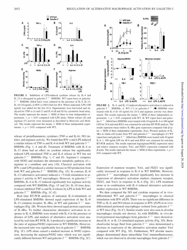
BMDMs (Fig. 1, A and B). Treatment of BMDMs with IL-4 orIL-13 alone had no effect on cytokine release but significantlyreduced LPS-stimulated TNF-� and IL-6 release in WT but notgalectin-3�/� BMDMs (Fig. 1, C and D). Arginase-1 competeswith NOS2 and mediates the alternative metabolic pathway of L-arginine to L-ornithine and urea (10). Classical activation withIFN-� and LPS produced a similar increase in NO production fromboth WT and galectin-3�/� BMDMs (Fig. 1E). In contrast, IL-4/IL-13 (alternative activation) induced a �5-fold stimulation in ar-ginase-1 activity in WT macrophages. However, in galectin-3�/�
BMDMs, IL-4 or IL-13 induced significantly less arginase activitycompared with WT BMDMs (Figs. 1F and 2A). IL-10 (true deac-tivation) inhibited TNF-� and IL-6 release by LPS in both WT andgalectin-3�/� BMDMs (Fig. 1C–E).
Real time RT-PCR analysis of untreated, IL-4-stimulated, orLPS-stimulated BMDMs showed equal expression of the IL-4/IL-13 common receptor, IL-4R�, in WT and galectin-3�/� mac-rophages (Fig. 2B). Western blots showed equal protein expressionof IL-4R�. To further characterize alternative activation in re-sponse to IL-4, BMDMs were treated with IL-4 in the presence orabsence of LPS, and markers of alternative activation were ana-lyzed by real-time RT-PCR. IL-4 produced a significant increase inthe ratio of arginase/NOS2 expression in WT BMDMs. However,the increased ratio was significantly less in galectin-3�/� BMDMs(Fig. 2C). LPS alone caused a marked increase in NOS2 expres-sion, decreasing the arginase/NOS2 ratio, which was not signifi-cantly different between WT and galectin-3�/� BMDMs (Fig. 2C).
Expression of mannose receptor, Ym1, and FIZZ1 was signifi-cantly increased in response to IL-4 in WT BMDMs. However,galectin-3�/� macrophages showed significantly less increase inexpression of alternative activation markers (mannose receptor,FIZZ1, and Ym1) in response to IL-4 (Fig. 2C). LPS treatmentalone or in combination with IL-4 reduced alternative activationmarker expression in WT BMDMs.
We then compared the NO and cytokine response of in vivo-differentiated WT and galectin-3�/� macrophages to classicalstimulation with IFN-�/LPS. There was no significant difference inTNF-�, IL-6, and NO release in response to IFN-�/LPS in in vivo-differentiated peritoneal macrophages isolated from galectin-3�/�
or WT mice (Fig. 3A–C). Similar results were also seen in alveolarmacrophages (results not shown). As with BMDMs, in vivo-de-rived peritoneal macrophages from galectin-3�/� mice showed re-duced IL-4- or IL-13-stimulated arginase activity (Fig. 3D). Bron-choalveolar lavage fluid from galectin-3�/� mice demonstrated adecrease in expression of the alternative activation marker Ym1compared with WT (Fig. 3E). Furthermore, WT alveolar macro-phages demonstrated dense intracellular Ym1 immunofluorescence,which was not observed in alveolar macrophages from galectin-3�/�
FIGURE 1. Inhibition of LPS-induced cytokine release by IL-4 andIL-13 is abrogated in galectin-3�/� BMDMs. WT (open bars) or galectin-3�/� BMDMs (filled bars) were cultured in the presence of IL-4, IL-13,IL-10 (10 ng/ml), or IFN-� (100 U/ml) for 48 h. Where indicated, LPS (100ng/ml) was added for the last 24 h. Supernatants were harvested and an-alyzed for TNF-� (A and C) and IL-6 (B and D) by cytometric bead array.The results represent the means � SEM of three to four independent ex-periments. �, p � 0.01 compared with LPS alone. Nitrite release (E) andarginase (F) activity were measured as described in Materials and Meth-ods. The results represent the means � SEM of three independent exper-iments. �, p � 0.01 compared with WT.
FIGURE 2. IL-4- and IL-13-induced alternative activation is reduced ingalectin-3�/� BMDMs. A, WT (E) or galectin-3�/� (F) BMDMs weretreated with IL-4 (0–10 ng/ml) for 24 h and arginase activity was deter-mined. The results represent the means � SEM of three independent ex-periments. �, p � 0.01 compared with WT. B, WT (open bars) and galec-tin-3�/� (filled bars) BMDMs were treated with 10 ng/ml IL-4 or 100 ng/mlLPS for 24 h and total RNA was extracted for real-time RT-PCR analysis. Theresults represent mean relative IL-4R� gene expression compared with �-ac-tin � SEM of three independent experiments. Inset, Western analysis of IL-4R� in whole-cell lysates from WT and galectin-3�/� macrophages (C) WT(open bars) and galectin-3�/� (filled bars) BMDMs were treated with 10 ng/mlIL-4 � 100 ng/ml LPS for 48 h and total RNA was extracted for real-timeRT-PCR analysis. The results represent log(arginase/NOS2 expression ratio)and relative mannose receptor, Ym1, and FIZZ1 expression compared with�-actin. The results represent the means � SEM of three experiments. �, p �0.01 compared with WT.
2652 REGULATION OF ALTERNATIVE MACROPHAGE ACTIVATION BY GALECTIN-3
by guest on August 20, 2018
http://ww
w.jim
munol.org/
Dow
nloaded from

mice (Fig. 3E). These data suggest that both in vitro- and in vivo-differentiated galectin-3�/� macrophages have a specific defect in IL-4/IL-13-stimulated alternative activation.
An IL-4-mediated galectin-3 autocrine loop promotes alternativemacrophage activation and is blocked by pharmacologicalinhibition of galectin-3 and CD98
BMDMs and human peripheral blood monocyte-derived macro-phages (PBMs) were treated with IL-4 for 48 h. Western blot anal-ysis shows that IL-4 or IL-13 stimulated the release of galectin-3into the culture media in both BMDMs and PBMs (Fig. 4A). Incontrast, LPS-treated BMDMs showed a marked down-regulationof galectin-3 expression in both cell lysates and supernatants.Thus, classically activated macrophages down-regulate galectin-3expression and show reduced galectin-3 expression on the cell sur-face. Bis-(3-deoxy-3-(3-methoxybenzamido)-�-D-galactopyrano-syl) sulfane is a specific and high-affinity inhibitor of galectin-3based on N-acetyllactosamine, the best natural ligand for galec-tin-3, (42) and is a specific and highly selective inhibitor of thecarbohydrate recognition domain of galectin-3 (Kd of 60 nM and�20-fold selectivity over other galectins, including galectin-1(37)). Coincubation with the galectin-3 inhibitor blocked IL-4-me-diated arginase activation and mannose receptor expression inBMDMs (Fig. 4B). This suggests that galectin-3 may be mediatingits effects at the cell surface via its carbohydrate-recognition do-
main. CD98 is a well-characterized macrophage receptor for ga-lectin-3 (34, 42). CD98 has been shown to promote PI3K activa-tion, which is dependent on association with �1-integrins and �1-integrin expression (35, 36). We therefore sought to establishwhether activation of CD98 by galectin-3 may mediate its effectson alternative activation. Cynaropicrin is a sesquiterpene lactonethat inhibits CD98 and �1-integrin-induced homotypic aggregationand down-regulates �1-integrin expression on macrophages (43).Cynaropicrin inhibited arginase activation in galectin-3�/� andWT BMDMs stimulated by IL-4 down to basal levels (Fig. 4B),suggesting that the effects of galectin-3 may be mediated via thissurface receptor. Neither cynaropicrin or bis-(3-deoxy-3-(3-me-thoxybenzamido)-�-D-galactopyranosyl) sulfane affected macro-phage number or viability as judged by cell counts, morphology,and trypan blue exclusion (data not shown).
IL-4-stimulated alternative macrophage activation is dependenton expression of galectin-3 and CD98 and activation of PI3K
We used the human monocytic cell line THP-1 to establishwhether galectin-3-dependent alternative activation was mediatedvia CD98. THP-1 cells were differentiated into a macrophage phe-notype by treatment with 100 ng/ml PMA for 24 h. As shownpreviously, differentiation of THP-1 cells with PMA caused a sig-nificant increase in expression of galectin-3 (Fig. 5A). This wasparalleled with an increase in expression of CD98. To confirm adirect association of galectin-3 with CD98, CD98 was immuno-precipitated from THP-1 whole-cell lysates using a goat polyclonalanti-CD98 Ab and was compared with normal goat IgG as control.Western blots were probed for galectin-3 using a mouse monoclo-nal anti-galectin-3 (A3A12). Fig. 5B shows that upon immunopre-cipitation of CD98 Ab and Western blotting for galectin-3, a band
IFNγγ/LPS01234
[TNF
-α]n
g/m
l
0
100
200
[IL-6
] pg/
ml
[Ure
a]µm
ole s
/mg/
hDC
IL-4 IL-13 IL-10IFNγ/LPS
5.0
0
[Nitr
ite]
mol
es/m
g
**
0
10
20
IL-4 IL-13 IL-10 IFNγ/LPS
A
IFNγ/LPS
B
wild typeGalectin-3-/-
IL-6TNFα
Nitrite Arginase
2.5n
LavageEYM-1
Gal-3-/-WT
Galectin-3-/-Wild type
YM-1
FIGURE 3. Galectin 3�/� peritoneal and alveolar macrophages displaynormal classical activation but impaired alternative activation. Peritoneal mac-rophages were stimulated with IFN-� (100 U/ml) for 48 h and LPS (100ng/ml) was added for the last 24 h. Supernatants were assayed for (A) TNF-�,(B) IL-6, and (C) nitrite. Cell lysates were assayed for arginase activity (D).The results represent the means � SEM of three experiments. �, p � 0.01compared with WT. E, PBS (1 ml) was administered intratracheally to WT andgalectin-3�/� mice and bronchoalveolar lavage fluid was analyzed for Ym1protein expression by Western blot analysis and immunofluorescence stainingof Ym1 in alveolar macrophages isolated from lavage fluid of WT (left panel)and galectin-3�/� mice (right panel) (scale bar: 5 �m).
FIGURE 4. Galectin-3 potentiates IL-4-induced alternative activationand is released from IL-4 treated cells. A, IL-4 stimulates galectin-3 ex-pression and release. Upper panel, Human PBMs and WT mouse BMDMswere incubated for 48 h with 10 ng/ml IL-4 or IL-13. Western blots fromcell supernatants were probed for galectin-3. Lower panel, WT BMDMswere treated for 48 h with IL-4 (0–10 ng/ml) in the presence or absence of100 ng/ml LPS. Western blots from cell supernatants and cell lysates wereprobed for galectin-3. B, BMDMs were treated with 10 ng/ml IL-4 for 24 hin the presence or absence of 5 �M of galectin-3 inhibitor (bis-(3-deoxy-3-(3-methoxybenzamido)-�-D-galactopyranosyl) sulfane) or 10 �M of cy-naropicrin, and arginase activity was measured in cell lysates. Results rep-resent the means � SEM of three independent experiments. �, p � 0.01compared with WT. Inset, Western blot of mannose receptor expression.
2653The Journal of Immunology
by guest on August 20, 2018
http://ww
w.jim
munol.org/
Dow
nloaded from

was visualized at �30 kDa (lane 2). This was not seen followingimmunoprecipitation with control IgG Ab (lane 4) and confirmsthat galectin-3 associates with CD98 in differentiated THP-1 cells.Moreover, incubation of THP-1 cells for 1 h with the galectin-3inhibitor (5 �M) before lysis blocked co-immunoprecitation ofgalectin-3 with CD98. We then sought to determine whether in-hibition of galectin-3 or CD98 expression could inhibit alternativeactivation. Four siRNA duplexes directed against human CD98 orgalectin-3 were tested in THP-1 cells. All four galectin-3 siRNAduplexes caused an �90% knockdown of galectin-3 protein ex-pression, and CD98 siRNA duplexes 1 and 3 reduced CD98 pro-tein expression by �95% (data not shown). In subsequent exper-
iments, galectin-3 duplex 1 and CD98 duplex 1 were used andcompared with a nontargeting control duplex. Results were con-firmed with galectin-3 duplex 4 and CD98 duplex 3 in all exper-iments (data not shown). siRNA-targeted inhibition of galectin-3or CD98 expression blocked IL-4-stimulated increase in mannosereceptor expression as judged by Western blot analysis and real-time RT-PCR (Fig. 5, C and D). Inhibition of galectin-3 functioneither by siRNA or with the galectin-3 inhibitor, and inhibition ofCD98 expression in THP-1 cells did not affect TNF-� release inresponse to IFN-�/LPS (Fig. 5E). As a whole, our data suggest thatalternative macrophage activation by IL-4 is driven by a galectin-3feedback mechanism, which activates CD98.
Galectin-3-mediated alternative activation involves CD98 andactivation of PI3K
Our previous work has shown that CD98 stimulates PI3K activa-tion (35, 36). siRNA-targeted inhibition of CD98 expression or
FIGURE 5. Alternative activation of human macrophages requires ga-lectin-3 and CD98. A, THP-1 cells were differentiated with 100 ng/mlPMA, and lysates were prepared for Western analysis at the time pointsindicated. Blots were probed for CD98, galectin-3, and �-actin. B, CD98and galectin-3 co-immunoprecipitate from THP-1 cell lysates. Normalizedprotein aliquots from lysates prepared from control THP-1 cells and THP-1cells treated for 1 h with 5 �M of galectin-3 inhibitor were immunopre-cipitated with 2 �g of goat anti-human CD98 and captured by protein-A/Gsepharose. Washed immunoprecipitates and whole-cell lysates (WCL)were blotted for galectin-3 and CD98. hc indicates immunoprecipitatingAb H chain. C, THP-1 cells differentiated for 24 h were transfected withindividual siRNA duplexes to human galectin-3 (duplex 1), human CD98(duplex 1), or control duplex as described in Materials and Methods. Forty-eight-hour posttransfection THP-1 cells were incubated with 10 ng/ml IL-4or 100 U/ml IFN-�/100 ng/ml LPS for 24 h. Cells lysates were resolved on12% SDS-PAGE gels. Blots were probed for mannose receptor (MR), ga-lectin-3 (Gal-3), CD98, and �-actin. Representative blots from three inde-pendent experiments are shown. D, THP-1 cells were differentiated andtransfected with siRNA as in C. Forty-eight-hour posttransfection THP-1cells were incubated with 10 ng/ml IL-4 for 24 h. Total-cell RNA wasextracted and mannose receptor gene expression was determined by real-time RT-PCR. Results are expressed as relative gene expression comparedwith �-actin and represent the means � SEM of three independent exper-iments. E, THP-1 cells differentiated and transfected with siRNA as in Cwere incubated with 100 U/ml IFN-�/100 ng/ml LPS in the presence orabsence or galectin-3 inhibitor (5 �M) for 24 h. TNF-� was measured byELISA. Results represent the means � SEM of three experiments.
FIGURE 6. Inhibition of PI3K blocks alternative activation. A, THP-1cells were transfected with siRNA against CD98 or control duplex andstimulated with 10 ng/ml IL-4 or 10 �g/ml galectin-3 for 24 h in thepresence or absence of 5 �M of galectin-3 inhibitor (bis-(3-deoxy-3-(3-methoxybenzamido)-�-D-galactopyranosyl) sulfane). Western blots of celllysates were probed with CD98, pPKB, and �-actin as indicated (bottompanel). B, Differentiated THP-1 cells were treated with 10 �M ofLY294002 20 min before addition of 10 ng/ml IL-4, 10 �g/ml 4F2, or 10�g/ml human recombinant galectin-3 for 20 min (top panel) or 24 h (bot-tom) as indicated. Western blots of cell lysates were probed with pPKB,total PKB, pSTAT6, mannose receptor (MR), and �-actin as indicated. C,Dose response to IL-4 in the presence of galectin-3 inhibitor. THP-1 cellswere treated with IL-4 for 24 h in the presence or absence of 5 �M ofgalectin-3 inhibitor (bis-(3-deoxy-3-(3-methoxybenzamido)-�-D-galacto-pyranosyl) sulfane). Western blots of cell lysates were probed with pPKBand �-actin as indicated. D, WT BMDMs were treated with IL-4 (0.1–100ng/ml) in the absence (F) or presence of 5 �M of galectin-3 inhibitor(bis-(3-deoxy-3-(3-methoxybenzamido)-�-D-galactopyranosyl) sulfane)(�) or 10 �M of LY294002 (E) for 24 h. Arginase activity was measuredfrom cell lysates as described in Materials and Methods. Results representthe means � SEM of three experiments.
2654 REGULATION OF ALTERNATIVE MACROPHAGE ACTIVATION BY GALECTIN-3
by guest on August 20, 2018
http://ww
w.jim
munol.org/
Dow
nloaded from
inhibition of galectin-3 carbohydrate binding by the galectin-3 in-hibitor both blocked IL-4 and galectin-3-stimulated PI3K activa-tion as measured by phosphorylation of PKB phosphorylation inTHP-1 cells (Fig. 6, A and C). Cross-linking CD98 with the mAb4F2 stimulated a sustained PI3K activation (evident at 20 min andmaintained at 24 h) and increased mannose receptor expression toa similar level as IL-4 in THP-1 cells (Fig. 6B). Blocking PI3Kactivity with LY294002 prevented IL-4-, galectin-3-, and 4F2-mediated PKB phosphorylation and mannose receptor expres-sion (Fig. 6B). LY294002 blocked IL-4-stimulated arginase ac-tivation in BMDMs to a level observed in galectin-3�/�
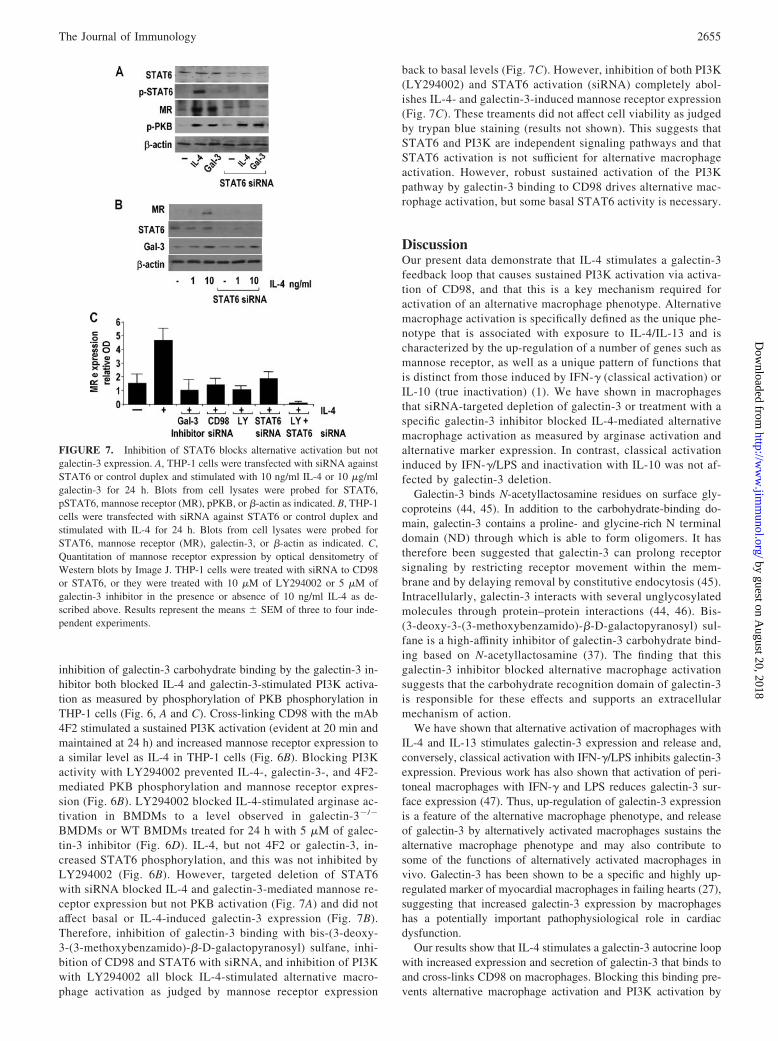
BMDMs or WT BMDMs treated for 24 h with 5 �M of galec-tin-3 inhibitor (Fig. 6D). IL-4, but not 4F2 or galectin-3, in-creased STAT6 phosphorylation, and this was not inhibited byLY294002 (Fig. 6B). However, targeted deletion of STAT6with siRNA blocked IL-4 and galectin-3-mediated mannose re-ceptor expression but not PKB activation (Fig. 7A) and did notaffect basal or IL-4-induced galectin-3 expression (Fig. 7B).Therefore, inhibition of galectin-3 binding with bis-(3-deoxy-3-(3-methoxybenzamido)-�-D-galactopyranosyl) sulfane, inhi-bition of CD98 and STAT6 with siRNA, and inhibition of PI3Kwith LY294002 all block IL-4-stimulated alternative macro-phage activation as judged by mannose receptor expression
back to basal levels (Fig. 7C). However, inhibition of both PI3K(LY294002) and STAT6 activation (siRNA) completely abol-ishes IL-4- and galectin-3-induced mannose receptor expression(Fig. 7C). These treaments did not affect cell viability as judgedby trypan blue staining (results not shown). This suggests thatSTAT6 and PI3K are independent signaling pathways and thatSTAT6 activation is not sufficient for alternative macrophageactivation. However, robust sustained activation of the PI3Kpathway by galectin-3 binding to CD98 drives alternative mac-rophage activation, but some basal STAT6 activity is necessary.
DiscussionOur present data demonstrate that IL-4 stimulates a galectin-3feedback loop that causes sustained PI3K activation via activa-tion of CD98, and that this is a key mechanism required foractivation of an alternative macrophage phenotype. Alternativemacrophage activation is specifically defined as the unique phe-notype that is associated with exposure to IL-4/IL-13 and ischaracterized by the up-regulation of a number of genes such asmannose receptor, as well as a unique pattern of functions thatis distinct from those induced by IFN-� (classical activation) orIL-10 (true inactivation) (1). We have shown in macrophagesthat siRNA-targeted depletion of galectin-3 or treatment with aspecific galectin-3 inhibitor blocked IL-4-mediated alternativemacrophage activation as measured by arginase activation andalternative marker expression. In contrast, classical activationinduced by IFN-�/LPS and inactivation with IL-10 was not af-fected by galectin-3 deletion.
Galectin-3 binds N-acetyllactosamine residues on surface gly-coproteins (44, 45). In addition to the carbohydrate-binding do-main, galectin-3 contains a proline- and glycine-rich N terminaldomain (ND) through which is able to form oligomers. It hastherefore been suggested that galectin-3 can prolong receptorsignaling by restricting receptor movement within the mem-brane and by delaying removal by constitutive endocytosis (45).Intracellularly, galectin-3 interacts with several unglycosylatedmolecules through protein–protein interactions (44, 46). Bis-(3-deoxy-3-(3-methoxybenzamido)-�-D-galactopyranosyl) sul-fane is a high-affinity inhibitor of galectin-3 carbohydrate bind-ing based on N-acetyllactosamine (37). The finding that thisgalectin-3 inhibitor blocked alternative macrophage activationsuggests that the carbohydrate recognition domain of galectin-3is responsible for these effects and supports an extracellularmechanism of action.
We have shown that alternative activation of macrophages withIL-4 and IL-13 stimulates galectin-3 expression and release and,conversely, classical activation with IFN-�/LPS inhibits galectin-3expression. Previous work has also shown that activation of peri-toneal macrophages with IFN-� and LPS reduces galectin-3 sur-face expression (47). Thus, up-regulation of galectin-3 expressionis a feature of the alternative macrophage phenotype, and releaseof galectin-3 by alternatively activated macrophages sustains thealternative macrophage phenotype and may also contribute tosome of the functions of alternatively activated macrophages invivo. Galectin-3 has been shown to be a specific and highly up-regulated marker of myocardial macrophages in failing hearts (27),suggesting that increased galectin-3 expression by macrophageshas a potentially important pathophysiological role in cardiacdysfunction.
Our results show that IL-4 stimulates a galectin-3 autocrine loopwith increased expression and secretion of galectin-3 that binds toand cross-links CD98 on macrophages. Blocking this binding pre-vents alternative macrophage activation and PI3K activation by
FIGURE 7. Inhibition of STAT6 blocks alternative activation but notgalectin-3 expression. A, THP-1 cells were transfected with siRNA againstSTAT6 or control duplex and stimulated with 10 ng/ml IL-4 or 10 �g/mlgalectin-3 for 24 h. Blots from cell lysates were probed for STAT6,pSTAT6, mannose receptor (MR), pPKB, or �-actin as indicated. B, THP-1cells were transfected with siRNA against STAT6 or control duplex andstimulated with IL-4 for 24 h. Blots from cell lysates were probed forSTAT6, mannose receptor (MR), galectin-3, or �-actin as indicated. C,Quantitation of mannose receptor expression by optical densitometry ofWestern blots by Image J. THP-1 cells were treated with siRNA to CD98or STAT6, or they were treated with 10 �M of LY294002 or 5 �M ofgalectin-3 inhibitor in the presence or absence of 10 ng/ml IL-4 as de-scribed above. Results represent the means � SEM of three to four inde-pendent experiments.
2655The Journal of Immunology
by guest on August 20, 2018
http://ww
w.jim
munol.org/
Dow
nloaded from

IL-4 and galectin-3. We have previously shown that CD98 stim-ulates PI3K activation via an association with �1-integrins (35,36). Our work shows that activation of CD98 is associated withcellular activation and transformation and is dependent on func-tional �1-integrin expression and focal adhesion kinase phosphor-ylation, causing elevation of intracellular phosphatidylinositol-3,4,5-triphosphate (PI(3,4,5)P3) and PKB activation (35, 36). Inthe present study, we suggest that CD98-mediated PI3K activationis a key mediator of alternative macrophage activation. The role ofCD98 was evaluated in experiments where CD98 function wasblocked using two strategies: inhibition of CD98 expression withsiRNA and by blocking CD98 function using the sesquiterpenelactone cynaropicrin. At the concentrations of cynaropicrin used inthis study no acute cytotoxic effects were noticed as determined byacridine orange/propidium iodide staining. This is in keeping withprevious studies with cynaropicrin on macrophages that showed nocytotoxicity at 1–10 �M concentrations (43). Inhibition of CD98/�1-integrin function and blocking PI3K activation with LY294002inhibited IL-4-, galectin-3-, and 4F2-stimulated alternative macro-phage activation, therefore demonstrating that activation of PI3Kis a key mediator of alternative activation downstream of CD98.Cynaropicrin has been shown to inhibit CD98 and �1-integrin-mediated responses in macrophages. This ability to inhibit integrinfunction in the absence of activating ligand (e.g., CD98/IL-4) mayexplain its ability to block basal arginase activity and mannosereceptor expression. We would hypothesize that CD98 through itsassociation with �1-integrins may be tonically driving PI3K acti-vation, which leads to a basal expression of mannose receptor.This expression can be further enhanced by activation of CD98 bygalectin-3. The galectin-3 inhibitor bis-(3-deoxy-3-(3-methoxy-benzamido)-�-D-galactopyranosyl) sulfane only inhibits this ele-vated response. This hypothesis is strengthened by our observa-tions that cynaropicirin blocks both basal and IL-4-stimulatedarginase activity in WT macrophages, whereas the galectin-3 in-hibitor only blocks the galectin-3-augmented response and inhibitsarginase activity to a level similar to that observed in galectin-3�/�
macrophages (Fig. 4B).Our results suggest that the effect of galectin-3 is not due to an
increase in expression of IL-4 receptors, as analysis of transcriptexpression and protein expression in whole-cell lysates from WTand galectin-3�/� macrophages showed no difference in IL-4R�.However, galectin-3 may affect receptor cycling and expression atthe cell surface (45). IL-4, but not 4F2 or galectin-3, increasedSTAT6 phosphorylation, and this was not inhibited by LY294002.However, IL-4- and galectin-3-stimulated alternative macrophageactivation was blocked with siRNA to STAT6, suggesting thatwhile STAT6 activation is permissive, it is not sufficient to drivealternative macrophage activation. Additionally, blocking STAT6with siRNA did not inhibit basal or IL-4-induced galectin-3 ex-pression. This would suggest that galectin-3 secretion is notSTAT6 dependent and may be controlled by an independent path-way. Taken together, our work suggests that PI3K is the mainpathway driving alternative activation in macrophages but thatsome STAT6 activity is required. The precise role of STAT6 inalternative activation requires further study. A number of studiessuggest that PI3K activation may be the final common pathway toalternative (M2) macrophage activation. The PI3K/PKB pathwayin macrophages negatively regulates NOS2 expression (48) andsuppresses LPS-induced inflammation in endotoxemic mice (49).Constitutive elevation of PI3K and PI(3,4,5)P3 in SHIP�/� miceproduces M2 skewing (50). These mice have high Ym1 concen-trations in the lung, which may represent an exaggerated manifes-tation of immune tolerance and healing, which contribute tochronic lung inflammation and fibrosis.
Our results provide a mechanistic link between IL-4, galectin-3,and CD98 in driving alternative macrophage activation. These re-sults may also be relevant to the formation of multinucleated giantcells (MGCs), a phenotype induced by IL-4 or IL-13 and associ-ated with chronic inflammatory and fibrotic diseases (30, 31). IL-4-induced MGC formation is dependent on �1- and �2-integrinexpression and is inhibited by function-blocking Abs and by in-hibitors of PI3K (51). Galectin-3 promotes monocyte–monocyteinteractions that ultimately lead to MGC formation. Furthermore,cross-linking CD98 with anti-CD98 mAbs promotes polykaryonformation (52). Therefore, modulation of galectin-3 expressionand interaction with CD98/integrins during macrophage differen-tiation may be important in the regulation of macrophage plasticityand control of polykaryon formation in chronic inflammatorydiseases.
Macrophages are involved in all stages of inflammatory processincluding fibrosis, tissue repair, and healing (53, 54). In progres-sive inflammatory injury, macrophage depletion results in amelio-ration of fibrosis. By contrast, depletion during recovery results ina failure of resolution, with persistence of cellular and matrix com-ponents of the fibrotic response occurring. Thus, macrophages playdistinct roles in injury and repair, highlighting that macrophagesmay be both pathogenic and beneficial depending on the timingand injury. A number of studies provide evidence for the associ-ation between alternative macrophage activation and enhanced fi-brosis (10, 14, 15, 55, 56). IL-4/IL-13-activated macrophages up-regulate several genes involved in the mechanisms of fibrosis (21,57, 58) and they stimulate production of fibronectin and other ma-trix proteins (14, 59, 60). Mice deficient in IL-4 and IL-13 or theircommon receptor IL-4R� show inhibition of alternative macro-phage activation correlating with decreased lung fibrosis (58).However, macrophage/neutrophil specific knockout of the IL-4/IL-13 common receptor, IL-4R�, does not prevent collagen dep-osition and hepatic fibrosis in Schistosoma mansoni-infected mice.In this model, it was suggested that alternative macrophage acti-vation down-regulates Th1 responses and that blocking alternativemacrophage activation enhances S. mansoni egg-induced inflam-mation with increased mortality due to Gram-negative septicemia(14).
Our previous work has shown that galectin-3-deficient micehave a reduced fibrotic phenotype (32), and other studies haveassociated galectin-3 expression with a worse outcome in myocar-dial fibrosis (27). However, in studies of diabetic nephropathy (61)and in asthma (62), galectin-3 expression is associated with a de-crease in tissue fibrosis. The divergent function of galectin-3 inthese disease models may reflect the dualistic function ofmacrophages.
Given the role that alternatively activated macrophages play indiverse disease pathologies, modulation of macrophage phenotypemay provide a novel approach to therapy. Inhibition of alternative(M2) macrophage activation may restrict fibrosis in granulomatousdiseases (10) and enhance host immunity against cancers (22). Indisease states where alternatively activated macrophages limit tis-sue injury or promote repair, it might be helpful to augment theiractivity, for example, in stabilizing atherosclerotic plaques. Fi-nally, our study provides insight into the mechanisms regulatingalternative macrophage activation and may provide moreclearly defined therapeutic targets in a wide range of humandiseases. Targeting the galectin-3/CD98/PI3K pathway withspecific inhibitors such as bis-(3-deoxy-3-(3-methoxybenz-amido)-�-D-galactopyranosyl) sulfane or cynaropicrin mayrepresent a novel therapeutic target for manipulating macro-phage phenotype in the treatment of cancer, chronic inflamma-tion, and fibrosis.
2656 REGULATION OF ALTERNATIVE MACROPHAGE ACTIVATION BY GALECTIN-3
by guest on August 20, 2018
http://ww
w.jim
munol.org/
Dow
nloaded from

AcknowledgmentsWe thank John Savill and John Iredale (University of Edinburgh) for help-ful discussions. We thank Judith Allen (University of Edinburgh) for pro-vision of the anti-Ym1 Ab.
DisclosuresThe authors have no financial conflicts of interest.
References1. Gordon, S. 2003. Alternative activation of macrophages. Nat. Rev. Immunol. 3:
23–35.2. Stout, R. D., C. Jiang, B. Matta, I. Tietzel, S. K. Watkins, and J. Suttles. 2005.
Macrophages sequentially change their functional phenotype in response tochanges in microenvironmental influences. J. Immunol. 175: 342–349.
3. Gordon, S., I. Fraser, D. Nath, D. Hughes, and S. Clarke. 1992. Macrophages intissues and in vitro. Curr. Opin. Immunol. 4: 25–32.
4. Mantovani, A., S. Sozzani, M. Locati, P. Allavena, and A. Sica. 2002. Macro-phage polarization: tumor-associated macrophages as a paradigm for polarizedM2 mononuclear phagocytes. Trends Immunol. 23: 549–555.
5. Munder, M., K. Eichmann, and M. Modolell. 1998. Alternative metabolic statesin murine macrophages reflected by the nitric oxide synthase/arginase balance:competitive regulation by CD4� T cells correlates with Th1/Th2 phenotype.J. Immunol. 160: 5347–5354.
6. Holscher, C., R. A. Atkinson, B. Arendse, N. Brown, E. Myburgh, G. Alber, andF. Brombacher. 2001. A protective and agonistic function of IL-12p40 in myco-bacterial infection. J. Immunol. 167: 6957–6966.
7. Louis, J., H. Himmelrich, C. Parra-Lopez, F. Tacchini-Cottier, and P. Launois.1998. Regulation of protective immunity against Leishmania major in mice.Curr. Opin. Immunol. 10: 459–464.
8. Munder, M., K. Eichmann, J. M. Moran, F. Centeno, G. Soler, and M. Modolell.1999. Th1/Th2-regulated expression of arginase isoforms in murine macrophagesand dendritic cells. J. Immunol. 163: 3771–3777.
9. Welch, J. S., L. Escoubet-Lozach, D. B. Sykes, K. Liddiard, D. R. Greaves, andC. K. Glass. 2002. TH2 cytokines and allergic challenge induce Ym1 expressionin macrophages by a STAT6-dependent mechanism. J. Biol. Chem. 277:42821–42829.
10. Hesse, M., M. Modolell, A. C. La Flamme, M. Schito, J. M. Fuentes,A. W. Cheever, E. J. Pearce, and T. A. Wynn. 2001. Differential regulation ofnitric oxide synthase-2 and arginase-1 by type 1/type 2 cytokines in vivo: gran-ulomatous pathology is shaped by the pattern of L-arginine metabolism. J. Im-munol. 167: 6533–6544.
11. Stein, M., S. Keshav, N. Harris, and S. Gordon. 1992. Interleukin 4 potentlyenhances murine macrophage mannose receptor activity: a marker of alternativeimmunologic macrophage activation. J. Exp. Med. 176: 287–292.
12. Nair, M. G., I. J. Gallagher, M. D. Taylor, P. Loke, P. S. Coulson, R. A. Wilson,R. M. Maizels, and J. E. Allen. 2005. Chitinase and Fizz family members are ageneralized feature of nematode infection with selective upregulation of Ym1 andFizz1 by antigen-presenting cells. Infect. Immun. 73: 385–394.
13. Raes, G., W. Noel, A. Beschin, L. Brys, P. de Baetselier, and G. H. Hassanzadeh.2002. FIZZ1 and Ym as tools to discriminate between differentially activatedmacrophages. Dev. Immunol. 9: 151–159.
14. Herbert, D. R., C. Holscher, M. Mohrs, B. Arendse, A. Schwegmann,M. Radwanska, M. Leeto, R. Kirsch, P. Hall, H. Mossmann, et al. 2004. Alter-native macrophage activation is essential for survival during schistosomiasis anddownmodulates T helper 1 responses and immunopathology. Immunity 20:623–635.
15. Nair, M. G., D. W. Cochrane, and J. E. Allen. 2003. Macrophages in chronic type2 inflammation have a novel phenotype characterized by the abundant expressionof Ym1 and Fizz1 that can be partly replicated in vitro. Immunol. Lett. 85:173–180.
16. Nelms, K., A. D. Keegan, J. Zamorano, J. J. Ryan, and W. E. Paul. 1999. TheIL-4 receptor: signaling mechanisms and biologic functions. Annu. Rev. Immunol.17: 701–738.
17. Keegan, A. D., K. Nelms, M. White, L. M. Wang, J. H. Pierce, and W. E. Paul.1994. An IL-4 receptor region containing an insulin receptor motif is importantfor IL-4-mediated IRS-1 phosphorylation and cell growth. Cell 76: 811–820.
18. O’Shea, J. J., M. Gadina, and R. D. Schreiber. 2002. Cytokine signaling in 2002:new surprises in the Jak/Stat pathway. Cell 109 (Suppl.): S121–S131.
19. Wynn, T. A. 2003. IL-13 effector functions. Annu. Rev. Immunol. 21: 425–456.20. Raes, G., L. Brys, B. K. Dahal, J. Brandt, J. Grooten, F. Brombacher, G. Vanham,
W. Noel, P. Bogaert, T. Boonefaes, et al. 2005. Macrophage galactose-type C-type lectins as novel markers for alternatively activated macrophages elicited byparasitic infections and allergic airway inflammation. J. Leukocyte Biol. 77:321–327.
21. Wynn, T. A. 2004. Fibrotic disease and the T(H)1/T(H)2 paradigm. Nat. Rev.Immunol. 4: 583–594.
22. Sica, A., T. Schioppa, A. Mantovani, and P. Allavena. 2006. Tumour-associatedmacrophages are a distinct M2 polarised population promoting tumour progres-sion: potential targets of anti-cancer therapy. Eur. J. Cancer 42: 717–727.
23. Hughes, R. C. 1997. The galectin family of mammalian carbohydrate-bindingmolecules. Biochem. Soc. Trans. 25: 1194–1198.
24. Sano, H., D. K. Hsu, L. Yu, J. R. Apgar, I. Kuwabara, T. Yamanaka,M. Hirashima, and F. T. Liu. 2000. Human galectin-3 is a novel chemoattractantfor monocytes and macrophages. J. Immunol. 165: 2156–2164.
25. Kuwabara, I., and F. T. Liu. 1996. Galectin-3 promotes adhesion of human neu-trophils to laminin. J. Immunol. 156: 3939–3944.
26. Barondes, S. H., D. N. Cooper, M. A. Gitt, and H. Leffler. 1994. Galectins:structure and function of a large family of animal lectins. J. Biol. Chem. 269:20807–20810.
27. Sharma, U. C., S. Pokharel, T. J. van Brakel, J. H. van Berlo, J. P. Cleutjens,B. Schroen, S. Andre, H. J. Crijns, H. J. Gabius, J. Maessen, and Y. M. Pinto.2004. Galectin-3 marks activated macrophages in failure-prone hypertrophiedhearts and contributes to cardiac dysfunction. Circulation 110: 3121–3128.
28. Liu, F. T., D. K. Hsu, R. I. Zuberi, I. Kuwabara, E. Y. Chi, and W. R. Henderson,Jr. 1995. Expression and function of galectin-3, a �-galactoside-binding lectin, inhuman monocytes and macrophages. Am. J. Pathol. 147: 1016–1028.
29. Dietz, A. B., P. A. Bulur, G. J. Knutson, R. Matasic, and S. Vuk-Pavlovic. 2000.Maturation of human monocyte-derived dendritic cells studied by microarrayhybridization. Biochem. Biophys. Res. Commun. 275: 731–738.
30. Helming, L., and S. Gordon. 2007. Macrophage fusion induced by IL-4 alterna-tive activation is a multistage process involving multiple target molecules. Eur.J. Immunol. 37: 33–42.
31. Okamoto, H., K. Mizuno, and T. Horio. 2003. Monocyte-derived multinucleatedgiant cells and sarcoidosis. J. Dermatol. Sci. 31: 119–128.
32. Henderson, N. C., A. C. Mackinnon, S. L. Farnworth, F. Poirier, F. P. Russo,J. P. Iredale, C. Haslett, K. J. Simpson, and T. Sethi. 2006. Galectin-3 regulatesmyofibroblast activation and hepatic fibrosis. Proc. Natl. Acad. Sci. USA 103:5060–5065.
33. Verrey, F., E. I. Closs, C. A. Wagner, M. Palacin, H. Endou, and Y. Kanai. 2004.CATs and HATs: the SLC7 family of amino acid transporters. Pflugers Arch.447: 532–542.
34. Dong, S., and R. C. Hughes. 1997. Macrophage surface glycoproteins binding togalectin-3 (Mac-2-antigen). Glycoconj. J. 14: 267–274.
35. Henderson, N. C., E. A. Collis, A. C. Mackinnon, K. J. Simpson, C. Haslett,R. Zent, M. Ginsberg, and T. Sethi. 2004. CD98hc (SLC3A2) interaction withbeta 1 integrins is required for transformation. J. Biol. Chem. 279: 54731–54741.
36. Rintoul, R. C., R. C. Buttery, A. C. Mackinnon, W. S. Wong, D. Mosher,C. Haslett, and T. Sethi. 2002. Cross-linking CD98 promotes integrin-like sig-naling and anchorage-independent growth. Mol. Biol. Cell 13: 2841–2852.
37. Cumpstey, I., A. Sundin, H. Leffler, and U. J. Nilsson. 2005. C2-symmetricalthiodigalactoside bis-benzamido derivatives as high-affinity inhibitors of galec-tin-3: efficient lectin inhibition through double arginine-arene interactions. An-gew. Chem. Int. Ed. Engl. 44: 5110–5112.
38. Colnot, C., M. A. Ripoche, G. Milon, X. Montagutelli, P. R. Crocker, andF. Poirier. 1998. Maintenance of granulocyte numbers during acute peritonitis isdefective in galectin-3-null mutant mice. Immunology 94: 290–296.
39. Duffield, J. S., S. J. Forbes, C. M. Constandinou, S. Clay, M. Partolina,S. Vuthoori, S. Wu, R. Lang, and J. P. Iredale. 2005. Selective depletion ofmacrophages reveals distinct, opposing roles during liver injury and repair.J. Clin. Invest. 115: 56–65.
40. Hart, S. P., G. J. Dougherty, C. Haslett, and I. Dransfield. 1997. CD44 regulatesphagocytosis of apoptotic neutrophil granulocytes, but not apoptotic lympho-cytes, by human macrophages. J. Immunol. 159: 919–925.
41. Corraliza, I. M., M. L. Campo, G. Soler, and M. Modolell. 1994. Determinationof arginase activity in macrophages: a micromethod. J. Immunol. Methods 174:231–235.
42. Dong, S., and R. C. Hughes. 1996. Galectin-3 stimulates uptake of extracellularCa2� in human Jurkat T-cells. FEBS Lett. 395: 165–169.
43. Cho, J. Y., A. R. Kim, H. G. Joo, B. H. Kim, M. H. Rhee, E. S. Yoo, D. R. Katz,B. M. Chain, and J. H. Jung. 2004. Cynaropicrin, a sesquiterpene lactone, as anew strong regulator of CD29 and CD98 functions. Biochem. Biophys. Res. Com-mun. 313: 954–961.
44. Dumic, J., S. Dabelic, and M. Flogel. 2006. Galectin-3: an open-ended story.Biochim. Biophys. Acta 1760: 616–635.
45. Partridge, E. A., C. Le Roy, G. M. Di Guglielmo, J. Pawling, P. Cheung,M. Granovsky, I. R. Nabi, J. L. Wrana, and J. W. Dennis. 2004. Regulation ofcytokine receptors by Golgi N-glycan processing and endocytosis. Science 306:120–124.
46. Shimura, T., Y. Takenaka, S. Tsutsumi, V. Hogan, A. Kikuchi, and A. Raz. 2004.Galectin-3, a novel binding partner of �-catenin. Cancer Res. 64: 6363–6367.
47. Sato, S., and R. C. Hughes. 1994. Regulation of secretion and surface expressionof Mac-2, a galactoside-binding protein of macrophages. J. Biol. Chem. 269:4424–4430.
48. Diaz-Guerra, M. J., A. Castrillo, P. Martin-Sanz, and L. Bosca. 1999. Negativeregulation by phosphatidylinositol 3-kinase of inducible nitric oxide synthaseexpression in macrophages. J. Immunol. 162: 6184–6190.
49. Schabbauer, G., M. Tencati, B. Pedersen, R. Pawlinski, and N. Mackman. 2004.PI3K-Akt pathway suppresses coagulation and inflammation in endotoxemicmice. Arterioscler. Thromb. Vasc. Biol. 24: 1963–1969.
50. Rauh, M. J., V. Ho, C. Pereira, A. Sham, L. M. Sly, V. Lam, L. Huxham,A. I. Minchinton, A. Mui, and G. Krystal. 2005. SHIP represses the generation ofalternatively activated macrophages. Immunity 23: 361–374.
51. McNally, A. K., and J. M. Anderson. 2002. �1 and �2 integrins mediate adhesionduring macrophage fusion and multinucleated foreign body giant cell formation.Am. J. Pathol. 160: 621–630.
52. Tajima, M., S. Higuchi, Y. Higuchi, N. Miyamoto, A. Uchida, M. Ito, M. Nishio,H. Komada, M. Kawano, S. Kusagawa, et al. 1999. Suppression of FRP-1/CD98-mediated multinucleated giant cell and osteoclast formation by an anti-FRP-1/CD98 mAb, HBJ 127, that inhibits c-src expression. Cell. Immunol. 193:162–169.
2657The Journal of Immunology
by guest on August 20, 2018
http://ww
w.jim
munol.org/
Dow
nloaded from

53. Nagaoka, T., Y. Kaburagi, Y. Hamaguchi, M. Hasegawa, K. Takehara,D. A. Steeber, T. F. Tedder, and S. Sato. 2000. Delayed wound healing in theabsence of intercellular adhesion molecule-1 or L-selectin expression.Am. J. Pathol. 157: 237–247.
54. Teder, P., R. W. Vandivier, D. Jiang, J. Liang, L. Cohn, E. Pure, P. M. Henson,and P. W. Noble. 2002. Resolution of lung inflammation by CD44. Science 296:155–158.
55. Endo, M., S. Oyadomari, Y. Terasaki, M. Takeya, M. Suga, M. Mori, andT. Gotoh. 2003. Induction of arginase I and II in bleomycin-induced fibrosis ofmouse lung. Am. J. Physiol. 285: L313–L321.
56. Grasemann, H., R. Schwiertz, S. Matthiesen, K. Racke, and F. Ratjen. 2005.Increased arginase activity in cystic fibrosis airways. Am. J. Respir. Crit. CareMed. 172: 1523–1528.
57. Fichtner-Feigl, S., W. Strober, K. Kawakami, R. K. Puri, and A. Kitani. 2006.IL-13 signaling through the IL-13�2 receptor is involved in induction of TGF-�1production and fibrosis. Nat. Med. 12: 99–106.
58. Liu, T., H. Jin, M. Ullenbruch, B. Hu, N. Hashimoto, B. Moore, A. McKenzie,N. W. Lukacs, and S. H. Phan. 2004. Regulation of found in inflammatory zone
1 expression in bleomycin-induced lung fibrosis: role of IL-4/IL-13 and media-tion via STAT-6. J. Immunol. 173: 3425–3431.
59. Doucet, C., D. Brouty-Boye, C. Pottin-Clemenceau, C. Jasmin, G. W. Canonica,and B. Azzarone. 1998. IL-4 and IL-13 specifically increase adhesion moleculeand inflammatory cytokine expression in human lung fibroblasts. Int. Immunol.10: 1421–1433.
60. Rishikof, D. C., D. A. Ricupero, P. P. Kuang, H. Liu, and R. H. Goldstein. 2002.Interleukin-4 regulates connective tissue growth factor expression in human lungfibroblasts. J. Cell. Biochem. 85: 496–504.
61. Pugliese, G., F. Pricci, C. Iacobini, G. Leto, L. Amadio, P. Barsotti, L. Frigeri,D. K. Hsu, H. Vlassara, F. T. Liu, and U. Di Mario. 2001. Accelerated diabeticglomerulopathy in galectin-3/AGE receptor 3 knockout mice. FASEB J. 15:2471–2479.
62. Lopez, E., V. del Pozo, T. Miguel, B. Sastre, C. Seoane, E. Civantos, E. Llanes,M. L. Baeza, P. Palomino, B. Cardaba, et al. 2006. Inhibition of chronic airwayinflammation and remodeling by galectin-3 gene therapy in a murine model.J. Immunol. 176: 1943–1950.
2658 REGULATION OF ALTERNATIVE MACROPHAGE ACTIVATION BY GALECTIN-3
by guest on August 20, 2018
http://ww
w.jim
munol.org/
Dow
nloaded from