research article angiotensin ii, vasopressin, and...
TRANSCRIPT

Research ArticleAngiotensin II, Vasopressin, and Collagen-IV Expression in theSubfornical Organ in a Case of Syndrome of Inappropriate ADH
Emilia M. Carmona-Calero,1,2 Juan M. González-Toledo,1 Leandro Castañeyra-Ruiz,1,3
Ibrahim González-Marrero,1 María Castañeyra-Ruiz,2 Héctor de Paz-Carmona,2
Agustín Castañeyra-Ruiz,2 Nélida Rancel-Torres,1 and Agustín Castañeyra-Perdomo1,2
1 Departamento de Anatomıa, Anatomıa Patologica e Histologıa, Facultad de Medicina, Universidad de La Laguna,Ofra s/n, 38071 La Laguna, Tenerife, Spain
2Departamento de Biotecnologıa, Instituto de Investigacion y Ciencias de Puerto del Rosario, C/Tenerife 35,35600 Puerto del Rosario, Fuerteventura, Isla Canarias, Spain
3 Departamento de Farmacologıa, Facultad de Medicina, Universidad de La Laguna, Ofra s/n, 38071 La Laguna,Tenerife, Islas Canarias, Spain
Correspondence should be addressed to Agustın Castaneyra-Perdomo; [email protected]
Received 28 May 2014; Revised 28 July 2014; Accepted 19 August 2014; Published 6 November 2014
Academic Editor: Carole Samango-Sprouse
Copyright © 2014 Emilia M. Carmona-Calero et al. This is an open access article distributed under the Creative CommonsAttribution License, which permits unrestricted use, distribution, and reproduction in any medium, provided the original work isproperly cited.
The syndrome of inappropriate antidiuretic hormone (SIADH) is a disease characterized by hyponatremia and hyperosmolarityof urine where vasopressin and angiotensin II are implicated in the alteration of salt water balance and cardiovascular and bloodpressure regulation. The aim of this study is to analyse the expression of substances related with cardiovascular and salt waterregulation in the subfornical organ in a case of SIADH. Two brains, one taken from a 66-year-old man with SIADH and the otherfrom a 63-year-old man without SIADH, were used. Immunohistochemical study was performed using anti-angiotensin II, anti-vasopressin, and anti-collagen-VI as primary antibodies. Angiotensin and vasopressin immunoreaction were found in neurons, inperivascular spaces, and in the ependymal layer in the subfornical organ in both cases. However, in the SIADH case, the angiotensinII and collagen-IV expression in the SFO were different suggesting this organ’s possible participation in the physiopathology ofSIADH.
1. Introduction
Diabetes insipidus (DI), syndrome of inappropriate antid-iuretic hormone (SIADH), and syndrome cerebral salt-wasting (CSW) are three pathologies with hyponatremiaand hyperosmolarity and differentiation between them isimportant to prescribe the most appropriate treatments[1, 2]. SIADH is a disease which is characterized by thehyponatremia and hyperosmolarity of urine [3–5]. There arewell known causes for this syndrome, such as neoplasmaticprocesses, disorders of the central nervous system, lung dis-eases, and the side effects of drugs. A study [6] of a large groupof patients has revealed that SIADH occurs in 3% of patientswith head and neck cancer, in 0.7% of patients with non-small-cell lung cancer, and in 15% of cases of small-cell lung
cancer [6]. The standard therapy for SIADH is to treat theunderlying malignant disease. If this is not possible or if thedisease has become refractory, other treatment methods areavailable such as water restriction, demeclocycline therapy,or, in severe cases, infusion of hypertonic saline togetherwith furosemide during treatment [6]. Total body waterand tonicity are strictly regulated by the renal action of theantidiuretic hormone (ADH), renin-angiotensin-aldosteronesystem, and norepinephrine and by the thirst mechanism.Abnormalities in water balance are manifested in SIADH assodium disturbances-hyponatremia and hypernatremia [6].On the other hand, the presence of VAS, AGII, and TH andtheir implication in cardiovascular, salt water balance andblood pressure regulation have long been described in thehypothalamus in man and different animal species [7–10].
Hindawi Publishing CorporationAdvances in EndocrinologyVolume 2014, Article ID 179795, 6 pageshttp://dx.doi.org/10.1155/2014/179795

2 Advances in Endocrinology
CA
CC
3V
SFO
HYP
THA
LV
SN
(a)
CA LV
CC
SFO
THA
HYP
SN
3V
(b)
(c) (d)
(e)
Figure 1: Frontal section photographs ((a), (b)) at Monroe foramen and SFO level. CA: caudate nucleus, CC: corpus callosum, HYP:hypothalamus, III: thirst ventricle, LV: lateral ventricle, SN: substantia nigra, SFO: subfornical organ, and THA: thalamus. ((a), (c)): NO-SIADH case, ((b), (d)): SIADH case, (e): middle sagittal section of brain indicating cutting orientation.
Several authors have also described the detection of tyrosinehydroxylase-immunoreactivity and vasopressin mRNA andANGII in the hypothalamus that could be related to hyper-tension and SIADH [11–14]. Furthermore, the subfornicalorgan (SFO) is a circumventricular organ located in themedial plane, below the commissure of fornix (Figures 1(a)and 1(b)), which contains neurons, glia, and a dense plexus ofhighly fenestrated capillaries, and is covered by an ependymallayer [15, 16]. Like other circumventricular organs, the SFOis characterized by the absence of a blood-brain barrier andthe presence of tanycytes capable of transporting substancesbetween the cerebrospinal fluid and plasma [15, 16]. The SFOfunction is connected with cardiovascular regulation andsalt water balance, and the increase of plasma vasopressin isregulated by the SFO angiotensinergic stimulus [9, 10, 17].Theaim of the present work is to analyse the expression of AGII,VAS, and collagen-IV (CIV), in the SFO in a case of SIADH.
2. Methods
The SFO of a 66-year-old man who had developed hypona-tremia and presented all the symptoms of SIADH andthe SFO of a 63-year-old man without SIADH, as a NO-SIADH case, were studied. Non-small-cell lung cancer wasthe cause of death in the SIADH case; in the other case theunderlying disease was cardioangiosclerosis and the patientdied of multiorgan failure. Two human brains came from theAnatomy Department of the University of La Laguna.
The brain areas containing the SFOwere cut in four seriesof coronal sections 10 𝜇m thick. The A series was stainedwith the Kluver-Barrera method.The B, C, and D series wereprocessed immunohistochemically; anti-vasopressin (VAS)(ICN Biomedicals, Inc., Catal. 6#:4717), anti-angiotensin II(AGII) [9], anti-AQP1 at 1 : 1000 (Ab9566Abcam,Cambridge,UK), and anti-collagen-IV (CIV) (SIGMA, T 2928) were

Advances in Endocrinology 3
F SFO
(a) (b)
(c) (f)(e)
(d)
SFO
F
SFO
F
ChP
Figure 2: Frontal section photographs of the human SFO immunostained with anti-angiotensin II. ((a), (c), and (e)) NO-SIADH case; ((b),(d), and (f)) SIADH case. ChP: choroid plexus, F: fornix, and SFO: subfornical organ. Bar: (a), (b) = 200𝜇m; (c), (d) = 60 𝜇m; (e), (f) = 20𝜇m.
used as primary antibodies. The anti-VAS, the anti-AGII,and the anti-CIV were diluted at 1 : 2000, 1 : 100, and 1 : 1000,respectively, in PBS-Triton with 2% normal goat serum; theincubation was for 24 h at room temperature, followed by the“DAKO StreptABCcomplex/HRP Duet, Mouse/Rabbit” pro-cedure. The reaction product was visualized by diaminoben-zidine reaction. Method specificity was controlled by omit-ting the primary antibody.
3. Results
3.1. Angiotensin II (AGII). AGII positive cells were located inall parts of the two SFOs and the immunoreactionwas presentin the perivascular spaces and the ependymal layer (Figures2(a), 2(c), and 2(e)). In the SIADH case, the intensity of stain-ing of labeled cells was low and redistributed, since the AGII
cells were found in the dorsolateral part of the SFO and to alesser degree in ventromedial parts. The perivascular spacesand ependymal layer also showed AGII immunoreaction inthe SIADH case (Figures 2(b), 2(d), and 2(f)).
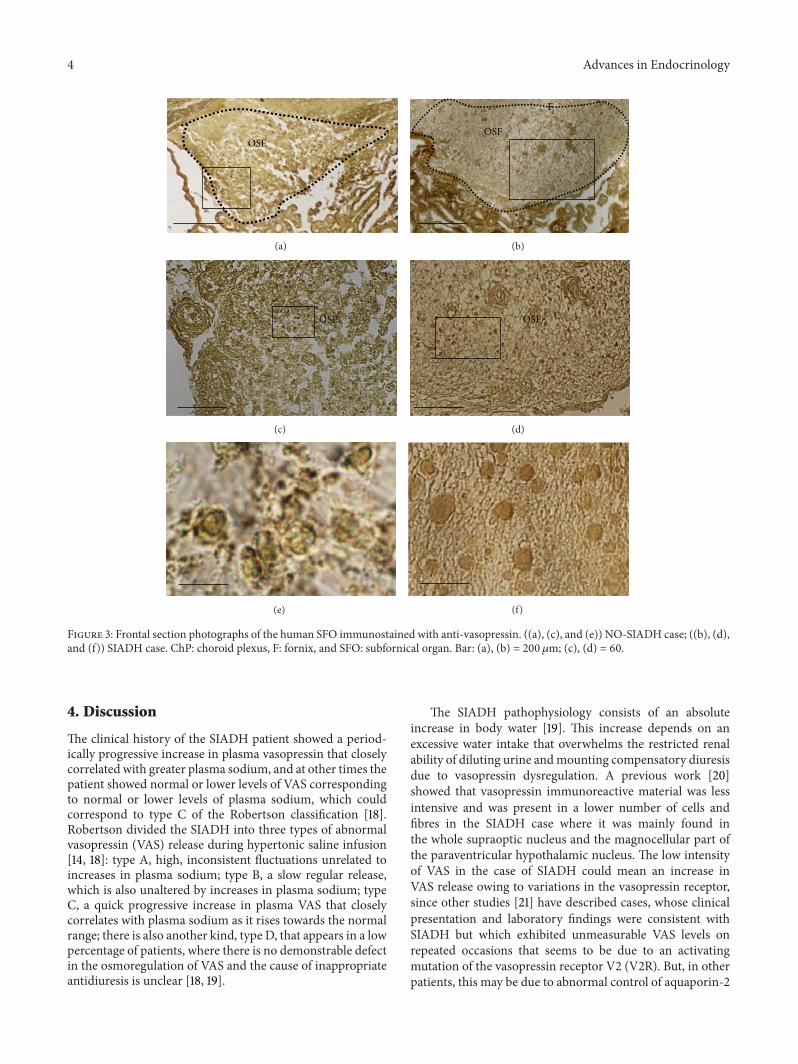
3.2. Vasopressin (VAS). In both of the two cases studied(syndrome and no-syndrome) the expression of VAS wassimilar in the SFOs, where the presence of clusters oflabeled vasopressin cells located in the lateral part of SFOwas detected. Immunoreactive material was also found inependymal cells and perivascular spaces (Figure 3).
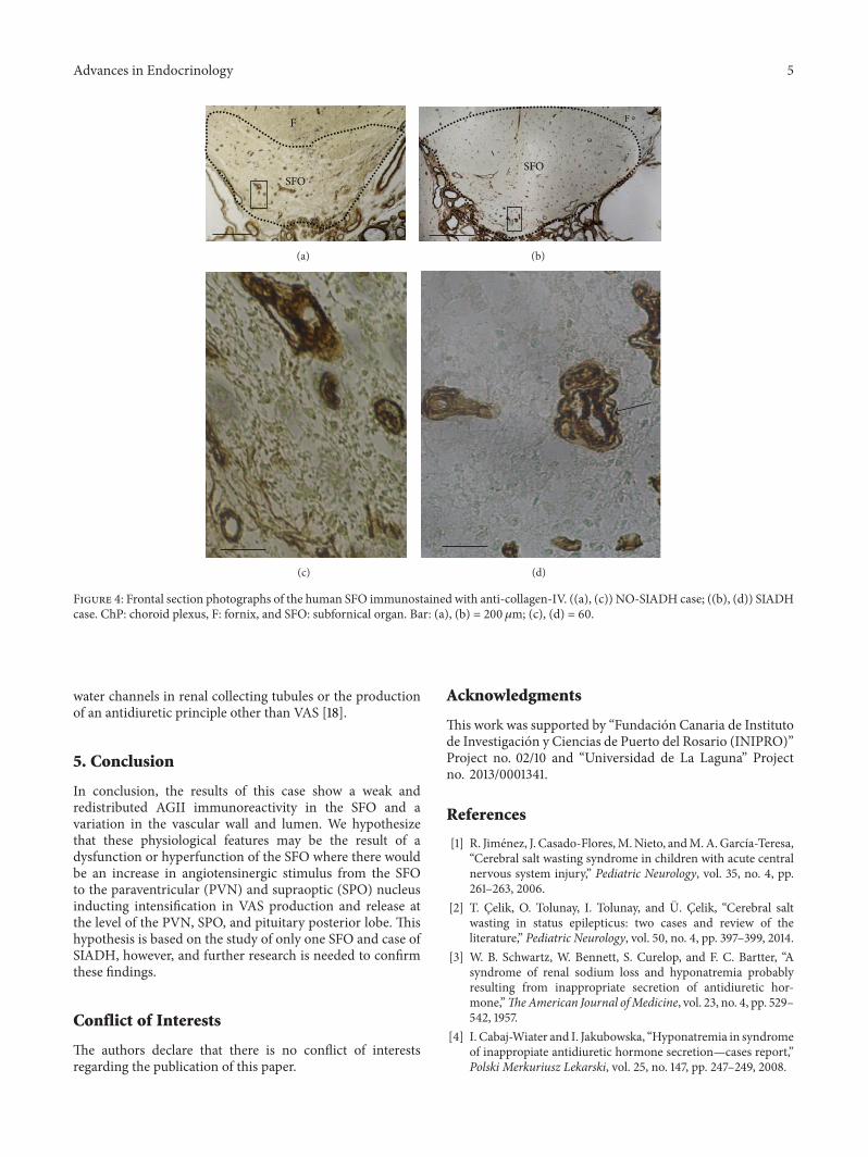
3.3. Collagen-IV (COL-IV). Collagen-IV was located at theSFO vessel wall of both cases (Figure 4). However, in theSIADHcase, amarkedly large number of vessels had irregularlumen and unfolded contours (Figures 4(b) and 4(d), arrow).
4 Advances in Endocrinology
OSF
(a)
F
OSF
(b)
OSF
(c)
OSF
(d)
(e) (f)
Figure 3: Frontal section photographs of the human SFO immunostained with anti-vasopressin. ((a), (c), and (e)) NO-SIADH case; ((b), (d),and (f)) SIADH case. ChP: choroid plexus, F: fornix, and SFO: subfornical organ. Bar: (a), (b) = 200𝜇m; (c), (d) = 60.
4. Discussion
The clinical history of the SIADH patient showed a period-ically progressive increase in plasma vasopressin that closelycorrelated with greater plasma sodium, and at other times thepatient showed normal or lower levels of VAS correspondingto normal or lower levels of plasma sodium, which couldcorrespond to type C of the Robertson classification [18].Robertson divided the SIADH into three types of abnormalvasopressin (VAS) release during hypertonic saline infusion[14, 18]: type A, high, inconsistent fluctuations unrelated toincreases in plasma sodium; type B, a slow regular release,which is also unaltered by increases in plasma sodium; typeC, a quick progressive increase in plasma VAS that closelycorrelates with plasma sodium as it rises towards the normalrange; there is also another kind, type D, that appears in a lowpercentage of patients, where there is no demonstrable defectin the osmoregulation of VAS and the cause of inappropriateantidiuresis is unclear [18, 19].
The SIADH pathophysiology consists of an absoluteincrease in body water [19]. This increase depends on anexcessive water intake that overwhelms the restricted renalability of diluting urine andmounting compensatory diuresisdue to vasopressin dysregulation. A previous work [20]showed that vasopressin immunoreactive material was lessintensive and was present in a lower number of cells andfibres in the SIADH case where it was mainly found inthe whole supraoptic nucleus and the magnocellular part ofthe paraventricular hypothalamic nucleus. The low intensityof VAS in the case of SIADH could mean an increase inVAS release owing to variations in the vasopressin receptor,since other studies [21] have described cases, whose clinicalpresentation and laboratory findings were consistent withSIADH but which exhibited unmeasurable VAS levels onrepeated occasions that seems to be due to an activatingmutation of the vasopressin receptor V2 (V2R). But, in otherpatients, this may be due to abnormal control of aquaporin-2
Advances in Endocrinology 5
F
SFO
(a)
F
SFO
(b)
(c) (d)
Figure 4: Frontal section photographs of the human SFO immunostained with anti-collagen-IV. ((a), (c)) NO-SIADH case; ((b), (d)) SIADHcase. ChP: choroid plexus, F: fornix, and SFO: subfornical organ. Bar: (a), (b) = 200𝜇m; (c), (d) = 60.
water channels in renal collecting tubules or the productionof an antidiuretic principle other than VAS [18].
5. Conclusion
In conclusion, the results of this case show a weak andredistributed AGII immunoreactivity in the SFO and avariation in the vascular wall and lumen. We hypothesizethat these physiological features may be the result of adysfunction or hyperfunction of the SFO where there wouldbe an increase in angiotensinergic stimulus from the SFOto the paraventricular (PVN) and supraoptic (SPO) nucleusinducting intensification in VAS production and release atthe level of the PVN, SPO, and pituitary posterior lobe. Thishypothesis is based on the study of only one SFO and case ofSIADH, however, and further research is needed to confirmthese findings.
Conflict of Interests
The authors declare that there is no conflict of interestsregarding the publication of this paper.
Acknowledgments
This work was supported by “Fundacion Canaria de Institutode Investigacion y Ciencias de Puerto del Rosario (INIPRO)”Project no. 02/10 and “Universidad de La Laguna” Projectno. 2013/0001341.
References
[1] R. Jimenez, J. Casado-Flores,M.Nieto, andM.A.Garcıa-Teresa,“Cerebral salt wasting syndrome in children with acute centralnervous system injury,” Pediatric Neurology, vol. 35, no. 4, pp.261–263, 2006.
[2] T. Celik, O. Tolunay, I. Tolunay, and U. Celik, “Cerebral saltwasting in status epilepticus: two cases and review of theliterature,” Pediatric Neurology, vol. 50, no. 4, pp. 397–399, 2014.
[3] W. B. Schwartz, W. Bennett, S. Curelop, and F. C. Bartter, “Asyndrome of renal sodium loss and hyponatremia probablyresulting from inappropriate secretion of antidiuretic hor-mone,”TheAmerican Journal ofMedicine, vol. 23, no. 4, pp. 529–542, 1957.
[4] I. Cabaj-Wiater and I. Jakubowska, “Hyponatremia in syndromeof inappropiate antidiuretic hormone secretion—cases report,”Polski Merkuriusz Lekarski, vol. 25, no. 147, pp. 247–249, 2008.

6 Advances in Endocrinology
[5] G. Decaux andW.Musch, “Clinical laboratory evaluation of thesyndrome of inappropriate secretion of antidiuretic hormone,”Clinical Journal of the American Society of Nephrology, vol. 3, no.4, pp. 1175–1184, 2008.
[6] J. B. Sorensen, M. K. Andersen, and H. H. Hansen, “Syndromeof inappropriate secretion of antidiuretic hormone (SIADH) inmalignant disease,” Journal of Internal Medicine, vol. 238, no. 2,pp. 97–110, 1995.
[7] H. E. de Wardener, “The hypothalamus and hypertension,”Physiological Reviews, vol. 81, no. 4, pp. 1599–1658, 2001.
[8] M. F. R. Ferrari, E. F. Coelho, K. L. G. Farizatto, G. Chadi,and D. R. Fior-Chadi, “Modulation of tyrosine hydroxylase,neuropeptide Y, glutamate, and substance P in ganglia and brainareas involved in cardiovascular control after chronic exposureto nicotine,” International Journal of Hypertension, vol. 2011,Article ID 216464, 9 pages, 2011.
[9] E. M. Carmona-Calero, H. Perez-Gonzalez, I. Martınez-Pena YValenzuela et al., “Effect of the arterial hypertension and capto-pril treatment on the angiotensin II content in the subfornicalorgan. A study in SHR rats,” Histology and Histopathology, vol.20, no. 1, pp. 135–138, 2005.
[10] I. Gonzalez-Marrero, L. Castaneyra-Ruiz, H. de Paz-Carmona,A. Castaneyra-Ruiz, J. M. Gonzalez-Toledo, and E. M.Carmona-Calero, “Variaciones del sistema angiotensina-vasopresina hipotalamico ante hipertension arterial y sutratamiento con captopril,” Majorensis, vol. 4, no. 1, pp. 15–18,2008.
[11] B. Mayinger and J. Hensen, “Nonpeptide vasopressin antago-nists: a new group of hormone blockers entering the scene,”Experimental and Clinical Endocrinology and Diabetes, vol. 107,no. 3, pp. 157–165, 1999.
[12] M. T. Panayotacopoulou, E. Goudsmit, J. J. van Heerikhuize,and D. F. Swaab, “Simultaneous detection of tyrosine hydro-xylase-immunoreactivity and vasopressin mRNA in neuronsof the human paraventricular and supraoptic nucleus,” BrainResearch, vol. 855, no. 1, pp. 181–185, 2000.
[13] M. Macova, J. Pavel, and J. M. Saavedra, “A peripherallyadministered, centrally acting angiotensin II AT2 antagonistselectively increases brain AT1 receptors and decreases braintyrosine hydroxylase transcription, pituitary vasopressin andACTH,” Brain Research, vol. 1250, pp. 130–140, 2009.
[14] K. Kaneko, T. Shioya, and K. Yabuta, “Inappropriate secretionof antidiuretic hormone and transient hypertension associatedwith Guillain-Barre syndrome,” Pediatric Neuroscience, vol. 15,no. 5, pp. 257–259, 1989.
[15] A. Castaneyra-Perdomo, G. Meyer, E. Carmona-Calero et al.,“The effects of chronic administration of captopril on themousesubfornical organ and area postrema,” Experimental Neurology,vol. 120, no. 1, pp. 145–148, 1993.
[16] A. Castaneyra-Perdomo, G. Meyer, and D. J. Heylings, “Earlydevelopment of the human area postrema and subfornicalorgan,” Anatomical Record, vol. 232, no. 4, pp. 612–619, 1992.
[17] J. P. Coble, R. F. Johnson, M. D. Cassell, A. K. Johnson, J.L. Grobe, and C. D. Sigmund, “Activity of protein kinase c-𝛼 within the subfornical organ is necessary for fluid intake inresponse to brain angiotensin,” Hypertension, vol. 64, pp. 141–148, 2014.
[18] G. L. Robertson, “Regulation of arginine vasopressin in thesyndrome of inappropriate antidiuresis,” The American Journalof Medicine, vol. 119, supplement 1, no. 7, pp. S36–S42, 2006.
[19] P. Esposito, G. Piotti, S. Bianzina, Y. Malul, and A. Dal Canton,“The syndrome of inappropriate antidiuresis: pathophysiology,
clinical management and new therapeutic options,” Nephron -Clinical Practice, vol. 119, no. 1, pp. c62–c73, 2011.
[20] E. M. Carmona-Calero, L. Castaneyra-Ruiz, I. Gonzalez-Marrero et al., “Vasopressin, angiotensin II and tyrosine-hydroxylase expression in the hypothalamus of the syndrome ofinappropriate ADH: a case report,”TheOpen Pathology Journal,vol. 6, no. 1, pp. 1–7, 2012.
[21] S. E. Gitelman, B. J. Feldman, and S. M. Rosenthal, “Nephro-genic syndrome of inappropriate antidiuresisa novel disorderin water balance in pediatric patients,”The American Journal ofMedicine, vol. 119, supplement 1, no. 7, pp. S54–S58, 2006.

Submit your manuscripts athttp://www.hindawi.com
Stem CellsInternational
Hindawi Publishing Corporationhttp://www.hindawi.com Volume 2014
Hindawi Publishing Corporationhttp://www.hindawi.com Volume 2014
MEDIATORSINFLAMMATION
of
Hindawi Publishing Corporationhttp://www.hindawi.com Volume 2014
Behavioural Neurology
EndocrinologyInternational Journal of
Hindawi Publishing Corporationhttp://www.hindawi.com Volume 2014
Hindawi Publishing Corporationhttp://www.hindawi.com Volume 2014
Disease Markers
Hindawi Publishing Corporationhttp://www.hindawi.com Volume 2014
BioMed Research International
OncologyJournal of
Hindawi Publishing Corporationhttp://www.hindawi.com Volume 2014
Hindawi Publishing Corporationhttp://www.hindawi.com Volume 2014
Oxidative Medicine and Cellular Longevity
Hindawi Publishing Corporationhttp://www.hindawi.com Volume 2014
PPAR Research
The Scientific World JournalHindawi Publishing Corporation http://www.hindawi.com Volume 2014
Immunology ResearchHindawi Publishing Corporationhttp://www.hindawi.com Volume 2014
Journal of
ObesityJournal of
Hindawi Publishing Corporationhttp://www.hindawi.com Volume 2014
Hindawi Publishing Corporationhttp://www.hindawi.com Volume 2014
Computational and Mathematical Methods in Medicine
OphthalmologyJournal of
Hindawi Publishing Corporationhttp://www.hindawi.com Volume 2014
Diabetes ResearchJournal of
Hindawi Publishing Corporationhttp://www.hindawi.com Volume 2014
Hindawi Publishing Corporationhttp://www.hindawi.com Volume 2014
Research and TreatmentAIDS
Hindawi Publishing Corporationhttp://www.hindawi.com Volume 2014
Gastroenterology Research and Practice
Hindawi Publishing Corporationhttp://www.hindawi.com Volume 2014
Parkinson’s Disease
Evidence-Based Complementary and Alternative Medicine
Volume 2014Hindawi Publishing Corporationhttp://www.hindawi.com