six-month healing success rates after endodontic treatment ... · pdf fileracine, wi), and a...
TRANSCRIPT
J Clin Exp Dent. 2016;8(3):e290-8. PURE clinical study - six-month healing success rates
e290
Journal section: Operative Dentistry and Endodontics Publication Types: Research
Six-month healing success rates after endodontic treatment using the novel GentleWave® System: The pure prospective multi-center clinical study
Asgeir Sigurdsson 1, Khang T. Le 2, Stacey M. Woo 3, Shahriar A. Rassoulian 4, Kimberly McLachlan 5, Farah Abbassi 6, Randy W. Garland 7
1 DDS, MS. Department of Endodontics, New York University College of Dentistry, New York2 DDS. Private Practices, Santa Ana, California 3 DDS, PhD. Private Practices, Whittier, California 4 DMD. Private Practices, Aliso Viejo, California 5 DMD, MSEd, MBA. Private Practices, Encinitas, California 6 DMD, MSD. Private Practices, Santa Ana, California 7 DDS. Private Practices, Encinitas, California
Correspondence:Department of EndodonticsNew York University College of Dentistry345 East 24 StreetNew York. NY 10010, [email protected]
Received: 24/09/2015Accepted: 04/11/2015
Abstract Background: This prospective multi-center (PURE) clinical study evaluated healing rates for molars after root ca-nal treatment employing the GentleWave® System (Sonendo, Inc., Laguna Hills, CA). Material and Methods: Eighty-nine patients met the inclusion criteria and consented for this clinical study after re-ferral for a root canal treatment. All enrolled patients were treated with the GentleWave System. Five endodontists performed the clinical procedures and follow-up evaluations. Pre-operative, intra-operative, and post-operative data were collected from the consented patients. Each patient was evaluated for clinical signs and symptoms. Two trained, blinded, and independent evaluators scored the subject tooth radiographs for apical periodontitis using the periapical index (PAI). The teeth classified as healing or healed were considered as a success and composed of a cumulative success rate of healing. Statistical analysis was performed by using the Fisher’s exact test, Pearson correlation, and multivariate logistic regression analyses of the pre-operative prognostic factors at 0.05 significance level. Results: Seventy-seven patients were evaluated at six months with a follow-up rate of 86.5%. The cumulative suc-cess rate of healing was 97.4%. Eleven prognostic factors were identified using bivariate analyses. Using logistic analyses, the two prognostic significant variables that were directly correlated to healing were the pre-operative presence of periapical index (p value=0.016), and single treatment visits (p value=0.024). Conclusions: In this six-month PURE clinical study, the cumulative success rate of healing was 97.4% when pa-tients were treated with the GentleWave® System.
Key words: Healing rate, root canal treatment, molar, GentleWave®, Sonendo®, Multisonic Ultracleaning™ .
doi:10.4317/jced.52779http://dx.doi.org/10.4317/jced.52779
Article Number: 52779 http://www.medicinaoral.com/odo/indice.htm© Medicina Oral S. L. C.I.F. B 96689336 - eISSN: 1989-5488eMail: [email protected] in:
PubmedPubmed Central® (PMC)ScopusDOI® System
Sigurdsson A, Le KT, Woo SM, Rassoulian SA, McLachlan K, Abbassi F, Garland RW. Six-month healing success rates after endodontic treat-Six-month healing success rates after endodontic treat-ment using the novel GentleWave® System: The pure prospective multi-center clinical study. J Clin Exp Dent. 2016;8(3):e290-8.http://www.medicinaoral.com/odo/volumenes/v8i3/jcedv8i3p290.pdf
J Clin Exp Dent. 2016;8(3):e290-8. PURE clinical study - six-month healing success rates
e291
IntroductionEndodontic treatment aims to remove vital and/or necro-tic tissue, bacteria and bacterial irritants from the root canal system, thereby promotes healing of the periapical area (1-2). Hence, complete root canal cleaning and dis-infection is essential to achieve healing of periradicular tissue and successful endodontic treatment (3). Many etiological factors affect the outcome of endodon-tic treatment (4). It is well accepted that current cleaning and shaping procedures cannot reach all the intricacies of the root canal system (5). As such, chemo-mechanical preparation and instrumentation do not always comple-tely eradicate the tissue or microbiota present in the ana-tomical complexities of the root canal system (6). Different irrigation techniques and devices have been developed to improve the cleaning of the root canal sys-tem, including ultrasonic irrigation, negative pressure irrigation, sonic irrigation, photo-induced photo-acous-tic streaming (PIPS), and laser technologies. However, the safety, efficacy, and/or reliability of all these techni-ques have been questioned in many studies (7-13). The positive pressure induced by some of these techniques may result in irrigant extrusion to the peri-apex, which may lead to severe patient trauma and post-operative pain (7-10). Further, tissue debris and biofilm cleaning of even contemporary techniques is often insufficient to provide an environment conducive for long term success (2,9-11). Furthermore, most of these techniques require increased dentin removal from the roots to facilitate the penetration of irrigants into the root canal system, which may weaken the remaining tooth and thereby also nega-tively affect long-term healing rates (12,13). The GentleWave® System (Sonendo, Inc., Laguna Hills, CA), which consists of a console and a treatment instru-ment should be capitalized, has been developed as a no-vel approach to clean and disinfect the root canal system (14-17). Haapasalo et al. (2014) demonstrated that the tissue dissolution efficacy of the GentleWave® System is at least eight times greater than that of conventional irrigation systems, ultrasonic irrigation, and EndoVac (14). Ma et al. (2015) performed micro-CT analysis and compared the cleaning efficiency of the GentleWave® System with passive ultrasonic system and conventio-nal needle irrigation configuration. The authors showed cleaning of the entire root canal system including the apical-third regions (15). The GentleWave System was the only technique that removed all the calcium hydroxi-de even in the apical thirds. However, these studies were performed in-vitro using extracted teeth. While in-vitro studies have demonstrated excellent results by the Gent-leWave® System with regards to canal cleanliness and safety, it is ultimately the in-vivo clinical studies that are needed for higher level evidence of the performance and benefits of any endodontic treatment strategy or device. The current study is the first clinical research that reports
the healing rates observed by five independent endodon-tists utilizing the GentleWave® System.
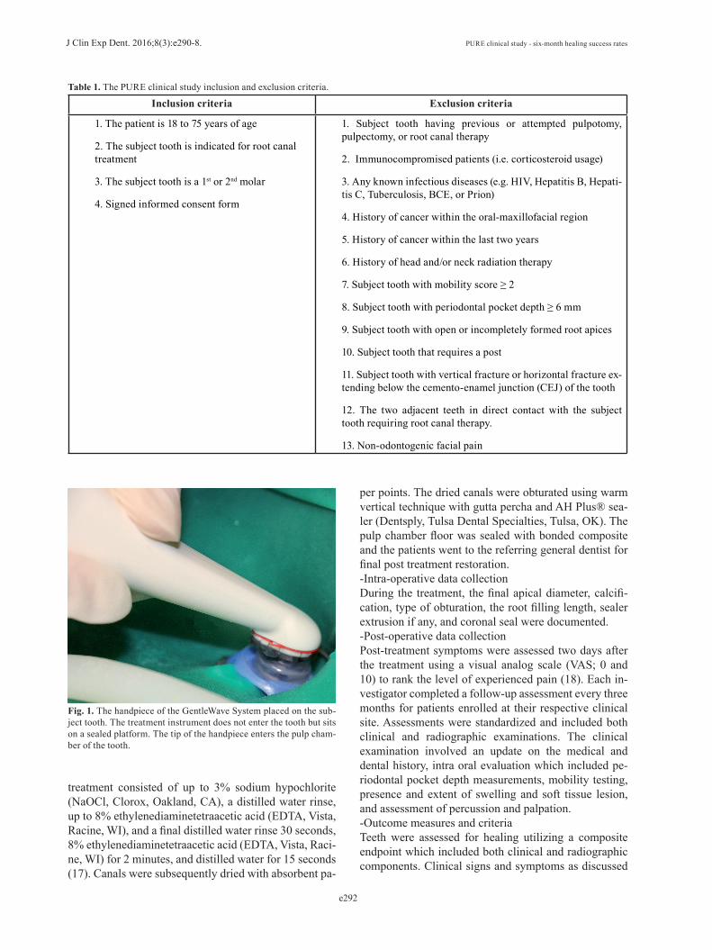
Material and Methods-Study cohortThe inception cohort comprised of eighty-nine patients who were referred for an endodontic treatment. The study protocol for the multi-center, prospective, non-significant risk clinical study was approved by an Institutional Re-view Board (Aspire Llc) and the study was carried out in accordance with the Declaration of Helsinki. The clinical study evaluated the healing rates of endodontic treatments performed using the GentleWave® System. The purpose of the study was explained to the patients and written infor-med consents were obtained. All the subjects adhered to previously defined inclusion and exclusion criteria stated in table 1. After initiation of the study, the subjects were given the opportunity to withdraw. A total of 89 teeth, one tooth per patient, were treated for the clinical study. -InterventionFive endodontists participated as investigators in the multi-center, prospective, non significant risk clinical study to assess the long-term performance of the So-nendo® endotherapy system (PURE). The investigators were trained for using the GentleWave System and per-formed a standardized treatment procedure at their inde-pendent clinical sites. Using standard coded data sheets, the collected redacted clinical and radiographic data pertained to each treated tooth before (pre-operative), during (intra-operative), and six-months after (post-ope-rative) the initial treatment. The data was directly trans-ferred to a database.-Pre-operative data collection Prior to treatment, the patients were clinically examined and radiographs were taken. Pulp and periradicular diag-nosis was completed and regarded. -Treatment procedureThe patient was anesthetized per standard techniques, the type of injection being at the discretion of the endo-dontist. The tooth was isolated with dental dam. Caries and existing restoration were removed. Missing tooth structures were built-up and a conservative straight-line access was performed. Patency was confirmed with #10 and #15 K type hand files (MANI K files, Utsunomi-ya, Japan) and the working length (defined as distance to the apical constriction of approximately 0.5-1 mm from the radiographic apex) was achieved using elec-tronic apex locator and confirmed with radiographs. Teeth were instrumented with a standardized minimal instrumentation protocol that included the use of hand files up to size ISO #20 and Protaper file F1 (Dentsply, Tulsa Dental Specialties, Tulsa, OK) regardless of the initial canal size. The GentleWave treatment instrument should be capitalized was then placed on the endodontic access opening of the molars as shown in figure 1. The
J Clin Exp Dent. 2016;8(3):e290-8. PURE clinical study - six-month healing success rates
e292
treatment consisted of up to 3% sodium hypochlorite (NaOCl, Clorox, Oakland, CA), a distilled water rinse, up to 8% ethylenediaminetetraacetic acid (EDTA, Vista, Racine, WI), and a final distilled water rinse 30 seconds, 8% ethylenediaminetetraacetic acid (EDTA, Vista, Raci-ne, WI) for 2 minutes, and distilled water for 15 seconds (17). Canals were subsequently dried with absorbent pa-
per points. The dried canals were obturated using warm vertical technique with gutta percha and AH Plus® sea-ler (Dentsply, Tulsa Dental Specialties, Tulsa, OK). The pulp chamber floor was sealed with bonded composite and the patients went to the referring general dentist for final post treatment restoration.-Intra-operative data collectionDuring the treatment, the final apical diameter, calcifi-cation, type of obturation, the root filling length, sealer extrusion if any, and coronal seal were documented. -Post-operative data collectionPost-treatment symptoms were assessed two days after the treatment using a visual analog scale (VAS; 0 and 10) to rank the level of experienced pain (18). Each in-vestigator completed a follow-up assessment every three months for patients enrolled at their respective clinical site. Assessments were standardized and included both clinical and radiographic examinations. The clinical examination involved an update on the medical and dental history, intra oral evaluation which included pe-riodontal pocket depth measurements, mobility testing, presence and extent of swelling and soft tissue lesion, and assessment of percussion and palpation. -Outcome measures and criteriaTeeth were assessed for healing utilizing a composite endpoint which included both clinical and radiographic components. Clinical signs and symptoms as discussed
Inclusion criteria Exclusion criteria
1. The patient is 18 to 75 years of age
2. The subject tooth is indicated for root canal treatment
3. The subject tooth is a 1st or 2nd molar
4. Signed informed consent form
1. Subject tooth having previous or attempted pulpotomy, pulpectomy, or root canal therapy
2. Immunocompromised patients (i.e. corticosteroid usage)
3. Any known infectious diseases (e.g. HIV, Hepatitis B, Hepati-tis C, Tuberculosis, BCE, or Prion)
4. History of cancer within the oral-maxillofacial region
5. History of cancer within the last two years
6. History of head and/or neck radiation therapy
7. Subject tooth with mobility score ≥ 2
8. Subject tooth with periodontal pocket depth ≥ 6 mm
9. Subject tooth with open or incompletely formed root apices
10. Subject tooth that requires a post
11. Subject tooth with vertical fracture or horizontal fracture ex-tending below the cemento-enamel junction (CEJ) of the tooth
12. The two adjacent teeth in direct contact with the subject tooth requiring root canal therapy.
13. Non-odontogenic facial pain
Table 1. The PURE clinical study inclusion and exclusion criteria.
Fig. 1. The handpiece of the GentleWave System placed on the sub-ject tooth. The treatment instrument does not enter the tooth but sits on a sealed platform. The tip of the handpiece enters the pulp cham-ber of the tooth.
J Clin Exp Dent. 2016;8(3):e290-8. PURE clinical study - six-month healing success rates
e293
previously were utilized for assessing the clinical com-ponent. Periapical index scoring (PAI) was utilized to assess the tooth using a periapical radiograph. The sco-res ranged from 1 (for normal periradicular tissue) to 5 (severe periodontitis with exacerbating features) (19). Based on clinical signs/symptoms and PAI scores, teeth were classified as healed, healing, or diseased (19-20). In summary, the diagnosed teeth were classified as fo-llows:(a) Healed – clinical normalcy other than tenderness to percussion accompanied by radiographic PAI scores of 1 or 2.(b) Healing – clinical normalcy other than tenderness to percussion accompanied by reduction in the size of peri-radicular lesion or reduction in PAI score.(c) Diseased – presence of clinical signs and symptoms accompanied by radiographic PAI score of 3 or higher or increase in the size of periradicular lesion or increase in PAI score. The teeth classified as healing or healed were considered as a success. The combined success of these cases was termed as healing rate. -Calibration of evaluators The radiographs were blindly evaluated by two expe-rienced endodontists. The images were coded and pro-vided to the evaluators after being randomized between different patients. Before evaluating the images, the two examiners evaluated a series of radiographs independent of the study sample that represented a wide range of pe-riapical lesions to account for inter-observer reliability (19). The Cohen’s kappa score was calculated. The exer-cise was independently performed three times to increa-se the calibration. In general, each visible root on the radiographs was assigned a PAI score. The highest PAI score for all the roots for a given tooth was considered as the PAI score of the tooth. This PAI score was consi-dered for further statistical evaluation. -Evaluating radiographs The two evaluators independently scored the radiogra-phs. After the independent scoring sessions, the exami-ners reached an agreement on the PAI scores if the scores of their independent evaluations differed. The consensus scores for all the radiograph images were considered as the true score and were used for statistical analysis. -Statistical analysis All the tests were performed as two-tailed with SPSS 15.0 (SPSS Inc., Chicago, IL) at 5% level of significan-ce. When analyzing, the event of interest was the suc-cess of healing of the tooth. A total of 34 variables were investigated. A univariate and bivariate analyses with percentage of frequencies and p-values was generated to characterize the study cohort. The bivariate analy-sis included outcome associations with pre-operative, intra-operative, and post-operative variables (Fisher exact test) to identify variables of interest. Spearson co-
efficients were calculated to determine any correlation between these variables to categorize potential outcome predictors. Finally, a multivariate analysis using logis-tic regression models was used to detect the significant outcome predictors. The odds ratio (OR) and confidence intervals (CI) were calculated.
Results-Examination reliabilityThe achieved Cohen’s kappa score for intra-observer agreement between the independent reviewers was 0.73- 0.75, indicating a good to very good agreement (19).-Recall and healing Eighty-nine patients met the inclusion criteria and con-sented to participate in the clinical study. Of the 89 pa-tients, 43.8% were male whereas 56.2% were female. 5.6% had a history of diabetes whereas 12.1% had a history of tobacco use. Oral hygiene of the study cohort was good (67.4%) or fair (32.6%).The successful recall of 77 of 89 teeth of the available patients represented an 86.5% recall rate. Of the 77 tee-th, 60 teeth (77.9%) were healed, 15 teeth (19.5%) were being healed, and two teeth (2.6%) were diseased. Ove-rall, 75 of 77 teeth (97.4%) were being healed six mon-ths after the GentleWave treatments. These results are summarized in table 2, table 2 continue.-Identifying predictor factorsTable 2 also provides an overview of the pre-operative, intra-operative, and post-operative factors. Pre-operative factors: None of the pre-operative fac-tors had a significant difference when compared to healing. The p values for gender, age, and oral hygiene were 0.86, 0.095, and 0.33, respectively. The following factors were also analyzed: periradicular diagnosis (p-value=0.787), pulp diagnosis (p value=0.487); PAI sco-re (p-value=0.573), pocket depth (p-value=0.560), pre-operative symptoms (p value =0.258), maxillary versus mandibular molars (p-value =0.207) and right versus left molars (p value=0.120). Inter-operative factors: A significant difference was ob-served (p-value=0.024) for the intra-operative factor related to single versus two-day endodontic treatment; the success rate of healing was correlated to single-visit treatments. Calcification (p value=0.221), sealer extru-sion (p-value=0.998) and root canal filling length (p-value=0.507) demonstrated no significant difference in regards to healing. Post-operative factors: At the six-month follow-up data was collected similar to that at the pre operative visit. Post-operative clinical symptoms (p-value=0.024) and post-operative PAI scores (p-value=0.0005) were signifi-cantly different. Periradicular diagnosis (p-value=0.096), pocket depth (p-value=0.756), and type of restoration (p-value=0.642) showed no significant difference. Further, as shown in table 3, the Pearson correlations
J Clin Exp Dent. 2016;8(3):e290-8. PURE clinical study - six-month healing success rates
e294
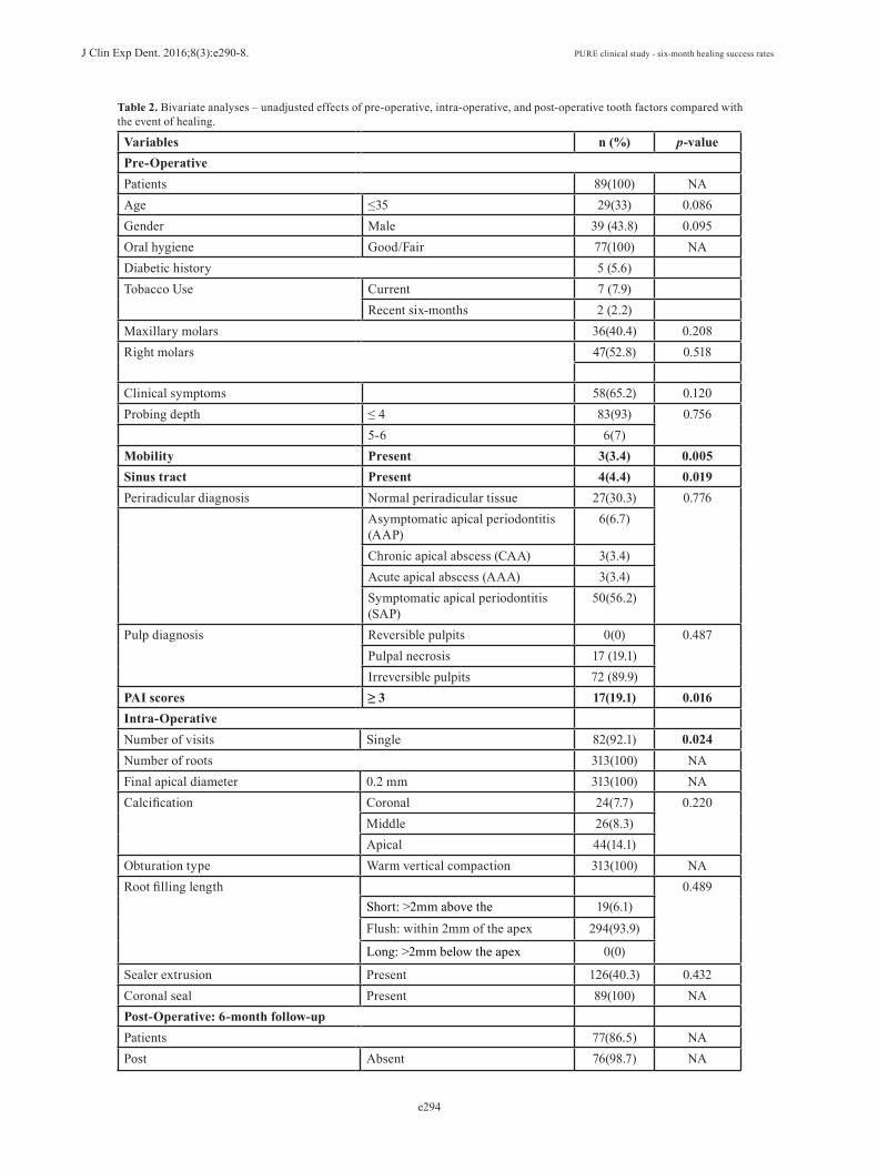
Table 2. Bivariate analyses – unadjusted effects of pre-operative, intra-operative, and post-operative tooth factors compared with the event of healing.
Variables n (%) p-valuePre-OperativePatients 89(100) NAAge ≤35 29(33) 0.086Gender Male 39 (43.8) 0.095Oral hygiene Good/Fair 77(100) NADiabetic history 5 (5.6)Tobacco Use Current 7 (7.9)
Recent six-months 2 (2.2)Maxillary molars 36(40.4) 0.208Right molars 47(52.8) 0.518
Clinical symptoms 58(65.2) 0.120Probing depth ≤ 4 83(93) 0.756
5-6 6(7)Mobility Present 3(3.4) 0.005Sinus tract Present 4(4.4) 0.019Periradicular diagnosis Normal periradicular tissue 27(30.3) 0.776
Asymptomatic apical periodontitis (AAP)
6(6.7)
Chronic apical abscess (CAA) 3(3.4)Acute apical abscess (AAA) 3(3.4)Symptomatic apical periodontitis (SAP)
50(56.2)
Pulp diagnosis Reversible pulpits 0(0) 0.487Pulpal necrosis 17 (19.1)Irreversible pulpits 72 (89.9)
PAI scores ≥ 3 17(19.1) 0.016Intra-OperativeNumber of visits Single 82(92.1) 0.024Number of roots 313(100) NAFinal apical diameter 0.2 mm 313(100) NACalcification Coronal 24(7.7) 0.220
Middle 26(8.3)Apical 44(14.1)
Obturation type Warm vertical compaction 313(100) NARoot filling length
0.489Short: >2mm above the 19(6.1)
Flush: within 2mm of the apex 294(93.9)
Long: >2mm below the apex 0(0)
Sealer extrusion Present 126(40.3) 0.432Coronal seal Present 89(100) NAPost-Operative: 6-month follow-upPatients 77(86.5) NAPost Absent 76(98.7) NA
J Clin Exp Dent. 2016;8(3):e290-8. PURE clinical study - six-month healing success rates
e295
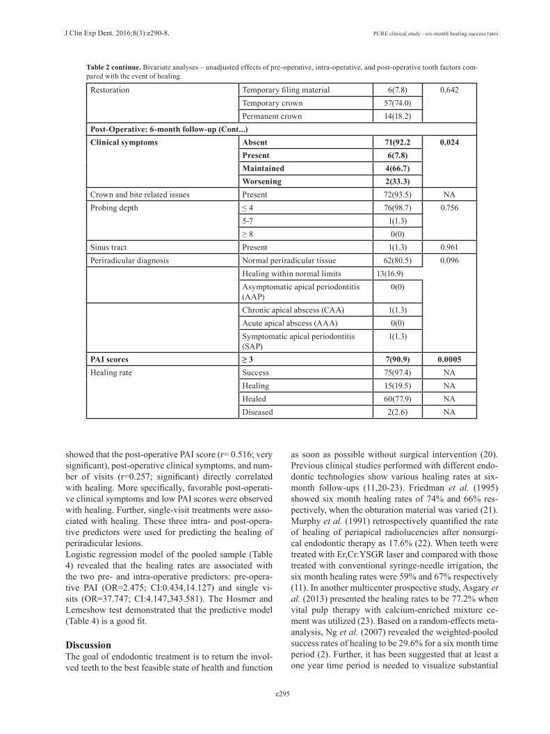
Restoration Temporary filing material 6(7.8) 0.642Temporary crown 57(74.0)Permanent crown 14(18.2)
Post-Operative: 6-month follow-up (Cont...)Clinical symptoms Absent 71(92.2 0.024
Present 6(7.8)Maintained 4(66.7)Worsening 2(33.3)
Crown and bite related issues Present 72(93.5) NAProbing depth ≤ 4 76(98.7) 0.756
5-7 1(1.3)≥ 8 0(0)
Sinus tract Present 1(1.3) 0.961Periradicular diagnosis Normal periradicular tissue 62(80.5) 0.096 Healing within normal limits 13(16.9)
Asymptomatic apical periodontitis (AAP)
0(0)
Chronic apical abscess (CAA) 1(1.3)Acute apical abscess (AAA) 0(0)Symptomatic apical periodontitis (SAP)
1(1.3)
PAI scores ≥ 3 7(90.9) 0.0005Healing rate Success 75(97.4) NA
Healing 15(19.5) NAHealed 60(77.9) NADiseased 2(2.6) NA
Table 2 continue. Bivariate analyses – unadjusted effects of pre-operative, intra-operative, and post-operative tooth factors com-pared with the event of healing.
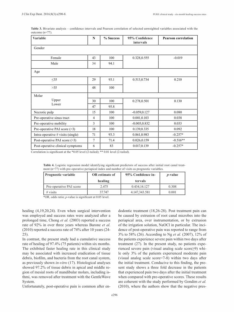
showed that the post-operative PAI score (r= 0.516; very significant), post-operative clinical symptoms, and num-ber of visits (r=0.257; significant) directly correlated with healing. More specifically, favorable post-operati-ve clinical symptoms and low PAI scores were observed with healing. Further, single-visit treatments were asso-ciated with healing. These three intra- and post-opera-tive predictors were used for predicting the healing of periradicular lesions. Logistic regression model of the pooled sample (Table 4) revealed that the healing rates are associated with the two pre- and intra-operative predictors: pre-opera-tive PAI (OR=2.475; CI:0.434,14.127) and single vi-sits (OR=37.747; CI:4.147,343.581). The Hosmer and Lemeshow test demonstrated that the predictive model (Table 4) is a good fit.
DiscussionThe goal of endodontic treatment is to return the invol-ved teeth to the best feasible state of health and function
as soon as possible without surgical intervention (20). Previous clinical studies performed with different endo-dontic technologies show various healing rates at six-month follow-ups (11,20-23). Friedman et al. (1995) showed six month healing rates of 74% and 66% res-pectively, when the obturation material was varied (21). Murphy et al. (1991) retrospectively quantified the rate of healing of periapical radiolucencies after nonsurgi-cal endodontic therapy as 17.6% (22). When teeth were treated with Er,Cr:YSGR laser and compared with those treated with conventional syringe-needle irrigation, the six month healing rates were 59% and 67% respectively (11). In another multicenter prospective study, Asgary et al. (2013) presented the healing rates to be 77.2% when vital pulp therapy with calcium-enriched mixture ce-ment was utilized (23). Based on a random-effects meta-analysis, Ng et al. (2007) revealed the weighted-pooled success rates of healing to be 29.6% for a six month time period (2). Further, it has been suggested that at least a one year time period is needed to visualize substantial
J Clin Exp Dent. 2016;8(3):e290-8. PURE clinical study - six-month healing success rates
e296
Variable N % Success 95% Confidence intervals
Pearson correlation
Gender
Female 43 100 0.328,0.555 -0.019 Male 34 94.1
Age
≤35 29 93.1 0.513,0.734 0.210
>35 48 100
Molar Upper Lower
30 100 0.278,0.501 0.13047 95.8
Necrotic pulp 15 100 -0.059,0.127 0.080Pre-operative sinus tract 4 100 0.001,0.103 0.038Pre-operative mobility 3 100 -0.005,0.832 0.033Pre-operative PAI score (>3) 18 100 0.139,0.335 0.092Intra-operative # visits (single) 71 93.3 0.861,0.983 -0.257*Post-operative PAI score (>3) 7 71.4 0.026,0.159 -0.516**Post-operative clinical symptoms 6 83 0.017,0.139 -0.257*
Table 3. Bivariate analysis – confidence intervals and Pearson correlation of selected unweighted variables associated with the outcome (n=77).
Correlation is significant at the *0.05 level (2-tailed); ** 0.01 level (2-tailed).
Prognostic variable OR estimate of
healing
95% Confidence in-
tervals
p-value
Pre-operative PAI score 2.475 0.434,14.127 0.308# visits 37.747 4.147,343.581 0.001
Table 4. Logistic regression model identifying significant predictors of success after initial root canal treat-ment (n=77) with pre-operative periapical index and number of visits as prognostic variables.
*OR, odds ratio; p-value is significant at 0.05 level.
healing (4,19,20,24). Even when surgical intervention was employed and success rates were analyzed after a prolonged time, Chong et al. (2003) reported a success rate of 92% in over three years whereas Barone et al. (2010) reported a success rate of 74% after 10 years (24-25). In contrast, the present study had a cumulative success rate of healing of 97.4% (75 patients) within six months. The exhibited faster healing rate in this clinical study may be associated with increased eradication of tissue debris, biofilm, and bacteria from the root canal system, as previously shown in-vitro (17). Histological analyses showed 97.2% of tissue debris in apical and middle re-gion of mesial roots of mandibular molars, including is-thmi, was removed after treatment with the GentleWave System. Unfortunately, post-operative pain is common after en-
dodontic treatment (18,26-28). Post treatment pain can be caused by extrusion of root canal microbes into the periapical area, over instrumentation, or by extrusion of the irrigation solution, NaOCl in particular. The inci-dence of post-operative pain was reported to range from 3% to 58% (26). According to Ng et al. (2007), 12% of the patients experience severe pain within two days after treatment (27). In the present study, no patients expe-rienced severe pain (visual analog scale score≥9) whi-le only 3% of the patients experienced moderate pain (visual analog scale score=7-8) within two days after the initial treatment. Conducive to this finding, the pre-sent study shows a three fold decrease in the patients that experienced pain two days after the initial treatment when compared with pre-operative scores. These results are coherent with the study performed by Gondim et al. (2010), where the authors show that the negative pres-
J Clin Exp Dent. 2016;8(3):e290-8. PURE clinical study - six-month healing success rates
e297
sure system resulted in significantly less post-operative pain (28). Interestingly, Charara et al. (2015) compared the GentleWave System to EndoVac and showed that both these negative pressure systems led to zero extru-sion to the periapical space in-vitro (16). In recent years, single-visit appointment regimens have reported numerous advantages including better patient acceptance, reduction of the inter-appointment infection risks, saving time, and cost (9,29). However, significant post-operative pain has been reported after single-visit root canal treatments and for teeth having necrotic pulp (9,29). Xaio et al. (2010) compared the healing rates of one-visit appointment with two-visit appointments and concluded that the healing rates were 68.4% and 64.5% respectively (29). Beus et al. (2012) demonstrated the prevalence of bacteria remaining in the root canal system when teeth were treated with single-visit regimens (9).On the contrary, in the present study using the Gent-leWave System, post-operative pain was not correlated with either single-visit appointment or with necrotic tee-th. Moreover, 72 patients showed success (98.6%) when treated with single-visit endodontics whereas 15 patients showed success (100%) when the teeth were necrotic. A fundamental factor that improves prognosis is the pre-servation of dentin structure in its native form (30). It is noteworthy that the present study utilized minimal en-dodontics by employing methods that minimally remove dentin structure while accessing the teeth and shaping the root canals. Previous studies showed that even when molars were shaped to #15/.04 in-vitro when using the GentleWave System, statistically significant clean root canal system was observed (14-17). However, the pre-sent clinical study utilized shaping to #20/.07 in order to facilitate standard obturation techniques. As shown in fi-gure 1, the treatment instrument of the GentleWave Sys-tem is placed in the pulp chamber of the molars. Since the treatment instrument should be capitalized does not have to enter the roots, the GentleWave System reduces the need for shaping of the roots using large instrumen-tation, hence practicing minimal endodontic technique with dentinal conservation. Details of the GentleWave technology are described elsewhere (16,17). Briefly, the technology employs a strong hydrodynamic cavitation cloud and generates a broad spectrum of sound waves that travel through the degassed treatment fluid and pro-pagates throughout the entire root canal system. The GentleWave System allows minimal instrumenta-tion, cleans the root canal system thoroughly, and pro-duces negative pressure in the root canal system (14-17). Therefore, the rare occurrence of post-operative symp-toms in the present study after the tooth is treated with the GentleWave System, is not surprising. In conclusion, root canal treatment utilizing the Gent-leWave System demonstrated a cumulative success rate for healing of 97.4% within six months of the initial
treatment. Long term follow-ups can improve the sta-tistical power and enable further investigation into prog-nostic factors for tooth healing following the root canal treatment. Additional in vivo studies are also needed to compare the healing rates acquired by the GentleWave System to those obtained with other conventional and contemporary endodontic techniques.
References1.Sjogren U, Hagglund B, Sundqvist G, Wing K. Factors affecting the long-term results of endodontic treatment. J Endod. 1990;16:498-504.2. Ng YL, Mann V, Rahbaran S, Lewsey J, Gulabivala K. Outcome of primary root canal treatment: systematic review of the literature – Part 1. Effects of study characteristics on probability of success. Int End J 2007;40:921-39.3. Loest C. Quality guidelines for endodontic treatment: consensus report of the european society of endodontology. Int Endod J. 2006; 39:921-30.4. Siqueira JF. Aetiology of root canal treatment failure: why well-treated teeth can fail. Int Endod J. 2001;34:1-10.5. Hulsmann M, Hahn W. Complications during root canal irrigation: literature review and case reports. Int Endod J. 2000;33:186-93.6. Haapasalo M, Endal U, Zandi H, Coil JM. Eradication of endodon-tic infection by instrumentation and irrigation solutions. Endod Top. 2005;10:77-102.7. Tay FR, Gu L, Schoeffel GJ, Wimmer C, Susin L, Zhang K, et al. Effect of vapor lock on root canal debridement by using a side-vented needle for positive-pressure irrigant delivery. J Endod. 2010;36:745-50.8. Muñoz HR, Camacho-Cuadra K. In vivo efficacy of three different endodontic irrigation systems for irrigant delivery to working length of mesial canals of mandibular molars J Endod. 2012;38:445-88.9. Beus C, Safavi K, Stratton J, Kaufman B. Comparison of the effect of two endodontic irrigation protocols on the elimination of bacteria from root canal system: a prospective, randomized cinical trial. J En-dod. 2012;38:1479-83.10. Li D, Jiang S, Yin X, Chang JWW, Ke J, Zhang C. Efficacy of nee-dle, ultrasonic, and endoactivator irrigation and photon-induced pho-toacoustic streaming in removing calcium hydroxide from the main ca-nal and isthmus: an in vitro micro-computed tomography and scanning electron microscopy study. Photomed and Las Surg. 2015;33:330-7.11. Martins MR, Carvalho MF, Vaz IP, Capelas JA, Martins MA, Gutk-necht N. Efficacy of Er, Cr:YSGG laser with endodontic radial firing. Lasers Med Sci. 2013;28:1049-55.12. Merino A, Estevez R, de Gregorio C, Cohenca N. The effect of different taper preparations on the ability of sonic and passive ultra-sonic irrigation to reach the working length in curved canals. Int End J. 2013;46:427-33.13. Saini HR, Tewari S, Sangwan P, Duhan J, Gupta A. Effect of Di-fferent Apical Preparation Sizes on Outcome of Primary Endodontic Treatment: A Randomized Controlled Trial. J Endod. 2012;38:1309-15.14. Haapasalo M, Wang Z, Shen Y, Curtis A, Patel P, Khakpour M. Tis-sue dissolution by a novel multisonic ultracleaning system and sodium hypochlorite. J Endod. 2014;40:1178-81.15. Ma J, Shen Y, Yang Y, Gao Y, Wan P, Gan Y, et al. In vitro study of calcium hydroxide removal from mandibular molar root canals using a GentleWave™ System. J Endod. 2015;41:553-8.16. Charara K, Friedman S, Sherman A, Kishen A, Malkhassian G, Khakpour M, et al. Assessment of apical extrusion during root canal procedure with the novel GentleWave System in a simulated apical environment. J Endod. 2016;42:135-9.17. Molina B, Glickman GN, Vandrangi P, Khakpour M. Histological evaluation of root canal debridement of human molars using the Gent-leWave™ System. J Endod. 2015;41:1701-5.18. Segura-Egea JJ, Cisneros-Cabello R, Llamas-Carreras JM, Ve-lasco-Ortega E. Pain associated with root canal treatment. Int End J. 2009;42:614-20.
J Clin Exp Dent. 2016;8(3):e290-8. PURE clinical study - six-month healing success rates
e298
19. Orstavik D, Kerekes K, Eriksen HM. The periapical index: a sco-ring system for radiographic assessment of apical periodontitis. Endod Dent Traumatol. 1986;2:20-34.20. Friedman S, Abitbol S, Lawrence HP. Treatment outcome in en-dodontics: the Toronto Study. Phase 1: initial treatment. J Endod. 2003;29:787-93.21. Friedman S, Lost C, Zarrabian M, Trope M. Evaluation of success and failure after endodontic therapy using a glass ionomer cement sea-ler. J Endod. 1995;21:384-90.22. Murphy WK, Kaugars GE, Collett WK, Dodds RN. Healing of periapical radiolucencies after nonsurgical endodontic therapy. Oral Surg Oral Med Oral Pathol. 1991;71:620-4.23. Asgary S, Eghbal MJ, Ghoddusi J, Yazdani S. One-year results of vital pulp therapy in permanent molars with irreversible pulpitis: an ongoing multicenter, randomized, non-inferiority clinical trial. Clin Oral Invest. 2013;17:431-9.24. Barone C, Dao TT, Basrani BB, Wang N, Friedman S. Treatment outcome in endodontics: the Toronto study—phases 3, 4, and 5: apical surgery. J Endod. 2010;36:28-35.25. Chong BS, Pitt Ford TR, Hudson MB. A prospective clinical study of Mineral Trioxide Aggregate and IRM when used as root-end filling materials in endodontic surgery. Int Endod J. 2003;36:520-6.26. Sathorn C, Parashos P, Messer H. The prevalence of postoperative pain and flare-up in single and multiple-visit endodontic treatment: a systematic review. Int End J. 2008;41:91-9.27. Ng YL, Glennon JP, Setchell DJ, Gulabivala K. Prevalence of and factors affecting post-obturation pain in patients undergoing root canal treatment. Int End J. 2004;37:381-391.28. Gondim E, Setzer FC, Dos Carmo CB, Kim S. Postoperative pain after the application of two different irrigation devices in a prospective randomized clinical trial. J Endod. 2010;36:1295-301.29. Xiao D, Zhang DH. A clinical study of one-visit endodontic treatment for infected root canals. Hua Xi Kou Qiang Yi Xue Za Zhi. 2010;28:57-60.30. Murdoch-Kinch CA, McLean ME. Minimally invasive dentistry. J Am Dent Assoc. 2003;134:87-95.
AcknowledgementsThe PURE clinical study is funded by Sonendo Inc. The authors would also like to thank Dr. Markus Haapasalo for his insightful comments on the manuscript.
Conflict of InterestAS and RG are consultants at Sonendo® Inc.