sox2 is essential for oligodendroglial proliferation …...4 83 genetic evidence suggests that sox2...
TRANSCRIPT
Accepted manuscripts are peer-reviewed but have not been through the copyediting, formatting, or proofreadingprocess.
Copyright © 2018 the authors
This Accepted Manuscript has not been copyedited and formatted. The final version may differ fromthis version. A link to any extended data will be provided when the final version is posted online.
Research Articles: Development/Plasticity/Repair
Sox2 is essential for oligodendroglial proliferation and differentiationduring postnatal brain myelination and CNS remyelination
Sheng Zhang1,2, Xiaoqing Zhu1,2, Xuehong Gui1, Christopher Croteau1, Lanying Song1,2, Jie Xu1, Aijun
Wang1,3, Peter Bannerman1,4 and Fuzheng Guo1,2
1Institute for Pediatric Regenerative Medicine, Shriners Hospitals for Children, Northern California, CA 958172Department of Neurology, School of Medicine, UC Davis, CA 958173Department of Surgery, School of Medicine, UC Davis, CA 958174Department of Cell Biology and Human Anatomy, School of Medicine, UC Davis, CA 495817
DOI: 10.1523/JNEUROSCI.1291-17.2018
Received: 10 May 2017
Revised: 11 December 2017
Accepted: 8 January 2018
Published: 15 January 2018
Author contributions: S.Z., X.Z., and F.G. designed research; S.Z., X.Z., X.G., C.C., L.S., J.X., A.W., P.B., andF.G. performed research; S.Z., X.Z., X.G., C.C., and F.G. analyzed data; S.Z., X.Z., and F.G. wrote the paper;A.W., P.B., and F.G. contributed unpublished reagents/analytic tools.
Conflict of Interest: The authors declare no competing financial interests.
supported by the NIH (R01NS094559 and R21NS093559 to F.G.) and research grants funded by ShrinersHospitals for Children (F.G.) and postdoctoral fellowship grant from Shriners Hospitals for Children (S.Z.). Wethank Dr. Q. Richard Lu (University of Cincinnati, OH) for his critical comments on the manuscript and DaffcarErol (UC Davis) for the editions. A. W. is supported in part by NIH R01NS100761.
Corresponding author: Fuzheng Guo, Department of Neurology, UC Davis School of Medicine, c/o ShrinersHospitals for Children, 2425 Stockton Blvd. Sacramento, CA 95817. Email: [email protected]
Cite as: J. Neurosci ; 10.1523/JNEUROSCI.1291-17.2018
Alerts: Sign up at www.jneurosci.org/cgi/alerts to receive customized email alerts when the fully formattedversion of this article is published.
1
Title Page 1
Journal section: Development/Plasticity/Repair 2
Title: Sox2 is essential for oligodendroglial proliferation and differentiation during 3
postnatal brain myelination and CNS remyelination 4
Abbreviated title: Sox2 regulates brain myelination and remyelination 5
Authors and affiliations: Sheng Zhang*1,2, Xiaoqing Zhu*1,2, Xuehong Gui1, 6
Christopher Croteau1, Lanying Song1,2, Jie Xu1, Aijun Wang1,3, Peter Bannerman1,4, 7
Fuzheng Guo**1,2 8
1, Institute for Pediatric Regenerative Medicine, Shriners Hospitals for Children, 9
Northern California, CA 95817 10
2, Department of Neurology, School of Medicine, UC Davis, CA 95817 11
3, Department of Surgery, School of Medicine, UC Davis, CA 95817 12
4, Department of Cell Biology and Human Anatomy, School of Medicine, UC Davis, CA 13
95817 14
* Co-first authors 15
** Corresponding author: Fuzheng Guo, Department of Neurology, UC Davis School of 16
Medicine, c/o Shriners Hospitals for Children, 2425 Stockton Blvd. Sacramento, CA 17
95817. Email: [email protected] 18
19
Number of figures: 10, Number of tables: 2 20
Number of words: abstract (225), significant statement (98), introduction (385), 21
discussion (1314). 22
Key words: neural stem cell factor, Sox2, oligodendroglial proliferation, differentiation, 23
regeneration, lineage progression and maturation, myelination, remyelination. 24
25
Acknowledgements: supported by the NIH (R01NS094559 and R21NS093559 to 26
F.G.) and research grants funded by Shriners Hospitals for Children (F.G.) and 27
postdoctoral fellowship grant from Shriners Hospitals for Children (S.Z.). We thank Dr. 28
Q. Richard Lu (University of Cincinnati, OH) for his critical comments on the manuscript 29
and Daffcar Erol (UC Davis) for the editions. A. W. is supported in part by NIH 30
R01NS100761. 31
32
2
Abstract (225 words) 33
In the central nervous system (CNS), myelination and remyelination depend on the 34
successful progression and maturation of oligodendroglial lineage cells including 35
proliferation and differentiation of oligodendroglial progenitor cells (OPCs). Previous 36
studies have reported that Sox2 transiently regulates oligodendrocyte (OL) 37
differentiation in the embryonic and perinatal spinal cord and appears dispensable for 38
myelination in the postnatal spinal cord. However, the role of Sox2 in OL development 39
in the brain has yet to be defined. We now report that Sox2 is an essential positive 40
regulator of developmental myelination in the postnatal murine brain of both sexes. 41
Stage-specific paradigms of genetic disruption demonstrated that Sox2 regulated brain 42
myelination by coordinating upstream OPC population supply and downstream OL 43
differentiation. Transcriptomic analyses further supported a crucial role of Sox2 in brain 44
developmental myelination. Consistently, oligodendroglial Sox2 deficient mice 45
developed severe tremors and ataxia, typical phenotypes indicative of hypomyelination, 46
and displayed severe impairment of motor function and prominent deficits of brain OL 47
differentiation and myelination persisting into the later CNS developmental stages. We 48
also found that Sox2 was required for efficient OPC proliferation and expansion and OL 49
regeneration during remyelination in the adult brain and spinal cord. Together, our 50
genetic evidences reveal an essential role of Sox2 in brain myelination and CNS 51
remyelination, and suggest that manipulation of Sox2 and/or Sox2-mediated 52
downstream pathways may be therapeutic in promoting CNS myelin repair. 53
54
55
56
57
3
Significance Statement (98 words) 58
Promoting myelin formation and repair has translational significance in treating myelin-59
related neurological disorders such as periventricular leukomalacia and multiple 60
sclerosis in which brain developmental myelin formation and myelin repair are severely 61
affected, respectively. In this report, analyses of a series of genetic conditional knockout 62
systems targeting different oligodendrocyte stages reveal a previously unappreciated 63
role of Sox2 in coordinating upstream proliferation and downstream differentiation of 64
oligodendroglial lineage cells in the mouse brain during developmental myelination and 65
CNS remyelination. Our study points to the potential of manipulating Sox2 and its 66
downstream pathways to promote oligodendrocyte regeneration and CNS myelin repair. 67
68
Introduction (385 words) 69
The transcription factor SRY (sex-determining region)-box 2 (Sox2) is a critical 70
transcription factor in regulating the properties of stem cells including neural stem cells 71
(Zhang and Cui, 2014), and it is the key determining factor for in vivo reprogramming of 72
differentiated neural cells into neural precursor cells (Heinrich et al., 2014; Niu et al., 73
2013). In the CNS, Sox2 was originally thought to inhibit the neuronal differentiation of 74
neural stem/progenitor cells (NSPCs) (Graham et al., 2003). However, later genetic 75
studies demonstrate that Sox2 positively regulates neuronal differentiation from NSPCs 76
(Episkopou, 2005; Ferri et al., 2004). 77
In oligodendroglial lineage cells, Sox2 has been reported to be absent from in 78
vitro OPCs (Kondo and Raff, 2004; Lyssiotis et al., 2007). Recent studies demonstrate 79
that in vivo OPCs constantly express low level of Sox2 (Dai et al., 2015; Shen et al., 80
2008) and propose that Sox2 maintains OPC proliferation and plays an inhibitory role in 81
OL differentiation and regeneration (Pedre et al., 2011; Shen et al., 2008). Until recently, 82
4
genetic evidence suggests that Sox2 is an essential regulator of OL terminal 83
differentiation, but dispensable for OPC proliferation and migration in the embryonic and 84
perinatal spinal cord (Hoffmann et al., 2014). A subsequent study by Zhao et al., shows 85
that Sox2 appears dispensable for developmental myelination in the postnatal spinal 86
cord; instead, it has a crucial role in recruiting adult OPCs into the chemical-induced 87
spinal demyelinating lesions during spinal cord myelin repair (Zhao et al., 2015). These 88
discrepant results strongly suggest that Sox2 may play a context-dependent role in 89
regulating CNS oligodendrocyte development and regeneration. In this regard, the 90
functions of Sox2 in brain myelination and remyelination have yet to be defined. 91
We found that Sox2 is expressed in all OPCs in the postnatal and adult CNS, 92
and that Sox2 is transiently upregulated in newly differentiated OLs during 93
developmental myelination and in newly regenerated OLs during remyelination. Using in 94
vivo gene conditional knockout (cKO), we demonstrate that Sox2 is essential not only 95
for OPC proliferation and population expansion, but also for downstream OL 96
differentiation during developmental myelination in the murine brain. We also 97
demonstrate that Sox2 is required for OPC proliferation and OL regeneration after 98
myelin damage in the adult brain and spinal cord. Our study suggests a context-99
dependent role of Sox2 in regulating CNS oligodendrocyte development and 100
regeneration. 101
102
Materials and Methods 103
Transgenic mice 104
All transgenic mice were maintained on a C57BL/6 background and covered by 105
Institutional Animal Care and Use Committee protocols approved by University of 106
California, Davis. The Cnp-Cre (Lappe-Siefke et al., 2003) (RRID: MGI_3051754) and 107
5
Rosa-EYFP (RRID: IMSR_JAX:006148) transgenic mice were described in our previous 108
study (Guo et al., 2012; Hammond et al., 2015). Sox10-Cre (RRID: IMSR_JAX:025807) 109
Pdgfra-CreERT2 (RRID: IMSR_JAX:018280), Sox2-CreERT2 (RRID: IMSR_JAX:017593) 110
and Sox2fl/fl (RRID: IMSR_JAX:013093) transgenic mice were purchased from Jackson 111
Laboratory. Both male and female mice were used in this study. We crossed Cre lines 112
with Sox2fl/fl mice to generate Sox2 conditioned knockout (cKO) mice, in which Cre 113
transgenes were maintained as heterozygosity (Cre+/-). We used non-Cre Sox2fl/fl as 114
Sox2 wild type (WT) mice or non-Cre control mice. In our study, we referred to the Cnp-115
Cre, Sox2fl/fl mice as Cnp-Sox2 cKO mice, Sox10-Cre, Sox2fl/fl as Sox10-Sox2 cKO 116
mice, and Pdgfrα-CreERT2, Sox2fl/fl mice treated with tamoxifen as Pdgfrα-Sox2 cKO 117
mice. In Sox2-CreERT2 mice, the Cre transgene is homologously knocked in the 118
endogenous locus of Sox2; therefore, Sox2-CreERT2, Sox2fl/+ mice would be Sox2 cKO 119
mice after tamoxifen injection and were referred to as Sox2-Sox2 cKO mice. 120
121
Tamoxifen preparation and administration 122
Tamoxifen (TM) (T5648; Sigma-Aldrich) was prepared as described in our previous 123
studies (Hammond et al., 2015; Lang et al., 2013). In the experimental designs of 124
developmental myelination, Pdgfrα-CreERT2, Sox2fl/fl mice and Sox2fl/fl controls were 125
intraperitoneally (i.p.) injected with tamoxifen at a dose of 100 μg/g body weight at time-126
points indicated in the figures. In the experimental designs of 127
demyelination/remyelination, adult (2-3 months old) Pdgfrα-CreERT2, Sox2fl/fl mice and 128
Sox2fl/fl controls were i.p. injected with 5-day course of daily tamoxifen at a dose of 1mg 129
per day. 130
131
BrdU or EdU preparation and administration 132
6
BrdU (B5002, Sigma) or EdU (A10044, Thermo Fisher Scientific) was dissolved in 0.9% 133
sterile saline at a concentration of 10 mg/ml. BrdU or EdU was i.p. injected to Sox2 cKO 134
and Sox2fl/fl control littermates at a dose of 100 ug/g body weight at time-points 135
indicated in the figures. 136
137
MOG-peptide35-55 EAE and cuprizone animal models of CNS demyelination 138
The procedures of myelin oligodendrocyte glycoprotein peptide 35-35 – induced 139
experimental autoimmune encephalomyelitis (MOG-peptide35-55 EAE) were conducted 140
as described in our previous study (Guo et al., 2012). Complete Freund’s adjuvant 141
(CFA)-immunized mice were used as CFA controls for MOG-peptide35-55 EAE. In the 142
MOG-peptide35-55 EAE animal model, multifocal inflammatory and demyelination lesions 143
predominantly appear in the lumbar segment of spinal cord. Tamoxifen was i.p. injected 144
to Pdgfrα-Sox2 cKO and Sox2fl/fl mice for 5 consecutive days starting when mice 145
showed a clinical score 2 or above (Guo et al., 2012). The cuprizone-induced 146
demyelination model was conducted according to our published protocols (Hammond et 147
al., 2015). In the cuprizone model, diffused demyelination lesions predominantly occur 148
in the forebrain corpus callosum. 149
150
Antibodies and Primers 151
The antibodies used in immunohistochemical staining and western blotting included: 152
Olig2 (AF2418, RRID: AB_2157554, 1:100; R&D Systems), Olig2 (18953, RRID: 153
AB_494617, 1:100;IBL), NG2 (AB5320, RRID:AB_91789, 1:200; Millipore), PDGFRα 154
(sc-338, RRID: AB_631064, 1:150; Santa Cruz Biotechnology), O4 (MAB345, RRID: 155
AB_94872, 1;200; Millipore), Sox2 (sc-17320, RRID: AB_2286684, 1:500; Santa Cruz 156
Biotechnology), beta-actin (3700, RRID: AB_2242334, 1:1000; Cell Signaling 157
7
Technology), Sox10 (sc-17342, RRID: AB_2195374, 1:100; Santa Cruz Biotechnology), 158
BrdU (sc-70441, RRID: AB_1119696, 1:100; Santa Cruz Biotechnology), Ki67(9129, 159
RRID: AB_10989986,1:200; Cell Signaling Technology), EYFP/GFP (06-896, RRID: 160
AB_310288, 1;500; Millipore), TCF7l2 (2569S , RRID: AB_2199816 ,1:200; Cell 161
Signaling Technology; sc-8632, RRID: AB_2199825,1;100; Santa Cruz Biotechnology), 162
MBP (NB600-717, RRID: AB_2139899, 1:200;Novus), SMI312(SMI-312R , RRID: 163
AB_2135329, 1:1000, Covance), active caspase-3 (G748A, RRID: AB_430875, 1:200; 164
Promega), pan-oligodendrocyte marker Clone CC1 (OP80, RRID: AB_213434, 1:200; 165
Calbiochem), APC (sc-896, RRID: AB_2057493, 1:100; Santa Cruz Biotechnology). Our 166
previous study (Lang et al., 2013) shows that the immunostaining patterns of APC and 167
clone CC1 antibodies are different and that APC is transiently expressed in 168
premyelinating oligodendrocytes. Subsequent study (Bin et al., 2016) demonstrates that 169
antibody clone CC1 binds Quaking 7, an RNA binding protein that is highly expressed in 170
myelinating oligodendrocytes. All secondary antibodies were DyLight 488- or 171
DyLight549-conjugated (Fab)2 fragments (from Jackson ImmunoResearch). Brdu, Edu 172
(Click-iT EdU imaging kits, Invitrogen C10339) and terminal deoxynucleotidyl 173
transferase dUTP nick end labeling (TUNEL) (Promega, G3250) immunostaining were 174
performed as previous study (Guo et al., 2011; Sohn et al., 2012). qPCR primers were 175
from the PrimerBank at pga.mgh.harvard.edu/primerbank/. 176
177
Primary OPC culture and in vitro differentiation, Western Blot, immunohistochemistry 178
and RT-qPCR 179
The procedures of the above-mentioned experiments were performed according to the 180
published protocols in our previous studies (Guo et al., 2012; Hammond et al., 2015; 181
Lang et al., 2013) 182
8
183
RNA sequencing and data analysis 184
Total RNA was extracted from forebrains of Pdgfrα-CreERT2, Sox2fl/fl mice (n=3, 185
tamoxifen injection at P6 and P7, forebrain harvested at P14) and non-Cre littermate 186
controls (n=3, tamoxifen injection at P6 and P7, forebrain harvested at P14) using 187
Qiagen RNeasy for lipid tissues (catalog #: 74804) with on-column DNase I digestion. 188
The quality of RNAs was determined by Agilent Bioanalyzer 2100 system. The cDNA 189
library was prepared using the NEBNext Ultra Directional RNA Library Prep Kit (# 190
E7420) for Illumina, and sequenced on the Illumina HiSeq 4000 sequencing platform. 191
Single-end clean reads were aligned to the reference genome (mouse genome mm10) 192
using TopHat v2.0.12. Differentially expressed genes (DEGs) were analyzed using 193
DESeq v1.10.1, and P < 0.05 was considered as differentially expressed genes. Gene 194
ontology (GO) analysis of the DEGs between Pdgfrα-Sox2 cKO and non-Cre controls 195
was performed using the NIH online tool of DAVID (https://david.ncifcrf.gov/). In our 196
RNA-seq results, the number of total clean reads was similar in Pdgfrα-Sox2 cKO 197
(1.29E+08, n=3) to those in non-Cre controls (1.20E+08, n=3, two-tailed Student’s t test, 198
p=0.63). Pearson correlation analysis showed that the intra-group and intergroup 199
variations were neglectable and demonstrated by very high correlation coefficient (R2 = 200
0.986-0.996). 201
202
Image acquisition and in vivo Sox2 density quantification 203
To quantify nuclear Sox2 density in OPCs and newly differentiated OLs, triple 204
immunohistochemical images of (Sox2, PDGFRα or NG2 and TCF7l2) (Fig. 1K, Fig. 8D, 205
G) projected 10-μm optical thickness were obtained using Nikon Confocal C1 and 206
imported to the NIH Image J for subsequent quantification of Sox2 density. The nuclei of 207
9
Sox2+/PDGFRα+ (or NG2+) OPCs and Sox2+/TCF7l2+ newly differentiated OLs were 208
outlined by using the “freeform” selection tool of Image J. The nuclear Sox2 expression 209
level was presented as Corrected Sox2 Fluorescence Density, which was calculated as 210
the value of Integrated Density – (Area of Selected Nucleus x Mean Fluorescence of 211
Background). The mean fluorescence of background was calculated by averaging at 212
least four non-staining locations on the same histological sections. At least 60 PDGFRα 213
(NG2)+ OPCs or TCF7l2+ OLs from 3 animals were quantified in this study. 214
215
Motor function assessment 216
Motor function was tested by measuring accelerating Rotarod retention times. The 217
parameter settings of the accelerating Rotarod in this study: starting speed = 4.0 218
rotations per minute (rpm), speed step = 1.3 rpm every 10 seconds, ending speed = 40 219
rpm. Mice were trained for three 5-minute sessions daily for 2 consecutive days 220
followed by data collection on the third day. The retention time of each mouse was 221
calculated by averaging the retention duration on the Rotarod of 3 trials. 222
223
Toluidine blue staining and transmission electron microscopy (TEM) 224
Tissue processing for semi-thin sections of Toluidine blue staining and ultra-thin 225
sections of TEM was adapted from our previous protocols (Guo et al., 2015; Sohn et al., 226
2017). In brief, postnatal 28 days Sox10-Cre, Sox2fl/fl and littermate Sox2fl/fl control mice 227
were anesthetized with ketamine and xylazine mixture and perfused with 4% (w/v) 228
paraformaldehyde, followed by 3% (w/v) glutaraldehyde. Corticospinal tracts at the 229
lumbar segments (~1-2 mm thick) were dissected under the stereoscope for further 230
process. Dissected specimens containing corticospinal tracts were washed with 0.2 M 231
sodium cacodylate buffer (pH7.2), post-fixed in 2% (w/v) aqueous osmium tetroxide for 232
10
2 hours and dehydrated through ascending alcohols, followed by washing in propylene 233
oxide. The resulting specimens were embedded in EMBed-812 resin. Semi-thin (500 234
nm) sections were cut on a Leica EM UC6 microtome and subjected for Toluidine blue 235
staining. Ultra-thin (70-80 nm) sections were cut on a Leica EM UC7 microtome and 236
collected on 12 mm Formvar-coated copper slot grids, double stained with uranyl 237
acetate and lead citrate, followed by imaging on a CM120 electron microscope. 238
239
Experimental Design and Statistical Analyses 240
For immunohistochemical quantification, 10 μm optical thickness sections were 241
obtained by confocal z-stacking and projected into one flattened image. The images 242
were from the same anatomic locations of Sox2 cKO and control mice, and at least 243
three histological sections were analyzed each animal. In the MOG-peptide EAE 244
experimental designs, due to the stochastic distribution of inflammatory infiltrations in 245
the spinal white matter, we quantified marker positive cells in the whole coronal sections 246
of lumbar spinal cord, rather than same anatomic locations. All the counting and 247
calculation were performed using the confocal EZ-C1 viewer application (Nikon). For 248
RT-qPCR quantification, the mRNA expression level of interested genes in each sample 249
was normalized to the internal control, housekeeping gene Hsp90. The equation of 2^(Ct 250
of Hsp90 – Ct of interested gene) was used for gene expression calculation. We used NIH Image J 251
to quantify protein expression levels by analyzing the scanned grey-scale films. The 252
level of protein of interest was normalized to the internal control β-actin. Two-group 253
comparisons were analyzed by two-tailed Student's t tests. Data were presented as 254
mean ± s.d. P value less than 0.05 was considered as significant difference. Multiple 255
comparisons were performed with one-way ANOVA, followed by Tukey’s post hoc test 256
to determine which two groups were significantly different. A P value less than 0.05 was 257
11
considered as significant difference. See Table 1 for detailed information of statistical 258
analyses including sample size, P value, t and degree of freedom. 259
260
261
Results 262
Sox2 is expressed in OPCs and transiently upregulated in newly differentiated, 263
premyelinating OLs during oligodendroglial lineage progression and maturation. 264
In the corpus callosum of neonatal pups of postnatal day 0 (P0) where no 265
differentiated OLs appear yet, Sox2 was observed in PDGFRα+ OPCs (Fig. 1A, 266
arrowheads) and brain lipid basic protein (BLBP)-positive astrocyte precursor cells (Fig. 267
1A, arrows). The observation of Sox2 expression in astroglial lineage cells was 268
consistent with previous data (Guo et al., 2011; Zhao et al., 2015) and was beyond the 269
scope of the current study. A previous study reported that Sox2 was expressed in early 270
postnatal OPCs and absent from adult OPCs (Zhao et al., 2015). Similarly, our 271
immunohistochemical data confirmed that early postnatal OPCs in the brain on P5 and 272
P14 expressed Sox2 (Fig. 1B, arrowheads). We found that OPCs in the adult brain 273
retained Sox2 expression (Fig. 1C, arrowheads, left panels). The expression of Sox2 in 274
adult OPCs was unequivocally demonstrated by the absence of immunoreactive signals 275
in the tamoxifen-induced Sox2 conditional knockout (cKO) brain of adult Pdgfra-276
CreERT2, Sox2fl/fl mice (Fig. 1B, arrows, right panels). Interestingly, Sox2 was also 277
expressed in differentiated CC1+ OLs in the forebrain on P14 (Fig. 1D, left, arrowheads) 278
and spinal cord on P10 (Fig. 1D, right, arrowheads), two representative time windows of 279
active myelination in the murine CNS. The proportion of CC1+ OLs that were Sox2-280
positive in the corpus callosum progressively decreased during postnatal brain 281
development (81.5% ± 7.0% at P7, 38.7% ± 5.8% at P13, 4.9% ± 1.7% at P60, mean ± 282
12
s.d.). Particularly, most myelin MBP+ (Fig. 1E, left) and CNP+ (Fig. 1E, right) OLs 283
expressed Sox2 in the forebrain on P7, a proximate time point that OL differentiation 284
starts in the murine brain. 285
Using TCF7l2 to mark newly differentiated OLs (Hammond et al., 2015), we 286
found that Sox2-expressing OLs (Sox2+/CC1+) were TCF7l2-positive in the forebrain 287
(Fig. 1F, arrowheads) and spinal cord (Fig. 1G, arrowheads). Quantification data 288
showed that greater than 90% of TCF7l2+ newly differentiated OLs were Sox2-positive 289
in the corpus callosum regardless of the time points we assessed (P7, P10, P14 and 290
P60), although the density of TCF7l2+ newly differentiated OLs was decreased over 291
time during developmental myelination, reflecting the gradually diminished rate of 292
oligodendrocyte generation (Hammond et al., 2015). 293
We found that Sox2 peaks at the onset of OLs differentiation. The Sox2 mRNA 294
level in primary differentiating OLs was significantly increased compared to that in 295
purified primary OPCs that had been isolated from neonatal mouse forebrains (Fig. H). 296
The Sox2 protein level was also increased in differentiating OLs after OPCs were 297
cultured in the differentiation medium for 1 day (Fig. 1I). The primary OPC culture and 298
differentiation system were validated by the sharp increases in the expression levels of 299
MBP mRNA (Fig. 1J) and protein (Fig. 1I) in differentiating OLs (MBP mRNA, ~700-fold 300
higher in D1 OLs vs OPCs, ~20,000-fold higher in D2 OLs vs OPCs and ~80,000-fold 301
higher in D4 OLs vs OPCs). Particularly, TCF7l2 was overlapped with Sox2 protein 302
expression in differentiating OLs at D1 and D2 (Fig. 1I), consistent with the 303
immunohistochemical observations (Fig. 1F, G). In line with the in vitro data, triple 304
immunohistochemistry of Sox2, TCF7l2 and PDGFRα (Fig. 1K) showed that the 305
corrected Sox2 fluorescence density was ~ 4-fold higher in TCF7l2+ newly differentiated 306
OLs than that in PDGFRα+ OPCs in the subcortical white matter tracts of P8 and P14 307
13
mice (Fig. 1L). Collectively, our data demonstrate that Sox2 is expressed in OPCs and 308
transiently upregulated in newly differentiated, premyelinating OLs along the 309
progression of oligodendroglial lineage during developmental myelination (Fig. 1M) and 310
indicate that Sox2 may play a crucial role in coordinating multiple steps of 311
oligodendroglial lineage progression. 312
313
Sox2 conditional knockout inhibits developmental myelination and OL 314
differentiation in the brain of Cnp-Cre, Sox2fl/fl mice. 315
Cre-LoxP-mediated genetic approach was used to study the role of Sox2 in the 316
progression of oligodendroglial lineage cells. We observed prominent hypomyelination 317
in the forebrain of Cnp-Cre, Sox2fl/fl (referred to as Cnp-Sox2 cKO) mice, compared to 318
non-Cre Sox2fl/fl controls on P14 (Fig. 2A-C). Quantification data showed that the 319
intensity of myelin basic protein (MBP) immunoreactive signals was significantly 320
reduced in the Cnp-Sox2 cKO forebrains (Fig. 2D) at the histological level, which was 321
confirmed by Western blot (Fig. 2E). In contrast, the intensity of SMI312+ axons was 322
indistinguishable between Cnp-Sox2 cKO and control mice (Fig. 2D, F), indicating that 323
the observed hypomyelination in the Cnp-Sox2 cKO brains is less likely due to a 324
diminution of myelinable axons. 325
Previous studies demonstrate that Cnp-Cre-mediated gene deletion primarily 326
occurs in the later stages of oligodendrocyte development (Dugas et al., 2010; Moyon et 327
al., 2016; Zhao et al., 2016). Using a Cnp-Cre reporter system, we confirmed that over 328
85% of PDGFRα+ OPCs were EYFP-negative in the subcortical white matter tract of P7 329
Cnp-Cre, Rosa26-EYFP reporter mice (Fig. 3A, arrowheads), and almost all EYFP+ 330
cells were O4+ differentiation-committed late OPCs and/or differentiated OLs (Fig. 3B, 331
arrowheads) (Miron et al., 2011). Consistent with the EYFP expression, Sox2 was intact 332
14
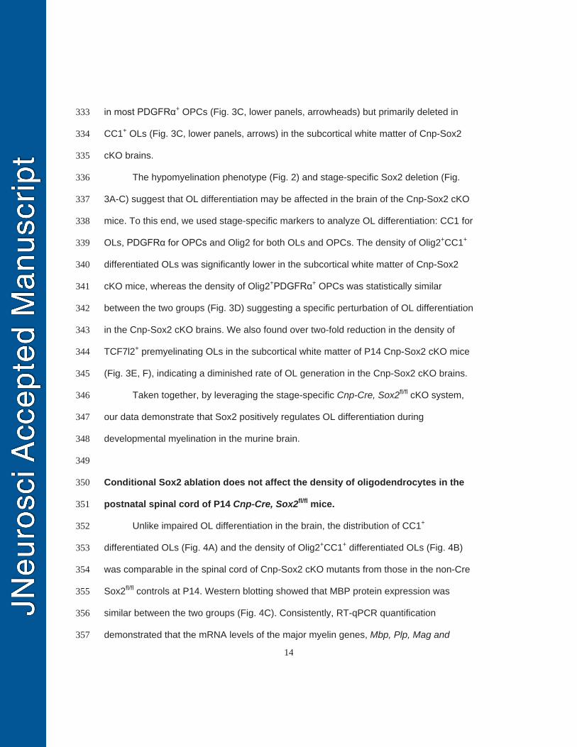
in most PDGFRα+ OPCs (Fig. 3C, lower panels, arrowheads) but primarily deleted in 333
CC1+ OLs (Fig. 3C, lower panels, arrows) in the subcortical white matter of Cnp-Sox2 334
cKO brains. 335
The hypomyelination phenotype (Fig. 2) and stage-specific Sox2 deletion (Fig. 336
3A-C) suggest that OL differentiation may be affected in the brain of the Cnp-Sox2 cKO 337
mice. To this end, we used stage-specific markers to analyze OL differentiation: CC1 for 338
OLs, PDGFRα for OPCs and Olig2 for both OLs and OPCs. The density of Olig2+CC1+ 339
differentiated OLs was significantly lower in the subcortical white matter of Cnp-Sox2 340
cKO mice, whereas the density of Olig2+PDGFRα+ OPCs was statistically similar 341
between the two groups (Fig. 3D) suggesting a specific perturbation of OL differentiation 342
in the Cnp-Sox2 cKO brains. We also found over two-fold reduction in the density of 343
TCF7l2+ premyelinating OLs in the subcortical white matter of P14 Cnp-Sox2 cKO mice 344
(Fig. 3E, F), indicating a diminished rate of OL generation in the Cnp-Sox2 cKO brains. 345
Taken together, by leveraging the stage-specific Cnp-Cre, Sox2fl/fl cKO system, 346
our data demonstrate that Sox2 positively regulates OL differentiation during 347
developmental myelination in the murine brain. 348
349
Conditional Sox2 ablation does not affect the density of oligodendrocytes in the 350
postnatal spinal cord of P14 Cnp-Cre, Sox2fl/fl mice. 351
Unlike impaired OL differentiation in the brain, the distribution of CC1+ 352
differentiated OLs (Fig. 4A) and the density of Olig2+CC1+ differentiated OLs (Fig. 4B) 353
was comparable in the spinal cord of Cnp-Sox2 cKO mutants from those in the non-Cre 354
Sox2fl/fl controls at P14. Western blotting showed that MBP protein expression was 355
similar between the two groups (Fig. 4C). Consistently, RT-qPCR quantification 356
demonstrated that the mRNA levels of the major myelin genes, Mbp, Plp, Mag and 357
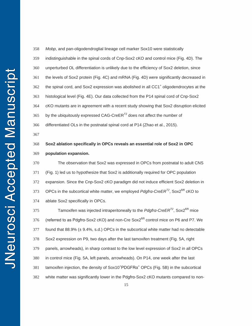
15
Mobp, and pan-oligodendroglial lineage cell marker Sox10 were statistically 358
indistinguishable in the spinal cords of Cnp-Sox2 cKO and control mice (Fig. 4D). The 359
unperturbed OL differentiation is unlikely due to the efficiency of Sox2 deletion, since 360
the levels of Sox2 protein (Fig. 4C) and mRNA (Fig. 4D) were significantly decreased in 361
the spinal cord, and Sox2 expression was abolished in all CC1+ oligodendrocytes at the 362
histological level (Fig. 4E). Our data collected from the P14 spinal cord of Cnp-Sox2 363
cKO mutants are in agreement with a recent study showing that Sox2 disruption elicited 364
by the ubiquitously expressed CAG-CreERT2 does not affect the number of 365
differentiated OLs in the postnatal spinal cord at P14 (Zhao et al., 2015). 366
367
Sox2 ablation specifically in OPCs reveals an essential role of Sox2 in OPC 368
population expansion. 369
The observation that Sox2 was expressed in OPCs from postnatal to adult CNS 370
(Fig. 1) led us to hypothesize that Sox2 is additionally required for OPC population 371
expansion. Since the Cnp-Sox2 cKO paradigm did not induce efficient Sox2 deletion in 372
OPCs in the subcortical white matter, we employed Pdgfrα-CreERT2, Sox2fl/fl cKO to 373
ablate Sox2 specifically in OPCs. 374
Tamoxifen was injected intraperitoneally to the Pdgfrα-CreERT2, Sox2fl/fl mice 375
(referred to as Pdgfrα-Sox2 cKO) and non-Cre Sox2fl/fl control mice on P6 and P7. We 376
found that 88.9% (± 9.4%, s.d.) OPCs in the subcortical white matter had no detectable 377
Sox2 expression on P9, two days after the last tamoxifen treatment (Fig. 5A, right 378
panels, arrowheads), in sharp contrast to the low level expression of Sox2 in all OPCs 379
in control mice (Fig. 5A, left panels, arrowheads). On P14, one week after the last 380
tamoxifen injection, the density of Sox10+PDGFRα+ OPCs (Fig. 5B) in the subcortical 381
white matter was significantly lower in the Pdgfrα-Sox2 cKO mutants compared to non-382
16
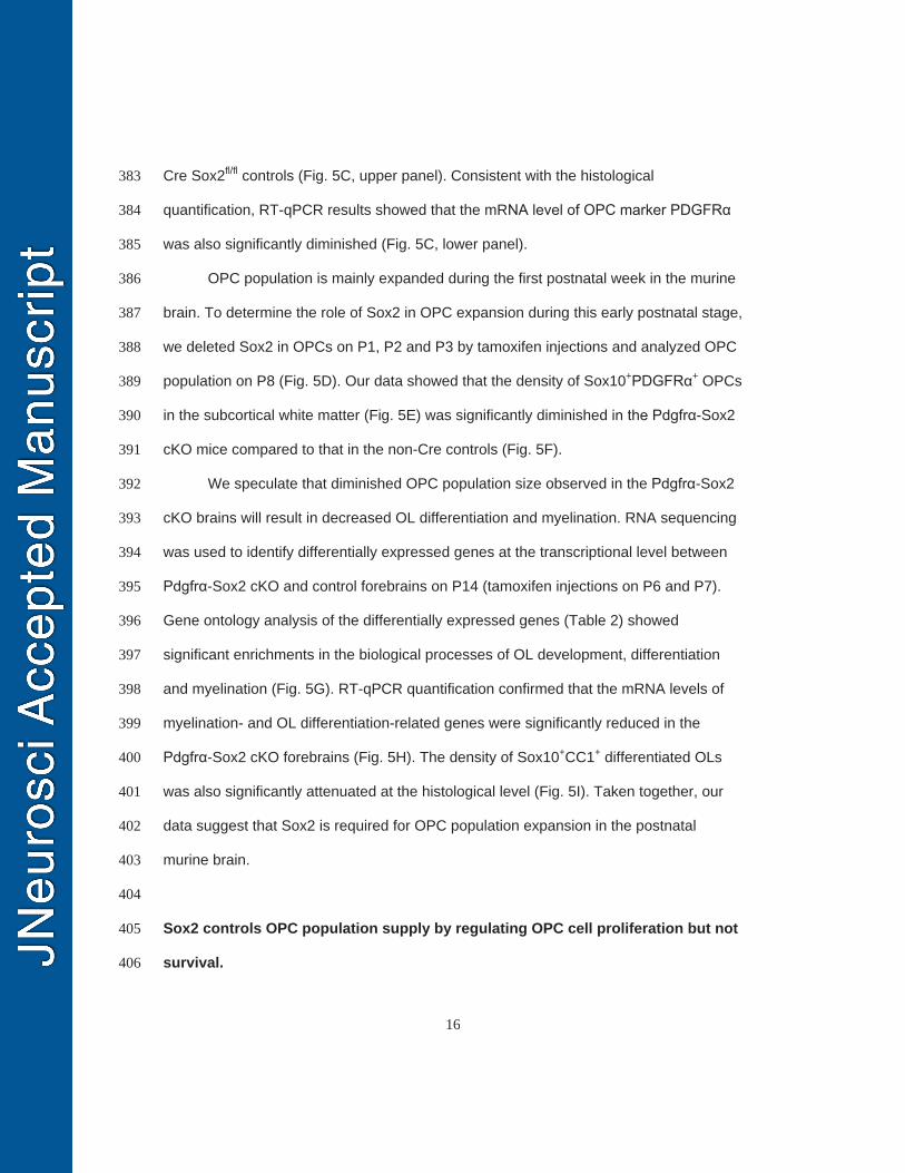
Cre Sox2fl/fl controls (Fig. 5C, upper panel). Consistent with the histological 383
quantification, RT-qPCR results showed that the mRNA level of OPC marker PDGFRα 384
was also significantly diminished (Fig. 5C, lower panel). 385
OPC population is mainly expanded during the first postnatal week in the murine 386
brain. To determine the role of Sox2 in OPC expansion during this early postnatal stage, 387
we deleted Sox2 in OPCs on P1, P2 and P3 by tamoxifen injections and analyzed OPC 388
population on P8 (Fig. 5D). Our data showed that the density of Sox10+PDGFRα+ OPCs 389
in the subcortical white matter (Fig. 5E) was significantly diminished in the Pdgfrα-Sox2 390
cKO mice compared to that in the non-Cre controls (Fig. 5F). 391
We speculate that diminished OPC population size observed in the Pdgfrα-Sox2 392
cKO brains will result in decreased OL differentiation and myelination. RNA sequencing 393
was used to identify differentially expressed genes at the transcriptional level between 394
Pdgfrα-Sox2 cKO and control forebrains on P14 (tamoxifen injections on P6 and P7). 395
Gene ontology analysis of the differentially expressed genes (Table 2) showed 396
significant enrichments in the biological processes of OL development, differentiation 397
and myelination (Fig. 5G). RT-qPCR quantification confirmed that the mRNA levels of 398
myelination- and OL differentiation-related genes were significantly reduced in the 399
Pdgfrα-Sox2 cKO forebrains (Fig. 5H). The density of Sox10+CC1+ differentiated OLs 400
was also significantly attenuated at the histological level (Fig. 5I). Taken together, our 401
data suggest that Sox2 is required for OPC population expansion in the postnatal 402
murine brain. 403
404
Sox2 controls OPC population supply by regulating OPC cell proliferation but not 405
survival. 406
17
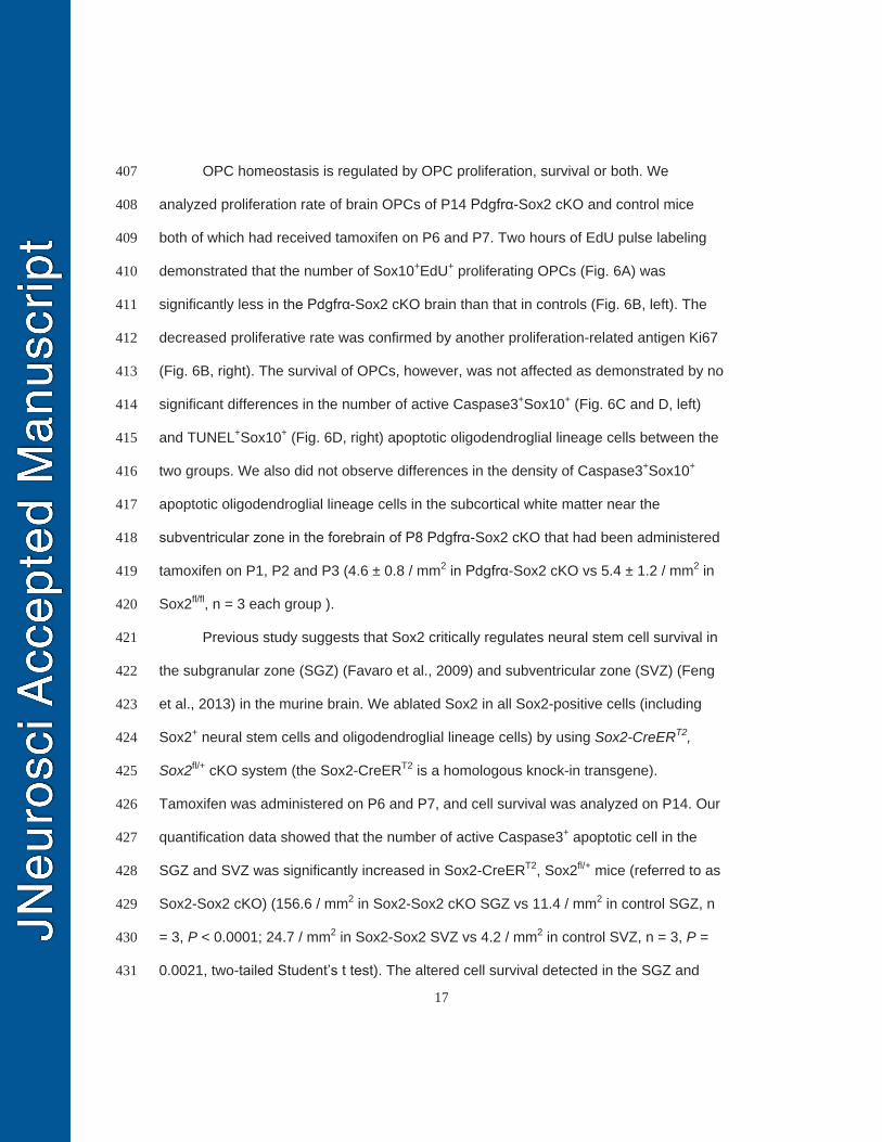
OPC homeostasis is regulated by OPC proliferation, survival or both. We 407
analyzed proliferation rate of brain OPCs of P14 Pdgfrα-Sox2 cKO and control mice 408
both of which had received tamoxifen on P6 and P7. Two hours of EdU pulse labeling 409
demonstrated that the number of Sox10+EdU+ proliferating OPCs (Fig. 6A) was 410
significantly less in the Pdgfrα-Sox2 cKO brain than that in controls (Fig. 6B, left). The 411
decreased proliferative rate was confirmed by another proliferation-related antigen Ki67 412
(Fig. 6B, right). The survival of OPCs, however, was not affected as demonstrated by no 413
significant differences in the number of active Caspase3+Sox10+ (Fig. 6C and D, left) 414
and TUNEL+Sox10+ (Fig. 6D, right) apoptotic oligodendroglial lineage cells between the 415
two groups. We also did not observe differences in the density of Caspase3+Sox10+ 416
apoptotic oligodendroglial lineage cells in the subcortical white matter near the 417
subventricular zone in the forebrain of P8 Pdgfrα-Sox2 cKO that had been administered 418
tamoxifen on P1, P2 and P3 (4.6 ± 0.8 / mm2 in Pdgfrα-Sox2 cKO vs 5.4 ± 1.2 / mm2 in 419
Sox2fl/fl, n = 3 each group ). 420
Previous study suggests that Sox2 critically regulates neural stem cell survival in 421
the subgranular zone (SGZ) (Favaro et al., 2009) and subventricular zone (SVZ) (Feng 422
et al., 2013) in the murine brain. We ablated Sox2 in all Sox2-positive cells (including 423
Sox2+ neural stem cells and oligodendroglial lineage cells) by using Sox2-CreERT2, 424
Sox2fl/+ cKO system (the Sox2-CreERT2 is a homologous knock-in transgene). 425
Tamoxifen was administered on P6 and P7, and cell survival was analyzed on P14. Our 426
quantification data showed that the number of active Caspase3+ apoptotic cell in the 427
SGZ and SVZ was significantly increased in Sox2-CreERT2, Sox2fl/+ mice (referred to as 428
Sox2-Sox2 cKO) (156.6 / mm2 in Sox2-Sox2 cKO SGZ vs 11.4 / mm2 in control SGZ, n 429
= 3, P < 0.0001; 24.7 / mm2 in Sox2-Sox2 SVZ vs 4.2 / mm2 in control SVZ, n = 3, P = 430
0.0021, two-tailed Student’s t test). The altered cell survival detected in the SGZ and 431
18
SVZ stem cell niche of the Sox2-Sox2 cKO mice is consistent with previous publications 432
(Favaro et al., 2009; Feng et al., 2013), which also supports the effectiveness of our cell 433
survival analysis. Double immunohistochemistry of active Caspase3 and lineage 434
specific markers demonstrated that apoptotic cells in the SGZ (Fig. 6G) and SVZ (not 435
shown) were GFAP+ neural stem cells but not doublecortin (DCX)+ neuroblasts nor 436
HuC/D+ neurons. 437
In agreement with the cell survival analysis in the Pdgfrα-Sox2 cKO system (Fig. 438
6C-D), we did not notice any changes in the number of active Caspase3+Sox10+ 439
apoptotic oligodendroglial lineage cells in SGZ and SVZ between Sox2-Sox2 cKO and 440
non-Cre controls (Fig. 6E-F, right panels). These data suggest that Sox2’s role in cell 441
survival is cell type-dependent: it regulates neural stem cell survival, but it is 442
dispensable for oligodendroglial lineage cell survival. 443
444
Oligodendrocyte differentiation and myelination are impaired in the Sox10-Cre, 445
Sox2fl/fl mutants, even in the later stages of brain development. 446
Using stage-specific Sox2 cKO paradigms, our experimental data suggest that 447
Sox2 regulates brain myelination by coordinating upstream OPC proliferation and 448
downstream OL differentiation. Therefore, we predict that Sox2 cKO in all 449
oligodendroglial lineage cells (both OPCs and OLs) will lead to the inhibition of OPC 450
expansion, OL differentiation, and brain myelination even in the later stages of brain 451
development. To support this prediction and also to strengthen our conclusion drawn 452
from Cnp-Sox2 cKO and Pdgfrα-Sox2 cKO mutants, we used Sox10-Cre (Matsuoka et 453
al., 2005) to ablate Sox2 in all oligodendroglial lineage cells and analyzed brain 454
myelination and OL differentiation at later developmental ages. 455
19
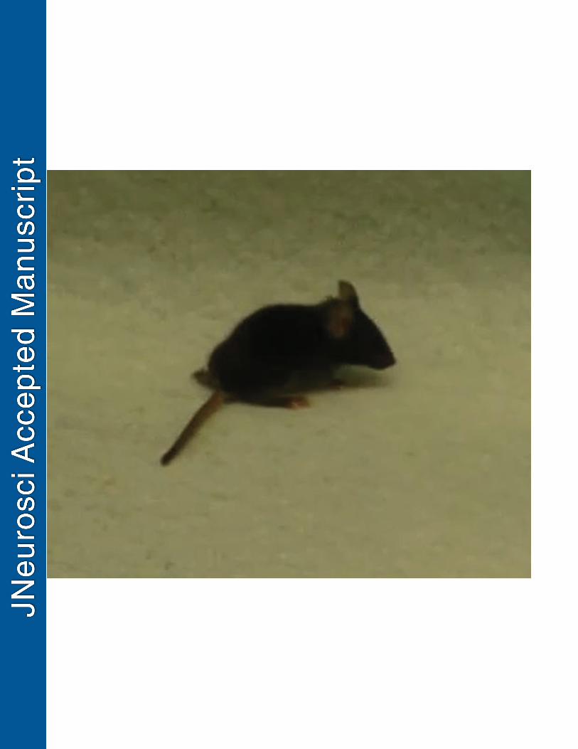
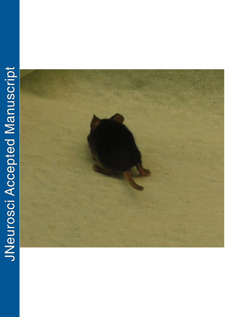
The Sox10-Cre, Sox2fl/fl (referred to as Sox10-Sox2 cKO) mice developed severe 456
tremors and ataxia, typical phenotypes reminiscent of CNS hypomyelination, by the 457
third postnatal weeks P21 (Movie 1), whereas littermate control mice did not show any 458
of the aforementioned behavioral phenotypes (Movie 2). Behavioral testing 459
demonstrated that Sox10-Sox2 cKO mice displayed severe motor function impairment, 460
evidenced by significantly less retention time on the accelerating rod (129.2 seconds, 461
non-Cre Sox2fl/fl control mice vs 2.1 seconds, Sox10-Sox2 cKO mice) (Fig. 7A). 462
The efficiency of Sox2 ablation in the Sox10-Sox2 cKO CNS was nearly 100% in 463
the oligodendroglial lineage cells including OPCs (Fig. 7B) and OLs (data not shown) 464
assessed on P21. The density of MBP+ myelin fibers in the Sox10-Sox2 cKO brains was 465
reduced by 50% of that in littermate controls, whereas the density of SMI312+ axons 466
was similar between the two groups (Fig. 7C). Western blotting (Fig. 7D, left panel) 467
demonstrated that the protein levels of MBP, CNP, and MAG were all significantly 468
decreased in the Sox10-Sox2 cKO brains compared to controls (Fig. 7D, right panel). In 469
line with the Western blotting data, RT-qPCR quantification showed that the 470
transcription levels of Mbp and mature OL-specific Plp isoform (exon3b containing Plp, 471
Plp-E3b) were reduced by > 50% of those in the control brain on P21 (Fig. 7E). 472
Immunohistochemical analysis demonstrated that the numbers of CC1+ mature OLs 473
(Fig. 7F) and PDGFRα+ OPCs were significantly decreased in the corpus callosum of 474
P21 Sox10-Sox2 cKO mice (Fig. 7G). Haploinsufficiency of Sox2 in regulating brain 475
oligodendroglial development was not observed, evidenced by the unaltered numbers of 476
OPCs and OLs in brains between Sox2fl/fl and Sox2 one-allele cKO (Sox10-Cre, Sox2fl/+) 477
mice (Fig. 7G). 478
Consistent with previous data derived from the embryonic and early postnatal 479
spinal cord (Hoffmann et al., 2014), the distribution of Olig1+ cells was similar between 480
20
Sox10-Sox2 cKO and Sox2fl/fl littermate controls within the spinal cord cross sections 481
(Fig. 7H), but the density of Olig1+ cells decreased by 30% (625 ± 66 / mm2 in Sox10-482
Sox2 cKO, 430 ± 47 / mm2 in controls, two-tailed Student’s t test, P = 0.0029, t = 4.82, 483
df = 6,) at the weaning age of P21, suggesting a dispensable role of Sox2 in 484
oligodendrocyte migration (Hoffman et al., 2014) and an essential role in 485
oligodendrocyte production. Our quantification data showed that the densities of 486
PDGFRα+ OPCs and CC1+ OLs were significantly reduced in the Sox10-Sox2 cKO 487
spinal cord than those in Sox2fl/fl controls on P21 (Fig. 7I). Toluidine blue staining of 488
semi-thin sections (Fig. 7J) and transmission electron microscopic images of ultra-thin 489
sections (Fig. 7K) showed that the myelinated axons were substantially fewer in the 490
corticospinal tract of Sox10-Sox2 cKO mutants compared to littermate controls on P28, 491
which is in line with the decreased density of mature OLs in the Sox10-Sox2 cKO spinal 492
cord. 493
Collectively, the in vivo data derived from the Cnp-Sox2 cKO, Pdgfrα-Sox2 cKO, 494
and Sox10-Sox2 cKO paradigms unequivocally demonstrate that Sox2 plays an 495
essential role in regulating oligodendroglial lineage progression and maturation during 496
brain developmental myelination. 497
498
Sox2 expression in oligodendroglial lineage cells during remyelination after 499
chemical-induced and autoimmunity-induced demyelination. 500
We assessed Sox2 expression in both cuprizone-induced demyelination and 501
MOG-peptide35-55-induced experimental autoimmune encephalomyelitis (EAE) animal 502
models (Guo et al., 2011). In the cuprizone-induced demyelinated corpus callosum, the 503
density of Sox2+ cells were substantially increased during the time window of active 504
oligodendrocyte regeneration (Hammond et al., 2015), for example at one week after 505
21
withdrawal of 6-week cuprizone treatment (6+1 wks) as shown in Fig. 8B, compared to 506
the normal-chow controls (Fig. 8A). Consistent with the observations from 507
developmental myelination (Fig. 1), Sox2 was also expressed in most TCF7l2+ newly 508
regenerated premyelinating OLs (Hammond et al., 2015) (Fig. 8C, arrowheads). Triple 509
immunohistochemistry of Sox2, NG2 and TCF7l2 (Fig. 8D, upper panels) showed that 510
the level of Sox2 in TCF7l2+ newly regenerated OLs was significantly higher than that in 511
NG2+ OPCs in the corpus callosum of mice maintaining on 6 consecutive weeks of 512
0.25% cuprizone diet (Fig. 8D, lower panel). In the MOG-EAE spinal cord in which OPC 513
proliferation and OL regeneration are consistently observed (Tripathi et al., 2010; Guo 514
et al., 2011), more Sox2+TCF7l2+ newly regenerated OLs were observed (Fig. 8F, 515
arrowheads) compared to scarce TCF7l2+ OLs in the complete Freund’s adjuvant (CFA) 516
control spinal cord (Fig. 8E, arrowheads) at day 21 (D21) post-MOG immunization (Fig. 517
8G). Similarly, Sox2 expression level in TCF7l2+ newly regenerated OLs were 518
significantly higher than that in NG2+ OPCs in the D21 spinal cord treated with MOG-519
peptide35-55 (Fig. 8H-I). These data suggest that Sox2 may play a role in oligodendrocyte 520
regeneration and remyelination during remyelination. 521
522
Sox2 is essential for brain remyelination after cuprizone-induced myelin damage. 523
Cuprizone-induced demyelination/remyelination in murine corpus callosum is a 524
well-established animal model for studying molecular mechanisms underlying OL 525
regeneration and myelin repair. Since constitutive Sox2 cKO affected brain 526
developmental myelination, we used the time-conditioned, tamoxifen-inducible Pdgfrα-527
CreERT2, Sox2fl/fl (Pdgfrα-Sox2 cKO) to study the role of Sox2 in OL regeneration and 528
remyelination. 529
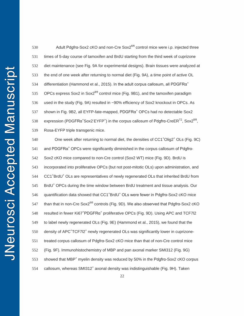
22
Adult Pdgfrα-Sox2 cKO and non-Cre Sox2fl/fl control mice were i.p. injected three 530
times of 5-day course of tamoxifen and BrdU starting from the third week of cuprizone 531
diet maintenance (see Fig. 9A for experimental designs). Brain tissues were analyzed at 532
the end of one week after returning to normal diet (Fig. 9A), a time point of active OL 533
differentiation (Hammond et al., 2015). In the adult corpus callosum, all PDGFRα+ 534
OPCs express Sox2 in Sox2fl/fl control mice (Fig. 9B1), and the tamoxifen paradigm 535
used in the study (Fig. 9A) resulted in ~90% efficiency of Sox2 knockout in OPCs. As 536
shown in Fig. 9B2, all EYFP-fate-mapped, PDGFRα+ OPCs had no detectable Sox2 537
expression (PDGFRα+Sox2-EYFP+) in the corpus callosum of Pdgfrα-CreERT2, Sox2fl/fl, 538
Rosa-EYFP triple transgenic mice. 539
One week after returning to normal diet, the densities of CC1+Olig2+ OLs (Fig. 9C) 540
and PDGFRα+ OPCs were significantly diminished in the corpus callosum of Pdgfrα-541
Sox2 cKO mice compared to non-Cre control (Sox2 WT) mice (Fig. 9D). BrdU is 542
incorporated into proliferative OPCs (but not post-mitotic OLs) upon administration, and 543
CC1+BrdU+ OLs are representatives of newly regenerated OLs that inherited BrdU from 544
BrdU+ OPCs during the time window between BrdU treatment and tissue analysis. Our 545
quantification data showed that CC1+BrdU+ OLs were fewer in Pdgfrα-Sox2 cKO mice 546
than that in non-Cre Sox2fl/fl controls (Fig. 9D). We also observed that Pdgfrα-Sox2 cKO 547
resulted in fewer Ki67+PDGFRα+ proliferative OPCs (Fig. 9D). Using APC and TCF7l2 548
to label newly regenerated OLs (Fig. 9E) (Hammond et al., 2015), we found that the 549
density of APC+TCF7l2+ newly regenerated OLs was significantly lower in cuprizone-550
treated corpus callosum of Pdgfrα-Sox2 cKO mice than that of non-Cre control mice 551
(Fig. 9F). Immunohistochemistry of MBP and pan axonal marker SMI312 (Fig. 9G) 552
showed that MBP+ myelin density was reduced by 50% in the Pdgfrα-Sox2 cKO corpus 553
callosum, whereas SMI312+ axonal density was indistinguishable (Fig. 9H). Taken 554
23
together, our data suggest that Sox2 is required for remyelination in the adult corpus 555
callosum after chemical-induced demyelination. 556
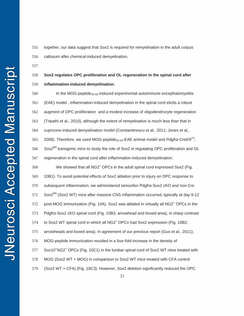
557
Sox2 regulates OPC proliferation and OL regeneration in the spinal cord after 558
inflammation-induced demyelination. 559
In the MOG-peptide35-55-induced experimental autoimmune encephalomyelitis 560
(EAE) model , inflammation-induced demyelination in the spinal cord elicits a robust 561
augment of OPC proliferation and a modest increase of oligodendrocyte regeneration 562
(Tripathi et al., 2010), although the extent of remyelination is much less than that in 563
cuprizone-induced demyelination model (Constantinescu et al., 2011; Jones et al., 564
2008). Therefore, we used MOG-peptide35-55-EAE animal model and Pdgfrα-CreERT2, 565
Sox2fl/fl transgenic mice to study the role of Sox2 in regulating OPC proliferation and OL 566
regeneration in the spinal cord after inflammation-induced demyelination. 567
We showed that all NG2+ OPCs in the adult spinal cord expressed Sox2 (Fig. 568
10B1). To avoid potential effects of Sox2 ablation prior to injury on OPC response to 569
subsequent inflammation, we administered tamoxifen Pdgfrα-Sox2 cKO and non-Cre 570
Sox2fl/fl (Sox2 WT) mice after massive CNS inflammation occurred, typically at day 9-12 571
post-MOG immunization (Fig. 10A). Sox2 was ablated in virtually all NG2+ OPCs in the 572
Pdgfrα-Sox2 cKO spinal cord (Fig. 10B3, arrowhead and boxed area), in sharp contrast 573
to Sox2 WT spinal cord in which all NG2+ OPCs had Sox2 expression (Fig. 10B2, 574
arrowheads and boxed area). In agreement of our previous report (Guo et al., 2011), 575
MOG-peptide immunization resulted in a four-fold increase in the density of 576
Sox10+NG2+ OPCs (Fig. 10C1) in the lumbar spinal cord of Sox2 WT mice treated with 577
MOG (Sox2 WT + MOG) in comparison to Sox2 WT mice treated with CFA control 578
(Sox2 WT + CFA) (Fig. 10C3). However, Sox2 deletion significantly reduced the OPC 579
24
density in the spinal cord of Pdgfrα-Sox2 cKO mice treated with MOG (Sox2 cKO + 580
MOG) (Fig. 10C2, C3), indicating that Sox2 is required for OPC population expansion 581
after inflammatory insults. Notably, the similar distribution patterns of OPCs within 582
inflammatory lesions were observed in the spinal cords between Pdgfrα-Sox2 cKO and 583
Sox2 WT mice with MOG (Fig. 10B2 vs B3 and Fig. 10C1 vs C2), indicating that Sox2 584
appears dispensable for OPC recruitment into the inflammation-induced demyelination 585
lesions. The number of Ki67+Olig2+ proliferating OPCs (Fig. 10D1-D2) was significantly 586
decreased in the spinal cord of Pdgfrα-Sox2 cKO mice treated with MOG (Sox2 cKO + 587
MOG) in comparison to Sox2 WT mice treated with MOG (Sox2 WT + MOG) (Fig. 10E). 588
Our previous study reports that the number of TCF7l2+APC+ newly regenerated 589
OLs increases in the spinal cord of MOG treatment (Fig. 10F) (Hammond et al., 2015). 590
Nevertheless, Sox2 deletion resulted in significant decrease in the generation of 591
TCF7l2+APC+ OLs in the spinal cord of Pdgfrα-Sox2 cKO mutants treated with MOG 592
(Fig. 10F). Altogether, these data suggest that Sox2 is essential for OPC proliferation 593
and OL regeneration in the spinal cord after inflammation-induced demyelination. 594
595
Discussion (1314 words) 596
There are several novel findings in this study: (1) Sox2 is upregulated in newly 597
differentiated OLs during developmental myelination and in newly regenerated OLs 598
during remyelination. (2) Sox2 is essential for brain developmental myelination by 599
regulating OPC proliferation and OL differentiation. (3) In the context of myelin repair, 600
Sox2 is required for OPC proliferation and/or OL regeneration not only in autoimmune-601
induced spinal cord demyelination lesions but also in chemical-induced brain 602
demyelination lesions. 603
25
In this study, we found that OPCs retain Sox2 expression in the adult murine 604
CNS. Previous study reports that Sox2+Olig2+ cells (presumably Sox2+ OPCs) can be 605
immunohistochemically detected in the adult human white matter and that these 606
Sox2/Olig2-positive cells can be differentiated into mature oligodendrocytes in vitro 607
(Oliver-De La Cruz et al., 2014). This report, together with our finding in the murine CNS, 608
suggests that Sox2 expression in adult OPCs is well conserved from rodent to human 609
CNS. However, the function of Sox2 in adult OPCs under normal conditions has not 610
been defined. Our study demonstrates that Sox2 is required for OPC proliferation and 611
population expansion during developmental myelination (Fig. 5A-F). Inspired by these 612
data, we hypothesize that Sox2 is required for adult OPC homeostasis and brain myelin 613
turnover and/or remodeling (Yeung et al., 2014; Zhang et al., 1999) and is essential for 614
OPCs’ response to various demyelinating insults, the latter of which is in agreement 615
with a recent report showing that Sox2 deletion prior to lysolecithin-induced focal 616
demyelination reduces progenitor response and recruitment to demyelinating lesions 617
(Zhao et al., 2015). 618
Interestingly, we also found that Sox2 is upregulated in a subpopulation of 619
differentiated OLs during developmental myelination and remyelination in the rodent 620
CNS. This upregulation is counterintuitive to the established concept that Sox2 621
maintains the “stemness” of neural stem/progenitor cells and is downregulated upon 622
progenitor differentiation (Graham et al., 2003). Specifically, Sox2 is transiently 623
upregulated in the differentiated oligodendrocytes that are TCF7l2-positive, which we 624
have previously identified as post-mitotic, newly formed (regenerated) OLs (Hammond 625
et al., 2015). Although the number of Sox2+ differentiated OLs are reduced over time 626
during developmental myelination, the percentage of Sox2+ cells among TCF7l2+ newly 627
formed OLs did not change (>90% at all time-points we assessed). Given the 628
26
asynchronous properties of in vivo OL differentiation, our quantitative data suggest that 629
all OLs express Sox2 at certain developmental stages during OL lineage progression. 630
Sox2 upregulation is necessary for OL lineage progression and maturation, as Cnp-631
Sox2 cKO resulted in diminished myelin formation (Fig. 2) and OL differentiation (Fig. 3) 632
in the brain. From a pre-translational perspective, it would be very important and 633
interesting to test whether dose-controlled, transient Sox2 overexpression in post-634
mitotic OPCs and/or differentiating OLs is sufficient to promote OL differentiation and/or 635
(re)myelination. 636
Our study suggests a previous unappreciated concept that Sox2 regulates CNS 637
myelin formation and repair in CNS region and context-dependent manners. Using 638
oligodendroglial specific Sox2 cKO mice, Hoffmann et al., demonstrate that Sox2 is 639
dispensable for OPC proliferation and migration; instead, it is required for OL 640
differentiation in the embryonic and perinatal spinal cord (P3) (Hoffmann et al., 2014). 641
More recently, Zhao et al., used the chicken beta-actin promoter and CMV enhancer-642
driven Cre to ubiquitously ablate Sox2 (Cag-CreERT2, Sox2fl/fl) and found that Sox2 643
plays a minor, if any, role in OL differentiation and myelination in the postnatal spinal 644
cord (Zhao et al., 2015). In our study, we employed Cnp-Cre to conditionally ablate 645
Sox2 in the later stages of OL development (Dugas et al., 2010; Moyon et al., 2016; 646
Zhao et al., 2016) and found a CNS region-dependent role of Sox2 in regulating OL 647
differentiation: it is required for brain OL differentiation (Fig. 3) and appears to play a 648
minor role in OL differentiation (or regulate the timing of OL differentiation) during 649
developmental myelination in the postnatal spinal cord (Fig. 4). The mechanisms 650
underlying the CNS region-dependent role of Sox2 are unclear. Previous study shows 651
that another SoxB1 family member Sox3 functions redundantly with Sox2 in regulating 652
spinal cord OL differentiation (Hoffmann et al., 2014). It is plausible that Sox3 may 653
27
compensate the loss of Sox2 during postnatal spinal cord development. However, the 654
compensatory effects of Sox3 on Sox2 loss-of-function is less likely to occur in the brain 655
myelination, as Sox10-Cre, Sox2fl/fl mutant mice display severe defects in motor function, 656
OL differentiation and brain developmental myelination (Fig. 7). Our results also indicate 657
that, in the context of remyelination, Sox2 is required for myelin repair in both adult brain 658
(Fig. 9) and spinal cord (Fig. 10) through regulating OPC population supply and/or OL 659
differentiation. Our remyelination study did not provide definitive evidence that Sox2 is 660
required for remyelination by directly regulating OL differentiation, as it does in brain 661
myelination. In this regard, time-conditioned, stage-specific Sox2 cKO or conditional 662
overexpression paradigms are needed to unequivocally define the role of Sox2 in 663
regulating OL differentiation itself after myelin damage. 664
Oligodendrocyte number was not affected in the spinal cord of the Cnp-Sox2 665
cKO mutants at P14 (Fig. 4) but was impaired in the spinal cord of the Sox10-Sox2 cKO 666
mutants even at later ages (Fig. 7H-K). This discrepancy presumably reflects the stage-667
specific Sox2 cKO in oligodendroglial lineage cells. In the Sox10-Sox2 cKO mutants, 668
Sox2 was initially ablated in the upstream proliferating OPCs whereas in the Cnp-Sox2 669
cKO mice, primarily in the differentiation-committed, late stage OPCs and OLs. The 670
reduced number of differentiated OL observed in Sox10-Sox2 cKO spinal cord is likely 671
due to the reduced proliferation and supply of OPCs as we noticed diminished density 672
of OPCs in the Sox10-Sox2 cKO (but not in Cnp-Sox2 cKO at P14) spinal cord. These 673
data are in line with our conclusion that Sox2 is required for OPC proliferation and 674
population expansion. 675
The working model in which Sox2 coordinates OPC proliferation and OL 676
differentiation is compatible with the one derived from neural stem/progenitor cells 677
(NSPCs) (Episkopou, 2005) and with the expression patterns of Sox2 along the 678
28
progression of oligodendroglial lineage (Fig. 1M). Previous studies have documented 679
that Sox2 genetic knockout (or knockdown) diminishes the proliferation of NSPCs under 680
self-renewal conditions and reduces neuronal differentiation from NSPCs under 681
neurogenic conditions (Cimadamore et al., 2013; Ferri et al., 2004). Mechanistically, 682
Sox2 interacts with Chd7, a chromatin remodeler to directly regulate a variety of 683
downstream signaling pathways including Shh and Notch signaling pathways in NSPCs 684
(Engelen et al., 2011), both of which are important regulators of oligodendroglial 685
development (He and Lu, 2013; Wheeler and Fuss, 2016). Interestingly, a recent study 686
shows that Chd7 regulates the onset of CNS myelination and remyelination through 687
interacting with Sox10 (He et al., 2016). Biochemical evidence suggests that Sox2 688
interacts with Sox10 to regulate Schwann cell development in the peripheral nervous 689
system (Arter and Wegner, 2015). Our unpublished data show that Sox2 is co-labeled 690
with Chd7 in oligodendroglial lineage cells at the histological level and displays similar 691
expression dynamics to that of Chd7 (He et al., 2016). Therefore, it is tempting to 692
hypothesize that Sox2 may regulate brain developmental myelination and CNS 693
remyelination through binding to Chd7-Sox10 and/or through the downstream signaling 694
pathways targeted by the putative Sox2-Chd7-Sox10 complex. Further studies are 695
needed to support or falsify this hypothesis. 696
In summary, a series of genetic experiments in our study demonstrate that Sox2 697
plays a crucial role in regulating OPC proliferation and OL differentiation during 698
postnatal brain development, although it appears to play a minor, transient role in OL 699
differentiation during postnatal spinal cord development. Furthermore, we show that 700
Sox2 is required for myelin repair by regulating OPC proliferation and/or OL 701
differentiation in both the demyelinated spinal cord and brain, suggesting a common 702
mechanism. Our study suggests that Sox2 may be a therapeutic target that can be 703
29
experimentally manipulated to promote OL regeneration and CNS remyelination. 704
Considering the established protocols of expressing Sox2 in the stem cell biology field, 705
it would be interesting to investigate whether Sox2 expression through genetic and/or 706
viral-mediated approaches is sufficient to promote endogenous CNS remyelination after 707
myelin damage. 708
709
References: 710
Arter, J., and M. Wegner. 2015. Transcription factors Sox10 and Sox2 functionally interact with 711
positive transcription elongation factor b in Schwann cells. J Neurochem. 132:384-393. 712
Bin, J.M., S.N. Harris, and T.E. Kennedy. 2016. The oligodendrocyte-specific antibody 'CC1' 713
binds Quaking 7. J Neurochem. 139:181-186. 714
Cimadamore, F., A. Amador-Arjona, C. Chen, C.T. Huang, and A.V. Terskikh. 2013. SOX2-715
LIN28/let-7 pathway regulates proliferation and neurogenesis in neural precursors. Proc 716
Natl Acad Sci U S A. 110:E3017-3026. 717
Constantinescu, C.S., N. Farooqi, K. O'Brien, and B. Gran. 2011. Experimental autoimmune 718
encephalomyelitis (EAE) as a model for multiple sclerosis (MS). Br J Pharmacol. 719
164:1079-1106. 720
Dai, J., K.K. Bercury, J.T. Ahrendsen, and W.B. Macklin. 2015. Olig1 function is required for 721
oligodendrocyte differentiation in the mouse brain. J Neurosci. 35:4386-4402. 722
Dugas, J.C., T.L. Cuellar, A. Scholze, B. Ason, A. Ibrahim, B. Emery, J.L. Zamanian, L.C. Foo, 723
M.T. McManus, and B.A. Barres. 2010. Dicer1 and miR-219 Are required for normal 724
oligodendrocyte differentiation and myelination. Neuron. 65:597-611. 725
Engelen, E., U. Akinci, J.C. Bryne, J. Hou, C. Gontan, M. Moen, D. Szumska, C. Kockx, W. van 726
Ijcken, D.H. Dekkers, J. Demmers, E.J. Rijkers, S. Bhattacharya, S. Philipsen, L.H. 727
Pevny, F.G. Grosveld, R.J. Rottier, B. Lenhard, and R.A. Poot. 2011. Sox2 cooperates 728
30
with Chd7 to regulate genes that are mutated in human syndromes. Nat Genet. 43:607-729
611. 730
Episkopou, V. 2005. SOX2 functions in adult neural stem cells. Trends Neurosci. 28:219-221. 731
Favaro, R., M. Valotta, A.L. Ferri, E. Latorre, J. Mariani, C. Giachino, C. Lancini, V. Tosetti, S. 732
Ottolenghi, V. Taylor, and S.K. Nicolis. 2009. Hippocampal development and neural 733
stem cell maintenance require Sox2-dependent regulation of Shh. Nat Neurosci. 12:1248-734
1256. 735
Feng, R., S. Zhou, Y. Liu, D. Song, Z. Luan, X. Dai, Y. Li, N. Tang, J. Wen, and L. Li. 2013. 736
Sox2 protects neural stem cells from apoptosis via up-regulating survivin expression. 737
Biochem J. 450:459-468. 738
Ferri, A.L., M. Cavallaro, D. Braida, A. Di Cristofano, A. Canta, A. Vezzani, S. Ottolenghi, P.P. 739
Pandolfi, M. Sala, S. DeBiasi, and S.K. Nicolis. 2004. Sox2 deficiency causes 740
neurodegeneration and impaired neurogenesis in the adult mouse brain. Development. 741
131:3805-3819. 742
Graham, V., J. Khudyakov, P. Ellis, and L. Pevny. 2003. SOX2 functions to maintain neural 743
progenitor identity. Neuron. 39:749-765. 744
Guo, F., P. Bannerman, E. Mills Ko, L. Miers, J. Xu, T. Burns, S. Li, E. Freeman, J.A. 745
McDonough, and D. Pleasure. 2015. Ablating N-acetylaspartate prevents leukodystrophy 746
in a Canavan disease model. Annals of neurology. 747
Guo, F., Y. Maeda, E.M. Ko, M. Delgado, M. Horiuchi, A. Soulika, L. Miers, T. Burns, T. Itoh, 748
H. Shen, E. Lee, J. Sohn, and D. Pleasure. 2012. Disruption of NMDA receptors in 749
oligodendroglial lineage cells does not alter their susceptibility to experimental 750
autoimmune encephalomyelitis or their normal development. J Neurosci. 32:639-645. 751
Guo, F., Y. Maeda, J. Ma, M. Delgado, J. Sohn, L. Miers, E.M. Ko, P. Bannerman, J. Xu, Y. 752
Wang, C. Zhou, H. Takebayashi, and D. Pleasure. 2011. Macroglial plasticity and the 753
31
origins of reactive astroglia in experimental autoimmune encephalomyelitis. J Neurosci. 754
31:11914-11928. 755
Hammond, E., J. Lang, Y. Maeda, D. Pleasure, M. Angus-Hill, J. Xu, M. Horiuchi, W. Deng, 756
and F. Guo. 2015. The Wnt effector transcription factor 7-like 2 positively regulates 757
oligodendrocyte differentiation in a manner independent of Wnt/beta-catenin signaling. J 758
Neurosci. 35:5007-5022. 759
He, D., C. Marie, C. Zhao, B. Kim, J. Wang, Y. Deng, A. Clavairoly, M. Frah, H. Wang, X. He, 760
H. Hmidan, B.V. Jones, D. Witte, B. Zalc, X. Zhou, D.I. Choo, D.M. Martin, C. Parras, 761
and Q.R. Lu. 2016. Chd7 cooperates with Sox10 and regulates the onset of CNS 762
myelination and remyelination. Nat Neurosci. 19:678-689. 763
He, L., and Q.R. Lu. 2013. Coordinated control of oligodendrocyte development by extrinsic and 764
intrinsic signaling cues. Neurosci Bull. 29:129-143. 765
Heinrich, C., M. Bergami, S. Gascon, A. Lepier, F. Vigano, L. Dimou, B. Sutor, B. Berninger, 766
and M. Gotz. 2014. Sox2-mediated conversion of NG2 glia into induced neurons in the 767
injured adult cerebral cortex. Stem Cell Reports. 3:1000-1014. 768
Hoffmann, S.A., D. Hos, M. Kuspert, R.A. Lang, R. Lovell-Badge, M. Wegner, and S. Reiprich. 769
2014. Stem cell factor Sox2 and its close relative Sox3 have differentiation functions in 770
oligodendrocytes. Development. 141:39-50. 771
Jones, M.V., T.T. Nguyen, C.A. Deboy, J.W. Griffin, K.A. Whartenby, D.A. Kerr, and P.A. 772
Calabresi. 2008. Behavioral and pathological outcomes in MOG 35-55 experimental 773
autoimmune encephalomyelitis. J Neuroimmunol. 199:83-93. 774
Kondo, T., and M. Raff. 2004. Chromatin remodeling and histone modification in the conversion 775
of oligodendrocyte precursors to neural stem cells. Genes Dev. 18:2963-2972. 776
32
Lang, J., Y. Maeda, P. Bannerman, J. Xu, M. Horiuchi, D. Pleasure, and F. Guo. 2013. 777
Adenomatous polyposis coli regulates oligodendroglial development. The Journal of 778
neuroscience : the official journal of the Society for Neuroscience. 33:3113-3130. 779
Lappe-Siefke, C., S. Goebbels, M. Gravel, E. Nicksch, J. Lee, P.E. Braun, I.R. Griffiths, and K.A. 780
Nave. 2003. Disruption of Cnp1 uncouples oligodendroglial functions in axonal support 781
and myelination. Nat Genet. 33:366-374. 782
Lyssiotis, C.A., J. Walker, C. Wu, T. Kondo, P.G. Schultz, and X. Wu. 2007. Inhibition of 783
histone deacetylase activity induces developmental plasticity in oligodendrocyte 784
precursor cells. Proc Natl Acad Sci U S A. 104:14982-14987. 785
Matsuoka, T., P.E. Ahlberg, N. Kessaris, P. Iannarelli, U. Dennehy, W.D. Richardson, A.P. 786
McMahon, and G. Koentges. 2005. Neural crest origins of the neck and shoulder. Nature. 787
436:347-355. 788
Miron, V.E., T. Kuhlmann, and J.P. Antel. 2011. Cells of the oligodendroglial lineage, 789
myelination, and remyelination. Biochimica et biophysica acta. 1812:184-193. 790
Moyon, S., J.L. Huynh, D. Dutta, F. Zhang, D. Ma, S. Yoo, R. Lawrence, M. Wegner, G.R. John, 791
B. Emery, C. Lubetzki, R.J. Franklin, G. Fan, J. Zhu, J.L. Dupree, and P. Casaccia. 2016. 792
Functional Characterization of DNA Methylation in the Oligodendrocyte Lineage. Cell 793
Rep. 794
Niu, W., T. Zang, Y. Zou, S. Fang, D.K. Smith, R. Bachoo, and C.L. Zhang. 2013. In vivo 795
reprogramming of astrocytes to neuroblasts in the adult brain. Nat Cell Biol. 15:1164-796
1175. 797
Oliver-De La Cruz, J., J. Carrion-Navarro, N. Garcia-Romero, A. Gutierrez-Martin, E. Lazaro-798
Ibanez, C. Escobedo-Lucea, R. Perona, C. Belda-Iniesta, and A. Ayuso-Sacido. 2014. 799
SOX2+ cell population from normal human brain white matter is able to generate mature 800
oligodendrocytes. PLoS One. 9:e99253. 801
33
Pedre, X., F. Mastronardi, W. Bruck, G. Lopez-Rodas, T. Kuhlmann, and P. Casaccia. 2011. 802
Changed histone acetylation patterns in normal-appearing white matter and early multiple 803
sclerosis lesions. J Neurosci. 31:3435-3445. 804
Shen, S., J. Sandoval, V.A. Swiss, J. Li, J. Dupree, R.J. Franklin, and P. Casaccia-Bonnefil. 2008. 805
Age-dependent epigenetic control of differentiation inhibitors is critical for remyelination 806
efficiency. Nat Neurosci. 11:1024-1034. 807
Sohn, J., P. Bannerman, F. Guo, T. Burns, L. Miers, C. Croteau, N.K. Singhal, J.A. McDonough, 808
and D. Pleasure. 2017. Suppressing N-Acetyl-l-Aspartate Synthesis Prevents Loss of 809
Neurons in a Murine Model of Canavan Leukodystrophy. J Neurosci. 37:413-421. 810
Sohn, J., V. Selvaraj, K. Wakayama, L. Orosco, E. Lee, S.E. Crawford, F. Guo, J. Lang, M. 811
Horiuchi, K. Zarbalis, T. Itoh, W. Deng, and D. Pleasure. 2012. PEDF is a novel 812
oligodendrogenic morphogen acting on the adult SVZ and corpus callosum. J Neurosci. 813
32:12152-12164. 814
Steelman, A.J., J.P. Thompson, and J. Li. 2012. Demyelination and remyelination in 815
anatomically distinct regions of the corpus callosum following cuprizone intoxication. 816
Neurosci Res. 72:32-42. 817
Tripathi, R.B., L.E. Rivers, K.M. Young, F. Jamen, and W.D. Richardson. 2010. NG2 glia 818
generate new oligodendrocytes but few astrocytes in a murine experimental autoimmune 819
encephalomyelitis model of demyelinating disease. J Neurosci. 30:16383-16390. 820
Wheeler, N.A., and B. Fuss. 2016. Extracellular cues influencing oligodendrocyte differentiation 821
and (re)myelination. Exp Neurol. 283:512-530. 822
Yeung, M.S., S. Zdunek, O. Bergmann, S. Bernard, M. Salehpour, K. Alkass, S. Perl, J. Tisdale, 823
G. Possnert, L. Brundin, H. Druid, and J. Frisen. 2014. Dynamics of oligodendrocyte 824
generation and myelination in the human brain. Cell. 159:766-774. 825
34
Zhang, S., and W. Cui. 2014. Sox2, a key factor in the regulation of pluripotency and neural 826
differentiation. World J Stem Cells. 6:305-311. 827
Zhang, S.C., B. Ge, and I.D. Duncan. 1999. Adult brain retains the potential to generate 828
oligodendroglial progenitors with extensive myelination capacity. Proc Natl Acad Sci U S 829
A. 96:4089-4094. 830
Zhao, C., Y. Deng, L. Liu, K. Yu, L. Zhang, H. Wang, X. He, J. Wang, C. Lu, L.N. Wu, Q. 831
Weng, M. Mao, J. Li, J.H. van Es, M. Xin, L. Parry, S.A. Goldman, H. Clevers, and Q.R. 832
Lu. 2016. Dual regulatory switch through interactions of Tcf7l2/Tcf4 with stage-specific 833
partners propels oligodendroglial maturation. Nat Commun. 7:10883. 834
Zhao, C., D. Ma, M. Zawadzka, S.P. Fancy, L. Elis-Williams, G. Bouvier, J.H. Stockley, G.M. 835
de Castro, B. Wang, S. Jacobs, P. Casaccia, and R.J. Franklin. 2015. Sox2 Sustains 836
Recruitment of Oligodendrocyte Progenitor Cells following CNS Demyelination and 837
Primes Them for Differentiation during Remyelination. J Neurosci. 35:11482-11499. 838
839
840
841
842
843
844
845
846
847
848
849
850
35
Figure Legends 851
Figure 1 - Sox2 is expressed in postnatal and adult OPCs and transiently upregulated 852
in premyelinating OLs. 853
A, confocal images showing Sox2 expression in PDGFRα+ OPCs (arrowheads) and 854
brain lipid basic protein (BLBP)+ astrocytic precursor cells (arrows) in the corpus 855
callosum close to the subventricular zone on postnatal day (P0), several days before 856
the presence of differentiated OLs. B, confocal images showing Sox2 is expressed in 857
PDGFRα+ OPCs in the brain corpus callosum at P5 (before OPC differentiation) and 858
P14 (peak OPC differentiation) during developmental myelination. Arrowheads point to 859
Sox2+PDGFRα+ OPCs. C, confocal images showing that Sox2 is expressed in adult 860
NG2+ OPCs at P60 (left panels, arrowheads) and that the expression is abolished in 861
Sox2 cKO brain of P60 Pdgfα-CreERT2, Sox2fl/fl mice that had been administered 862
tamoxifen 5 days prior to tissue harvest (right panels, arrows). D, double 863
immunostaining of Sox2 and differentiated OL marker CC1 in forebrain corpus callosum 864
and spinal cord. Arrowheads point to Sox2+CC1+ OLs. E, confocal images showing 865
Sox2 expression in myelin gene CNP- and MBP-positive differentiated OLs 866
(arrowheads). Blue, DAPI. F-G, triple immunostaining of Sox2, CC1 and TCF7l2 in the 867
corpus callosum (F) and spinal cord (G) showing that Sox2-expressing OLs (Sox2+CC1+) 868
are TCF7l2+ newly differentiated OLs (TCF7l2+CC1+). Arrowheads are representative of 869
the triple positive cells (Sox2+TCF7l2+CC1+). H-J, Sox2 mRNA (H) and protein (I) levels 870
in purified, mouse primary OPCs and differentiating OLs at day 1 (D1), 2 and 4 (n=3 at 871
each time point). The differentiation of primary OPCs is verified by the sharp increase of 872
MBP protein (I) and mRNA (J) from D1 through D4. Note that Sox2 protein expression 873
is overlapped with TCF7l2 in differentiating OLs (I). K-L, representative images of triple 874
immunostaining of Sox2, TCF7l2 and PDGFRα in the forebrain corpus callosum (K) and 875
36
quantification of corrected Sox2 fluorescence density in TCF7l2+ newly differentiated 876
OLs and PDGFRα+ OPCs in P8 and P14 brains (L). Arrowheads and arrows in K point 877
to Sox2+TCF7l2+ OLs and Sox2+PDGFRα+ OPCs, respectively. M, schematic drawing 878
illustrating Sox2 expression patterns along the progression and maturation of 879
oligodendroglial lineage. OPC markers PDGFRα and NG2 are progressively 880
downregulated whereas OL markers CC1, proteolipid protein (Plp), myelin basic protein 881
(MBP) and CNPase (CNP) are progressively upregulated. One-way ANOVA with 882
Tukey’s post hoc test (H) and two-tailed Student’s t test (L); *, p<0.05, **, p<0.01, ***, 883
p<0.001 (see Table 1 for the details of the statistical analyses for Figs. 1-10). Scale 884
bar: A, B, C, E, K, 10 μm; D, F, G, 20 μm. 885
886
Figure 2 - Cnp-Cre, Sox2fl/fl mice display brain hypomyelination on postnatal day 14. 887
A-B, grey-scale images of MBP immunostaining showing decreased density of myelin 888
fibers in the corpus callosum at the cingular cortical areas (A) and in the subcortical 889
white matter tracts (B). C, Single and merged channels of confocal images of MBP and 890
SMI312 in the subcortical white matter and the overlaying cortex. Note that most 891
SMI312+ axons (green) lack MBP+ myelin (red) in the Cnp-Cre, Sox2fl/fl mice (lower 892
right), in sharp contrast to extensive co-labeling of MBP and SMI312 in the Sox2fl/fl 893
littermate controls (upper right). D, quantification of relative densities of MBP and 894
SMI312 immunoreactive signals (n=4 Cnp-Cre, Sox2fl/fl, n=5 Sox2fl/fl mice). Two-tailed 895
Student’s t test, ** p<0.01, ns, not significant. E, representative image of Western 896
blotting analyses of Sox2 and MBP protein from Cnp-Cre, Sox2fl/fl (cKO) and Sox2fl/fl 897
(WT) forebrains. β-actin serves as an internal loading control. F, high power confocal 898
images of MBP and SMI312 in the cortical areas of layers 5/6 of P14 Cnp-Cre, Sox2fl/fl 899
and Sox2fl/fl control brains. Scale bars: B, 200 μm; C, 50 μm, F, 10 μm. 900
37
901
Figure 3 – Oligodendrocyte differentiation is inhibited in the brain of Cnp-Cre Sox2fl/fl 902
mice. 903
A, fate-mapping of Cnp-Cre-expressing cells in the subcortical white matter of P7 Cnp-904
Cre, Rosa-EYFP mice. Note that many PDGFRα+ OPCs are EYFP negative 905
(arrowheads). B, many EYFP+ fate-mapped cells are O4+ differentiation-committed, late 906
OPCs and/or immature oligodendrocytes (arrowheads) in the subcortical white matter of 907
P7 Cnp-Cre, Rosa-EYFP mice. C, triple immunostaining of CC1, PDGFRα and Sox2 in 908
the corpus callosum. Arrows and arrowheads point to representative CC1+ OLs and 909
PDGFRα+ OPCs, respectively. In the Sox2 cKO corpus callosum (lower panels), many 910
OPCs retain Sox2 expression, whereas no OLs have detectable Sox2. D, quantification 911
of Olig2+CC1+ OLs and Olig2+PDGFRα+ OPCs in the subcortical white matter tract. 912
Olig2 is a pan-oligodendroglial marker expressed in both OPCs and OLs. E-F, density 913
(cell # /mm2) (E) and representative images (F) of TCF7l2-expressing, newly 914
differentiated OLs in the subcortical white matter tracts (dotted areas in F). 915
Animals used in C-F are P14 old. n=4 Cnp-Cre Sox2fl/fl and n=6 Sox2fl/fl (D, E), two-916
tailed Student’s t test, ** p<0.01, ns, not significant. Scale bars: A, B, C, 50 μm; F, 100 917
μm. 918
919
Figure 4 – Oligodendrocyte number appears normal in the spinal cord of P14 Cnp-Cre 920
Sox2fl/fl mice. 921
A, low power confocal images showing the distribution of CC1+ differentiated OLs in the 922
spinal cord. B, quantification of Olig2+CC1+ differentiated OLs in the spinal cord (n=4 923
Cnp-Cre Sox2fl/fl, n=6 Sox2fl/fl). C, representative images of Western blotting of MBP, 924
Sox2 and the internal loading control β-actin in the spinal cord. D, RT-qPCR 925

38
quantification of mRNA levels of Sox2, Mbp, proteolipid protein (Plp), myelin-associated 926
protein (Mag), myelin-associated oligodendrocyte basic protein (Mobp) and Sox10 (n=4 927
Cnp-Cre Sox2fl/fl n=8 Sox2fl/fl). E, representative confocal images and quantification 928
showing Sox2 is completely deleted in CC1+ OLs in the spinal cord of Cnp-Cre Sox2fl/fl 929
mice (n=4 Cnp-Cre Sox2fl/fl n=3 Sox2fl/fl). Arrowheads point to Sox2+CC1+ OLs. Two-930
tailed Student’s t test, ** p<0.01, *** P<0.001, ns, not significant. Spinal cord tissues are 931
harvested at P14 old. Scale bars: A, 500 μm; E, 10 μm. 932
933
Figure 5 – Sox2 is required for OPC population expansion. 934
A, confocal images depicting Sox2 expression in PDGFRα+ OPCs (left panels, 935
arrowheads) in the corpus callosum of P9 Sox2fl/fl mice (left panels, arrowheads), and 936
Sox2 is abolished in PDGFRα+ OPCs in the corpus callosum of Pdgfrα-Cre:Sox2 cKO 937
mice (right panels, arrowheads).Boxed areas are shown at higher magnification in the 938
bottom. B, representative confocal images of Sox10+PDGFRα+ OPCs (arrowheads) in 939
the P14 corpus callosum. Boxed areas are shown at higher magnification in the bottom. 940
C, histological quantification of Sox10+PDGFRα+ OPCs and Sox10+ pan-941
oligodendroglial lineage cells in the subcortical white matter (upper panel, n=7, Pdgfrα-942
Sox2 cKO, n=7 control) and RT-qPCR quantification of Sox2 and Pdgfrα mRNA levels 943
in the P14 forebrains (lower panel, n=4, Pdgfrα-Cre:Sox2 cKO, n=4 control). D, 944
immunostaining showing that Sox2 is ablated in PDGFRα+ OPCs in the corpus callosum 945
of P8 Pdgfrα-Cre:Sox2 cKO mice that had received tamoxifen at P1-P3 (arrowhead, 946
right panel). E-F, representative images of triple immunostaining of Sox10, PDGFRα 947
and CC1 (E) and quantification of marker+ cells (F) in the P8 corpus callosum. N=3 948
Sox2fl/fl, n=3 Pdgfrα-Sox2 cKO. G, significant enriched gene ontology (GO) terms from 949
GO analyses using the differentially expressed genes derived from RNA-sequencing 950
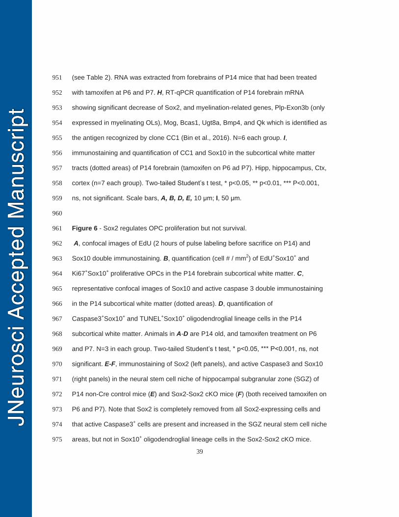
39
(see Table 2). RNA was extracted from forebrains of P14 mice that had been treated 951
with tamoxifen at P6 and P7. H, RT-qPCR quantification of P14 forebrain mRNA 952
showing significant decrease of Sox2, and myelination-related genes, Plp-Exon3b (only 953
expressed in myelinating OLs), Mog, Bcas1, Ugt8a, Bmp4, and Qk which is identified as 954
the antigen recognized by clone CC1 (Bin et al., 2016). N=6 each group. I, 955
immunostaining and quantification of CC1 and Sox10 in the subcortical white matter 956
tracts (dotted areas) of P14 forebrain (tamoxifen on P6 ad P7). Hipp, hippocampus, Ctx, 957
cortex (n=7 each group). Two-tailed Student’s t test, * p<0.05, ** p<0.01, *** P<0.001, 958
ns, not significant. Scale bars, A, B, D, E, 10 μm; I, 50 μm. 959
960
Figure 6 - Sox2 regulates OPC proliferation but not survival. 961
A, confocal images of EdU (2 hours of pulse labeling before sacrifice on P14) and 962
Sox10 double immunostaining. B, quantification (cell # / mm2) of EdU+Sox10+ and 963
Ki67+Sox10+ proliferative OPCs in the P14 forebrain subcortical white matter. C, 964
representative confocal images of Sox10 and active caspase 3 double immunostaining 965
in the P14 subcortical white matter (dotted areas). D, quantification of 966
Caspase3+Sox10+ and TUNEL+Sox10+ oligodendroglial lineage cells in the P14 967
subcortical white matter. Animals in A-D are P14 old, and tamoxifen treatment on P6 968
and P7. N=3 in each group. Two-tailed Student’s t test, * p<0.05, *** P<0.001, ns, not 969
significant. E-F, immunostaining of Sox2 (left panels), and active Caspase3 and Sox10 970
(right panels) in the neural stem cell niche of hippocampal subgranular zone (SGZ) of 971
P14 non-Cre control mice (E) and Sox2-Sox2 cKO mice (F) (both received tamoxifen on 972
P6 and P7). Note that Sox2 is completely removed from all Sox2-expressing cells and 973
that active Caspase3+ cells are present and increased in the SGZ neural stem cell niche 974
areas, but not in Sox10+ oligodendroglial lineage cells in the Sox2-Sox2 cKO mice. 975
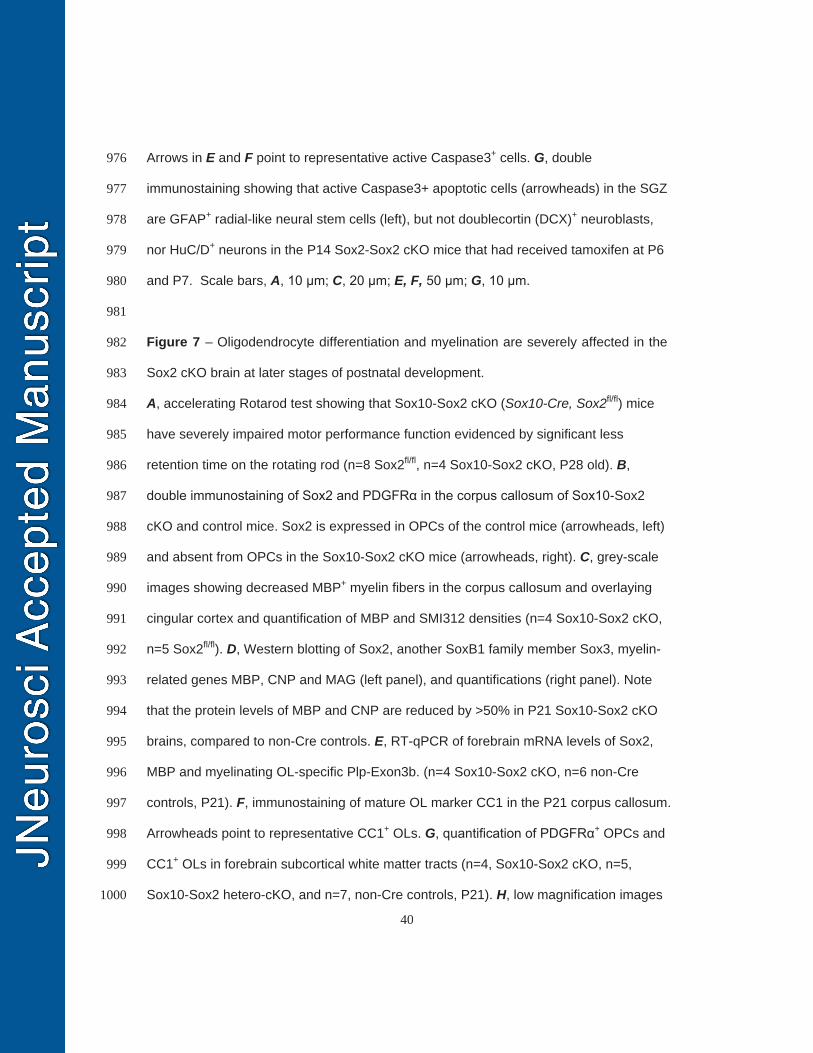
40
Arrows in E and F point to representative active Caspase3+ cells. G, double 976
immunostaining showing that active Caspase3+ apoptotic cells (arrowheads) in the SGZ 977
are GFAP+ radial-like neural stem cells (left), but not doublecortin (DCX)+ neuroblasts, 978
nor HuC/D+ neurons in the P14 Sox2-Sox2 cKO mice that had received tamoxifen at P6 979
and P7. Scale bars, A, 10 μm; C, 20 μm; E, F, 50 μm; G, 10 μm. 980
981
Figure 7 – Oligodendrocyte differentiation and myelination are severely affected in the 982
Sox2 cKO brain at later stages of postnatal development. 983
A, accelerating Rotarod test showing that Sox10-Sox2 cKO (Sox10-Cre, Sox2fl/fl) mice 984
have severely impaired motor performance function evidenced by significant less 985
retention time on the rotating rod (n=8 Sox2fl/fl, n=4 Sox10-Sox2 cKO, P28 old). B, 986
double immunostaining of Sox2 and PDGFRα in the corpus callosum of Sox10-Sox2 987
cKO and control mice. Sox2 is expressed in OPCs of the control mice (arrowheads, left) 988
and absent from OPCs in the Sox10-Sox2 cKO mice (arrowheads, right). C, grey-scale 989
images showing decreased MBP+ myelin fibers in the corpus callosum and overlaying 990
cingular cortex and quantification of MBP and SMI312 densities (n=4 Sox10-Sox2 cKO, 991
n=5 Sox2fl/fl). D, Western blotting of Sox2, another SoxB1 family member Sox3, myelin-992
related genes MBP, CNP and MAG (left panel), and quantifications (right panel). Note 993
that the protein levels of MBP and CNP are reduced by >50% in P21 Sox10-Sox2 cKO 994
brains, compared to non-Cre controls. E, RT-qPCR of forebrain mRNA levels of Sox2, 995
MBP and myelinating OL-specific Plp-Exon3b. (n=4 Sox10-Sox2 cKO, n=6 non-Cre 996
controls, P21). F, immunostaining of mature OL marker CC1 in the P21 corpus callosum. 997
Arrowheads point to representative CC1+ OLs. G, quantification of PDGFRα+ OPCs and 998
CC1+ OLs in forebrain subcortical white matter tracts (n=4, Sox10-Sox2 cKO, n=5, 999
Sox10-Sox2 hetero-cKO, and n=7, non-Cre controls, P21). H, low magnification images 1000
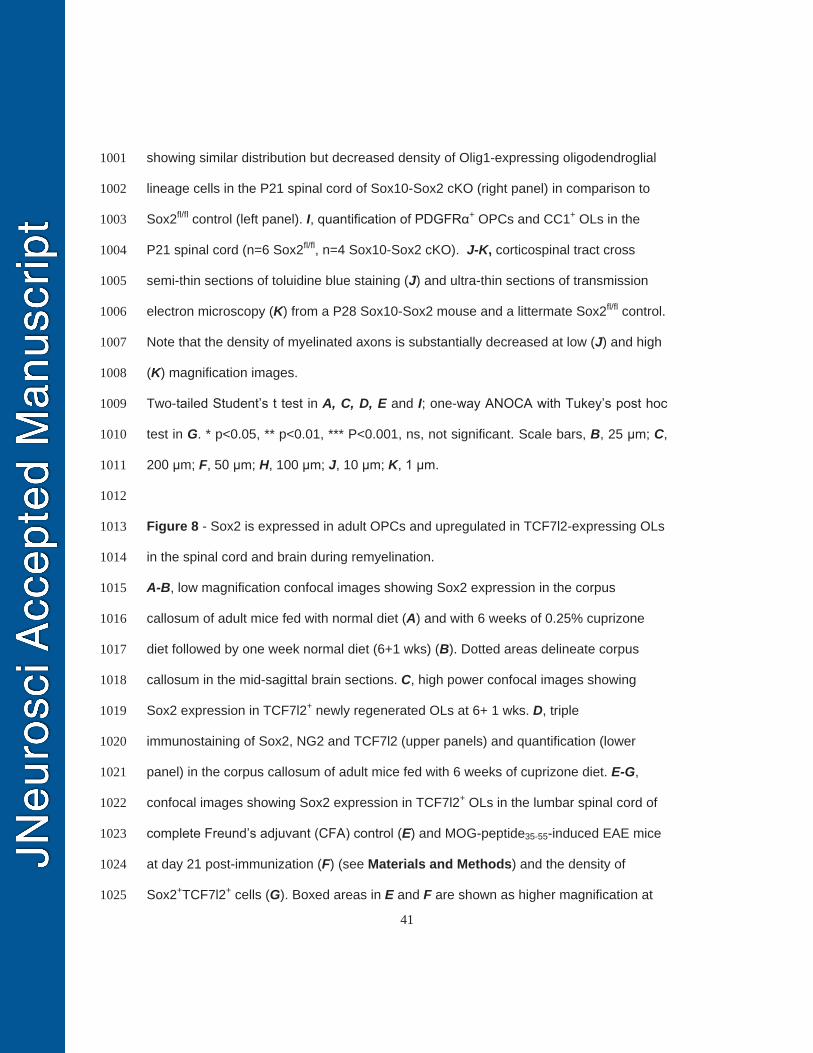
41
showing similar distribution but decreased density of Olig1-expressing oligodendroglial 1001
lineage cells in the P21 spinal cord of Sox10-Sox2 cKO (right panel) in comparison to 1002
Sox2fl/fl control (left panel). I, quantification of PDGFRα+ OPCs and CC1+ OLs in the 1003
P21 spinal cord (n=6 Sox2fl/fl, n=4 Sox10-Sox2 cKO). J-K, corticospinal tract cross 1004
semi-thin sections of toluidine blue staining (J) and ultra-thin sections of transmission 1005
electron microscopy (K) from a P28 Sox10-Sox2 mouse and a littermate Sox2fl/fl control. 1006
Note that the density of myelinated axons is substantially decreased at low (J) and high 1007
(K) magnification images. 1008
Two-tailed Student’s t test in A, C, D, E and I; one-way ANOCA with Tukey’s post hoc 1009
test in G. * p<0.05, ** p<0.01, *** P<0.001, ns, not significant. Scale bars, B, 25 μm; C, 1010
200 μm; F, 50 μm; H, 100 μm; J, 10 μm; K, 1 μm. 1011
1012
Figure 8 - Sox2 is expressed in adult OPCs and upregulated in TCF7l2-expressing OLs 1013
in the spinal cord and brain during remyelination. 1014
A-B, low magnification confocal images showing Sox2 expression in the corpus 1015
callosum of adult mice fed with normal diet (A) and with 6 weeks of 0.25% cuprizone 1016
diet followed by one week normal diet (6+1 wks) (B). Dotted areas delineate corpus 1017
callosum in the mid-sagittal brain sections. C, high power confocal images showing 1018
Sox2 expression in TCF7l2+ newly regenerated OLs at 6+ 1 wks. D, triple 1019
immunostaining of Sox2, NG2 and TCF7l2 (upper panels) and quantification (lower 1020
panel) in the corpus callosum of adult mice fed with 6 weeks of cuprizone diet. E-G, 1021
confocal images showing Sox2 expression in TCF7l2+ OLs in the lumbar spinal cord of 1022
complete Freund’s adjuvant (CFA) control (E) and MOG-peptide35-55-induced EAE mice 1023
at day 21 post-immunization (F) (see Materials and Methods) and the density of 1024
Sox2+TCF7l2+ cells (G). Boxed areas in E and F are shown as higher magnification at 1025
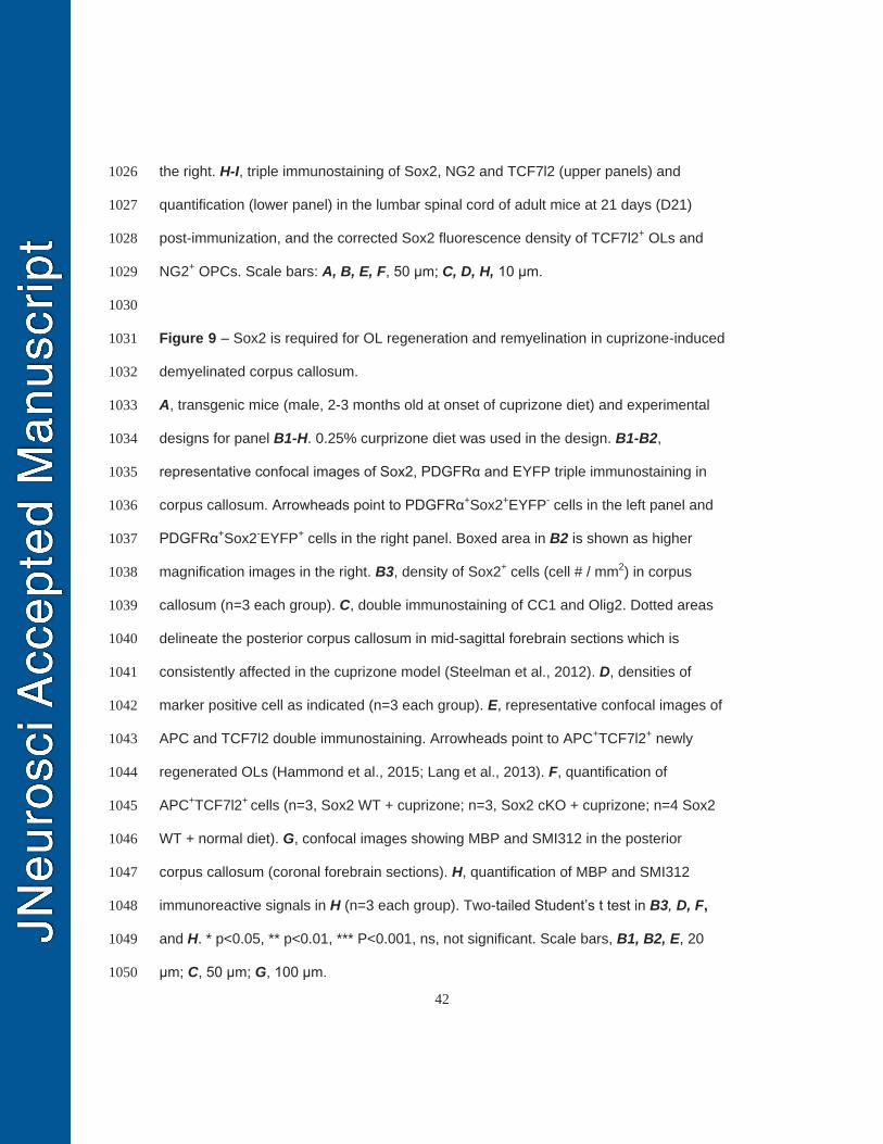
42
the right. H-I, triple immunostaining of Sox2, NG2 and TCF7l2 (upper panels) and 1026
quantification (lower panel) in the lumbar spinal cord of adult mice at 21 days (D21) 1027
post-immunization, and the corrected Sox2 fluorescence density of TCF7l2+ OLs and 1028
NG2+ OPCs. Scale bars: A, B, E, F, 50 μm; C, D, H, 10 μm. 1029
1030
Figure 9 – Sox2 is required for OL regeneration and remyelination in cuprizone-induced 1031
demyelinated corpus callosum. 1032
A, transgenic mice (male, 2-3 months old at onset of cuprizone diet) and experimental 1033
designs for panel B1-H. 0.25% curprizone diet was used in the design. B1-B2, 1034
representative confocal images of Sox2, PDGFRα and EYFP triple immunostaining in 1035
corpus callosum. Arrowheads point to PDGFRα+Sox2+EYFP- cells in the left panel and 1036
PDGFRα+Sox2-EYFP+ cells in the right panel. Boxed area in B2 is shown as higher 1037
magnification images in the right. B3, density of Sox2+ cells (cell # / mm2) in corpus 1038
callosum (n=3 each group). C, double immunostaining of CC1 and Olig2. Dotted areas 1039
delineate the posterior corpus callosum in mid-sagittal forebrain sections which is 1040
consistently affected in the cuprizone model (Steelman et al., 2012). D, densities of 1041
marker positive cell as indicated (n=3 each group). E, representative confocal images of 1042
APC and TCF7l2 double immunostaining. Arrowheads point to APC+TCF7l2+ newly 1043
regenerated OLs (Hammond et al., 2015; Lang et al., 2013). F, quantification of 1044
APC+TCF7l2+ cells (n=3, Sox2 WT + cuprizone; n=3, Sox2 cKO + cuprizone; n=4 Sox2 1045
WT + normal diet). G, confocal images showing MBP and SMI312 in the posterior 1046
corpus callosum (coronal forebrain sections). H, quantification of MBP and SMI312 1047
immunoreactive signals in H (n=3 each group). Two-tailed Student’s t test in B3, D, F, 1048
and H. * p<0.05, ** p<0.01, *** P<0.001, ns, not significant. Scale bars, B1, B2, E, 20 1049
μm; C, 50 μm; G, 100 μm. 1050
43
1051
Figure 10 – Sox2 regulates OPC proliferation and OL regeneration in the spinal cord in 1052
response to inflammatory insults. 1053
A, transgenic mice (female, 2-3 months old) and experimental designs for panels B1-F. 1054
Experimental autoimmune encephalomyelitis (EAE) was induce at day 0 by immunizing 1055
study mice with myelin oligodendrocyte glycoprotein (MOG) peptide35-55 emulsified in 1056
the complete Freund’s adjuvant (CFA) (see Materials and Methods) (Guo et al., 2012). 1057
Seven days after the last tamoxifen injection, lumbar spinal cord was harvested for 1058
analysis. B1-B3, double immunostaining of Sox2 and NG2 in the lumbar spinal cords of 1059
each group. Boxed areas are shown at higher power at the right. Images of B2 and B3 1060
delineate demyelination lesions with extensive inflammatory infiltrations indicated by 1061
dense DAPI nuclear counterstaining. Arrowheads in B1 and B2 point to Sox2+NG2+ 1062
OPCs. Note that NG2+ OPCs in Sox2 cKO have no Sox2 expression (B3), but distribute 1063
within the demyelination lesions in a similar pattern to that in Sox2 WT mice (B2). C1-1064
C3, confocal images of NG2 and Sox10 double immunostaining (C1 and C2) and 1065
quantification of Sox10+NG2+ OPCs (C3). Arrowheads point to NG2+Sox10+ OPCs. 1066
Boxed areas are shown as higher magnification images at the right. D1-E, 1067
representative confocal images of Ki67 and Olig2 (D1 and D2), and quantifications (E). 1068
F, density of APC+TCF7l2+ newly regenerated OLs. N=3 in each group. Two-tailed 1069
Student’s t test, * p<0.05, ** p<0.01. Scale bars: 20 μm, applied to all. 1070
1071
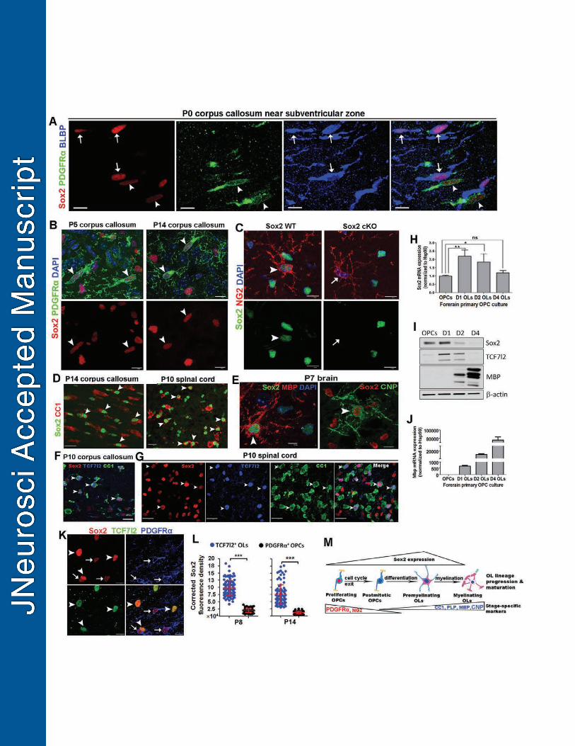
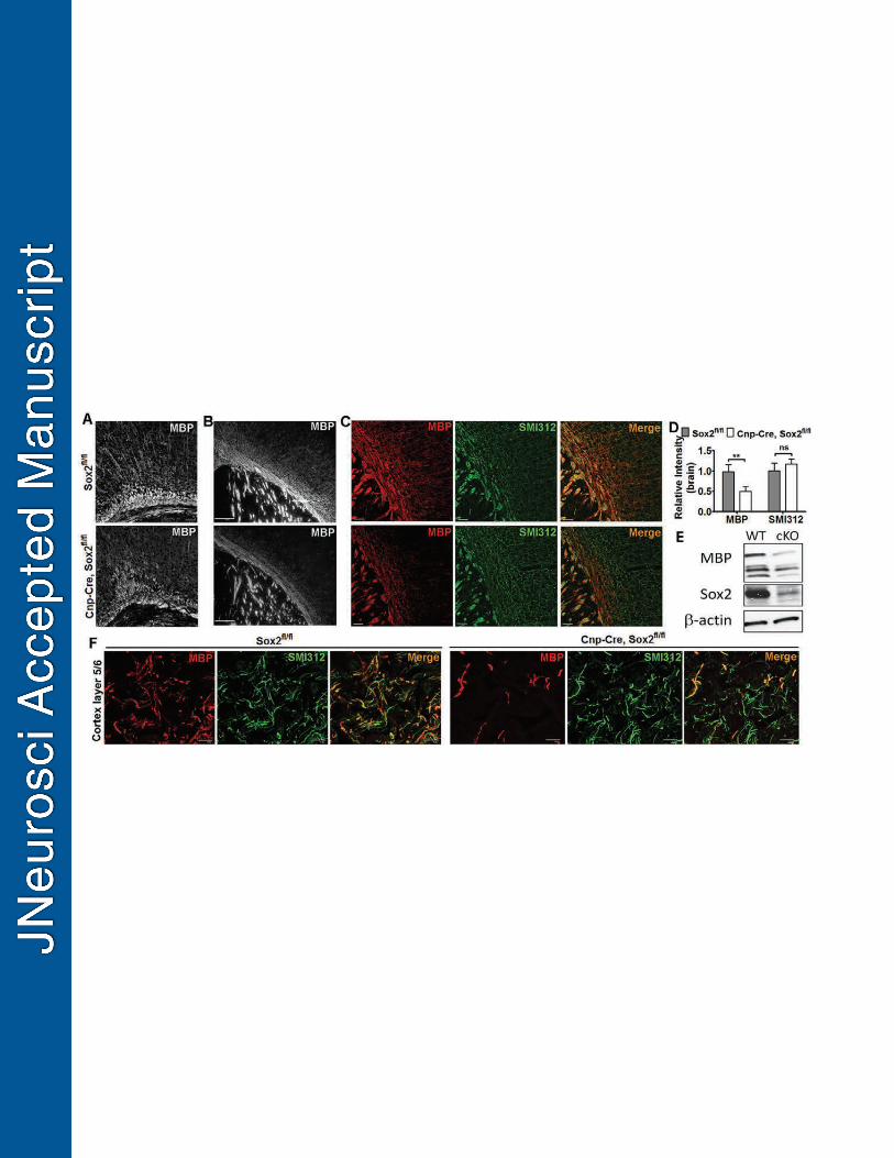
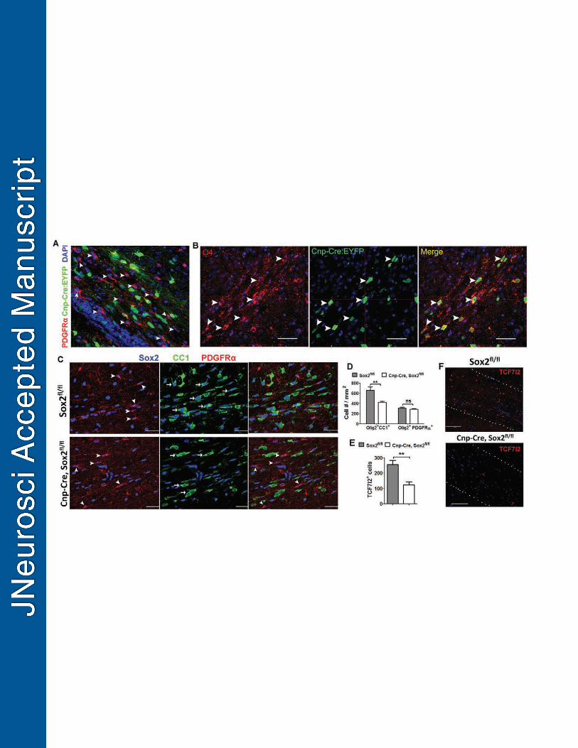
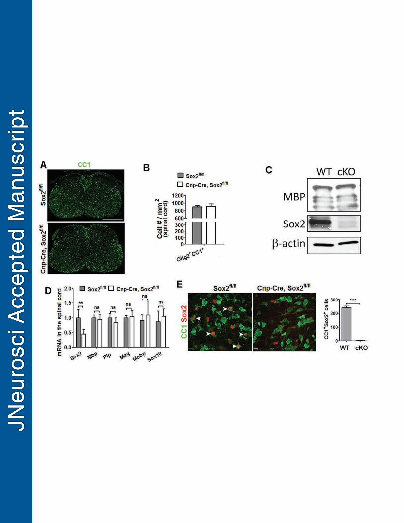
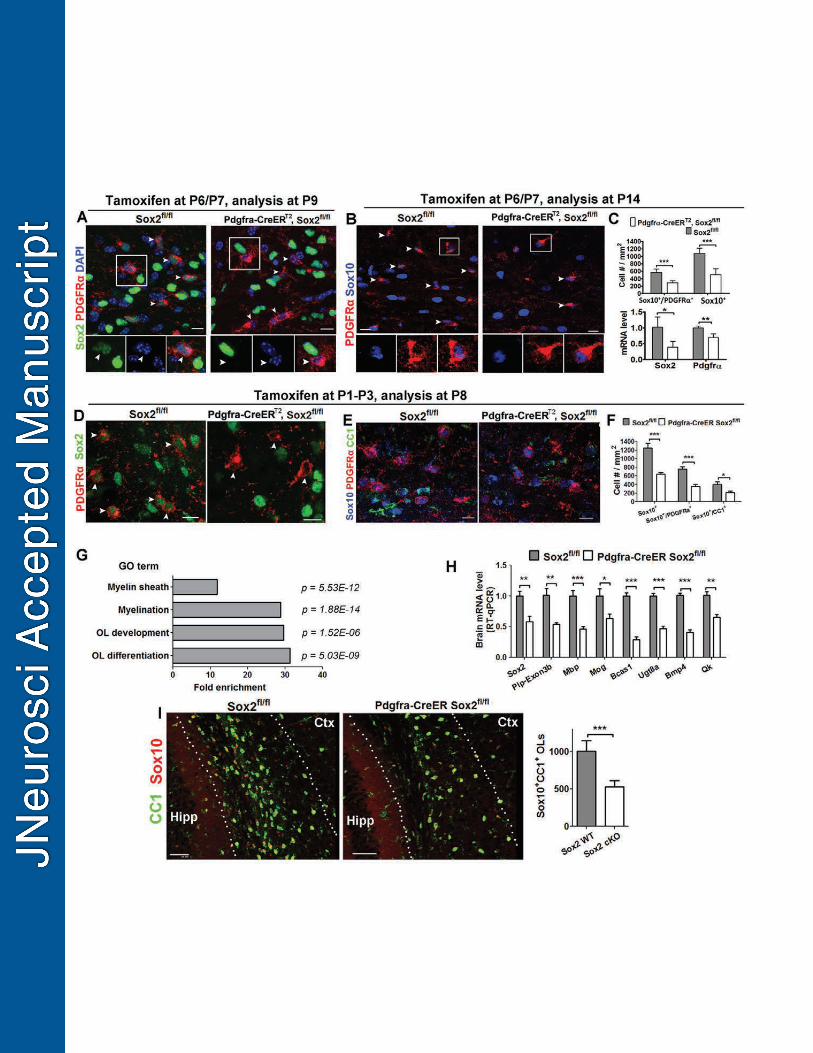
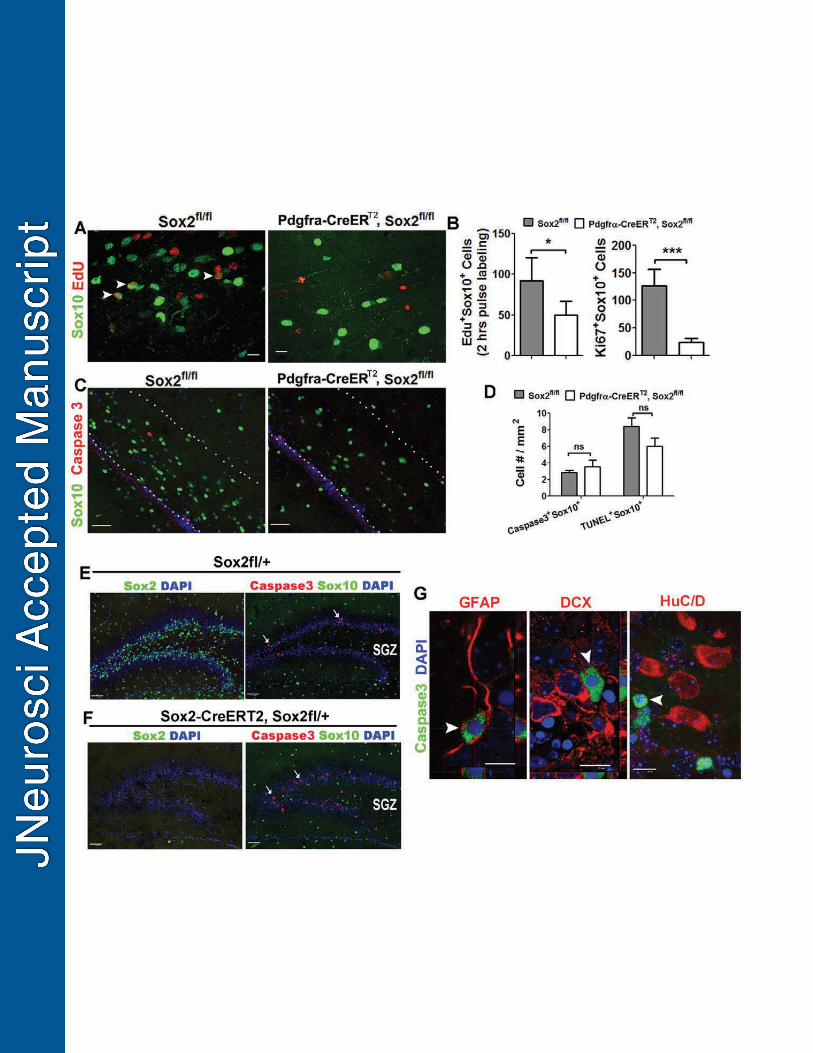
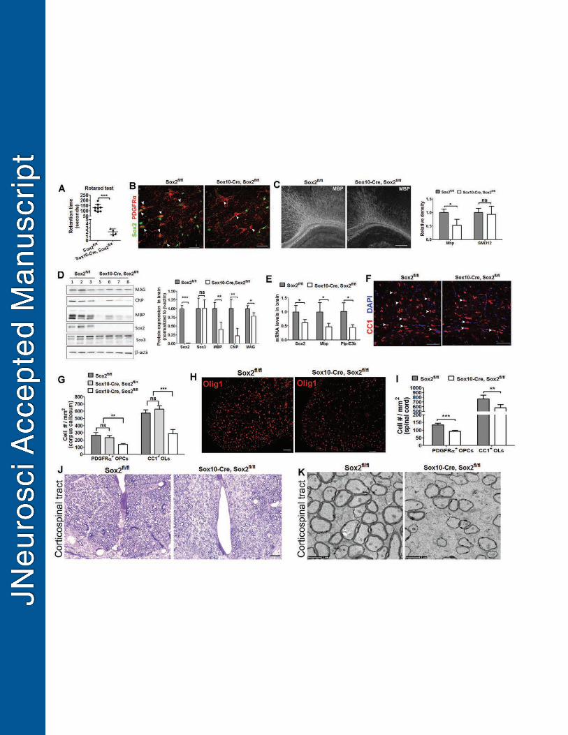
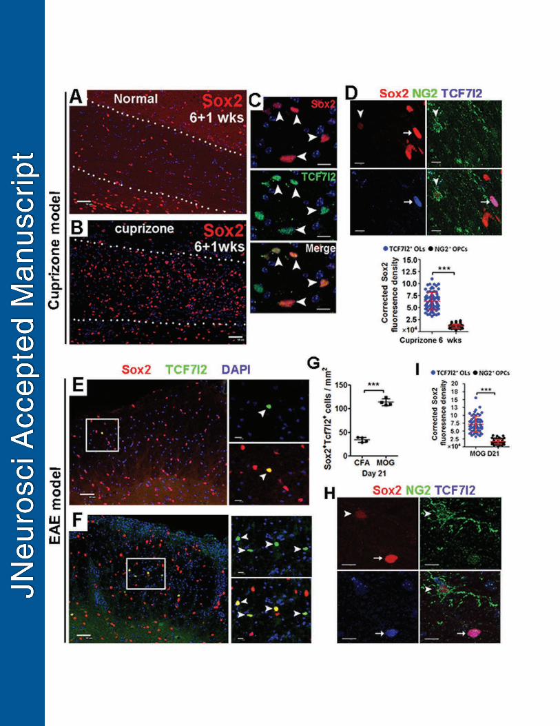
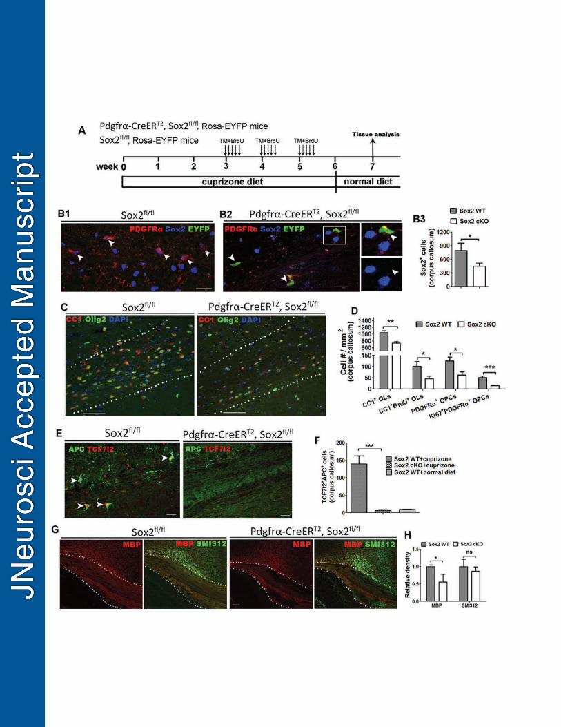
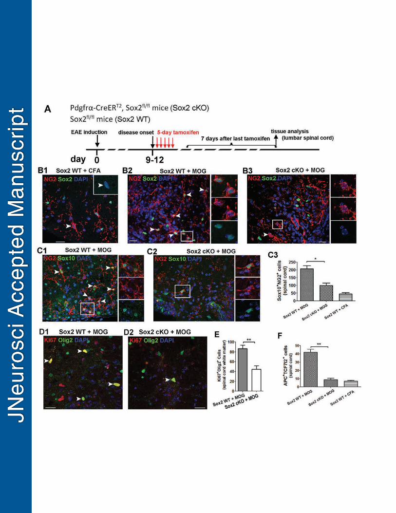

1
Table 1 – Detailed information of statistical analyses.
Figures Sample size (n) Statistical Methods P value, t and degree of freedom (df)
Fig. 1G
n=3 independent
experiments
at each time point
one-way ANOVA (P =
0.0062, F3,8 = 8.193)
with Tukey's post test
OPCs vs D1 OLs, ** P < 0.01,
OPCs vs D2 OLs, *P < 0.05
OPCs vs D4 OLs, P > 0.05
Fig. 1K
n = 145 OPCs and 116
OLs from 3 mice at P8,
n = 125 OPCs and 101
OLs from 3 mice at P14
two-tailed unpaired
Student’s t-test
P8, ***, P < 0.0001 (t = 34.33, df = 259)
P14, ***, P < 0.0001 (t = 14.87, df = 124)
Fig. 2D
n = 5 sox2fl/fl
n = 4 Cnp-Cre, Sox2fl/fl
two-tailed unpaired
Student’s t-test
MBP, ** P < 0.002 (t = 4.779, df = 7)
SMI312, P = 0.178 ( t = 1.497, df = 7)
Fig. 3D
n = 6 Sox2fl/fl
n = 4 Cnp-Cre, Sox2fl/fl
two-tailed unpaired
Student’s t-test
Olig2+CC1+, ** P = 0.0012 (t = 4.879, df = 8)
Olig2+PDGFRa+, P = 0.2314 (t = 1.295, df = 8)
Fig. 3E
n = 6 sox2fl/fl
n = 4 Cnp-Cre, Sox2fl/fl
two-tailed unpaired
Student’s t-test P = 0.0041 (t = 3.975, df = 8)
Fig. 4B
n = 6 Sox2fl/fl
n = 4 Cnp-Cre, Sox2fl/fl
two-tailed unpaired
Student’s t-test
Olig2+CC1+, P = 0.3783 (t = 0.9326, df = 8)
Fig. 4D
n = 8 Sox2fl/fl
n = 4 Cnp-Cre, Sox2fl/fl
two-tailed unpaired
Student’s t-test
Sox2, ** P = 0.0055 (t = 3.5424, df = 10)
Mbp, P = 0.5097 (t = 0.6837, df = 10)
Plp, P = 0.119 (t = 1.705, df = 10)
Mag, P = 0.6681 (t = 0.4417, df = 10)
Mobp, P = 0.3381 (t = 1.006, df = 10)
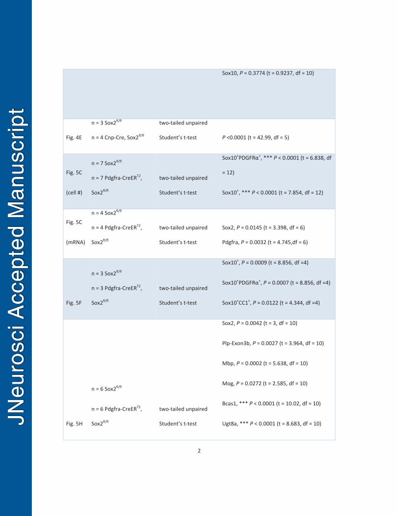
2
Sox10, P = 0.3774 (t = 0.9237, df = 10)
Fig. 4E
n = 3 Sox2fl/fl
n = 4 Cnp-Cre, Sox2fl/fl
two-tailed unpaired
Student’s t-test P <0.0001 (t = 42.99, df = 5)
Fig. 5C
(cell #)
n = 7 Sox2fl/fl
n = 7 Pdgfra-CreERT2,
Sox2fl/fl
two-tailed unpaired
Student’s t-test
Sox10+PDGFRα+, *** P < 0.0001 (t = 6.838, df
= 12)
Sox10+, *** P < 0.0001 (t = 7.854, df = 12)
Fig. 5C
(mRNA)
n = 4 Sox2fl/fl
n = 4 Pdgfra-CreERT2,
Sox2fl/fl
two-tailed unpaired
Student’s t-test
Sox2, P = 0.0145 (t = 3.398, df = 6)
Pdgfra, P = 0.0032 (t = 4.745,df = 6)
Fig. 5F
n = 3 Sox2fl/fl
n = 3 Pdgfra-CreERT2,
Sox2fl/fl
two-tailed unpaired
Student’s t-test
Sox10+, P = 0.0009 (t = 8.856, df =4)
Sox10+PDGFRα+, P = 0.0007 (t = 8.856, df =4)
Sox10+CC1+, P = 0.0122 (t = 4.344, df =4)
Fig. 5H
n = 6 Sox2fl/fl
n = 6 Pdgfra-CreERT2,
Sox2fl/fl
two-tailed unpaired
Student’s t-test
Sox2, P = 0.0042 (t = 3, df = 10)
Plp-Exon3b, P = 0.0027 (t = 3.964, df = 10)
Mbp, P = 0.0002 (t = 5.638, df = 10)
Mog, P = 0.0272 (t = 2.585, df = 10)
Bcas1, *** P < 0.0001 (t = 10.02, df = 10)
Ugt8a, *** P < 0.0001 (t = 8.683, df = 10)
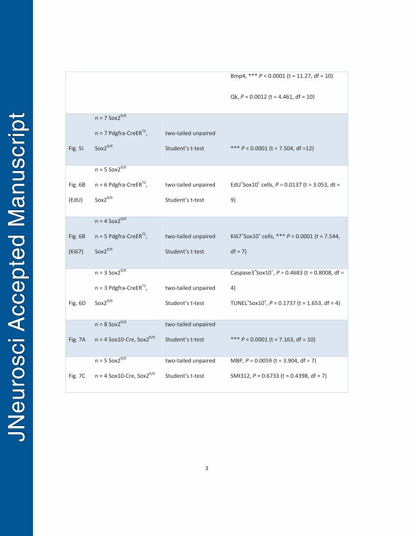
3
Bmp4, *** P < 0.0001 (t = 11.27, df = 10)
Qk, P = 0.0012 (t = 4.461, df = 10)
Fig. 5I
n = 7 Sox2fl/fl
n = 7 Pdgfra-CreERT2,
Sox2fl/fl
two-tailed unpaired
Student’s t-test *** P < 0.0001 (t = 7.504, df =12)
Fig. 6B
(EdU)
n = 5 Sox2fl/fl
n = 6 Pdgfra-CreERT2,
Sox2fl/fl
two-tailed unpaired
Student’s t-test
EdU+Sox10+ cells, P = 0.0137 (t = 3.053, dt =
9)
Fig. 6B
(Ki67)
n = 4 Sox2fl/fl
n = 5 Pdgfra-CreERT2,
Sox2fl/fl
two-tailed unpaired
Student’s t-test
Ki67+Sox10+ cells, *** P < 0.0001 (t = 7.544,
df = 7)
Fig. 6D
n = 3 Sox2fl/fl
n = 3 Pdgfra-CreERT2,
Sox2fl/fl
two-tailed unpaired
Student’s t-test
Caspase3+Sox10+, P = 0.4683 (t = 0.8008, df =
4)
TUNEL+Sox10+, P = 0.1737 (t = 1.653, df = 4)
Fig. 7A
n = 8 Sox2fl/fl
n = 4 Sox10-Cre, Sox2fl/fl
two-tailed unpaired
Student’s t-test *** P < 0.0001 (t = 7.163, df = 10)
Fig. 7C
n = 5 Sox2fl/fl
n = 4 Sox10-Cre, Sox2fl/fl
two-tailed unpaired
Student’s t-test
MBP, P = 0.0059 (t = 3.904, df = 7)
SMI312, P = 0.6733 (t = 0.4398, df = 7)
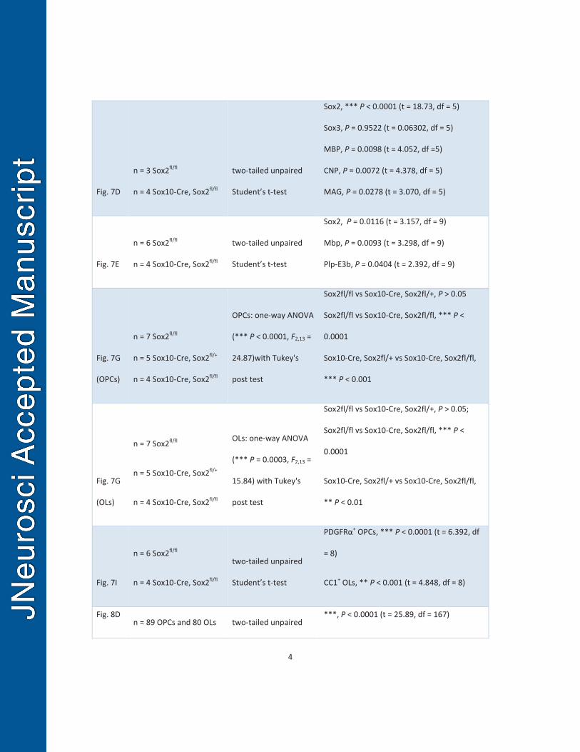
4
Fig. 7D
n = 3 Sox2fl/fl
n = 4 Sox10-Cre, Sox2fl/fl
two-tailed unpaired
Student’s t-test
Sox2, *** P < 0.0001 (t = 18.73, df = 5)
Sox3, P = 0.9522 (t = 0.06302, df = 5)
MBP, P = 0.0098 (t = 4.052, df =5)
CNP, P = 0.0072 (t = 4.378, df = 5)
MAG, P = 0.0278 (t = 3.070, df = 5)
Fig. 7E
n = 6 Sox2fl/fl
n = 4 Sox10-Cre, Sox2fl/fl
two-tailed unpaired
Student’s t-test
Sox2, P = 0.0116 (t = 3.157, df = 9)
Mbp, P = 0.0093 (t = 3.298, df = 9)
Plp-E3b, P = 0.0404 (t = 2.392, df = 9)
Fig. 7G
(OPCs)
n = 7 Sox2fl/fl
n = 5 Sox10-Cre, Sox2fl/+
n = 4 Sox10-Cre, Sox2fl/fl
OPCs: one-way ANOVA
(*** P < 0.0001, F2,13 =
24.87)with Tukey's
post test
Sox2fl/fl vs Sox10-Cre, Sox2fl/+, P > 0.05
Sox2fl/fl vs Sox10-Cre, Sox2fl/fl, *** P <
0.0001
Sox10-Cre, Sox2fl/+ vs Sox10-Cre, Sox2fl/fl,
*** P < 0.001
Fig. 7G
(OLs)
n = 7 Sox2fl/fl
n = 5 Sox10-Cre, Sox2fl/+
n = 4 Sox10-Cre, Sox2fl/fl
OLs: one-way ANOVA
(*** P = 0.0003, F2,13 =
15.84) with Tukey's
post test
Sox2fl/fl vs Sox10-Cre, Sox2fl/+, P > 0.05;
Sox2fl/fl vs Sox10-Cre, Sox2fl/fl, *** P <
0.0001
Sox10-Cre, Sox2fl/+ vs Sox10-Cre, Sox2fl/fl,
** P < 0.01
Fig. 7I
n = 6 Sox2fl/fl
n = 4 Sox10-Cre, Sox2fl/fl
two-tailed unpaired
Student’s t-test
PDGFRα+ OPCs, *** P < 0.0001 (t = 6.392, df
= 8)
CC1+ OLs, ** P < 0.001 (t = 4.848, df = 8)
Fig. 8D n = 89 OPCs and 80 OLs two-tailed unpaired
***, P < 0.0001 (t = 25.89, df = 167)
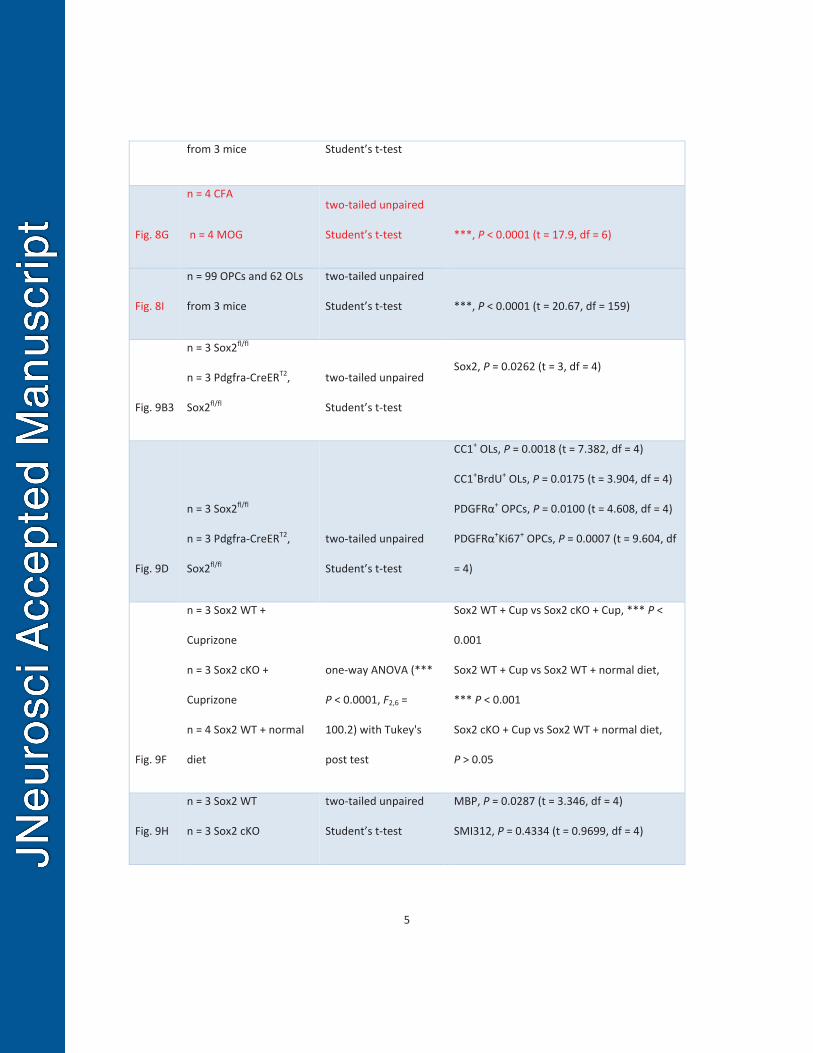
5
from 3 mice Student’s t-test
Fig. 8G
n = 4 CFA
n = 4 MOG
two-tailed unpaired
Student’s t-test ***, P < 0.0001 (t = 17.9, df = 6)
Fig. 8I
n = 99 OPCs and 62 OLs
from 3 mice
two-tailed unpaired
Student’s t-test ***, P < 0.0001 (t = 20.67, df = 159)
Fig. 9B3
n = 3 Sox2fl/fl
n = 3 Pdgfra-CreERT2,
Sox2fl/fl
two-tailed unpaired
Student’s t-test
Sox2, P = 0.0262 (t = 3, df = 4)
Fig. 9D
n = 3 Sox2fl/fl
n = 3 Pdgfra-CreERT2,
Sox2fl/fl
two-tailed unpaired
Student’s t-test
CC1+ OLs, P = 0.0018 (t = 7.382, df = 4)
CC1+BrdU+ OLs, P = 0.0175 (t = 3.904, df = 4)
PDGFRα+ OPCs, P = 0.0100 (t = 4.608, df = 4)
PDGFRα+Ki67+ OPCs, P = 0.0007 (t = 9.604, df
= 4)
Fig. 9F
n = 3 Sox2 WT +
Cuprizone
n = 3 Sox2 cKO +
Cuprizone
n = 4 Sox2 WT + normal
diet
one-way ANOVA (***
P < 0.0001, F2,6 =
100.2) with Tukey's
post test
Sox2 WT + Cup vs Sox2 cKO + Cup, *** P <
0.001
Sox2 WT + Cup vs Sox2 WT + normal diet,
*** P < 0.001
Sox2 cKO + Cup vs Sox2 WT + normal diet,
P > 0.05
Fig. 9H
n = 3 Sox2 WT
n = 3 Sox2 cKO
two-tailed unpaired
Student’s t-test
MBP, P = 0.0287 (t = 3.346, df = 4)
SMI312, P = 0.4334 (t = 0.9699, df = 4)
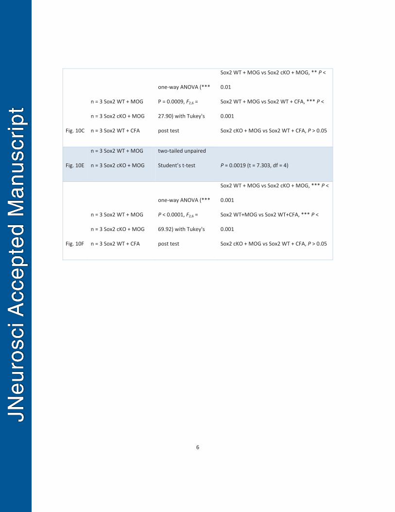
6
Fig. 10C
n = 3 Sox2 WT + MOG
n = 3 Sox2 cKO + MOG
n = 3 Sox2 WT + CFA
one-way ANOVA (***
P = 0.0009, F2,6 =
27.90) with Tukey's
post test
Sox2 WT + MOG vs Sox2 cKO + MOG, ** P <
0.01
Sox2 WT + MOG vs Sox2 WT + CFA, *** P <
0.001
Sox2 cKO + MOG vs Sox2 WT + CFA, P > 0.05
Fig. 10E
n = 3 Sox2 WT + MOG
n = 3 Sox2 cKO + MOG
two-tailed unpaired
Student’s t-test P = 0.0019 (t = 7.303, df = 4)
Fig. 10F
n = 3 Sox2 WT + MOG
n = 3 Sox2 cKO + MOG
n = 3 Sox2 WT + CFA
one-way ANOVA (***
P < 0.0001, F2,6 =
69.92) with Tukey's
post test
Sox2 WT + MOG vs Sox2 cKO + MOG, *** P <
0.001
Sox2 WT+MOG vs Sox2 WT+CFA, *** P <
0.001
Sox2 cKO + MOG vs Sox2 WT + CFA, P > 0.05
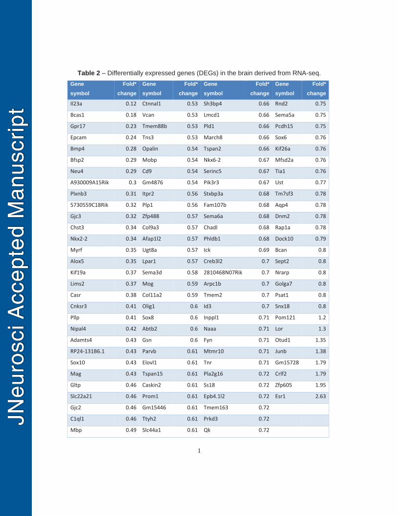
1
Table 2 – Differentially expressed genes (DEGs) in the brain derived from RNA-seq. Gene symbol
Fold* change
Gene symbol
Fold* change
Gene symbol
Fold* change
Gene symbol
Fold* change
Il23a 0.12 Ctnnal1 0.53 Sh3bp4 0.66 Rnd2 0.75
Bcas1 0.18 Vcan 0.53 Lmcd1 0.66 Sema5a 0.75
Gpr17 0.23 Tmem88b 0.53 Pld1 0.66 Pcdh15 0.75
Epcam 0.24 Tns3 0.53 March8 0.66 Sox6 0.76
Bmp4 0.28 Opalin 0.54 Tspan2 0.66 Kif26a 0.76
Bfsp2 0.29 Mobp 0.54 Nkx6-2 0.67 Mfsd2a 0.76
Neu4 0.29 Cd9 0.54 Serinc5 0.67 Tia1 0.76
A930009A15Rik 0.3 Gm4876 0.54 Pik3r3 0.67 Ust 0.77
Plxnb3 0.31 Itpr2 0.56 Stxbp3a 0.68 Tm7sf3 0.78
5730559C18Rik 0.32 Plp1 0.56 Fam107b 0.68 Aqp4 0.78
Gjc3 0.32 Zfp488 0.57 Sema6a 0.68 Dnm2 0.78
Chst3 0.34 Col9a3 0.57 Chadl 0.68 Rap1a 0.78
Nkx2-2 0.34 Afap1l2 0.57 Phldb1 0.68 Dock10 0.79
Myrf 0.35 Ugt8a 0.57 Ick 0.69 Bcan 0.8
Alox5 0.35 Lpar1 0.57 Creb3l2 0.7 Sept2 0.8
Kif19a 0.37 Sema3d 0.58 2810468N07Rik 0.7 Nrarp 0.8
Lims2 0.37 Mog 0.59 Arpc1b 0.7 Golga7 0.8
Casr 0.38 Col11a2 0.59 Tmem2 0.7 Psat1 0.8
Cnksr3 0.41 Olig1 0.6 Id3 0.7 Snx18 0.8
Pllp 0.41 Sox8 0.6 Inppl1 0.71 Pom121 1.2
Nipal4 0.42 Abtb2 0.6 Naaa 0.71 Lor 1.3
Adamts4 0.43 Gsn 0.6 Fyn 0.71 Otud1 1.35
RP24-131B6.1 0.43 Parvb 0.61 Mtmr10 0.71 Junb 1.38
Sox10 0.43 Elovl1 0.61 Tnr 0.71 Gm15728 1.79
Mag 0.43 Tspan15 0.61 Pla2g16 0.72 Crlf2 1.79
Gltp 0.46 Caskin2 0.61 Ss18 0.72 Zfp605 1.95
Slc22a21 0.46 Prom1 0.61 Epb4.1l2 0.72 Esr1 2.63
Gjc2 0.46 Gm15446 0.61 Tmem163 0.72
C1ql1 0.46 Ttyh2 0.61 Prkd3 0.72
Mbp 0.49 Slc44a1 0.61 Qk 0.72
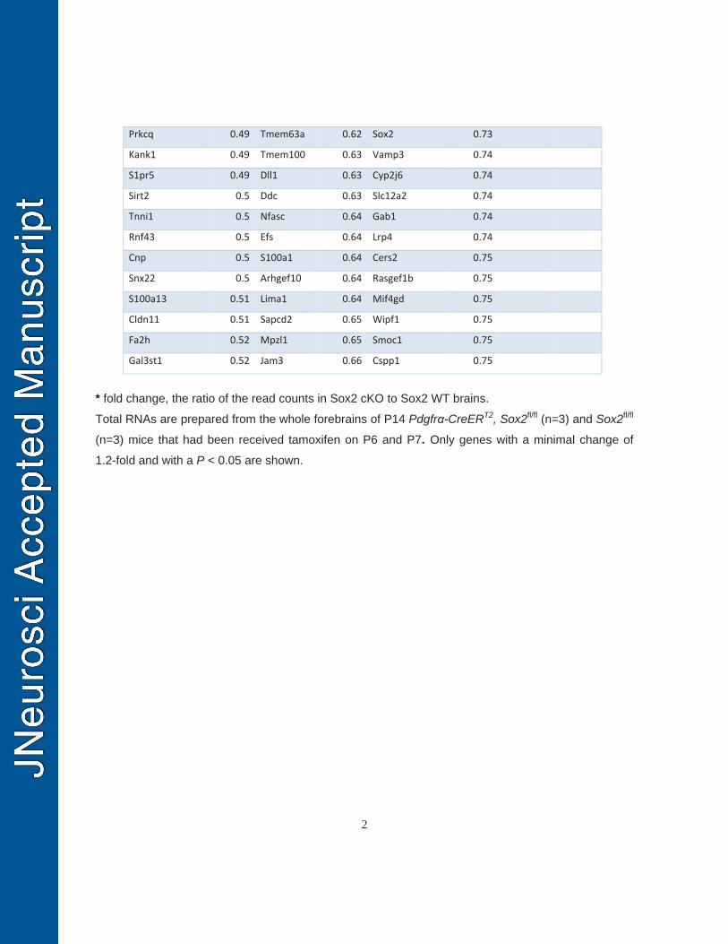
2
Prkcq 0.49 Tmem63a 0.62 Sox2 0.73
Kank1 0.49 Tmem100 0.63 Vamp3 0.74
S1pr5 0.49 Dll1 0.63 Cyp2j6 0.74
Sirt2 0.5 Ddc 0.63 Slc12a2 0.74
Tnni1 0.5 Nfasc 0.64 Gab1 0.74
Rnf43 0.5 Efs 0.64 Lrp4 0.74
Cnp 0.5 S100a1 0.64 Cers2 0.75
Snx22 0.5 Arhgef10 0.64 Rasgef1b 0.75
S100a13 0.51 Lima1 0.64 Mif4gd 0.75
Cldn11 0.51 Sapcd2 0.65 Wipf1 0.75
Fa2h 0.52 Mpzl1 0.65 Smoc1 0.75
Gal3st1 0.52 Jam3 0.66 Cspp1 0.75
* fold change, the ratio of the read counts in Sox2 cKO to Sox2 WT brains.
Total RNAs are prepared from the whole forebrains of P14 Pdgfrα-CreERT2, Sox2fl/fl (n=3) and Sox2fl/fl
(n=3) mice that had been received tamoxifen on P6 and P7. Only genes with a minimal change of
1.2-fold and with a P < 0.05 are shown.