staging advanced pca and imaging response...
TRANSCRIPT

Staging Advanced PCa and
imaging response assessmentJelle Barentsz
Geert Villiers and Anwar Padhani
Prostate MR Reference/Expert Center, Nijmegen, NL

Conflict of interest

I can bestubborn

NODES

N-Staging: When?

N-Staging: When?
EAU-Guidelines (2016)
N-staging in i.m.- and high-risk patients (>5%)
in general:
PSA >10 ng/mL
OR Gleason score >6
Or cT3 or higher

N-Staging: Where?

N-Staging: Where?Obturator 30%
External Iliac 30%
Internal iliac 30%
Presacral 7%
Pararectal 4%
Common Iliac 1-2%
Variable spread: no single sentinal node

Pathways of spread
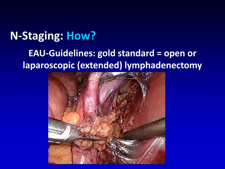
N-Staging: How?
EAU-Guidelines: gold standard = open or laparoscopic (extended) lymphadenectomy

ePLND
1. No benefit in clinical outcome

ePLND
1. No benefit in clinical outcome
2. More complications

ePLND
1. No benefit in clinical outcome
2. More complications
3. But accurate assessment of cancer spread

N-Staging: How?
40% of ⨁ LN not detected with PLND
Heesakkers, Radiology 2008
59%
20%
6%
11%
4%

Recent own data 16/26 (62%) ⨁ LN not removed
Pre-operative Post-operative

Pre-operative Post-operative
Recent own data 16/26 (62%) ⨁ LN not removed

Pre-operative Post-operative
Recent own data 16/26 (62%) ⨁ LN not removed
What is the relevance of very small ⨁ LN?
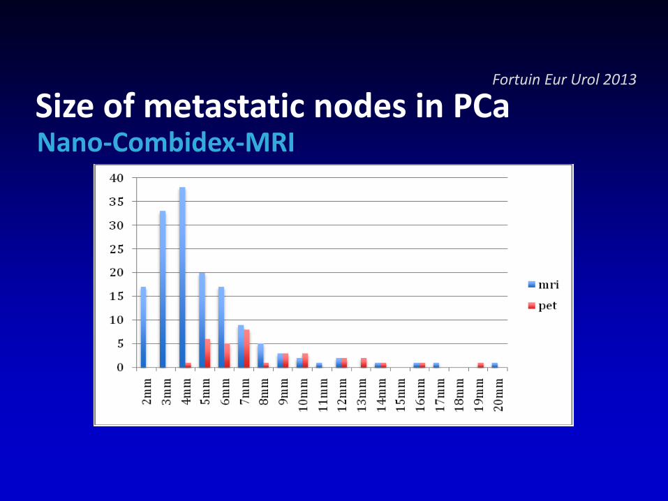
Size of metastatic nodes in PCaNano-Combidex-MRI
Fortuin Eur Urol 2013

N-Staging: How: Imaging?

N-Staging: How: Imaging?
- CT
- DWI
- Nano-MRI
- MRI
- PET-CT

N-Staging: How: Imaging?
- Sensitivity for metastatic nodes: ~40%CT: 42% (range 5%-94%)MRI: 39% (range 6% -83%)
Specificity : ~80%CT: 82% (range 80% - 83%)MRI: 82% (range 79% - 83%)

N-Staging: How: Imaging?
- Sensitivity for metastatic nodes: ~40%CT: 42% (range 5%-94%)MRI: 39% (range 6% -83%)
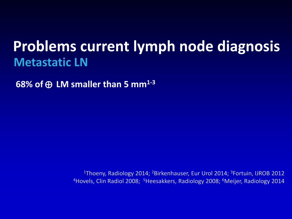
Problems current lymph node diagnosis
1Thoeny, Radiology 2014; 2Birkenhauser, Eur Urol 2014; 3Fortuin, IJROB 20124Hovels, Clin Radiol 2008; 5Heesakkers, Radiology 2008; 6Meijer, Radiology 2014
68% of⨁ LM smaller than 5 mm1-3
Metastatic LN
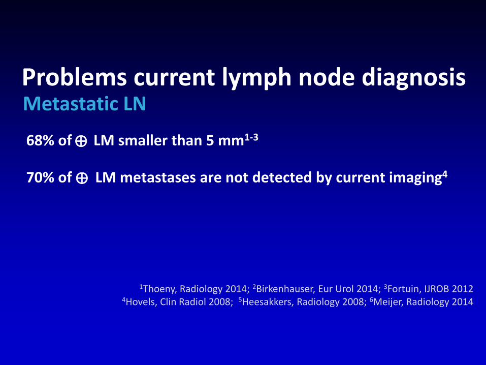
Problems current lymph node diagnosis
1Thoeny, Radiology 2014; 2Birkenhauser, Eur Urol 2014; 3Fortuin, IJROB 20124Hovels, Clin Radiol 2008; 5Heesakkers, Radiology 2008; 6Meijer, Radiology 2014
68% of⨁ LM smaller than 5 mm1-3
70% of⨁ LM metastases are not detected by current imaging4
Metastatic LN

Problems current lymph node diagnosis
1Thoeny, Radiology 2014; 2Birkenhauser, Eur Urol 2014; 3Fortuin, IJROB 20124Hovels, Clin Radiol 2008; 5Heesakkers, Radiology 2008; 6Meijer, Radiology 2014
68% of⨁ LM smaller than 5 mm1-3
40% of⨁ LM not detected with PLND5
50% of⨁ LM outside COTG radiotherapy CTV6
70% of⨁ LM metastases are not detected by current imaging4
Metastatic LN


N-Staging: How?
- CT
- DWI
- Nano-MRI
- MRI
- PET-CT

Small lymph node Decreased diffusion(High signal on b600)
Eiber, J Magn Reson Imaging 2011;33:1160Beer, Mol Imaging Biol 2011;13:352

N-Staging: How?
Technique (3Tesla)
3D-T1 + 3D-T2 + DWI (b0, b500, b1000)
3 readers, correlation DWI - morphology
only normal-sized nodes (< 8 mm)
Results (per patient)
73% sensitivity, 86% specificity
missed nodes <5 mm (majority <3 mm)

N-Staging: How?
Technique (3Tesla)
3D-T1 + 3D-T2 + DWI (b0, b500, b1000)
3 readers, correlation DWI - morphology
only normal-sized nodes (< 8 mm)
Results (per patient)
73% sensitivity, 86% specificity
missed nodes <5 mm (majority <3 mm)

N-Staging: How?
- CT
- DWI
- Nano-MRI
- MRI
- PET-CT

ferumoxtran-10Nano-MRI
Combidex/Sinerem


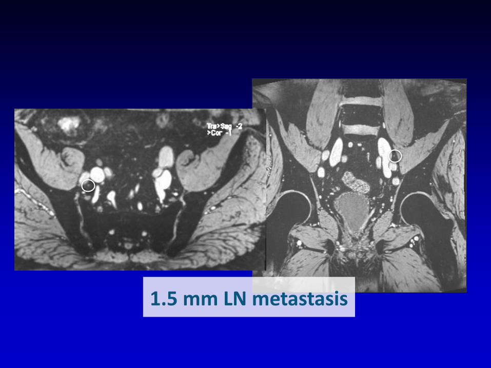
1.5 mm LN metastasis

1.5 mm LN metastasis

1.5 mm LN metastasis

Sytematic Review: QUADAS-2
Medvalue 2016
Study True positives
False positives
False negatives
True negatives
Specificity[95% CI] Sensitivity [95% CI]
Harisinghani 2003 33 2 0 45 1.00 [0.89, 1.00] 0.96 [0.85, 0.99]
Heesakkers 2008 50 23 11 291 0.82 [0.70, 0.91] 0.93 [0.89, 0.95]
Triantafyllou 2013 12 9 8 46 0.60 [0.36, 0.81] 0.84 [0.71, 0.92]
Pooled Sensitivity 88%
Pooled specificity 93%

Not (yet) available

It will soon be available

N-Staging: How?
- CT
- DWI
- Nano-MRI
- MRI
- PET-CT

68Ga-PSMA PET-CT vs other tracers
Yu, Am J Nucl Med Mol Imag 2014
0
10
20
30
40
50
60
70
Choline Acet FDG PSMA
Px
RTh

68GA-PSMA PET-CT

68Ga PSMA PET-CT
Prostate cancer
Kidneys
Salivatary Glands
68Ga(HBED-CC)-antibody
Small bowel
Afshar-Oromieh, Eur J Nucl Med Mol Imag 2014

11C-Choline PET-CT vs nano-MRI 2005
Fortuin Eur Urol 2013
0
5
10
15
20
25
30
35
40
1 2 3 4 5 6 7 8 9 10 11 12 13 14 15
Choline MRL Ansje
Nodal Size

11C-Ch-PET-CT vs nano-MRI 2005 and 2017
1 2 3 4 5 6 7 8 9 10 11 12 13 14 15
MRL Ansje Choline
0
5
10
15
20
25
30
35
40
45
50
1 2 3 4 5 6 7 8 9 10 11 12 13 14 15
Choline MRL MRL Ansje
Nodal Size

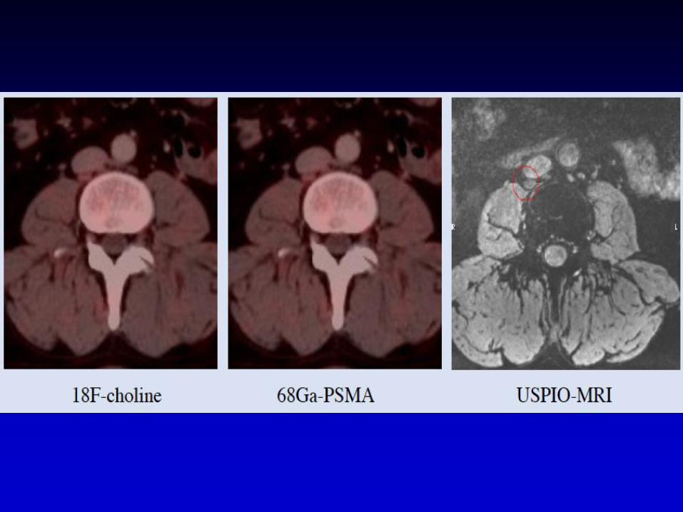
11C-Ch- vs 68Ga-PSMA PET-CT and MRI
1 2 3 4 5 6 7 8 9 10 11 12 13 14 15
MRL Ansje Choline
1 2 3 4 5 6 7 8 9 10 11 12 13 14 15
MRL MRL Ansje Choline
0
5
10
15
20
25
30
35
40
45
50
1 2 3 4 5 6 7 8 9 10 11 12 13 14 15
MRL MRL Ansje Choline PSMA
Nodal Size

68Ga-PSMA PET-CT and nano-MRI 2017
1 2 3 4 5 6 7 8 9 10 11 12 13 14 15
MRL Ansje Choline
1 2 3 4 5 6 7 8 9 10 11 12 13 14 15
MRL MRL Ansje Choline
1 2 3 4 5 6 7 8 9 10 11 12 13 14 15
MRL PSMA MRL Ansje Choline
1 2 3 4 5 6 7 8 9 10 11 12 13 14 15
MRL PSMA
Nodal Size

Afshar-Oromieh, Eur J Nucl Med Mol Imag 2015
68Ga PSMA PET-CT
0%
20%
40%
60%
80%
100%
120%
% positive

68Ga PSMA PET-CT
12/30 high-risk patients; 53/608 LN metastases
LN Staging
Buddaus, Eur Urol 2015

68Ga PSMA PET-CT
12/30 high-risk patients; 53/608 LN metastases
LN Staging
Buddaus, Eur Urol 2015
Sensitivity: 33%Specificity: 100%PPV: 100%NPV: 53%

68Ga PSMA PET-CT
12/30 high-risk patients; 53/608 LN metastases
LN Staging
Buddaus, Eur Urol 2015
Sensitivity: 33%Specificity: 100%PPV: 100%NPV: 53%
Median size detected LN: 13.6 mmMedian size undetected LN: 4.3 mm

68Ga PSMA PET-CT
12/30 high-risk patients; 53/608 LN metastases
LN Staging
Buddaus, Eur Urol 2015
Smallest positive LN: 4.0 mm

BONE

WHOLE BODY MRI

MET-RADS: WB-MRI sequences; by indications
Sequence description Core protocol Extensions for
comprehensive
assessments
Clinical use Lesion detection &
characterization
Response
assessment1 Whole spine – sagittal, T1W, TSE Yes Yes 2 Whole spine – sagittal, STIR (preferred) or fat
suppressed T2W
Yes Yes
3 Whole body (vertex to mid thighs) –T1W, GRE Dixon
technique. Fat image reconstructions are mandatory.
Axial or coronal Axial and coronal
4 Whole body (skull base to mid-thighs) – axial, diffusion
weighted, STIR fat suppression, contiguous slicing,
multiple stations.
2 b-values 3 b-values
5 Whole body (vertex to mid thighs) – axial, T2W, TSE No
fat suppression
Option Yes
6 Regional assessments including small field of view
images, brain studies and contrast enhancement
No Yes
Padhani AR, et al. METastasis Reporting and Data System for Prostate Cancer: Practical Guidelines for Acquisition, Interpretation, and Reporting of Whole-body MRI Evaluations of Multiorgan Involvement in Advanced Prostate Cancer (MET-RADS-P). Eur Urol. 2017
Jan;71(1):81-92.

Short MRI protocol in <30 mins (detection); Comprehensive MRI protocol in 45min (response)
T1W-GRE57 sec
T1W-SE2.14 min
STIR-SE3.20 min
DWI b50-900: 17.01 mins
b900 MIP b900 MPR
1.5T

WB-MRI detects more malignant lesions/ patient than bone scans
04Oct13 01Oct13
74 m CRPC on Enzalutamide. PSA 0.4 ng/ml3 lesions on planar bone scan; 6 lesions on WB-MRI

74 m CRPC, PSA 0.4: residual active diseaseIrregular/thick hyperintense rim on T2W-FS images, ↑cellularity on
DWI: suspicious of active disease despite suppressed PSA
T2W-FS T1W CT T1W-F%
b900T1W
RT atrophy74 mCRPC,Rx Enzalutamide PSA 0.4ng/ml

WB-MRI outperforms bone scans in detecting bone metastases & is as good as CT for lymph node evaluations
Lecouvet, F et al. Can Whole-body MRI with diffusion-weighted imaging replace Tc 99m bone scanning and computed tomography for single-step detection of metastases in patients with high-risk prostate cancer? Euro Urol 2012; 62:68-75
▪ 100 men with high risk prostate cancer
▪ 5 independent reviewers (CT = 2; WB-MRI = 2; BS+x-ray = 1)
▪ Metastases prevalence (best value comparator): 68 patients
▪ MRI detected bone metastases in 7-8 of 55 BS negative patients
Sensitivity Specificity PPV NPV
Lymph node metastases (prevalence 44 patients)
CT 77-82 95-96 92-95 84-87
WB-MRI 77-82 96-98 94-97 84-87
Bone metastases (prevalence 51 patients)
BS + x-ray 86 98 98 87
WB-MRI 98-100 98-100 98-100 98-100

Hamstra DA, et al, J Clin Oncol 2007: 25:4104-4109
Biological processes involved in therapy
induced changes in DWI
27Feb12
23July12
1500 µm2/s
95 centile of Ex1
Syngo.via Frontier MR Total Tumor Load software; Siemens Healthineers
No Rx effect
Highly likely response
Likely response
95%

Choline-PET/CT, MRI, and planar bone
scan for bone metastasis detection in
prostate cancer
Shen G, Deng H, Hu S, Jia Z. Comparison of choline-PET/CT, MRI, SPECT, and bone scintigraphy in the diagnosis of bone metastases in patients with prostate cancer: a meta-analysis. Skeletal
Radiol. 2014 Nov;43(11):1503-13.
FCH-PET/CT MRI BS

68Ga PSMA PET-CT and DWI WB-MRI

68Ga PSMA PET-CT and DWI WB-MRI

68Ga PSMA PET-CT and DWI WB-MRI

68Ga PSMA PET-CT and DWI WB-MRI
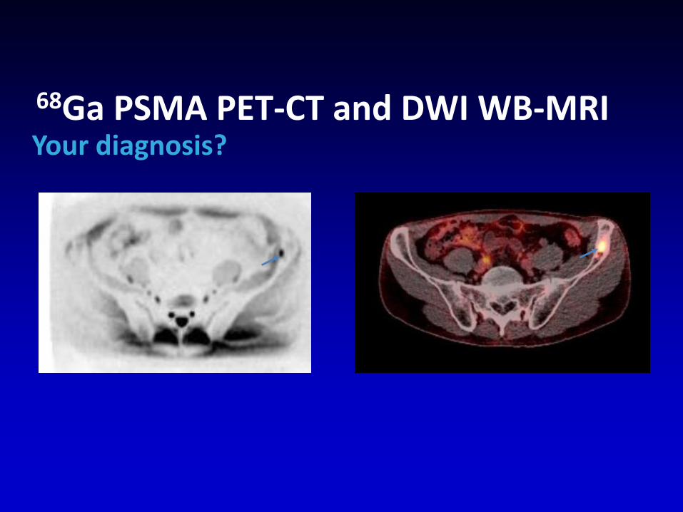
68Ga PSMA PET-CT and DWI WB-MRIYour diagnosis?


Post-operative 68Ga PSMA PET-CTpT2b N1 Mx, PSA NADIR 0.4, in 3 months 0.8
✓ 68Ga PSMA: Left positive node
✓ Rest normal


✓ Left ureter


✓ Right positive node

Current imaging tools:
fit for purpose (detection & response)?

mCRPC. Rx Enzalutamide. 2°resistance
ScreeningPSA 45.5
Week 13PSA 0.6
Week 25PSA 0.3
Retroperitoneal nodes. WB-MRI - no bone metastases
BS = no lesions
Retroperitoneal nodes improved. WB-MRI = 1 new bone metastasis
BS = no lesions
Retroperitoneal nodes improved. WB-MRI = no lesions
BS = no lesions
WB-DWI BS

mCRPC. Rx Enzalutamide. Oligo-progression
Week 25; PSA 0.3 ng/ml
b900
STIR
B900MIP
T1W
T1Wb900

Week 25
PSA 0.3
Week 37
PSA 0.4
Week 49
PSA 1.5
Retroperitoneal nodes improved. WB-MRI = 1 new bone metastasis
BS = no lesions
Retroperitoneal nodes progressing. New lymphoedema.
WB-MRI = 7 bone lesionsBS = 7 lesions (1 on posterior
projection)
mCRPC. Rx Enzalutamide. 2°resistance
Retroperitoneal nodes worse. WB-MRI = 5 bone lesions
BS = 2 lesions (outside flare period; needs confirmation)
Actual date ofOligo-progression
Poly-metastatic progression confirmed
Recorded date of progression
WB-DWI BS
The need to wait to confirm poly metastatic BS lesions (PCWG 2/3) before declaring
progression results in (unacceptable) delays in starting the next therapy

MET-RADS-P template report
Padhani AR, et al. METastasis Reporting and Data System for Prostate Cancer: Practical Guidelines for Acquisition, Interpretation, and Reporting of Whole-body MRI Evaluations of Multiorgan Involvement in Advanced Prostate Cancer (MET-RADS-P). Eur Urol. 2017
Jan;71(1):81-92.

MET-RADS: standard for WB-MRI in metastatic cancer
▪ Develop criteria to assess response of metastatic bone disease
METRADS:https://youtu.be/kDZjmERFFuk

Progression criteria on morphology
Lecouvet FE, et al. MRI for response assessment in metastatic bone disease. Eur Radiol. 2013; 23(7):1986-97

Appendix table 3. MET-RADS-P regional response assessment categories 4-5Response
Assessment
Category (RAC)
Classification Region Description
4Likely to be
progressing
Local, nodal and visceral
• Changes depicting tumour progression that do not meet RECIST v1.1/PCWG criteria for progression (see below)
Bone
• Evidence of worsening disease, but not enough to fulfil criteria for RAC 5. • Equivocal appearance of new lesion(s) • No change in size but increasing SI on high b-value images (with ADC values <1400
µm2/s) consistent with possible disease progression*• Relapse disease: re-emergence of lesion(s) that previously disappeared or enlargement
of lesion(s) lesions that had partially regressed/stabilized with prior treatments• Imaging depicted bone lesions that might be clinically significant (therefore excludes
asymptomatic fractures in non-critical bones) • Soft tissue in spinal canal causing narrowing not associated with neurological findings
and not requiring radiotherapy
5Highly likely to
be progressing
Local, nodal and visceral
• Changes depicting tumour progression that meet RECIST v1.1/PCWG criteria for unequivocal progression (see below)
Bone
• New critical fracture(s)/cord compression requiring radiotherapy/surgical intervention → only if confirmed as malignant by MRI signal characteristics
• Unequivocal new focal/diffuse area(s) of metastatic infiltration in regions of prior normal marrow
• Unequivocal increase in number/size of focal lesions• Evolution of focal lesions to diffuse neoplastic pattern• Appearance/increasing soft tissue associated with bone disease• New lesions/regions of high signal intensity on high b-value images with ADC value
between 600-1000 µm2/s*ADC cut-off values determined by measurements of untreated lesions [21,36,37] ; ** based on the reproducibility of ADC values of <20% [38,39].
Padhani AR, et al. METastasis Reporting and Data System for Prostate Cancer: Practical Guidelines for Acquisition, Interpretation, and Reporting of Whole-body MRI Evaluations of Multiorgan Involvement in Advanced Prostate Cancer (MET-RADS-P). Eur Urol. 2017
Jan;71(1):81-92.

67 yo male with metastatic castrate resistant prostate cancer Abiraterone and Zoladex
Progression on morphology & DWI/ADC maps
Primary resistance to AR directed Rx
29April16 30Aug16 29April16 30Aug16 29April16 30Aug16

67 yo male with metastatic castrate resistant prostate cancer
Abiraterone and Zoladex
Primary resistance to AR directed Rx29April16 30Aug16

67 yo male with metastatic castrate resistant prostate cancer Abiraterone and zoladex
Progression on morphology & DWI/ADC maps
Tumor vol: 155 mL Tumor vol: 536 mL
Ex1
Ex2
1500 µm2/s
95 cent of Ex1
Syngo.via Frontier MR Total Tumor Load software; Siemens Healthcare

Response criteria on morphologic MRI
Lecouvet FE, et al. MRI for response assessment in metastatic bone disease. Eur Radiol. 2013; 23(7):1986-97

Appendix table 3. MET-RADS-P regional response assessment categories 1-3Response
Assessment
Category (RAC)
Classification Region Description
1Highly likely to
be responding
Local, nodal and visceral
• Consistent with RECIST v1.1/PCWG criteria for unequivocal response (partial/complete). See below.
Bone
• Return of normal marrow in areas previously infiltrated by focal/diffuse metastatic infiltration
• Decrease in number/size of focal lesions• Evolution diffuse neoplastic pattern to focal lesions• Decreasing soft tissue associated with bone disease• Dense lesion sclerosis (edge to edge), sharply defined, very thin/disappearance of
hyperintense rim on T2W-FS images• The emergence of intra/peri-tumoural fat within/around lesions (fat dot/halo signs)• Previously evident lesion shows increase in ADC from ≤1400 µm2/s to >1400 µm2/s * • ≥40% increase in ADC from baseline with corresponding decrease in high b-value SI;
and morphological findings consistent with stable or responding disease**
2Likely to be
responding
Local, nodal and visceral
• Changes depicting tumour response that do not meet RECIST v1.1/PCWG criteria for partial or complete response (see below)
Bone
• Evidence of improvement, but not enough to fulfil criteria for RAC 1. For example:• Previously evident lesions showing increases in ADC from ≤1000 µm2/s to <1400
µm2/s*• >25% but <40% increase in ADC from baseline with corresponding decrease in high b-
value SI; and morphological findings consistent with stable or responding disease**3 No change All • No observable change
*ADC cut-off values determined by measurements of untreated lesions [21,36,37] ; ** based on the reproducibility of ADC values of <20% [38,39].
Padhani AR, et al. METastasis Reporting and Data System for Prostate Cancer: Practical Guidelines for Acquisition, Interpretation, and Reporting of Whole-body MRI Evaluations of Multiorgan Involvement in Advanced Prostate Cancer (MET-RADS-P). Eur Urol. 2017
Jan;71(1):81-92.

Metastatic Prostate Cancer (bone & nodes)Pre & post 4x 4x docetaxel, goserelin & prednisolone
27Feb12 23July12
T1W b900T2W T1W b900T2W
Retroperitoneal nodes and 2 sites of cord
compression (PSA 93.8 ng/ml)Looks worse on T1W. Can’t tell on T2W.
Cord compression better. Excellent
response on DWI. PSA 9.9 ng/ml.
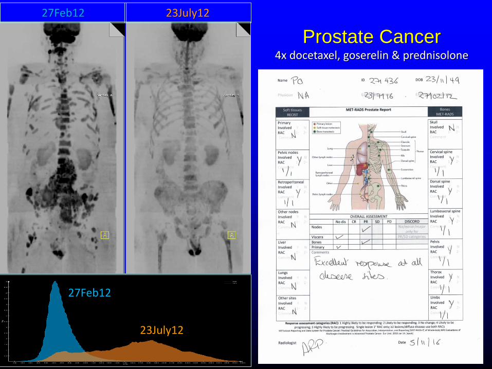
Prostate Cancer4x docetaxel, goserelin & prednisolone
27Feb12 23July12
27Feb12
23July12

Hamstra DA, et al, J Clin Oncol 2007: 25:4104-4109
Biological processes involved in therapy
induced changes in DWI
27Feb12
23July12
1500 µm2/s
95 centile of Ex1
Syngo.via Frontier MR Total Tumor Load software; Siemens Healthineers
mCNPC4x docetaxel, goserelin & prednisolone
No Rx effect
Highly likely response
Likely response
95%

Take home messages
1. The Bone Scan has had its time
2. WB-MRI with DWI is emerging
3. METRADS is quite time consuming and a challenge for wide-spread use
4. New tracers (68Ga PSMA) PET-CT are emerging, role in diagnosis and follow-up has to be established

FUTURE1. For nodes nano (Combidex) MRI
2. For bone and local recurrence; 68Ga PSMA and/or WB-MRI (incl. DWI)
METRADS: https://youtu.be/kDZjmERFFuk