supplement to produced under an educational grant from...
TRANSCRIPT

November 2007
Supplement to Produced under an educational grant from Medtronic CardioVascular.
Evolving Techniques
for Improving Outcomes in
ThoracicAortic
DiseasePitfalls in Pre-Case Planning for Stent Grafting
Dynamic ECG-Gated CTA of the Thoracic Aorta
Pushing the Envelope With Complex TEVAR
Coverage of the Left Subclavian Artery During TEVAR
Endovascular Treatment of Aortic Dissections
Traumatic Thoracic Aortic Transection
Establishing an Acute Aortic Treatment Center
ThoracicAortic
Disease
Evolving Techniques
for Improving Outcomes in

3 Pitfalls in Pre-Case Planning for Thoracic Stent GraftingWhat to consider to ensure clinical success.BY W. ANTHONY LEE, MD
7 Dynamic ECG-Gated CTA of the Thoracic AortaInsights into using 4D imaging when designing the next-generationthoracic endografts. BY AARON M. FISCHMAN, MD, AND ROBERT A. LOOKSTEIN, MD
11 Pushing the Envelope With Complex TEVARLessons learned, creative solutions, and new developments. BY FRANK J. CRIADO, MD
19 Coverage of the Left Subclavian Artery During TEVARHow to predict and prevent neurological complications.BY RACHEL CLOUGH, MRCS (ENG); EITAN HELDENBERG, MD;MOHAMAD HAMADY, FRCR; AND NICK CHESHIRE, MD, FRCS
22 Endovascular Treatment of Aortic DissectionsProvision of evidence through trial data.BY MATT THOMPSON, MD, FRCS; DAVID SAYER, MB BS;IAN LOFTUS, MD, FRCS; AND ROB MORGAN, FRCR
25 Traumatic Thoracic Aortic TransectionEarly outcomes favor endovascular repair over open repair.BY GALE L. TANG, MD, AND MARK K. ESKANDARI, MD
28 Establishing an Acute Aortic Treatment CenterRapid patient transportation, diagnosis, and introduction of therapyby a dedicated multidisciplinary team can reduce the significantmortality of acute aortic syndromes. BY ALAN B. LUMSDEN, MD, DEBRA J. CRAWFORD, RN;ERIC K. PEDEN, MD; BRYAN T. CROFT, MBA; MICHAEL J. REARDON, MD; JEFF E. KALINA, MD; FAISAL N.MASUD, MD; AND KRISTOFER M. CHARLTON-OUW, MD, FACS
Cover image courtesy of Robert A. Lookstein, MD.
ThoracicAortic Disease
Evolving Techniques for
Improving Outcomes in
ThoracicAortic Disease
Evolving Techniques for
Improving Outcomes in

NOVEMBER 2007 I SUPPLEMENT TO ENDOVASCULAR TODAY I 3
Pitfalls in Pre-Case Planningfor Thoracic Stent GraftingWhat to consider to ensure clinical success.
BY W. ANTHONY LEE, MD
Accurate preoperative assessment is critical to theearly and late success of thoracic endovascularaortic repair (TEVAR) (Figure 1). It can be saidthat 90% of the “battle” is won or lost before
stepping into the operating room. Although repair of anuncomplicated mid-descending thoracic aneurysm is fairlystraightforward, most thoracic pathologies lie close to thearch vessels proximally and/or the mesenteric vessels dis-tally. Meticulous attention to detail and proficiency inadvanced endovascular skills are required to safely com-plete these procedures and avoid the myriad of potentialpitfalls that can lead to lethal complications.
Although experience with abdominal endografts is use-ful, TEVAR is sufficiently different due to the extreme tor-tuosity of the thoracic aorta that is not easily correctedwith stiff wires, the greater hemodynamic forces in thearch, the remote location of the pathology, and the signifi-cantly increased risk of iatrogenic dissection and aorticinjury that is not readily surgically accessible (Figure 2).
Even though the principles outlined in this article maybe broadly applied to the repair of a variety of thoracicpathologies, certain problems, such as dissections andtraumatic transections, require special considerations thatare beyond the scope of the present discussion. It shouldbe noted that the only currently approved indication forTEVAR is for degenerative aortic aneurysms with at least a2-cm landing zone distal to the left common carotidartery and proximal to the celiac artery.
PITFALLS AND HOW TO AVOID THEMImaging
The importance of proper cross-sectional imaging can-not be overemphasized. Although this may be with eitherCT or MR imaging, the gold standard is CTA. The large vol-umetric data sets currently available with 32- and 64-multi-slice detector scanners can be rendered into a variety of 3Dreconstruction formats and enable unprecedented mor-phologic and dimensional anatomic analysis. Two suchpostprocessing systems include the TeraRecon AquariusWorkstation (San Mateo, CA) and the M2S (West Lebanon,
NH). Today, there is almost no role for invasive, convention-al angiography. Use of multiplanar reformations and center-line measurements have become almost routine in the pre-operative assessment and planning for TEVAR. Having saidthat, “garbage in, garbage out,” and nothing can compen-sate for a poorly performed study. The ideal CTA should beacquired with ≤2-mm collimation, uniform arterial opacifi-cation, no venous contamination, and cover from the baseof the neck to the femoral heads.
Device SelectionIt is worth noting that, unlike endovascular abdominal
aortic repairs, when sizing for thoracic repairs, the centerlinemeasurements tend to underestimate the actual path thatthe device will take. If device lengths are matched exactly tothe centerline analysis, the actual length of aortic coveragetends to be short by 3 to 5 cm. Therefore, the intervention-ist should always select a longer device if on the borderbetween choosing one size shorter or longer and haveextensions available.
Figure 1. Medtronic Talent (Medtronic CardioVascular,
Endovascular Innovations,Santa Rosa,CA) thoracic stent graft
being deployed to repair a distal arch thoracic aneurysm.

4 I SUPPLEMENT TO ENDOVASCULAR TODAY I NOVEMBER 2007
EVOLVING TECHNIQUES IN THORACIC AORTIC DISEASE
Anesthetic TechniqueMost patients with thoracic aortic disease
have multiple cardiovascular comorbidities,and the procedures should be performedwith anesthesia. The importance of main-taining an absolute sterile technique withproper airflow conditions of a formal oper-ating room environment cannot be over-stated to avoid a life-threatening graft infec-tion. TEVAR should be performed undergeneral or regional anesthesia. General anes-thesia has clear benefits with its control ofthe airway, respiration, blood pressure, andpatient movement. However, for coopera-tive patients, regional anesthesia is a viableoption, especially for those with pulmonaryinsufficiency. Despite the obvious torsion,stretching, and movements of the thoracicaorta by the delivery catheter, and the widehemodynamic fluctuations that can occurwith transient proximal balloon occlusion,the patients do not seem to feel this. Forhemodynamic monitoring, a radial arterialcatheter should be routinely placed in theright wrist because left subclavian arterycoverage is required in more than one thirdof cases to gain an adequate proximal land-ing zone.
Posterior Circulation IschemiaFor proximal aortic aneurysms, the anatomy of the verte-
bral arteries and the posterior cerebral circulation should beassessed. This study may be obtained at the same time aswhen the rest of the body is imaged using CTA. In 60% to70% of cases, the left vertebral artery is dominant, and inapproximately 2% to 3% of cases, one of the vertebral arter-ies terminates as the posterior inferior cerebellar artery with-out joining the basilar artery (Figure 3). In these situations,prophylactic revascularization is strongly recommended ifcoverage of the left subclavian artery is planned to prevent apotentially devastating posterior circulation ischemicstroke.1 Patients with a previous left internal mammarycoronary bypass graft should also undergo revascularizationto avoid myocardial ischemia. Only very rarely is sympto-matic upper-extremity ischemia an indication for subclavianartery bypass, which can be performed electively after thethoracic repair.
StrokeStrokes remain one of the most devastating complica-
tions during TEVAR. In most case series, the incidence ofclinically evident cerebrovascular accidents range from 4%
to 5%. This risk seems to be increased inthe subset of patients who require zone 2coverage (extension to left commoncarotid artery). Although unsubstantiated,other anatomic factors that may play a roleinclude the severity of atheromatous dis-ease in the arch and calcifications of theorigins of the great vessels. However, theseare nonmodifiable factors that may be use-ful for preoperative risk stratification and indeciding whether to offer a repair, but if apatient presents with a life-threatening tho-racic aortic problem, these factors wouldnot by themselves be exclusionary. Last,operator technique plays a critical role inminimizing the risk of stroke during TEVAR.These include meticulous guidewire andcatheter hygiene (frequent wiping of thewires, flushing of catheters, avoidance ofbubbles during any injections), minimizingmanipulation of the delivery system nearthe arch, and avoidance of balloon moldingunless clinically indicated to achieve a seal.
Spinal Cord IschemiaPlacement of a prophylactic spinal
drainage catheter is a controversial topicthat is beyond the scope of this article. It should be noted,however, that in the absence of clearly proven risk factors,routine spinal drainage should be instituted except whenthere are obvious contraindications (such as coagulopathyand focal disease, such as a penetrating ulcer that requires<50% thoracic aortic coverage). The risk of serious iatro-
Figure 3. Posterior views of 3D reconstructions of the verte-
brobasilar circulation. Surface shaded model (A), thin maxi-
mum-intensity projection (B). Note the dominant left verte-
bral artery and the diminutive right vertebral artery termi-
nating before joining the basilar artery.
A B
Figure 2. Iatrogenic retro-
grade thoracic aortic dissec-
tion after TEVAR.The patient
was asymptomatic, and the
dissection was discovered on
postoperative CTA.The dis-
section was electively
repaired with ascending aor-
tic and arch replacement with
distal anastomosis directly
onto the endograft (TAG).

genic hemorrhagic complications fromspinal catheter placement has rangedfrom 0% to 3%.2 On the other hand, ifexpectant management is followed,patients should be monitored in anintensive care setting for at least 24hours, with hourly lower-extremitymotor-sensory examinations and amechanism for immediate spinaldrainage available around the clock.
AccessThe beginning of any TEVAR proce-
dure is to gain secure access to the aorta.The best side of entry for the endograftis selected based on preoperative evalu-ation of the access vessels. The currentlycommercially available TAG (Gore &Associates, Flagstaff, AZ) device requiresintroducer sheaths with outer diametersranging from 23 F to 27 F, and two otherthoracic endograft systems that areundergoing premarket approval process(TX2, Cook Medical, Bloomington, IN;Talent Thoracic, Medtronic Cardio-Vascular, Santa Rosa, CA) have similarprofiles. There is a relatively higher pro-portion of women among patients who present with tho-racic aortic pathologies compared to abdominalaneurysms.3 This subset of patients has smaller iliofemoralvessels that are frequently involved with calcific occlusivedisease. This combination results in approximately 15% ofcases requiring an iliac conduit when the access vessels donot allow safe insertion of the delivery catheter.4 This con-duit is constructed through a lower-quadrant retroperi-toneal approach with a 10-mm prosthetic tube graft anas-tomosed to the distal common iliac artery in an end-to-sidemanner (Figure 4). At the conclusion of the procedure, thisconduit may be either (1)simply transected at its base,(2) left long and tuckedunderneath the inguinal liga-ment to be reaccessed in thefuture, or (3) converted as aniliofemoral bypass if thefemoral pulse is weak. Thethreshold for performing aniliac conduit should be low.Iliac injuries, ranging fromasymptomatic iliac dissectionto life-threatening iliac rup-tures, are the single most pre-
ventable life-threatening complications dur-ing endovascular aortic repair. Often, in thetime it takes to perform all the adjunctivetechniques, such as angioplasties and serialdilations to manage a compromised accessvessel, an iliac conduit can be easily per-formed in most instances. It should be keptin mind that even if the delivery catheter orsheath were to be eventually advanced intothe aorta, this represents only half the battle.A significant proportion of iliac injuries actu-ally occur during retraction of the sheathafter the device is deployed and can have asignificant adverse impact on an otherwisesuccessful thoracic repair.
Lesion LocalizationOccasionally, the disease may not be easily
seen on the aortogram if the aneurysm islined with thrombus or if a penetrating ulceris overlying the aortic lumen. In these situa-tions, accuracy of the preoperative measure-ments in determining the location of thelesion relative to pertinent vessels or otherradiographically visible structures is criticalbecause accurate placement of the endograftis almost entirely dependent on them.
Alternatively, adjunctive imaging techniques, such asintravascular ultrasound, may be helpful in localizing thelesion.
Aortic ArchFor accurate positioning of the endograft near the aortic
arch, the origins of the great vessels must be clearly visual-ized. In cases of marginal proximal landing zone, critical mil-limeters of seal may be lost by improper localization of theleft common carotid artery. Simple widening of the aorticarch using a left anterior oblique projection may not be
NOVEMBER 2007 I SUPPLEMENT TO ENDOVASCULAR TODAY I 5
Figure 4. The iliac conduit. A
Dacron tube graft is anasto-
mosed to the distal common
iliac artery and brought out
through a separate incision at
the level of the femoral artery
(A).This is directly accessed
using a standard Seldinger
technique to introduce the
delivery sheath (B).
A
B
Figure 5. The concept of avoiding the apex of the arch in the deployment of a thoracic endo-
graft.The endograft being deployed either in the proximal arch, even with the left subclavian
artery being covered, or distal to the apex to land the device within a straight segment of the
aorta (A, B). A suboptimal deployment at the apex of the arch (C).
BA C

6 I SUPPLEMENT TO ENDOVASCULAR TODAY I NOVEMBER 2007
EVOLVING TECHNIQUES IN THORACIC AORTIC DISEASE
sufficient to achieve maximum separation of the great ves-sels because of the complex and unpredictable distortionsthat are typically present in these aortas. Therefore, the 3Dplane of the proximal delimiting artery relative to its adja-cent vessels should be carefully assessed so that the optimalorthogonal projection of the image intensifier can be deter-mined. As a matter of technique, the apex of the archremains the Achilles’ heel of TEVAR. All devices have a min-imum radius of curvature below which the device fails toconform along the inner convexity of the arch. This malap-position can lead to type IA endoleaks and, rarely, devicecollapse.5 Therefore, if there is an adequate length of non-aneurysmal proximal aorta, the endograft should bedeployed sufficiently proximal to the apex of the arch, evenif the left subclavian artery must be covered, or entirely dis-tal (“downslope”) to it so as to land the device in a parallelsegment of the aorta (Figure 5).
Distal Thoracic AortaThe distal limit of TEVAR is the celiac artery. It is not
uncommon for the mesenteric vessels to originate at ananterolateral angle and a full 90º lateral projection, as is typi-cally performed to visualize the mesenteric arteries, may notbe optimal. Similar to optimal imaging of the arch vessel, thebest orientation of the image intensifier can be determinedon the axial images of the preoperative CT scan.
Aortic CoverageThe instructions for use for a particular device notwith-
standing, 3 cm is the minimum length of proximal or distalneck that should be used if anatomically possible, and 5 cmshould be the minimum device-to-device overlap length.The interventionist should always err on the side of coveringmore than less of the thoracic aorta. For proximal lesionsnear the arch, the centerline length may not be relevant indetermining whether a seal is likely to be achieved. Forexample, in fusiform lesions, the limiting factor is the lengthof apposition along the lesser curve, and in saccularaneurysms or penetrating ulcers, with defects that may besituated anywhere along the circumference of the aorticlumen, the actual length of graft coverage from its mostproximal extent to the start of the aortic defect is the rele-vant parameter.
Thoracoabdominal TortuosityThoracic aortic aneurysms may present with at least three
tandem segments of significant tortuosity—the abdominalaorta, the distal thoracic aorta, and the arch. Pushability islost after passage of each successive segment due to the seri-al frictional resistance and the noncoaxial vector forcestransmitted along the delivery catheter. Although, in theory,a stiffer guidewire may remedy this situation, even the
stiffest wires available today (eg, Lunderquist, Cook Medical)are inadequate in certain cases, and the only option is to usea transbrachiofemoral wire (Figure 6).
CONCLUSIONSafe performance of TEVAR requires skill sets learned
from abdominal endovascular aortic repair and additionaltechniques that are unique to the treatment of the thoracicaorta due to its remote location, greater tortuosity, largerand stiffer delivery catheters, and the more severe hemody-namic conditions in which these devices must be deployed.However, meticulous attention to detail and careful pre-operative planning can avert most technical pitfalls andresult in a successful repair. ■
W. Anthony Lee, MD, is Associate Professor of Surgery,Division of Vascular Surgery and Endovascular Therapy,University of Florida, in Gainesville, Florida. He has disclosedthat he is a paid consultant to Cook and Medtronic. Dr. Leemay be reached at (352) 273-5484;[email protected].
1. Feezor RJ, Martin TD, Hess PJ, et al. Risk factors for perioperative stroke during thoracicendovascular aortic repairs (TEVAR). J Endovasc Ther. 2007;14:568-573.2. Weaver KD, Wiseman DB, Farber M, et al. Complications of lumbar drainage after thoracoabdom-inal aortic aneurysm repair. J Vasc Surg. 2001;34:623-627.3. Feezor RJ, Huber TS, Martin TD, et al. Perioperative differences between endovascular repair ofthoracic and abdominal aortic diseases. J Vasc Surg. 2007;45:86-89.4. Makaroun MS, Dillavou ED, Kee ST, et al. Endovascular treatment of thoracic aortic aneurysms:results of the phase II multicenter trial of the GORE TAG thoracic endoprosthesis. J Vasc Surg.2005;41:1-9.5. Steinbauer MG, Stehr A, Pfister K, et al. Endovascular repair of proximal endograft collapse aftertreatment for thoracic aortic disease. J Vasc Surg. 2006;43:609-612.
Figure 6. The transbrachiofemoral wire technique. A
guidewire is introduced from the right brachial artery over a
long guide catheter or sheath and snared from a femoral
approach (A).The thoracic endograft is advanced over the
guidewire applying firm tension at both ends (B). Having two
points of fixation such as these provides a stronger rail than
even the most stiff guidewire can provide.The long guide
sheath (55 cm) can be used to inject contrast for control
angiography during the deployment (C).
BA
C

NOVEMBER 2007 I SUPPLEMENT TO ENDOVASCULAR TODAY I 7
Dynamic ECG-GatedCTA of the Thoracic AortaInsights into using 4D imaging when designing the next-generation thoracic endografts.
BY AARON M. FISCHMAN, MD, AND ROBERT A. LOOKSTEIN, MD
Surgical repair of thoracic aortic disease traditionallyhas been associated with high perioperative mortali-ty and an extensive recovery period. Although longconsidered the gold standard, there are significant
risks and morbidity associated with thoracic aortic surgery,including infection, pulmonary embolus, extended hospitalcourse, and spinal cord ischemia.1 Due to advances inendovascular devices and techniques, thoracic endovascularrepair procedures (TEVAR) are becoming a promising alter-native.2 Endovascular repair has its own unique challenges—particularly device endoleaks, stent graft misplacement, sub-sequent migration and fracture, aortic perforation, and iliacartery trauma (Figures 1 and 2).3
Determining which patients are suitable candidates forendovascular therapy is critical to procedural success and tothe reduction of postoperative complications.4 TEVAR pro-cedures have been shown to be associated with greatercomplications than endovascular abdominal aortic repair(EVAR).5 Because of this increased risk, preoperative imagingof the thoracic aorta is of paramount importance inimproving outcomes in these patients.
THE GOLD STANDARD?Digital subtraction angiography remains the gold stan-
dard for arterial imaging, but CT is increasingly utilized as anoninvasive alternative. The use of CTA has dramaticallyfacilitated device sizing6 and can also predict problems suchas migration and endoleaks when evaluating the proximaland distal seal zones of an endograft.7
Even with 3D reconstruction of these data sets, limita-tions still exist. In particular, the acquired data are notdynamic and only evaluate one point in the cardiac cycle.Currently, measurements for stent grafts are based onanatomic criteria using data from 2D/3D CTA, which hasbeen criticized because it only provides a single temporalmeasurement and does not account for aortic walldynamics and volume changes.8 Due to its anatomicalposition, the thoracic aorta is particularly predisposed tohave wall motion throughout the cardiac cycle.9 Thephase of the cardiac cycle in which aortic diameter is
measured on traditional CTA is unknown. Utilizing nongat-ed CTA for preoperative imaging results in inconsistentstent graft sizing.4 ECG gating limits motion artifact andallows for optimal selection of the appropriate reconstruc-tion interval.
4D IMAGINGUnderstanding the dynamic changes in aortic volume
may offer improvements in patient selection and reducepostoperative complications. Understanding the dynamicchanges in aortic volume may offer improvements inpatient selection and reduce postoperative complications.Previous studies have provided insight regarding the dynam-ic changes in the aorta using techniques such as intravascu-lar ultrasound and MRA.10-12 4D CTA, originally used toquantify wall motion and cardiac chamber changes such asejection fraction, has more recently been praised for its highspatial resolution in comparison to other modalities and its
Figure 1. A 43-year-old man sustained blunt trauma to the
descending thoracic aorta during a high-speed motor vehicle
crash, which resulted in thoracic aortic disruption. He subse-
quently underwent TEVAR.The 3D and maximum-intensity
projection images from 3-month follow-up CTA show col-
lapse and infolding of the stent graft.
(Courtesy of Irina Oyfe.)

feasibility in being able to measure diameter in cross sec-tion and area changes in a dynamic aorta.8
4D CTA: SINGLE-CENTER EXPERIENCEBetween March 2006 and January 2007, our imaging lab
studied 40 patients (age range, 22-81 years) with a clinicaldiagnosis of acute aortic syndrome. These patients under-went ECG-gated 4D CTA of the thoracic aorta on a 64-sliceSiemens Somatom Sensation 64 (Siemens Medical SolutionsUSA, Inc., Malvern, PA).
The entire aorta from the heart to the iliac bifurcationwas imaged during a single breath hold phase of 20 sec-onds. The CTA protocol used a 1.2-mm collimation X 32detector array, a pitch of .2, and radiation exposureparameters set at 140 kV and CTDI of 20.10 mGy. Foreach patient, intravascular nonionic contrast (100 mL,Isovue 370, Bracco Diagnostics, Princeton, NJ) followed bya 50-mL saline chaser bolus, was injected at a flow rate of4 mL/s. The scan started with bolus-triggering softwarewith a threshold of80 HU over baseline,the ROI in theascending aorta, anda rotation speed of330 ms. ECG-trig-gered retrospectivereconstructions werecreated at 10 equi-distant time points(0% to 90%) over theR-R cardiac cycle. Nomedication was usedto stabilize ordecrease thepatients’ heart rates.
Data AnalysisThe data sets (section thickness, 1.5 mm; increment, 1
mm) from each patient were loaded into a dedicated car-diovascular 3D/4D workstation (Aquarius WS, TeraRecon,Inc., San Mateo, CA) and postprocessed. The volume andlength of the thoracic aorta for each patient, defined fromthe level of the aortic root to the ostium of the celiac artery,was measured during all 10 phases of the cardiac cycle. TheTeraRecon workstation allows the user to select an angio-graphic volume (Figure 3).
Subsequently, the volumes of both the ascendingaorta (defined from the aortic root to the origin of theleft subclavian artery) and descending aorta (definedfrom the origin of the left subclavian artery to the celiacartery) were measured individually. Maximum and mini-mum volumes were obtained from data obtained ateach point during the cardiac cycle. The absolute vol-ume change was used to calculate a percentage changein each aorta. These data were compiled for the totalthoracic aorta and both the ascending and descendingportions.
Preliminary FindingsVolumetric and longitudinal changes were observed for
every patient. All 40 patients demonstrated a rapid expan-sion during early diastole, and a gradual return to baselineduring the remainder of diastole.
Volumetric changes during the cardiac cycle wereobserved for every patient (average, 18.7 mL; range, 6-61.1mL). Every patient demonstrated a rapid volume increase ofthe thoracic aorta (mean, 7.4%; range, 1.6%-18.2%) duringearly diastole and a gradual return to baseline throughoutthe remainder of diastole. Figure 4 demonstrates the totalblood volume of the thoracic aorta during each of the car-diac phases.
Similar findings were observed when the thoracic
8 I SUPPLEMENT TO ENDOVASCULAR TODAY I NOVEMBER 2007
EVOLVING TECHNIQUES IN THORACIC AORTIC DISEASE
Figure 2. Thoracic stent graft migration due to aortic remodel-
ing after TEVAR with the Relay investigational device (Bolton
Medical, Inc., Sunrise, FL). Preoperative image (A). One-month
postoperative image (B). Cranial stent migration of the distal
device with kinking of the distal seal zone 1 year after initial
placement (C).
Figure 3. Volumetric analysis (A) and Centerline tool (B) on an
Aquarius Workstation (TeraRecon, Inc.).
A B
A B C
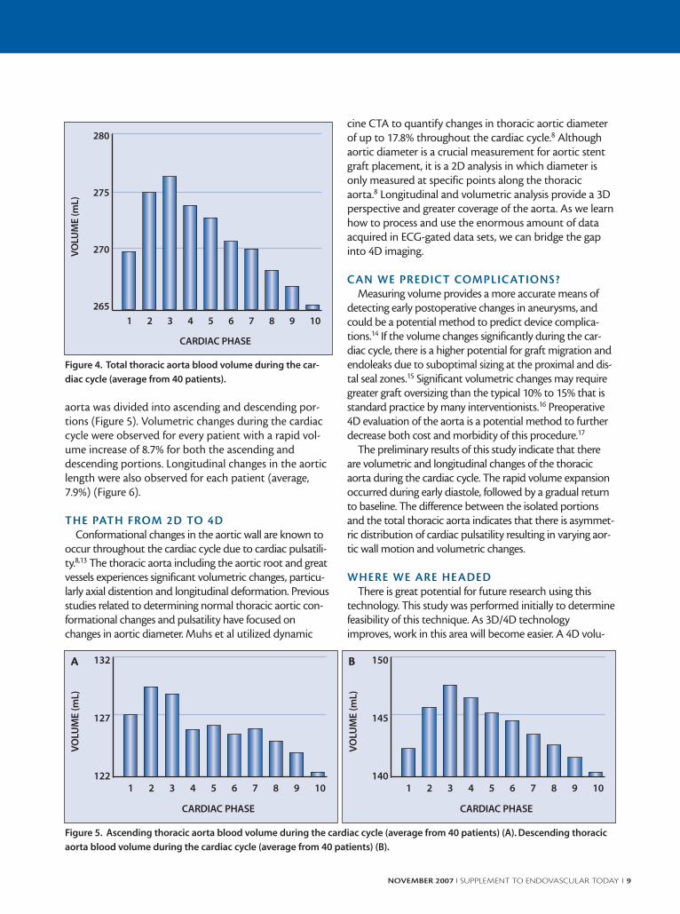
aorta was divided into ascending and descending por-tions (Figure 5). Volumetric changes during the cardiaccycle were observed for every patient with a rapid vol-ume increase of 8.7% for both the ascending anddescending portions. Longitudinal changes in the aorticlength were also observed for each patient (average,7.9%) (Figure 6).
THE PATH FROM 2D TO 4DConformational changes in the aortic wall are known to
occur throughout the cardiac cycle due to cardiac pulsatili-ty.8,13 The thoracic aorta including the aortic root and greatvessels experiences significant volumetric changes, particu-larly axial distention and longitudinal deformation. Previousstudies related to determining normal thoracic aortic con-formational changes and pulsatility have focused onchanges in aortic diameter. Muhs et al utilized dynamic
cine CTA to quantify changes in thoracic aortic diameterof up to 17.8% throughout the cardiac cycle.8 Althoughaortic diameter is a crucial measurement for aortic stentgraft placement, it is a 2D analysis in which diameter isonly measured at specific points along the thoracicaorta.8 Longitudinal and volumetric analysis provide a 3Dperspective and greater coverage of the aorta. As we learnhow to process and use the enormous amount of dataacquired in ECG-gated data sets, we can bridge the gapinto 4D imaging.
CAN WE PREDICT COMPLICATIONS?Measuring volume provides a more accurate means of
detecting early postoperative changes in aneurysms, andcould be a potential method to predict device complica-tions.14 If the volume changes significantly during the car-diac cycle, there is a higher potential for graft migration andendoleaks due to suboptimal sizing at the proximal and dis-tal seal zones.15 Significant volumetric changes may requiregreater graft oversizing than the typical 10% to 15% that isstandard practice by many interventionists.16 Preoperative4D evaluation of the aorta is a potential method to furtherdecrease both cost and morbidity of this procedure.17
The preliminary results of this study indicate that thereare volumetric and longitudinal changes of the thoracicaorta during the cardiac cycle. The rapid volume expansionoccurred during early diastole, followed by a gradual returnto baseline. The difference between the isolated portionsand the total thoracic aorta indicates that there is asymmet-ric distribution of cardiac pulsatility resulting in varying aor-tic wall motion and volumetric changes.
WHERE WE ARE HEADEDThere is great potential for future research using this
technology. This study was performed initially to determinefeasibility of this technique. As 3D/4D technologyimproves, work in this area will become easier. A 4D volu-
NOVEMBER 2007 I SUPPLEMENT TO ENDOVASCULAR TODAY I 9
280
275
270
265
VO
LUM
E (
mL
)
CARDIAC PHASE
1 2 3 4 5 6 7 8 9 10
132
127
122
VO
LUM
E (
mL
)
CARDIAC PHASE
1 2 3 4 5 6 7 8 9 10
150
145
140
VO
LUM
E (
mL
)
CARDIAC PHASE
1 2 3 4 5 6 7 8 9 10
Figure 4. Total thoracic aorta blood volume during the car-
diac cycle (average from 40 patients).
Figure 5. Ascending thoracic aorta blood volume during the cardiac cycle (average from 40 patients) (A). Descending thoracic
aorta blood volume during the cardiac cycle (average from 40 patients) (B).
A B

metric analysis can provide a stronger characterization ofthe relationship between aortic wall motion, compliance,and contractility. It has been shown that the wall stress ofthe thoracic aorta decreases linearly with age.18 Healthypatients with more compliant aortas will likely demon-strate greater volume changes during the cardiac cycle.Although most aneurysmal aortas needing repair (open orendovascular) are diseased with large amounts of athero-sclerotic plaque, great potential exists for providing this 4Devaluation in the preoperative evaluation of healthyyounger patients with acute aortic injury, such as aortictransection and dissection. We believe this technique canhave great impact on the care of patients with acute aorticinjury because the aorta in these patients is subject to thegreatest volumetric changes during the cardiac cycle.19
Potential exists for providing a more durable endovascularrepair for acute traumatic aortic injury for a larger numberof patients in the near future.20,21
This technology may facilitate the development ofdevices that may be used in the ascending and transversearch. The ability to quantify the forces on the arch vesselsduring the cardiac cycle may encourage the development ofbranch vessel technology. This technology may offer theability to identify impending rupture by measuring compli-ance in aneurysmal aortas as a function of volumetricchange. Last, this technology may become a novel tech-nique for surveillance of chronic dissection by being able torecord not only diameter changes, but compliance changesover time.
SUMMARYECG-gated 4D CTA offers a noninvasive modality to
observe and perform volumetric analyses of the thoracicaorta, creating potential to improve endovascular devicedesign and ultimately endovascular repair. By understandinghow wall motion and compliance can be quantified in both
normal and diseased aortas, significant advancements maybe made in the endovascular treatment of thoracic aorticpathology. ■
Aaron M. Fischman, MD, is from the Department ofRadiology at Mount Sinai Medical Center in New York City,New York. He has disclosed that he holds no financial interestin any product or manufacturer mentioned herein. Dr.Fischman may be reached at (212) 241-7409; [email protected].
Robert A. Lookstein, MD, is from the Department ofInterventional Radiology at Mount Sinai Medical Center inNew York City, New York. He has disclosed that he holds nofinancial interest in any product or manufacturer mentionedherein. Dr. Lookstein may be reached at (212) 241-7409;[email protected].
1. Kawaharada N, Morishita K, Kurimoto Y, et al. Spinal cord ischemia after elective endovascularstent-graft repair of the thoracic aorta. Eur J Cardiothorac Surg. 2007;31:998-1003; discussion. 2. Makaroun MS, Dillavou ED, Kee ST, et al. Endovascular treatment of thoracic aortic aneurysms:results of the phase II multicenter trial of the GORE TAG thoracic endoprosthesis. J Vasc Surg.2005;41:1-9.3. Therasse E, Soulez G, Giroux MF, et al. Stent-graft placement for the treatment of thoracic aorticdiseases. Radiographics. 2005;25:157-173.4. Sprouse LR II, Meier GH III, Parent FN, et al. Is three-dimensional computed tomography recon-struction justified before endovascular aortic aneurysm repair? J Vasc Surg. 2004;40:443-447.5. Leurs LJ, Bell R, Degrieck Y, et al. Endovascular treatment of thoracic aortic diseases: combinedexperience from the EUROSTAR and United Kingdom Thoracic Endograft registries. J Vasc Surg.2004;40:670-679; discussion 9-80.6. Parker MV, O’Donnell SD, Chang AS, et al. What imaging studies are necessary for abdominalaortic endograft sizing? A prospective blinded study using conventional computed tomography, aor-tography, and three-dimensional computed tomography. J Vasc Surg. 2005;41:199-205.7. Roubin GD, Dake MD, Semba CP. Current status of three-dimensional spiral CT scanning forimaging the vasculature. Radiol Clin N Am. 1995;33:51-70.8. Muhs BE, Vincken KL, van Prehn J, et al. Dynamic cine-CT angiography for the evaluation of thethoracic aorta: insight in dynamic changes with implications for thoracic endograft treatment. Eur JVasc Endovasc Surg. 2006;32:532-536.9. Feezor RJ, Huber TS, Martin TD, et al. Perioperative differences between endovascular repair ofthoracic and abdominal aortic diseases. J Vasc Surg. 2007;45:86-89.10. Koschyk DH, Meinertz T, Hofmann T, et al. Value of intravascular ultrasound for endovascularstent-graft placement in aortic dissection and aneurysm. J Cardiac Surg. 2003;18:471-477.11. Pereles FS, McCarthy RM, Baskaran V, et al. Thoracic aortic dissection and aneurysm: evalua-tion with nonenhanced true FISP MR angiography in less than 4 minutes. Radiology. 2002;223:270-274.12. Goldfarb JW, Holland AE, Heijstraten FM, et al. Cardiac-synchronized gadolinium-enhanced MRangiography: preliminary experience for the evaluation of the thoracic aorta. Magn Reson Imag.2006;24:241-248.13. Schurink GW, Aarts NJ, Malina M, et al. Pulsatile wall motion and blood pressure in aneurysmswith open and thrombosed endoleaks–comparison of a wall track system and M-mode ultrasoundscanning: an in vitro and animal study. J Vasc Surg. 2000;32:795-803.14. Lookstein RA. Impact of CT angiography on endovascular therapy. Mount Sinai J Med.2003;70:367-374.15. Schurink GW, Aarts NJ, van Baalen JM, et al. Stent attachment site-related endoleakage afterstent graft treatment: an in vitro study of the effects of graft size, stent type, and atherosclerotic wallchanges. J Vasc Surg. 1999;30:658-667.16. Teutelink A, Muhs BE, Vincken KL, et al. Use of dynamic computed tomography to evaluate pre-and postoperative aortic changes in AAA patients undergoing endovascular aneurysm repair. JEndovasc Ther. 2007;14:44-49.17. Holzenbein J, Kretschmer G, Glanzl R, et al. Endovascular AAA treatment: expensive prestige oreconomic alternative? Eur J Vasc Endovasc Surg. 1997;14:265-272.18. Bader H. Dependence of wall stress in the human thoracic aorta on age and pressure. Circ Res.1967;20:354-361.19. Hundley WG, Kitzman DW, Morgan TM, et al. Cardiac cycle-dependent changes in aortic areaand distensibility are reduced in older patients with isolated diastolic heart failure and correlate withexercise intolerance. J Am Coll Cardiol. 2001;38:796-802.20. Kato N, Dake MD, Miller DC, et al. Traumatic thoracic aortic aneurysm: treatment with endovas-cular stent-grafts. Radiology. 1997;205:657-662.21. Semba CP, Kato N, Kee ST, et al. Acute rupture of the descending thoracic aorta: repair withuse of endovascular stent-grafts. J Vasc Interv Radiol. 1997;8:337-342.
10 I SUPPLEMENT TO ENDOVASCULAR TODAY I NOVEMBER 2007
EVOLVING TECHNIQUES IN THORACIC AORTIC DISEASE
405
400
395
390
AO
RT
IC L
EN
GT
H (
mm
)
CARDIAC CYCLE
1 2 3 4 5 6 7 8 9 10
Figure 6. Thoracic aorta length during the cardiac cycle (aver-
age from 40 patients).

NOVEMBER 2007 I SUPPLEMENT TO ENDOVASCULAR TODAY I 11
Pushing the EnvelopeWith Complex TEVARLessons learned, creative solutions, and new developments.
BY FRANK J. CRIADO, MD
Developments in thoracic endovascularaneurysm repair (TEVAR) have now gainedtheir rightful place at the forefront of mod-ern-day cardiovascular medicine.
Advancements with thoracic endograft technologiesand rapidly evolving new techniques have revolution-ized the entire field of thoracic aortic surgery. One les-son that took time and considerable experience to learnwas the necessary clear understanding that—unlikeabdominal aortic aneurysm (AAA) endovascular proce-dures—TEVAR can be more complex and dangerousand, at times, extraordinarily difficult. An importantreason for this difficulty is anatomy, and another is thewide variety of thoracic aortic pathologies being con-sidered for endovascular treatment (Table 1).
Complex TEVAR is a term used to denote the per-formance of these procedures on patients who presentwith one or more anatomy- or disease-related factorsleading to increased technical difficulty and to a higher
risk for intra- and postoperative complications. Suchfactors and conditions are numerous; this article focus-es on some of the most important considerationsaccording to the author’s 10-year TEVAR experiencewith more than 400 procedures.
C A S E S T U D I E SComplex TEVAR can perhaps be best described with
two case examples.
Case 1A 71-year-old man was referred for treatment of a
large juxtasubclavian thoracic aortic aneurysm (TAA)measuring 8.5 cm in diameter (Figure 1). He had a histo-ry of severe aortoiliac occlusive disease and previousbilateral iliac artery stenting, creating a very difficult sit-uation for endovascular access. Treatment of his TAAinvolved placing a single 11.5-cm-long Talent ThoracicStent Graft (Medtronic CardioVascular, EndovascularInnovations, Santa Rosa, CA) with proximal bare springsthat crossed the origin of the left subclavian artery(LSA) but did not exclude the vessel. It was immediatelyrealized that proximal fixation length was not adequate,but access impediments precluded placement of aproximal extension cuff. Postoperatively, a type 1endoleak was identified on CT (Figure 2). Transbrachialcatheterization and coiling of the endoleak nidusproved feasible, resulting in complete thrombosis of theentire TAA sac. However, this outcome proved short-Figure 1. A large juxtasubclavian TAA.
• Aneurysms
• Dissection
• Intramural hematoma
• Penetrating ulcers
• Ductus aneurysms
• Pseudoaneurysms
• Traumatic injuries
• Embologenic plaques
TABLE 1. EXAMPLES OFTHORACIC AORTIC LESIONS

lived because the endoleak recurred 4 months later,with dramatic enlargement of the TAA sac and evidenceof distal migration of the stent graft with diameterexpansion of the neck just beyond the LSA origin.Definitive repair required preliminary transposition/bypass of the left common carotid artery (LCCA) with acrossover right-to-left carotid-carotid bypass and liga-tion of the proximal LCCA (Figure 3), followed 2 weekslater by placement of an access conduit attached to thedistal descending thoracic aorta (Figure 4) via left tho-racotomy that enabled delivery and deployment of anendograft cuff that extended the repair proximally tothe origin of the innominate artery. Complete andsecure exclusion of the TAA was finally obtained. Thepatient recovered and did well after the procedure butsuccumbed to lung cancer 2 years later.
Case 1 CommentaryThoracic aneurysms adjacent to or in the aortic arch
represent a challenge. The most common “mistake”
relates to the operator’s willingness to compromise tosimplify the procedure. It is clear now that there can beno compromise at the time of obtaining a sufficient (2cm) length of proximal neck for fixation and seal if anoptimal and durable result is to be achieved, particular-ly in the region of the aortic arch knuckle.
Transposition/bypass of the LSA and/or the LCCAshould have been considered from the outset. Thispatient’s serious aortoiliac access issues complicated theTEVAR procedure enormously. Use of a descending tho-racic aorta conduit is unusual, but it does represent oneadditional available option. Antegrade transcarotid ortrans-subclavian access would not have worked becauseof the proximal location of the TAA.
Case 2A relatively healthy 61-year-old woman was referred
for treatment of a large ductus (saccular) TAA arisingfrom the distal aortic arch. The lesion was directlyacross the origin of the LSA and a short distance
12 I SUPPLEMENT TO ENDOVASCULAR TODAY I NOVEMBER 2007
EVOLVING TECHNIQUES IN THORACIC AORTIC DISEASE
Figure 2. Angiographic result (A) and postoperative CT image (B) and three-dimensional reconstruction (C) confirming pres-
ence of a significant endoleak.
Figure 3. Crossover right-to-left carotid-carotid retropharyngeal bypass and ligation of LCCA just below anastomosis (arrow).
A B C
A B C

beyond the LCCA (Figure5). The need for endo-graft coverage of thesetwo vessels seemed likely,if not inevitable. Thestrategy consisted of pre-emptive percutaneouscatheterization of theLCCA with retrogradeadvancement of amicropuncture wire intothe ascending aorta,where it was initiallyparked (Figure 6). TEVARinvolved deployment ofa Talent Thoracic StentGraft, resulting in near-complete exclusion ofthe vessel (Figure 7).Restoration of normalpatency and antegradeflow into the LCCArequired placement of an
appropriately sized balloon-expandable stent across theorigin of the artery and protruding (in its proximal one-third) into the lumen of the aortic arch, breaking theendograft seal in a focal area just behind the stent graft(Figure 8). Retrograde insertion (into the carotid artery)of a short 6-F introducer sheath was necessary for thisprocedure. The outcome was excellent, and the patientcontinues to do well 3 years after the intervention.
Case 2 Commentary Arch branch preservation, not debranching, has been
a major focus of ours recently. The technique originatedas a troubleshooting efforton two patients who hadunintentional coverage ofthe LCCA.1 The technicalrequirements to perform ret-rograde stenting of theLCCA and LSA have provedrelatively simple and versa-tile. They have also proved(so far) to be safe anddurable. Although thepotential to create anendoleak from loss of endo-graft apposition in the areaoccupied by the stent deviceis an obvious concern, wehave not yet observed any
such occurrence thus far. The advantages of theseapproaches over the more conventional cervical bypass-es are significant because the patient is spared the needfor preliminary or concomitant additional operations,and they allow the surgeon to resolve it all in one oper-ative session. Patients with thick and very short aorticnecks may not be good candidates for percutaneousretrograde stenting of the LCCA.
COM P L E X T E VARThe most significant issues relate to access, anatomy,
presence of aortic branches, the aortic arch, and certaintreatment indications such as acute aortic dissection.
Access Access is universally acknowledged to constitute the
first crucial step in a TEVAR procedure and is potential-ly a significant impediment. Women comprise >30% ofthe thoracic patient population, contributing to theaccess challenges because of their small femoral and
NOVEMBER 2007 I SUPPLEMENT TO ENDOVASCULAR TODAY I 13
A B
Figure 4. Stump (arrow) left
behind after excision of access
conduit (10-mm Dacron) that
was anastomosed to the side
of the distal descending tho-
racic aorta.
Figure 5. Large ductus arch TAA. Note the relationship with
the LSA and the LCCA.
A B
Figure 6. Retrograde percutaneous catheterization of the LCCA with micropuncture set and
advancement of wire into the ascending aorta (arrows). Note also the presence of the diag-
nostic catheter coming from the left brachial artery sheath.

iliac arteries. Experience has taught us many importantlessons and a few fundamental principles—particularlythat probing the femoral-iliac arteries with Coons dila-tors (Cook Medical, Bloomington, IN) should be a rou-tine procedure whenever the interventionist is in doubtabout the adequacy of the access vessels. The dictum is,“The time to do it is when you think you might needone.” Iliac conduits for endovascular access are impor-tant tools, with reported use in 15% to 30% of TEVARprocedures at the present time (Figure 9). Not surpris-ingly, technical considerations and attention to detailare paramount to achieve the results and to avoid com-plications and pitfalls.2 Although it is a convenient dis-posal technique, partially excising the conduit at theend of the operation can leave a sizeable stump behindthat may be later diagnosed as a pseudoaneurysm, lead-ing to confusion and unnecessary concern (Figure 10).For that reason, an effort should be made to leave assmall a stump as possible. At times, it is best to bringthe graft down under the inguinal ligament and attachit to the common femoral artery as a permanentlyimplanted iliofemoral bypass (Figure 11). Thoracicendograft introduction and delivery through a trans-
carotid approach is an unusual butpotentially useful access option forpatients with difficult aortoiliac arter-ies. The author has had an opportunityto perform this maneuver several timesrecently and has been impressed withthe relative ease and adequacy of thistechnique. The right carotid arterywould appear to be the best choice.The proximal common carotid arterytends to be a large and often normalartery that allows passage of a largesheath without difficulty. However, thisapproach is only workable for endovas-cular treatment of aortic lesions distalto the LSA.
AO RT I C ANATOMY This anatomy must be carefully assessed in every
case, of course, because it can impose significant diffi-culty at the time of endovascular navigation and devicedelivery. Marked S-shaped angulations in the distalthoracic and proximal abdominal aorta can be particu-larly challenging.
The Aortic Arch and Its BranchesThis area deserves our utmost attention. TEVAR
implies dealing with the arch and branches in at least30% of cases.3 Creating an adequate proximal
14 I SUPPLEMENT TO ENDOVASCULAR TODAY I NOVEMBER 2007
EVOLVING TECHNIQUES IN THORACIC AORTIC DISEASE
A B
Figure 7. Coverage of the LCCA origin by the Talent Thoracic Stent Graft.
B
Figure 8. Stenting of the
LCCA origin restored normal
antegrade flow.
A
• Simple coverage-exclusion (LSA)
• Cervical bypasses and transpositions (all)
• Retrograde stenting (LSA and LCCA)
• Retrograde endobranching (potentially all)
• Transthoracic ascending-aorta–based bypasses (all)
TABLE 2. MANAGEMENT OPTIONS FOR AORTICARCH BRANCHES
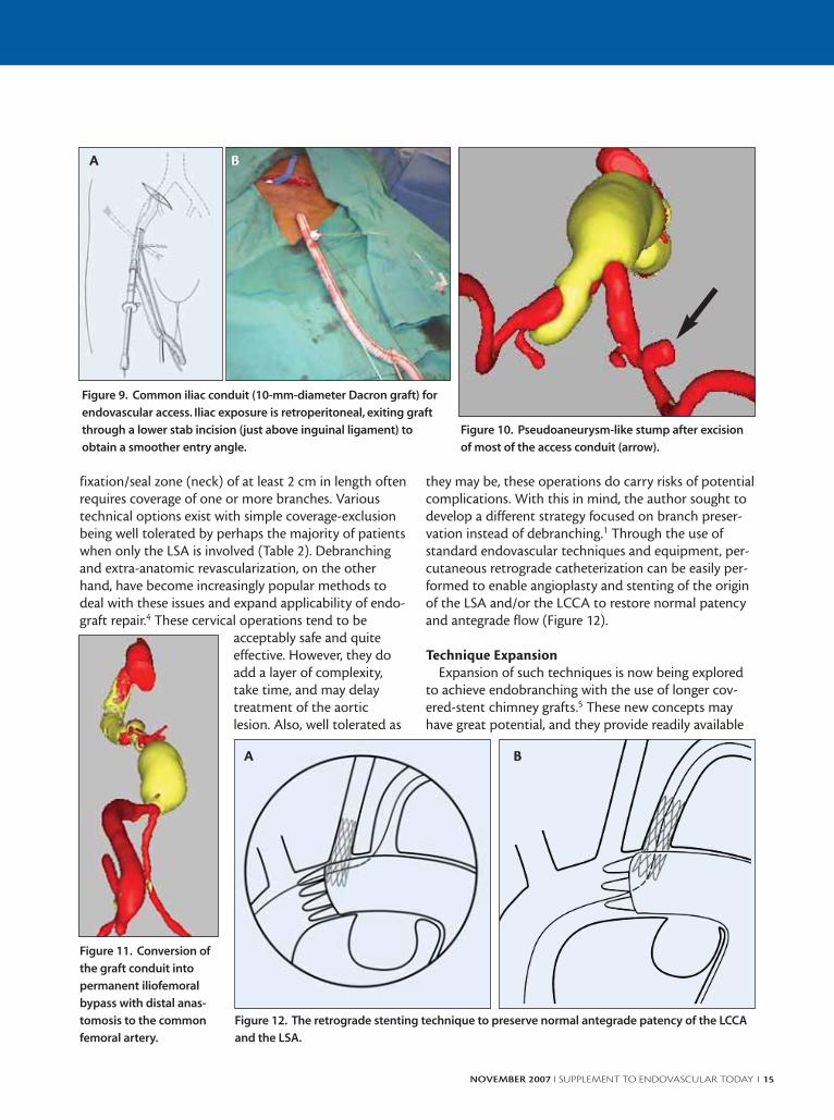
fixation/seal zone (neck) of at least 2 cm in length oftenrequires coverage of one or more branches. Varioustechnical options exist with simple coverage-exclusionbeing well tolerated by perhaps the majority of patientswhen only the LSA is involved (Table 2). Debranchingand extra-anatomic revascularization, on the otherhand, have become increasingly popular methods todeal with these issues and expand applicability of endo-graft repair.4 These cervical operations tend to be
acceptably safe and quiteeffective. However, they doadd a layer of complexity,take time, and may delaytreatment of the aorticlesion. Also, well tolerated as
they may be, these operations do carry risks of potentialcomplications. With this in mind, the author sought todevelop a different strategy focused on branch preser-vation instead of debranching.1 Through the use ofstandard endovascular techniques and equipment, per-cutaneous retrograde catheterization can be easily per-formed to enable angioplasty and stenting of the originof the LSA and/or the LCCA to restore normal patencyand antegrade flow (Figure 12).
Technique Expansion Expansion of such techniques is now being explored
to achieve endobranching with the use of longer cov-ered-stent chimney grafts.5 These new concepts mayhave great potential, and they provide readily available
NOVEMBER 2007 I SUPPLEMENT TO ENDOVASCULAR TODAY I 15
A B
Figure 9. Common iliac conduit (10-mm-diameter Dacron graft) for
endovascular access. Iliac exposure is retroperitoneal, exiting graft
through a lower stab incision (just above inguinal ligament) to
obtain a smoother entry angle.
Figure 10. Pseudoaneurysm-like stump after excision
of most of the access conduit (arrow).
Figure 11. Conversion of
the graft conduit into
permanent iliofemoral
bypass with distal anas-
tomosis to the common
femoral artery.
Figure 12. The retrograde stenting technique to preserve normal antegrade patency of the LCCA
and the LSA.
A B

tools for partial or even total endovascular archrepair—all done in one operative session and sparingthe patient the need for multiple operations (Figure 13).However, many questions remain, including the poten-tial for compression against the aortic wall by the tho-racic endograft, the creation of endoleaks, and graftintegrity issues resulting possibly from interactionbetween the two devices over time. Ultimately, trulybranched endograft technologies may emerge as theideal endovascular solutions for the arch and the viscer-al segment of the aorta. Unfortunately, these technolo-gies are going to be complex and unlikely to becomewidely available for several years.
Total Transthoracic Debranching This procedure is another technical option that can
be used to enable complete endograft relining andrepair of the entire arch. Through a median sternotomyapproach, an ascending aorta-based bypass to theinnominate artery and the LCCA can be relatively easilyperformed because it involves partial (side) clamping ofthe aorta only. Graft branching into the LSA may alsobe possible. For patients who cannot tolerate such anoperation or patients who have anatomical impedi-ments, a totally extrathoracic approach that involvesthe use of the right femoral artery as the inflow sourcecan be considered (Figure 14).
Endograft Conformity at the Arch Knuckle This is another important issue because sharp bends
at the apex of the aortic arch represent an enormouschallenge for currently available devices, which tend notto conform well to such geometries. Aortic wall malap-position along the lesser curve is particularly problem-atic, leading potentially to vessel injury and perforation,
retrograde type A dissection, type 1endoleak, and even endograft compressionand collapse. Some devices are more proneto such problems than others. The surgeonmust strive to avoid these potentially seri-ous complications by not landing at or inthe knuckle area. Instead, proximal fixationshould take place just distal to the bend or(likely) more proximally within the trans-verse arch, which, of course, often impliesthat you must deal with one or morebranches.
The Visceral Segment This area is receiving increasing attention
as the scope of TEVAR continues to grow.Endovascular treatment of juxtaceliac TAA
and thoracoabdominal aortic aneurysm (TAAA) withinvolvement of one or more visceral and renal arteries isbeing reported more often at present. The most com-mon complexity that operators are likely to encounterrelates to TAA lesions that are in close proximity to theceliac artery origin (Figure 15). Deployment precision atthe distal fixation site is paramount and perhaps a littlemore difficult to achieve when compared with theaction at the proximal neck. Some TEVAR systems arecertainly better than others in this regard (Figure 16).Endograft coverage of the celiac artery can be per-formed if necessary, and would appear to be well toler-ated by the majority of patients. However, anecdotal
16 I SUPPLEMENT TO ENDOVASCULAR TODAY I NOVEMBER 2007
EVOLVING TECHNIQUES IN THORACIC AORTIC DISEASE
A B
Figure 13. Chimney-graft endobranching techniques showing two differ-
ent options.
Figure 14. Femoral-based bypasses to achieve total extratho-
racic debranching of the aortic arch.

reports of serious complicationshave brought into appropriatefocus the potential dangers ofsuch a maneuver, with no suremeans today of predicting its safe-ty in an additional case.
The risk of unintentional cover-age of the superior mesentericartery (SMA) is something theauthor finds more worrisome.Covering the SMA would be, ofcourse, catastrophic, especiallywhen excluding the celiac arteryas well. It has become our routineto catheterize the SMA (from thefemoral artery) in such cases andkeep an indwelling catheter with-in the vessel while completing theTEVAR procedure (Figure 16).Having retrograde endovascularaccess to the SMA provides for areadily available pathway for bal-loon dilatation and stenting torestore patency quickly in the faceof encroachment by the thoracic endograft.
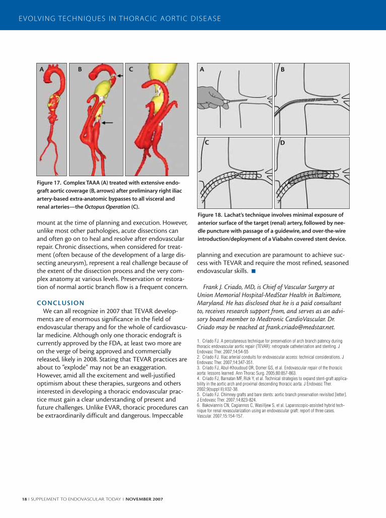
Extra-Anatomic Intra-Abdominal BypassesIntra-abdominal bypass to two or more of the viscer-
al-renal arteries (used with endovascular grafting invarious hybrid combinations) can be formidable opera-tions but are generally considered to represent less of aphysiologic insult than direct surgical thoracoabdomi-
nal repair with prolonged aorticclamping (Figure 17). This maybe especially true for olderpatients with significant medicalcomorbidities and a large TAA.Mario Lachat, MD, of Switzer-land6 has made a significant con-tribution to the field with hisVORTEC (Viabahn OpenRebranching Technique) strategythat promises to simplify (orobviate) the performance ofsome of the most difficult anas-tomoses, particularly those tothe renal arteries that tend to bethe most challenging aspect ofsuch operations (Figure 18).
TEVAR for Type B AorticDissection
TEVAR for type B aortic dissec-tion is, almost by definition, com-plex and high risk both in the set-tings of acute and chronic dissec-
tions. Acute dissection challenges are multiple, oftenleading to higher-than-usual risks of serious complica-tions, including aortic perforation and rupture, retro-grade type A dissection, and issues related to theanatomical complexities derived from the presence of along and spiraling false lumen that needs to be carefullyidentified and separated from the true lumen.Experience and refined endovascular skills are para-
NOVEMBER 2007 I SUPPLEMENT TO ENDOVASCULAR TODAY I 17
Figure 15. Aneurysm formation in the distal
thoracic aorta in close proximity to visceral
vessels.
Figure 16. Juxtaceliac aneurysm (A) treated with the Talent Thoracic Stent Graft landing precisely just above the origin of the
celiac artery (B). Retrograde (transfemoral) catheterization of the SMA performed to provide access pathway for vessel stenting
in case of unintentional coverage by endograft (C, D).
A C DB
mount at the time of planning and execution. However,unlike most other pathologies, acute dissections canand often go on to heal and resolve after endovascularrepair. Chronic dissections, when considered for treat-ment (often because of the development of a large dis-secting aneurysm), represent a real challenge because ofthe extent of the dissection process and the very com-plex anatomy at various levels. Preservation or restora-tion of normal aortic branch flow is a frequent concern.
CO N C L USI O NWe can all recognize in 2007 that TEVAR develop-
ments are of enormous significance in the field ofendovascular therapy and for the whole of cardiovascu-lar medicine. Although only one thoracic endograft iscurrently approved by the FDA, at least two more areon the verge of being approved and commerciallyreleased, likely in 2008. Stating that TEVAR practices areabout to “explode” may not be an exaggeration.However, amid all the excitement and well-justifiedoptimism about these therapies, surgeons and othersinterested in developing a thoracic endovascular prac-tice must gain a clear understanding of present andfuture challenges. Unlike EVAR, thoracic procedures canbe extraordinarily difficult and dangerous. Impeccable
planning and execution are paramount to achieve suc-cess with TEVAR and require the most refined, seasonedendovascular skills. ■
Frank J. Criado, MD, is Chief of Vascular Surgery atUnion Memorial Hospital-MedStar Health in Baltimore,Maryland. He has disclosed that he is a paid consultantto, receives research support from, and serves as an advi-sory board member to Medtronic CardioVascular. Dr.Criado may be reached at [email protected].
1. Criado FJ. A percutaneous technique for preservation of arch branch patency duringthoracic endovascular aortic repair (TEVAR): retrograde catheterization and stenting. JEndovasc Ther. 2007;14:54-552. Criado FJ. Iliac arterial conduits for endovascular access: technical considerations. JEndovasc Ther. 2007;14:347-351.3. Criado FJ, Abul-Khoudoud OR, Domer GS, et al. Endovascular repair of the thoracicaorta: lessons learned. Ann Thorac Surg. 2005;80:857-863.4. Criado FJ, Barnatan MF, Rizk Y, et al. Technical strategies to expand stent-graft applica-bility in the aortic arch and proximal descending thoracic aorta. J Endovasc Ther.2002;9(suppl II):II32-38.5. Criado FJ. Chimney grafts and bare stents: aortic branch preservation revisited [letter].J Endovasc Ther. 2007;14:823-824.6. Bakoviannis CN, Cagiannos C, Wasilljew S, et al. Laparoscopic-assisted hybrid tech-nique for renal revascularization using an endovascular graft: report of three cases.Vascular. 2007;15:154-157.
18 I SUPPLEMENT TO ENDOVASCULAR TODAY I NOVEMBER 2007
EVOLVING TECHNIQUES IN THORACIC AORTIC DISEASE
Figure 17. Complex TAAA (A) treated with extensive endo-
graft aortic coverage (B, arrows) after preliminary right iliac
artery-based extra-anatomic bypasses to all visceral and
renal arteries—the Octopus Operation (C).
A B C
Figure 18. Lachat’s technique involves minimal exposure of
anterior surface of the target (renal) artery, followed by nee-
dle puncture with passage of a guidewire, and over-the-wire
introduction/deployment of a Viabahn covered stent device.
A B
DC

NOVEMBER 2007 I SUPPLEMENT TO ENDOVASCULAR TODAY I 19
Coverage of theLeft Subclavian ArteryDuring TEVARHow to predict and prevent neurological complications.
BY RACHEL CLOUGH, MRCS (ENG); EITAN HELDENBERG, MD; MOHAMAD HAMADY, FRCR;
AND NICK CHESHIRE, MD, FRCS
Thoracic endovascular aortic repair has become aviable option for treating multiple aorticpathologies. Large series document mortalityand morbidity rates that are substantially lower
than those of comparable open repair.1-4
Adequate proximal and distal landing zones are fun-damental to successful endovascular repair, and inabilityto achieve these can predispose to type I and type IIendoleak. Traditional series, such as by Dake et al, andmore recent series (Riesenman et al, Stanley et al, Saitoet al) have suggested that to achieve satisfactory proxi-mal fixation, the landing zone must include at least 20mm of healthy aortic tissue.5-8 With advancements inendovascular devices and user expertise, the indicationsfor thoracic stenting have grown, and the proximallanding zone has advanced to the distal and, in somecases, the proximal, aortic arch. It can be quickly real-ized that a landing zone within this position will neces-sitate coverage of one or more of the aortic arch vessels.This article focuses on the coverage of the left subcla-vian artery and the neurological complications that mayoccur as a consequence of this.
Anatomical studies, such as that of Biglioli et al, revealthe importance of the left subclavian artery in the per-fusion, via the left vertebral artery, of both the spinalcord and the brain (Figure 1).9 Isolated coverage of theleft subclavian artery in the presence of normal arterialanatomy may not necessarily precipitate neurologicalsequelae due to the presence of collateral networksboth within the brain via the circle of Willis and withinthe spinal cord via the contributions of the intercostal,Adamkiewicz, and lumbar arteries to the anterior spinalartery.
Early series, such as those of Hauseggar et al, Burks etal, and Görich et al, published in 2000-2001, wereamong the first to extend the landing zone to within
the proximal aortic arch.10-12 In their combined series ofmore than 30 thoracic stent grafts, there was no inci-dence of neurological complications. At our institution,in our initial series of 14 in which endovascular repairnecessitated coverage of the left subclavian artery, wereported a single embolic intracranial event with fullresolution of symptoms and no occurrence of spinalcord injury or vertebrobasilar insufficiency. Further cen-ters from across Europe revealed similarly encouragingresults.13-17
More recently, large, multicenter series have emerged.The combined series of Thompson et al (containing ourexperience with the Valiant Thoracic Stent Graft[Medtronic CardioVascular, Endovascular Innovations,Santa Rosa, CA]), Fattori et al, Leurs et al, andMakaroun et al, consists of more than 1,000 patients
Figure 1. Extensive collateral network.

20 I SUPPLEMENT TO ENDOVASCULAR TODAY I NOVEMBER 2007
EVOLVING TECHNIQUES IN THORACIC AORTIC DISEASE
and reveals stroke rates of 2.3% to 3.9% and rates ofspinal cord injury of 1.7% to 3.3% (Figure 2).18-21 Therewas little or no mention of the subclavian steal phe-nomenon.
S T RO K EThe etiology of intracranial injury is likely multifactor-
ial. Fattori et al and Thompson et al both showed a sig-nificant association between the coverage of the leftsubclavian artery and the incidence of stroke.18,19 Feezoret al reiterate this finding and, in addition, illustrate thatof the nine strokes that occurred in their series, fiveoccurred in the posterior circulation in individuals whohad not undergone a previous revascularization proce-dure.22
Other series, such as that of Reece et al, suggest thatstent and wire manipulation within the arch may be animportant factor, illustrated by three embolic intracra-nial events within the distribution of the anteriorintracranial circulation in their series.23 Thompson et aland Makaroun et al both showed a clustering of strokesin individuals with a high proximal atherothromboticload, a finding that adds weight to the conclusions ofReece et al.
Feezor et al found that 56% of individuals with astroke during thoracic stent grafting had documentedintraoperative hypotension with a systolic blood pres-sure of <80 mm Hg.22
S P I N AL CO R D I N J U RYOur current series of more than 70 visceral hybrid
repairs serves to illustrate the successful management oflong-segment aortic disease and, in addition, the ever-present risk of spinal cord ischemia.
The registry series of Thompson et al, Fattori et al,and Leurs et al revealed an association between thelength of the aortic segment coverage and the rate ofspinal cord injury. In addition, in the four cases of spinalischemia described by Makaroun et al, two patients hadundergone previous aortobifemoral repairs.21 The pub-lished series to date declare no specific associationbetween coverage of the left subclavian artery and theincidence of spinal cord ischemia, but this artery’s con-tribution to the anterior spinal artery and thus the col-lateral spinal network cannot be disputed.
PR E V E N T I O N O F N E U RO LO G I C AL I N J U RYPre-emptive revascularization of the left subclavian
artery often follows careful preoperative evaluation ofboth the local and intracranial anatomy using one or acombination of CTA, MRA, DSA, or Duplex. Indicationsfor revascularization include an aberrant or dominant
left vertebral artery, a hypoplastic right vertebral artery,a deficient circle of Willis, and current use of the leftinternal mammary artery as a coronary artery bypassgraft.22,23
Revascularization may take the form of either a leftcommon carotid artery to left subclavian artery bypassor left subclavian artery to left common carotid arterytransposition. The latter is frequently described in theliterature with good results. Bypass becomes a moreattractive option if the left vertebral artery takes earlyorigin from the left subclavian artery or when the leftinternal mammary artery is incorporated into a coro-nary bypass grafting procedure. Technically more chal-lenging, bypass requires division of the anterior scalenemuscle and identification of the left subclavian arterymore distally. Compared to proximal covered stents, aproximal bare stent configuration may allow precisedeployment and help to fix the graft in the case of ashort neck (<2 cm) without compromising flowthrough the left subclavian artery.
In addition to revascularization, flow to the left sub-clavian artery may be maintained via the use of baremetal stents for proximal fixation. These devices allowsecure proximal fixation with coverage of the vessel, andtheir use in this location is currently evolving.
Systematic review of the two procedures reveals simi-lar rates of mortality and morbidity between the twotechniques with recognized complications includingnerve injury, stroke, lymphatic damage, and hematomaformation.24
Methods for preventing spinal cord injury duringendovascular thoracic aortic repair focus on maintain-ing an adequate arterial perfusion pressure with anappreciation of its relationship to the cerebrospinalfluid (CSF) pressure; the length of aortic coverage can-not be easily manipulated. Weigang et al institute neu-
Figure 2. Combined registry data by complication type.

rophysiologic monitoring and CSF pressure monitoringfor all elective cases.25 They have shown that with anincrease in CSF pressure, there is also a change in theevoked potentials and that institution of CSF drainageat this point can prevent spinal cord injury.
CO N C L USI O N Our current series of more than 200 thoracic endo-
grafts serves to illustrate both the success of this thera-peutic intervention and the growing evidence that cov-erage of the left subclavian artery is associated with ahigher incidence of neurological injury. Furthermore, wehave seen that coverage of the left subclavian arterywithout its ligation may precipitate complex typeIa/type II endoleak within the proximal region of thegraft.
The concept of total endovascular repair is evolving,and the literature reveals promising early reports of theuse of branch and fenestrated grafts within thisanatomical location.8,26-28
With relation to coverage of the left subclavian arteryand the incidence of neurological injury, it is clear thatthere is an association, but whether this relationship iscausal or purely a function of more extensive diseaseremains to be elucidated. ■
Rachel Clough, MRCS (Eng), is from the Vascular Unit,Imperial Healthcare, St. Mary’s Campus, London, England.She has disclosed that she holds no financial interest in anyproduct or manufacturer mentioned herein. Dr. Cloughmay be reached at +011 44 77364 5486; [email protected].
Eitan Heldenberg, MD, is a consultant vascular surgeonwith the Vascular Unit, Imperial Healthcare, St. Mary’sCampus, London, England. He has disclosed that he holdsno financial interest in any product or manufacturer men-tioned herein. Dr. Heldenberg may be reached at [email protected].
Mohamad Hamady, FRCR, is from the Department ofInterventional Radiology, Imperial Healthcare, St. Mary’sCampus, London, England. He has disclosed that he holdsno financial interest in any product or manufacturer men-tioned herein. Dr. Hamady may be reached at +011 44207 886 2282; [email protected].
Nick Cheshire, MD, FRCS, is Professor, Vascular Unit,Imperial Healthcare, St. Mary’s Campus, London, England.He has disclosed that his department receives researchfunding from Medtronic. Professor Cheshire may bereached at +011 44 207 886 1086; [email protected].
1. Brandt M, Hussel K, Walluscheck KP, et al. Stent-graft repair versus open surgery forthe descending aorta: a case-control study. J Endovasc Ther. 2004;11:535-538.
2. Schoder M, Grabenwöger M, Hölzenbein T, et al. Endovascular repair of the thoracicaorta necessitating anchoring of the stent graft across the arch vessels. J ThoracCardiovasc Surg. 2006;131:380-387.3. Najibi S, Terramani TT, Weiss VJ, et al. Endoluminal versus open treatment of descend-ing thoracic aortic aneurysms. J Vasc Surg. 2002;36:732-737.4. Weigang E, Hartert M, von Samson P, et al. Thoracoabdominal aortic aneurysm repair:interplay of spinal cord protecting modalities. Eur J Vasc Endovasc Surg. 2005;30:624-631.5. Dake MD, Miller DC, Mitchell RS, et al. The “first generation” of endovascular stent-grafts for patients with aneurysms of the descending thoracic aorta. J Thorac CardiovascSurg. 1998;116:689-703;discussion 703-704.6. Riesenman PJ, Farber MA, Mendes RR, et al. Coverage of the left subclavian artery dur-ing thoracic endovascular aortic repair. J Vasc Surg. 2007;45:90-94;discussion 94-95.7. Stanley BM, Semmens JB, Lawrence-Brown MM, et al. Fenestration in endovasculargrafts for aortic aneurysm repair: new horizons for preserving blood flow in branch ves-sels. J Endovasc Ther. 2001;8:16-24.8. Saito N, Kimura T, Odashiro K, et al. Feasibility of the Inoue single-branched stent-graftimplantation for thoracic aortic aneurysm or dissection involving the left subclavian artery:short- to medium-term results in 17 patients. J Vasc Surg. 2005;41:206-212;discussion212.9. Biglioli P, Roberto M, Cannata A, et al. Upper and lower spinal cord blood supply: thecontinuity of the anterior spinal artery and the relevance of the lumbar arteries. J ThoracCardiovasc Surg. 2004;127:1188-1192.10. Hausegger KA, Oberwalder P, Tiesenhausen K et al. Intentional left subclavian arteryocclusion by thoracic aortic stent-grafts without surgical transposition. J Endovasc Ther.2001;8:472-476.11. Burks JA Jr, Faries PL, Gravereaux EC, et al. Endovascular repair of thoracic aorticaneurysms: stent-graft fixation across the aortic arch vessels. Ann Vasc Surg. 2002;16:24-28.12. Görich J, Asquan Y, Seifarth H, et al. Initial experience with intentional stent-graft cov-erage of the subclavian artery during endovascular thoracic aortic repairs. J Endovasc Ther.2002;9(suppl2):II39-II43.13. Tiesenhausen K, Hausegger KA, Oberwalder P, et al. Left subclavian artery manage-ment in endovascular repair of thoracic aortic aneurysms and aortic dissections. J CardSurg. 2003;18:429-435.14. Lambrechts D, Casselman F, Schroeyers P, et al. Endovascular treatment of thedescending thoracic aorta. Eur J Vasc Endovasc Surg. 2003;26:437-444.15. Sunder-Plassmann L, Scharrer-Pamler R, Liewald F, et al. Endovascular exclusion ofthoracic aortic aneurysms: mid-term results of elective treatment and in contained rupture.J Card Surg. 2003;18:367-374.16. Rehders TC, Petzsch M, Ince H, et al. Intentional occlusion of the left subclavian arteryduring stent-graft implantation in the thoracic aorta: risk and relevance. J Endovasc Ther.2004;11:659-666.17. Caronno R, Piffaretti G, Tozzi M, et al. Intentional coverage of the left subclavian arteryduring endovascular stent graft repair for thoracic aortic disease. Surg Endosc.2006;20:915-918.18. Thompson M, Ivaz S, Cheshire N, et al. Early results of endovascular treatment of thethoracic aorta using the valiant endograft. Cardiovasc Intervent Radiol. 2007;30:1130-1138.19. Fattori R, Nienaber CA, Rousseau H, et al. Results of endovascular repair of the tho-racic aorta with the Talent Thoracic stent graft: the Talent Thoracic Retrospective Registry. JThorac Cardiovasc Surg. 2006;132:332-339.20. Leurs LJ, Bell R, Degrieck Y, et al. Endovascular treatment of thoracic aortic diseases:combined experience from the EUROSTAR and United Kingdom Thoracic Endograft reg-istries. J Vasc Surg. 2004;40:670-679;discussion 679-680.21. Makaroun MS, Dillavou ED, Kee ST, et al. Endovascular treatment of thoracic aorticaneurysms: results of the phase II multicenter trial of the GORE TAG thoracic endoprosthe-sis. J Vasc Surg. 2005;41:1-9.22. Feezor RJ, Martin TD, Hess PJ, et al. Risk factors for perioperative stroke during tho-racic endovascular aortic repairs (TEVAR). J Endovasc Ther. 2007;14:568-573.23. Reece TB, Gazoni LM, Cherry KJ, et al. Reevaluating the need for left subclavian arteryrevascularization with thoracic endovascular aortic repair. Ann Thorac Surg. 2007;84:1201-1205;discussion 1205.24. Cina CS, Safar HA, Lagana A, et al. Subclavian carotid transposition and bypass graft-ing: consecutive cohort study and systematic review. J Vasc Surg. 2002;35:422-429.25. Weigang E, Hartert M, Siegenthaler MP, et al. Perioperative management to improveneurologic outcome in thoracic or thoracoabdominal aortic stent-grafting. Ann ThoracSurg. 2006;82:1679-1687.26. Inoue K, Hosokawa H, Iwase T, et al. Aortic arch reconstruction by transluminallyplaced endovascular branched stent graft. Circulation. 1999;100(suppl):II316-II321.27. McWilliams RG, Murphy M, Hartley D, et al. In situ stent-graft fenestration to preservethe left subclavian artery. J Endovasc Ther. 2004;11:170-174.28. Peterson BG, Resnick SA, Morasch MD, et al. Aortic arch vessel stenting: a single-center experience using cerebral protection. Arch Surg. 2006;141:560-563;discussion563-564.
NOVEMBER 2007 I SUPPLEMENT TO ENDOVASCULAR TODAY I 21

22 I SUPPLEMENT TO ENDOVASCULAR TODAY I NOVEMBER 2007
EVOLVING TECHNIQUES IN THORACIC AORTIC DISEASE
Endovascular Treatmentof Aortic DissectionsProvision of evidence through trial data.
BY MATT THOMPSON, MD, FRCS; DAVID SAYER, MB BS; IAN LOFTUS, MD, FRCS;
AND ROB MORGAN, FRCR
Endovascular techniques have been enthusiasti-cally applied to the thoracic aorta during thelast decade, and in many diseases, endovascularsurgery has gained preference over conventional
open procedures. This change in clinical practice hasbeen driven by patient and physician preference, thebelief that modern technology can improve medicalpractice, the evolution of most surgical procedures toencompass minimally invasive alternatives, commercialdrivers, and clinical outcome data. Interestingly, endo-vascular repair of the thoracic aorta has become anestablished treatment modality with a relatively poorevidence base, because there have been none of the tra-ditional randomized trials that often accompany theintroduction of new therapies.
There are good explanations for the lack of random-ized data when the clinical outcomes for pathologieswith established indications for treatment are comparedbetween endovascular procedures and open thoracicsurgery.1 Case series and registry data would appear toshow that the endovascular procedures offer significant-ly lower mortality, morbidity, and paraplegia rates whencompared to open thoracic repair of thoracic aneurysmsor acute dissections.2 The IRAD registry, an independentregistry of patients with dissections, has provided somedata regarding the long-term results of acute type B dis-sections.3 The lack of randomized trials does, however,pose a problem for endovascular techniques becausemuch of the evidence base is composed of small retro-spective series and registries that do not offer sufficientdetail to facilitate the subgroup analysis that is manda-tory to refine indications for treatment. This situation isnot helped by the stringent inclusion and exclusion cri-teria of trials that are designed for regulatory purposesbecause they often do not reflect the types of lesionsencountered in day-to-day clinical practice.
As a result of the problems encountered with thequality and quantity of the published literature, it hasbecome difficult to determine the outcome of endovas-
cular repair for differing indications within the thoracicaorta. These challenges are of particular relevance tothoracic aortic dissections due to their differing presen-tations, differing indications for treatment, and varyingoutcomes, which often reflect the initial presentation.Progress in treating thoracic dissections can only bemade by improving the evidence base. Many clinicianshave suggested that this process should be facilitatedby the endovascular industry providing “not-for-profit”support of clinical trials and registries.4 This articlebriefly reviews the current issues regarding endovascu-lar therapy of type B aortic dissections and outlinessome of the current trials in this area. The indicationsfor treatment discussed represent the criteria used atthe St. George’s Vascular Institute in our routine clinicalpractice.
INDICATI ONS F OR TRE ATMENT ANDARE A S OF UNCERTAINTYAcute Dissections
There appears to be a general consensus that patientswith acute type B dissection have robust indications forendovascular therapy if they present with complica-tions (ie, rupture, end organ ischemia), unremittingpain, or persistent hypertension. A number of serieshave reported reasonable short-term outcomes in thesecohorts, although long-term results are less well docu-mented, as is the morphologic fate of the false lumenover the longer term.5 Several technical details of acutedissection therapy remain unresolved, such as thelength of thoracic aorta that should be covered,whether bare-metal stents have a role to play in stimu-lating remodeling and reducing paraplegia by limitingthe number of intercostals occluded,6 whether there areany specific outcomes that could be related to graftdesign (eg, retrograde type A dissection and proximalfixation methodology), and defining whether endo-grafts should have differing designs in dissections ascompared to aneurysms.
NOVEMBER 2007 I SUPPLEMENT TO ENDOVASCULAR TODAY I 23
In recent years, several investigators have suggestedthat asymptomatic acute type B dissections might ben-efit from endovascular repair in preventing acute andlong-term complications.7 The rationale for this propo-sition revolves around the outcome for patients withasymptomatic dissection. Several historical studies havesuggested that aortic-related death was high in thisgroup of patients and that mortality (from acute com-plications or late aortic rupture) might be reduced byendovascular repair. It might be argued that newer datahave revealed a low rate of complications in patientswith stable, uncomplicated type B dissections8,9 andthat the key issue in this group of patients will be todefine a subgroup that exhibits a high complicationrate that might benefit from aortic repair in the acutephase.10 Careful morphologic studies and long-term fol-low-up will be required to define these groups, butsome useful data have recently been published, whichidentify some simply applied criteria that predict rapidaortic expansion.
Chronic DissectionsIndications for repair of chronic dissections have usu-
ally been limited to the onset of complications and anaortic diameter exceeding 5.5 cm to 6 cm. Again, therewas enthusiasm for attempting endovascular repair ofasymptomatic chronic type B dissections to preventlong-term complications that was addressed in theINSTEAD trial.7 The early results of this study did notdemonstrate any difference in medically treated andendovascularly treated patients, but long-term out-comes have not yet been released.
The literature surrounding the results of endovascularrepair for chronic dissections is particularly poor. Manystudies combine acute and chronic dissections, and thelong-term fate of the aorta in chronic dissections treat-ed endovascularly remains undefined. There have beenanecdotal reports that the false lumen below the stentmay continue to expand after treatment and that therate of repeated intervention is high.11 This is an areathat requires careful documentation to facilitate effec-tive therapy. Again, procedural details require refine-ment because there is still uncertainty regarding theextent of aorta that should be covered, whether cover-ing the entry tear is sufficient or if all re-entry tearsshould be treated, and whether aortic remodeling canbe predicted by the morphology of the lesion.
C U R R E N T T R I A L SClearly, there are a number of significant issues con-
cerning the natural history and treatment of type B aor-tic dissection that require resolution by carefully
planned and executed clinical trials. A number of trialsare sponsored by industry partners; we describe a selec-tion of these trials.
VIRTUE The VIRTUE study is a prospective single-arm clinical
registry designed to evaluate the Valiant Thoracic StentGraft (Medtronic CardioVascular, EndovascularInnovations, Santa Rosa, CA) in the treatment of acute,subacute, and chronic type B dissections. The study issponsored by Medtronic, and the Principal Investigatoris Professor Matt Thompson. The study plans to recruit100 patients with type B dissection, treated with theValiant Thoracic Stent Graft from 18 European centers.The primary endpoint is procedure-, device-, or dis-ease-related mortality at 12 months, but more impor-tantly, patients will be evaluated by serial imaging up to36 months after the procedure; the results of CT imag-ing will be evaluated by a core lab. This study will pro-vide reliable data regarding the morphology of theaorta after endovascular treatment, and in particular,may help to define the results of endovascular therapyin subacute and chronic cases. Similar registry data arebeing acquired by both Bolton Medical (Sunrise, FL)and LeMaitre Vascular, Inc. (Burlington, MA).
ADSORB The ADSORB study is a randomized trial of patients
with acute uncomplicated type B dissection, and thePrincipal Investigator is Professor J. Brunkwall. Thestudy will recruit 270 patients from 30 European sitesand randomize them to endovascular repair using theTAG prosthesis (Gore & Associates, Flagstaff, AZ) orbest medical therapy. Primary endpoints are focusedon aortic morphology after treatment, and secondaryendpoints include dissection and all-cause mortality;imaging will be interpreted by a core lab. In the light ofrecent data defining the outcome of uncomplicateddissection, randomizing this subgroup of patientsmight be considered controversial to some. However,like many randomized trials, the acquisition of dataregarding the natural history of medically treated dis-ease will be invaluable and will help to define sub-groups of patients with uncomplicated dissection whowill benefit from treatment.
Zenith Dissection Endovascular System The Zenith dissection system (Cook Medical,
Bloomington, IN) combines a covered proximal endo-graft (TX2) with an uncovered, open mesh distal com-ponent (TZD). The Zenith system is proposed to facili-tate remodeling of the aorta with effective closure of

24 I SUPPLEMENT TO ENDOVASCULAR TODAY I NOVEMBER 2007
EVOLVING TECHNIQUES IN THORACIC AORTIC DISEASE
the entry site without coverage of a long length ofaorta. The system has a number of potential advantagesand is attractive in concept. A trial of 40 patients at sixEuropean and Australian sites is planned to evaluate theeffectiveness of the system in treating acute type B aor-tic dissection. Endpoints for this study include survivalat 30 days, as well as clinical utility, incidence and rateof adverse events, mortality, and factors related to mor-bidity at 12 months.
CO N C L USI O N SEndovascular treatment of thoracic dissections is a
challenging field that requires a steady stream of robustdata in order to evolve. The studies we have describedwill provide good quality data on the natural history oftype B dissections and their response to treatment atdiffering time periods. Acquisition of such data isimportant and should be encouraged. ■
Matt Thompson, MD, FRCS, is Professor of VascularSurgery, St. George’s Vascular Institute, St. George’sHospital, London, United Kingdom. He has disclosed thathe is a paid consultant to Medtronic and Cook. ProfessorThompson may be reached at +44 208 725 3205;[email protected].
David Sayer, MB BS, is an academic F2 trainee at St.George’s Vascular Institute, St. George’s Hospital, London,United Kingdom. He has disclosed that he holds no finan-cial interest in any product or manufacturer mentionedherein. Dr. Sayer may be reached at +44 208 725 3205.
Ian Loftus, MD, FRCS, is a consultant vascular surgeonat St. George’s Vascular Institute, St. George’s Hospital,London, United Kingdom. He has disclosed that he is apaid consultant to Medtronic. Mr. Loftus may be reachedat +44 208 725 3205.
Rob Morgan, FRCR, is a consultant vascular radiologistat St. George’s Vascular Institute, St. George’s Hospital,London, United Kingdom. He has disclosed that he is apaid consultant to Medtronic. Dr. Morgan may be reachedat +44 208 725 3205.
1. Sayed S, Thompson MM. Endovascular repair of the descending thoracic aorta: evi-dence for the change in clinical practice. Vascular. 2005;13:148-157.2. Leurs LJ, Bell R, Degrieck Y, et al. Endovascular treatment of thoracic aortic diseases:combined experience from the EUROSTAR and United Kingdom Thoracic Endograft reg-istries. J Vasc Surg. 2004;40:670-679; discussion 679-680.3. Tsai TT, Fattori R, Trimarchi S, et al. Long-term survival in patients presenting with typeB acute aortic dissection: insights from the International Registry of Acute Dissection.Circulation. 2006;114:2226-2231.4. Powell JT. How much evidence is needed for the introduction of new technologies intoclinical practice in vascular surgery? Eur J Vasc Endovasc Surg. 2006;31:1-2.5. Schoder M, Czerny M, Cejna M, et al. Endovascular repair of acute type B aortic dissec-tion: long-term follow-up of true and false lumen diameter changes. Ann Thorac Surg.2007;83:1059-1066.6. Nienaber CA, Kische S, Zeller T, et al. Provisional extension to induce complete attach-ment after stent-graft placement in type B aortic dissection: the PETTICOAT concept. JEndovasc Ther. 2006;13:738-746.7. Nienaber CA, Zannetti S, Barbieri B, et al. INvestigation of STEnt grafts in patients withtype B Aortic Dissection: design of the INSTEAD trial—a prospective, multicenter,European randomized trial. Am Heart J. 2005;149:592-599.8. Estrera AL, Miller CC 3rd, Safi HJ, et al. Outcomes of medical management of acutetype B aortic dissection. Circulation. 2006;114(1 suppl):I384-I389.9. Winnerkvist A, Lockowandt U, Rasmussen E, et al. A prospective study of medicallytreated acute type B aortic dissection. Eur J Vasc Endovasc Surg. 2006;32:349-355.10. Song JM, Kim SD, Kim JH, et al. Long-term predictors of descending aorta aneurys-mal change in patients with aortic dissection. J Am Coll Cardiol. 2007;50:799-804.11. Thompson M, Ivaz S, Cheshire N, et al. Early results of endovascular treatment of thethoracic aorta using the valiant endograft. Cardiovasc Intervent Radiol. 2007;30:1130-1138. Epub Aug 21 2007.

NOVEMBER 2007 I SUPPLEMENT TO ENDOVASCULAR TODAY I 25
Traumatic ThoracicAortic TransectionEarly outcomes favor endovascular repair over open repair.
BY GALE L. TANG, MD, AND MARK K. ESKANDARI, MD
Traumatic thoracic aor-tic transection (TTAT),also known as bluntaortic injury, from
deceleration injuries, such asmotor vehicle collisions or falls,is the second most commoncause of death from blunt trau-ma.1 Patients surviving trans-port to the hospital usuallyhave a contained rupture orpseudoaneurysm of thedescending thoracic aorta atthe isthmus just distal to thetakeoff of the left subclavian artery. They frequently havemultiple associated injuries complicating the managementof their aortic injury. Although aortography previously wasthe gold standard for diagnosis of TTAT, it has recentlybeen challenged by CT angiography (Figure 1A). Strictblood pressure and heart rate control significantly decreasethe risk of rupture of the pseudoaneurysm and fatal exsan-guinations, allowing stabilization of other injuries.2
However, when concomitant head or spinal injuries arepresent, this strategy may result in suboptimal neurologicoutcomes. In addition, a small portion of patients experi-ence ruptures while awaiting stabilization before definitiverepair.
OPEN REPAIRSuccessful open surgical repair of traumatic injury was
first reported in 1959 by Passaro and Pace.3 Open surgicalrepair is carried out through a left thoracotomy with sin-gle-lung ventilation and aortic cross-clamping, followed byprimary repair or placement of an interposition graft.Patients with associated significant pulmonary contusionsor acute lung injury may not be able to tolerate single-lungventilation. Aortic cross-clamp time is directly related tospinal cord ischemia and the development of the poten-tially devastating complication of paraplegia. Mortalityrates range from 5% to 28% after open repair, with paraple-
gia rates secondary to spinal cord ischemia between 0%and 14%.4,5 Several adjuncts for spinal protection havebeen described, including use of the Gott shunt, left heartbypass, and total cardiopulmonary bypass. These adjunctstrade spinal cord protection for the need for low-dose orhigh-dose systemic heparinization, which may be con-traindicated in patients with multiple injuries. Procedure-specific complications include recurrent laryngeal nervepalsy, phrenic nerve palsy, and cardiac tamponade.
ENDOVASCULAR REPAIRThe availability of thoracic endografting technology has
resulted in a dramatic shift in the standard of care for thetreatment of TTAT. After the first report by Dake et al in1997,6 trauma centers in the US and worldwide havemoved away from open repair in favor of endovascularstent graft placement. Endovascular repair (TEVAR) is sig-nificantly less invasive for the trauma patient. Advantagesof endovascular stent graft placement include avoidance ofthoracotomy, single-lung ventilation, and aortic cross-clamp time. It can be performed without systemicheparinization and has been done entirely percutaneously.7
If necessary, it may be performed under local or regionalanesthesia. When reported, operation time and estimatedblood loss are less for endovascular repair than for openrepair.
Figure 1. A CT scan with intravenous contrast. Preprocedural image shows a blunt aortic
injury with associated mediastinal and periaortic hematoma, as well as a right chest wall
contusion (A). Postprocedural image shows endovascular repair with the endograft in
place, sealing the injured segment of the descending thoracic aorta (B).
A B

26 I SUPPLEMENT TO ENDOVASCULAR TODAY I NOVEMBER 2007
EVOLVING TECHNIQUES IN THORACIC AORTIC DISEASE
Operators should have technical expertise inthoracic stent grafting, as well as experience withlarge-bore (16 F to 24 F) vascular access, and theability to manage vascular access complications,especially iliac rupture, which may lead to rapidblood loss. A combined operating room angio-suite with anesthetic support is likely the safestenvironment for the patient. Although a varietyof devices kept “on the shelf” facilitate emergentrepair of this injury, frequently, patients may betemporized with strict blood pressure and heartrate control until appropriate devices are avail-able for endovascular repair.
A wide variety of devices have been used forthis purpose, including custom (homemade)devices, proximal abdominal aortic extensioncuffs, and thoracic stent grafts. Recommendeddevice oversizing is 10% to 15% larger than thenative aorta. The approach is usually femoral but mayrequire iliac or aortic exposure for conduit placement,especially in young patients with small iliac arteries (6.2mm for 16-F sheath to 9.2 mm for 24-F sheath). Aortic oriliac exposure may also be favored if the patient requiresan exploratory laparotomy for other injuries. Secondaryaccess for diagnostic angiography may be gained from theopposite groin or from the brachial artery. A variety oftechniques have been described for improved accuracy ofdeployment, including apnea, adenosine arrest, and con-trolled hypotension, although with the current commer-cially available devices, none of these strategies is likelynecessary.8 Procedure-specific complications include needfor open conversion, endoleak, access complications(thrombosis, dissection, avulsion, or rupture), contrastnephropathy, and graft collapse, as reported with the GoreTAG endoprosthesis (Gore & Associates, Flagstaff, AZ).
PUBLISHED RE SULTSMultiple small case series have been published demon-
strating the safety and efficacy of stent grafts for TTAT.Several nonrandomized comparisons between open andendovascular repair have also been published.6,9,10 Muchof the published literature originates from Europe wheremore devices are available. A meta-analysis performed byour group of the last 5 years (2001-2006) showedimproved early outcomes for stroke, paraplegia, anddeath after endovascular repair when compared withopen repair (Figure 1B).10 These results were presented atthe 61st Annual Society for Vascular Surgery meeting inJune 2007 and are summarized in Table 1. All the pub-lished reports are retrospective and nonrandomized, withmean injury severity scores and ages of the patients simi-lar between groups, thus suggesting that outcome com-
parisons between the groups are valid.Although mortality rates were cut in half by TEVAR
(7.6% vs 15.2%), they were still higher than those seen forthoracic stent grafting for descending thoracic aorticaneurysms,11 reflecting the patients’ serious associatedinjuries. Stroke rates were also significantly less afterTEVAR than for open repair, or for thoracic stent graftingfor descending thoracic aortic aneurysms. This probablyreflects the decreased incidence of significant atheroscle-rotic arch disease in the younger trauma patient popula-tion.
Most strikingly, the paraplegia risk is lower than afteropen repair. Just one case of paraplegia has been reportedthus far out of more than 400 published cases.9,10,12,13 Theparaplegia risk may be lessened because only a short seg-ment of aorta is covered, there is no cross-clamp time,and the location of the injury is away from the significantintercostal arteries. The single case of paraplegia and twocases of transient paraparesis, all of whom had the originof the left subclavian artery covered, however, indicatethat patients are still at risk for this complication afterstent graft repair. Because there is no possibility of inter-costal artery reimplantation during stent graft repair,adjunctive measures, such as elevation of mean arterialpressure and placement of a lumbar drain to increase thespinal perfusion pressure, are indicated in patients whodevelop paraparesis or paraplegia after endovascularrepair.
The origin of the left subclavian artery is covered inapproximately 30% of cases.10 This is well tolerated inmost patients, with only a small percentage developingsymptomatic claudication or upper-extremity ischemia,which can usually be addressed at a later time after otherinjuries have resolved. Left subclavian artery coverage,
TEVAR (n) Open Repair (n) P Value
Mean age 41.3 (355) 38.9 (278) <.1
Mean ISS 39.8 (245) 36 (207) <.1
Mortality 28/7.6% (370) 50/15.2% (329) .0076
Paraplegia 0/0% (370) 18/5.5% (329) <.0001
Stroke 3/.81% (370) 15/5.1% (294) .0028
Technical success 96.5% (370) 98.5% (329) .58
Procedure-specificcomplications 44 (370) 50 (294) .030
Presented at: SVS Annual Meeting; June 7-10, 2007; Baltimore, Maryland.
TABLE 1. 30-DAY OUTCOMES AFTERTEVAR AND OPEN REPAIR

however, may be associated with an increased stroke risk.In addition, the vertebral artery is a collateral pathway forspinal cord perfusion via the anterior spinal artery.Therefore, pre-emptive left carotid-subclavian arterytransposition or bypass may be indicated in patients whoare stable enough to tolerate an additional procedure andwho have a dominant left vertebral artery, previousinfrarenal abdominal aortic repair, and/or occludedhypogastric arteries, putting them at increased risk forposterior circulation hypoperfusion or for spinal cordischemia. In patients who have a left internal mammaryartery-based coronary bypass graft, a left carotid-subcla-vian artery bypass with proximal subclavian artery ligationor embolization is preferred over left carotid-subclavianartery transposition to preserve flow through the coro-nary bypass graft.
TECHNICAL LIMITATIONSIn general, patients with TTAT tend to be young and,
therefore, frequently have small aortic diameters (16 to 20mm) and a tighter aortic arch curvature than patientswith degenerative thoracic aortic aneurysms. This, cou-pled with excessive device oversizing, can lead to the seri-ous complication of poor lesser curve wall apposition andstent graft collapse, as reported with the Gore TAG.13
Additional problems may be encountered when usingproximal abdominal aortic extension cuffs that are avail-able in smaller diameters. The relatively short lengths ofthese devices may require multiple cuffs (2 to 4 mm) tocover the lesion adequately, predisposing the risk of a typeIII endoleak. Moreover, the short delivery system ofabdominal aortic cuffs may not reach the proximal tho-racic aorta in tall patients when using a routine femoralapproach. Lastly, younger trauma patients may have smalliliac and brachial arteries, as well as the propensity todevelop intense vasospasm that may lead to access sitecomplications. Currently, in the US, TEVAR represents anoff-label usage of commercially available devices.Development of lower-profile delivery systems and small-er-diameter devices for this application will likely improvethe safety and efficacy of endovascular repair.
Long-term outcomes are clearly a concern in a patientpopulation that has notoriously poor follow-up. Becausethe endograft is usually being placed in a surroundinghealthy aorta, early and midterm results have tended toshow minimal migration and healing of the pseudo-aneurysm. However, young patients have decades of lifeexpectancy within which their aorta may enlarge overtime, which may ultimately result in loss of fixation. Inaddition, material fatigue, such as stent fractures and fab-ric fatigue, as well as component separation, worsen overtime. Lastly, it is not yet clear whether thoracic stent grafts
are more or less prone to long-term infectious complica-tions than interposition grafts. Fortunately, these prob-lems will likely arise years after the original trauma whenthey may be addressed in a more elective and controlledfashion without the confounding influence of associatedinjuries. Patients need to be made aware of the necessityof life long follow-up, and operators have a responsibilityto keep track of patients receiving TEVAR.
CONCLUSIONSTEVAR has significant early advantages over open repair
in terms of death, stroke, and paraplegia rates. None of thecurrently available devices are designed for this specificapplication, suggesting that as devices continue to evolve,perioperative morbidity for TEVAR should improve overtime. Long-term follow-up data will be critical for assessingthe overall durability of this procedure, which will in turndetermine whether it should supplant open surgery asfirst-line therapy for blunt thoracic aortic injuries. ■
Gale L. Tang, MD, is a Vascular Surgery Fellow, Division ofVascular Surgery, Northwestern University Feinberg Schoolof Medicine, in Chicago, Illinois. She has disclosed that sheholds no financial interest in any product or manufacturermentioned herein. Dr. Tang may be reached at (312) 695-2716.
Mark K. Eskandari, MD, is Associate Professor, Division ofVascular Surgery, Northwestern University Feinberg Schoolof Medicine, in Chicago, Illinois. He has disclosed that he is apaid consultant to Gore, Cordis, Abbott Vascular, Medtronic,Terumo, and Boston Scientific. Dr. Eskandari may be reachedat (312) 695-2716; [email protected].
1. Smith RS, Chang FC. Traumatic rupture of the aorta: still a lethal injury. Am J Surg.1986;152:660-663.2. Hemmila MR, Arbabi S, Rowe SA, et al. Delayed repair for blunt thoracic aortic injury: is itreally equivalent to early repair? J Trauma. 2004;56:13-23.3. Passaro E, Jr, Pace WG. Traumatic rupture of the aorta. Surgery. 1959;46:787-791.4. Jahromi AS, Kazemi K, Safar HA, et al. Traumatic rupture of the thoracic aorta: cohort studyand systematic review. J Vasc Surg. 2001;34:1029-1034.5. von Oppell UO, Dunne TT, De Groot MK, et al. Traumatic aortic rupture: twenty-year meta-analysis of mortality and risk of paraplegia. Ann Thorac Surg. 1994;58:585-593.6. Kato N, Dake MD, Miller DC, et al. Traumatic thoracic aortic aneurysm: treatment withendovascular stent-grafts. Radiology. 1997;205:657-662.7. Peterson BG, Matsumura JS, Morasch MD, et al. Percutaneous endovascular repair ofblunt thoracic aortic transection. J Trauma. 2005;59:1062-1065.8. Tehrani HY, Peterson BG, Katariya K, et al. Endovascular repair of thoracic aortic tears. AnnThorac Surg. 2006;82:873-877; discussion 877-878.9. Midgley PI, Mackenzie KS, Corriveau MM, et al. Blunt thoracic aortic injury: a single insti-tution comparison of open and endovascular management. J Vasc Surg. 2007;46:662-468.10. Tang GL, Tehrani HY, Usman A, et al. Reduced mortality, paraplegia, and stroke with stentgraft repair of blunt aortic transections: a modern meta-analysis. Journal of Vascular Surgery.2007;in press.11. Makaroun MS, Dillavou ED, Kee ST, et al. Endovascular treatment of thoracic aorticaneurysms: results of the phase II multicenter trial of the GORE TAG thoracic endoprosthesis. JVasc Surg. 2005;41:1-9.12. Neschis DG, Moaine S, Gutta R, et al. Twenty consecutive cases of endograft repair oftraumatic aortic disruption: lessons learned. J Vasc Surg. 2007;45:487-492.13. Cambria RP, Ramaiah V, Bavaria J, et al. Stent graft repair (TEVAR) of complex thoracicaortic pathology: a multicenter, prospective trial. Presented at: SVS Annual Meeting; June 7-10, 2007; Baltimore, Maryland.
NOVEMBER 2007 I SUPPLEMENT TO ENDOVASCULAR TODAY I 27

28 I SUPPLEMENT TO ENDOVASCULAR TODAY I NOVEMBER 2007
EVOLVING TECHNIQUES IN THORACIC AORTIC DISEASE
Establishing an AcuteAortic Treatment CenterRapid patient transportation, diagnosis, and introduction of therapy by a dedicated multi-disciplinary team can reduce the significant mortality of acute aortic syndromes.
BY ALAN B. LUMSDEN, MD, DEBRA J. CRAWFORD, RN; ERIC K. PEDEN, MD; BRYAN T. CROFT, MBA;
MICHAEL J. REARDON, MD; JEFF E. KALINA, MD; FAISAL N. MASUD, MD;
AND KRISTOFER M. CHARLTON-OUW, MD
There is increasing recognition of the need forexpeditious care in a variety of life-threateningillnesses. Whereas rapidly getting a patient toan emergency room has long been associated
with management of the critically ill trauma patient(“scoop and run,” “the golden hour”), there has beenless emphasis on moving patients rapidly through thehospital after they arrive on site. It is the combinationof increasingly efficient transportation to the hospitalcoupled with rapid diagnosis and emergent introduc-tion of therapy that is currently being widely embraced.Door-to-balloon times are now broadly documented,reported, and recognized as a quality marker for man-agement of acute myocardial infarction. Likewise, earlytreatment of stroke has resulted in development ofstroke centers where stroke recognition and early inter-vention are hallmarks of excellence. In our institution,acute stroke therapy and speed of recanalization foracute myocardial infarction have broken down adminis-trative, processing, and physician barriers so that wecan focus on reducing morbidity by streamlining trans-portation, diagnosis, and initiation of appropriate ther-apy. It is against this backdrop that we believe it is time-ly to focus these resources on rapid treatment ofpatients with acute aortic syndromes.
R AT I O NALEAcute aortic syndromes consist of aortic dissection,
aortic rupture, intramural hematoma, penetratingulcers, and acute aortic occlusion, all of which can beimmediately life-threatening and mandate a highlyskilled, multidisciplinary team of physicians andresources for optimal outcomes.
These emergencies are immediately life threatening,with mortality rising with increasing time after the
acute episode. This is particularly the case with one ofthe most common aortic emergencies—Stanford typeA (DeBakey I and II) aortic dissection. With this diagno-sis, the mortality rate increases at a rate of 1% per hour.In fact, type A dissection is more fatal than acutemyocardial infarction if not treated emergently.
We have established a new type of critical care centerat the Methodist Hospital in Houston, Texas: the acuteaortic treatment center (AATC). The focus of this cen-ter is rapid transportation, diagnosis, and introductionof therapy for patients with acute aortic syndromes.The center will also focus on patient and physician edu-cation regarding acute aortic syndromes. This educa-tion process is imperative for rapid delivery of patientsto the center.
The keys to successful treatment of acute aortic dis-ease, therefore, include early diagnosis, rapid institutionof medical therapy, endovascular or surgical interven-tion, high-quality cardiovascular anesthesia, selectivecustomized cardiovascular intensive care unit (CVICU)care, and outcomes tracking.
W H Y S H O U L D A H OS P I TA L D E V E LO P AN A ATC ?
The concept of an AATC is relatively new, focusing onacute aortic emergencies and creating a differentiatedmarket strategy. A successful AATC will position theinstitution for upcoming acute aortic clinical trials.There is then the potential to generate interesting out-comes and human-interest stories for public relationsand state-of-the-art research opportunities, as well asthe potential to differentiate from other competinghospitals with a strong market position in thoracic aor-tic disease.
AATC development is primarily an organizational

NOVEMBER 2007 I SUPPLEMENT TO ENDOVASCULAR TODAY I 29
issue, with minimal capital outlayrequired. Most tertiary hospitalsalready have the necessary com-ponents (Table 1), such as state-of-the-art imaging suites in anoperating room environmentwhere both open- and catheter-based therapy can be performedin the presence of a cardiovascu-lar anesthesiologist, which is theideal scenario for acute aorticcare. An AATC leverages the hos-pital’s existing rapid-responseservices infrastructure, creatingshorter door-to-balloon times foracute myocardial infarction andrapid intervention at the strokecenter.
K E Y P H Y S I C I A NR E S O U R CE S
There are five essential physi-cian positions that must be inplace to effectively operate anAATC: (1) an emergency roomphysician to function as a pointperson in the emergency roomand act as an interface with EMTservices; (2) the Medical Directorof the CVICU, who would coordi-nate care for emergent stabiliza-tion; (3) a CT radiologist to pro-vide emergent interpretation ofCT imaging; (4) an on-call sur-geon, servicing the AACT, whowill triage based on CT images;and (5) cardiac and vascular sur-geons who work in close collabo-ration and are part of the samedepartment of cardiovascularsurgery. This group comprises theprimary reviewing and triagingteam.
I M P L E M E N T I N G C L I N I C A L PAT H WAY SAND PROTO CO L S
In addition to the dedicatedAATC physician team, severalother elements must be put inplace. A clinical pathway fromdiagnosis through emergent
FIGURE 1. FROM DIAGNOSIS OF ACUTE AORTIC SYNDROME TOMEDICAL OR SURGICAL TREATMENT IN ≤60 MINUTES
Time of arrival in ER ___________
ER physician examines patient
Labs: BUN, creatinine with i-Stat
CT of chest without contrast
CT of chest with contrast
Creatine >1.6or iodine allergy
Acute Aorta?Time ___
Activate grouppage
ED physician calls CV surgeon andCVICU intensivist
CV surgeon on reviews CT anddecides patient disposition
CV surgeon calls CVICU intensivist with plan
CVICU or OR? If OR, open orendovascular?
2nd tier group page
Arrival of patient to ORor CVICU Time_____
ERprotocolfor patientevaluation
Yes
Yes No
No
In-house radiologist contacts ER physician with CT findings
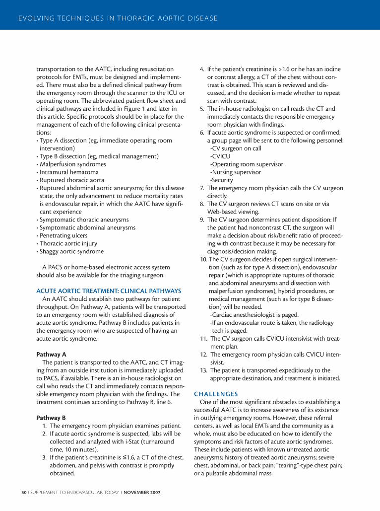
30 I SUPPLEMENT TO ENDOVASCULAR TODAY I NOVEMBER 2007
EVOLVING TECHNIQUES IN THORACIC AORTIC DISEASE
transportation to the AATC, including resuscitationprotocols for EMTs, must be designed and implement-ed. There must also be a defined clinical pathway fromthe emergency room through the scanner to the ICU oroperating room. The abbreviated patient flow sheet andclinical pathways are included in Figure 1 and later inthis article. Specific protocols should be in place for themanagement of each of the following clinical presenta-tions:• Type A dissection (eg, immediate operating room
intervention)• Type B dissection (eg, medical management)• Malperfusion syndromes• Intramural hematoma• Ruptured thoracic aorta• Ruptured abdominal aortic aneurysms; for this disease
state, the only advancement to reduce mortality ratesis endovascular repair, in which the AATC have signifi-cant experience
• Symptomatic thoracic aneurysms• Symptomatic abdominal aneurysms• Penetrating ulcers• Thoracic aortic injury• Shaggy aortic syndrome
A PACS or home-based electronic access systemshould also be available for the triaging surgeon.
ACUTE AORTIC TREATMENT: CLINICAL PATHWAYSAn AATC should establish two pathways for patient
throughput. On Pathway A, patients will be transportedto an emergency room with established diagnosis ofacute aortic syndrome. Pathway B includes patients inthe emergency room who are suspected of having anacute aortic syndrome.
Pathway AThe patient is transported to the AATC, and CT imag-
ing from an outside institution is immediately uploadedto PACS, if available. There is an in-house radiologist oncall who reads the CT and immediately contacts respon-sible emergency room physician with the findings. Thetreatment continues according to Pathway B, line 6.
Pathway B1. The emergency room physician examines patient. 2. If acute aortic syndrome is suspected, labs will be
collected and analyzed with i-Stat (turnaroundtime, 10 minutes).
3. If the patient’s creatinine is ≤1.6, a CT of the chest,abdomen, and pelvis with contrast is promptlyobtained.
4. If the patient’s creatinine is >1.6 or he has an iodineor contrast allergy, a CT of the chest without con-trast is obtained. This scan is reviewed and dis-cussed, and the decision is made whether to repeatscan with contrast.
5. The in-house radiologist on call reads the CT andimmediately contacts the responsible emergencyroom physician with findings.
6. If acute aortic syndrome is suspected or confirmed,a group page will be sent to the following personnel:
-CV surgeon on call-CVICU-Operating room supervisor-Nursing supervisor-Security
7. The emergency room physician calls the CV surgeondirectly.
8. The CV surgeon reviews CT scans on site or viaWeb-based viewing.
9. The CV surgeon determines patient disposition: Ifthe patient had noncontrast CT, the surgeon willmake a decision about risk/benefit ratio of proceed-ing with contrast because it may be necessary fordiagnosis/decision making.
10. The CV surgeon decides if open surgical interven-tion (such as for type A dissection), endovascularrepair (which is appropriate ruptures of thoracicand abdominal aneurysms and dissection withmalperfusion syndromes), hybrid procedures, ormedical management (such as for type B dissec-tion) will be needed.-Cardiac anesthesiologist is paged.-If an endovascular route is taken, the radiologytech is paged.
11. The CV surgeon calls CVICU intensivist with treat-ment plan.
12. The emergency room physician calls CVICU inten-sivist.
13. The patient is transported expeditiously to theappropriate destination, and treatment is initiated.
CHALLENGE SOne of the most significant obstacles to establishing a
successful AATC is to increase awareness of its existencein outlying emergency rooms. However, these referralcenters, as well as local EMTs and the community as awhole, must also be educated on how to identify thesymptoms and risk factors of acute aortic syndromes.These include patients with known untreated aorticaneurysms; history of treated aortic aneurysms; severechest, abdominal, or back pain; “tearing”-type chest pain;or a pulsatile abdominal mass.

The AATC will likely also have to establish and coordi-nate the rapid transport mechanisms with appropriatereimbursement, and then, as previously indicated, educatethe ambulance service staff regarding identification of acuteaortic syndromes. A key element of the education fortransport services regards the concept of permissivehypotension for ruptured aneurysms.
A public awareness campaign should be designed withthe goal of driving patient requests for transportation tothe AATC should they feel symptoms. A dedicated, AATC-specific Web site helps to build community awareness ofthe facility’s program. Patient-oriented information aboutaortic disease and treatment options should be displayedprominently.
Drs. Michael E. DeBakey, Stanley Crawford, and DentonCooley first tackled what was formerly a prohibitive surgi-cal disease and developed contemporary surgical tech-niques to treat the thoracic and abdominal aorta. Dr.DeBakey was the first to classify aortic dissection, a classifi-cation that bears his name to this day. Due to the legacy ofthese physicians and a long history of innovation in treatingaortic disease, The Methodist Hospital in Houston is partic-ularly well suited for development of this new concept. ■
Alan B. Lumsden, MD, is Professor of Surgery, TheMethodist DeBakey Heart Center, Houston, Texas. He may bereached at (713) 441-6201; [email protected].
Debra J. Crawford, RN, is Clinical Manager, Department of
Cardiovascular Surgery, The Methodist DeBakey Heart Center,Houston, Texas. She may be reached at (713) 441-6556;[email protected].
Eric K. Peden, MD, is Associate Professor, Acting Chief ofVascular Surgery, Department of Cardiovascular Surgery, TheMethodist DeBakey Heart Center, Houston, Texas. He may bereached at (713) 441-5200; [email protected].
Bryan T. Croft, MBA, is Vice President of Operations, TheMethodist DeBakey Heart Center in Houston, Texas. He maybe reached at (713) 441-1183; [email protected].
Michael J. Reardon, MD, is Professor of CardiothoracicSurgery, Weill Medical College of Cornell University, Chief ofCardiac Surgery, Department of Cardiovascular Surgery, TheMethodist DeBakey Heart Center, Houston, Texas. He may bereached at (713) 441-5200; [email protected].
Jeff E. Kalina, MD, is Associate Medical Director ofEmergency Medicine, Chairman of Disaster Response, WeillMedical College of Cornell University. He may be reached at(713) 441-4467; [email protected].
Faisal N. Masud, MD, is Associate Professor of ClinicalAnesthesiology, Weill College of Cornell University, MedicalDirector, Cardiovascular Intensive Care Unit, The MethodistDeBakey Heart Center, Houston, Texas. He may be reached at(713) 441-3620; [email protected].
Kristofer M. Charlton-Ouw, MD, is a Research Fellow,Department of Cardiovascular Surgery, The MethodistDeBakey Heart Center, Houston, Texas. He may be reached at(713) 441-6572; [email protected].
• Rapid identification and transport system
• Education of emergency medical technicians and local area emergency rooms on recognition of acute aortic syndromes
• Emergency room group with extensive experience in diagnosis and resuscitation of patients with acute aortic emergencies
• On-site 64-slice CT scanner in emergency room for immediate, fast imaging to permit emergent case triage
• Dedicated cardiovascular operating rooms and hybrid suites
• Goal of emergency room to treatment initiation time of <1 hour
• Highly experienced cardiovascular surgeons
• Highly experienced endovascular surgeons
• Surgeons working in close collaboration as a team of physicians for triaging cases and in surgical procedures
• History of integrating endovascular and open approaches
• Highly experienced, board-certified cardiovascular anesthesiologists
• Comprehensive operating room to CVICU care, with 24-hour in-house intensivist care
• Availability of new technologies for endovascular therapy of acute aortic syndromes
• Outcomes database
TABLE 1. COMPONENTS OF A SUCCESSFUL AATC AND PROGRAM
NOVEMBER 2007 I SUPPLEMENT TO ENDOVASCULAR TODAY I 31

UC200803460 EN