tendinitis and bursitis around knee joint & role of usg
TRANSCRIPT
Tendinitis and Bursitis
around knee joint &
Role of USG
Dr Shrenik Shah
Consultant Orthopaedic Surgeon
Shrey Hospital, Ahmedabad
Cyst and masses around knee
Popliteal cyst
Meniscal cyst
Ganglion cyst
Bursitis
Synovial chondromatosis
Synovial sarcoma
Villonodular synovitis
Tendinitis around knee
Infrapatellar tendinitis
Jumper’s knee
Hoffa’s fat pad syndrome
Popliteal tendinitis
PATB (Pes Anserinus Tendinitis & Bursitis) Syndrome
Muscle/ Tendon rupture
• Quadriceps
• Gastrocnemius
• Hamstring
• Patellar Tendon
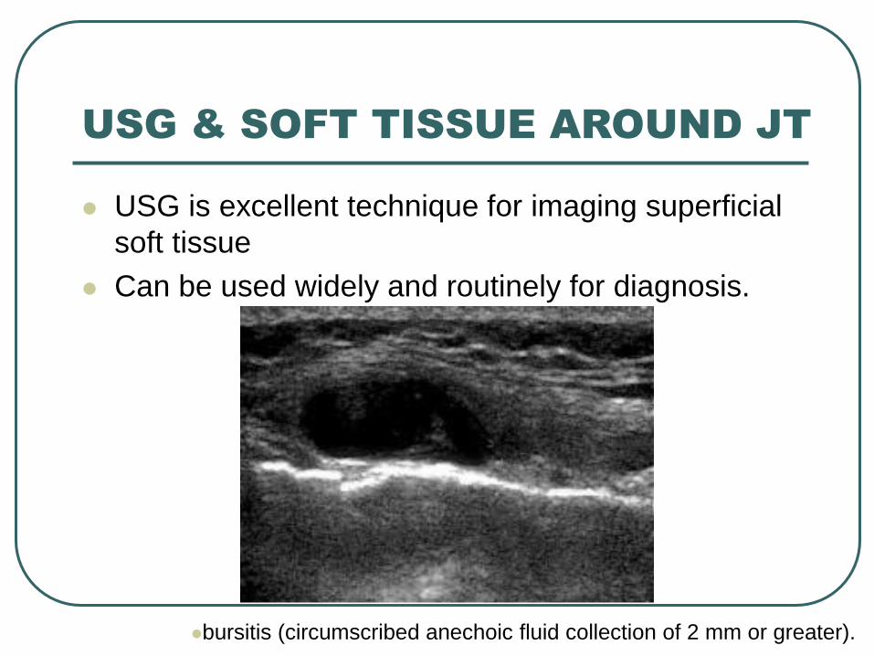
USG & SOFT TISSUE AROUND JT
USG is excellent technique for imaging superficial
soft tissue
Can be used widely and routinely for diagnosis.
bursitis (circumscribed anechoic fluid collection of 2 mm or greater).
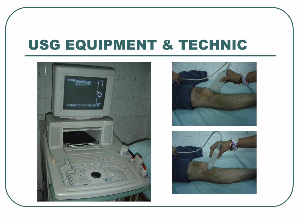
USG EQUIPMENT & TECHNIC
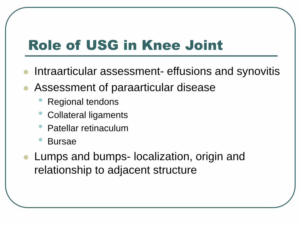
Role of USG in Knee Joint
Intraarticular assessment- effusions and synovitis
Assessment of paraarticular disease
• Regional tendons
• Collateral ligaments
• Patellar retinaculum
• Bursae
Lumps and bumps- localization, origin and
relationship to adjacent structure
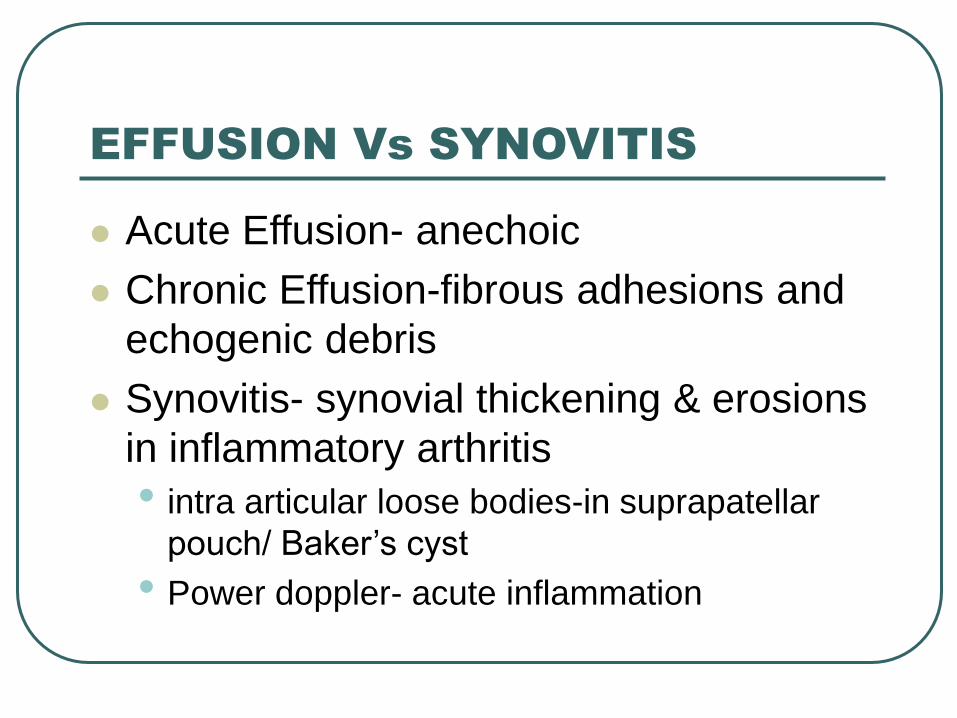
EFFUSION Vs SYNOVITIS
Acute Effusion- anechoic
Chronic Effusion-fibrous adhesions and
echogenic debris
Synovitis- synovial thickening & erosions
in inflammatory arthritis
• intra articular loose bodies-in suprapatellar
pouch/ Baker’s cyst
• Power doppler- acute inflammation
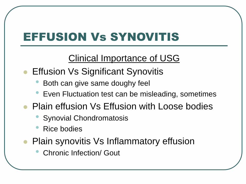
Clinical Importance of USG
Effusion Vs Significant Synovitis
• Both can give same doughy feel
• Even Fluctuation test can be misleading, sometimes
Plain effusion Vs Effusion with Loose bodies
• Synovial Chondromatosis
• Rice bodies
Plain synovitis Vs Inflammatory effusion
• Chronic Infection/ Gout
EFFUSION Vs SYNOVITIS
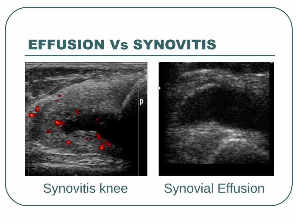
Synovitis knee Synovial Effusion
EFFUSION Vs SYNOVITIS
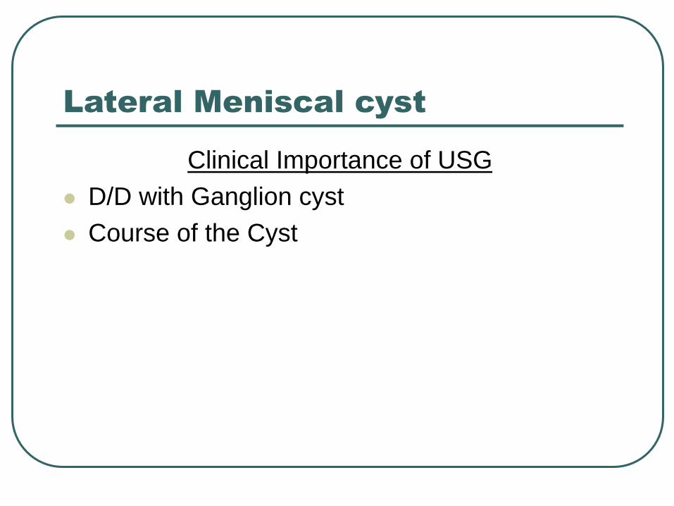
Lateral Meniscal cyst
Clinical Importance of USG
D/D with Ganglion cyst
Course of the Cyst
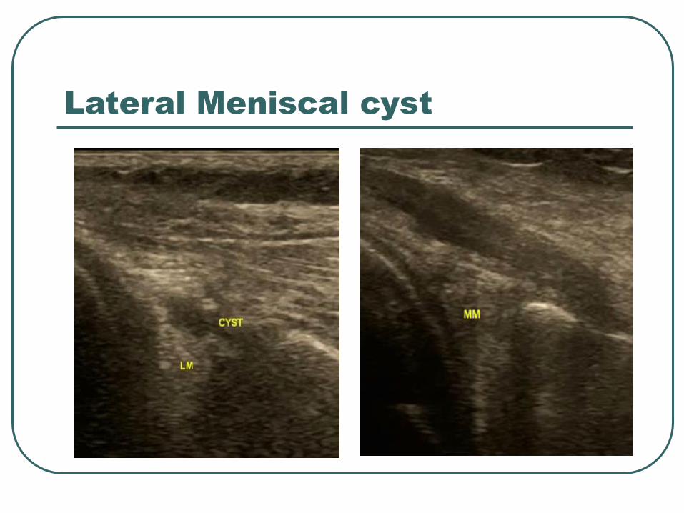
Lateral Meniscal cyst
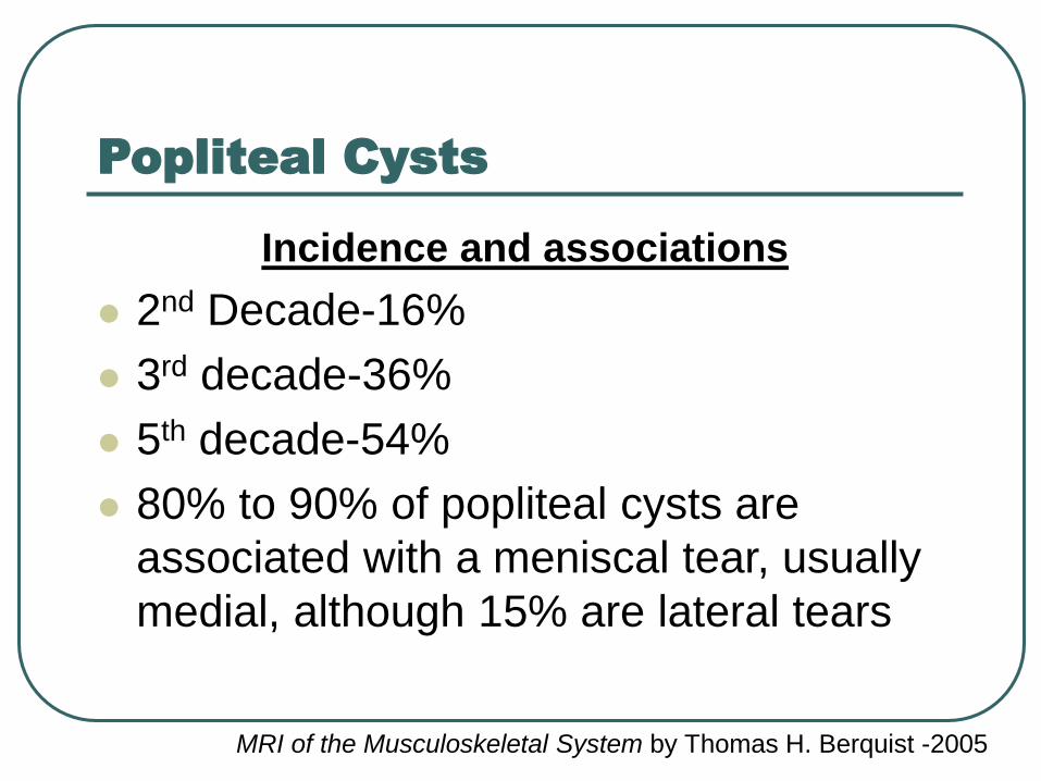
Popliteal Cysts
Incidence and associations
2nd Decade-16%
3rd decade-36%
5th decade-54%
80% to 90% of popliteal cysts are
associated with a meniscal tear, usually
medial, although 15% are lateral tears
MRI of the Musculoskeletal System by Thomas H. Berquist -2005
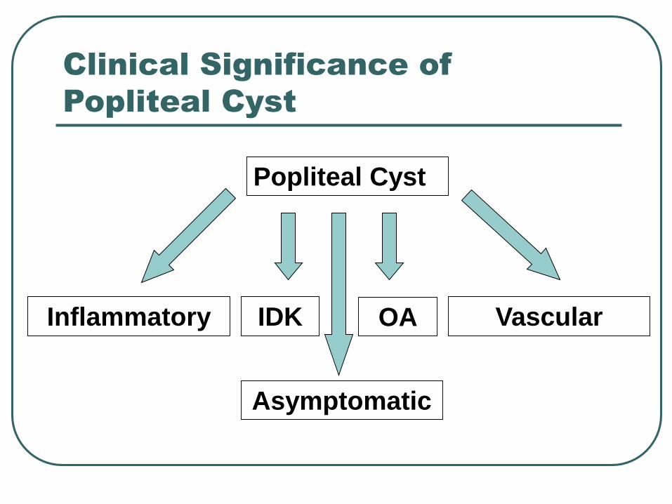
Clinical Significance of
Popliteal Cyst
Popliteal Cyst
OA Vascular
Asymptomatic
IDKInflammatory
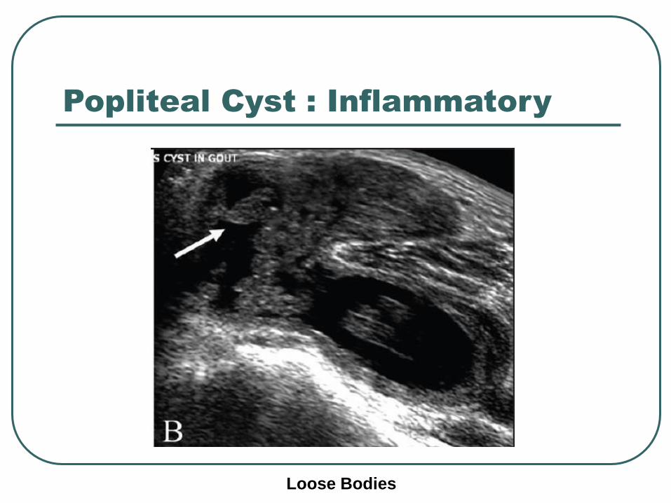
Popliteal Cyst : Inflammatory
Loose Bodies
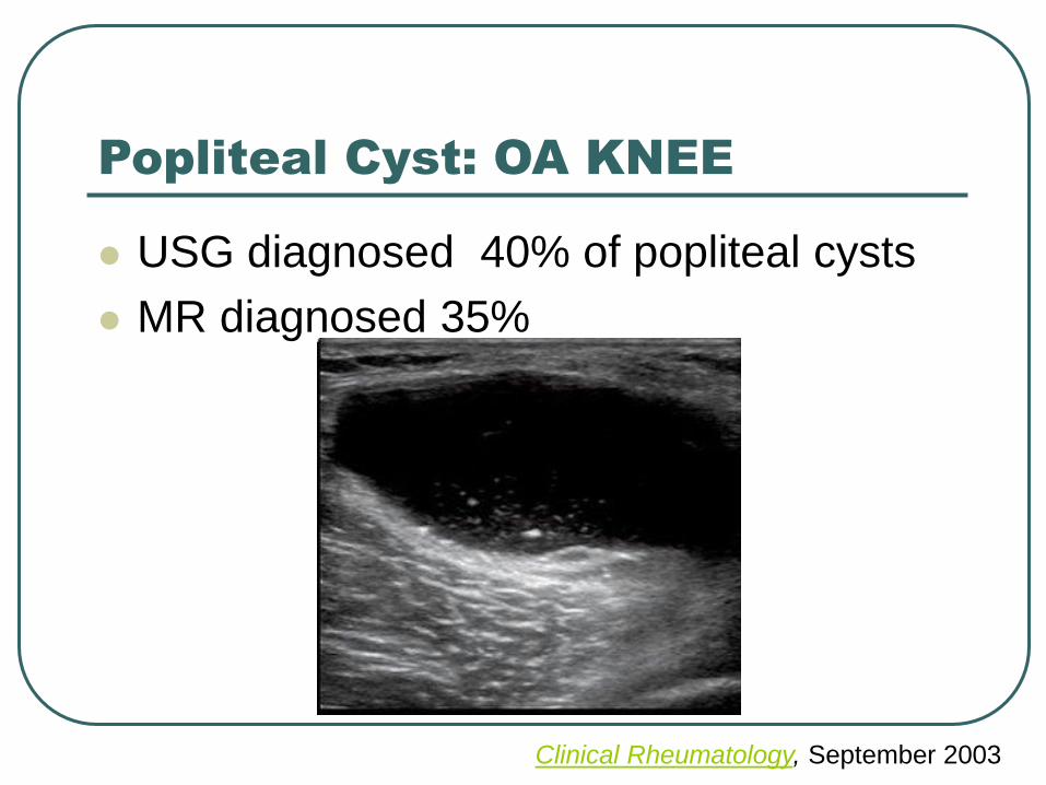
Popliteal Cyst: OA KNEE
USG diagnosed 40% of popliteal cysts
MR diagnosed 35%
Clinical Rheumatology, September 2003
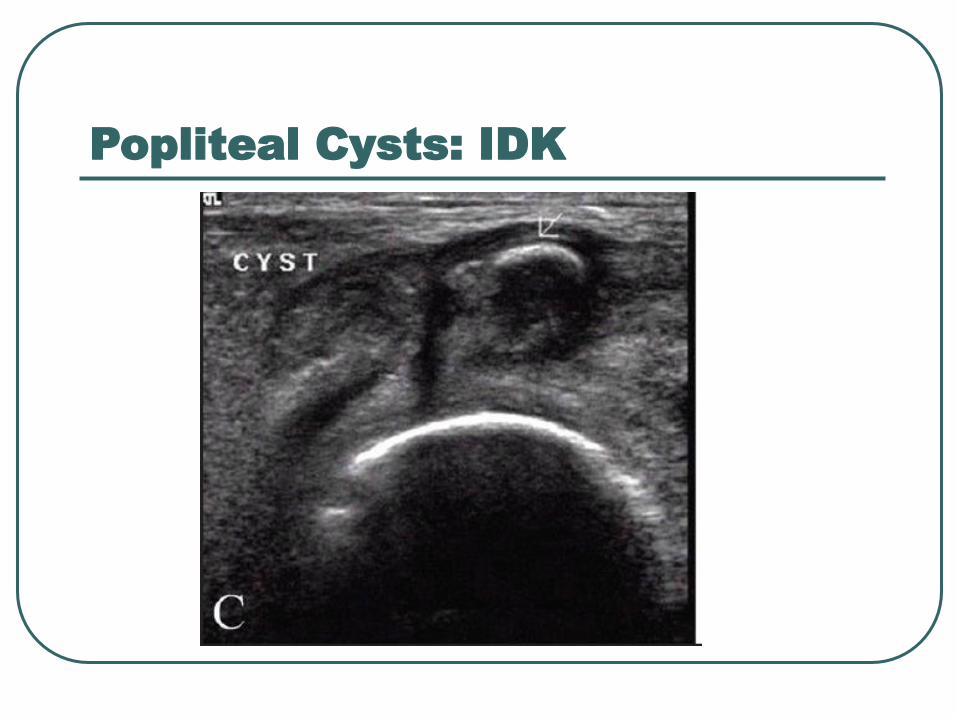
Popliteal Cysts: IDK
Previous meniscectomy
Articular cartilage damage
• chondromalacia patella
• degenerative arthritis
Collateral and cruciate ligament injury
Popliteal Cysts: IDK
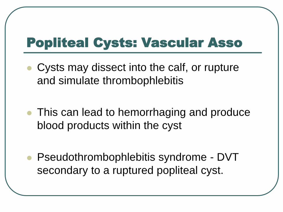
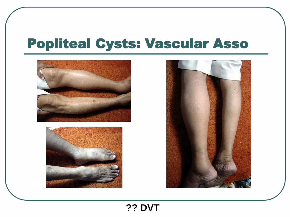
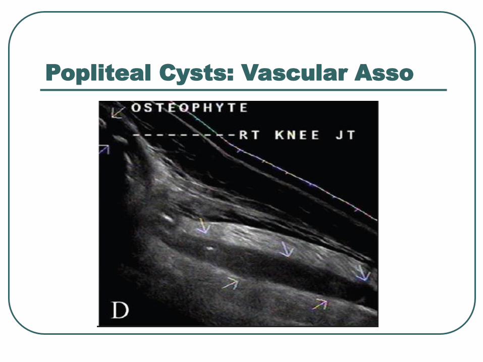
Popliteal Cysts: Vascular Asso
Cysts may dissect into the calf, or rupture
and simulate thrombophlebitis
This can lead to hemorrhaging and produce
blood products within the cyst
Pseudothrombophlebitis syndrome - DVT
secondary to a ruptured popliteal cyst.
Popliteal Cysts: Vascular Asso
?? DVT
Popliteal Cysts: Vascular Asso
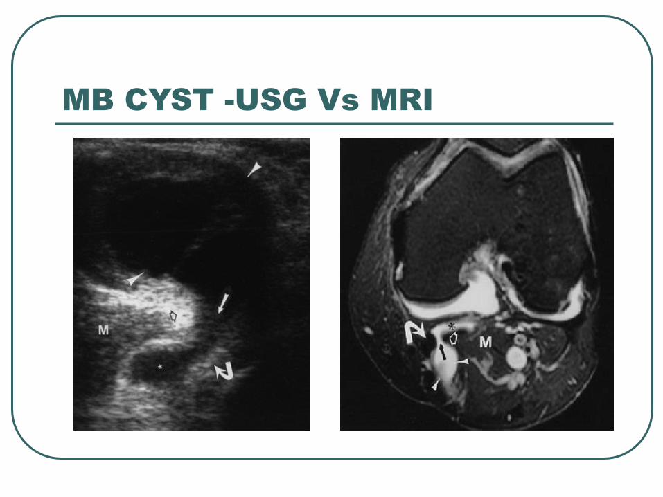
MB CYST -USG Vs MRI
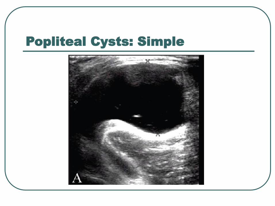
Popliteal Cysts: Simple
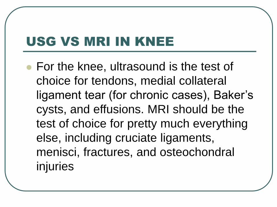
USG VS MRI IN KNEE
For the knee, ultrasound is the test of
choice for tendons, medial collateral
ligament tear (for chronic cases), Baker’s
cysts, and effusions. MRI should be the
test of choice for pretty much everything
else, including cruciate ligaments,
menisci, fractures, and osteochondral
injuries
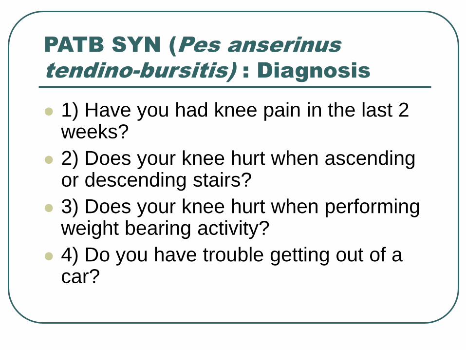
PATB SYN (Pes anserinus
tendino-bursitis) : Diagnosis
1) Have you had knee pain in the last 2 weeks?
2) Does your knee hurt when ascending or descending stairs?
3) Does your knee hurt when performing weight bearing activity?
4) Do you have trouble getting out of a car?
Pes anserinus tendino-bursitis:
clinical and imaging corelation
37 clinical PATB patients,
• USG3 Anserine bursitis
1 Pes Anserinus tendinitis
clinical PATB syndrome with type 2 DM
28.6% had PA tendinitis
Scand J Rheumatol 2000

PES ANSERINUS TENDINITIS
RIGHT 5 MM-THICKENING + LOSS
OF NORMAL ECHOTEXTURE LEFT 3.5 MM
PATB AND OA KNEE
“Pain of OA could have a cause due to periarticular problems, such as anserine bursitis”
Local corticosteroid injection resulted in complete relief of pain only in those patients with US findings of PATB.
USG exam can serve as a useful tool to determine the usage of NSAIDs Vs Acetaminophen
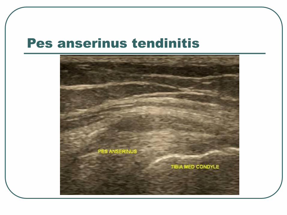
Pes anserinus tendinitis
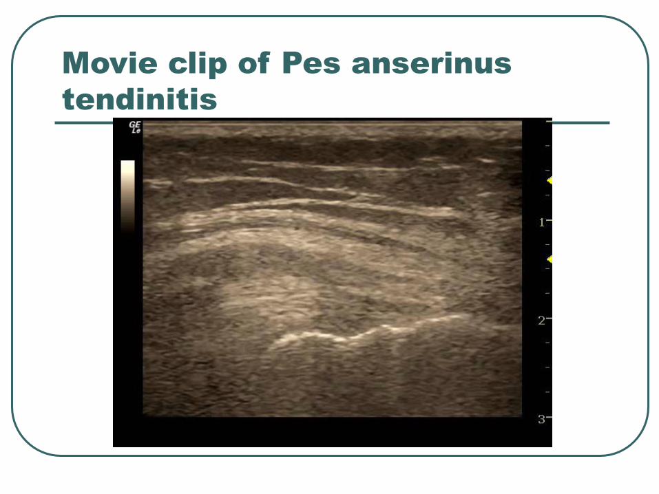
Movie clip of Pes anserinus
tendinitis
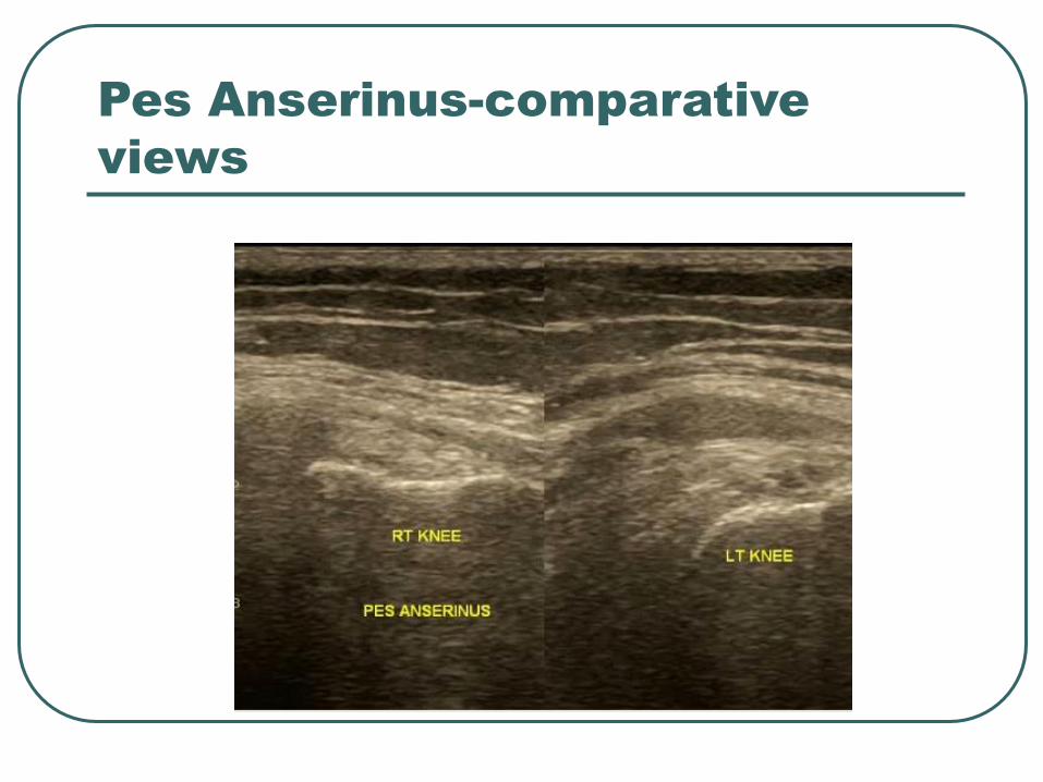
Pes Anserinus-comparative
views
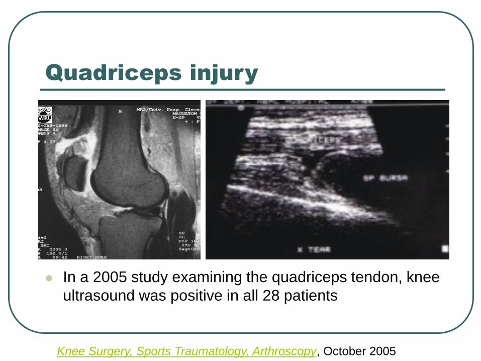
Quadriceps injury
In a 2005 study examining the quadriceps tendon, knee
ultrasound was positive in all 28 patients
Knee Surgery, Sports Traumatology, Arthroscopy, October 2005
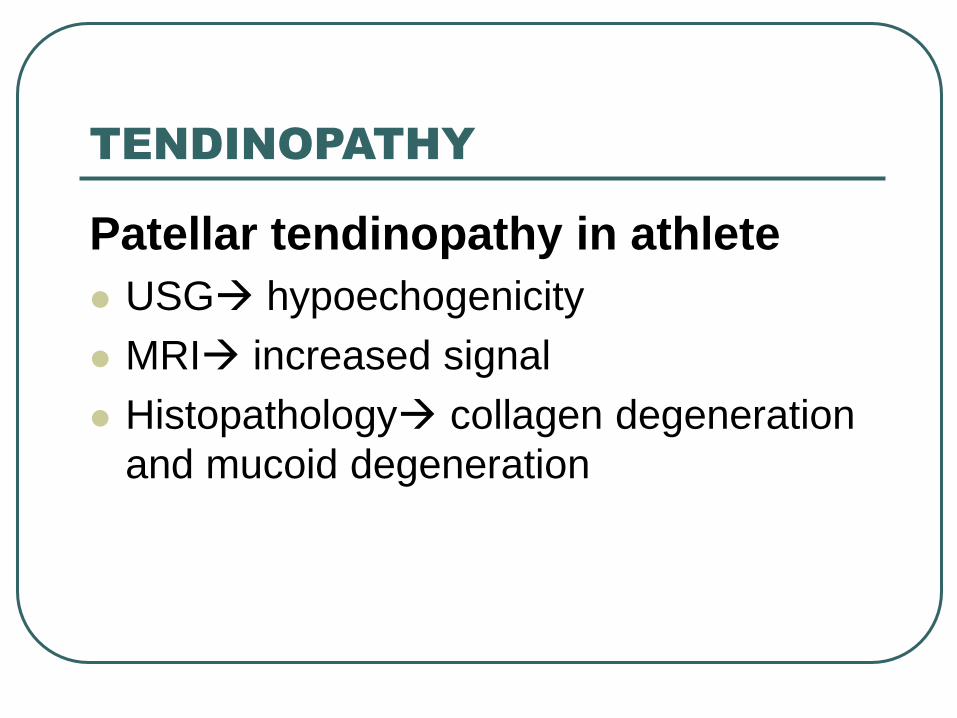
TENDINOPATHY
Patellar tendinopathy in athlete
USG hypoechogenicity
MRI increased signal
Histopathology collagen degeneration
and mucoid degeneration
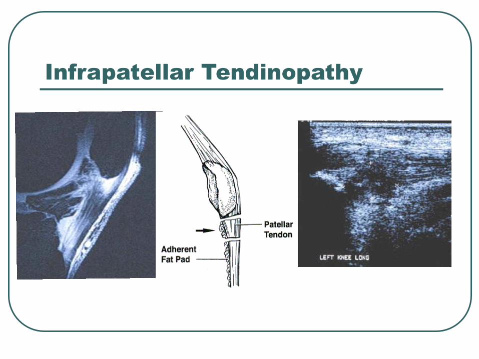
Infrapatellar Tendinopathy
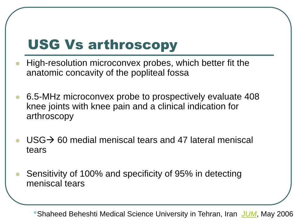
USG Vs arthroscopy
High-resolution microconvex probes, which better fit the anatomic concavity of the popliteal fossa
6.5-MHz microconvex probe to prospectively evaluate 408 knee joints with knee pain and a clinical indication for arthroscopy
USG 60 medial meniscal tears and 47 lateral meniscal tears
Sensitivity of 100% and specificity of 95% in detecting meniscal tears
•Shaheed Beheshti Medical Science University in Tehran, Iran JUM, May 2006
Carry home message
USG is noninvasive, economical
Accessible next door investigation
Under utilised, operator dependent
Scores over MRI in tendon imaging
Dynamic and real time study
Ability to compare contra lateral side
Various planes can be traced
Advantage of repetitive study
Ease of documentation

Thank You