the class mesomycetozoea: a heterogeneous … · the species in these genera has several...
TRANSCRIPT
16 Aug 2002 13:56 AR AR168-MI56-14.tex AR168-MI56-14.SGM LaTeX2e(2002/01/18)P1: GJC10.1146/annurev.micro.56.012302.160950
Annu. Rev. Microbiol. 2002. 56:315–44doi: 10.1146/annurev.micro.56.012302.160950
First published online as a Review in Advance on May 7, 2002
THE CLASS MESOMYCETOZOEA: A HeterogeneousGroup of Microorganisms at the Animal-FungalBoundary∗
Leonel Mendoza,1 John W. Taylor,2 and Libero Ajello31Medical Technology Program, Department of Microbiology and Molecular Genetics,Michigan State University, East Lansing Michigan, 48824-1030;e-mail: [email protected] of Plant and Microbial Biology, University of California, Berkeley,California 94720-3102; e-mail: [email protected] for Disease Control and Prevention, Mycotic Diseases Branch, AtlantaGeorgia 30333; e-mail: [email protected]
Key Words Protista, Protozoa, Neomonada, DRIP, Ichthyosporea
■ Abstract When the enigmatic fish pathogen, the rosette agent, was first found tobe closely related to the choanoflagellates, no one anticipated finding a new group oforganisms. Subsequently, a new group of microorganisms at the boundary between an-imals and fungi was reported. Several microbes with similar phylogenetic backgroundswere soon added to the group. Interestingly, these microbes had been considered to befungi or protists. This novel phylogenetic group has been referred to as the DRIP clade(an acronym of the original members:Dermocystidium, rosette agent,Ichthyophonus,and Psorospermium), as the class Ichthyosporea, and more recently as the classMesomycetozoea. Two orders have been described in the mesomycetozoeans: the Der-mocystida and the Ichthyophonida. So far, all members in the order Dermocystida havebeen pathogens either of fish (Dermocystidiumspp. and the rosette agent) or of mam-mals and birds (Rhinosporidium seeberi), and most produce uniflagellated zoospores.Fish pathogens also are found in the order Ichthyophonida, but so are saprotrophic mi-crobes. The Ichthyophonida species do not produce flagellated cells, but many produceamoeba-like cells. This review provides descriptions of the genera that comprise theclass Mesomycetozoea and highlights their morphological features, pathogenic roles,and phylogenetic relationships.
CONTENTS
INTRODUCTION . . . . . . . . . . . . . . . . . . . . . . . . . . . . . . . . . . . . . . . . . . . . . . . . . . . . . 316MEMBERS OF THE CLASS MESOMYCETOZOEA. . . . . . . . . . . . . . . . . . . . . . . . 322
Order Ichthyophonida. . . . . . . . . . . . . . . . . . . . . . . . . . . . . . . . . . . . . . . . . . . . . . . . 322
∗The U.S. Government has the right to retain a nonexclusive royalty-free license in and toany copyright covering this paper.
315
Ann
u. R
ev. M
icro
biol
. 200
2.56
:315
-344
. Dow
nloa
ded
from
arj
ourn
als.
annu
alre
view
s.or
gby
NE
SLi2
on
08/2
2/08
. For
per
sona
l use
onl
y.
16 Aug 2002 13:56 AR AR168-MI56-14.tex AR168-MI56-14.SGM LaTeX2e(2002/01/18)P1: GJC
316 MENDOZA ¥ TAYLOR ¥ AJELLO
Order Dermocystida. . . . . . . . . . . . . . . . . . . . . . . . . . . . . . . . . . . . . . . . . . . . . . . . . . 330RELATIONSHIPS OF THE MESOMYCETOZOEANSWITH THE OTHER CLADES ARISING NEAR THEANIMAL-FUNGAL DIVERGENCE . . . . . . . . . . . . . . . . . . . . . . . . . . . . . . . . . . . . . 335
CLOSE RELATIVES OF THE MESOMYCETOZOEA ANDTHE EVOLUTION OF TRAITS COMMON TO ALL . . . . . . . . . . . . . . . . . . . . . . . 336
FUTURE DIRECTIONS OF RESEARCH INTHE MESOMYCETOZOEA . . . . . . . . . . . . . . . . . . . . . . . . . . . . . . . . . . . . . . . . . . . 338
INTRODUCTION
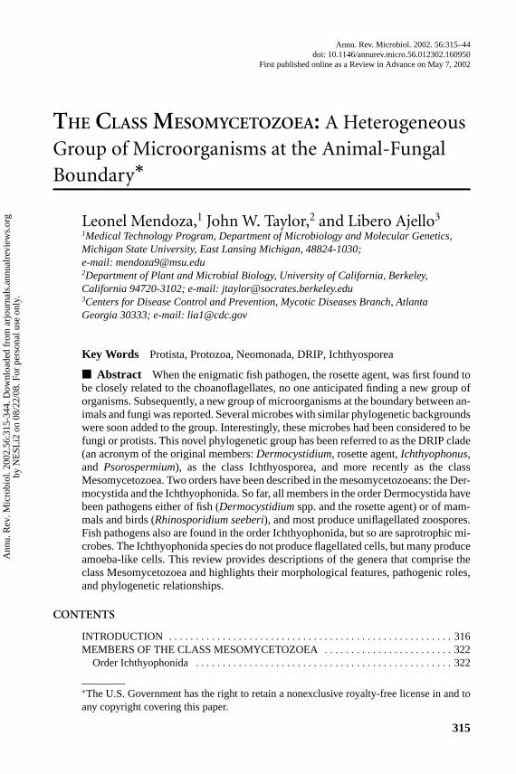
Led by Haeckel’s proposal that the metazoans may have had an ancestor within theunicellular protists, numerous studies utilizing morphology as well as molecularand phylogenetic analyses have supported his concept (18, 48, 51, 56). Those stud-ies were validated in 1993 when Wainright et al. (95) used phylogenetic analysesof small subunit ribosomal DNA (18S SSU rDNA) sequences to conclude that themetazoans were a monophyletic group that shared ancestry with the choanoflag-ellates. They also reported that animals and fungi might have had a more recentcommon ancestor than either group had with plants, alveolates, or stramenopiles(14, 95). First Spanggaard et al. (84) and then Ragan et al. (77) reported that anew group of parasitic and saprotrophic protists had been found near the animaland fungal divergence. Later, others verified their findings (8, 18, 19, 30, 39, 44).These investigators confirmed that previously unclassified animal parasites andsaprotrophic microbes grouped together as a new protistan monophyletic cladelocated near the point where the animals had diverged from the fungal boundary(Figure 1). Early on Ragan et al. referred to those microorganisms as the DRIPclade (an acronym forDermocystidium, rosette agent,IchthyophonusandPsoros-permium). Later Cavalier-Smith placed them in the class Ichthyosporea (18), andmore recently Mendoza et al. (57) established the class Mesomycetozoea to ac-commodate them. The location of this group at the divergence between animalsand fungi was significant because it indicated that this unique group of microorgan-isms arose near the time that animals had diverged from fungi (Figure 1), providingadditional organisms for comparative studies that could reveal the nature of theprogenitor of the animal and the fungi, two of the kingdoms of multicellular andmacroscopic organisms (17, 39, 77).
Examination of Figure 1 shows that the class Mesomycetozoea is a mono-phyletic group composed of two strongly supported clades, the orders Ichthyo-phonida and Dermocystida. However, as often is the case, the relationships of theMesomycetozoea to other broad taxa are poorly supported in molecular phyloge-netic analyses. Incorporating phenotypical data, Cavalier-Smith (17, 18) held thatthe ancestors of the animals and fungi were not mesomycetozoeans but unicellularflagellate organisms in the choanoflagellates. He based his conclusions on the factsthat the mesomycetozoeans did not possess chitin or flagellate stages, a conceptthat recently was proven to be incorrect.
Ann
u. R
ev. M
icro
biol
. 200
2.56
:315
-344
. Dow
nloa
ded
from
arj
ourn
als.
annu
alre
view
s.or
gby
NE
SLi2
on
08/2
2/08
. For
per
sona
l use
onl
y.
16 Aug 2002 13:56 AR AR168-MI56-14.tex AR168-MI56-14.SGM LaTeX2e(2002/01/18)P1: GJC
THE MESOMYCETOZOEANS 317
Figure 1 A phylogenetic analysis made by neighbor joining in Phylogenetic AnalysisUsing Parsimony (PAUP) using distances estimated by maximum likelihood (transi-tions/transversions estimated by ML; emperical nulceotide frequencies among sitevariation allowed comparable to the HKY85 model). The thickened branches are sup-ported by 90% or greater of 1000 bootstrapped datasets. The topologies and supportare similar to those found with parsimony analysis, with the exception of the place-ment ofNuclearia moebiusi, which can move as far as the branch subtending animals+ choanoflagellaes+Mesomycetozoea. The scale for percent nucleotide substitutionper nucleotide is given on the branch toOchromonas danica.
Originally, Cavalier-Smith included the mesomycetozoeans in the class Icht-hyosporea (18) (Table 1). This class was based on the fact that the originalmembers of the DRIP clade described by Ragan et al. (77) were all fish parasites.However, new members of the group includingAmoebidium parasiticum, Anu-rofeca richardsi, Pseudoperkinsus tapetis, Rhinosporidium seeberi, Sphaerosoma
Ann
u. R
ev. M
icro
biol
. 200
2.56
:315
-344
. Dow
nloa
ded
from
arj
ourn
als.
annu
alre
view
s.or
gby
NE
SLi2
on
08/2
2/08
. For
per
sona
l use
onl
y.

16 Aug 2002 13:56 AR AR168-MI56-14.tex AR168-MI56-14.SGM LaTeX2e(2002/01/18)P1: GJC
318 MENDOZA ¥ TAYLOR ¥ AJELLO
TABLE 1 Current classification of the class Mesomycetozoea including new members
Kingdom Protozoa (Cavalier-Smith 1997)Subkingdoms
1-Archezoa (Giardia, Chilomastix, Retortamonas)2-Eozoa (Trichomonas)3-Neozoa (four Infrakingdoms)
a) Sarcodina (Amoeba, Acanthamoeba, Entamoeba, Dictyostelium)b) Alveolata (Perkinsus, Eimeria, Plasmodium, Babesia, Paramecium, Tetrahymena)c) Actinopoda (Acanthocystis, Radiozoa)d) Neomonada, Cavalier-Smith 1997
Phylum NeomonadaSubphylaa) Apusozoa (Apusomonas)b) Isomita (Nephromyces)c) Mesomycetozoa (four Classes) stat. nov.
Class 1 Choanoflagellatea (Sphaeroeca)Class 2 Corallochytrea (Corallochytrium)Class 3 Mesomycetozoea (em. Mendoza et al. 2001)–(Ichthyosporea, Cavalier-Smith 1998)
Order Dermocystida Order Ichthyophonidaa)Dermocystidiumspp. a)Amoebidium parasiticumb) Rhinosporidium seeberi b) Anurofeca richardsic) Rosette agent c)Ichthyophonusspp.
d) Pseudoperkinsus tapetise)Psorospermium haeckelif ) Sphaerosoma articumg) LKM51
Class 4 Cristidiscoidea, Order Nucleariida (Nuclearia)
arcticum, and the isolate LKM51 are not fish parasites, which renders the termIcthyosporea inappropriate. In addition, the finding thatIchthyophonus hoferihaschitin in its cell walls (83) and thatR. seeberipossesses at least one of the chitinsynthase genes (40) suggested that this group of microbes may also have chitin in itscell walls, as do the fungi and some stramenopilans (60). This result did not comeas a surprise because several investigators had previously suggested the presenceof this polymer in the cell walls of some mesomycetozoeans (40, 83). The mainproblem has been that this polymer is not abundant in Mesomycetozoeans’ cellwalls. Thus it was difficult to demonstrate its presence in this group of protista (61).Moreover, chitin has always been associated with fungi but not with the protista,which in part discouraged some investigators from seeking it in organisms otherthan fungi (9). Mulisch (61) defied this trend by reporting that several protists pos-sess chitin in their cell walls including the filamentous stramenopilans (60, 84).This finding strongly supported the reports of the presence of this polymer inI. hoferiandR. seeberi, and by extrapolation in all of the other mesomycetozoeans(9, 40, 83). Based on these facts, we recently emended Cavalier-Smith’s originalproposal and introduced the new class Mesomycetozoea (57), a name more suitablefor this group of microbes (Figure 1). Accordingly, we use this term throughout our
Ann
u. R
ev. M
icro
biol
. 200
2.56
:315
-344
. Dow
nloa
ded
from
arj
ourn
als.
annu
alre
view
s.or
gby
NE
SLi2
on
08/2
2/08
. For
per
sona
l use
onl
y.

16 Aug 2002 13:56 AR AR168-MI56-14.tex AR168-MI56-14.SGM LaTeX2e(2002/01/18)P1: GJC
THE MESOMYCETOZOEANS 319
review. The subphylum Choanozoa was also emended to subphylum Mesomyce-tozoa. Because the choanoflagellates were the only group between animals andfungi at that time, the epithet Chonozoa was previously introduced (17). With theinclusion of related nearby microbes, the term Mesomycetozoa (between animalsand fungi), originally proposed by Herr et al. (39), was considered to be moreappropriate.
Based on phylogenetic analyses of 18S small subunit rDNA genes, the class Me-somycetozoea comprises 10 different parasitic and saprophytic microbes. They aremembers of the generaAmoebidium, Anurofeca, Dermocystidium, Ichthyophonus,Pseudoperkinsus, Psorospermium, Rhinosporidium, Sphaerosoma, and two as yetunnamed microbes, the “rosette agent” and “clone LKM51” (Figure 1). Each ofthe species in these genera has several morphological characteristics in commonwith species in the other genera of the clade, but each has unique characteristics aswell (Table 2). Although members of this group are typically thought to be aquaticpathogens of fish, there are exceptions:R. seeberi, the only member of the class
TABLE 2 Members of orders Dermocystida and Ichthyophonida and the only species of theclass Corallochytrea (Cora) with highlights of their key attributes
MitocondrialTaxa cristae Life cycle traits Hosts
DermocystidaDermocystidiumspp Flat Cysts with endospores Fish
uniflagellate zoosporesRosette agent Flat? Spherules with endospores Fish
uniflagellate zoosporesRhinosporidium seeberi Flat Sporangium with endospores Mammals,
uniflagellate zoospores? Birds
IchthyophonidaAmoebidium parasiticum Flat Sporangium, sporangiospores Insects
amoebic stage CrustaceansIchthyophonus hoferi Tubular Hyphae, plasmodium, spores Fish
amoebic stagePsorospermium haeckeli Flat Ovoid shell-bearing spores Crayfish
amoebic stageAnurofeca richardsi Flat? Spherules with endospores Anural larvaePseudoperkinsus tapetis Unknown Spherules with spores Clams
uniflagellate zoospores?Sphaerosoma articum Flat Spherules with endospores Saprotrophic?Isolate LKM51 Unknown Unknown Saprotrophic?
Corallochytrea
Corallochytrium Flat? Spherules with spores Saprotrophiclimacisporum amoebic stage
?= not clear.
Ann
u. R
ev. M
icro
biol
. 200
2.56
:315
-344
. Dow
nloa
ded
from
arj
ourn
als.
annu
alre
view
s.or
gby
NE
SLi2
on
08/2
2/08
. For
per
sona
l use
onl
y.

16 Aug 2002 13:56 AR AR168-MI56-14.tex AR168-MI56-14.SGM LaTeX2e(2002/01/18)P1: GJC
320 MENDOZA ¥ TAYLOR ¥ AJELLO
known to cause infections in mammals and birds;Amoebidiumspecies, a genusof saprotrophic organisms;A. richardsi, whose role in diseases of anuran larvaeis not clear;Ps. tapetis, a possibly nonpathogenic species associated with clams;S. arcticum, usually a saprotrophic organism; and the isolate termed LKM51, aputatively saprotrophic eukaryote found in phytoplankton.
Nothing is known regarding Mesomycetozoea’s geographical distribution ortheir relationships with their natural environments. Thus, their epidemiologicalfeatures and their interactions with other microbes in their ecological niches arealso largely unknown. Because most mesomycetozoeans are animal parasites,what we do know about their cell cycles was learned from studies conductedon their parasitic stages. These studies have been of pivotal importance for the par-tial construction of their life cycles (5, 64, 83, 92, 97) (Figure 2). For instance, itwas demonstrated that the species of the generaAmoebidium, Ichthyophonus, andPsorospermiumdeveloped amoeba-like cells in vitro. Based on these studies it wasspeculated that the amoeba-like cells could be the infecting propagules in nature(83, 92, 97) (Figure 2). Likewise, in vitro theDermocystidiumspp. and the rosetteagent developed uniflagellated zoospores, indicating that they could serve as theinfectious propagules [(37, 65) K.D. Arkush, personal communication] (Figure 2).The major contribution of these studies is the finding that during their parasiticstages, at least one phenotypic form of the mesomycetozoean species could initiatea new cycle outside their hosts.
In spite of these findings, their true life cycles in nature remain a mystery.Sexual development in the Mesomycetozoea has yet to be reported. This is duein part to the fact that most mesomycetozoeans have only been studied in theirparasitic stages rather than in culture. Thus, sexual fusion, gamete formation,meiosis, and other major important genetic traits have yet to be found or induced.It is important to note that some investigators reported the presence of multiplenuclei during the parasitic stages of some mesomycetozoeans during the formationof new spores. In addition, little is known about their feeding habits. It has beenfound, however, that during their parasitic stagesD. salmonis(65),I. hoeferi(45),P. haeckeli(89, 91), andR. seeberi(59) absorb nutrients from the hosts throughthe mesomycetozoean’s cell walls, a finding that might suggest a similar behaviorduring their environmental stages. However, more studies are necessary to validatethis assumption.
The epithets used to identify the phenotypic stages of the members of the classMesomycetozoea varied according to the type of microorganism the investigatorsthought them to be. For instance, mycological terminology was used to identify thestructures ofA. parasiticum, A. richardsi, I. hoferi, andR. seeberi, all of them stud-ied by mycologists (5, 21, 38, 62, 98, 99). The terms they used included endospores,hyphae, sporangia, spores, sporangiospores, thalli, and others. In contrast, proto-zoological names such as amoeba, cyst, plasmodium, sporocyst, and zoosporewere used by protistologists to identify the structures formed byA. parasiticum,D. salmonis, I. hoferi, P. haeckeli, and the rosette agent (14, 24, 42, 62, 64, 75).With their inclusion in the class Mesomycetozoea, standardization of the names
Ann
u. R
ev. M
icro
biol
. 200
2.56
:315
-344
. Dow
nloa
ded
from
arj
ourn
als.
annu
alre
view
s.or
gby
NE
SLi2
on
08/2
2/08
. For
per
sona
l use
onl
y.

16 Aug 2002 13:56 AR AR168-MI56-14.tex AR168-MI56-14.SGM LaTeX2e(2002/01/18)P1: GJC
THE MESOMYCETOZOEANS 321
Figure 2 Depiction of the putative life cycle of members of the orders Dermocystida(left) and Ichthyophonida (right). Members of the order Dermocystida develop spheri-cal cells with endospores (stage 1). In vitro the released endospores (stage 2) give riseto uniflagellated zoospores (stage 3). When the zoopores (infecting units) infect thehost, they encyst (stage 4) and increase in size (stages 4, 5) and undergo cleavage intoendospores (stage 1). The endospores can also be directly released within the host’sinfected tissues, and the cycle is repeated inside the hosts (stages 1, 4, 5, 1). Mem-bers of the order Ichthyophonida develop spherical (Ichthyophonusspp.) or ovoid cellsin infected tissues (Psorospermium haekeli) or on its hosts (Amoebidium parasiticum)(stage 1). In vitro (stage 1) the hatching of spore receptacles occurs from the ovoid cells(stage 2); the receptacles containing spores (stage 3) then rupture and release their en-dospores (stage 4), which develop into motile amoeboid cells (infecting units) (stage 5).The amoeboid cell reach the hosts and develops into a small receptacle (stage 6) thatlater generates a hard cell wall with endospores (stages 7, 1). The endospores can alsobe released within their hosts, repeating the cycle (stages 1, 6, 7, 1). Note that in thegenusIchthyophonus, the development of hyphae that produce spherical cells withendopores (in vitro) is a feature, so far, not encountered in the other members of theorder (62, 64, 83). For the order Ichthyophonida the cell cycle was adapted from Vogt& Rugg (92).
used to describe their phenotypic stages would be of importance in studying themembers of this class. In this review, however, we continue to use the traditionalnomenclature until a consensus is reached regarding their terminology.
We provide a brief description of the genera that are currently in the class Me-somycetozoea, highlighting their similarities and differences, with the intentionof covering the latest known developments of the mesomycetozoeans and intro-ducing the microbiological community to this novel class of microorganisms andtheir morphological, pathogenic, and phylogenetic relationships.
Ann
u. R
ev. M
icro
biol
. 200
2.56
:315
-344
. Dow
nloa
ded
from
arj
ourn
als.
annu
alre
view
s.or
gby
NE
SLi2
on
08/2
2/08
. For
per
sona
l use
onl
y.
16 Aug 2002 13:56 AR AR168-MI56-14.tex AR168-MI56-14.SGM LaTeX2e(2002/01/18)P1: GJC
322 MENDOZA ¥ TAYLOR ¥ AJELLO
MEMBERS OF THE CLASS MESOMYCETOZOEA
Order Ichthyophonida
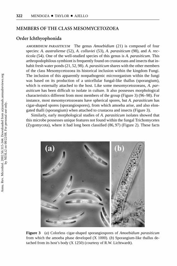
AMOEBIDIUM PARASITICUM The genusAmoebidium(21) is composed of fourspecies:A. australiense(52), A. colluviei (53), A. parasiticum(98), andA. rec-ticola (54). One of the well-studied species of this genus isA. parasiticum. Thisarthropodophilous symbiont is frequently found on crustaceans and insects that in-habit fresh water ponds (21, 52, 98).A. parasiticumshares with the other membersof the class Mesomycetozoea its historical inclusion within the kingdom Fungi.The inclusion of this apparently nonpathogenic microorganism within the fungiwas based on its production of a unicellular fungal-like thallus (sporangium),which is externally attached to the host. Like some mesomycetozoeans,A. par-asiticumhas been difficult to isolate in culture. It also possesses morphologicalcharacteristics different from most members of the group (Figure 3) (96–98). Forinstance, most mesomycetozoeans have spherical spores, butA. parasiticumhascigar-shaped spores (sporangiospores), from which amoeba arise, and also elon-gated thalli (sporangium) when attached to crustacea and insects (Figure 3).
Similarly, early morphological studies ofA. parasiticumisolates showed thatthis microbe possesses unique features not found within the fungal Trichomycetes(Zygomycota), where it had long been classified (86, 97) (Figure 2). These facts
Figure 3 (a) Colorless cigar-shaped sporangiospores ofAmoebidium parasiticumfrom which the amoeba phase developed (X 1000). (b) Sporangium-like thallus de-tached from its host’s body (X 1250) (courtesy of R.W. Lichtwardt).
Ann
u. R
ev. M
icro
biol
. 200
2.56
:315
-344
. Dow
nloa
ded
from
arj
ourn
als.
annu
alre
view
s.or
gby
NE
SLi2
on
08/2
2/08
. For
per
sona
l use
onl
y.
16 Aug 2002 13:56 AR AR168-MI56-14.tex AR168-MI56-14.SGM LaTeX2e(2002/01/18)P1: GJC
THE MESOMYCETOZOEANS 323
prompted some investigators to hypothesize thatA. parasiticumrepresents a non-fungal evolutionary lineage possibly derived from a protozoan ancestor (54, 96).The finding of Whisler (97) thatA. parasiticumforms amoebal cysts that give riseto several motile amoebas, which later encyst to form sporangium-like structures,provided the strongest evidence for this hypothesis. Although other investigatorssupported this new position (74, 85), those studies did not resolve the phylogeneticconnection of this genus with other living organisms.
The phylogenetic affinities ofA. parasiticumwere almost simultaneously re-solved when first Ustinova et al. (86) and later Benny & O’Donnell (12) sequencedthe 18S SSU rDNA ofA. parasiticum. Using phylogenetic analyses, these two in-dependent groups of researchers found thatA. parasiticumis a member of theclass Mesomycetozoea and far from the fungal class Trichomycetes of the king-dom Fungi. In their phylogenetic trees,A. parasiticumwas always closely related toI. hoferi. Interestingly, ultrastructural studies have indicated thatA. parasiticumpossesses mitochondria with flat cristae (9, 98), a finding that contrasts with thetubular cristae found onI. hoferi’s mitochondria (77). Our phylogenetic analy-ses confirm thatA. parasiticumis the sister clade toI. hoferi and I. irregularis(Figure 1). These independent analyses showed thatA. parasiticumbelongs tothe order Ichthyophonida, family Ichthyophonae in the class Mesomycetozoea(Table 1). Unfortunately, the other three species of the genusAmoebidiumhavenot been phylogenetically investigated and are yet to be classified.
ANUROFECA RICHARDSI Anurofeca richardsiwas originally held to be an alga anddescribed under the namePrototheca richardsi(11, 99). It was first reported byRichards in 1958 (79) in feces of the larval state ofRana pipiensand implicatedas a gut parasite of anural larvae (10, 79). Indeed,A. richardsi is isolated onlyfrom the guts of anuran larvae (99). Although the occurrence ofA. richardsi iscorrelated with the presence of amphibian larvae in ponds, its role as a pathogenof anuran larvae is still unclear (8).
The genusAnurofecawas proposed by Baker et al. in 1999 (8).Anurofecarichardsi produces nonpigmented spherical cells 2–20µm in diameter. Severalsmall, round endospore-like cells develop within the spherical cells and are re-leased after the cell wall breaks open. The inclusion ofA. richardsiwithin the algalgenusProtothecawas originally motivated by its morphological resemblance tomembers of that genus (Figure 4). Some investigators, however, noticed severalinconsistencies. For instance,A. richardsigrows weakly onProtothecaisolationmedia, whereas theProthecaspecies can be easily cultured on that medium (73).It was also noticed that while otherProtothecaspecies antigenically cross-reactedwith each other,A. richardsishowed weak or no cross-reactions (99). Nonetheless,the final position of this algal-like organism was resolved by neither its morpho-logical nor by its antigenic features.
A. richardsi’s phylogenetic connection with the class Mesomycetozoea recentlywas revealed by Baker et al. (8). Using molecular approaches, they found thatA.richardsi formed the sister clade toI. hoferiand was closely related toP. haeckeli,
Ann
u. R
ev. M
icro
biol
. 200
2.56
:315
-344
. Dow
nloa
ded
from
arj
ourn
als.
annu
alre
view
s.or
gby
NE
SLi2
on
08/2
2/08
. For
per
sona
l use
onl
y.

16 Aug 2002 13:56 AR AR168-MI56-14.tex AR168-MI56-14.SGM LaTeX2e(2002/01/18)P1: GJC
324 MENDOZA ¥ TAYLOR ¥ AJELLO
Figure 4 (a) Scanning electron microscopy ofAnurofeca richardsishowing its spher-ical phenotype. Bacteria and debris are observed around the cells (X 5000). (b) Trans-mission electron microscopy ofA. richardsi showing numerous vesicles within itscytoplasm (X 8720) (courtesy of T.J.C. Beebee).
far away from the algae. In our phylogenetic treeA. richardsiis closely related tothe clone LKM51 isolate, and together they are the sister clade ofPseudoperkin-sus tapetisplus Sphaerosoma arcticum. Mendoza et al. (57) placedA. richardsiwithin the phylum Neomonada, subphylum Mesomycetozoa (previously knownas subphylum Choanozoa), class Mesomycetozoea, order Ichthyophonida, familyIchthyophonae (Table 1) (Figure 1).
ICHTHYOPHONUS HOFERI AND I. IRREGULARIS Ichthyophonus hoferi(72) was un-til recently considered to be a species of unknown taxonomic placement. Owingto its spherical in vivo morphologic features,I. hoferi was considered to be eithera fungus or a protozoan, a taxonomic history that it shares with the other mem-bers of the class Mesomycetozoea. At least three other species,I. gastrophilum(20),I. intestinalis(50), andI. lotae(49), have also been described. More recently,however,I. gastrophilumwas found to be synonymous withI. hoferi, whereasI. intestinalisand I. lotae were found to be related to the order Enthomophtho-rales in the kingdom Fungi (49, 62). This placement partly explains why earlyinvestigators consideredI. hoferi to be a zygomycetous fungus.
Since the early twentieth century many reports of infections caused byI. hoferiin freshwater and marine fishes have been published. At least 14 marine and 6freshwater fishes were found infected withI. hoferi worldwide. This microor-ganism frequently affects the internal organs of the infected animals such as theheart, liver, muscles, and spleen.I. hoferi occurs in infected tissues as spherical,thick-walled, multinucleated cells referred to as cysts, spores, and/or resting spores(45) (Figure 5). This organism can easily be recovered in pure culture from tissuesamples of infected hosts (Figure 5).
Ann
u. R
ev. M
icro
biol
. 200
2.56
:315
-344
. Dow
nloa
ded
from
arj
ourn
als.
annu
alre
view
s.or
gby
NE
SLi2
on
08/2
2/08
. For
per
sona
l use
onl
y.

16 Aug 2002 13:56 AR AR168-MI56-14.tex AR168-MI56-14.SGM LaTeX2e(2002/01/18)P1: GJC
THE MESOMYCETOZOEANS 325
Figure 5 (A) Ichthyophonus hoferi’s “microspores” released from the tips ofits hyphae. (B) Histological section of herring infected withI. hoferi depicting(a) epidermis, (b) body musculature, and (c) I. hoferi spores (X 100) (courtesy ofR.M. Kocan).
Ichthyophonus hoferihas been the only species of the genusIchthyophonusrec-ognized to cause disease in fish. However, it had been reported for some time thata variety of fishes were infected with anIchthyophonus-like organisms (38, 75).Based on morphological criteria and phylogenetic DNA sequences, Rand et al. (76)found that an unusual species ofIchthyophonus, morphologically different fromI. hoferi, had been recovered from yellowtail flounders (Limanda feruginea) inNova Scotia. When the 18S SSU rDNA from this particular isolate was sequenced,it was found to be distinct from two previously sequenced and geographically dis-tant isolates ofI. hoferi recovered from infected fish. These investigators proposedthat their isolates were a new species, which they namedIchthyophonus irregu-laris. This revealed that the genusIchthyophonuscomprises more than one speciesand that all may be fish pathogens.
The controversy over the phylogenetic placement ofI. hoferi came to an endwhen Spanggaard et al. (84) isolated the 18S SSU rDNA from this pathogenand found that it was related to the choanoflagellates. Later, when Ragan et al.(77) included more taxa in their analysis, it was found thatI. hoferi was nota choanoflagellate but a member of the class Mesomycetozoea. In contrast tosome members of this class,I. hoferi can be easily cultured (64, 75, 76, 83), andit possesses mitochondria with tubular cristae (9, 77). Cultivation studies haveshown that at a low pH the organism developed hyphal forms, but when shiftedto higher pHs (7–9) it developed motile amoeba-like forms (64). The studieswere later confirmed by others (83). This finding indicated that the infectingunits of this pathogen could be its amoeba-like form. The ability to developmotile amoeba-like forms was also found in some other members of theorder Ichthyophonida (89, 91, 92, 97), but so far not in members of the orderDermocystida.
Ann
u. R
ev. M
icro
biol
. 200
2.56
:315
-344
. Dow
nloa
ded
from
arj
ourn
als.
annu
alre
view
s.or
gby
NE
SLi2
on
08/2
2/08
. For
per
sona
l use
onl
y.

16 Aug 2002 13:56 AR AR168-MI56-14.tex AR168-MI56-14.SGM LaTeX2e(2002/01/18)P1: GJC
326 MENDOZA ¥ TAYLOR ¥ AJELLO
PSEUDOPERKINSUS TAPETIS Pseudoperkinsus tapetiswas originally described asPerkinsus atlanticusin samples isolated from clams infected with aPerkinsus-likemicroorganism [(29) B. Novoa & A. Figueras, personal communication]. Althoughthe morphological features that separatePs. tapetisfrom Perkinsusspp. wererelatively minor, with the use of molecular techniques it was evident thatPs. tapetisbelongs to the new class Mesomycetozoea, phylogenetically far from the alveolategenusPerkinsus(9, 17, 18, 71) (Table 1). With this information, Figueras et al.(29) recommended a new genus to accommodate the organism originally namedP. atlanticusby his group. He proposed the genus and the speciesPseudoperkinsustapetis(29). Confirming evidence thatPs. tapetisis distinct from the members ofthe genusPerkinsuscame also from molecular studies using 5.8S ribosomal RNA(7, 31, 41, 46) onP. marinus, P. atlanticus, and otherPerkinsusspecies. Thosestudies revealed that thePerkinsusspp. clustered together with the alveolates andthat they were all different from the DNA sequences ofPs. tapetisdeposited in theGenBank by Figueras et al. (29).
Although morphologically thePs. tapetisandPerkinsusspp. are indistinguish-able, the fact thatPs. tapetisdid not cause high mortality in species of clamssusceptible toPerkinsusspp. provided a phenotypic difference that correlated withthe molecular divergence (B. Novoa & A. Figueras, personal communication).However, the virulence ofPs. tapetisto several other species of clams is still underinvestigation (68, 69).P. tapetisis spherical with several round vesicles in its cyto-plasm (Figure 6). It can be recovered easily from clams and cultivated in syntheticmedia. It is interesting to note that in thioglycolate, this organism increases in sizeand produces spherical inclusions within its cytoplasm, similar to those observedin the hypnospores of the species classified in the genusPerkinsus. The production
Figure 6 (a) Fresh mount preparation ofPseudoperkinsus tapetisfrom Spanish clamcultures (X 250). (b) Detail of the spherules developed byPs. tapetisfrom culturesamples (X 400) (courtesy of A. Figueras & B. Novoa).
Ann
u. R
ev. M
icro
biol
. 200
2.56
:315
-344
. Dow
nloa
ded
from
arj
ourn
als.
annu
alre
view
s.or
gby
NE
SLi2
on
08/2
2/08
. For
per
sona
l use
onl
y.

16 Aug 2002 13:56 AR AR168-MI56-14.tex AR168-MI56-14.SGM LaTeX2e(2002/01/18)P1: GJC
THE MESOMYCETOZOEANS 327
of amoeba-like cells, typical of the order Ichthyophonida, has not yet been foundin Ps. tapetis(B. Novoa & A. Figueras, personal communication). However, Ord´as& Figueras (67) reported that one of their isolates, identified initially asP. atlanti-cusand later considered to bePs. tapetis, produced uniflagellated zoospores. Ifconfirmed, this report would be the first evidence for flagellated cells in the orderIcthyophonida. The observation of flagella inPs. tapetis, however, is controversialbecausePerkinsusspp. are well known to develop uniflagellated zoospores, raisingthe possibility that thePs. tapetiscultures had been contaminated with aPerkinsussp. Contamination is plausible because, as mentioned above, both microbes aremorphologically indistinguishable, and they often have been recovered simultane-ously in culture (B. Novoa, personal communication). Nonetheless, the finding ofuniflagellated cells in this microbe (67) warrants further study.
Pseudoperkinsus tapetiswas found to belong in the class Mesomycetozoeawhen its 18S SSU rDNA sequence was used for phylogenetic analysis (29).Figueras and colleagues found thatPs. tapetisis part of the class Ichthyophonidain the phylum Neomonada and the sister taxon ofS. arcticum, as is also shownin our analysis (Table 1) (Figure 1). More importantly, its DNA sequences werefound to possess a 99.5% identity withS. arcticum, a finding that may indicatethatPs. tapetisis a member of the genusSphaerosomaand not in a separate genus.Additional data, however, from other molecules are needed to validate this claim.
PSOROSPERMIUM HAECKELI Haeckel (33) first reported crayfish infections causedby P. haeckeliin 1857 (33), but he did not propose a name for the etiologic agent.In 1883 Hilgendorf (42) named the organismP. haeckelias a tribute to Haeckel.This organism mainly infects crayfish and has been reported in the Americas, themajority of European countries, as well as in Siberia (23, 47, 62, 90, 93). OriginallyP. haeckeliwas considered to be either a fungus, a protist, or an alga based onits morphological similarities with fungi and algae (63). Owing to the taxonomicuncertainties ofP. haeckeliit has always been referred to as “the enigmatic parasiteof freshwater crayfish.”
Psorospermium haeckelimay not be the only member of the genus. In addition,Psorospermium orconectis(24) and aPsorospermiumsp. from Siberia (93) havebeen described, although the morphological features ofP. orconectiswere similarto those ofP. haeckeliand the two may be conspecific. Based on size, morphology,and histology, Vogt (89) recently stated that the genusPsorospermiumis probablycomposed of at least six morphotypes, each one perhaps representing differentpathogenic species.
Psorospermium haeckeliaffects the connective tissues around the gut of infectedcrayfish. It also has been reported to affect muscles and other tissues. In the infectedareasP. haeckeliappears as a 45× 90 µm–diameter ovoid and elongated sporewith a refractive cell wall enclosing refringent globules of different sizes in theircytoplasm (Figure 7). Its oval morphology contrasts with the other members of theclass, which have spherical phenotypes. The development of a binucleate amoeba-like stage has been used in the past to classify this parasite within the Protozoa
Ann
u. R
ev. M
icro
biol
. 200
2.56
:315
-344
. Dow
nloa
ded
from
arj
ourn
als.
annu
alre
view
s.or
gby
NE
SLi2
on
08/2
2/08
. For
per
sona
l use
onl
y.

16 Aug 2002 13:56 AR AR168-MI56-14.tex AR168-MI56-14.SGM LaTeX2e(2002/01/18)P1: GJC
328 MENDOZA ¥ TAYLOR ¥ AJELLO
Figure 7 (a) Fresh mount preparation of connective tissue from crayfish infectedwith the ovoid cells ofPsorospermiun haekeli(X 400). Note several globular vesicleswithin the cell’s cytoplasm. (b) Elongate spore ofP. haekelifrom the connective tissueof a crayfish (X 1000) (courtesy of G. Vogt).
(81, 32). This feature was corroborated by Vogt (89), who also induced the hatchingof slowly moving amoeboid cells from oval spores obtained from crayfish tissueinfected withP. haeckeli. P. haeckeliin all constructed phylogenetic trees has beenfound to be closely related toI. hoferi. The two species have amoeba-like bodiesand pathogenicity for marine animals. They differ, however, in thatP. haeckelipossesses flat mitochondrial cristae rather than the tubular form found inI. hoferi(92). Mitochondrial ultrastructure was considered to be a key characteristic prior tothe development of molecular phylogenetics, but it seems that the morphologicalcharacter is not as stable as was thought, judging from the apparent shift from flatto tubular cristae in the case ofI. hoferi (87, 101).
The mystery of its taxonomic affinities was resolved when Ragan et al. (77)amplified the 18S SSU rDNA molecule from this pathogen. They found thatP. haeckeliwas not a fungus but that it was part of the DRIP clade. In that studyP. haeckeliclustered close toI. hoferi, a finding confirmed later by Cavalier-Smith(18), Baker et al. (8), Herr et al. (39), Fredericks et al. (30), and others. In ourphylogenetic treeP. haeckelilies at the base of the order Ichthyophonida, with thewhole order resting on a well-supported branch (Figure 1).
SPHAEROSOMA ARCTICUM Sphaerosoma arcticumis a new member of the Me-somycetozoea. The story of how this organism was first found is an example
Ann
u. R
ev. M
icro
biol
. 200
2.56
:315
-344
. Dow
nloa
ded
from
arj
ourn
als.
annu
alre
view
s.or
gby
NE
SLi2
on
08/2
2/08
. For
per
sona
l use
onl
y.
16 Aug 2002 13:56 AR AR168-MI56-14.tex AR168-MI56-14.SGM LaTeX2e(2002/01/18)P1: GJC
THE MESOMYCETOZOEANS 329
of the rapid expansion of our understanding of the Mesomycetozoea. During anexpedition to the high Arctic Dr. Bjarne Landfald and collaborators, while investi-gating unusual bacteria in cold marine habitats, isolated a microorganism from theamphipodGammarus setosus. They recovered in culture a spherical eukaryotic mi-croorganism (B. Landfald, personal communication). Because the host from whichthe organism had been isolated did not show pathological changes, it was not clearif S. arcticumwas a parasite, a harmless protist, or that perhapsG. setosushadonly an accidental association with this spherical organism, e.g., a food organism.
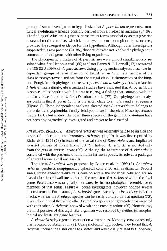
In cultureS. arcticumdevelops spherical structures containing several individualcells, which at maturity release∼100 smaller cells (B. Landfald, personal commu-nication). Electron microscopic studies ofS. arcticumshowed that the sphericalbodies, with well-defined internal spherical cells, were similar to the morpho-logical features of the algal species ofChlorella andPrototheca(Figure 8). Theinternal cells contained mitochondria with flat cristae and a well-defined nucleus.Attempts to infect related amphipod species withS. arcticumwere unsuccess-ful (B. Landfald, personal communication). However, the fact thatS. arcticum’s18S SSU rDNA sequence is 99.5% similar to thePs. tapetissequence suggests
Figure 8 Transmission electron microscopy section ofSphaerosoma arcticumfromcultured samples. The presence of numerous spores within the cytoplasm is its mainphenotypic characteristic. The morphological features thatS. arcticumshares with thespecies of the algal generaProtothecaandChlorellaare strikingly similar (X 26,400)(courtesy of B. Landfald).
Ann
u. R
ev. M
icro
biol
. 200
2.56
:315
-344
. Dow
nloa
ded
from
arj
ourn
als.
annu
alre
view
s.or
gby
NE
SLi2
on
08/2
2/08
. For
per
sona
l use
onl
y.
16 Aug 2002 13:56 AR AR168-MI56-14.tex AR168-MI56-14.SGM LaTeX2e(2002/01/18)P1: GJC
330 MENDOZA ¥ TAYLOR ¥ AJELLO
not only thatPs. tapetismay be perhaps a species of the genusSpaerosomabutalso thatS. arcticummay be a microbe associated with clams. Therefore, one canspeculate thatS. arcticummay be a nonpathogenic species associated with clams,as in the case ofPs. tapetis. This might explain in part why Landfald could notinfect amphipods withS. arcticum. Phylogenetic analysis of theS. arcticum’s 18SSSU rDNA showed that this organism is a member of the class Mesomycetozoea(B. Landfald, personal communication). In our analysisS. arcticumandPs. tapetisform the sister clade toA. richardsiplus the LKM51 isolate, and both groups arewell supported within the order Ichthyophonida (Figure 1).
ISOLATE LKM51 Isolate LKM51 is known only from an 18S SSU rDNA sequenceobtained from DNA isolated from phytoplankton (86a). Thus, mesomycetozoeanshave joined the many archaea, eubacteria, and uncultivated eukaryotic microbeswhose presence is known only from environmental DNA, but which could wellcomprise 70% of the earth’s microbiota (3, 43, 80, 94). Recently van Hannen et al.(86a), while investigating the correlation between the biomass of bacteria andthe biomass of protozoans in phytoplankton samples, found that the affiliation of∼20% of the 18S SSU rDNA sequences investigated during this study could not bequickly determined. One of these sequences, clone LKM51, was later grouped withthe mesomycetozoeans. Although the morphological features and other character-istics of the organism from which the DNA was isolated were not investigated, thiswas the first evidence that some mesomycetozoeans could be planktonic. Whetherisolate LKM51 represents a free-living planktonic organism or simply part ofits life cycle is not known, but were it shown to be free living, it would be theonly mesomycetozoean that is not an obligate parasite. Phylogenetic analysis ofthe LKM51 clone showed that it is the sister clade ofA. richardsiand well sup-ported within the order Ichthyophonida (86a). Our analysis gave the same results(Figure 1).
Order Dermocystida
DERMOCYSTIDIUM SPP. The genusDermocystidiumis the sister taxon ofRhi-nosporidium. The latter genus comprises approximately 12 species, all of whichcause deadly infections in fish and other marine animals (22, 27, 37, 66). TheDer-mocystidiumspp. are characterized by their spherical sporangia (cysts) and theproduction of endospore-like structures (Figure 9). Such features are also encoun-tered in the other members of the order Dermocystida (Table 1). These similaritiescould well explain why early investigators called attention to the morphologicalrelationship between theDermocystidiumspp. and their sister taxonR. seeberi(39). Owing to their morphological features, theDermocystidiumspp. were pre-viously considered to be either members of the haplosporeans, the fungi, or theapicomplexa groups (9, 26, 62, 70, 77, 88, 100).
Because of their intractability to cultivation, the ecological distribution ofDer-mocystidiumspp. is poorly known. It is believed that fish are infected through
Ann
u. R
ev. M
icro
biol
. 200
2.56
:315
-344
. Dow
nloa
ded
from
arj
ourn
als.
annu
alre
view
s.or
gby
NE
SLi2
on
08/2
2/08
. For
per
sona
l use
onl
y.

16 Aug 2002 13:56 AR AR168-MI56-14.tex AR168-MI56-14.SGM LaTeX2e(2002/01/18)P1: GJC
THE MESOMYCETOZOEANS 331
Figure 9 (a) Histological section of a fish’s gills infected with a sporangium ofDermocystidium salmoniscontaining numerous endospores (X 400). (b) Histologicalsection of an enlargement ofD. salmonis’s sporangium. Note the presence of well-developed endospores, a feature also observed inRhinosporidium seeberi(X 800)(courtesy of K.D. Arkush).
contact with the endospores released from infected fish. The spherical sporangiaof Dermocystidiummeasure 200–400µm in diameter and contain hundreds ofendospores. They are readily observed in infected gills (Figure 9). These sphericalsporangia are produced in great numbers to the point of making it impossible forthe infected fish to take up oxygen and they finally succumb to their infections.However, the presence of uniflagellate zoospores inD. salmonis(65) suggeststhat motile zoospores could be the infective propagules of theDermocystidiumspecies. Recently it has also been found that the rosette agent develops uniflagel-late zoospores (K.D. Arkush, personal communication). The rosette agent is also amember of the order Dermocystida and also causes a disease that primarily affectsfish gills.
Early reports ofDermocystidiumspp. suggested that the species of that genuscould cause infections not only in fish but also in amphibians (14, 15, 25). Basedon its spherical parasitic stage and the ecological distribution of its hosts, however,other investigators speculated that the true etiology of the spherical structures inamphibians were not theDermocystidiumspecies but members of a new genus(14). Accordingly, the genusDermosporidiumwas created to accommodate theDermocystidiumspp. in infected amphibians (14). However, based on morpho-logical characteristics, Herr et al. (39) speculated thatDermosporidium granulo-sum(14) andDermosporidium ranarum(15) should be identified asR. seeberiin
Ann
u. R
ev. M
icro
biol
. 200
2.56
:315
-344
. Dow
nloa
ded
from
arj
ourn
als.
annu
alre
view
s.or
gby
NE
SLi2
on
08/2
2/08
. For
per
sona
l use
onl
y.
16 Aug 2002 13:56 AR AR168-MI56-14.tex AR168-MI56-14.SGM LaTeX2e(2002/01/18)P1: GJC
332 MENDOZA ¥ TAYLOR ¥ AJELLO
amphibians rather than as species of the newly proposed genusDermosporidiumorspecies of the genusDermocystidium. Nevertheless, the phylogenetic connectionof the amphibian parasite withR. seeberihas yet to be established by comparisonof DNA sequences. It is important to note that the species ofDermocystidiumarealso phenotypically similar to those of the genusPerkinsus. Thus, thePerkinsusspecies have been studied in the past as members of the genusDermocystidium.A good example of misidentification isP. marinus, which was once erroneouslyincluded in the genusDermocystidiumasD. marinus(71). We now know, however,that members of both genera are phylogenetically distant (71).
The taxonomic affinities of theDermocystidiumspp. were resolved whenRagan et al. (77) sequenced their 18S SSU rDNAs. Those studies revealed that thesefish pathogens possess mitochondria with flat cristae and were related to other fishparasites. Later, Herr et al. (39) found that the genusDermocystidiumwas the sistertaxon ofR. seeberiand was closely related to the rosette agent. That study, in part,explained why the phenotypic features of theDermocystidiumspp. had been pre-viously confused withR. seeberiin the tissues of their infected hosts (14, 15, 25).Cavalier-Smith (18) placed the genusDermocystidiumin the order Dermocys-tida along with the rosette agent;R. seeberiwas added later (57) (Figure 1)(Table 1).
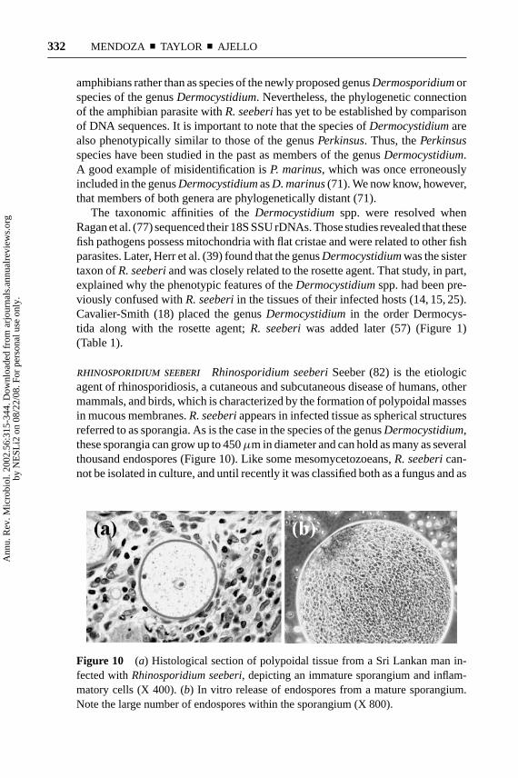
RHINOSPORIDIUM SEEBERI Rhinosporidium seeberiSeeber (82) is the etiologicagent of rhinosporidiosis, a cutaneous and subcutaneous disease of humans, othermammals, and birds, which is characterized by the formation of polypoidal massesin mucous membranes.R. seeberiappears in infected tissue as spherical structuresreferred to as sporangia. As is the case in the species of the genusDermocystidium,these sporangia can grow up to 450µm in diameter and can hold as many as severalthousand endospores (Figure 10). Like some mesomycetozoeans,R. seeberican-not be isolated in culture, and until recently it was classified both as a fungus and as
Figure 10 (a) Histological section of polypoidal tissue from a Sri Lankan man in-fected withRhinosporidium seeberi, depicting an immature sporangium and inflam-matory cells (X 400). (b) In vitro release of endospores from a mature sporangium.Note the large number of endospores within the sporangium (X 800).
Ann
u. R
ev. M
icro
biol
. 200
2.56
:315
-344
. Dow
nloa
ded
from
arj
ourn
als.
annu
alre
view
s.or
gby
NE
SLi2
on
08/2
2/08
. For
per
sona
l use
onl
y.

16 Aug 2002 13:56 AR AR168-MI56-14.tex AR168-MI56-14.SGM LaTeX2e(2002/01/18)P1: GJC
THE MESOMYCETOZOEANS 333
a protozoan (5, 6, 23). The possible development of uniflagellated zoospores byR. seeberi, similar to those ofDermocystidiumspp. and the rosette agent, is cur-rently under investigation (D. McMeekin & L. Mendoza, unpublished data).
Light and electron microscopic analyses in the past 100 years have indicatedthat R. seeberihas a complex in vivo life cycle (5, 22) that is initiated with therelease of endospores into its host’s tissues from spherical bodies (100–450µm)referred to as sporangia (Figure 10). Once implanted, the endospores increase insize and progressively develop into juvenile, intermediate, and finally mature spo-rangia with endospores (59). The endospores are then released and the in vivocycle is reinitiated. These analyses, however, did not provide clues regardingR. seeberi’s taxonomic affinities. In 1999 a team of investigators from MichiganState University, Emory University, University of California, Berkeley, and theUniversity of Paradeniya, Sri Lanka (39), using phylogenetic analysis, found thatthe 18S SSU rDNA ofR. seeberifrom humans with rhinosporidiosis clusteredwith the DRIPs, then a recently discovered group of fish parasites. Their datashowed thatR. seeberiwas the sister taxon of twoDermocystidiumspp. andthat this trio was close to the rosette agent. This study was later corroborated byFredericks et al. (30), whose 18S SSU rDNA sequence from a dog with rhi-nosporidiosis proved to be identical to that of the human isolate sequenced byHerr et al. (39). That finding strongly supported the view thatR. seeberimay bea monotypic genus. Although Frederick et al. (30) reported thatR. seeberipos-sessed mitochondria with tubular cristae, Herr et al. and Mendoza and colleagues(39, 58) demonstrated thatR. seeberidid indeed have mitochondria with flatcristae.
The phylogenetic home ofR. seeberihas been controversial for over a century.When Seeber described the first known infection caused byR. seeberiin 1900(82), he believed that this spherical microorganism was a coccidium. Later, otherinvestigators, using morphological and staining procedures, suggested that thepathogen was more closely related to members of the kingdom Fungi than tomembers of the kingdom Protoctista. Investigations mainly focused on the in vivohistopathological features ofR. seeberibecause it could not be cultivated. None ofthose investigations, however, provided clues as to the true nature of this pathogen.This frustration led other investigators to propose extreme views such as thatR. seeberiwas a carbohydrate waste product resulting from the ingestion of tapioca(1) or a cyanobacterium in the genusMicrocystis (2). The recent finding thatR. seeberi’s 18S SSU rDNA clustered with a novel clade of fish parasites in thedivergence between animals and fungi ended 100 years of taxonomic uncertainties.R. seeberidiffers from the other mesomycetozoeans in that it is the only memberpathogenic to mammals and birds. Correspondingly,R. seeberihas several featuresin common with the other mesomycetozoeans: (a) It was previously classifiedas a fungus or a different type of protozoan; (b) it was associated with aquaticenvironments; (c) it produced spherical structures containing several daughter cells(endospores); and (d) R. seeberiand some other mesomycetozoeans are intractableto culture (Table 1).
Ann
u. R
ev. M
icro
biol
. 200
2.56
:315
-344
. Dow
nloa
ded
from
arj
ourn
als.
annu
alre
view
s.or
gby
NE
SLi2
on
08/2
2/08
. For
per
sona
l use
onl
y.

16 Aug 2002 13:56 AR AR168-MI56-14.tex AR168-MI56-14.SGM LaTeX2e(2002/01/18)P1: GJC
334 MENDOZA ¥ TAYLOR ¥ AJELLO
ROSETTE AGENT This organism is an obligate intracellular fish parasite. The in-fections it causes were first described by Harrell et al. (35) in net-pen-rearedchinook salmon (Onchorhynchus tshawytscha). No generic name was proposed atthat time. It was only referred to as the rosette agent because of the false impressionthat it clustered as a six-celled organism, resembling a rosette, in infected tissues(28). Based on morphological findings, Harrell et al. (35) pointed out that thisunique salmon pathogen could either be a fungus, a protozoan, or an alga.
Infected salmon develop severe anemia and lymphocytosis. Swollen kidneysand spleens occur. Gram-stained smears of the infected tissue showed gram-positive spherical structures∼5–7µm in diameter. The spherical organisms werefound within the macrophages of infected kidneys and spleens. Transmission elec-tron microcopy of the infected areas revealed spherical cells with multilayered cellwalls, vacuoles, and a prominent nucleus (Figure 11). More recently, morpholog-ical and molecular studies of several isolates of the rosette agent showed thattheir 18S SSU rDNAs were identical. That study suggested that the rosette agentrepresented a new protozoan genus and species. Details of this proposal will bepublished elsewhere (K. D. Arkush, personal communication).
Experimental infections using rosette agent endospores taken from infectedsalmon tissues failed. Recently, however, Arkush at the University of Cali-fornia, Davis, induced the production of uniflagellated zoospores by the cells
Figure 11 (a) Transmission electron microscopy section of a salmon kidney’s inter-sticium infected with the rosette agent (X 27250). (b) Transmission electron microscopyshowing the rosette agent undergoing division by progressive internal cleavage of itscytoplasm (X 24500). (c) Enlargement of a rosette agent’s spherule showing two mi-tochondria with flat cristae (X 34024) (courtesy of R.A. Elston & K.D. Arkush).
Ann
u. R
ev. M
icro
biol
. 200
2.56
:315
-344
. Dow
nloa
ded
from
arj
ourn
als.
annu
alre
view
s.or
gby
NE
SLi2
on
08/2
2/08
. For
per
sona
l use
onl
y.

16 Aug 2002 13:56 AR AR168-MI56-14.tex AR168-MI56-14.SGM LaTeX2e(2002/01/18)P1: GJC
THE MESOMYCETOZOEANS 335
of the rosette agent. The zoospores were approximately 1–2µm in diameter andnearly spherical. They had only one flagellum∼10µm long with a typical 9+ 2configuration of microtubules. When the zoospores were used to infect salmon, theanimals developed lesions comparable to those in natural infections (K. D. Arkush,personal communication). In addition, the rosette agent has been successfully cul-tured using the chinook salmon embryo cell line CHSE-214 at 15◦C (4).
The rough phylogenetic relationships of the rosette agent were first revealedwhen Kerk et al. (44) characterized its18S SSU rDNA. Spanggaard et al. (84)corroborated this finding. Both studies revealed that the rosette agent was closelyrelated to the choanoflagellates. Ragan et al. (77) established that this pathogenwas part of a novel clade of parasites located between the divergence of fungiand animals, a finding corroborated by others (8, 12, 19, 29, 30, 39, 57, 101). In ourphylogenetic analyses the rosette agent is the sister clade toR. seeberiandDermo-cystidiumspp., all possessing flat mitochondrial cristae and all closely related toother members of the order Ichthyophonida (Figure 1). Interestingly, Arkush et al.(4) found that the rosette agent possessed tubular mitochondrial cristae. However, aclose evaluation of their electron microscopic figures and new photographic data in-dicated that the rosette agent might have mitochondria with flat cristae (Figure 11c).
RELATIONSHIPS OF THE MESOMYCETOZOEANSWITH THE OTHER CLADES ARISING NEAR THEANIMAL-FUNGAL DIVERGENCE
The report of Ragan et al. (77) that the mesomycetozoeans form a monophyleticclade near the animal-fungal divergence was an unexpected finding. The exact po-sition of this group, however, remains controversial. Ragan et al. (77) found that,depending on the sequences added to their phylogenetic trees, the mesomyceto-zoeans are either the sister group to the Animal kingdom plus the choanoflagel-lates or the sister group to the animals plus the choanoflagellates plus the fungi.Cavalier-Smith (18) argued that the mesomycetozoeans are the sister group tochoanoflagellates plusCorallochytrium, and not to choanoflagellates plus bothCorallochytriumand animals as inferred by Ragan et al. (77). Broader phylo-genetic studies tend to confirm Cavalier-Smith’s position (30, 39). Phylogeneticstudies including additional members of the order Ichthyophonida, suggested thatthe mesomycetozoeans are the sister clade to the choanoflagellates and that to-gether they are the sister group to animals and, with the inclusion of the animals,to the fungi (12, 29, 86). Our own phylogenetic analyses, using all the microbesin previous studies, indicated that mesomycetozoeans are indeed the sister groupto the choanoflagellates plusCorallochytriumand theNuclearia(Figure 1), thussupporting previous interpretations (18). However, it must be kept in mind thatnone of the deeper branches relating the Mesomycetozoea to other clades are wellsupported.
Based on the data published by several investigators (44, 77), Cavalier-Smith (18) proposed two different orders within the mesomycetozoeans, the
Ann
u. R
ev. M
icro
biol
. 200
2.56
:315
-344
. Dow
nloa
ded
from
arj
ourn
als.
annu
alre
view
s.or
gby
NE
SLi2
on
08/2
2/08
. For
per
sona
l use
onl
y.
16 Aug 2002 13:56 AR AR168-MI56-14.tex AR168-MI56-14.SGM LaTeX2e(2002/01/18)P1: GJC
336 MENDOZA ¥ TAYLOR ¥ AJELLO
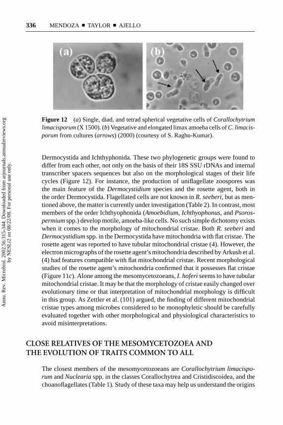
Figure 12 (a) Single, diad, and tetrad spherical vegetative cells ofCorallochytriumlimacisporum(X 1500). (b) Vegetative and elongated limax amoeba cells ofC. limacis-porumfrom cultures (arrows) (2000) (courtesy of S. Raghu-Kumar).
Dermocystida and Ichthyphonida. These two phylogenetic groups were found todiffer from each other, not only on the basis of their 18S SSU rDNAs and internaltranscriber spacers sequences but also on the morphological stages of their lifecycles (Figure 12). For instance, the production of uniflagellate zoospores wasthe main feature of theDermocystidiumspecies and the rosette agent, both inthe order Dermocystida. Flagellated cells are not known inR. seeberi, but as men-tioned above, the matter is currently under investigation (Table 2). In contrast, mostmembers of the order Ichthyophonida (Amoebidium, Ichthyophonus, andPsoros-permiumspp.) develop motile, amoeba-like cells. No such simple dichotomy existswhen it comes to the morphology of mitochondrial cristae. BothR. seeberiandDermocystidiumspp. in the Dermocystida have mitochondria with flat cristae. Therosette agent was reported to have tubular mitochondrial cristae (4). However, theelectron micrographs of the rosette agent’s mitochondria described by Arkush et al.(4) had features compatible with flat mitochondrial cristae. Recent morphologicalstudies of the rosette agent’s mitochondria confirmed that it possesses flat cristae(Figure 11c). Alone among the mesomycetozoeans,I. hoferiseems to have tubularmitochondrial cristae. It may be that the morphology of cristae easily changed overevolutionary time or that interpretation of mitochondrial morphology is difficultin this group. As Zettler et al. (101) argued, the finding of different mitochondrialcristae types among microbes considered to be monophyletic should be carefullyevaluated together with other morphological and physiological characteristics toavoid misinterpretations.
CLOSE RELATIVES OF THE MESOMYCETOZOEA ANDTHE EVOLUTION OF TRAITS COMMON TO ALL
The closest members of the mesomycetozoeans areCorallochytrium limacispo-rumandNucleariaspp. in the classes Corallochytrea and Cristidiscoidea, and thechoanoflagellates (Table 1). Study of these taxa may help us understand the origins
Ann
u. R
ev. M
icro
biol
. 200
2.56
:315
-344
. Dow
nloa
ded
from
arj
ourn
als.
annu
alre
view
s.or
gby
NE
SLi2
on
08/2
2/08
. For
per
sona
l use
onl
y.

16 Aug 2002 13:56 AR AR168-MI56-14.tex AR168-MI56-14.SGM LaTeX2e(2002/01/18)P1: GJC
THE MESOMYCETOZOEANS 337
of the Mesomycetozoea. AlthoughC. limacisporumis not a mesomycetozoean, itis included in this review for its phylogenetic proximity to that group.
Corallochytrium limacisporumis a novel type of saprotrophic marine protist(78). This organism was originally isolated from a coral reef in the Indian Ocean(78). Like some members of the class Mesomycetozoea, it is a spherical, single-celled organism, 4.5–20.0µm in diameter that undergoes several binary fissions tolater release numerous elongated daughter cells (up to 32 daughters per single cell)(Figure 12).C. limacisporumreleases its endospores through one or more poresin its cell wall, recalling the behavior ofR. seeberi. However, inR. seeberithere isonly one exit pore (59). The elongated released spores are amoeba-like and havea slow sinusoidal movement (Figure 12). The production of an amoebic stage is acharacteristic shared with mesomycetozoeans in the order Ichthyophonida. In thatorderAmoebidium parasiticumalso produces elongated spores and has an amoeba-like stage.C. limacisporumapparently possess mitochondria with flat cristae (19),but photomicrographs depicting these organelles have not been published, andtheir morphology needs verification (Table 2).
Using phylogenetic analyses of 18S SSU rDNA, Cavalier-Smith & Allsopp (19)reported thatC. limacisporumwas closely related to the choanoflagellates and tothe only member of the mesomycetozoeans known at that time, the rosette agent; itwas not a thraustochytrid, the group in which it had previously been placed. Basedon this result, Cavalier-Smith later created a new class, the Corallochytrea (18).Our phylogenetic analyses confirmed Cavalier-Smith’s placement of this microbeoutside of the mesomycetozoeans and close to the choanoflagellates (Table 1)(Figure 1).
Cavalier-Smith (16–18) placed the nucleariid amoeba in the phylum Neomon-ada, subphylum Mesomycetozoa (new subphylum that replaces the subphylumChoanozoa), class Cristidiscoidea, order Nucleariia, closely associated withC. limacisporumand the mesomycetozoeans (Table 1). According to the taxo-nomic classification of Cavalier-Smith (18), they all share the same phylum andsubphylum (Table 1). This was confirmed by Zettler et al. (101) and by our ownphylogenetic analyses (Figure 1). Members of the nucleariid amoeba are classi-fied according to their morphological characteristics, especially those related tolocomotion styles and feeding habits. Mitochondrial cristae morphology is impor-tant in the classification of nucleariid amoebae, which contain flat and discoidalmitochondrial cristae, the least common of the mitochondrial types. Based onphylogenetic analyses, Zettler et al. (101) speculated that the nucleariid amoebamight have developed discoidal mitochondrial cristae independently from othermicrobes that possess this type of cristae, again pointing out the evolutionary plas-ticity of this characteristic. The recent placement of the nucleariid amoeba nearthe mesomycetozoeans, choanoflagellates, and corallochytrians, and the rate atwhich new groups are being found at the animal-fungal boundary, suggest thatmore microbes at the same location will be found. BothCorallochytriumandNu-clearia spp. have amoeba or amoeba-like cells and neither have flagella, whereasanimals, choanoflagellates, some fungi, and some Mesomycetozoea retain flagella.
Ann
u. R
ev. M
icro
biol
. 200
2.56
:315
-344
. Dow
nloa
ded
from
arj
ourn
als.
annu
alre
view
s.or
gby
NE
SLi2
on
08/2
2/08
. For
per
sona
l use
onl
y.
16 Aug 2002 13:56 AR AR168-MI56-14.tex AR168-MI56-14.SGM LaTeX2e(2002/01/18)P1: GJC
338 MENDOZA ¥ TAYLOR ¥ AJELLO
Assuming that it is much easier to lose a flagellum than to gain one, it seems thattheCorallochytriumandNucleariaspp. must have lost flagella independently ofsimilar losses in the Ichthyophonida and several phyla of the kingdom Fungi (87).
Amoebal motility, like the flagellar type, appears to be an ancestral trait found inamoebal-flagellates like the plasmoidal slime molds and their relatives (9, 18, 87).Even in the kingdom Fungi, when flagellated zoospores, especially those of theBlastocladiales, are trapped under cover glass and microscope slides, amoebalmotility is observed (55). Cell walls also appear to be ancestral, being found in thechoanoflagellates, fungi, the Mesomycetozoea, theCorallochytrium, andNucle-aria spp., but not in the animals. Surely, cell walls were lost in animals. The ances-tors of all groups at the animal-fungal boundary must have been unicellular, andonly in the animals and fungi did multicellularity develop and become widespread.In the MesomycetozoeaI. hoferi is filamentous, but the filaments are cenocytic andlack regular septa. They resemble the fungal hyphae in the Blastocladiales, Ento-mophthorales, Mucorales, Glomales, and other zygomycete orders. The membersof those fungal orders similarly lack the multicellularity and macroscopic mor-phology typical of fungi in the Ascomycota and Basidiomycota. Multicellularityand macroscopic structures clearly developed independently in several lineages,including the green, brown, and red plants, in addition to fungi and animals. Fur-ther study may find macroscopic Mesomycetozoea. Most Mesomycetozoea areassociated with animals as parasites or commensals. We speculate thatSphaero-somaand the LKM51 isolate are saprotrophic and free-living. It seems likely thatother saprotrophic Mesomycetozoea will be found when methods of cultivatingthese organisms are applied to samples taken from nature.
FUTURE DIRECTIONS OF RESEARCH INTHE MESOMYCETOZOEA
What can we expect from the Mesomycetozoea in the near future, in addition to thediscovery of more members? The cultivation of more members seems likely, andwith it the possibility of more detailed comparisons of mesomycetozoean devel-opment with that of the other groups at the animal-fungal boundary. For example,the problem of mitochondrial form might be solved, as well as discovery of moreflagellated species in the order Dermocystida. Cultivation also increases the pos-sibility of gathering sequences from additional DNA regions in hopes of resolvingthe relationships among Mesomycetozoea, fungi, animals, and choanoflagellates.For example, Loytynoja & Milikovitch (55) have compared sequences of mito-chondrial ADP-ATP carriers to question the relationship of fungi and animals assister taxa, albeit without including the Mesomycetozoea or choanoflagellates. Itwould be worth extending the analysis to include these groups. The geological tim-ing of divergence of the Mesomycetozoea, fungi, and animals is also an importanttopic. Several estimates of the date when fungi diverged from animals have beenmade, ranging from∼1 billion (13) to 1.6 billion yeas ago (36). Again, the class
Ann
u. R
ev. M
icro
biol
. 200
2.56
:315
-344
. Dow
nloa
ded
from
arj
ourn
als.
annu
alre
view
s.or
gby
NE
SLi2
on
08/2
2/08
. For
per
sona
l use
onl
y.

16 Aug 2002 13:56 AR AR168-MI56-14.tex AR168-MI56-14.SGM LaTeX2e(2002/01/18)P1: GJC
THE MESOMYCETOZOEANS 339
Mesomycetozoea is not represented in these studies, and additional sequencesmight lead to new estimates of the timing of the divergence of these taxa. Culti-vation also introduces the possibility of genomic analyses, and given the obviousinterest in the origin of the Animal Kingdom, a strong case can be made for ob-taining the genome of a mesomycetozoean for comparative genomics. Importantcontributions can be made, however, with traditional approaches. For example,the life cycle of no member of the Mesomycetozoea is known completely. Inves-tigations to determine if gametes are produced, and the nuclear division needed toproduce them, as well as the fusion of gametes to form zygotes, would be mostwelcome, as they could bring genetics to the class.
ACKNOWLEDGMENTS
We thank all the investigators who shared their unpublished and in press infor-mation with us. We also thank Mat´ıas J. Cafaro and Roger A. Herr for theircontributions to some sections of this review.
The Annual Review of Microbiologyis online at http://micro.annualreviews.org
LITERATURE CITED
1. Ahluwalia KB. 1992. New interpretationsin rhinosporidiosis, enigmatic disease ofthe last nine decadesJ. Submicrosc. Cytol.Pathol.24:109–14
2. Ahluwalia KB. 1999. Culture of the or-ganism that causes rhinosporidiosis.J.Laryngol. Otol.113:523–28
3. Amann RI, Ludwing W, Schleifer KH.1995. Phylogenetic identification andinsitu detection of individual microbialcells without cultivation.Microbiol. Rev.59:143–69
4. Arkush KD, Frasca S Jr, Hedrick RP.1998. Pathology associated with the ros-ette agent, a systemic protist infectingsalmonid fishes.J. Aquat. Anim. Health10:1–11
5. Arseculeratne SN, Ajello L. 1998.Rhi-nosporidium seeberi. In Topley and Wil-son’s Microbiology and Microbial Infec-tions, ed. L Ajello, RJ Hay, 4:595–643.London/Sydney/Auckland: Arnold
6. Ashworth JH. 1923. OnRhinosporidiumseeberi(Wernecki, 1903) with special ref-erence to its sporulation and affinities.
Trans. R. Soc. London Edinb.53:301–42
7. Azevedo C. 1989. Fine structure ofPerkinsus atlanticusn. sp. (Apicomplexa,Perkinsea) parasite of the clamRuditapesdecussatesfrom Portugal.J. Parasitol.75:627–35
8. Baker GC, Beebee TJC, Ragan MA. 1999.Prototheca richardsi, a pathogen of anu-ran larvae, is related to a clade of protis-tan parasites near the animal-fungal diver-gence.Microbiology145:1777–84
9. Beakes GW. 1998. Relationships betweenlower fungi and protozoa. See Ref. 22a,pp. 351–73
10. Beebee TJC. 1991. Purification of anagent causing growth inhibition in anu-ran larvae and its identification as a uni-cellular unpigmented alga.Can. J. Zool.69:2146–53
11. Beebee TJC, Wong ALC. 1993. Stimu-lation of cell division and DNA replica-tion in Prototheca richardsiby passagethrough larval amphibian guts.Parasitol-ogy107:119–24
Ann
u. R
ev. M
icro
biol
. 200
2.56
:315
-344
. Dow
nloa
ded
from
arj
ourn
als.
annu
alre
view
s.or
gby
NE
SLi2
on
08/2
2/08
. For
per
sona
l use
onl
y.

16 Aug 2002 13:56 AR AR168-MI56-14.tex AR168-MI56-14.SGM LaTeX2e(2002/01/18)P1: GJC
340 MENDOZA ¥ TAYLOR ¥ AJELLO
12. Benny GL, O’Donnell K. 2000.Amoe-bidium parasiticumis a protozoan, nota trichomycete (Zygomycota).Mycologia92:1133–37
13. Berbee ML, Taylor JW. 2001. Fungalmolecular evolution: gene trees and ge-ologic time. In The Mycota: Systemat-ics and Evolution, ed. D McLaughlin,E McLaughlin P Lemke, VII, part B:229–45. Berlin: Springer-Verlag
14. Broz O, Privora M. 1951. Two skin par-asites ofRana temporaria: Dermocystid-ium ranaeGuyenot & Naville andDermo-sporidium granulosumn. sp.Parasitology24:65–69
15. Carini A. 1940. Sobre um pasasito semel-hante ao “Rhinosporidium” encontradoem quistos da pele de uma “hyla.”Arq.Inst. Biol. Sao Paulo11:93–96
16. Cavalier-Smith T. 1997. Amoeboflagel-lates and mitochondrial cristae in eukary-ote evolution: megasystematics of the newprotozoan subkingdoms Eozoa and Neo-zoa.Arch. Protistenkunde.147:237–58
17. Cavalier-Smith T. 1998. A revised six-kingdom system of life.Biol. Rev.73:203-66
18. Cavalier-Smith T. 1998. Neomonada andthe origin of animal and fungi. See Ref.22a, pp. 375–407
19. Cavalier-Smith T, Allsopp MTEP. 1996.Corallochytrium, an enigmatic non-flag-ellated protozoan related to choanoflag-ellates.Eur. J. Protistol.32:306–10
20. Caullery M, Menil F. 1905. Sur les hap-losporidies parasites de poisons marins.Cr. Seanc. Soc. Biol.58:640–43
21. Cienkowski L. 1861. Ueber parasitischeSchlauche auf Crustaceen und einigenInsektenlarven (Amoebidium parasiticumm.). Bot. Ztg.19:169–74
22. Chuan E, Kannan-Kutty M. 1975.Rhi-nosporidium seeberi: spherules and theirsignificance.Pathology7:133–37
22a. Coombs GH, Vickerman K, Sleigh MA,Warren A, eds. 1998.Evolutionary Rela-tionships Among Protozoa, Vol. 56. Dor-drecht, The Netherlands: Kluwer
23. Daniels RA. 1980. Distribution and sta-tus of crayfish in the Pit River drainage,California.Crustaceana38:131–38
24. de la Herran R, Garrido MA, Navas RJ,Rejon CR, Rejon MR. 2000. Molecularcharacterization of the ribosomal RNAgene region ofPerkinsus atlanticus: itsuse in phylogenetic analysis and as a tar-get for a molecular diagnosis.Parasitol-ogy120:345–53
25. Dukerly JS. 1914.Dermocystidium pus-ula Perez, parasitic on trutta fario.Zool.Anz.44:179–82
26. Dykova L, Lom J. 1992. New evidenceof fungal nature ofDermocystidium koiHoshina and Sahara, 1950.J. Appl. Ich-thyol.8:180–85
27. Eiras JC, Silva-Souza AT. 2000. ADer-mocystidium infection in Trichomycte-russp. (Osteichthyes, Trichomycteridae).Parasite7:323–26
28. Elston RA, Harrell L, Wilkinson MT.1986. Isolation and in vitro characteri-zation of Chinook salmon (Oncorhyncustshawytscha) rosette agent.Aquaculture56:1–21
29. Figueras A, Lorenzo G, Ordas MC, GouyM, Novoa B. 2000. Sequence of the smallsubunit ribosomal RNA gene ofPerkinsusatlanticus-like isolated from carpet shellclam in Galicia, Spain.Mar. Biotechnol.2:419–28
30. Fredericks DN, Jolley JA, Lepp PW,Kosek JC, Relman DA. 2000.Rhino-sporidium seeberi: a human pathogenfrom a novel group of aquatic protistanparasites.Emerg. Infect. Dis.6:273–82
31. Goggin CL. 1994. Variation in the inter-nal transcribed spacers and 5.8S riboso-mal RNA from 5 isolates of marine par-asitesPerkinsus(Protist, Apicomplexa).Mol. Biochem. Parasitol.65:179–82
32. Gunter V, Rug M. 1999. Life stagesand tentative life cycle ofPsorospermiumhaeckeli, a species of the novel DRIPsclade from the animal-fungal dichotomy.J. Exp. Zool.283:31–42
33. Haeckel E. 1857. Ueber die gewebe des
Ann
u. R
ev. M
icro
biol
. 200
2.56
:315
-344
. Dow
nloa
ded
from
arj
ourn
als.
annu
alre
view
s.or
gby
NE
SLi2
on
08/2
2/08
. For
per
sona
l use
onl
y.
16 Aug 2002 13:56 AR AR168-MI56-14.tex AR168-MI56-14.SGM LaTeX2e(2002/01/18)P1: GJC
THE MESOMYCETOZOEANS 341
flusskrebses.Arch. Anat. Physiol. Wiss.Med.24:469–568
34. Deleted in proof35. Harrell LW, Elston RA, Scott TM, Wilkin-
son MT. 1986. A significant new systemicdisease of net-pen reared chinook salmon(Oncorhyncus tshawytscha) brook stock.Aquaculture55:249–62
36. Heckman DS, Geiser DM, Eidell BR,Stauffer RL, Kardos NL, Hedges SB.2001. Molecular evidence for the earlycolonization of land by fungi and plants.Science293:1129–33
37. Hedrick RP, Friedman CS, Modin J. 1989.Systemic infection in Atlantic salmonSalmo salarwith a Dermocystidium-likespecies.Dis. Aquat. Org.7:171–77
38. Herman RL. 1984.Ichthyophonus-likeinfection in newts (Notophthalmus viri-descensrafinisque).J. Wildl. Dis.20:55–56
39. Herr AR, Ajello L, Taylor JW, Arseculer-atne SN, Mendoza L. 1999. Phylogeneticanalysis ofRhinosporidium seeberi’s 18Ssmall-subunit ribosomal DNA groups thispathogen among members of the protoc-tistant mysomycetozoan clade.J. Clin.Microbiol. 37:2750–54
40. Herr AR, Ajello L, Mendoza L. 1999. Chi-tin synthase class 2 (CHS2) gene from thehuman and animal pathogenRhinospori-dium seeberi. Proc. 99th Gen. Meet. Am.Soc. Microbiol.p. 296 (Abstr.)
41. Deleted in proof42. Hilgendorf F. 1883. Bemerkungen ¨uber
die sogenanntekrebspest, insbesondereuberPsorospermium haeckelispec. nova.Sitz. Gess. Nat. Freun. Berlin9:179–83
43. Hugenholtz P, Goebel BM, Pace NR.1998. Impact of culture-independent stu-dies on the emerging phylogenetic viewof bacterial diversity.J. Bacteriol. 180:4765–74
44. Kerk D, Gee A, Standish M, WainwrightPO, Drum AS, et al. 1995. The rosetteagent of Chinook salmon (Oncorhyncustshawytscha) is closely related to choa-noflagellates, as determined by phyloge-
netic analyses of its small ribosomal sub-unit RNA. Mar. Biol. 122:187–92
45. Kocan RM, Hershberger P, Mehl T, ElderN, Bradley M, et al. 1999. Pathogenicityof Ichthyosphonus hoferifor laboratory-reared pacific herringClupea pallasiandits early appearance in wild Puget Soundherring.Dis. Aquat. Org.35:23–29
46. Kotob SI, McLaughlin SM, van Berkum P,Faisal M. 1999. Discrimination betweentwo Perkinsusspp. isolated from the soft-shell clam,Mya arenaria, by sequenceanalysis of two internal transcribed spacerregion and the 5.8S ribosomal RNA gene.Parasitology119:363–68
47. Krucinska J, Simon E. 1968. On the para-sites and epibionts of the branchial cavityin crayfish at Wrocaw and vicinity.Prze-gald. Zool.12:288–90
48. Leeddale GF. 1974. How many are thekingdoms of organisms?Taxon32:261–70
49. Leger L. 1924. Sur un organisme du typeIchthyophonusparasite du tube digestifde la Lote d’eau douce.Cr. Hebd. Seanc.Acad. Sci. Paris197:785–87
50. Leger L, Hesse E. 1923. Sur un cham-pignon du typeIchthyophonusparasite del’intestin de la truite.Cr. Hebd. Seanc.Acad. Sci. Paris176:420–22
51. Levine ND. 1970. Taxonomy of the sporo-zoa.J. Parasitol.56:208–9
52. Lichtwardt RW. 1997. Costa Rican gutfungi (Trichomycetes) infecting lotic in-sect larvae.Rev. Biol. Trop.45:1339–83
53. Lichtwardt RW, Arenas J. 1996. Tri-chomycetes in aquatic insects from south-ern Chile.Micologia88:844–57
54. Lichtwardt RW, Williams MC. 1990. Tri-chomycete gut fungi in Australian aquaticinsect larvae.Can. J. Bot.1057–74
55. Loytynoja A, Milikovitch MC. 2001. Mo-lecular phylogenetic analyses of the mito-chondrial ADP-ATP carriers: the Plantae/Fungi/Metazoa trichotomy revisitedProc.Natl. Acad. Sci. USA98:10202–7
56. Margulis L. 1996. Archeal-eubacterial
Ann
u. R
ev. M
icro
biol
. 200
2.56
:315
-344
. Dow
nloa
ded
from
arj
ourn
als.
annu
alre
view
s.or
gby
NE
SLi2
on
08/2
2/08
. For
per
sona
l use
onl
y.

16 Aug 2002 13:56 AR AR168-MI56-14.tex AR168-MI56-14.SGM LaTeX2e(2002/01/18)P1: GJC
342 MENDOZA ¥ TAYLOR ¥ AJELLO
mergers in the origin of eukarya: phylo-genetic classification of life.Proc. Natl.Acad. Sci. USA93:1071–76
57. Mendoza L, Ajello L, Taylor JW. 2001.The taxonomic status ofLacazia loboiandRhinosporidium seeberihas been finallyresolved with the use of molecular tools.Rev. Iberoam. Micol.18:95–98
58. Mendoza L, Herr AR, Ajello L. 2001.Causative agent of rhinosporidiosis.J.Clin. Microbiol. 39:413–15
59. Mendoza L, Herr RA, Arseculeratne SN,Ajello L. 1999.In vitro studies on the me-chanism of endospore release byRhino-sporidium seeberi. Mycopathologia148:9–15
60. Mort-Bontemps M, Gay L, Fevre M.1997.CHS2, a chitin synthase gene fromthe OomyceteSaprolegnia monoica. Mi-crobiol. 143:2009–20
61. Mulisch M. 1993. Chitin in protistan or-ganisms.Eur. J. Protistol.29:1–18
62. Neish GA, Hughes GC. 1980. Fungal dis-eases of fishes, Book 6. InDiseases ofFishes, ed. SF Snieszco, HR Axelrod,pp. 61–100. USA/New Jersey/Gt. Britain/Surrey: TFH
63. Nylund V, Westman K, Laintmaa K.1983. Ultrastructure and taxonomic po-sition of the crayfish parasitePsoro-spermium haeckeliHeilgendorf.Freshw.Crayfish5:307–14
64. Okamoto N, Nakase K, Suzuki H, NakaiY, Fujii K, Sano T. 1985. Life history andmorphology of Ichthyophonus hoferiinvitro. Fish Pathol.20:273–85
65. Olson RE, Dungan CF, Holt RA. 1991.Water-borne transmission ofDermocys-tidium salmonisin the laboratory.Dis.Aquat. Org.12:41–48
66. Olson RE, Holt RA. 1995. The gill path-ogenDermocystidium salmonisin Oregonsalmonids.J. Aquat. Anim. Health7:111–17
67. Ordas MC, Figueras A. 1998.In vitro cul-ture ofPerkinsus atlanticus, a parasite ofthe carpet shell clamRuditapes decus-sates. Dis. Aquat. Org.33:129–36
68. Ordas MC, Novoa B, Faisal M, Mclaugh-lin S, Figueras A. 2001. Proteolytic ac-tivity of culturedPseudoperkinsus tapetisextracellular products.Comp. Biol. Phys-iol. B 130:199–206
69. Ordas MC, Novoa B, Figueras A. 2000.Modulation of the chemiluminescence re-sponse of Mediterranean mussel (Mytilusgalloprovencialis) haematocytes.FishShellfish Immunol.10:611–22
70. Paperna I, Kim SH. 1996. Ultrastructureof the etiological agent of systemic granu-loma in goldfish.Dis. Aquat. Org.26:43–47
71. Perkins FO. 1996. The structure ofPerkin-sus marinus(Mackin, Owen and Collier,1950) Levine, 1978 with comments onthe taxonomy and phylogeny ofPerkin-susspp.J. Shellfish Res.15:67–87
72. Plehn M, Mulsow K. 1911. Der erregerder “taumelkrankheir” der salmoniden.Zentbl. Bakt. ParasitKde, Abt.58:63–68
73. Pore RS. 1985.Protothecataxonomy.My-copathologia90:129–39
74. Porter D, Smiley R. 1979. RibosomalRNA molecular weights of trichomycetesand zygomycetes.Exp. Mycol.3:188–93
75. Rand TG. 1994. An unusual form ofIchthyophonus hoferi(Icthyophonales:Ichthyophonaceae) from yellowtail floun-der Limanda ferrugineafrom the NovaScotia shelf.Dis. Aquat. Org.18:21–28
76. Rand TG, White K, Cannone JJ, GutellRR, Murphy CA, Ragan MA. 2000.Ichthyophonus irregularissp. nov. fromthe yellowtail flounderLimanda ferrug-inea from the Nova Scotia shelf.Dis.Aquat. Organ.41:31–36
77. Ragan MA, Goggins CL, Cawthorn RJ,Cerenius L, Jamienson AVC, et al. 1998.A novel clade of protistan parasites nearthe animal-fungal divergence.Proc. Natl.Acad. Sci. USA93:11907–12
78. Raghu-Kumar S. 1987. Occurrence of thetraustochytrid,Corallochytrium limacis-perumgen. et sp. nov. in the coral reef la-goons of the Lakshadweep Islands in theArabian Sea.Bot. Mar.30:83–89
Ann
u. R
ev. M
icro
biol
. 200
2.56
:315
-344
. Dow
nloa
ded
from
arj
ourn
als.
annu
alre
view
s.or
gby
NE
SLi2
on
08/2
2/08
. For
per
sona
l use
onl
y.
16 Aug 2002 13:56 AR AR168-MI56-14.tex AR168-MI56-14.SGM LaTeX2e(2002/01/18)P1: GJC
THE MESOMYCETOZOEANS 343
79. Richards CM. 1958. The inhibition ofgrowth in crowdedRana pipienstadpoles.Physiol. Zool.31:138–51
80. Rondon MR, Augus PR, Bettermann AD,Brady SF, Grossman TH, et al. 2000.Cloning the soil metagenome: a strategyfor accessing the genetic and functionaldiversity of uncultured microorganisms.Appl. Environ. Microbiol.66:2541–47
81. Rug M, Vogt G. 1994. Developmentalstages of the crayfish parasitePsorosper-mium haeckeliin thoraxic arteries ofAsta-cus astacus. J. Invert. Pathol.64:153–55
82. Seeber GR. 1900.Un nuevo sporozuarioparasito del hombre. Dos casos encontra-dos en polypos nasals. MD thesis, Univ.Nacional Buenos Aires, Imprenta Libreria“Boullosa”
83. Spanggaard B, Huss HH, BrescianniJ. 1995. Morphology ofIchthyophonushoferiassessed by light and scanning elec-tron microscopy.J. Fish Dis.18:567–77
84. Spanggaard B, Skouboe P, Rossen L,Taylor JW. 1996. Phylogenetic relation-ships of the intercellular fish pathogenIchthyophonus hoferi, and fungi, choano-flagellates and the rosette agent.Mar. Biol.126:109–15
85. Trotter MJ, Whistler HC. 1965. Chemi-cal composition of the cell wall ofAmoe-bidium parasiticum. Can. J. Bot.43:869–76
86. Ustinova I, Kienitz L, Huss VAR. 2000.Hyaloraphidium curvatumis not a greenalga, but a lower fungus;Amoebidiumparasiticumis not a fungus, but a memberof the DRIPs.Protist151:253–62
86a. van Hannen EJ, Mooij W, van AgterveldMP, Gons HJ, van Laanbroek P. 1999.Detritus-dependent development of themicrobial community in an experimen-tal system: quality analysis by denaturinggradient gel electrophoresis.Appl. Envi-ron. Microbiol.65:2478–84
87. Vickerman K. 1998. Revolution amongthe protozoa. See Ref. 22a, pp. 1–24
88. Vıtovec SCJ, Lom J, Kubu JHF. 1974.Dermocystidiosis—a gill disease of the
carp due toDermocystidium cyprinin.sp.J. Fish Biol.6:689–99
89. Vogt G. 1999. In vitro induction of furtherdevelopment of sporocysts of the crayfishparasitePsorospermium haeckeli.Freshw.Crayfish12:319–34
90. Vogt G, Keller M, Brandis D. 1996.Occurrence ofPsorospermium haeckeliin the stone crayfishAustropotamobiustorrentium from a population naturallymixed with the noble crayfishAstacus as-tacus. Dis. Aquat. Org.25:233–38
91. Vogt G, Rug M. 1995. Microscopicanatomy and histochemistry of the cray-fish parasitePsorospermium haeckely.Dis. Aquat. Org.21:79–90
92. Vogt G, Rug M. 1999. Life stages and ten-tative life cycle ofPsorospermium haeck-eli, a species of a novel DRIPs cladefrom the animal-fungal dichotomy.J. Exp.Zool.283:31–42
92a. von Dietheilm-Scheer D. 1979.Psoros-permium orconectisn. sp. a new parasitein Orconectes limosus. Arch. Protistenkd.121:381–91
93. Voronin VN. 1975. On findings ofPsoros-permium lacustris. Izvest. Gos. Nauch.Instit. Oz. Irech. Ryb. Khoz.(Leningrad)93:125–26
94. Ward DM, Weller R, Bateson MM. 1990.16S rDNA sequences reveal numerous un-cultured microorganisms in natural com-munity.Nature345:63–65
95. Wainright PO, Hinkle G, Sogin ML,Stickel SK. 1993. Monophyletic origins ofthe metazoans: an evolutionary link withfungi. Science260:340–42
96. Whisler HC. 1962. Culture and nutritionof Amoebidium parasiticum. Am. J. Bot.29:193–99
97. Whisler HC. 1968. Developmental con-trol of Amoebidium parasiticum. Dev.Biol. 17:562–70
98. Whisler HC, Fuller M. 1968. Preliminaryobservations on the holdfast ofAmoebid-ium parasiticum. Micologia60:1068–79
99. Wong A, Beebee T. 1994. Identificationof a unicellular, non-pigmented alga
Ann
u. R
ev. M
icro
biol
. 200
2.56
:315
-344
. Dow
nloa
ded
from
arj
ourn
als.
annu
alre
view
s.or
gby
NE
SLi2
on
08/2
2/08
. For
per
sona
l use
onl
y.

16 Aug 2002 13:56 AR AR168-MI56-14.tex AR168-MI56-14.SGM LaTeX2e(2002/01/18)P1: GJC
344 MENDOZA ¥ TAYLOR ¥ AJELLO
that mediates growth inhibition in anu-ran tadpoles: a new species of the genusPrototheca(Chlorophyceae: Chlorococ-cales).Hydrobiologia277:85–96
100. Wootten R, Mcvicar AH. 1982.Dermo-cystidiumfrom cultured eels,Anguilla an-
guilla L., in Scotland.J. Fish Dis.5:215–22
101. Zettler LAA, Nerad TA, O’Kelly CJ,Sogin ML. 2001. The nucleariid amoeba:more protists at the animal-fungal bound-ary.J. Eukaryot. Microbiol.48:293–97
Ann
u. R
ev. M
icro
biol
. 200
2.56
:315
-344
. Dow
nloa
ded
from
arj
ourn
als.
annu
alre
view
s.or
gby
NE
SLi2
on
08/2
2/08
. For
per
sona
l use
onl
y.
P1: FRK
August 9, 2002 8:38 Annual Reviews AR168-FM
Annual Review of MicrobiologyVolume 56, 2002
CONTENTS
FRONTISPIECE, Evelyn M. Witkin xii
CHANCES AND CHOICES: COLD SPRING HARBOR 1944–1955,Evelyn M. Witkin 1
FUNCTION OF PSEUDOMONAS PORINS IN UPTAKE AND EFFLUX,Robert E. W. Hancock and Fiona S. L. Brinkman 17
THE BITTERSWEET INTERFACE OF PARASITE AND HOST:LECTIN-CARBOHYDRATE INTERACTIONS DURING HUMAN INVASION
BY THE PARASITE ENTAMOEBA HISTOLYTICA, William A. Petri Jr.,Rashidul Haque, and Barbara J. Mann 39
HEAVY METAL MINING USING MICROBES, Douglas E. Rawlings 65
MICROSPORIDIA: BIOLOGY AND EVOLUTION OF HIGHLY REDUCED
INTRACELLULAR PARASITES, Patrick J. Keeling and Naomi M. Fast 93
BACTERIOCINS: EVOLUTION, ECOLOGY, AND APPLICATION,Margaret A. Riley and John E. Wertz 117
EVOLUTION OF DRUG RESISTANCE IN CANDIDA ALBICANS,Leah E. Cowen, James B. Anderson, and Linda M. Kohn 139
BIOTERRORISM: FROM THREAT TO REALITY, Ronald M. Atlas 167
BIOFILMS AS COMPLEX DIFFERENTIATED COMMUNITIES, P. Stoodley,K. Sauer, D. G. Davies, and J. W. Costerton 187
MICROBIAL COMMUNITIES AND THEIR INTERACTIONS IN SOIL AND
RHIZOSPHERE ECOSYSTEMS, Angela D. Kent and Eric W. Triplett 211
TRANSITION METAL TRANSPORT IN YEAST, Anthony Van Ho,Diane McVey Ward, and Jerry Kaplan 237
INTEINS: STRUCTURE, FUNCTION, AND EVOLUTION, J. Peter Gogarten,Alireza G. Senejani, Olga Zhaxybayeva, Lorraine Olendzenski,and Elena Hilario 263
TYPE IV PILI AND TWITCHING MOTILITY, John S. Mattick 289
THE CLASS MESOMYCETOZOEA: A HETEROGENEOUS GROUP OF
MICROORGANISMS AT THE ANIMAL-FUNGAL BOUNDARY,Leonel Mendoza, John W. Taylor, and Libero Ajello 315
METABOLIC DIVERSITY IN AROMATIC COMPOUND UTILIZATION BY
ANAEROBIC MICROBES, Jane Gibson and Caroline S. Harwood 345vi
Ann
u. R
ev. M
icro
biol
. 200
2.56
:315
-344
. Dow
nloa
ded
from
arj
ourn
als.
annu
alre
view
s.or
gby
NE
SLi2
on
08/2
2/08
. For
per
sona
l use
onl
y.
P1: FRK
August 9, 2002 8:38 Annual Reviews AR168-FM
CONTENTS vii
THE MOLECULAR BIOLOGY OF WEST NILE VIRUS: A NEW
INVADER OF THE WESTERN HEMISPHERE, Margo A. Brinton 371
MICROBIAL DEGRADATION OF POLYHYDROXYALKANOATES,Dieter Jendrossek and Rene Handrick 403
MENACING MOLD: THE MOLECULAR BIOLOGY OF ASPERGILLUS
FUMIGATUS, Axel A. Brakhage and Kim Langfelder 433
WHAT ARE BACTERIAL SPECIES? Frederick M. Cohan 457
GENOME REMODELING IN CILIATED PROTOZOA, Carolyn L. Jahn andLawrence A. Klobutcher 489
COMMON PRINCIPLES IN VIRAL ENTRY, Minna M. Poranen,Rimantas Daugelavicius, and Dennis H. Bamford 521
CROSS-SPECIES INFECTIONS AND THEIR ANALYSIS, Man-Wah Tan 539
BACTERIAL CHROMOSOME SEGREGATION, Geoffrey C. Draperand James W. Gober 567
IMPACT OF GENOMIC TECHNOLOGIES ON STUDIES OF BACTERIAL GENE
EXPRESSION, Virgil Rhodius, Tina K. Van Dyk, Carol Gross,and Robert A. LaRossa 599
CONTROL OF CHROMOSOME REPLICATION IN CAULOBACTER CRESCENTUS,Gregory T. Marczynski and Lucy Shapiro 625
THE PREVALENCE AND MECHANISMS OF VANCOMYCIN RESISTANCE
IN STAPHYLOCOCCUS AUREUS, Timothy R. Walsh and Robin A. Howe 657
POLIOVIRUS CELL ENTRY: COMMON STRUCTURAL THEMES IN VIRAL
CELL ENTRY PATHWAYS, James M. Hogle 677
PRIONS AS PROTEIN-BASED GENETIC ELEMENTS, Susan M. Uptainand Susan Lindquist 703
MECHANISMS OF SOLVENT TOLERANCE IN GRAM-NEGATIVE BACTERIA,Juan L. Ramos, Estrella Duque, Marıa-Trinidad Gallegos,Patricia Godoy, Marıa Isabel Ramos-Gonzalez, Antonia Rojas,Wilson Teran, and Ana Segura 743
GROWING OLD: METABOLIC CONTROL AND YEAST AGING,S. Michal Jazwinski 769
INDEXES
Subject Index 793Cumulative Index of Contributing Authors, Volumes 52–56 833Cumulative Index of Chapter Titles, Volumes 52–56 836
ERRATA
An online log of corrections to Annual Review of Microbiology chapters(if any, 1997 to the present) may be found at http://micro.annualreviews.org/
Ann
u. R
ev. M
icro
biol
. 200
2.56
:315
-344
. Dow
nloa
ded
from
arj
ourn
als.
annu
alre
view
s.or
gby
NE
SLi2
on
08/2
2/08
. For
per
sona
l use
onl
y.