the role of rad4 in dna repair and its interplay with
TRANSCRIPT
BearWorks BearWorks
MSU Graduate Theses
Summer 2018
The Role of RAD4 in DNA Repair and Its Interplay with Telomeres The Role of RAD4 in DNA Repair and Its Interplay with Telomeres
in Tetrahymena thermophila in Tetrahymena thermophila
Emily Nischwitz Missouri State University, [email protected]
As with any intellectual project, the content and views expressed in this thesis may be
considered objectionable by some readers. However, this student-scholar’s work has been
judged to have academic value by the student’s thesis committee members trained in the
discipline. The content and views expressed in this thesis are those of the student-scholar and
are not endorsed by Missouri State University, its Graduate College, or its employees.
Follow this and additional works at: https://bearworks.missouristate.edu/theses
Part of the Cell Biology Commons, and the Molecular Biology Commons
Recommended Citation Recommended Citation Nischwitz, Emily, "The Role of RAD4 in DNA Repair and Its Interplay with Telomeres in Tetrahymena thermophila" (2018). MSU Graduate Theses. 3294. https://bearworks.missouristate.edu/theses/3294
This article or document was made available through BearWorks, the institutional repository of Missouri State University. The work contained in it may be protected by copyright and require permission of the copyright holder for reuse or redistribution. For more information, please contact [email protected].

THE ROLE OF RAD4 IN DNA REPAIR AND ITS INTERPLAY WITH
TELOMERES IN TETRAHYMENA THERMOPHILA
A Masters Thesis
Presented to
The Graduate College of
Missouri State University
TEMPLATE
In Partial Fulfillment
Of the Requirements for the Degree
Master of Science Cell and Molecular Biology
By
Emily Nischwitz
August 2018
ii
THE ROLE OF RAD4 IN DNA REPAIR AND ITS INTERPLAY WITH
TELOMERES IN TETRAHYMENA THERMOPHILA
Biomedical Sciences
Missouri State University, August 2018
Master of Science in Cell and Molecular Biology
Emily Nischwitz
ABSTRACT
Telomeres are repetitive parts of the genome that act as a protective end cap to the
chromosomes. Telomeres are critical to the integrity and stability of the genome,
therefore, ensuring that their sequence is maintained, even after damage, is crucial. Much
of the pioneering work responsible for explaining telomeres has been conducted in
ciliates, specifically in Tetrahymena thermophila. Telomeres in T. thermophila have a
high amount of tandem thymine repeats (GGGGTT) and, thus, are susceptible to
ultraviolet light (UV) induced lesions called pyrimidine dimers, which must be repaired
by nucleotide excision repair (NER). In humans, Xeroderma Pigmentosum C (XPC) is a
protein that helps recognize DNA damage in NER. The Tetrahymena thermophila
homolog to XPC (RAD4) showed strong evolutionary conservation to higher level
eukaryotes making it an ideal model organism. RAD4 expression was depleted by cloning
and expressing a short hairpin RNA gene that will target RAD4. After both UV and
hydrogen peroxide treatment, Rad4 depleted cells had reduced survivability. Moreover, a
DIG-labeled detection assay was developed for detection of telomere length, without the
use of radioactivity. Telomere length increased in the absence of Rad4 which reveals its
role in telomere maintenance. This work ultimately provides two new resources to the
fields of NER and telomeres by means of Rad4 knockdown T. thermophila strains, and
DIG-labeled telomere detection assay.
KEYWORDS: DNA repair, telomeres, RAD4, XPC, NER, Tetrahymena thermophila,
DIG labeling
This abstract is approved as to form and content
_______________________________
Dr. Joshua J. Smith
Chairperson, Advisory Committee
Missouri State University
iii
THE ROLE OF RAD4 IN DNA REAPIR AND ITS INTERPLAY WITH
TELOMERES IN TETRAHYMENA THERMOPHILA
By
Emily Nischwitz
A Masters Thesis
Submitted to the Graduate College
Of Missouri State University
In Partial Fulfillment of the Requirements
For the Degree of Master of Science in Cell and Molecular Biology
August 2018
Approved:
_______________________________________
Joshua J. Smith, PhD
_______________________________________
Randi J. Ulbricht, PhD
_______________________________________
Colette M. Witkowski, PhD
_______________________________________
Julie J. Masterson, PhD: Dean, Graduate
College
In the interest of academic freedom and the principle of free speech, approval of this thesis indicates the
format is acceptable and meets the academic criteria for the discipline as determined by the faculty that
constitute the thesis committee. The content and views expressed in this thesis are those of the student-
scholar and are not endorsed by Missouri State University, its Graduate College, or its employees.

iv
ACKNOWLEDGEMENTS
I want to thank the entire Biomedical Sciences Department for their years of
support. Each professor and staff member has made my education a fruitful and life
changing endeavor. Thank you to the graduate college for thesis funding and support.
I would also like to acknowledge Dr. Benjamin Linger for his guidance and
advice in the design of DIG labeled telomere detection system, and Dr. Wayne Mitchell
for his assistance and guidance in the statistical analysis within this thesis. Also, a special
thank you to the members of Smith Lab who have assisted in this project: Inyeong Lee,
Taylor Walker, and Lynsie Daniels.
Additionally, each of my committee members has been invaluable. Dr. Smith’s
mentorship and guidance has made me into the scientist and person I am today. He has
created an impression on me that will last a life time, and I am eternally grateful for all of
the opportunities he has given me. Both Dr. Ulbricht and Dr. Witkowski have fostered
such growth in me as both a scientist and writer, and specifically Dr. Ulbricht’s advice
both in regards to my project and life will be something I will take with me for years.
Without the support of my friends and family this would not have been possible.
With all my love, I would like to thank my mother Maria Nischwitz and my father James
Nischwitz for always encouraging and supporting me in the pursuit of my education and
dreams.

v
TABLE OF CONTENTS
Introduction ..........................................................................................................................1
Telomeres .................................................................................................................1
Telomere and Telomerase Discovery ......................................................................3
Shelterin Proteins .....................................................................................................5
Telomere Proteins in Tetrahymena thermophila .....................................................8
Telomere Dysregulation...........................................................................................9
Nucleotide Excision Repair ...................................................................................11
Nucleotide Excision Repair at the Telomeres ........................................................18
Purpose Statement ..................................................................................................19
Experimental Procedures ...................................................................................................21
Bioinformatics – Phylogenetic Tree Design and shRNA Primer Design ..............21
shRNA Plasmid Construction ................................................................................22
Electroporation of DH10B Escherichia coli with pBT1-shRNA:RAD4 ...............24
Lysozyme Boil Plasmid Isolation and Ethanol Precipitation ................................24
Restriction Enzyme Digest Confirmation of pBT1-shRNA:RAD4.......................25
Linearization of pBT1-shRNA:RAD4 for Biolistic Transformation .....................26
Cell Culture Maintenance and Strains Used ..........................................................26
Biolistic Transformation of Tetrahymena thermophila .........................................27
Knock Down Confirmation via Whole-Cell PCR .................................................29
Total RNA Isolations .............................................................................................30
Quantitative Reverse Transcriptase Polymerase Chain Reaction (qRT-PCR) ......31
Survivability Assay of Rad4 Knock Down Strains in T. thermophila ...................32
T. thermophila Genomic DNA Isolation ...............................................................32
DIG-Labeled Telomere Probe Detection Assay ....................................................33
Statistical Analysis .................................................................................................35
Results ...............................................................................................................................36
Conservation of Shelterin and XPC/RAD4 Proteins .............................................36
Quantification of Tetrahymena thermophila RAD4 Expression Levels Before
and After Damage ..................................................................................................39
Established Rad4 Knock Down Strains in T. thermophila ....................................45
Verification of Rad4 Knock Down Strains in T. thermophila ...............................48
Effect of UV and Hydrogen Peroxide on Rad4 Knock Down Survivability .........54
Experimental Design and Optimization of DIG-labeled Telomere Probe
Detection Assay .....................................................................................................54
Telomere Length in Rad4 Knock Down Strain .....................................................63
Discussion ..........................................................................................................................66
The Role of RAD4 in NER and BER .....................................................................66
Rad4 Knock Down Reveals Important Role in NER and BER .............................67
Development of a DIG-Labeled Telomere Probe Detection Assay .......................68

vi
The Effect of RAD4 on Telomeres.........................................................................70
Future Directions ...................................................................................................71
References ..........................................................................................................................75
vii
LIST OF TABLES
Table 1. Primer Sequences .................................................................................................23
Table 2. Tetrahymena thermophila Strains ........................................................................28
Table 3. Shelterin Proteins InterPro Domain Analysis ......................................................37
Table 4. XPC/Rad4 InterPro Domain Analysis .................................................................38

viii
LIST OF FIGURES
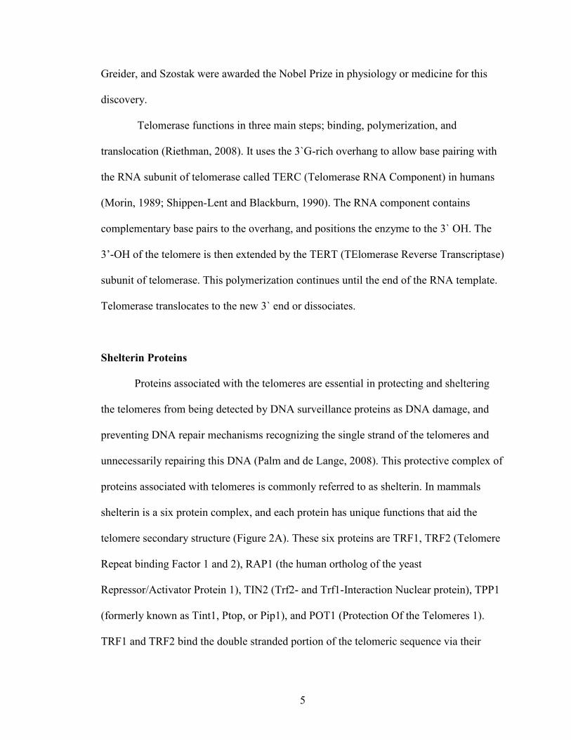
Figure 1. Telomere Structure and Associated Proteins ........................................................2
Figure 2. Shelterin Proteins..................................................................................................6
Figure 3. Nucleotide Excision Repair Pathway .................................................................12
Figure 4. Structure of XPC/RAD23B ................................................................................15
Figure 5. Maximum Likelihood Phylogenetic Tree of XPC/Rad4 ....................................40
Figure 6. Quantification of RAD4 Expression After Ultraviolet (UV) Treatment ............42
Figure 7. Quantification of RAD4 Expression After Hydrogen Peroxide (H2O2)
Treatment ...........................................................................................................................43
Figure 8. Quantification of RAD4 Expression after Methyl Methanesulfonate (MMS)
Treatment ...........................................................................................................................44
Figure 9. RAD4 Expression Quantification After Hydrogen Peroxide, Methyl
Methanesulfonate, and Ultraviolet Light Treatment ..........................................................46
Figure 10. Construction of pBT1-shRNA:RAD4 ..............................................................47
Figure 11. Digest of pBT1-shRNA:RAD4 ........................................................................49
Figure 12. Knock Down Confirmation via Whole-Cell PCR ............................................50
Figure 13. Quantification of RAD4 Expression in the Rad4 Knock Down Strains ...........51
Figure 14. Quantification of RAD4 Expression in Knock Down Strains ..........................53
Figure 15. Effects of Rad4 Knock Down on Survival After UV Treatment .....................55
Figure 16. Effects of Rad4 Knock Down on Survival After H2O2 Treatment ...................56
Figure 17. Development and Optimization of DIG-Labeled Telomere Probe Detection
Assay ..................................................................................................................................58
Figure 18. Optimization of gDNA Isolations for DIG-Labeled Telomere Probe Detection
Assay ..................................................................................................................................59
ix
Figure 19. Optimization of Blotting Conditions for DIG-Labeled Telomere Probe
Detection Assay. ................................................................................................................61
Figure 20. Telomere Detection in Wild Type and Rad4 Knock Down Strains .................62
Figure 21. Telomere Quantification in Wild Type and Rad4 Knock Down Strains ..........64
1
INTRODUCTION
Telomeres
Each cell in the human body contains approximately seven billion base pairs of
deoxyribose nucleic acid (DNA) (Gillooly et al., 2015). This DNA contains all of the
genetic information, and encodes all components of life (Watson and Crick, 1953).
Genomic DNA is organized around a complex of proteins called histones that ultimately
form nucleosomes that organize themselves into linear chromosomes. At the terminus of
these chromosomes, there is a critical and unique structure called the telomere that is
critical for the stability and integrity of the genome.
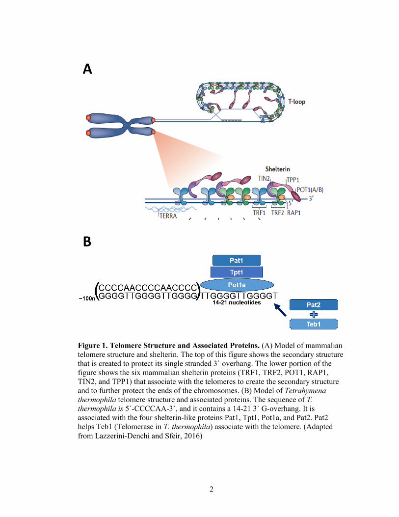
Both the sequence and structure of telomeres assist in genome protection (Figure
1A). Telomeric sequence across organisms is a simple sequence, repeated one hundred to
one thousand times. The human telomeric repeat sequence is 5`-TTAGGG-3` (Moyzis et
al., 1988). The sequence can vary, based on the organism, but it is typically G-rich which
allows for the formation of G-quadruplexes. The formation of a G-quadraplex is
stabilized based on the ability for guanines to bond to one another. This G-quandruplex
has been hypothesized to play an important role in the capping and protecting function of
telomeres (Henderson et al., 1987). Another unique component to the structure of
telomeric sequence is the 3` single stranded G-rich overhang. This overhang is present
due the inability of the replication machinery to fully replicate the linear chromosome to
its terminus, leaving a shortened 5’ end (Henderson and Blackburn, 1989). The 3` single
stranded end of the telomere invades the upstream DNA duplex, base-pairing with the
2
Figure 1. Telomere Structure and Associated Proteins. (A) Model of mammalian
telomere structure and shelterin. The top of this figure shows the secondary structure
that is created to protect its single stranded 3` overhang. The lower portion of the
figure shows the six mammalian shelterin proteins (TRF1, TRF2, POT1, RAP1,
TIN2, and TPP1) that associate with the telomeres to create the secondary structure
and to further protect the ends of the chromosomes. (B) Model of Tetrahymena
thermophila telomere structure and associated proteins. The sequence of T.
thermophila is 5`-CCCCAA-3`, and it contains a 14-21 3` G-overhang. It is
associated with the four shelterin-like proteins Pat1, Tpt1, Pot1a, and Pat2. Pat2
helps Teb1 (Telomerase in T. thermophila) associate with the telomere. (Adapted
from Lazzerini-Denchi and Sfeir, 2016)
A
B
3
homologous telomere sequence and creating a structure called a T-loop (Griffith et al.,
1999).
Telomeres have three widely conserved functions across organisms (Riethman,
2008). First, this sequence protects the ends of the chromosomes from degradation and
being recognized as damage. Second, telomeres help organize chromosomes during
meiosis to facilitate appropriate chromosome recombination and segregation (Bass et al.,
1997). The third conserved function is reliant on the ability for the telomeric sequence to
be extended by a ribonucleoprotein reverse transcriptase called telomerase (Greider and
Blackburn, 1985). Without this unique DNA polymerase-independent extension
mechanism, each round of replication would lead to telomere shortening, and threaten
exposure of critical portions of the genome. Shortening of the telomeres will also lead to
cellular senescence (Olovnikov, 1973).
Telomere and Telomerase Discovery
The discovery of telomeres and telomerase has a rich and collaborative history
that involved a variety of model organisms and scientists. Originally theorized by
McClintock, telomeres were named after their Latin origin telo (end) and mere (part)
(McClintock, 1931; Mullner, 1938). Much of the pioneering work in telomere research
has been conducted in the ciliate Tetrahymena thermophila. Due to its complex genome,
it has a high number of chromosomes which in turn yields a large amount of telomeric
DNA. Furthermore, this ciliate has nuclear dimorphism that splits the genetic information
into a transcriptionally silent nucleus (micronucleus) and transcriptionally active nucleus
(macronucleus) with 250 to 300 45N small chromosomes (Yao et al., 1981). Telomere
4
sequences specific to T. thermophila (5`-CCCCAA-3’) are found at the end of
chromosomes in the macronucleus and micronucleus (Blackburn and Gall, 1978). The
macronuclear DNA of T. thermophila undergoes a fragmentation step which results in
minichromosomes. These minichromsomes were observed to have telomeric sequence
added to them, but the mechanism for the addition was unclear (Blackburn et al., 1983).
The repetitive telomeric sequence was originally found on ribosomal DNA (rDNA),
which is contained within approximately 9000 of minichromsomes (Figure 1; Yao et al.,
1979).
Similar identification and characterization of telomeric sequence were found in a
wide variety of ciliates (Katzen et al., 1981; Klobutcher et al., 1981; Boswell et al.,
1982). Blackburn and Szostak studied the telomeric sequence of Saccharomyces
cerevisae to ensure the translation of the original findings in T. thermophila and other
ciliates translated to other organisms (Shampay et al., 1984). There are particular portions
of the S. cerevisae genome that are circular, and highly unstable when linearized. Upon
the experimental addition of the rDNA sequence from T. thermophila these linear
segments were stable. This reinforced the hypothesis of the stabilizing properties of the
telomeric sequence (Szostak and Blackburn, 1982).
Telomeric DNA is added to the termini of the telomeres of T. thermophila by a
terminal transferase named telomerase. Because telomerase was first discovered in
Tetrahymena, its telomerase is one of the best characterized telomerase ribonucleoprotein
(Greider and Blackburn, 1985). Telomerase has two subunits that contribute to the
functional structure; one is the RNA portion serving as a template sequence and the other
is the catalytically active facilitating the addition of sequence. In 2009 Blackburn,
5
Greider, and Szostak were awarded the Nobel Prize in physiology or medicine for this
discovery.
Telomerase functions in three main steps; binding, polymerization, and
translocation (Riethman, 2008). It uses the 3`G-rich overhang to allow base pairing with
the RNA subunit of telomerase called TERC (Telomerase RNA Component) in humans
(Morin, 1989; Shippen-Lent and Blackburn, 1990). The RNA component contains
complementary base pairs to the overhang, and positions the enzyme to the 3` OH. The
3’-OH of the telomere is then extended by the TERT (TElomerase Reverse Transcriptase)
subunit of telomerase. This polymerization continues until the end of the RNA template.
Telomerase translocates to the new 3` end or dissociates.
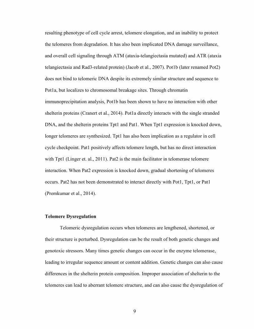
Shelterin Proteins
Proteins associated with the telomeres are essential in protecting and sheltering
the telomeres from being detected by DNA surveillance proteins as DNA damage, and
preventing DNA repair mechanisms recognizing the single strand of the telomeres and
unnecessarily repairing this DNA (Palm and de Lange, 2008). This protective complex of
proteins associated with telomeres is commonly referred to as shelterin. In mammals
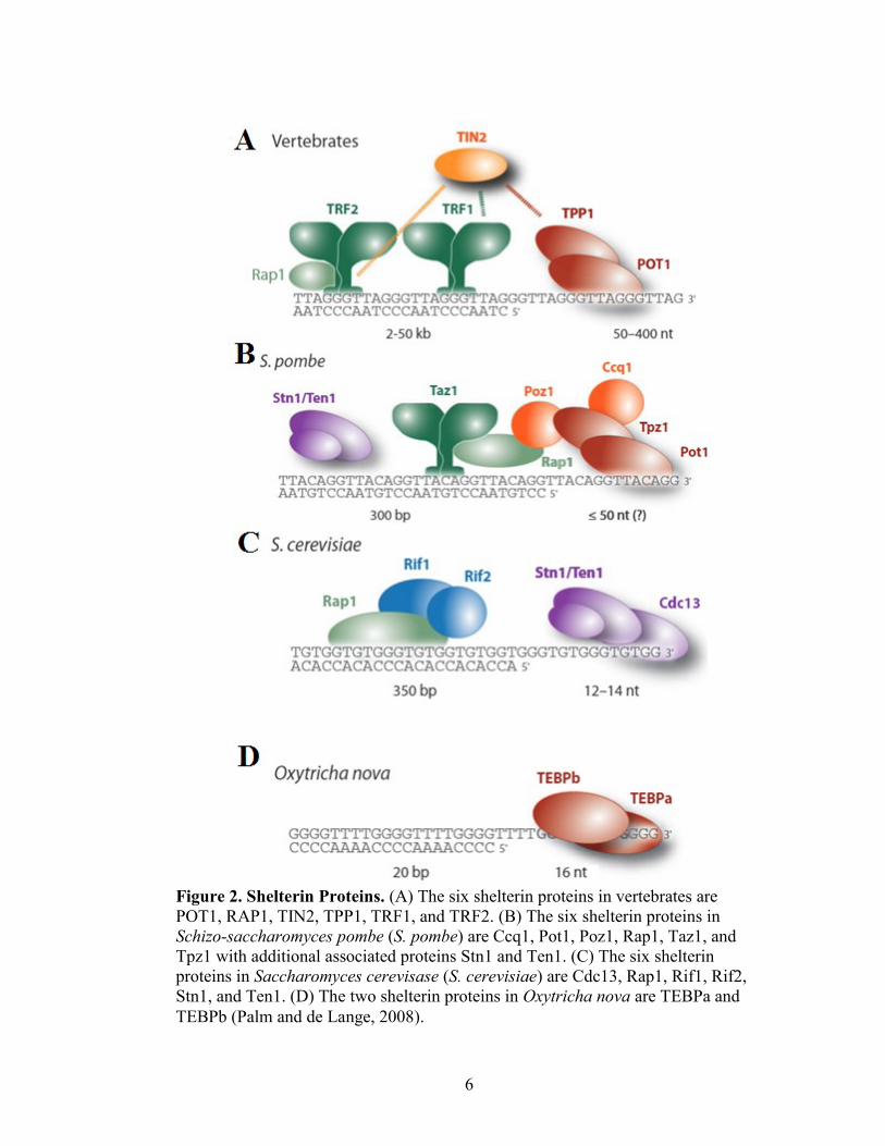
shelterin is a six protein complex, and each protein has unique functions that aid the
telomere secondary structure (Figure 2A). These six proteins are TRF1, TRF2 (Telomere
Repeat binding Factor 1 and 2), RAP1 (the human ortholog of the yeast
Repressor/Activator Protein 1), TIN2 (Trf2- and Trf1-Interaction Nuclear protein), TPP1
(formerly known as Tint1, Ptop, or Pip1), and POT1 (Protection Of the Telomeres 1).
TRF1 and TRF2 bind the double stranded portion of the telomeric sequence via their
6
Figure 2. Shelterin Proteins. (A) The six shelterin proteins in vertebrates are
POT1, RAP1, TIN2, TPP1, TRF1, and TRF2. (B) The six shelterin proteins in
Schizo-saccharomyces pombe (S. pombe) are Ccq1, Pot1, Poz1, Rap1, Taz1, and
Tpz1 with additional associated proteins Stn1 and Ten1. (C) The six shelterin
proteins in Saccharomyces cerevisase (S. cerevisiae) are Cdc13, Rap1, Rif1, Rif2,
Stn1, and Ten1. (D) The two shelterin proteins in Oxytricha nova are TEBPa and
TEBPb (Palm and de Lange, 2008).
7
Myb binding motif, forming homodimers (Bianchi et al., 1997; Broccoli et al., 1997).
RAP1 binds to TRF2 in the shelterin complex (Li et al., 2000). Without TRF2, RAP1 is
highly unstable protein, and cannot localize to telomeric DNA (Celli and de Lange,
2005). TIN2 is a critical scaffold to the overall integrity of the shelterin complex, binding
TRF1, TRF2, and TPP1 (Ye et al., 2004). TPP1 connects TIN2 and POT1, and is a direct
positive regulator of telomerase activity (Xin et al., 2007). POT1 binds to the G-rich
strand of the single stranded portion of the telomere, and protects it from degradation by
nucleases (Lei et al., 2004). POT1 also functions as a negative regulator of telomerase
activity (Kelleher et al., 2005). POT1 was initially identified due to its sequence
homology to TEBP alpha and beta (Telomere End Binding protein alpha and beta) in
Oxytricha nova (Figure 2D). Of the six shelterin proteins, it holds the highest amount of
sequence similarity across model organisms (Baumann and Cech, 2001).
Schizosaccharomyces pombe has six shelterin proteins that have a very similar
functional and structural architecture to vertebrate shelterin: Taz1 (Telomere-Associated
in Schizosaccharomyces pombe), Rap1 (Repressor/Activator site binding Protein 1), Poz1
(Pot1-associated in Schizosaccharomyces pombe), Tpz1 (TPP1 homolog in
Schizosaccharomyces pombe), Ccq1 (Coiled-Coil protein Quantitatively enriched), and
Pot1 (Protection of Telomeres 1) (Figure 2B). Taz1 has similar characteristics to TRF1/2,
and binds double stranded telomere repeat as a homodimer (Cooper et al., 1997). Rap1
resembles the functionality of Rap1 and serves as a bridge from Taz1 to Poz1 (Kanoh and
Ishikawa, 2001). Poz1, Tpz1, and Ccq1 serve as a bridge between Taz1 and Pot1. Ccq1
has been associated with regulation in meiotic division and plays a role in the recruitment
of telomerase (Flory et al., 2004; Tomita and Cooper, 2008). Pot1 was originally
8
discovered is S. pombe due to its sequence homology to the previously discovered TEBP
alpha and beta (Telomere End Binding Protein) in Oxytricha nova. This telomere protein
in S. pombe also binds to 3` G overhang, and possess similar regulatory functions as
POT1 in vertebrates (Baumann and Cech, 2001).
Although shelterin proteins typically do not have close sequence homology,
shelterin complex of proteins in other organisms have shown to possess functional
homology. In Saccharomyces cerevisiae, three proteins associate with the G-rich single
stranded overhang: Cdc13 (Cell Division Cycle 13), Stn1 (Suppressor of cdc ThirteeN 1),
and Ten1 (TElomeric Pathways with STn 1) (Gao et al., 2007) (Figure 2C). Cdc13 assists
in the recruitment of telomerase, while Stn1 inhibits the binding of telomerase to the
telomeres (Pennock et al., 2001; Puglisi et al., 2008). Rap1 (Repressor/ Activator site
binding Protein 1) binds directly to the double stranded portion of the S. cerevisae
telomere. Rap1 has the binding partners Rif1 and 2 (Rap1 Interaction Factor 1 and 2)
which directly regulate telomere length (Marcand et al., 1997). As the binding of Rif1
and Rif2 increase association to Rap1, this complex inhibits the ability of telomerase to
access the telomeres (Hirano et al., 2009).
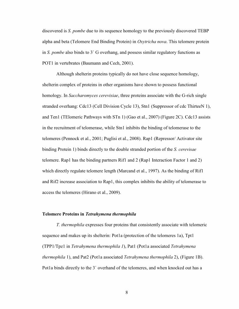
Telomere Proteins in Tetrahymena thermophila
T. thermophila expresses four proteins that consistently associate with telomeric
sequence and makes up its shelterin: Pot1a (protection of the telomeres 1a), Tpt1
(TPP1/Tpz1 in Tetrahymena thermophila 1), Pat1 (Pot1a associated Tetrahymena
thermophila 1), and Pat2 (Pot1a associated Tetrahymena thermophila 2), (Figure 1B).
Pot1a binds directly to the 3` overhand of the telomeres, and when knocked out has a
9
resulting phenotype of cell cycle arrest, telomere elongation, and an inability to protect
the telomeres from degradation. It has also been implicated DNA damage surveillance,
and overall cell signaling through ATM (ataxia-telangiectasia mutated) and ATR (ataxia
telangiectasia and Rad3-related protein) (Jacob et al., 2007). Pot1b (later renamed Pot2)
does not bind to telomeric DNA despite its extremely similar structure and sequence to
Pot1a, but localizes to chromosomal breakage sites. Through chromatin
immunoprecipitation analysis, Pot1b has been shown to have no interaction with other
shelterin proteins (Cranert et al., 2014). Pot1a directly interacts with the single stranded
DNA, and the shelterin proteins Tpt1 and Pat1. When Tpt1 expression is knocked down,
longer telomeres are synthesized. Tpt1 has also been implication as a regulator in cell
cycle checkpoint. Pat1 positively affects telomere length, but has no direct interaction
with Tpt1 (Linger et. al., 2011). Pat2 is the main facilitator in telomerase telomere
interaction. When Pat2 expression is knocked down, gradual shortening of telomeres
occurs. Pat2 has not been demonstrated to interact directly with Pot1, Tpt1, or Pat1
(Premkumar et al., 2014).
Telomere Dysregulation
Telomeric dysregulation occurs when telomeres are lengthened, shortened, or
their structure is perturbed. Dysregulation can be the result of both genetic changes and
genotoxic stressors. Many times genetic changes can occur in the enzyme telomerase,
leading to irregular sequence amount or content addition. Genetic changes can also cause
differences in the shelterin protein composition. Improper association of shelterin to the
telomeres can lead to aberrant telomere structure, and can also cause the dysregulation of
10
telomerase. Genotoxic stressors can damage the actual sequence of the DNA, causing
single stranded or double stranded breaks, or lesions to individual bases.
Dysregulation that causes lengthened or shortened telomeres is associated with
human disease and overall cellular dysfunction. One of the first diseases shown to be
associated with short telomeres was dyskeratosis congenital due to a mutation in the
transcriptional processing of telomerase. (Mitchell et al., 1999). Dyskeratosis patients
have a higher propensity to develop cancer in comparison to the rest of the population
(Alter et al., 2009). Shortened telomeres in all tissues are commonly associated with
various types of lung diseases including pulmonary fibrosis and emphysema (Armanios,
2012; Stanley et al., 2015). These patients have an increased incidence of organ failure
that accounts for about 90% of the deaths in this population (Dokal, 2000). Patients with
longer telomeres are also predisposed to a variety of diseases including melanoma and
glioma. Mutations in the POT1, TPP1, and RAP1 genes, which encode components of
the shelterin complex, have also been associated with these types of cancers (Robles-
Espinoza et al., 2014; 6, Kocak et al., 2014).
The inability to repair telomeric sequence after damage can also lead to aberrant
telomeres. Because of the unique secondary structure and sequence of telomeres, damage
is often processed differently at the telomeres in comparison to other parts of the genome.
Typically, DNA repair machinery must interact with histones, but at the telomeres the
repair machinery must also interact with shelterin proteins. An additional structural
difference is the T-loop formed by the single stranded DNA invading upstream
homologous double stranded DNA. Usually, DNA damage surveillance protein would
recognize the single stranded 3’ end as DNA damage, but to maintain the integrity of the
11
telomere structure this should not be recognized as damage at this location of the
genome.
If telomeres become critically short, this can induce non-homologous end joining.
This is the only in the most dire of situations when cells are in distress. This can result in
telomere-telomere fusions and ultimately overall genome instability (Stewart et al.,
2012). Telomeres can also combat shortening through a telomerase independent method
called alternative lengthening of the telomeres. Telomeric sequence undergoes
homologous recombination of neighboring sequence to increase the overall length of the
telomeres (Cesare and Reddel, 2010). Additionally, the three types of DNA excision
repair (base excision repair, mismatch repair, and nucleotide excision repair) have been
observed and studied at the telomeres (Jia et al., 2015). Nucleotide excision repair is
particularly necessary because of the composition of telomere sequence, yet is
understudied at this site of the genome.
Nucleotide Excision Repair
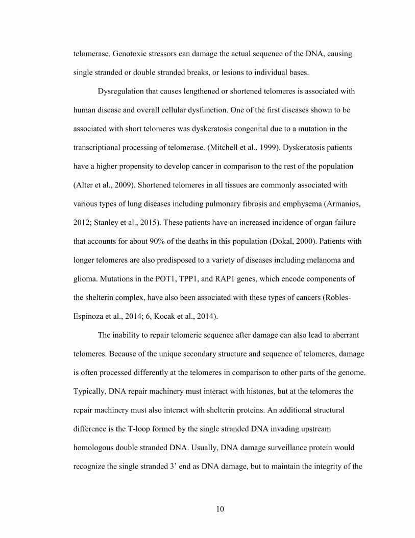
Nucleotide excision repair (NER) recognizes and repairs damage that occurs to a
single strand of DNA due to physical and chemical mutagens in the environment and
inside of the cell. UV light is a typical exogenous, physical mutagen that induces damage,
generating both cyclobutane pyrimidine dimers (CPDs) and pyrimidine(6-4) pyrimidone
photoproducts (6-4PP), that are repaired by NER (Figure 3). Adducts caused by
environmental, chemical mutagens are benzo[α]pyrene and various aromatic amines, and
are also repaired by NER (Szymkowski et al., 1993). All types of damage listed above
typically have one major characteristic in common; interaction with DNA distorts the
12
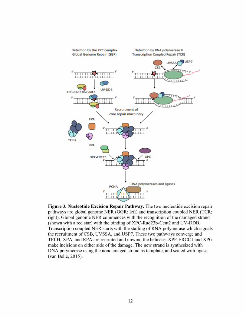
Figure 3. Nucleotide Excision Repair Pathway. The two nucleotide excision repair
pathways are global genome NER (GGR; left) and transcription coupled NER (TCR;
right). Global genome NER commences with the recognition of the damaged strand
(shown with a red star) with the binding of XPC-Rad23b-Cent2 and UV-DDB.
Transcription coupled NER starts with the stalling of RNA polymerase which signals
the recruitment of CSB, UVSSA, and USP7. These two pathways converge and
TFIIH, XPA, and RPA are recruited and unwind the helicase. XPF-ERCC1 and XPG
make incisions on either side of the damage. The new strand is synthesized with
DNA polymerase using the nondamaged strand as template, and sealed with ligase
(van Belle, 2015).
13
double helix and thermodynamically destabilize DNA duplexes (Sugasawa et al., 2001).
Because the recognition proteins in the NER pathway detect this helical distortion rather
than the actual damaged base, NER is able to recognize a wide range of damage. This is a
unique quality in comparison to other DNA damage repair pathways, which typically
recognize the irregular structure and damage of the DNA. Along with this unique
recognition characteristic, the discovery of the proteins within the NER pathway is
different from other DNA repair pathways. Many of the proteins within the nucleotide
excision repair pathway were discovered through studies of patients with Xeroderma
Pigmentosum.
Xeroderma Pigmentosum patients cannot perform nucleotide excision repair
(NER) due to a lack of one of the XP repair proteins. Researchers study these patients to
better understand the overall pathway of NER and the pathobiology of Xeroderma
Pigmentosum both clinically and through cellular culture (Cleaver, 1968). The hallmark
phenotypic characteristic of this genetic disorder is extreme sensitivity to sunlight.
Patients can have over a 2000-fold increase in the risk of skin cancer (DiGiovanna and
Kramer, 2012). Many times, patients also have developmental delay, neurological
damage, and overall increased risk of cancer (Cleaver, 2005). This phenotypic
manifestation is likely due to the accumulation of DNA damage due to the inability for
the damage to either be recognized or repaired by NER. In addition to Xeroderma
Pigmentosum, mutations in various specific XP proteins can lead to Cockayne syndrome
and trichothiodystrophy (Lehmann, 2003). Studies with XP patients have made great
headway into understanding the NER pathway.
14
There are two types of nucleotide excision repair processes: global genome and
transcription coupled repair. Transcription coupled repair as the name implies, focuses on
repairing genes undergoing active transcription (Hanawalt and Spivak, 2008). Global
genome repair is responsible for repairing both transcriptionally active and inactive parts
of the genome (Gillet and Schärer, 2006). The primary difference between these two
types of repair is the damage recognition step. Transcription coupled repaired responds
largely to the RNA polymerase stalled at a site of damage in an actively transcribed
portion of the genome (Figure 3; TCR pathway). The recognition factors specific to TCR
are CSB (Cockayne Syndrome group B), UVSSA (UV-Stimulated Scaffold protein A),
and USP7 (Ubiquitin-Specific-Processing protease 7).
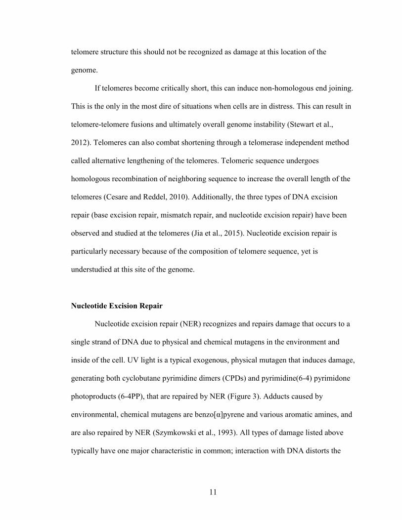
Global genome repair begins with specific recognition factors of XPC-RAD23B
(Xeroderma Pigmentosum C, RADiation sensitive 23B) and if necessary UV-DDB
(Ultraviolet-Damaged DNA B) that recognize thermodynamically destabilized duplexes
(Evans et al., 1997; Sugasawa et al., 2001) (Figure 3 GGR pathway). XPC, known as
RAD4 (RADiation sensitive 4) in both yeast and Tetrahymena thermophila, contains two
key functional domains: transglutamase homology domain (TGD) and Beta-hairpin
domains 1-3 (BHD 1-3) (Figure 4). Both TGD and BHD1 anchor XPC and RAD23 to the
DNA at the site of damage. BHD2 and 3 bind the undamaged strand encircling the two
nucleotides across from the damage. This ability to bind the undamaged strand allows
XPC-RAD23B to recognize a large variety of DNA damages (Min and Pavletich, 2007).
While the domains of XPC assist in physically binding DNA, the main function of a large
amount of helical destabilization. The recognition of CPD damage is facilitated by UV-
DDB complex (Tang et al., 2000). DDB2 creates a significant kink in the DNA duplex,
15
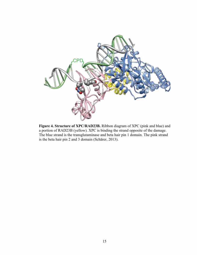
Figure 4. Structure of XPC/RAD23B. Ribbon diagram of XPC (pink and blue) and
a portion of RAD23B (yellow). XPC is binding the strand opposite of the damage.
The blue strand is the transglutaminase and beta hair pin 1 domain. The pink strand
is the beta hair pin 2 and 3 domain (Schärer, 2013).

16
and this distortion is more recognizable to XPC-RAD23B (Scrima et al., 2008).
For both global and transcription coupled NER, the pathways converge once the
damage is recognized, and initial protein complexes are stabilized. The next step required
is the recruitment of the pre-incision complex. The pre-incision initiates with TFIIH,
which is recruited to the site of damage by the presence of the recognition proteins. The
TFIIH complex has a total of 10 subunits, with its catalytic proteins XPB and XPD which
perform helicase activity (Evans, 1997; Tapias et al., 2004). The XPB functions to pry
open the DNA duplex with the facilitation of XPC that allows loading of THIIH (Coin et
al., 2008). XPD translocates along the single stranded DNA opening it, and stalls at the
location of the damage (Sugasawa et al., 2009). This stalling causes the rest of the pre-
incision complex to assemble, which includes XPA, RPA, and XPG; (Wakasugi and
Sancar, 1998; Volker et al., 2001). Once these proteins are recruited the XPC-RAD23B
complex dissociates, XPA then plays a key role in the placement and positioning of many
of the nucleotide excision repair proteins including ERCC1-XPF, TFIIH, XPC-RAD23B
complex, DDB2, and PCNA (Li et al., 1994; Park et al., 1995; Bunick et al., 2006;
Wakasugi et al., 2009; Gilljam et al., 2012). RPA binds to and coats the ssDNA on the
opposite side of the damage for a span of about 30 nucleotides, which is about the size of
the damaged strand that will be removed (Fan and Pavletich, 2012). The placement of the
RPA proteins helps to position ERCC1-XPF and XPG to allow for a dual incision to
remove the damaged sequence (de Laat et al., 1998). ERCC1-XPF are recruited to this
complex via XPA, and creates the first incision at the 5` end of the sequence (Li et al.,
1994). This first incision is required at the 5` end before the second incision can be
performed. XPG will only bind when a ssDNA stretch at the 3` end is present which is
17
created by this first incision (Hohl et al., 2003). The second incision at the 3` end is made
by XPG (Fagbemi et al., 2011).
The incision made by ERCC1-XPF create a 3` hydroxyl group that can be used by
DNA polymerase to fill in the gap created. There are three polymerases that have been
associated with NER: Polymerase ĸ, Ɛ, and δ. Pol ĸ which is typically associated with
translesion synthesis has been shown to be involved in NER (Ogi and Lehmann, 2006).
When Pol Ɛ and δ expression is knocked down, there is a 50% reduction of repair
synthesis. Each polymerase has specific proteins that facilitate the polymerase association
to the DNA strand (Ogi et al., 2010). The 5` phosphate created by XPG will be ligated to
the newly extended DNA fragment by DNA ligase I or DNA ligase IIIα to seal the
backbone. DNA ligase IIIα is active in both dormant and actively replicating cells, while
DNA ligase I is active in replicating cells (Moser et al., 2007).
Once the DNA has been successfully repaired, NER proteins dissociate, and the
cell is able to resume its regular function. This is critical to both actively transcribed and
silent portions of the genome. If NER does not occur in areas that are being actively
transcribed this could lead to a point mutation, or the inability of RNA polymerase to be
able to transcribe the gene successfully. In silent portions of the genome, this repair is
critical because remaining damage could be passed to the next generation of cells. These
mutations can cause phenotypic difference and overall genome instability. One silent
portion of the gnome that is critical to genome stability is the telomeres. NER is
important to maintain telomeric DNA, but thus far has been understudied.
18
Nucleotide Excision Repair at the Telomeres
Limited studies have been conducted with nucleotide excision repair at the
telomeres. CPDs occur seven-fold higher at telomeres than the bulk genome in human
fibroblast cells (Rochette and Brash, 2010). CPDs are removed 1.5-fold faster at the
telomeres over genomic DNA, suggesting that NER at the telomeres is unique and
efficient (Parikh et al., 2015). However, contradictory data has been published currently
call into question if repair is due to nucleotide excision repair pathways or to other repair
pathways (Parikh et al., 2015; Rochette and Brash, 2010). For example, primary mouse
fibroblasts deficient in XPB and chronically exposed to low amounts of hydrogen
peroxide, an agent that induces base excision repair (BER), have shorter telomeres and
higher amounts of telomere degradation (Ting et al., 2010). A similar phenotype has been
observed in XPB/XPD deficient lymphoblastoids treated with hydrogen peroxide
(Gopalakrishnan et al., 2010). It is possible that these BER-related phenotypes may
actually be related to NER since NER proteins can play a role in the BER pathway (Melis
et al., 2013).
NER proteins are also directly involved in telomere maintenance via shelterin.
XPF and the shelterin protein TRF2 have an intricate interaction; when XPF is over
expressed, shorter telomere fragments occur, and less TRF2 interaction with the telomere
is observed (Wu et al., 2008). Additionally, ERCC1/XPF is one of the endonucleases
responsible for removing the 3` overhang of telomeres if left uncapped. TRF2 serves as
the protector against the ERCC1/XPF endonuclease activity in regularly capped
telomeres (Zhu et al., 2003).

19
NER proteins may also help regulate telomerase itself. Xpc-/- knock out Mus
musculus have chronically longer telomeres, while double knock out M. musculus of Xpc
and Terc (catalytic subunit of telomerase) show lengthened telomeres. This surprising
lengthening of telomeres with the Xpc-/-G1-G3-/- is likely due to alternative lengthening of
the telomeres (ALT) (Stout and Blasco, 2013).
Currently, still much about the UV response and XPC activity at the telomere is
unknown. Due to the unique structure, sequence, and critical role of telomeres (Figure 1),
it is likely that the repair pathways are very individualistic to this part of the genome in
comparison to typical genomic sequence. Thus far no nucleotide repair studies at the
telomere have ever been conducted in Tetrahymena thermophila but it is an extremely
well characterized model organism in telomere biology studies, and will allow DNA
repair at this are of the genome to be addressed.
Purpose Statement
The major goal of this research project was to understand the behavior of RAD4,
the homolog of XPC in T. thermophila, both generally in the genome as well as at the
telomeres. RAD4 is a critical DNA damage recognition protein in the nucleotide excision
repair pathway in the global genome pathway. The process of nucleotide excision repair
at the telomeres is not well known, yet is intriguing because of the telomeres unique
architecture and sequence. The telomere sequence in T. thermophila (3`-GGGGTT-5`) is
especially prone to UV damage due to the frequency of thymine dinucleotides that may
undergo UV-induced dimerization, which is typically repaired by nucleotide excision
repair. Similarly, the mammalian telomeric sequence (TTAGGG) is also susceptible to
20
damage repaired by NER due to its dipyrimidine sequence. Thus, the integrity of
telomeres may be affected if the nucleotide excision repair pathway is disrupted.
To begin these studies, XPC and RAD4 sequences were analyzed by a variety of
bioinformatics tools to define their homology. RAD4 expression levels were observed
after T. thermophila exposure to mutagens, including UV, hydrogen peroxide, and MMS,
to induce DNA damage recognized and repaired by nucleotide excision repair, base
excision repair, and double stranded break repair, respectively. To further characterize
RAD4, a RAD4 knock down was established via short hairpin RNA. Cellular growth after
UV light and hydrogen peroxide exposure was measured to determine how RAD4
depletion affects the ability to recognize damaged DNA. Finally, telomere length and
integrity were measured in the wild type and RAD4 depleted cells, before and after UV
treatment via a unique DIG-labeled probe telomere detection assay. It was anticipated
that the nucleotide excision repair pathway will be further characterized in the model
organism, T. thermophila, providing further understanding of how nucleotide excision
repair helps to maintain telomeres.
21
EXPERIMENTAL PROCEDURES
Bioinformatics – Phylogenetic Tree Design and shRNA Primer Design
RAD4 nucleotide sequence, amino acid sequence, exon and intron predictions,
and paralogs were collected from Tetrahymena Genome Database (TGD; Stover et al.,
2012). The paralog obtained from TGD was RAD4 (TTHERM_00825460; Altschul et al.,
1997). Orthologs were obtained using National Center for Biotechnology Information
Basic Local Alignment Search Tool (NCBI BLAST; Altschul et al., 1997) search from
RAD4. The following proteins were obtained as the top ortholog hits: NP_001460.1
(Homo sapiens), NP_034377.2 (Mus musculus), ABI54461.1 (Danio rerio), BAA76953.1
(Xenopus laevis), NP_504491.1 (Caenorhabditis elegans), AGA18917.1 (Drosophila
melanogaster), XP_637925.1 (Dictyostelium discoideum), AJS88329.1 (Saccharomyces
cerevisiae), NP_587828.1 (Schizosaccharomyces pombe) NP_564012.1 (Arabidopsis
thaliana), XP_002267875.1 (Vitis vinifera), and ABN13872.1 (Aspergillus niger).
Domain analysis was performed on these amino acid sequences with InterPro
(Finn et al., 2017). Additionally, sequences were analyzed for evolutionary conservation
with Multiple Sequence Alignment-CLUSTALW (Li et al., 2015), and Molecular
Evolutionary Genetic Analysis 7.0 (MEGA7; Kumar et al., 2016). MEGA 7.0 was used
to create a maximum likelihood tree using bootstrap consensus with 500 replicates.
Sequences were analyzed with Tree-based Consistency Objective Function For alignment
Evaluation (TCOFFEE; Notredame et al., 2000) to determine the appropriate location for
knock down primer design (data not shown). Primers (shRNA) were analyzed using
Integrate DNA Technologies OligoAnalyzer 3.1 (https://www.idtdna.com/calc/analyzer).
22
Domain analysis was performed on the shelterin proteins with InterPro of Homo
sapiens, Saccharomyces cerevisiae, Schizosaccharomyces pombe, and Tetrahymena
thermophila. The following proteins were analyzed for Homo sapiens: POT1
(NP_056265.2), RAP1 (ABA64473.1), TIN2 (AAF18439.1), TPP1 (NP_001075955.1)
TRF1 (NP_059523.2), and TRF2 (NP_059523.2); Saccharomyces cerevisiae: CDC13
(NP_010061.1), RAP1 (NP_014183.1), RIF1 (NP_009834.4), RIF2 (NP_013558.3);
Schizosaccharomyces pombe: CCQ1 (NP_588210.1), POT1 (NP_594453.1), POZ1
(NP_594428.1), RAP1 (NP_596285.1), TAZ1 (NP_594047.1), and TPZ1
(NP_001342857.1); Tetrahymena thermophila: POT1 (TTHERM_000378989), TPT1
(TTHERM_00523050), PAT1 (TTHERM_00013120), and PAT2
(TTHERM_00049470).
shRNA Plasmid Construction
The appropriate pBT1-YFG vector (from the work of Kyle Cottrell in 2012) was
combined with the specific RAD4 sense and antisense primers to construct a theoretical
plasmid with Gene Construction Kit 3.0 (textco biosoftware). To phosphorylate the
shRNA oligos, 200 pmol of the sense and antisense RAD4 oligos (Table 1), 10 units of
T4-PNK (New England Biolabs), 1X PNK buffer (New England Biolabs), and 1 mM
ATP were combined and brought to a final volume of 50 µL. The reaction was incubated
at 37°C for 60 minutes, and then 70°C for ten minutes, 10 µL of this phosphorylation
reaction was combined with 1XSSC and brought to a final volume of 25 µL. To anneal
these oligos, this reaction was heated at 75°C for five minutes, and cooled in a 23°C

23
Table 1. Primer Sequences.
Target Primer Sequence (5` to 3`)
ACT1 Forward: TGAATTAAAGGCTTACAAGGAATC
Reverse: CACACTTCATGATAGAGTTGAAGG
DIG-ACT1 /5DigN/TGAATTAAAGGCTTACAAGGAATC
DIG-Telomere /5DigN/CCCCAACCCCAACCCCAACCCCAACCCCAA
HHP1 Forward: TTAGCAATGATAAACCTTAGAC
Reverse: TGTGTAAAGAGATTTTCCATC
RAD4 qRT-PCR Forward: AGAGCTGCACGTTTTTCAGATATG (1032F)
Reverse: TGACAGCATTTGGTCAAATAAATCA (1271R)
shRNA
Confirmation
Forward: ATGAATGATATAAATGAAGAGTGGC
Reverse: TGTTATGTGAATGAAGTTAATTGGG
24
annealed RAD4 oligos at 1:5 and 2:5 (vector:insert) molar ratios. These ligation reactions
contained 1X T4 DNA ligase buffer (New England Biolabs) and 400 units T4 DNA
ligase (New England Biolabs). Ligation reactions were incubated at 14°C overnight, and
kept in at -20°C for long term storage.
Electroporation of DH10B Escherichia coli with pBT1-shRNA:RAD4
DH10B E. coli cells were combined with 1 µL of the pBT1-RAD4shRNA ligation
reaction, and transferred to a 2-mm electroporation cuvette (Fisher) taking care to avoid
the introduction of any bubbles. Samples were placed in the electroporation chamber
(Bio-Rad Gene Pulser II Electroporation System) and electroporated at 2.5kV, 200 ohms,
25 µF. Samples were recovered in 1 mL of SOC media (Invitrogen) at 37°C for 60
minutes, and 100 µL of each sample was plated on a LB+AMP plates (1% bacto tryptone,
0.5% yeast extract, 1% NaCl, 1.5% bacterial agar with 100 µg/µL ampicillin), and
incubated at 37°C for 12-20 hours.
Lysozyme Boil Plasmid Isolation and Ethanol Precipitation
Plasmid-containing bacteria were grown on LB+AMP plates (1% bacto tryptone,
0.5% yeast extract, 1% NaCl, 1.5% bacterial agar with 100 µg/µL ampicillin). Two
milliliter (miniprep) and 25 mL (midiprep) cultures were grown in LB+AMP liquid
media (1% bacto tryptone, 0.5% yeast extract, 1% NaCl, with 100 µg/µL ampicillin), and
incubated overnight at 37°C in at 220 rpm. Cultures were centrifuged for two minutes
(Sorvall Legend X1 Centrifuge, Thermo Fisher Scientific) at 13300 rpm (miniprep) or
6000 rpm (midiprep) for ten minutes. The remaining media was removed. Cellular pellet
25
was resuspended in 350 µL or 3.5 mL sucrose lysis buffer (8% sucrose, 0.5% Trion X-
100, 50 mM EDTA, 10 mM Tris-pH 8) and 10 mg/mL lysozyme (Fisher Scientific).
Samples were transferred to 1.5 mL microcentrifuge tubes, incubated at room
temperature for five minutes, and boiled for one minute. Samples were spun at maximum
speed for 15 minutes, and cellular debris was removed. To precipitate DNA, 220 µL of
isopropanol and 40 µL 3 M sodium acetate were added and incubated for five minutes.
Plasmid isolates were spun at maximum speed for 5 minutes, and washed with 70%
ethanol. Samples were combined and resuspended in 50 µL (miniprep) or 300 µL
(midiprep) TE (10 mM Tris-HCl, 1 mM EDTA-pH 8).
Midipreps were incubated with 10 mg/mL RNase A (AppliChem Panreac) at
37°C for 3 hours. Samples then underwent a phenol chloroform extraction. Purified
samples were combined with 1/10th 3 M sodium acetate and 2.5 times 100% chilled
ethanol, and incubated at -20°C overnight. Samples were centrifuged at maximum speed
for 10 minutes and the supernatant was poured off. The pellet was washed with 70%
ethanol, and centrifuged for 10 minutes at maximum speed. The supernatant was
discarded, and carefully removed. The plasmid was resuspended in 150 µL double
distilled water.
Restriction Enzyme Digest Confirmation of pBT1-shRNA:RAD4
The isolated plasmid was digested with SacI (New England Biolabs) and XhoI
(New England Biolabs) overnight at 37°C. Samples were electrophoresed on a 1.0%
agarose gel in TAE at 120V for 30 minutes, and visualized with the KODAK Gel Logic
200 Imaging System. The strain considered positive for pBT1-shRNA:RAD4 after
26
analysis was grown up in a 2 mL culture in LB+AMP liquid media (1% bacto tryptone,
0.5% yeast extract, 1% NaCl, with 100 µg/µL ampicillin). For long term storage, glycerol
stocks were made by combining culture and 10% glycerol in a 1:1 ratio, and stored at -
80°C.
Linearization of pBT1-shRNA:RAD4 for Biolistic Transformation
Gene Construction Kit 3.0 was used find the best restriction enzymes to linearize
the knock down plasmid after the E. coli transformation. A 200 µL sample was prepared
with 1X SmartCut Buffer (NEB), 2.5 µL XhoI, 2.5 µL SacI, and 100 µg plasmid DNA
and incubated over night at 37°C. The sample was centrifuged at maximum speed for 15
minutes. The precipitate was washed with 70% ethanol, and centrifuged for five minutes.
Ethanol was removed, and sample was allowed to dry at room temperature. Sample was
quantified on the NanoDrop spectrometer (NanoDrop 2000 Spectrophotometer, Thermo
Fisher Scientific), and sample was adjusted to 2.0 µg/µL in water. One µL sample was
run on a 1.0% agarose gel in TAE at 120V for 30 minutes, and visualized with the
KODAK Gel Logic 200 Imaging System to ensure proper digestion sizes before biolistic
transformation.
Cell Culture Maintenance and Strains Used
Tetrahymena thermophila were grown in 2% PPY media (0.02 g/mL protease
peptone, 0.002 g/mL yeast extract) and 1XPSF (100 µg/mL penicillin, 100 µg/mL
streptomycin, 0.25 µg/mL amphotericin B, Thermo Fisher Scientific). Cultures grown in
plates (96-, 48-, and 24-well plates) were incubated in humidity chambers at 30°C.
27
Cultures grown in larger quantities for RNA isolation, survivability assay, gDNA
isolations, and any other experiments were grown at 30°C at 100 rpm. Concentrations of
cells throughout experimentation were estimated via a hemocytometer. Stock cultures
were either continued or established in 1% PPY+10 mM Tris and 1XPSF. Stock cultures
were maintained approximately every 4-6 months by transferring 100 µL of the original
stock to a new 1% PPY+10 mM Tris and 1XPSF. All strains used during the course of
this project are described in Table 2. Strains designated with a “CU” indicate that the
strain was obtained from the Tetrahymena Stock Center located at Cornell University.
Biolistic Transformation of Tetrahymena thermophila
The gold beads were prepared by weighing out 30 mg of Au particles, and 70%
ethanol was added. The beads were vortexed for three to five minutes, centrifuged at
maximum speed for five seconds and the supernatant was discarded. These steps were
repeated three times.
To prepare the cells, a culture was started by inoculating 25 mL culture of 2%
PPY media (0.02 g/mL protease peptone, 0.002 g/mL yeast extract and 1XPSF (100
µg/mL penicillin, 100 µg/mL streptomycin, 0.25 µg/mL amphotericin B, Thermo Fisher
Scientific) culture with a 0.5 mL of stock culture of either CU428, CU522, or CU725
(Table 2). The next day a culture was diluted to a final concentration of 1 to
3x10⁵cell/mL at the time of the transformation. The next day the culture was centrifuged
at 3000 rpm for three minutes (Sorvall Legend X1 Centrifuge, Thermo Fisher Scientific).
The supernatant was decantated and the pellet resuspended in 10 mM Tris and
28
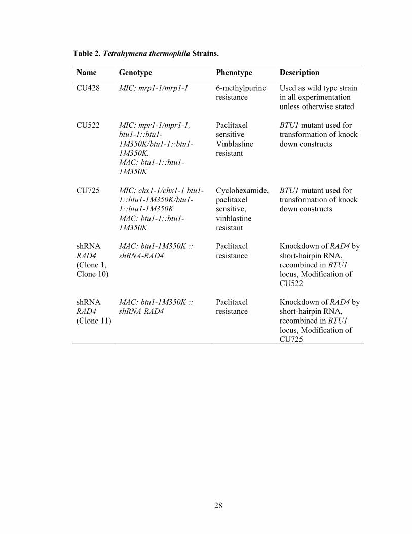
Table 2. Tetrahymena thermophila Strains.
Name Genotype Phenotype Description
CU428 MIC: mrp1-1/mrp1-1 6-methylpurine
resistance Used as wild type strain
in all experimentation
unless otherwise stated
CU522 MIC: mpr1-1/mpr1-1,
btu1-1::btu1-
1M350K/btu1-1::btu1-
1M350K.
MAC: btu1-1::btu1-
1M350K
Paclitaxel
sensitive Vinblastine
resistant
BTU1 mutant used for
transformation of knock
down constructs
CU725 MIC: chx1-1/chx1-1 btu1-
1::btu1-1M350K/btu1-
1::btu1-1M350K
MAC: btu1-1::btu1-
1M350K
Cyclohexamide,
paclitaxel
sensitive,
vinblastine
resistant
BTU1 mutant used for
transformation of knock
down constructs
shRNA RAD4 (Clone 1,
Clone 10)
MAC: btu1-1M350K ::
shRNA-RAD4 Paclitaxel
resistance Knockdown of RAD4 by
short-hairpin RNA,
recombined in BTU1
locus, Modification of
CU522
shRNA RAD4 (Clone 11)
MAC: btu1-1M350K ::
shRNA-RAD4 Paclitaxel
resistance Knockdown of RAD4 by
short-hairpin RNA,
recombined in BTU1
locus, Modification of
CU725
29
centrifuged again for 3 minutes. The 10 mM Tris was decanted, and the cell pellet was
resuspended in 10 mM Tris pH. Cultures were then placed in 30°C without shaking for
14 to 20 hours. Media was pre-warmed at 30°C for transformations.
The gold beads had 5 µg of the linearized knock down plasmid, 25 µL 2.5 M
calcium chloride, and 10 µL 100 mM spermidine added. The mixture was vortexed for 30
minutes at 4°C. The mixture was centrifuged, the supernatant was removed, and 70%
ethanol was added; this was repeated with 100% ethanol. The steel macrocarrier holder,
plastic macrocarrier, plastic cap, and metal stopping screens were sterilized by dipping in
100% ethanol. The rupture disks were sterilized by dipping in 100% isopropanol.
Prepared gold samples were added to the macrocarrier. Ten mM HEPES was
placed on a filter and 1x10⁷ of cells were added. The gene gun (Bio-Rad PDS-
1000/HeTM Biolistic Particle Delivery System) was set up the as follows: rupture disk
(1100 PSI for 900 PSI) has a 3/8 inch gap to the macrocarrier assembly,stopping screen
was in the third position, and was placed in the macrocarrier assembly, and cell plate
station was in the bottom most position. The vacuum pump was turned on and a vacuum
was created and pressure to 25-26 mmHg, and then shot. Cell were allowed to recover in
2% PPY media (0.02 g/mL protease peptone, 0.002 g/mL yeast extract) for seven hours
at 30°C, and subsequently plated in 96-well plates. Cells were selected with 20 µM
Paclitaxel (Fisher Scientific). After three to seven days, surviving Tetrahymena were
placed in 24-well plates with 40 µM Paclitaxel.
Knock Down Confirmation via Whole-Cell PCR
Both CU428 and shRNA-RAD4 Tetrahymena thermophila strains were grown in
30
24-well plates and treated with 20 µM Paclitaxel for 2-3 days. PCR reactions were made
by adding 3 µL of growing cells to 1X GoTaq MasterMix (Promega), 10 pmol shRNA
forward and reverse confirmation primers to a final reaction volume of 25 µL. A similar
reaction was performed for the positive control but in place of the growing cells a 1:200
dilution of the linearized shRNA-RAD4 plasmid was used. Samples were heated to 95°C
for 2 minutes and then cycled through amplification 32 times at 95°C for 45 seconds,
60.6°C for 45 second, and 72°C for 1 minute 45 seconds. The samples are left at 72°C for
5 minutes, and the temperature was lowered to 4°C (MJ Mini Personal Thermocycler,
Bio Rad). Products were run on a 1.5% agarose gel in TAE at 120V for 30 minutes and
visualized with the KODAK Gel Logic 200 Imaging System.
Total RNA Isolations
Tetrahymena thermophila cultures (CU428 and shRNA-RAD4 knock down
strains) were grown to 1x10⁵cell/mL. Cells were centrifuged for 3 minutes at 3000 rpm
(Sorvall Legend X1 Centrifuge, Thermo Fisher Scientific), and washed with 10 mM Tris.
Cells that necessitated UV treatment were resuspended in 10 mL of 10 mM Tris and
treated with 100 J/m2 UV, and were allowed to recover for two hours. Total RNA was
isolated with the Qaigen RNeasy mini kit according to the manufacturers
recommendations. Briefly, cells were thoroughly resuspended in 600 µL RLT Buffer
with 143 mM β-mercaptoethanol. One thousand µL 70% Ethanol was added and the
solution was transferred to RNeasy spin column. Column was centrifuged at maximum
speed for 15 seconds. The flow through was discarded, and 350 µL Buffer RW1 and
centrifuged at maximum speed for 15 seconds. Then 80 µL of RDD solution with 30 U
31
RNase-free DNase I (Qiagen). This sample was incubated on the column at room
temperature for 15 minutes, and 350 µL Buffer RW1 was added and centrifuge at
maximum speed for 15 seconds. Then 500 µL of Buffer RPE was added and centrifuged
at maximum speed for 15 seconds. Flow through was discarded and 500 µL of Buffer
RPE was added and centrifuged at maximum speed for 2 minutes. The column is
transferred to a new collection tube and 50 µL of RNase free water (Qiagen). RNA
samples were quantified (NanoDrop 2000 Spectrophotometer, Thermo Fisher Scientific),
and samples were stored at -80°C.
Quantitative Reverse Transcriptase Polymerase Chain Reaction (qRT-PCR)
cDNA was created by combining 1X AMV Buffer (Promega), 5 mM MgCl2, 2 µL
10 mM dNTPs (Promega), 1 µL RNasin-RNase Inhibitor (Thermo Fisher Scientific), 7.5
units AMV Reverse Transcriptase (Promega), and 5 µM Oligo dTVN (IDT), and then 2
µg of Total RNA isolation was added to aliquoted master mix and incubated at 42°C for
25 minutes, 99°C for 5 minutes, and 4°C for 5 minutes.
Quantitate Reverse Transcriptase polymerase chain reactions were created by
adding 1X SsoFast EvaGreen (Bio Rad), 10 µM of forward and reverse primer (qRT-
PCR RAD4, shRNA Confirmation, HHP1 or ACT1; Table 1) were added to 1 µL cDNA,
water or gDNA. These samples were placed in the Bio Rad CFX Connect Real-Time
Detection System. They were heated to 95°C for 2 minutes and then the following three
steps 32 times: 95°C for 45 seconds, 54°C for 45 second, and 72°C for 1 minute 45
seconds. The samples are left at 72°C for 5 minutes, and allowed to heat to their full
melting temperatures. The Bio Rad CFX Connect Real-Time Detection Software
32
determines the Starting Quantities (SQ) of qRT-PCR products on the basis of the gDNA
dilutions with the ACT1 primers. The samples were then normalized to the HHP1
expression levels for the respective treatments, and made relative to either no treatment or
wild type cells.
Survivability Assay of Rad4 Knock Down Strains in T. thermophila
Tetrahymena thermophila cultures (CU428 and shRNARAD4 knock down
strains) were grown to 1x10⁵cells/mL, and the treated with various levels of UVC
treatment (0, 25, 50, 75, and 100 J/m2). Treatment occurred in 10 mM Tris in the CL-
1000 Ultraviolet Crosslinker, UVP, and one mL of treated cells was added to 9 mL 10
mM Tris, and vortexed. This process was repeated, and 0.6 mL of this dilution was added
to 29.4 mL 2% PPY+1XPSF. Cells were plated in 96-well plates and incubated in
humidity chambers at 30°C for 7 days. After 7 days, wells with significant growth were
counted. No UV treatment (0 J/m2) was considered 100% survivability.
T. thermophila Genomic DNA Isolation
Tetrahymena thermophila cultures (CU428 and shRNA-RAD4 knock down
strains) were grown to 1x10⁵cells/mL. Cells were centrifuged for 3 minutes at 3000 rpm
(Sorvall Legend X1 Centrifuge, Thermo Fisher Scientific), and were washed with 10 mM
Tris. Samples treated with UV in the CL-1000 Ultraviolet Crosslinker, UVP with 100
J/m2 UVC. Samples were isolated from untreated, 0, 1, 2, 3, and 4 hours after UV
treatment. After samples are centrifuge for 3 minutes at 3000 rpm (Sorvall Legend X1
Centrifuge, Thermo Fisher Scientific). The pellet was washed with 10 mM Tris then
33
lysed with 3.5 mL Urea Lysis Buffer. 700 µL of the lysate was aliquoted to 1.5 mL
microcentrifuge tubes and DNA was phenol chloroform extracted twice. Aqueous
samples were frozen overnight at -20°C. Samples were thawed and 0.88 M NaCl, and
equal volumes of isopropanol were added. Samples were centrifuged (Spectrafuge 24D
Digital Microcentrifuge) at maximum speed for 10 minutes. Supernatant was decanted
and 500 µL of the pellet was washed with 70% ethanol at room temperature for two
minutes. Samples were centrifuged for three minutes at maximum speed, the supernatant
decanted and the DNA pellet was dried. Fifty µL of Tris-EDTA (100 mM Tris-HCl, 10
mM EDTA) buffer was added to each DNA pellet, and all samples containing the same
treatment and strain are combined. Genomic DNA (gDNA) samples were incubated with
10 mg/mL RNase A (AppliChem Panreac) at 37°C for 3 hours. Samples were quantified
with a NanoDrop spectrometer (NanoDrop 2000 Spectrophotometer, Thermo Fisher
Scientific).
DIG-Labeled Telomere Probe Detection Assay
gDNA (5 µg) were digested overnight at 37°C with HindIII-HF (NEB) in 1X
SmartCut Buffer. Digests were electrophoresed on a 1.5% agarose gel for 3 hours (or
until migrated three-fourths down the gel for maximum separation) at 70V, then stained
with 10 mg/mL ethidium bromide for 5 to 10 minutes, and visualized with the KODAK
Gel Logic 200 Imaging System. Any gel not containing sample is removed, and gel is
destained for 15 minutes in water and then depurinated in 0.125 N HCl for 20 minutes,
and rinsed with water. Gel was washed in denaturing solution (1.5 M NaCl, 0.5 NaOH)
for 20 minutes, and rinsed with water. Gel was washed in neutralizing solution (1.5 M
34
NaCl, 0.5 M Tris base) for 20 minutes, and rinsed. The agarose gel was placed in a Pyrex
dish with 10X SSC (3.0 M NaCl, 0.3 M Sodium Citrate, pH adjusted to 7.0 with 1 N
HCl) on top of the wick (made from stacked chromatography paper, Whatman and paper
towels), then topped with a piece of prewet nitrocellulose membrane (Whatman Nytran
SuPerCharge Membrane), two pieces of chromatography paper, 3 inches of paper towels,
and a glass weight. This transfer apparatus was left overnight for approximately 12 hours.
The next day, the nitrocellulose membrane was UV cross linked (CL-1000 Ultraviolet
Crosslinker, UVP) at 1200 J/m2 twice, and stored at 4°C.
All incubation of the nitrocellulose membrane occurred in a rotating hybridization
oven at the specified temperature. The membrane was washed in 0.1X SSC/0.1% SDS for
one hour at 65°C. Membrane was prehybridized with 10X Denhardt’s Solution,
(ThermoFisher Scientific), 6X SSC, 0.1% SDS for an hour at 40°C. The Digoxygenin
(DIG)-Telomere and Digoxygenin (DIG)-ACT1 telomere probes (Table 1) at 100 µM
was added and allowed to hybridize for approximately 12 to 15 hours. The hybridization
solution was removes and the membrane was washed with three times with 6X
SSC/0.1%SDS at 42°C for two minutes, ten minutes, and another ten minutes. The
membrane was washed at 45°C in 6X SSC/0.1%SDS for twenty minutes.
The membrane was blocked in 0.5% milk in TBST with 0.002% Tween 20
(Biorad) for one hour, and then incubated for 1.5 hours in TBST with 1:1000 anti-Dig
antibody (Anti-Dig-POD, Fab fragments Roche). The membrane was washed in new
TBST for five minutes, 15 minutes, and another 15 minutes, and finally for 15 minutes in
TBS. To visualize the restriction fragment lengths, the membrane was covered in a one to
one ratio of SuperSignal West Dura Stable Peroxide Buffer and SuperSignal West Dura
35
Lumino/Enhancer Solution (Thermo Scientific). The membrane was visualized using
both the LI-COR Odyssey FC (5 and 15 minutes exposures) and the Agfa CP1000
Automatic Film Processor (10 seconds, 30 seconds, 1 minute, 5 minutes, and 30 minutes
exposure).
Statistical Analysis
For statistical analysis of the qRT-PCR data, a One-Way ANOVA was performed
with IMB SPSS 20.0. To test the differences between untreated expression levels and 0,
1, 2, 3, and 4 hours after treatment a Tukey’s HSDs were calculated. Error bars were
calculated by plotting plus or minus the standard error of the mean.
For statistical analysis of the survivability assays, a One-Way ANOVA was
performed with IMB SPSS 20.0. To test the difference between no treatment and the
various condition types and between the same treatments a Tukey’s HSDs was
conducted. Error bars were calculated by plotting plus or minus the standard error of the
mean. The effect size was calculated with a t-test assuming equal variances with the
equation (t2-1)/(t2+df+1).
For statistical analysis of the knock down confirmation qRT-PCR, a One-way
ANOVA was performed with IMB SPSS 20.0. To test the difference between wild type
and knock down strains treated and untreated a Tukey’s HSDs was conducted.

36
RESULTS
Conservation of Shelterin and XPC/RAD4 Proteins
Shelterin proteins from Tetrahymena thermophila, Homo sapiens, Saccharomyces
cerevisiae, and Schizosaccharomyces pombe were analyzed for the presence of predicted
domains and sites using InterPro (Table 3). Upon analysis many of the shelterin protein
sequences did not contain any identifiable domains (data not shown). The most conserved
domains amongst the shelterin proteins were found in the POT1 proteins and POT1
associated proteins. Outside of those domains there was large divergence in type and
position of domains in these proteins between model organisms. Even though many of
the functions are conserved, it is sometimes difficult to find homologs based on amino
acid sequence in other organisms.
Rad4 (TTHERM_00825460) amino acid sequence in T. thermophila was
analyzed with BLAST to find homologous amino acid sequences in frequently studied
model organisms. There was a conservation in domain type and position in all the model
organisms studied (see Bioinformatics in Experimental Procedures). Because of this
quality, any research conclusions of RAD4 in Tetrahymena thermophila can be more
readily applied across organisms rather than shelterin, which diverges significantly. Ras4
(XPC) homologs were acquired from Homo sapiens, Mus musculus, Danio rerio,
Xenopus laevis, Caenorhabditis elegans, Drosophila melanogaster, Dictyostelium
discoideum, Saccharomyces cerevisiae, Schizosaccharomyces pombe, Zea mays,
Arabidopsis thaliana, Vitis vinifera, and Aspergillus niger (Table 4). All identified
homologs contain four characteristic domains of Rad4 proteins: transglutaminase-like
37
Table 3. Shelterin Proteins InterPro Domain Analysis
1SSDBD,POT1/Cdc13: single stranded DNA binding domain, POT1/Cdc13; EFH: EF-
Hand; TRBF, Dim: Telomere Repeat Binding Factor, Dimerization; RBD: Rap1 Binding
Domain; BRCT: BRCA1 C Terminus; DBD: DNA binding domain
Gene Diagram Size (aa)
Schizosaccharomyces
pombeRAP1 693
BRCT: 81-105
DBD: 495-602
Schizosaccharomyces
pombeTRF1 663
TRBF,Dim: 125-351
RBD: 452-496
Myb: 557-612
Saccharomyces
cerevisiaeRIF1 1916 Rif1: 234-649
Schizosaccharomyces
pombeCCQ1 555
SSDBD,POT1/ Cdc13: 27-
171
SSDBD,POT1: 203-338
Saccharomyces
cerevisiaeCDC13 924
Cdc13: 16-241
SSDBD,POT1/ Cdc13:
448-579
Saccharomyces
cerevisiaeRAP1 827
BRCT: 121-208
Myb: 355-415
RBD: 490-543
TRF2/RBD: 551-626
Homo sapiens RAP1 399BRCT: 18-100
Myb: 132-195
TRF2/RBD 322-397
Homo sapiens TIN2 354 TRF1 BD: 20-201
Homo sapiens TRF1 439TRBF,Dim: 77-269
Myb: 375-432
Homo sapiens TRF2 542TRBF,Dim: 77-269
TRBF2,RBD: 317-358
Myb: 484-541
Tetrahymena
thermophilaTPT1 992
EFH: 34-105, 157-192,
193-228, 794-865, 919-
990
Homo sapiens POT1 634SSDBD,POT1/ Cdc13: 11-
141
SSDBD,POT1: 152-296
SpeciesDomain
Locations
Tetrahymena
thermophilaPOT1 576
SSDBD,POT1/ Cdc13:
111-218
1
1
38
Table 4. XPC/Rad4 InterPro Domain Analysis
1Blue: protozoa, Red: metazoa, Green: helminth, Purple: fungi, Yellow: plants 2TGF: transglutaminase-like fold, β-Hairpin Domain 1-3, TG: Transglutaminase
Gene Diagram Size (aa)
TGF: 495-618
βHD1: 623-675
βHD2: 677-737
βHD3:744-818
930XPCMus musculus
Tetrahymena
thermophilaRAD4 934
Xenopus laevis XPC 1063
Drosophila
melanogasterXPC 1293
Saccharomyces
cerevisiaeRAD4
TGF: 542-622
βHD1: 707-759
βHD2: 707-759
βHD3:815-889
Homo sapiens XPC 872
TGF: 433-557
βHD1: 562-614
βHD2: 616-676
βHD3:683-757
TGF: 651-753
βHD1: 758-810
βHD2: 812-872
βHD3: 879-953
Danio rerio XPC 879
TGF: 430-561
βHD1: 566-618
βHD2: 620-679
βHD3: 686-760
TGF: 689-822
βHD1: 827-881
βHD2: 883-938
βHD3: 945-1091
Caenorhabditis
elegansXPC 1119
TGF: 689-822
βHD1: 827-881
βHD2: 883-938
βHD3: 945-1091
754
TGF: 285-425
βHD1: 430-488
βHD2: 490-543
βHD3: 551-626
Dictyostelium
discoideumRAD4 967
TGF: 689-822
βHD1: 827-881
βHD2: 883-938
βHD3: 945-1091
Zea mays RAD4 862
TG: 198-262
TGF: 435-545
βHD1: 550-601
βHD2: 603-665
βHD3: 672-746
Schizosaccharomyces
pombeRHP42 686
TGF: 289-436
βHD1: 440-499
βHD2: 501-564
βHD3: 571-645
Vitis vinifera RAD4 952
TG: 231-297
TGF: 466-622
βHD1: 627-678
βHD2: 680-743
βHD3: 750-824
Arabidopsis
thailanaRAD4 865
TG: 191-254
TGF: 379-538
βHD1: 543-594
βHD2: 596-659
βHD3: 666-740
Aspergillus niger RAD4 945
TGF: 356-508
βHD1: 519-576
βHD2: 578-641
βHD3: 648-722
SpeciesDomain
Locations
1
1 2
2
39
fold (TGF), Beta-hairpin loop 1 (βHD1), βHD2, and βHD 3 (Table 4). An additional
transglutaminase domain was present near the N-terminus in the three plant species
analyzed (Zea mays, Arabidopsis thaliana, and Vitis vinifera).
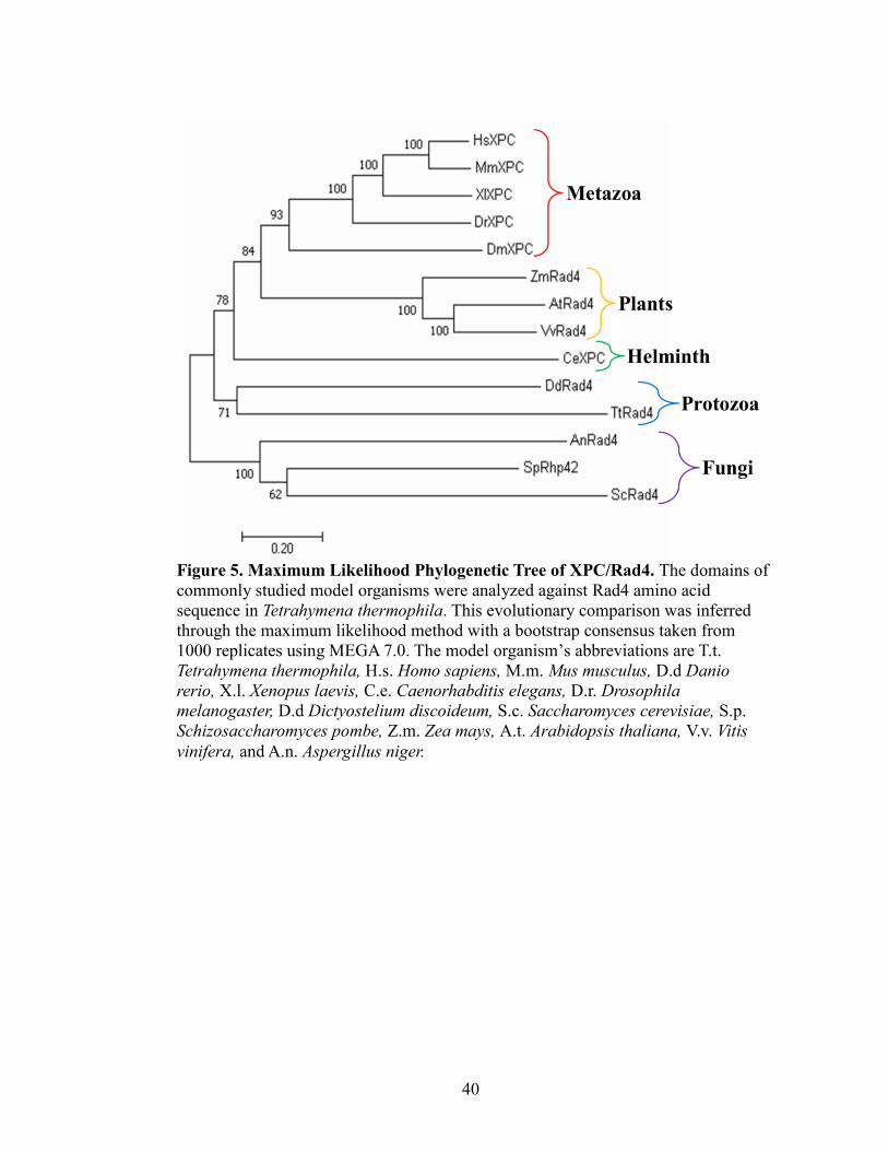
To further analyze the evolutionary conservation of Rad4 homologs and reinforce
the validity of Tetrahymena thermophila as a effective model organism for studying
RAD4, these same protein sequences were organized by phylogeny with MEGA 7.0
(Figure 5). There are two large initial diverges that occurred in evolution: one clade
contains metazoa, plants, helminth, and protozoa, and the other clade contains fungi. The
metazoa (H. sapiens, M. musculus, X. laevis, D. rerio, and D. melanogaster) grouped
together. The only helminth, C. elegans, grouped alone. The two protozoa D. discoideum
and T. thermophila diverged together off of the first clade. The three fungi in the other
clade contained A. niger, S. pombe, and S. cerevisiae. Because the T. thermophila is
diverging in the first clade with metazoa, plants, and helminth, its sequence is more
evolutionarily conserved than the sequence of the fungi Rad4 proteins.
Quantification of Tetrahymena thermophila RAD4 Expression Levels Before and
After Damage
To more completely understand the role of RAD4 in DNA repair, RAD4
expression levels were measured by qRT-PCR after three different genotoxic stressors
that lead to different type of DNA damage, repaired by different mechanisms: UV light
causes pyrimidine dimers that are repaired by nucleotide excision repair. Hydrogen
peroxide treatment causes oxidation of the nucleotide bases and hydrolysis of the
phosphodiester that is repaired by base excision repair. Methyl methanesulfonate (MMS)
40
Figure 5. Maximum Likelihood Phylogenetic Tree of XPC/Rad4. The domains of
commonly studied model organisms were analyzed against Rad4 amino acid
sequence in Tetrahymena thermophila. This evolutionary comparison was inferred
through the maximum likelihood method with a bootstrap consensus taken from
1000 replicates using MEGA 7.0. The model organism’s abbreviations are T.t.
Tetrahymena thermophila, H.s. Homo sapiens, M.m. Mus musculus, D.d Danio
rerio, X.l. Xenopus laevis, C.e. Caenorhabditis elegans, D.r. Drosophila
melanogaster, D.d Dictyostelium discoideum, S.c. Saccharomyces cerevisiae, S.p.
Schizosaccharomyces pombe, Z.m. Zea mays, A.t. Arabidopsis thaliana, V.v. Vitis
vinifera, and A.n. Aspergillus niger.
41
adds methyl groups to DNA strands that ultimately leads to double stranded breaks
repaired by homologous recombination or nonhomologous end joining (Weeden and
Asselin-Labat, 2018).
To analyze the qRT-PCR data, an amplification curve, melt peak, and agarose gel
visualizing a set of the qRT-PCR RAD4 (Figure 6A, 7A, 8A) and HHP1 (H1/HP1-like;
Figure 6C, 7C, and 8C) products. The analysis of HHP1 was included as a housekeeping
gene which maintains expression levels before and after genotoxic stressors. This allowed
for later data normalization. An amplification curve measures the relative florescent units
(RFU) during each cycle of amplification. In all treatments the amplification curves show
a variety of RAD4 expression levels. HHP1 showed relatively consistent amount of
amplification amongst treatments. Additionally, in these curves, actin amplification
curves are included (colored in red). This creates a standard curve using 1000 ng, 100 ng,
10 ng, and 1 ng of genomic DNA with actin primers.
Melt peaks were analyzed for both the RAD4 and HHP1 as indicator of a singular
product. One peak indicates that a single product is in the reaction that is melting or
dissociating at a specific temperature. The melt peaks showed specific peaks representing
RAD4 (Figure 6B, 7B, and 8B) and HHP1 (Figure 6D, 7D, and 8D) in one peak and
ACT1 control in the other, indicating that there was specific amplification of the
appropriate products during qRT-PCR.
The products of both RAD4 and HHP1 were also separated by agarose gel
electrophoresis. A single band at the expected length with RAD4 (186 base pairs) and
HHP1 (216 base pairs) was observed on the gel (Figure 6E,7E, 8E), further suggesting
specific amplification of RAD4 and HHP1 products by qRT-PCR. Also, both the RAD4
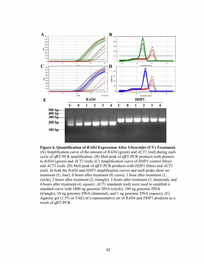
42
Figure 6. Quantification of RAD4 Expression After Ultraviolet (UV) Treatment.
(A) Amplification curve of the amount of RAD4 (green) and ACT1 (red) during each
cycle of qRT-PCR amplification. (B) Melt peak of qRT-PCR products with primers
to RAD4 (green) and ACT1 (red). (C) Amplification curve of HHP1 control (blue)
and ACT1 (red). (D) Melt peak of qRT-PCR products with HHP1 (blue) and ACT1
(red). In both the RAD4 and HHP1 amplification curves and melt peaks show no
treatment (U; line), 0 hours after treatment (0; cross), 1 hour after treatment (1;
circle), 2 hours after treatment (2; triangle), 3 hours after treatment (3; diamond), and
4 hours after treatment (4; square). ACT1 standards (red) were used to establish a
standard curve with 1000 ng genomic DNA (circle), 100 ng genomic DNA
(triangle), 10 ng genomic DNA (diamond), and 1 ng genomic DNA (square). (E)
Agarose gel (1.5% in TAE) of a representative set of RAD4 and HHP1 products as a
result of qRT-PCR.
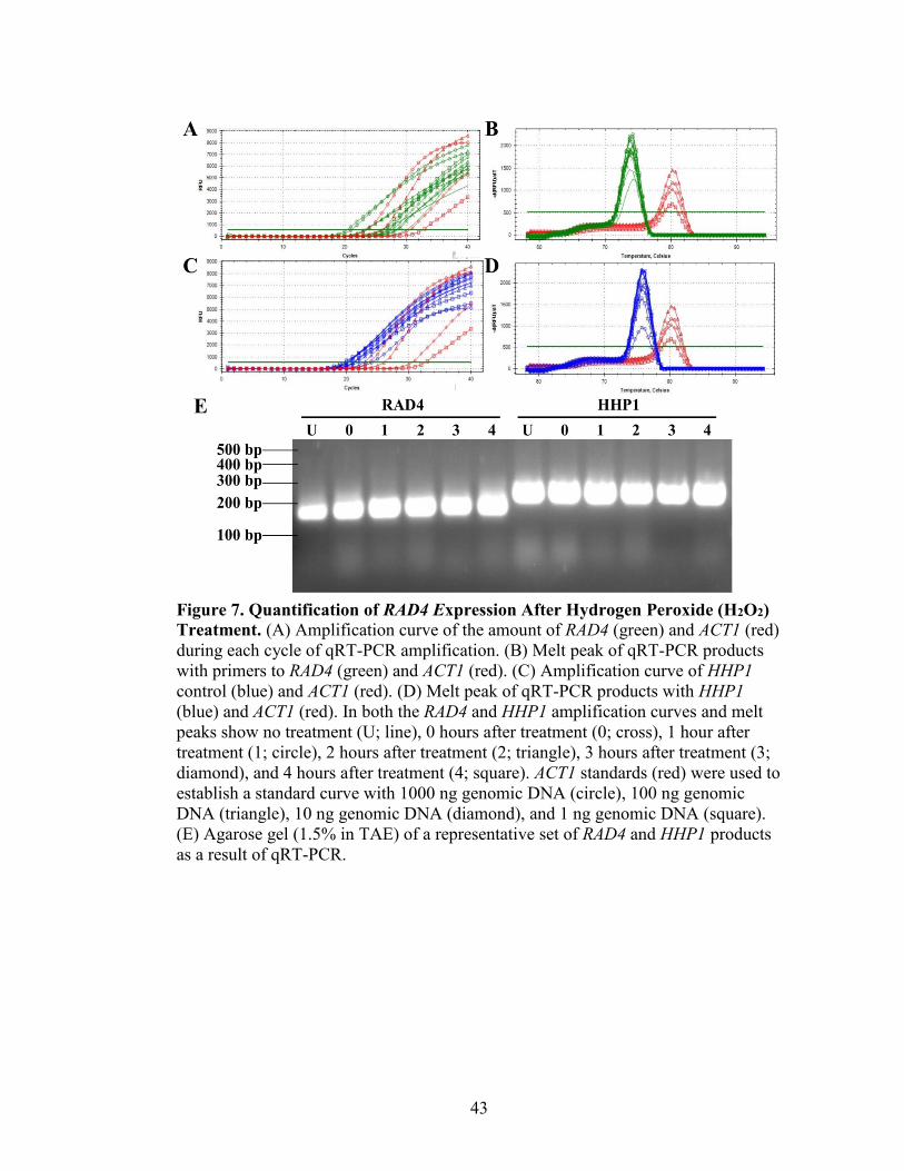
43
Figure 7. Quantification of RAD4 Expression After Hydrogen Peroxide (H2O2)
Treatment. (A) Amplification curve of the amount of RAD4 (green) and ACT1 (red)
during each cycle of qRT-PCR amplification. (B) Melt peak of qRT-PCR products
with primers to RAD4 (green) and ACT1 (red). (C) Amplification curve of HHP1
control (blue) and ACT1 (red). (D) Melt peak of qRT-PCR products with HHP1
(blue) and ACT1 (red). In both the RAD4 and HHP1 amplification curves and melt
peaks show no treatment (U; line), 0 hours after treatment (0; cross), 1 hour after
treatment (1; circle), 2 hours after treatment (2; triangle), 3 hours after treatment (3;
diamond), and 4 hours after treatment (4; square). ACT1 standards (red) were used to
establish a standard curve with 1000 ng genomic DNA (circle), 100 ng genomic
DNA (triangle), 10 ng genomic DNA (diamond), and 1 ng genomic DNA (square).
(E) Agarose gel (1.5% in TAE) of a representative set of RAD4 and HHP1 products
as a result of qRT-PCR.

44
Figure 8. Quantification of RAD4 Expression After Methyl Methanesulfonate
(MMS) Treatment. (A) Amplification curve of the amount of RAD4 (green) and
ACT1 (red) during each cycle of qRT-PCR amplification. (B) Melt peak of qRT-
PCR products with primers to RAD4 (green) and ACT1 (red). (C) Amplification
curve of HHP1 control (blue) and ACT1 (red). (D) Melt peak of qRT-PCR products
with HHP1 (blue) and ACT1 (red). In both the RAD4 and HHP1 amplification
curves and melt peaks show no treatment (U; line), 0 hours after treatment (0;
cross), 1 hour after treatment (1; circle), 2 hours after treatment (2; triangle), 3 hours
after treatment (3; diamond), and 4 hours after treatment (4; square). ACT1
standards (red) were used to establish a standard curve with 1000 ng genomic DNA
(circle), 100 ng genomic DNA (triangle), 10 ng genomic DNA (diamond), and 1 ng
genomic DNA (square). (E) Agarose gel (1.5% in TAE) of a representative set of
RAD4 and HHP1 products as a result of qRT-PCR.
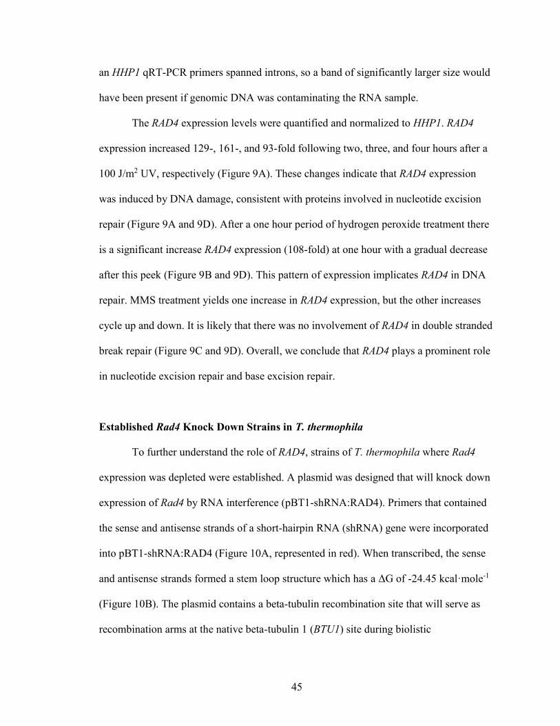
45
an HHP1 qRT-PCR primers spanned introns, so a band of significantly larger size would
have been present if genomic DNA was contaminating the RNA sample.
The RAD4 expression levels were quantified and normalized to HHP1. RAD4
expression increased 129-, 161-, and 93-fold following two, three, and four hours after a
100 J/m2 UV, respectively (Figure 9A). These changes indicate that RAD4 expression
was induced by DNA damage, consistent with proteins involved in nucleotide excision
repair (Figure 9A and 9D). After a one hour period of hydrogen peroxide treatment there
is a significant increase RAD4 expression (108-fold) at one hour with a gradual decrease
after this peek (Figure 9B and 9D). This pattern of expression implicates RAD4 in DNA
repair. MMS treatment yields one increase in RAD4 expression, but the other increases
cycle up and down. It is likely that there was no involvement of RAD4 in double stranded
break repair (Figure 9C and 9D). Overall, we conclude that RAD4 plays a prominent role
in nucleotide excision repair and base excision repair.
Established Rad4 Knock Down Strains in T. thermophila
To further understand the role of RAD4, strains of T. thermophila where Rad4
expression was depleted were established. A plasmid was designed that will knock down
expression of Rad4 by RNA interference (pBT1-shRNA:RAD4). Primers that contained
the sense and antisense strands of a short-hairpin RNA (shRNA) gene were incorporated
into pBT1-shRNA:RAD4 (Figure 10A, represented in red). When transcribed, the sense
and antisense strands formed a stem loop structure which has a ΔG of -24.45 kcal·mole-1
(Figure 10B). The plasmid contains a beta-tubulin recombination site that will serve as
recombination arms at the native beta-tubulin 1 (BTU1) site during biolistic
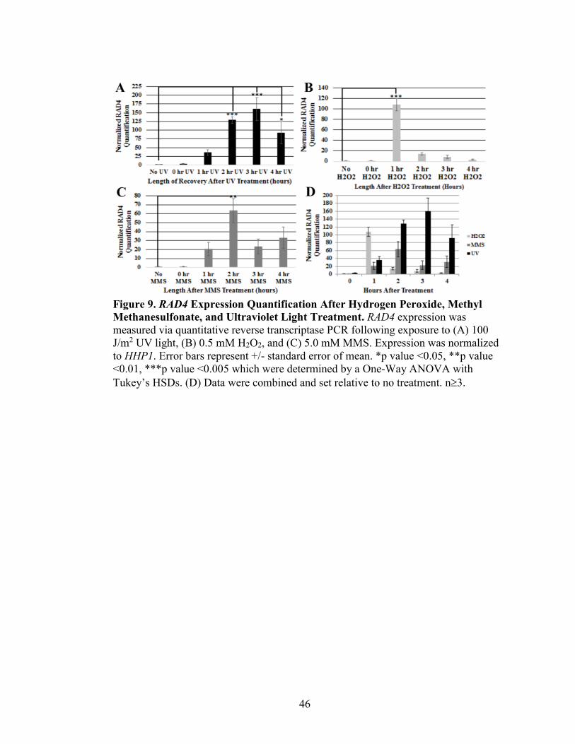
46
Figure 9. RAD4 Expression Quantification After Hydrogen Peroxide, Methyl
Methanesulfonate, and Ultraviolet Light Treatment. RAD4 expression was
measured via quantitative reverse transcriptase PCR following exposure to (A) 100
J/m2 UV light, (B) 0.5 mM H2O2, and (C) 5.0 mM MMS. Expression was normalized
to HHP1. Error bars represent +/- standard error of mean. *p value <0.05, **p value
<0.01, ***p value <0.005 which were determined by a One-Way ANOVA with
Tukey’s HSDs. (D) Data were combined and set relative to no treatment. n3.
47
Figure 10. Construction of pBT1-shRNA:RAD4. (A) pBT1-shRNA:RAD4
plasmid map containing a red segment with RAD4 sense and antisense primers, light
blue segment in the beta-tubulin 1 (BTU1) recombination site, and various restriction
enzyme sites. (B) Stem-loop representation of the Rad4 hairpin structure (21
nucleotides). The ΔG is -24.45 kcal·mole-1. (C) Sequence and diagram representation
of stem-loop sequence. Purple represents the linker region. Green represents the
Rad4 sense sequence and red represents the Rad4 antisense sequence. Yellow
represents the loop portion and XhoI restriction enzyme site.
48
transformation into T. thermophila. (Figure 10A, represented in light blue). When
transformed into DH10B cells, pBT1-shRNA:RAD4 produced 1 CFU with 10 ng of
plasmid, and 14 CFUs with 50 ng of plasmid. Negative controls were analyzed; pBT1-
YFG with no insert and no ligase produced 2 CFUs with 10 ng of plasmid, and 9 CFUs
with 50 ng of plasmid.
DNA was isolated from DH10B containing pBT1-shRNA:RAD4 and cloning was
confirmed by restriction digest. The shRNA insertion has a unique XhoI cut site (Figure
10C), therefore, digestion with XhoI, and a restriction enzyme that uniquely cuts within
the backbone (SacI) will produce unique fragments. The predicted products of the
XhoI/SacI digest are 1070 and 4214 base pairs. Only one clone contained the expected
products (Figure 11, clone 4), and was selected for further use. The shRNA plasmid DNA
was linearized with KpnI and SacI (data not shown). This linearized product was then
transformed and selected in T. thermophila via paclitaxel. Whole-cell PCR was
performed to confirm the presence of pBT1-shRNA:RAD4 (Figure 12). Samples not
transformed were expected to amplify only the BTU1 (native) locus (1750 bp), while
cells that successfully incorporated the Rad4 shRNA knock down will amplify a 500 bp
region containing the shRNA insert. Products corresponding to amplification of the
shRNA were most intensely amplified in the digested plasmid (positive control), and
CU522 clone 1 and 10 and CU725 clone 11. These clones were selected as Rad4
knockdowns subjected to further analysis.
Verification of Rad4 Knock Down in T. thermophila
To verify that expression of RAD4 is affected by introduction of the Rad4 shRNA,
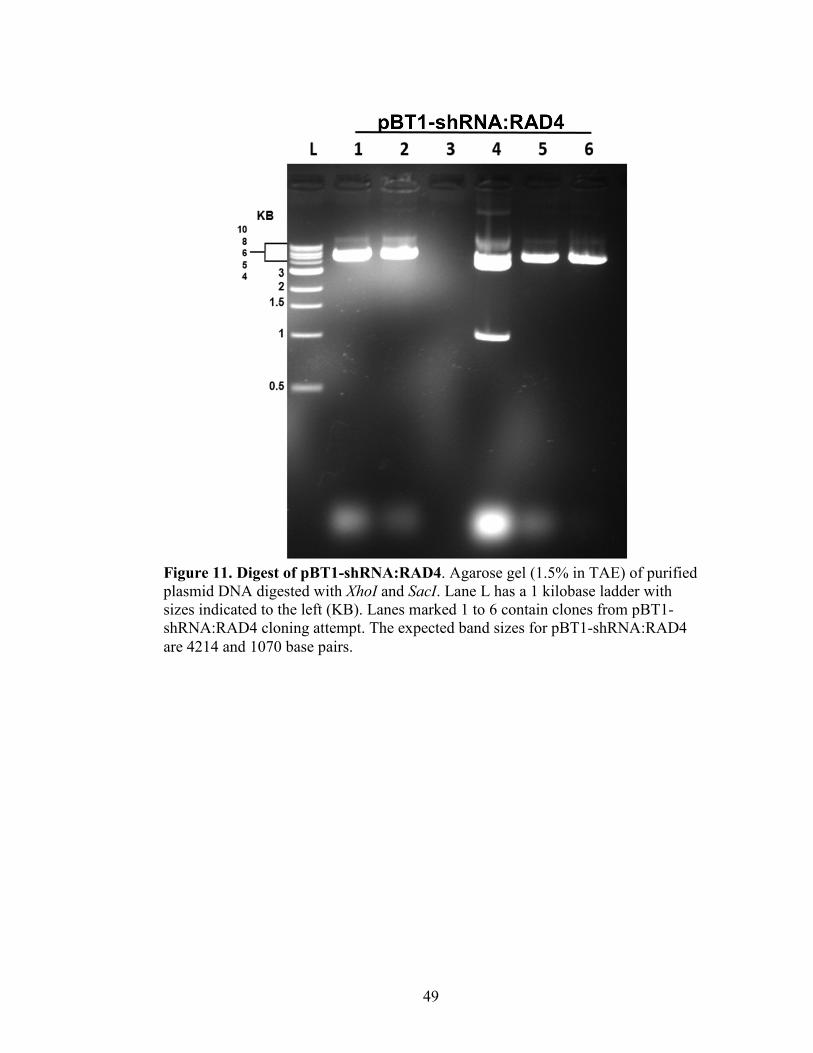
49
Figure 11. Digest of pBT1-shRNA:RAD4. Agarose gel (1.5% in TAE) of purified
plasmid DNA digested with XhoI and SacI. Lane L has a 1 kilobase ladder with
sizes indicated to the left (KB). Lanes marked 1 to 6 contain clones from pBT1-
shRNA:RAD4 cloning attempt. The expected band sizes for pBT1-shRNA:RAD4
are 4214 and 1070 base pairs.
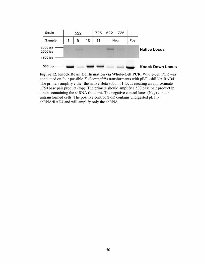
50
Figure 12. Knock Down Confirmation via Whole-Cell PCR. Whole-cell PCR was
conducted on four possible T. thermophila transformants with pBT1-shRNA:RAD4.
The primers amplify either the native Beta-tubulin 1 locus creating an approximate
1750 base pair product (top). The primers should amplify a 500 base pair product in
strains containing the shRNA (bottom). The negative control lanes (Neg) contain
untransformed cells. The positive control (Pos) contains undigested pBT1-
shRNA:RAD4 and will amplify only the shRNA.
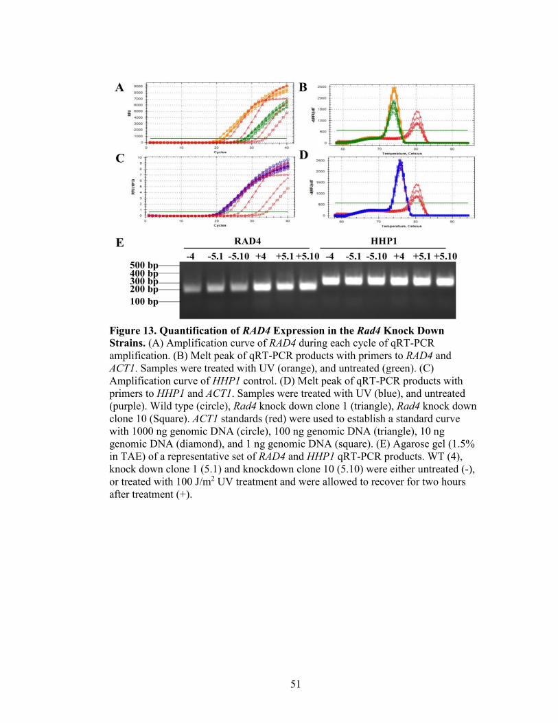
51
Figure 13. Quantification of RAD4 Expression in the Rad4 Knock Down
Strains. (A) Amplification curve of RAD4 during each cycle of qRT-PCR
amplification. (B) Melt peak of qRT-PCR products with primers to RAD4 and
ACT1. Samples were treated with UV (orange), and untreated (green). (C)
Amplification curve of HHP1 control. (D) Melt peak of qRT-PCR products with
primers to HHP1 and ACT1. Samples were treated with UV (blue), and untreated
(purple). Wild type (circle), Rad4 knock down clone 1 (triangle), Rad4 knock down
clone 10 (Square). ACT1 standards (red) were used to establish a standard curve
with 1000 ng genomic DNA (circle), 100 ng genomic DNA (triangle), 10 ng
genomic DNA (diamond), and 1 ng genomic DNA (square). (E) Agarose gel (1.5%
in TAE) of a representative set of RAD4 and HHP1 qRT-PCR products. WT (4),
knock down clone 1 (5.1) and knockdown clone 10 (5.10) were either untreated (-),
or treated with 100 J/m2 UV treatment and were allowed to recover for two hours
after treatment (+).
52
qRT-PCR was performed on wild type and RAD4 knock down clones 1 and 10. Based on
phenotypic results, qRT-PCR analysis was not performed on CU725 clone 11. In all
treatments the amplification curves showed a variety of RAD4 expression
levels. (Figure 13A). The HHP1 house keeping genes showed tightly grouped
amplification regardless of the treatment, (blue: UV, purple: no UV) as expected (Figure
13C). Actin 1 was amplified from defined amounts of genomic DNA to provide a
standard curve for detection (Figure 13, red). Isolated melt peaks appear for RAD4,
HHP1, and ACT1, indicating specific products for each (Figure 13B and 13D).
Furthermore, electrophoresis of the qRT-PCR products of both RAD4 and HHP1 showed
a single amplicon at the appropriate size for each product (186 and 278 bp, respectively;
Figure 13E). Both the specific melt peak, and the singular amplicon on the agarose gel
suggest specific amplification of RAD4 and HHP1 products by qRT-PCR.
RAD4 expression levels in the knock down strains were quantified, and
normalized to HHP1 (Figure 14). Rad4 knock down clone 1 showed 50% reduction in
expression after UV exposure (p = 0.001), when compared to wild type, indicating
successful knockdown of RAD4 expression by the shRNA. This difference in expression
was seen more drastically following UV treatment because there was a larger fold
difference. After UV treatment, clone 10 did not show a reduction that was significant.
This was likely due to the variance between the amplification that occurred in the
samples during qRT-PCR. Because this strain had inconclusive results, phenotypic
characterization of this strain was still conducted. Overall, Clone 1 was considered the
most effective knock down strain.
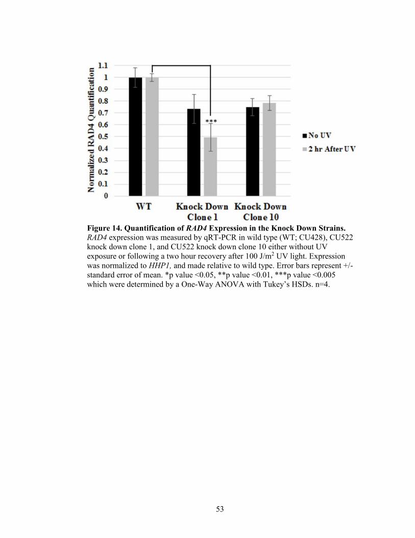
53
Figure 14. Quantification of RAD4 Expression in the Knock Down Strains.
RAD4 expression was measured by qRT-PCR in wild type (WT; CU428), CU522
knock down clone 1, and CU522 knock down clone 10 either without UV
exposure or following a two hour recovery after 100 J/m2 UV light. Expression
was normalized to HHP1, and made relative to wild type. Error bars represent +/-
standard error of mean. *p value <0.05, **p value <0.01, ***p value <0.005
which were determined by a One-Way ANOVA with Tukey’s HSDs. n=4.
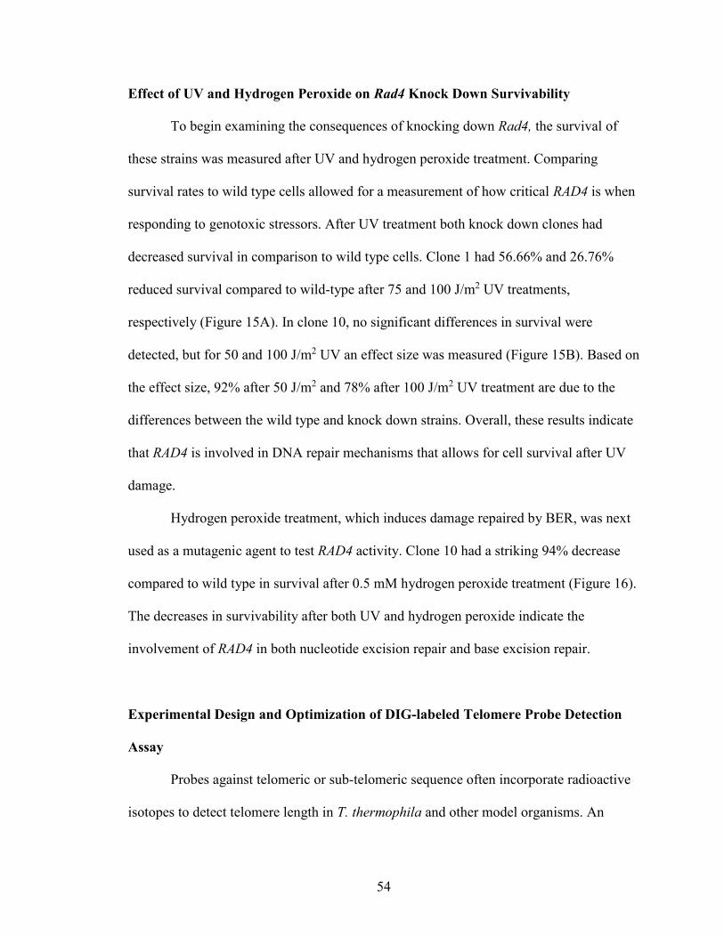
54
Effect of UV and Hydrogen Peroxide on Rad4 Knock Down Survivability
To begin examining the consequences of knocking down Rad4, the survival of
these strains was measured after UV and hydrogen peroxide treatment. Comparing
survival rates to wild type cells allowed for a measurement of how critical RAD4 is when
responding to genotoxic stressors. After UV treatment both knock down clones had
decreased survival in comparison to wild type cells. Clone 1 had 56.66% and 26.76%
reduced survival compared to wild-type after 75 and 100 J/m2 UV treatments,
respectively (Figure 15A). In clone 10, no significant differences in survival were
detected, but for 50 and 100 J/m2 UV an effect size was measured (Figure 15B). Based on
the effect size, 92% after 50 J/m2 and 78% after 100 J/m2 UV treatment are due to the
differences between the wild type and knock down strains. Overall, these results indicate
that RAD4 is involved in DNA repair mechanisms that allows for cell survival after UV
damage.
Hydrogen peroxide treatment, which induces damage repaired by BER, was next
used as a mutagenic agent to test RAD4 activity. Clone 10 had a striking 94% decrease
compared to wild type in survival after 0.5 mM hydrogen peroxide treatment (Figure 16).
The decreases in survivability after both UV and hydrogen peroxide indicate the
involvement of RAD4 in both nucleotide excision repair and base excision repair.
Experimental Design and Optimization of DIG-labeled Telomere Probe Detection
Assay
Probes against telomeric or sub-telomeric sequence often incorporate radioactive
isotopes to detect telomere length in T. thermophila and other model organisms. An
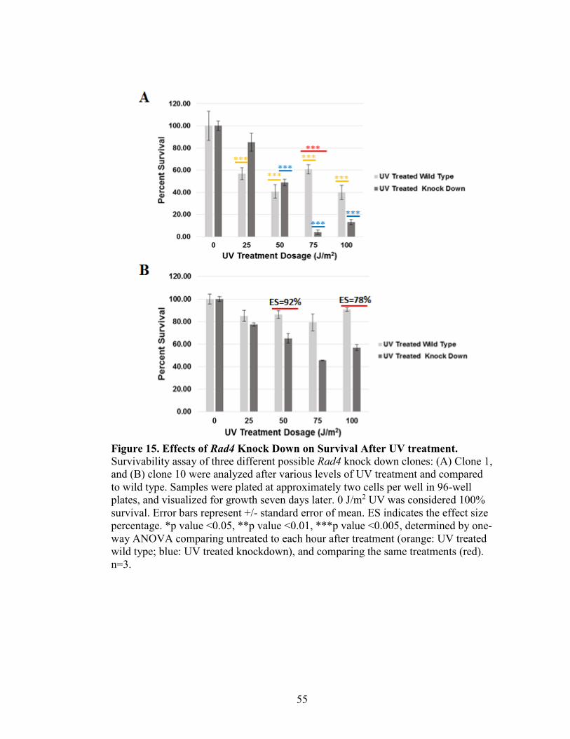
55
Figure 15. Effects of Rad4 Knock Down on Survival After UV treatment.
Survivability assay of three different possible Rad4 knock down clones: (A) Clone 1,
and (B) clone 10 were analyzed after various levels of UV treatment and compared
to wild type. Samples were plated at approximately two cells per well in 96-well
plates, and visualized for growth seven days later. 0 J/m2 UV was considered 100%
survival. Error bars represent +/- standard error of mean. ES indicates the effect size
percentage. *p value <0.05, **p value <0.01, ***p value <0.005, determined by one-
way ANOVA comparing untreated to each hour after treatment (orange: UV treated
wild type; blue: UV treated knockdown), and comparing the same treatments (red).
n=3.
56
Figure 16. Effects of Rad4 Knock Down on Survival after H2O2 Treatment.
Survivability assays of Rad4 knock down clone 10 were analyzed after various
levels of hydrogen peroxide treatment (X-axis) and compared to wild type. Samples
were plated at approximately two cells per well in 96-well plates, and visualized for
growth seven days later. No hydrogen peroxide (0 mM) was considered 100%
survival. Error bars represent +/- standard error of mean. *p value <0.05, **p value
<0.01, ***p value <0.005 determined by one-way ANOVA comparing untreated to
each hour after treatment (orange: UV treated wild type; blue: UV treated
knockdown), and comparing the same treatments (red). n=3.
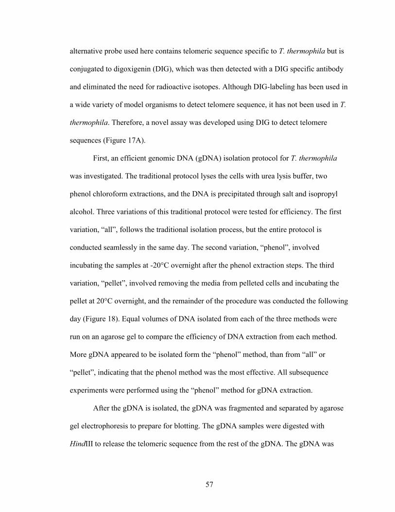
57
alternative probe used here contains telomeric sequence specific to T. thermophila but is
conjugated to digoxigenin (DIG), which was then detected with a DIG specific antibody
and eliminated the need for radioactive isotopes. Although DIG-labeling has been used in
a wide variety of model organisms to detect telomere sequence, it has not been used in T.
thermophila. Therefore, a novel assay was developed using DIG to detect telomere
sequences (Figure 17A).
First, an efficient genomic DNA (gDNA) isolation protocol for T. thermophila
was investigated. The traditional protocol lyses the cells with urea lysis buffer, two
phenol chloroform extractions, and the DNA is precipitated through salt and isopropyl
alcohol. Three variations of this traditional protocol were tested for efficiency. The first
variation, “all”, follows the traditional isolation process, but the entire protocol is
conducted seamlessly in the same day. The second variation, “phenol”, involved
incubating the samples at -20°C overnight after the phenol extraction steps. The third
variation, “pellet”, involved removing the media from pelleted cells and incubating the
pellet at 20°C overnight, and the remainder of the procedure was conducted the following
day (Figure 18). Equal volumes of DNA isolated from each of the three methods were
run on an agarose gel to compare the efficiency of DNA extraction from each method.
More gDNA appeared to be isolated form the “phenol” method, than from “all” or
“pellet”, indicating that the phenol method was the most effective. All subsequence
experiments were performed using the “phenol” method for gDNA extraction.
After the gDNA is isolated, the gDNA was fragmented and separated by agarose
gel electrophoresis to prepare for blotting. The gDNA samples were digested with
HindIII to release the telomeric sequence from the rest of the gDNA. The gDNA was

58
Figure 17. Development and Optimization of DIG-Labeled Telomere Probe
Detection Assay. (A) This experimental schematic was developed to detect
telomeric sequence without the use of a radioactive labeled probe. (B) A probe
designed specifically for telomeric sequence in T. thermophila (GGGGTT) was
conjugated to the digoxigenin (DIG) to be detected by a horseradish peroxidase
(HRP) conjugated anti-DIG antibody. The antibody when reacted with hydrogen
peroxide releases a chemiluminescent product which is detected on the blot
(Sambrook and Russell, 2001).
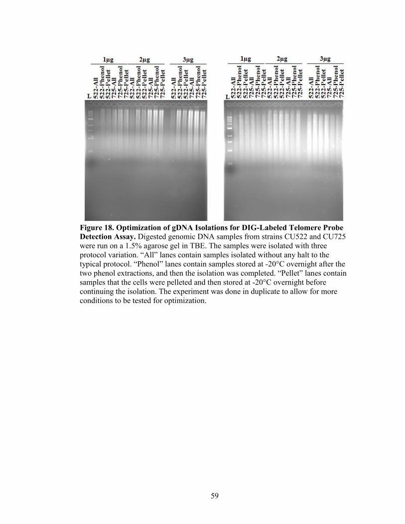
59
Figure 18. Optimization of gDNA Isolations for DIG-Labeled Telomere Probe
Detection Assay. Digested genomic DNA samples from strains CU522 and CU725
were run on a 1.5% agarose gel in TBE. The samples were isolated with three
protocol variation. “All” lanes contain samples isolated without any halt to the
typical protocol. “Phenol” lanes contain samples stored at -20°C overnight after the
two phenol extractions, and then the isolation was completed. “Pellet” lanes contain
samples that the cells were pelleted and then stored at -20°C overnight before
continuing the isolation. The experiment was done in duplicate to allow for more
conditions to be tested for optimization.
60
separated by agarose gel electrophoresis at a low voltage (70 V) over three to four hours,
to maximize separation. An agarose gel was loaded with one, two, and three µg of DNA,
and at least three µg was used in subsequent gels. The initial test showed more intensity
than subsequent experiments (data not shown). The predicted issue with this was the use
of different Nanodrop spectrophotometers. The Nanodrop 2000 gave consistently higher
readings than the ImplenNanoPhotometer NP80, therefore, less DNA was digested and
ran on the gel when read with the Nanodrop 2000. This issue was resolved by use of the
ImplenNanoPhotometer NP80 for sample analysis, and five µg of DNA.
To prepare the blot, the agarose gels were transferred to nitrocellulose membrane
and crosslinked. The gels were stained with ethidium bromide prior to transfer, in order
to estimate amounts of DNA per lane. The blot was prepared for hybridization by a pre-
hybridization incubation that prevents non-specific binding of the probe to the membrane.
The DIG-labeled telomeric probe was hybridized to the blot overnight by incubating at a
temperature at least 42°C. After hybridization, the blot was washed to remove non-
specifically bound probe from the membrane. Different wash temperatures were tested
(42, 45, and 50°C) and optimal signal was detected at 45°C, therefore, this temperature
was used in future assays (Figure 19).
Next, the blot was blocked, then incubated with the anti-DIG antibody (Figure
17B). Optimization of antibody detection occurred by varying blocking agents and
antibody concentration. Li-cor blocking agent and 0.5% milk in TBST were tested for
their blocking ability. 0.5% milk in TBST allowed optimal signal and thus was used in
future assays (Figure 19). The anti-DIG antibody conjugated to horseradish peroxidase
was added in 1:500 and 1:1000 dilutions. The 1:1000 dilution showed an optimal signal,

61
Figure 19. Optimization of Blotting Conditions for DIG-Labeled Telomere
Probe Detection Assay. Strips of membrane with digested genomic DNA were
treated with a variety of conditions to find the optimal combination for future assays.
The values listed above the nitrocellulose membranes are the varying temperatures
used for the wash directly following overnight probe hybridization at 42°C. Li-Cor
blocking agent (LC) and 0.5% Milk in TBST (Milk) served as antibody blocking
agents. The 1:500 and 1:1000 indicate the anti-DIG antibody dilutions.
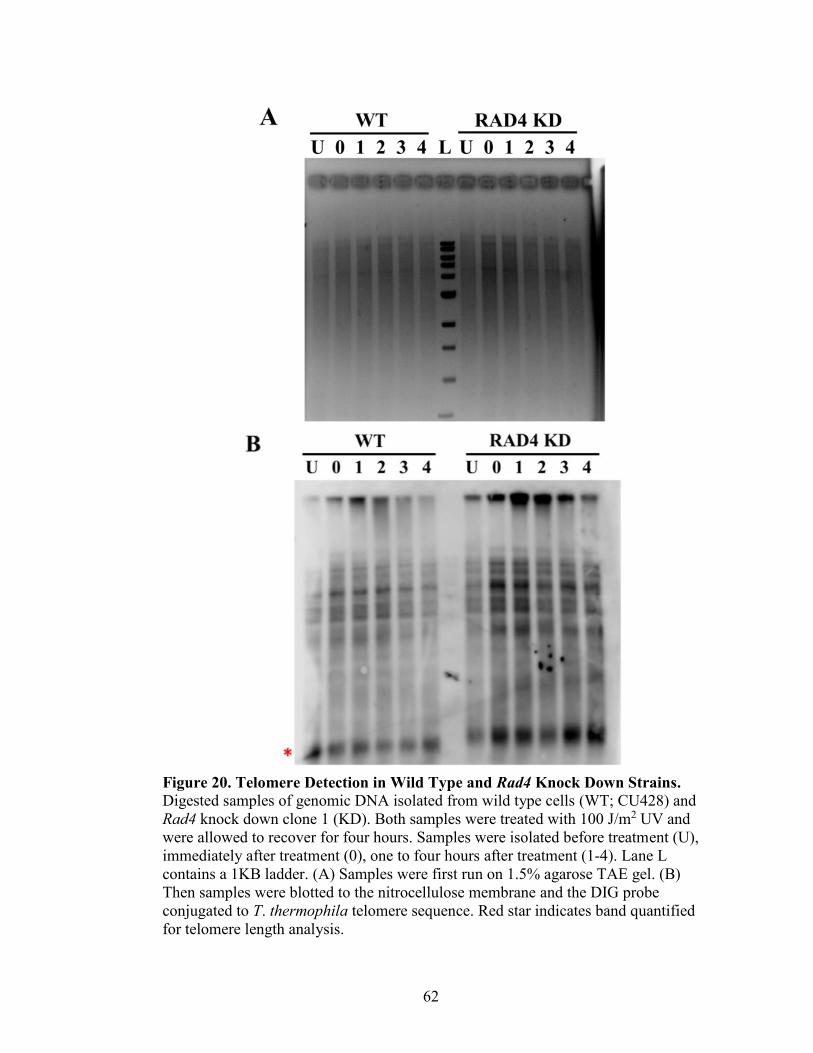
62
Figure 20. Telomere Detection in Wild Type and Rad4 Knock Down Strains. Digested samples of genomic DNA isolated from wild type cells (WT; CU428) and
Rad4 knock down clone 1 (KD). Both samples were treated with 100 J/m2 UV and
were allowed to recover for four hours. Samples were isolated before treatment (U),
immediately after treatment (0), one to four hours after treatment (1-4). Lane L
contains a 1KB ladder. (A) Samples were first run on 1.5% agarose TAE gel. (B)
Then samples were blotted to the nitrocellulose membrane and the DIG probe
conjugated to T. thermophila telomere sequence. Red star indicates band quantified
for telomere length analysis.

63
and was used in future assays (Figure 17B and Figure 19). The membrane was then
exposed to SuperSignal West Dura Stable Peroxide Buffer and SuperSignal West Dura
Lumino/Enhancer Solution, and was visualized using both the LI-COR Odyssey FC and
the Agfa CP1000 Automatic Film Processor. Although the LI-COR required a longer
amount of exposure to acquire comparable images to the film processor (data not shown),
the LI-COR images were able to achieve a quality image (Figure 19 and 20).
Telomere Length in Rad4 Knock Down Strain
The optimized DIG-labeled telomere probe detection assay was used to
characterize the differences in telomere composition between wild type (WT) and a Rad4
knock down strain (RAD4 KD). Telomeres from both untreated and UV treated samples
from wild type cells and Rad4 knock down clone 1 were analyzed. UV treated cells were
exposed to 100 J/m2 UV, and allowed to recover for four hours. Genomic DNA (5 µg)
was loaded onto an agarose gel and electrophoresed (Figure 20A).
After samples were transferred to a nitrocellulose membrane, telomeres were
visualized with the anti-DIG antibody. As expected, multiple bands show a
heterogeneous mixture of telomere sizes. Probing of this blot for actin 1 as a loading
control was also attempted. Unfortunately, it appears that more optimal probe design or
conditions need to be made for this probe as no detection was possible (data not shown).
In place of this loading control, gDNA streak intensity was quantified and used for
normalization (Figure 20A). The intensity of the lowest band on the nitrocellulose
membrane, likely representative of the telomeres on rDNA minichromosomes, was
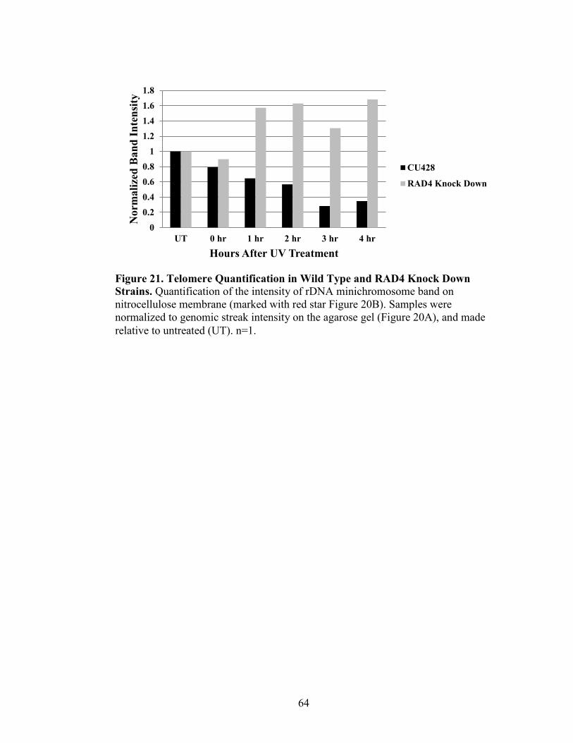
64
Figure 21. Telomere Quantification in Wild Type and RAD4 Knock Down
Strains. Quantification of the intensity of rDNA minichromosome band on
nitrocellulose membrane (marked with red star Figure 20B). Samples were
normalized to genomic streak intensity on the agarose gel (Figure 20A), and made
relative to untreated (UT). n=1.
0
0.2
0.4
0.6
0.8
1
1.2
1.4
1.6
1.8
UT 0 hr 1 hr 2 hr 3 hr 4 hr
Norm
ali
zed
Ban
d I
nte
nsi
ty
Hours After UV Treatment
CU428
RAD4 Knock Down
65
measured as a representative telomere length for the cell (Figure 20B, indicated with a
red star). The intensity of this band indicates the number of telomeric repeats of rDNA
minichromosomes (Figure 21). After UV treatment, the telomeres in the wild type
gradually decrease in size after UV treatment, while the Rad4 knock down strain
telomeres increase in size after UV treatment. At the end of the time course a 0.35
relative decrease and 1.68 relative increase respectively occurred in telomere length. This
indicated that RAD4 regulates telomere length in T. thermophila.
66
DISCUSSION
The Role of RAD4 in NER and BER
The overall purpose of this research is to understand the role of RAD4 in DNA
repair and its interplay with telomeres in Tetrahymena thermophila. It was anticipated
that, due to telomeres unique structure, nucleotide excision repair would occur differently
than in other areas of the genome. Analysis of the proteins associated with telomeres
(shelterin) revealed that overall shelterin proteins did not possess a large amount of
sequence conservation between organisms (Table 3). This lack of conservation makes it
challenging to find translatable results between organisms. The only detectable common
domains in the shelterin proteins in T. thermophila are Pot1a and Tpt1. However,
XPC/Rad4 proteins have strong conservation of the beta hair pin domain 1-3 and
transglutaminase domain, and are an attractive point of study between T. thermophila and
more complex model organisms (Table 4, Figure 5).
RAD4 expression levels in T. thermophila were measured after exposure to DNA
damaging agents (UV, H2O2 and MMS) to determine the involvement of Rad4 in DNA
repair (Figure 9). RAD4 expression significantly increases after both UV and hydrogen
peroxide treatment then gradually decreases, a typical expression pattern for DNA repair
genes, which expression spikes in response to the damage and then begins to return to
basal levels (Ramsey et al., 2004; Joshua Smith, unpublished data; Figure 9D).
Previously RAD4 has been implicated in the recognition of damage in global genome
nucleotide excision repair in response to UV damage (Evans et al., 1997). Rad4 also
plays a role in base excision repair by binding Ogg1, the base excision repair damage
67
recognition protein, and increasing the affinity of Ogg1 to damage (Melis et al., 2013). At
their peaks, RAD4 expression levels after UV and hydrogen peroxide treatment rose 129-
and 108-fold, respectively. These drastic increases in RAD4 expression in response to UV
and oxidative damage are likely related to its role in nucleotide excision repair (NER) and
base excision repair (BER) in T. thermophila. These findings validate T. thermophila as a
model organism in the study of NER and BER.
Rad4 Knock Down Reveals Important Role in NER and BER
Rad4 levels were depleted in Tetrahymena to further characterize its role in NER
and BER through stable short hair pin RNA construct (Figure 10). Knock down of Rad4
was confirmed in these strains through measurement of RAD4 expression levels by qRT-
PCR (Figure 14). There was a significant reduction (51%) in Rad4 transcription levels in
clone 1, indicating successful knock down of Rad4 by the shRNA. Although not
significant, there was an apparent reduction in expression levels in clone 10. The lack of
significance is likely due to the variances between the amplification of samples during
qRT-PCR.
Survivability after mutagenic treatment was used to measure the ability of T.
thermophila to repair the DNA damage, rather than enter apoptosis. UV treatment
decreased survivability upon Rad4 depletion, indicating that T. thermophila are not able
to repair UV-induced DNA damage with NER (Figure 15A). Clone 10 showed
insignificant results in both expression reduction and UV treatment. Interestingly,
survivability appeared to be dose dependent, as knock down clone 1 had more reduction
in survivability than clone 10, where Rad4 knock down and survivability after UV was
68
reduced, but not significant. Knock down clone 10 had significantly decreased
survivability after hydrogen peroxide treatment (Figure 16), indicating that even subtle
Rad4 depletion has dramatic impacts on the BER pathway. Altogether, T. thermophila
Rad4 likely contributes to the ability of NER and BER system to recognize damage and
recruit the additional proteins needed to repair the damage. When Rad4 is absent, the
damage accumulates, and the cells enter apoptosis.
Development of a DIG-Labeled Telomere Probe Detection Assay
The establishment of an assay to measure telomere length via a DIG-labeled
telomere-specific probe was a cornerstone of this project (Figure 17). The ability to work
with radioactive isotopes is limited to institutions or scientists with the facilities,
equipment, clearance and training to do so, therefore the ability to detect telomeres
without radioactive labeled probes should open telomere biology studies to a wider range
of institutions and laboratories.
The design of DIG-based telomere detection required the collaboration of many
resources and scientists. Dr. Benjamin Linger was consulted on the length and possible
sequence of the probe, and telomere isolation techniques. The blotting and probing
protocol was combined from different resources, including those contributed by Dr.
Smith, Dr. Ulbricht, and a variety of biotechnical companies that have DIG labeling
products. Time and effort was devoted to understanding current radiolabeled methods of
telomere detection in T. thermophila previous work of from Dr. Carolyn Price and Dr.
Linger. Considerations from these sources helped develop the ideal conditions for probe
69
design, gDNA isolation, hybridization, blotting, probe detection, and interpretation of the
results from the telomere detection assay.
While designing the DIG-based telomere detection assay, steps of the protocol
were identified as potential targets of optimization as the procedure was refined. Three
different gDNA isolation conditions were attempted, and the technique with the most
intense gDNA streak on an agarose gel after HindIII digestion was selected (Figure 18).
Phenol chloroform extraction and ethanol precipitation in one day from freshly isolated
cells or those pelleted frozen overnight was inferior to performing two consecutive
phenol chloroform extractions on freshly isolated cells and freezing the aqueous layer
overnight before precipitation the next day. The key difference between these protocols is
likely the freeze-thaw step that may increase the solubility of DNA.
After optimization of the gDNA isolation procedure, multiple hybridization and
blotting conditions for probing and detection were considered (Figure 19). The wash
temperatures after hybridization of the probe were varied from 42°C to 50°C. The most
distinctive telomeric banding was seen at 45°C, suggesting that this temperature allows
optimal removal of background, without disrupting interaction between the probe and
gDNA. Detection of the DIG-labeled probe was optimized by varying the blocking agent
and anti-DIG antibody concentration (Figure 19). Li-cor blocking solution seemed to be
ineffective since the image of the blot was completely dark at all washing temperatures
and antibody concentrations, with no distinct bands. Blocking in 0.5% milk in TBST
allowed distinct bands to appear in lanes with gDNA, indicating that this condition is
optimal for allowing binding of the anti-DIG antibody to the DIG-labeled probe, but
preventing non-specific binding of the antibody to the blot. Incubation of the blot in
70
1:1000 anti-DIG antibody resulted in clearer bands on the blot than in a 1:500 dilution of
the antibody, suggesting that incubation of the blot in the more dilute concentration of
antibody is optimal for probe detection. Altogether, the washing step at 45°C, the
antibody in a 1:1000 dilution, and the blocking buffer of 0.5% milk in TBST made
optimal conditions for probe washing and detection.
The Effect of RAD4 on Telomeres
With the DIG-labeled telomere probe detection assay optimized, telomeres from
wild type and Rad4 knock down T. thermophila were analyzed to determine the effects of
depleted Rad4 levels on telomere composition (Figure 20 and 21). In tandem with
telomere detection, a actin probe was designed to be used, providing an internal control
for the amount of gDNA in each lane. Unfortunately, the actin probe had no discernable
bands, indicating that probe design, hybridization, or washing conditions need to be
further optimized. Instead, an alternative loading control was developed where gDNA
amount was quantified from ethidium bromide stains of each gel, prior to blotting (Figure
20A). Telomeres on rDNA minichromsome were selected for quantification by
measuring the intensity of the band, which is an indication of the amount of telomere
repeats (indicated by a red star; Figure 20B). After UV exposure, the Rad4 knock down
clone 1 has longer telomeres than wild type strain (Figure 21). This is consistent with
previous literature which showed that XPC knock outs resulted in longer telomeres after
UV exposure in Mus musculus (Stout and Blasco, 2013). In wild type cells, UV exposure
leads to shortened telomeres, whereas cells that are deficient in XPC, which are also
71
deficient in NER and BER, actually extend telomeres after UV exposure, which has also
been supported in the literature (Ramírez et al., 2002).
The Rad4 depletion in T. thermophila potentially stresses the cells stimulating
alternative lengthening of the telomeres. This mechanism helps to add telomeric sequence
when the cells are under duress. Alternatively, the presence of unrepaired pyrimidine
dimers could be hypothesized to cause shelterin proteins to not bind as effectively to the
damaged DNA. Since shelterin proteins block telomerase access to the telomeres,
disrupting their binding leads to aberrant lengthening of telomeres (Palm and de Lange,
2008). It is evident that Rad4 in T. thermophila plays a critical role in length regulation
and telomere maintenance.
Future Directions
This work contributes a significant step forward in our understanding of the
interactions of Rad4, and more generally DNA repair, and telomere regulation.
Moreover, the work opens a wide-range of avenues for future studies. To more
completely verify Rad4 knock down clone 1 and 10 are depleting the protein as well as
the transcript after UV and hydrogen peroxide treatments, GFP or RFP tagged RAD4
could be transformed in the rlp29 locus and visualized. Additionally, creating a complete
knock out of RAD4 would allow for clear characterization of cells without the function in
NER and telomere repair.
Further phenotypic characterization of Rad4 knock downs may clarify the
conclusions made here or provide additional information. Specifically, hydrogen
peroxide treatment of the most significantly affected Rad4 knock down (clone 1) is
72
expected to produce an even more pronounced reduction in survivability after hydrogen
peroxide treatment. Also, an increase in the amount of UV survivability assays will
increase the power of the experiment and determine if the interesting, yet not statistically
significant, observed effects between the wild type and Rad4 knock downs were due to
variability in the assay or sample, or due to subtle effects of the inefficient Rad4 knock
down.
Only one telomere assay was able to be performed since a large amount of time
and resources were spent on development of the assay. Additional trials of this telomere
detection assay will allow statistical analysis of the telomere length and strengthen the
conclusions made in this one impressive experiment. It would also be interesting to
compare telomere lengths in both clone 1 and clone 10, to see if the knock down
efficiency effects telomere length. Since this study consistently shows Rad4-dependent
BER phenotypes, it would be interesting to see the effects on telomere composition after
hydrogen peroxide treatment. Previous studies have shown that telomeres shorten after
hydrogen peroxide treatment (Ting et al., 2010). After hydrogen peroxide treatment, it is
possible we would observe a very similar phenotype after UV treatment, but if there is a
lack of change it would show NER specificity. Also, it would be advantageous to extend
the time course for the gDNA isolations until telomere lengths return to untreated levels.
Importantly, this study provides Missouri State University and other institutions
with the ability to study telomeres without radioactive labeled probes. An interesting
augmentation to this protocol could be using a probe to the subtelomeric region, rather
than the telomere repeat. These studies have been performed by the Price lab with
forward and reverse radioactive labeled probes specific to the subtelomeric sequence of

73
specific telomeres in T. thermophila (Jacob et al., 2004). The advantage of this design is a
potentially cleaner blot, showing specific telomere length, instead of a heterogeneous
mixture of telomeres. An internal rDNA probe on the minichromosome could be used for
normalization. Additionally, the optimization of the actin probe would be another
effective loading control that could be optimized as an appropriate loading control.
At the genesis of this project, there were hopes of obtaining a variety of tagged
strains from the Price lab. The particular strains of interest we would have liked to have
studied. The likely most important is the TERT knockout construct and strain (Linger et
al., 2011). This would serve as an excellent control for cells that would have shorter
telomeres. The overall ultimate goal of this research is to find a potential shelterin
binding partner to RAD4. Because this is a recognition protein that would be the first to
interact with damaged telomeric DNA, a binding partner could be expected (Jia et al.,
2015). To study this interaction there are two possible approaches, co-
immunopercipitation of tagged Rad4 or tagged shelterin proteins. Currently the Price lab
has POT2-HA, POT1-TAP, and TPT1-TAP (Cranert et al., 2014; Jacob et al., 2007;
Linger et al., 2011). Because currently tagging Rad4 has proven problematic (Emily
Nischwitz and Rachel Mullner unpublished data), it would be beneficial to acquire those
tagged strains for further studies. Additionally, knockout strains of POT2, POT1, and
TPT1 also have shown interesting length phenotypes that could be studied with the newly
establish DIG-labeled probe telomere detection assay (Cranert et al., 2014; Jacob et al.,
2007; Linger et al., 2011).
Overall, this project added resources to the scientific community to be able to
study DNA repair and telomere biology. The establishment of characterized Rad4 knock

74
down strains allow researchers to continuing the study of NER in T. thermophila within
the macronucleus and micronucleus. Additionally, the newly established DIG-labeled
telomere probe detection assay allows for Tetrahymena labs to be able to study telomere
length and composition in the context of DNA repair and any other field.
.
75
REFERENCES
Alter, B.P., Giri, N., Savage, S.A., and Rosenberg, P.S. (2009). Cancer in dyskeratosis
congenita. Blood 113, 6549-6557.
Altschul, S.F., Stephen, F., Madden, T.L., Schäffer, A.A., Zhang, J., Zhang, Z., Miller,
W., and Lipman, D.J. (1997). Gapped BLAST and PSI-BLAST: a new generation of
protein database search programs. Nucleic Acids Res. 25, 3389-3402.
Armanios, M. (2012). Telomerase and idopathic pulmonary fibrosis. Mutat. Res. 730, 52-
58.
Bass, H.W., Marshall, W.F., Sedat, J.W., Agard, D.A., and Cande, W.Z. (1997).
Telomeres cluster de novo before the initation of synapsis: a three-dimensional spatial
analysis of telomere positions before and during meiotic phrophase. J. Cell Biol. 137, 5-
18.
Bauman, P., and Cech, T.R. (2001). Pot1, the putative telomere end-binding protein in
fission yeast and humans. Science 292, 1171-1175.
Bianchi, A., Smith, S., Chong, L., Elias, P., and de Lange, T. (1997). TRF1 is a dimer and
bends telomeric DNA. EMBO. 18, 5735-5744.
Blackburn, E.H., Budarf, M.L., Challoner, P.B., Cherry, J.M., Howard, E.A., Katzen,
A.L., Pan, W.C., and Ryan, T. (1983). DNA termini in ciliate macronuclei. Quant. Biol.
47, 1195-1207.
Blackburn, E.H., and Gall, J.G. (1978). A tandemly repeated sequence at the termini of
the extrachromosomal ribosomal RNA genes in Tetrahymena. J. Mol. Biol. 120, 33-53.
Boswell, R.E., Klobutcher, L.A., and Prescott, D.M. (1982). Inverted terminal repeats are
added to genes during macronuclear development in Oxytricha nova. Proc. Natl. Acad.
Sci. USA. 79, 3255-3259.
Broccoli, D., Smogorzewska, A., Chong, L., and de Lange, T. (1997). Human telomeres
contain two distinct Myb-related proteins, TRF1 and TRF2. Nat. Genet. 17, 231-235.
Bunick, C.G., Miller, M.R., Fuller, B.E., Fanning, E., and Chazin, W.J. (2006).
Biochemial and structural domain analysis of xeroderma pigmentosum complementation
group C protein. Biochem. 45, 14965-14979.
Cranert, S., Heyse, S., Linger, B., Lescasse, R., and Price, C. (2014). Tetrahymena Pot2
is a developmentally regulated paralog of Pot1 that localizes to chromosome breakage
sites but not to telomeres. Eukaryot. Cell 13, 1519-1529.
76
Celli, G., and de Lange, T. (2005). DNA processing not required for ATM-mediated
telomere damage response after TRF2 depletion. Nat. Cell Biol. 7, 712-718.
Cesare, A.J., and Reddel, R.R. (2010). Alternative lengthening of telomeres: models,
mechanisms and implications. Nat. Rev. Genet. 11, 319-330.
Cleaver, J.E. (1968). Defective repair replication of DNA in xeroderma pigmentosum.
Nature 218, 652-656.
Cleaver, J.E. (2005). Cancer in xeroderma pigmentosum and related disorder of DNA
repair. Nat. Rev. Cancer 5, 564-573.
Coin, F., Oksenych, V., Mocquet, V., Groh, S., Blattner, C., and Egly, J.M. (2008).
Nucleotide excision repair drive by the dissociation of CAK from TFIIH. Mol. Cell 31, 9-
20.
Cooper, J.P., Nimmo, E.R., Allshire, R.C., and Cech, T.R. (1997). Regulation of telomere
and function by a Myb-domain protein in fission yeast. Nature 385, 744-747.
DiGiovanna, J.J., and Kraemer, K.H. (2012). Shining a light on xeroderma pigmentosum.
J. Invest. Dermatol. 132, 785-796.
de Laat, W.L., Appeldoorn, E., Sugasawa, K., Weterings, E., Jaspers, N.G., and
Hoeijmakers, J.H. (1998). DNA-binding polarity of human replication protein A
positions nucleases in nucleotide excision repair. Genes Dev. 12, 2598-2609.
Dokal, I. (2000). Dyskeratosis congenital in all its forms. Br. J. Haematol. 110, 768-779.
Evans, E., Moggs, J.G., Hwang, J.R., Egly, J.M., and Wood, R.D. (1997). Mechanism of
open complex and dual incision formation by human nucleotide excision repair factors.
EMBO J. 16, 6559-6573.
Fagbemi, A.F., Orelli, B., and Schärer, O.D. (2011). Regulation of endonuclease activity
in human nucleotide excision repair. DNA Repair 10, 722-729.
Fan, J., and Pavletich, N.P. (2012). Structure and conformational change of a replication
protein A heterotrimer bound to ssDNA. Genes Dev. 26, 2337-2347.
Finn, R.D., Attwood, T.K., Babbitt, P.C., Bateman, A., Bork, P., Bridge, A.J., Chang, H-
Y., Dosztanyi, Z., El-Gebali, S., Fraser, M., et al. (2017). InterPro in 2017-beyond protein
family and domain annotations. Nucleic Acids Res. 45, D190-D199
Flory, M.R., Carson, A.R., Muller, E.G., and Aebersold, R. (2004). An SMC-domain
protein in fission yeast links telomeres to the meiotic centrosome. Mol. Cell 16, 619-630.
77
Gao, H., Cervantes, R.B., Mandel, E.K., Otero, J.H., and Lundblad, V. (2007). RPA-like
proteins mediate yeast telomere function. Nat. Struct. Mol. Biol. 14, 208-214.
Gillet, L.C., and Schärer, O.D. (2006). Molecular mechanisms of mammalian global
genome nucleotide excision repair. Chem. Rev. 106, 253-276.
Gilljam, K.M., Muller, R., Liabakk, N.B., and Otterlei, M. (2012). Nucleotide excision
repair is associated with the replisome and its efficiency depends on a direct interaction
between XPA and PCNA. PloS ONE 7, e49199
Gillooly, J.F., Hein, A., and Damiani, R. (2015). Nuclear DNA content varies with cell
size across human cell types. Cold Spring Harb. Perspect. Biol. 7, a019091.
Gopalakrishnan, K., Low, G.K.M., Ting, A.P.L., Srikanth, P., Slijepcevic, P., and Hande,
M.P. (2010). Hydrogen peroxide induced genomic instability in nucleotide excision
repair-deficient lymphoblastoid cells. Genome Integrity 16, 1-14.
Greider, C.W., and Blackburn, E.H. (1985). Identification of a specific telomere terminal
transferase activity in Tetrahymena extracts. Cell 43, 405-413.
Griffith, J.D., Comeau, L., Rosenfield, S., Stansel, R.M., Bianchi, A., Moss, H., and de
Lange, T. (1999). Mammalian telomeres end in a large duplex loop. Cell 97, 503- 514.
Hanawalt, P.C., and Spivak, G. (2008). Transcription-coupled DNA repair: two decades
of progress and surprises. Nat. Rev. Mol. Cell Biol. 9, 958-970.
Henderson, E., Hardin, C.C., Walk, S.K., Tinoco, I., and Blackburn, E.H. (1987).
Telomeric DNA oligonucleotides form novel intramolecular structures containing
guanine-guanine base pairs. Cell 51, 899-908.
Henderson, E.R., and Blackburn, E.H. (1989). An overhang 3` terminus is a conserved
feature of telomeres. Mol. Cell Biol. 9, 345-348.
Hirano, Y., Fukunaga, K., and Sugimoto, K. (2009). Rif1 and rif2 inhibit localization of
tel1 to DNA ends. Cell 126, 63-77.
Hohl, M., Thorel, F., Clarkson, S.G., and Schärer, O.D. (2003). Structural determinants
for substrate binding and catalysis by the structure-specific endonuclease XPG. J. Biol.
Chem. 278, 19500-19508.
Jacob, N.K., Stout, A.R., and Price, C.M. (2004). Modulation of telomere length
dynamics by the subtelomeric region of Tetrahymena telomeres. Mol. Biol. of the Cell
15, 3719-3728.
78
Jacob, N.K., Lescasse, R., Linger, B.R., and Price, C.M. (2007). Tetrahymena POT1a
regulates telomere length and prevents activation of a cell cycle checkpoint. Mol. Cell
Biol. 27, 1592-1601.
Jia, P., Her, C., and Chai, W. (2015). DNA excision repair at telomeres. DNA Repair
(Amst.) 36. 137-145.
Kanoh, J., and Ishikawa, F. (2001). spRap1 and spRif1, recruited to telomeres by Taz1,
are essential for telomere function in fission yeast. Curr. Biol. 11, 1624-1630.
Katzen, A.L., Cann, G.M., and Blackburn, E.H. (1981). Sequence specific fragmentation
of macronuclear DNA in a holotrichous ciliate. Cell 24, 313-320.
Kelleher, C., Kurth, I., and Linger, J. (2005). Human protection of telomeres 1 (POT1) is
a negative regulator of telomerase activity in vitro. Mol. Cell Biol. 25, 808-818.
Kumar, S., Stecher, G., and Tamura, K. (2016). MEGA7: Molecular Evolutionary
Genetic Analysis Version 7.0 for bigger datasets. Mol. Biol. Evol. 33, 1870-1874.
Klobutcher, L.A., Swanton, M.T., Donini, P., and Prescott, D.M. (1981). All gene-sized
DNA molecules in four species of hypotrichs have the same terminal sequence and an
unusual 3` terminus. Proc. Natl. Acad. Sci. USA. 78, 3015-3019.
Kocak, H., Ballew, B.J., Bisht, K., Eggebeen, R., Hicks, B.D., Suman, S., O’Neil, A.,
Giri, N., Maillard, I., Alter, B.P., et al. (2014). Hoyeraal-Hreidarsson syndrome caused by
a germline mutation in the TEL patch of the telomere protein TPP1. Genes Dev. 28,
2090-2102.
Lazzerini-Denchi, E. and Sfeir, A. (2016). Stop pulling my strings-what telomeres taught
us about the DNA damage response. Nat. Rev. Mol. Cell Biol. 17, 364-378.
Lehmann, A.R. (2003). DNA repair-deficient diseases, xeroderma pigmentosum,
Cockayne syndrome and trichothiodystrophy. Biochimie 85, 1101-1111.
Lei, M., Podell, E.R., and Cech, T.R. (2004). Structure of human POT1 bound to
telomeric single-stranded DNA provides a model for chromosome end-protection. Nat.
Struct. Mol. Biol. 11, 1223-1229.
Li, B., Oestreich, S., and de Lange, T. (2000). Identification of human Rap1: implications
for telomere evolution. Cell 101, 471-483.
Li, L., Elledge, S.J., Peterson, C.A., Bales, E.S., and Legerski, R.J. (1994). Specific
association between the human DNA repair proteins XPA and ERCC1. Proc. Natl. Acad.
Sci. 91, 5012-5016.
79
Li, W., Cowley, A., Uludag, M., Gur, T., McWilliam, H., Squizzato, S., Park, Y.M., Buso
N., and Lopez, R. (2015). The EMBL-EBI bioinformatics web and programmatic tools
framework. Nucleic Acids Res. 43, W580-W584.
Linger, B.R., Morin, G.B., and Price, C.M. (2011). The Pot1a-associated proteins Tpt1
and Pat1 coordinate telomere protection and length regulation in Tetrahymena. MBoC.
22, 4161-4170.
Marcand, S., Gilson, E., and Shore, D. (1997). A protein-counting mechanism for
telomere length regulation in yeast. Science 275, 986-990.
McClintock, B. (1931). Cytological observation of deficiencies involving known genes,
translocations and inversion in Zea mays. Missouri Agr. Exp. Sta. Res. Bull. 163, 1-48.
Melis, J.P.M., van Steeg, H., and Luijten, M. (2013). Oxidative DNA damage and
nucleotide excision repair. Antioxidants and Redox Signaling. 18, 2409-2419.
Min, J.H., and Pavletich, N.P. (2007). Recognition of DNA damage by the Rad4
nucleotide excision repair protein. Nature 499, 570-575.
Mitchell, J.R., Wood, E., and Collins, K. (1999). A telomerase component is defective in
the human disease dyskeratosis congenital. Nature 402, 551-555.
Morin, G.B. (1989). The human telomere terminal transferase enzyme is a
ribonucleoprotein that synthesizes TTAGGG repeats. Cell 59, 521-529.
Moser, J., Kool, H., Giakzidis, I., Caldecott, K., Mullenders, L.H., and Fousteri, M.I.
(2007). Sealing of chromosomal DNA nicks during nucleotide excision repair requires
XRCCI and DNA ligase III α in a cell-cycle-specific manner. Mol. Cell 27, 311-323.
Moyzis, R.K., Buckingham, J.M., Cram, L.S., Dani, M., Deavn, L.L., Jones, M.D.,
Meyne, J., Ratliff, R.L., and Wu, J.R. (1988). A highly conserved repetitive DNA
sequence (TTAGGG)n, present at the telomeres of human chromosomes. Proc. Natl.
Acad. Sci. USA. 85, 6622-6626.
Mullner, H.J. (1938). The remaking of chromosomes. Collecting Net. 13, 181-198.
Notredame, C., Higgins, D.G., and Heringa, J. (2000). T-Coffee: A novel method for fast
and accurate multiple sequence alignment. J. of Mol. Biol. 302, 205-217.
Ogi, T., and Lehmann, A.R. (2006). The Y-family DNA polymerase kappa functions in
mammalian nucleotide excision repair. Nat. Cell Biol. 8, 640-642.
80
Ogi, T., Limsirichaikul, S., Overmeer, R.M., Volker, M., Takenaka, K., Cloney, R.,
Nakazawa, Y., Niimi, A., Miki, Y., Jaspers, N.G., et al. (2010). Three DNA polymerases,
recruited by different mechanisms, carry out NER repair synthesis in human cells. Mol.
Cell 37, 714-727.
Olovnikov, A.M. (1973). A theory of marginotomy. The incomplete copying of template
margin in enzymic synthesis of polynucleotides and biological significance of the
phenomenon. J. Theor. Biol. 41, 181-190.
Palm, W., and de Lange, T. (2008). How shelterin protects mammalian telomeres. Annu.
Rev. Genet. 42, 301-334.
Parikh, D., Fouquerel, E., Murphy, C.T., Wang, H., and Opresko, P. (2015). Telomeres
are partly shielded from ultraviolet-induced damage and proficient for nucleotide
excision repair of photoproducts. Ncomms. 6, 8214.
Park, C.H., Mu, D., Reardon, J.T., and Sancar, A. (1995). The general transcription-repair
factor TFIIH is recruited to the excision repair complex by the XPA protein independent
of the TFIIE transcription factor. J. Biol. Chem. 270, 4896-4902.
Pennock, E., Buckley, K., and Lundblad, V. (2001). Cdc13 deliver separate complexes to
the telomere for end protection and replication. Cell 104, 387-396.
Premkumar, V.L., Cranert, S., Linger, B.R., Morin, G.B., Minium, S., and Price, C.
(2014). The 3` overhangs at Tetrahymena thermophila telomeres are packaged by four
proteins, Pot1a, Tpt1, Pat1, and Pat 2. Eukaryot. Cell 13, 240-245.
Puglisi, A., Bianchi, A., Lemmens, L., Damay, P., and Shore, D. (2008). Distinct roles
for yeast Stn1 in telomere capping and telomerase inhibition. EMBO J. 27, 2328-2339.
Ramírez, R., Carracedo, J., Jimenez, R., Canela, A., Herrera, E., Aljama, P., and Blasco,
M.A. (2002). Massive telomere loss in an early event of DNA damage-induced apoptosis.
J. Biol. Chem. 278, 836-842.
Ramsey, K.L., Smith, J.J., Dasgupta, A., Maqani, N., Grant, P., and Auble, D.T. (2004)
The NEF4 complex regulates Rad4 levels and utilizes Snf2/Swi2-relatd ATPase activity
for nucleotide excision repair. Mol. Cell Biol. 24, 6362-6378.
Riethman, H. (2008). Human telomere structure and biology. Annu. Rev. Genom. Hum.
Genet. 9, 1-19.
Robles-Espinoza, C.D., Harland, M., Ramsay, A.J., Aoude, L.G., Quesada, V., Ding, Z.,
Pooly, K.A. Pritchard, A.L., Tiffen, J.C. Petljak, M., et al. (2014). POT1 loss-of-function
variants predispose to familial melanoma. Nat. Genet. 46, 478-481.
81
Rochette, P.J., and Brash, D.E. (2010). Human telomeres are hypersentivie to UV-
induced DNA damage and refractory to repair. PLOS Genet. 6, e1000926.
Sambrook, J., and Russell, R.W. (2001) Molecular cloning: a laboratory manual, 3rd ed.
Cold Spring Harbor Lab. Press.
Schärer, O.D. (2013). Nucleotide excision repair in eukaryotes. Cold Spring Harb.
Perspect. Biol. 5, a012609.
Scrima, A., Konickova, R., Czyzewski, B.K., Kawasaki, Y., Jeffrey, P.D., Groisman, R.,
Nakatani, Y., Iwai, S., Pavletich, N.P., and Thoma, N.H. (2008). Structural basis of UV
DNA-damage recognition by the DDB1-DDB2 complex. Cell 135, 1213-1223.
Shampay, J., Szostak, J.W., and Blackburn, E.H. (1984). DNA sequences of telomeres
maintained in yeast. Nature 305, 189-193.
Shippen-Lentz, D., and Blackburn, E.H. (1990). Functional evidence for an RNA
template in telomerase. Science. 247, 546-552.
Stout, G.J., and Blasco, M.A. (2013). Telomere length and telomerase activity impact the
UV sensitivity syndrome Xeroderma Pigmentosum C. Cancer Res. 73, 1844-1854.
Stanley, S.E., Chen, J.J., Podlevsky, J.D., Alder, J.K. Handel, N.N., Mathias, R.A., Qi,
X., Rafaels, N.M., Wise, R.A., Silverman, E.K., et al. (2015). Telomerase mutations in
smokers with severe emphysema. J. Clin. Invest. 125, 563-570.
Stewart, J.A., Chaiken, M.F., Wang, F., and Price, C.M. (2012). Maintaining the end:
roles of telomere proteins in end-protection, telomere replication and length regulation.
Mutat. Res. 730, 12-19.
Stover, N.A., Punia, R.S., Bowen, M.S., Dolins, S.B., and Clark, T.G. (2012).
Tetrahymena Genome Database Wiki: a community-maintained model organism
database. Database (Oxford). 20, bas007.
Sugasawa, K., Okamoto, T., Shimizu, Y., Masutani, C., Iwai, S., and Hanaoka, F. (2001).
A multistep damage recognition mechanism for global genomic nucleotide excision
repair. Genes Dev. 15, 507-521.
Sugasawa, K., Akagi, J., Nishi, R., Iwai, S., and Hanaoka, F. (2009). Two-step
recognition of DNA damage for mammalian nucleotide excision repair: directional
binding of the XPC complex and DNA strand scanning. Mol. Cell 36, 642-653.
Szostak, J.W. and Blackburn, E.H. (1982). Cloning yeast telomeres on linear plasmid
vectors. Cell 29, 245-255.
82
Szymkowski, D.E., Lawrence, C.W., and Wood, R.D. (1993). Repair by human cell
extract of single (6-4) and cyclobutane thymine-thymine photoproducts in DNA. Proc.
Natl.Acad. Sci. USA. 90, 9823-9827.
Tang, J.Y., Hwang, B.J., Ford, J.M., Hanawalt, P.C., and Chu, G. (2000). Xeroderma
pigmentosum p48 gene enhances global genomic repair and suppresses UV-induced
mutagenesis. Mol. Cell 5, 737-744.
Tapias, A., Auriol, J., Forget, D., Enzlin, J.H., Schärer, O.D., Coin, F., Coulombe, B., and
Egly, J.M. (2004). Ordered conformational changes in damaged DNA induced by
nucleotide excision repair factors. J. Biol. Chem. 279, 19074-19083.
Ting, A.P.L., Low, G.K.M., Gopalakrishnan, K., and Hande, M.P. (2010). Telomere
attrition and genomic instability in xeroderma pigmentosum type-b deficient fibroblasts
under oxidative stress. J. Cell. Mol. Med. 14, 403-416.
Tomita, K., and Cooper, J.P. (2008). Fission yeast Ccq1 is telomerase recruiter and local
checkpoint controller. Genes Dev. 22, 3461-3474.
Van Belle, G.J.C. (2015). Differential pathway control in nucleotide excision repair.
Volker, M., Mone, M.J., Karmakar, P., van Hoffen, A., Schul, W., Vermeulen, W.,
Hoeijmakers, J.H., van Driel, R., van Zeeland, A.A., and Mullenders, L.H. (2001).
Sequential assembly of the nucleotide excision repair factors in vivo. Mol. Cell 8, 213-
224.
Wakasugi, M., and Sancar, A. (1998). Assembly, subunit composition, and footprint of
human DNA repair excision nuclease. Proc. Natl. Acad. Sci. 95, 6669-6674.
Wakasugi, M., Kasashima, H., Fukase, Y., Imura, M., Imai, R., Yamada, S., Cleaver,
J.E., and Matsunaga, T. (2009). Physical and functional interaction between DDB and
XPA in nucleotide excision repair. Nucleic Acids Res. 37, 516-525.
Watson, J.D. and Crick, F.H.C. (1953). Molecular structure of nucleic acids. Nature 171,
737-738.
Weeded, C.E. and Asselin-Labat, M.L. (2018). Mechanisms of DNA damage repair in
adult stem cells and implications for cancer formation. Biochem. Biophys. Acta. 1864,
89-101.
Wu, Y., Mitchell, T.R., and Zhu, X.D. (2008). Human XPF controls TRF2 and telomere
length maintenance through distinctive mechanisms. Mech. Ageing Dev. 129, 602-610.
Xin, H., Liu, D., Wan, M., Safari, A., Kim, H., Sun, W., O’Connor, M.S., and Songyang,
Z. (2007). TPP1 is a homologue of ciliate TEBP-beta and interacts with POT1 to recruit
telomerase. Nature 445, 559-562.
83
Yao, M.C., Blackburn, E., and Gall, J.G. (1979). Amplification of the rRNA genes in
Tetrahymena. Quant. Biol. 43, 1293–1296.
Yao, M.C., Blackburn, E., and Gall, J. (1981) Tandemly repeated C-C-C-C-A-A
hexanucleotide of Tetrahymena rDNA is present elsewhere in the genome and may be
related to the alteration of the somatic genome. J. Cell Biol. 90, 515–520.
Ye, J.Z., Hockemeyer, D., Krutchinsky, A.N., Loayaz, D., Hooper, S.M. Chait, B.T., and
de Lange, T. (2004). POT1-interacting protein PIP1: a telomere length regulator that
recruits POT1 to the TIN2/TRF1 complex. Genes Dev. 18, 1649-1654.
Zhu, X.D., Niedernhofer, B.K. Mann, M., Hoeijmakers, J., and de Lange, T. (2003).
ERCC1/XPF removes the 3` overhang from uncapped telomeres and represses formation
of telomeric DNA-containing double minute chromosomes. Mol. Cell12, 1489-1496.