universidad de sevilla · 2018-06-08 · summary dna replication and transcription take place on...
TRANSCRIPT

STUDIES ON THE EFFECTS OF PERSISTENT RNA PRIMING ON
DNA REPLICATION AND GENOMIC STABILITY
RUTH STUCKEYJune 2014
Tesis DoctoralUniversidad de Sevilla

STUDIES ON THE EFFECTS OF PERSISTENT
RNA PRIMING ON DNA REPLICATION AND
GENOMIC STABILITY
Trabajo realizado en el Departamento de Genética, Facultad de Biología,
Universidad de Sevilla y en el Departamento de Biología Molecular,
CABIMER, para optar al grado de Doctor en Biología por la Licenciada
RUTH STUCKEY.
SEVILLA, 2014
La doctoranda El director de tesis
Ruth Stuckey Ralf Erik Wellinger

SUMMARY
DNA replication and transcription take place on the same DNA template, and the correct
interplay between these processes ensures faithful genome duplication. DNA replication must
be highly coordinated with other cell cycle events, such as segregation of fully replicated DNA
in order to maintain genomic integrity. Transcription generates RNA:DNA hybrids, transient
intermediate structures that are degraded by the ribonuclease H (RNaseH) class of enzymes.
RNA:DNA hybrids can form R-loops, three-stranded, thermodynamically stable forms of the
RNA:DNA hybrid, which have been shown to challenge replication and genome integrity.
Replication is initiated during S phase from defined replication origins and requires the activity
of specialized DNA “primases” to provide the RNA to prime DNA synthesis. However, it has
been shown that RNA:DNA hybrids can function to initiate replication in bacteriophage T7,
E.coli plasmids, or mitochondrial DNA, Here we describe, for the first time in a eukaryotic
genome, the formation of replication intermediates that are indicative of RNA:DNA hybrid-
mediated replication in the ribosomal DNA of S. cerevisiae. These unscheduled replication
events were transcription dependent and induced by increased torsional stress due to the
elimination of Top1 activity. We named this process “transcription-initiated replication” (TIR)
and suggest that it may have important roles in genetic diseases and evolution.
By genetic dissection we demonstrate that cells lacking RNaseH activity depend on
homologous recombination and post-replicative repair pathways in order to deal with the
deleterious impact of R-loops. Special emphasis is given to the observation that the MRC1-
complex, considered as a mediator of the replication checkpoint, is very important to tolerate
the lack of RNaseH activities. Our data indicate that replication bypass of R-loops may rely on
the Mrc1-dependent but Rad53-independent stabilization of replication forks, or suggest that
the MRC1-complex has a yet to be defined role in genomic stability.

Finally, we show that R-loops constrain chromosome segregation and nucleolar organisation.
As a consequence, the action of the phosphatase Cdc14 (a key player in mitotic exit) is
constrained and accordingly, we observe a misregulation of B-type cyclins. Thereby, R-loops
lead to premature entry into S-phase and promote apoptotic events.
The absence of RNaseH activity had previously been linked to embryonic lethality in mice
lacking RNaseH1 activity, and the neurological disorder Aicardi-Goutieres syndrome (AGS) in
humans lacking RNaseH2 activity. The findings presented in this thesis extend these
observations and highlight the importance of proficient R-loop processing in genome stability
and evolution.
RESUMEN
La replicación y la transcripción del ADN suceden al mismo tiempo y en la misma molécula de
ADN de modo que su correcta interacción asegura la duplicación precisa del material genético.
La replicación del ADN se debe también coordinar con otros eventos del ciclo celular, como la
segregación de los cromosomas replicados para, de este modo, mantener la estabilidad del
genoma. La transcripción forma híbridos de ARN:ADN, estructuras intermediarias transitorias
que son degradadas por unas enzimas denominadas ribonucleasas H (RNasaH). Los “R-loops”
de triple hebra son formas termodinámicamente estables de los híbridos de ARN:ADN cuya
acumulación puede comprometer la replicación e integridad del genoma.
La replicación del ADN se inicia durante la fase S a partir de orígenes de replicación bien
definidos y requiere la actividad de primasas especializadas para generar cebadores de ARN
para la síntesis del ADN. No obstante en procariotas como el bacteriófago T7 o plásmidos de
E.coli, y en el ADN mitocondrial, los híbridos ARN:ADN pueden iniciar replicación fuera del
origen. En esta tesis, describimos por primera vez en un genoma eucariota la formación de
intermediarios de replicación que indican una iniciación de replicación mediada por híbridos
ARN:ADN en el ADN ribosómico de S. cerevisiae. Estos eventos de replicación no programadas
son dependientes de la transcripción e inducidos por el aumento de estrés torsional como
consecuencia de la eliminación de la actividad de Top1. Nombramos este proceso replicación
iniciada por transcripción (TIR pos sus siglas en inglés) y sugerimos que estos eventos pueden
ser altamente mutagénicos siendo de particular relevancia en enfermedades genéticas así
como para la evolución.
Mediante análisis genético demostramos que las células que no poseen actividades RNasaH
depende de las vías de reparación de daño en el ADN de la recombinación homologa y la

reparación post-replicativa para enfrentarse a los impactos perjudiciales de los R-loops. De
particular importancia es el hecho que el complejo MRC1, un mediador del checkpoint
replicativo, es fundamental para tolerar la falta de actividad de RNasaH. Nuestros datos
indican que el “bypass” replicativo de los R-loops podría depender de la estabilidad de las
horquillas de replicación mediada por Mrc1 pero independiente de Rad53 o puede apuntar a
un papel novedoso y sin definir del complejo MRC1.
Por último, demonstramos que los R-loops provocan dificultades en la segregación de los
cromosomas y la organización nucleolar. Como consecuencia, decrece la acción de la fosfatasa
Cdc14 (un factor clave en la salida de mitosis) y, en concordancia, observamos un
misregulación de las ciclinas de tipo B. Así, la acumulación de R-loops lleva a una entrada
prematura en la fase S y promueven eventos apoptóticos. En estudios previos se ha
relacionado la ausencia de la actividad RNasaH con mortalidad embrionaria en ratones que no
poseen RNasaH1 y la enfermedad neurológica del síndrome de Aicardi-Goutières (AGS) en
humanos que no poseen RNasaH2. Los hallazgos presentados en esta tesis amplían este
conocimiento y destacan la importancia del procesamiento de los R-loops en la estabilidad del
genoma y la evolución.

ACKNOWLEDGEMENTS
I´d like to express my gratitude to many special people for helping me achieve this thesis and for their
support during my time spent both in- and outside of Cabimer.
First and foremost I would like to express my sincere gratitude to Ralf for all his guidance and wise
words, for his continual patience, encouragement and never-ending scientific ideas.
To my surrogate mother, Mª Carmen (Vicky), for teaching me everything she knows. To Marta
(Cristina), who has been my rock in so many ways...os quiero.
To Néstor, for accompanying me on adventures in the lab and in los jereles. To Román, for his
constant good mood and entertainment. To “las burbujitas” Marina and Mª Jóse, I couldn’t have got
through the days without you. To Hayat and Migue, for keeping me company and keeping me sane.
To “las madres” Ana and Macarena, for all the advice. To RW past and present – Julia (come
back!), Elena, April, Nabila, Sergio and Eli. I must also thank Hélène for all her help over the
years.
To my adoptive family los Clemente-Ruíz. And a huge thank-you to my “real” family, the Stuckeys.
Thank you doesn´t seem sufficient to express my gratitude for everything you´ve done for me over the
years, for your unconditional love and support, and for always believing in me. I truly owe you so much.
A mi jerezano favorito, Bito. Perdóname todas las noches que llegaba tarde a casa, todas las veces que
estabas esperándome fuera en el coche, y los “tengo que pasar por Cabimer”. Pero sobre todo, gracias por
aguantarme y quererme...sin ti nada tiene sentido.
I´d like to dedicate this doctoral thesis to my son, Eidan.
This work was supported by a JAE-Predoc grant from CSIC.
“If you don´t ask, you don´t get”. Paul Vincent Stuckey

INDEX
Page number
1. Abbreviations 1
2. List of Figures and Tables 4
3. Introduction 8
DNA replication and the cell cycle 8
Origins of replication and replication initiation 8
Replication priming 9
RNA:DNA hybrids 10
RNaseH enzymes 11
What happens if R-loops are not removed? 12
RNA:DNA hybrid-primed replication 13
Importance of RNA:DNA hybrid removal by RNase H enzymes 15
Other means of removing RNA:DNA hybrids 16
Topoisomerase 1 activity and inhibitors 17
Organisation of Ribosomal DNA 19
The replication-transcription conflict 20
Pathways that resolve constrained replication 21
Cell cycle checkpoints 23
4. Objectives 27
5. Results: 28
Results - Chapter 1 - Transcription-initiated DNA Replication in Yeast 28
Yeast lacking RNaseH activity are sensitive to the Top1 inhibitor CPT 28
Yeast lacking RNaseH activity suffer from increased genome instability 30
Genome stability of the rDNA is particularly affected in RNH- mutants 33
Impaired R-loop processing leads to aberrant DNA replication fork 36
progression
Impaired R-loop processing leads to origin independent replication initiation 39
Lack of Top1 is crucial for origin-independent replication initiation events 43
Unscheduled replication initiation events at rDNA are RNA PolI 46
transcription-dependent
Discussion - Chapter 1 50
Consequences of Persistent R-loops 50
R-loops Promote Origin-Independent Replication 50
Persistent R-loops Particularly Affect the Stability of rDNA 52
Origin-Independent Replication Outside of S-phase 53
R-loops Provoke Replication Fork Pausing 53
Removal of R-loops Protects Genome Integrity 54
Results - Chapter 2 - RNH coding genes: genetic interactions reveal a link 56
to genome stability and nucleolar function
Genetic interactions of RNH enzymes with DNA replication and repair factors 56
TIR is Rad51 independent 58
PRR, NER but not NHEJ is required for the repair of CPT mediated DNA 59
damage
Nucleolar activity and integrity is linked to CPT sensitivity 64
Discussion - Chapter 2 68
HR and PRR are Critical Pathways in Yeast Lacking RNaseH Activity 68
Mrc1 is important for viability in the absence of RNaseH activities 69
Loss of Rnh2 Activity is more detrimental than Loss of Rnh1 71
Persistent R-loops may impede TCR 72
Nucleolar Function Affects CPT Sensitivity and Viability 72
Results - Chapter 3 - Yeast lacking RNaseH activity exhibit altered cell 73
cycle progression
RNH lacking cells suffer from premature S-phase entry 73
R-loop formation partially overcomes cdc7-4 temperature sensitivity 75
RNH- mutants are not held in G2/M in the absence of Mrc1 activity 77
G2/M DNA damage- and morphogenesis checkpoints fail to hold RNH- 80
cells in G2/M
The degradation of cyclin Clb2 is delayed in RNH- mutants 82
Nucleolar Cdc14 is constrained in RNH- mutants 84
RNH- mutants do not respond to the spindle assembly checkpoint (SAC) 86
R-loops are responsible for chromosome segregation defects 88
RNH- mutants are prone to premature re-budding and apoptosis 93
Discussion -Chapter 3 96
Lack of RNaseH Activity Leads to Abnormal Cell Cycle Transitions at 96
G2/M and G1/S
RNH- Mutants are partially defective in nuleolar Cdc14 release 96
Persistent R-loops Impede Chromosome Segregation 98
RNH- mutants are prone to premature re-budding and apoptosis 99

R-loop-Mediated Replication Cannot By-Pass the Need for Canonical 100
Origin Firing
Critical Role of the MRC1-Complex in Yeast Lacking RNaseH Activity 101
Loss of RNaseH Activity Does Not Activate the Rad53-Dependent S-phase 102
Checkpoint
RNaseH Enzymes Play A Critical Role in Preventing Aneuploidy 102
6. Conclusions 104
7. Materials and Methods 106
1. MEDIA 106
1.1 Bacterial Media 106
1.2 Yeast Media 106
2. STRAINS AND GROWTH CONDITIONS 107
2.1 Escherichia coli strains 107
2.2 Saccharomyces cerevisiae strains 107
2.3 Genetic analyses 108
2.4 Growth conditions 108
2.5 Degron strains 108
3. TRANSFORMATIONS 109
3.1 Transformation of bacteria 109
3.2 Transformation of yeast 110
3.3 Plasmid isolation from E.coli cells 110
3.4 Yeast DNA extraction 110
4. VIABILITY ASSAYS 110
4.1 Growth rate determination 110
4.2 Viability assays 111
4.3 Survival assays 111
4.4 Halo assays 111
4.5 Cell size distribution 112
5. RECOMBINATION AND MUTATION ASSAYS 112
5.1 A-like faker assay (ALF) 112
5.2 Interrupted LEU2 recombination assay 113
5.3 Ribosomal DNA recombination assay 113
5.4 Laur mutation assay 114
5.5 Canavanine mutation assay 114
6. CELL CYCLE SYNCHRONIZATION AND PROGRESSION ANALYSIS 114
6.1 Alpha factor synchronization 114
6.2 Flow cytometry analysis of cell cycle progression 115
6.3 Nocodazole synchronization 115
6.4 Induction of AID degron strains 116
7. SOUTHERN BLOT ANALYSIS OF DNA FRAGMENTS 116
7.1 Genomic DNA extraction 116
7.2 Alkaline transfer 117

7.3 DNA hybridization 117
7.4 Signal quantification 117
7.5 Analysis of extrachromosomal rDNA circles 117
8. BI-DIMENSIONAL GEL ELECTROPHORESIS (2D-agarose gels) 118
8.1 Characterization of replication intermediates 121
8.2 Characterization of RF pausing sites 121
9. CLAMPED HOMOGENEOUS ELECTRIC FIELD (CHEF) GEL 122
ELECTROPHORESIS
9.1 Agarose plug preparation 122
9.2 Analysis of replicating chromosomes 122
9.3 rDNA array repeat length determination 123
10. MICROSCOPY 123
10.1 Fixation of cells 123
10.2 Classification of nuclear phenotypes 124
10.3 Quantification of Rad52-YFP foci 124
10.4 Determination of nucleolar Rad52-YFP foci co-localization 124
10.5 Analysis of Rebudding 125
10.6 Methylene blue staining of dead cells 125
11. IMMUNOFLUORESCENCE 125
11.1 Cdc14 and α-tubulin staining 125
11.2 RNA:DNA hybrid detection 126
12. ANALYSIS OF ROS AND APOPTOSIS 126
13. PROTEIN ANALYSIS 127
13.1 Protein extraction 127
13.2 Western blot analysis 127
13.3 Analysis of Clb2 levels 128
13.4 Analysis of Sic1 levels 128
13.5 Analysis of Rad53 phosphorylation 129
13.6 Confirmation of AID-tagged protein depletion 129
8. Annexes: 135
I. Drugs and Reagents 135
II. Composition of buffers and solutions 137
III. Published Articles 139
9. Bibliography 140

1
1. ABBREVIATIONS
2D-gel two-dimensional agarose gel electrophoresis
4NQO 4-nitroquinoline 1-oxide
AGS Aicardi-Goutières syndrome
AID auxin-inducible degron
ALF a-like faker
AMP ampicillin
APC/C anaphase-promoting complex/cyclosome
ARS autonomously replicating sequence
BER base excision repair
BIR break induced replication
CDK cyclin dependent kinase
CPT camptothecin
CSR class switch recombination
DAPI 4´,6-diamidino-2-phenylindole
dCTP deoxycytosine triphosphate
DNA deoxyribonucleic acid
dNTPs deoxyribonucleoside triphosphates
DSB double-strand break
ERC extrachromosomal rDNA circles
EtBr ethidium bromide
FACS fluorescence-activated cell sorting
FRDA Friedrich´s ataxia
GCRs gross chromosomal rearrangements
GEN geniticin
GFP green fluorescent protein
HR homologous recombination
HU hydroxyurea
HYG hygromycin
IAA indole acetic acid
Kb kilobase

2
LOH loss of heterozygosity
MCM minichromosome maintenance complex MMS methyl methanosulphonate
mRNA messenger RNA
mRNP messenger ribonucleoprotein particles
NAT nourseothricin (clonNAT)
NER nucleotide excision repair
NHEJ non-homologous end joining
OD optical density
ORF open reading frame
PCR polymerase chain reaction
PFGE pulsed-field gel electrophoresis
Pol polymerase
pre-IC pre-initiation complex
pre-RC pre-replication complex
PRR post-replication repair
rDNA ribosomal DNA
RDR recombination-dependent repair
RF replication fork
RFB replication fork barrier
RFP replication fork pausing
RI replication intermediate
RNA ribonucleic acid
RNaseH Ribonuclease H endonuclease
rNMP ribonucleotide monophosphate
RNR ribonucleotide reductase
rNTP ribonucleoside triphosphates
ROS reactive oxygen species
rRNA ribosomal RNA
SCA1 spinocerebellar ataxia
SGD Saccharomyces Genome Database
SSB single-stranded DNA
TAR transcription-associated recombination

3
TLS translesion synthesis
TNR trinucleotide repeat
Top1cc Top1-DNA cleavage complex
tRNA transfer RNA
UV ultraviolet radiation
YFP yellow fluorescent protein
WT wild-type

4
2. LIST OF FIGURES AND TABLES
Introduction Page
Figure 1. Schematic representation of origin firing. 9
Figure 2. Schematic representation of a replication fork. 10
Figure 3. Schematic and electron micrograph of an R- loop. 11
Figure 4. Schematic representation of mtDNA replication as an example of 14
transcription-primed DNA replication.
Figure 5. Mutations in nucleic acid removing enzymes can cause 16
Aicardi-Goutieres syndrome.
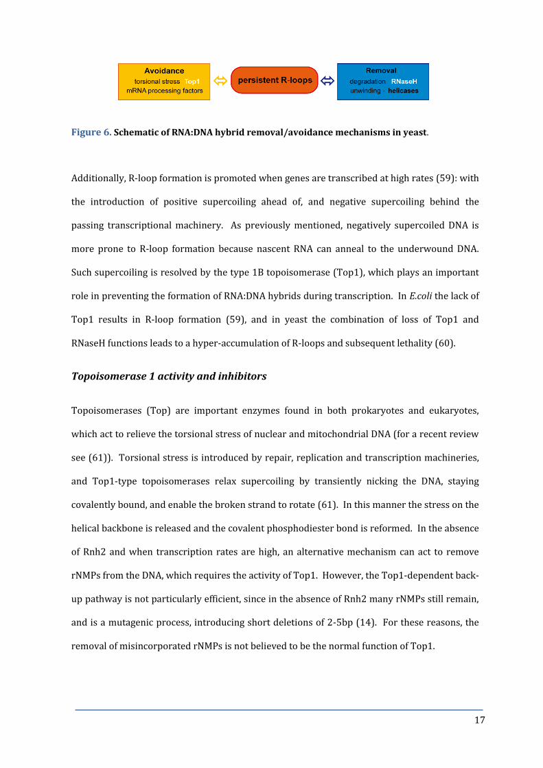
Figure 6. Schematic of RNA:DNA hybrid removal/avoidance mechanisms in yeast. 17
Figure 7. Molecular structure and schematic of mode of action of 18
camptothecin (CPT), a Top1 specific inhibitor.
Figure 8. The ribosomal DNA is compartmentalized within the nucleolus. 20
Figure 9. Schematic of recombinational repair pathways. 23
Figure 10. Schematic of the G1/S, intra-S, and G2/M checkpoint responses. 25
Chapter 1
Figure 11. Yeast lacking RNase H activity are sensitive to replication 29
stress and DNA damage independent of RAD5 and SSD1.
Figure 12. RNaseH activity prevents genome instability. 32

5
Figure 13. Loss of RNase H activity affects genome stability of the rDNA 35
array.
Figure 14. CPT treatment of rnh1Δ rnh2Δ mutant cells causes an increase of 37
DNA damage at late S-phase.
Figure 15. CPT treatment of rnh1Δ rnh2Δ mutant cells provokes replication 39
fork pausing at late S-phase.
Figure 16. CPT treatment of rnh1Δ rnh2Δ mutant cells provokes rARS- 40
independent replication initiation at late S-phase.
Figure 17. rARS-independent replication initiation is only observed in RNH- 41
mutants following CPT treatment.
Figure 18. Characterization of RFP sites and bubble arcs in CPT-treated 42
rnh1∆ rnh2∆ cells.
Figure 19. Confirmation of functionality of the 9Myc-Top1AID degron. 44
Figure 20. Absence of Top1 activity in RNH- mutant cells leads to RF pausing 45
and replication re-initiation.
Figure 21. CPT sensitivity of rnh1∆ rnh2∆ is related to rDNA transcription by 47
RNA PolI.
Figure 22. ARS-independent replication initiation is dependent on 35S 48
rDNA transcription by RNA PolI.
Figure 23. Model for ‘Transcription Initiated Replication’ in yeast rDNA. 49

6
Chapter 2
Figure 24. Synthetic lethal and synthetic sick interactions with the 57
rnh1Δ rnh2Δ double mutant.
Figure 25. Rad51 is not needed for the formation of replication bubbles by TIR. 59
Table 1. Analysis of the CPT sensitivity of rnh1Δ rnh2Δ triple mutants. 61
Figure 26. Growth at higher temperatures further sensitizes rnh1∆ rnh2∆ mutants 66
to CPT.
Chapter 3
Figure 27. Yeast lacking RNase H activity show a premature S-phase transition. 73
Figure 28. Yeast lacking RNaseH activity cannot by-pass the need for canonical 76
origin firing.
Figure 29. Mrc1-dependent replication fork stability is important in yeast lacking 78
RNaseH activity.
Figure 30. CPT treatment of the RNH- mutants does not activate the S-phase 82
Rad53-dependent checkpoint.
Figure 31. Absence of RNaseH affects the timing of activity of multiple cyclins. 83
Figure 32. Reduced nucleolar Cdc14 is released in RNH- mutants 85
Figure 33. RNH- mutants do not respond to the spindle assembly checkpoint. 88
Figure 34. Analysis of replication status of RNH- mutants by CHEF. 90
Figure 35. Yeast lacking RNase H activity show DNA segregation defects. 92
Figure 36. Nocodazole treatment of RNH- mutants causes re-budding and apoptosis. 94

7
Materials & Methods
Figure 37. Schematic illustration of the auxin inducible degron (AID) system. 109
Figure 38. An α-factor halo assay to test for bar1Δ phenotype of a yeast strain. 112
Figure 39. Plasmid pRS314LB direct repeat and chromosomal 113
leu2-k::ADE2-URA3::leu2-k recombination systems.
Figure 40. Schematic representation of two-dimensional gel analysis (2D-gel). 118
Figure 41. S-phase delay of the RNH- double mutant in minimal medium 119
lacking adenine.
Table 2. List of Saccharomyces cerevisiae strains used in this thesis. 130
Table 3. List of primers used in this thesis. 134
Table 4. Light excitation and emission conditions for fluorescence 123
microscopy.
8
3. INTRODUCTION
DNA replication and the cell cycle
DNA replication is a highly regulated process responsible for the accurate duplication of a cell´s
genetic material once per cell cycle, which is subsequently segregated into an identical
daughter cell. The mechanisms controlling this process are described by the four stage cell
cycle, in which the two major events of DNA replication (S phase) and chromosome
segregation and cytokinesis (M phase), are separated by two gap phases, known as G1 and G2.
In eukaryotes, DNA synthesis occurs during the S phase of the cell cycle.
DNA replication requires the action of DNA polymerases, to synthesize a new DNA strand
complementary to the original template strand. This mechanism is conserved from
prokaryotes to eukaryotes and is known as semi-conservative DNA replication. DNA
replication initiation must be highly coordinated with other cell cycle events, including the
repair of damaged DNA and segregation of fully replicated DNA to the daughter cell, to
maintain genomic integrity.
Origins of replication and replication initiation
DNA replication is initiated at specific sites, known as origins of replication (ori), throughout
the genome. Initiation from multiple origins allows eukaryotes to multiply their large
chromosomes in an appropriate time (for a review see (1)). The yeast Saccharomyces
cerevisiae (S. cerevisiae), a unicellular fungal eukaryote, has a genome of approximately 12.1Mb
with over three hundred origins of replication, referred to as autonomously replicating
sequences (ARS), and a doubling time in rich media of approximately 90 minutes (2). ARSs
consist of a short consensus sequence that acts as a site of recognition and assembly for the
Origin of Replication Complex (ORC). The ORC is associated with the ARS throughout the cell
cycle, and acts as a platform for sequential recruitment of the pre-replicative complex (pre-RC)

9
components Cdc6, Cdt1 and the Mcm2-7 helicase complex, a process known as replication
licensing (Figure 1). Once the pre-RC is assembled, the Mcm2-7 helicase complex is activated
by Cdc7 phosphorylation and can unwind DNA, converting the pre-RC into a pre-initiation
complex (pre-IC) and the origin is fired. The activity of the major cyclin dependent kinase
(CDK), Cdc28, directs the formation of pre-RCs. In G1 phase, Cdc28 activity is absent,
permitting the formation of pre-RCs but these are not competent to fire (3). Cdc28 then blocks
the formation of new pre-RCs until cells have passed through the G2 and M phases of the
current cell cycle (4). This Cdc28 control acts to regulate replication initiation, ensuring that
each origin is activated, or fired, just once per cell cycle.
Figure 1. Schematic representation of origin firing. ORC is bound to replication origins
throughout the cell cycle. During G1 phase of the cell cycle, Cdc6 binds to ORC-DNA. Cdc6 and Cdt1
bring MCM complexes to the origin, promoting the opening of the MCM ring, so it can encircle DNA.
Cdc6 ATP hydrolysis promotes closing of the MCM ring and the release of Cdt1 and Cdc6. Orc1 ATP
hydrolysis promotes release of ORC from the MCM2-7 complex. Cdc6 and Cdt1 are no longer
required and are removed from the nucleus or degraded. Cdc7 phosphorylates MCM2-7 that can
now slide on DNA, and MCMs (and associated proteins, GINS and Cdc45) unwind DNA to expose
template DNA. At this point replisome assembly is completed and replication in initiated. Schematic
taken from (5).
Replication priming
Following origin firing, the two strands are separated and factors required for DNA synthesis,
such as the yeast replicative polymerases alpha (α), delta (δ) and epsilon (ε), now have access
to the template DNA and can undertake DNA synthesis. However, the replicative polymerases

10
need a 3´-hydroxyl group to extend from and require the prior production of RNA primers by
specialized polymerases known as primases. This necessity means that the replicative
polymerases can only advance in a 5´ to 3´ direction along a template strand. As such, the
leading strand is synthesized in the same direction as the movement of the replication fork
(RF) in a continuous manner, by DNA Polymerase (DNA Pol) ε (6). The lagging strand,
however, is synthesized in the opposite direction to the movement of the RF as discrete
segments of replicated DNA, known as Okazaki fragments of approximately 150 nucleotides in
eukaryotic cells (Figure 2). The primase synthesizes an RNA primer, of approximately 10-12
nucleotides, and DNA Polα then adds some 20 nucleotides of DNA, allowing the lagging strand
polymerase DNA Polδ to extend from the primers formed and produce an Okazaki fragment.
The RNA primers must subsequently be removed before the fragments of replicated DNA can
be joined by the action of DNA ligase into a continuous fully replicated complementary strand.
Figure 2. Schematic representation of a replication fork. . DNA Polε (blue) synthesizes the
leading strand in the 5´to 3´direction in a continuous manner. For the lagging strand, DNA Polα-
primase first synthesizes an RNA fragment of about 10 nt (red) and then extends that with 20–30 nt
of DNA (orange). DNA Polδ extends the primer to a length of 200–300 nucleotides (green) until it
reaches the already synthesized fragment downstream. Joining of the Okazaki fragments involves
additional enzymes, such as FEN1 and DNA ligase. Adapted from (7).
RNA:DNA hybrids
RNA:DNA hybrids are frequently occurring intermediate structures, formed by base pairing
between a ssDNA and its complementary RNA strand. Such hybrids exist transiently during
normal replication, as part of the primers for DNA synthesis (as Okazaki fragments), and are

11
also formed during telomere elongation and transcription. For example, during transcription,
the two strands of the DNA double helix are separated to form a transcription bubble and the
synthesizing RNA forms a short-lived RNA:DNA hybrid of 8bp with the template DNA strand,
leaving the non-template DNA to loop out as single-stranded DNA behind the elongating RNA
polymerase (8). Under normal conditions this hybrid is a temporary structure and the RNA
transcript is removed and further processed and packaged into a ribonucleoprotein particle.
However, in some cases the nascent RNA can reanneal to its DNA complement, and form an R-
loop (Figure 3). R-loops are a three-stranded, thermodynamically stable form of the RNA:DNA
hybrid, formed by base pairing between the hybrid and the displaced ssDNA strand. Certain
conditions can favour the formation of R-loops. For instance, negatively supercoiled DNA (9)
and G-rich sequences (10) are more prone to form R-loops since both facilitate the opening up
of the DNA double helix.
BA
Figure 3. Schematic (A.) and electron micrograph (B.) of an R- loop. Electron micrograph
taken from (11). R-loop is indicated by arrowhead.
RNaseH enzymes
Ribonuclease H endonucleases (RNaseH) specifically hydrolyze the RNA moiety when annealed
to a complementary DNA. All living organisms possess at least one RNaseH activity to remove
RNA:DNA hybrids (reviewed in (12)). S. cerevisiae possess two RNaseHs: the monomer Rnh1
(encoded by RNH1) and the heterotrimeric protein complex Rnh2 (formed by the gene

12
products of RNH201, the catalytic subunit, and RNH202 and RNH203 accessory subunits) (13).
Although they seem to have some overlapping functions, Rnh1 specifically recognises
RNA:DNA hybrids with stretches of 4 or more consecutive ribonucleotides (rNMPs), whereas
Rnh2 can remove hybrids and has an additional activity capable of removing rNMPs covalently
attached to DNA, such as those misincorporated into DNA during replication of the genome
(14). For instance, DNA Polα lacks 3´-5´exonuclease proofreading activity and includes an
average of 1 rNTP per 625 bases of replicated DNA (15).
What happens if R-loops are not removed?
Persistent R-loops have been linked to various forms of genomic instability (for a review see
(16)) and may be lethal if not resolved. The looped out ssDNA of the R-loop structure is
exposed and more susceptible to damage than dsDNA. For this reason, R-loops have been
referred to as “fragile” sites, since they are more likely to suffer base lesions, such as
deamination (17), which may lead to mutations or strand breaks (18); R-loop forming regions
have also been linked to hyper-recombination (19,20).
Mutations in genes that are involved in the packaging of nascent RNA into a ribonucleoprotein
particle increase the likelihood of R-loop formation (21,22), and include genes with roles in
transcription and RNA processing and export, such as THO/TREX (19), and the ASF/SF2
splicing factor (20). Mutations in these genes are associated with transcription-dependent
genomic instability phenotypes, such as transcription-associated recombination (TAR) (23),
and such instability can be suppressed by the overexpression of Rnh1 (19,21), demonstrating
that the instability is due to the presence of RNA:DNA hybrids. Furthermore, the R-loop
structures themselves may hinder DNA metabolism, blocking transcription elongation (24) or
RF progression (25,26).
R-loops have been associated with the instability of trinucleotide repeat (TNR) sequences
(27,28). Their formation has been demonstrated in vitro at the disease-associated TNR (CAG)n,

13
(28) in spinocerebellar ataxia disease (SCA1), and (GAA)n, in Friedrich´s ataxia (FRDA) (11),
and thus R-loops have been linked to these and other TNR diseases, including myotonic
dystrophy (DM1) and fragile X type A (FRAXA) (11,28).
Despite their detrimental consequences to genomic stability, R-loop structures also play some
important physiological roles. For instance, they promote transcription termination of RNA
PolII genes, such as the human β-actin gene (29), and aid class switch recombination (CSR) of
immunoglobulin (Ig) genes, responsible for the diversification of Ig isotypes in mammalian B
lymphocytes (30). The looped out ssDNA of the R-loop at the highly repetitive switch regions
is specifically attacked by activation-induced cytidine deaminase (AID), an enzyme that
deaminates cytidine residues (31), and leads to the formation of DSBs (32) necessary for CSR
to take place.
There is also growing evidence associating R-loops with epigenetic modifications and control
of gene expression. For example, R-loops may protect against DNA methylation and have been
shown to form at CpG islands (CGI) in gene promoters (33). Interestingly, AID-mediated
demethylation of DNA has been shown to be important for epigenetic reprogramming of
mammalian cells (reviewed in (34)) and linked to the pluripotency of stem cells (15,35).
Furthermore, R-loops may favour chromatin accessibility through a reduced affinity for
histones (36) and recently, R-loop formation has also been shown to trigger histone 3 S10
phosphorylation (H3S10P) and linked to chromatin compaction (37).
RNA:DNA hybrid-primed replication
In eukaryotes, DNA Polα and its´ intrinsic primase activity initiates RNA-primed DNA synthesis.
However, in the case of prokaryotic and mitochondrial DNA, RNA Pol transcripts existing as
stable R-loops can function as primers for the extension of DNA synthesis. For example, R-
loops can function as origins of replication for T4 and T7 bacteriophages (38), and for ColE1-
type plasmids in E.coli (39), where replication is sensitive to rifampcin, an RNA Pol inhibitor.

14
Mitochondrial DNA is a good example of a transcription-primed DNA replication mechanism
(for a review see (40)). Replication origins in mtDNA are highly conserved from yeast to
humans and consist of a promoter for the initiation of transcription by RNA Pol and a high GC
content downstream. In the case of mammalian mtDNA, transcription from the light-strand
promoter (LSP) opens up the heavy-strand origin (OH) (41) and produces a stable and
persistent RNA:DNA hybrid (42) (Figure 4), that once processed, can be used as a primer for
extension by DNA Polγ (43).
Figure 4. Schematic representation of mtDNA replication, as an example of transcription-
primed DNA replication. Transcription from the light-strand promoter creates an RNA:DNA
hybrid, which acts as a primer for the initiation of DNA replication of the mitochondrial genome.
Apart from RNA:DNA-primed replication, transcription has also been shown to bypass the
need for replication initiation factors in E.coli (44). Under normal conditions, E.coli
chromosomal DNA replication is initiated from one specific origin (oriC), whose opening up is
essential for the assembly of the replisomes and depends on the replication initiation factor
DnaA. However, mutants capable of initiating constitutive stable DNA replication in the
absence of DnaA were identified (44) and the mutation was mapped to the RNH locus (rnhA)
(45). oriC independent replication was transcription-dependent suggesting that stabilized R-
loops provided access for factors needed for replication initiation. Importantly, rnhA mutants
15
cannot bypass the growth defect of primase deficient dnaG mutants (46), indicating that
replication has to initiate from a primase generated RNA primer.
Importance of RNA:DNA hybrid removal by RNase H enzymes
The RNaseH enzymes specifically remove RNA:DNA hybrids. Eukaryotic RNases H1 and H2 are
important participants in maintaining genome stability by resolving R-loops that form during
transcription, and in the case of RNase H2, by initiating the removal of rNMPs in DNA, making
an excision on the 5´ side of the rNMP. Misincorporated rNTPs must be removed by DNA
repair pathways, since they are more mutagenic than mispaired dNTPs due to the reactive
hydroxyl 2´group on the ribose ring (47), and their presence in the template strand can cause
the RF to stall in vitro (6,48) and sensitize the DNA backbone to spontaneous breaks (49).
The importance of RNaseH activity is exemplified by the fact that deletion of RNaseH1 in
drosophila and mice results in embryonic lethality due to the inability to amplify mitochondrial
DNA (mtDNA) (50). Furthermore, deletion of RNaseH2B in mice causes embryonic lethality
with an observed accumulation of single ribonucleotides in the DNA (51). Mutation in any of
the subunits of the human Rnh2 complex can lead to Aicardi-Goutieres syndrome (AGS) (52), a
severe but rare autosomal recessive neurological disorder (Figure 5). Patients manifest basal
ganglia calcification, cerebral atrophy (loss of tissue), chronic cerebrospinal fluid
lymphocytosis (increase of lymphocytes in the cerebrospinal fluid), and characteristic
chilblains of fingers, toes and ears. AGS can also be caused by mutations in other enzymes with
roles in the removal of nucleic acid species, such as SAMHD1 that reduces dNTP pools and
TREX1 (53), a 3´ to 5´ DNA exonuclease, deletion of which leads to the accumulation of
fragments of ssDNA of 60-65bp in the endoplasmic reticulum (ER). The accumulation of nucleic
acids can trigger the inappropriate activation of the innate immune system (IFNα response)
(for a review see (54)), with the body responding as if to a viral infection, and many of the
clinical features of AGS parallel those for a viral infection.

16
IFN -
triggered
innate
immune
response
A
B
Figure 5. Mutations in nucleic acid removing enzymes can cause Aicardi-Goutieres
syndrome. A. Loss of AGS-related protein activity leads to nucleic acid accumulation, which
triggers an innate immune response. TREX1 – human exonuclease; degrades ss- and dsDNA.
SAMHD1 – converts dNTP to a nucleoside and a triphosphate. Schematic adapted from (55). B.
Characteristic phenotypes of AGS patients include chilblains of the ears, toes and fingers (photos
taken from www.aicardi-goutieres.org.), and calcification of the basal ganglia. The MRI scan shows
rarified white matter, characteristic of neonatal AGS (56).
Other means of removing RNA:DNA hybrids
Since R-loops are a threat to genomic integrity, bacteria and eukaryotic cells possess different
mechanisms to prevent the formation of R-loops (Figure 6). Yeast RNH double deletion
mutants are viable, indicating that cells have other means of processing RNA:DNA hybrids.
These may include helicases such as the yeast (and mammalian) Pif1, which has been shown to
unwind RNA:DNA hybrids in vitro (57), and the yeast Sen1 (known as Senataxin in mammals),
whose absence has been shown to result in an accumulation of RNA:DNA hybrids downstream
of the poly(A) signal (58).
17
Figure 6. Schematic of RNA:DNA hybrid removal/avoidance mechanisms in yeast.
Additionally, R-loop formation is promoted when genes are transcribed at high rates (59): with
the introduction of positive supercoiling ahead of, and negative supercoiling behind the
passing transcriptional machinery. As previously mentioned, negatively supercoiled DNA is
more prone to R-loop formation because nascent RNA can anneal to the underwound DNA.
Such supercoiling is resolved by the type 1B topoisomerase (Top1), which plays an important
role in preventing the formation of RNA:DNA hybrids during transcription. In E.coli the lack of
Top1 results in R-loop formation (59), and in yeast the combination of loss of Top1 and
RNaseH functions leads to a hyper-accumulation of R-loops and subsequent lethality (60).
Topoisomerase 1 activity and inhibitors
Topoisomerases (Top) are important enzymes found in both prokaryotes and eukaryotes,
which act to relieve the torsional stress of nuclear and mitochondrial DNA (for a recent review
see (61)). Torsional stress is introduced by repair, replication and transcription machineries,
and Top1-type topoisomerases relax supercoiling by transiently nicking the DNA, staying
covalently bound, and enable the broken strand to rotate (61). In this manner the stress on the
helical backbone is released and the covalent phosphodiester bond is reformed. In the absence
of Rnh2 and when transcription rates are high, an alternative mechanism can act to remove
rNMPs from the DNA, which requires the activity of Top1. However, the Top1-dependent back-
up pathway is not particularly efficient, since in the absence of Rnh2 many rNMPs still remain,
and is a mutagenic process, introducing short deletions of 2-5bp (14). For these reasons, the
removal of misincorporated rNMPs is not believed to be the normal function of Top1.

18
Incomplete Top1 action has been shown to be a natural source of DNA damage, such as DNA
single strand breaks (SSBs), which can be converted to DSBs during replication (62).
Camptothecin (CPT) is a Top1 specific inhibitor that acts by trapping the Top1 after nick
formation on the DNA as a cleavage complex (Top1cc; Figure 7A), binding at the Top1-DNA
interface, and thus impedes religation of the nick (Figure 7B) (63). Water-soluble derivatives
of CPT are commonly employed as anti-tumour drugs, such as topotecan for the treatment of
ovarian cancer (64).
The CPT sequestered, covalently bound, 90kDa Top1 must be removed from the 3´ end before
the DSB can be repaired and replication resumed. Specialized enzymes, such as the tyrosyl-
DNA phosphodiesterase, Tdp1 (65), as well as the Rad1-Rad10 and Mus81 endonucleases (66),
can remove Top1cc. In addition, the homologous recombination machinery has been reported
to be involved in the repair of Top1-mediated lesions (67).
Figure 7. Camptothecin (CPT) is a Top1 specific inhibitor. A. Molecular structure of CPT. The
lactone ring in CPT is important for the drug’s biological activity, active as a “closed” α-
hydroxylactone form and inactive as the “open” carboxylate form. The lactone ring can rapidly
open at physiological (or higher) pH. B. Schematic of CPT mode of action. Under normal
conditions, the covalent Top1cc, formed in the action of nicking, are short-lived and reversible.
However, under some circumstances, such as upon treatment with CPT and deriviatives, the Top1cc
is stabilized and the ligation stage is impaired, leading to the introduction of a SSB. Adapted from
(64).

19
Organisation of Ribosomal DNA
The ribosomal DNA (rDNA) is compartmentalized within the nucleolus, a crescent-shaped sub-
compartment of the nucleus (Figure 8A), which is the site of rDNA transcription and ribosome
assembly, essential processes for the cell since cell growth is directly dependent on the rate of
protein synthesis (68). Top1 is enriched at the nucleolus (69), and Top1´s activity at this site is
particularly important to relieve torsional stress, since rDNA transcription by RNA PolI can
account for approximately 80% of the total transcription in yeast (68). The highly transcribed
rDNA is more prone to RNA:DNA hybrid formation, and accordingly R-loop formation in the
rDNA have been shown to be enhanced in top1 mutants in yeast (60).
The ribosomal locus of S. cerevisiae consists of a single array of 150-200 copies of a 9.1kb
repeat unit located in the middle of chromosome XII (Figure 8B). In contrast to yeast, the rDNA
repeats of higher eukaryotes are located in multiple nucleolar organizing regions (NORs). One
yeast repeat unit consists of the RNA PolI transcribed 35S gene that encodes the 35S precursor
rRNA, which is processed into the mature 18S, 5.8S and 26S rRNAs, and the RNA PolIII
transcribed (in opposite direction) 5S gene, respectively. Two non-transcribed intergenic
spacers (NTS1 and NTS2) separate the 35S and 5S rRNA sequences. The NTS regions contain
cis-regulatory elements for the control of DNA replication, which include a replication fork
barrier (RFB) and an origin of replication (ARS), respectively (for a review of the rDNA
organisation, see (70)). The RFB is polar, allowing RFs to pass if moving in the same direction
as transcription of the 35S gene, but blocks over 90% of advancing forks from opposing
direction (71). RFBs appear to be a highly conserved feature of rDNAs, confirmed in a number
of other organisms, including humans (72).

20
Figure 6
A B
RFB ARS
CAR
Figure 8. A. Fluorescent microscopy image to show the nucleolus. The nucleolus is seen in red
(Nop1-mRFP), and the nucleus in blue (DAPI stain). B. Schematic representation of the rDNA locus
of S.cerevisiae. A single rDNA unit measures 9.1kb and contains two transcribed genes – 35S
transcribed by Pol I, processed to mature 18S, 5.8S and 25S species, and 5S by Pol III. NTS, non-
transcribed spacer; RFB, replication fork barrier; ARS, origin of replication; CAR, cohesion
attachment region.
In addition, each rDNA repeat contains a cohesion attachment region (CAR) located proximal
to the 5S gene in the NTS2 (73). Cohesion is an evolutionarily conserved complex that contains
several members of the Smc (structural maintenance of chromosomes) family. The association
of cohesion is thought to hold sister chromatids together during S phase, to regulate
recombination between repeats, until their controlled separation and segregation during
mitosis (74). Smc proteins are also found in condensin complexes, and there is an intimate
relationship between cohesin and condensin functions (cohesin and condensin are reviewed in
(74)), both of which are important for the correct segregation of the rDNA array (75). The
presence of cohesion within the tandem repeated rDNA array may limit the template available
for recombinational repair of a DNA break (73) and therefore is important for maintaining
rDNA repeat stability (75).
The replication-transcription conflict
The polarity of the ribosomal RFB allows the replication and transcription machineries to
move in the same direction. However, in other regions of the genome, this is not the case such
that replication and transcription machineries can collide (76,77) leading to RF stalling or

21
arrest (reviewed in (78,79)). It has been suggested that head-on collisions are more
detrimental than co-directional collisions (80), and consequently, highly expressed genes tend
to be transcribed with the same polarity as RF progression (71,81). For example, RFs were
shown to pause at tRNA genes when the direction of transcription was opposite to the
direction of RF progression (82). Therefore, eukaryotes have evolved mechanisms to help
prevent head-on collisions (for a recent review see (83)), explaining the presence of RFBs in
the highly transcribed rDNA (71).
Pathways that resolve constrained replication
In addition to transcription-induced impediments, the RF must deal with other DNA-bound
proteins, secondary structures, and frequently occurring DNA lesions caused by various
exogenous and endogenous sources, which can cause RF stalling. Blocks to replication can lead
to RF collapse if not resolved and result in DNA strand breaks. As such, a plethora of repair
factors and pathways exist to remove DNA lesions and facilitate RF progression, in order to
maintain genomic integrity. The choice of which repair system to use depends on both the
type of lesion and on the cell-cycle phase (reviewed in (84)).
Continuously produced reactive oxygen species (ROS), a by-product of normal cellular
metabolism, can modify bases by oxidation, and such oxidative base lesions can block the
progress of DNA and RNA polymerases (85,86). The Base Excision Repair (BER) pathway acts
to repair damage to individual bases, including methylation, deamination and
depurination/depyrimidation (for a review of BER see (87)). In contrast, Nucleotide Excision
Repair (NER) acts to remove bulky DNA adducts that cause a structural deformation of the
DNA helix, such as DNA intrastrand and interstrand crosslinks, and pyrimidine dimers that can
be produced by ultraviolet (UV) radiation (for a review of NER see (88)). A specific sub-
pathway of NER can repair lesions that impede RNA Pol progression through transcribed

22
genes. A stalled RNA Pol at the lesion, for example helix-distorting lesions, appears to be the
signal for Transcription-coupled repair (TC-NER) (reviewed in (89)).
However, the most commonly formed lesions resulting from stalled or collapsed RFs are
double-strand breaks (DSBs). The two major pathways for repair of DSBs are non-homologous
end joining (NHEJ) and homologous recombination (HR). NHEJ is error-prone, ligating
together the broken DNA ends with little or no homology, often resulting in loss or gain of
sequence at the site of repair (reviewed in (90)), while HR repairs the DSB with high fidelity,
using the sister chromatid or homologous chromosome as template. NHEJ has been shown to
be active throughout the cell cycle, although it is particularly active in G1 (91), whereas HR is
restricted to the S and G2 phases, when the sister chromatid is available to act as the template
for this mode of repair (reviewed in (92)).
The Mre11/Rad50/ Xrs2 (MRX) complex functions in both HR and NHEJ, where Rad50 holds
DSB ends together to favour NHEJ (93). In yeast, HR is initiated with processing of the ends of
the break by the MRX complex to generate 3′-ssDNA. Rad51 then searches for the homologous
sequence and facilitates strand invasion of the ssDNA at the homologous sequence, allowing
the DNA Pol to extend the 3´ end using the homologous sequence as a template.
There are at several different mechanisms of homologous recombination that can be used to
repair a chromosomal DSB in yeast cells, including double strand break repair (DSBR),
synthesis-dependent strand annealing (SDSA), single-strand annealing (SSA) and break-
induced replication (BIR) (see Figure 9) (reviewed in (94)).

23
Figure 9. . Recombinational repair pathways. Schematic highlighting key factors in double-
strand break repair (DSBR), synthesis-dependent strand annealing (SDSA), single-strand annealing
(SSA) and break-induced replication (BIR) homology-dependent recombinational repair pathways.
Adapted from (34).
Eukaryotic cells also possess two damage tolerance mechanisms that depend on the activities
of Rad6 and Rad18 to allow the RF to by-pass blocking lesions (95,96). Damage tolerance can
be mediated by the error-prone TLS, where a specialized polymerase can replicate across the
DNA lesion (97), or the error-free Rad5-dependent pathway, that uses the undamaged sister
chromatid via template switching (98) to re-prime replication downstream of the lesion. The
deletion of both template switch and TLS pathways were shown to be essential to tolerate
misincorporated rNMPs in the DNA of yeast lacking RNaseH activity upon replicative stress
(99).
Cell cycle checkpoints
Surveillance mechanisms known as checkpoints exist, which act to detect problems that may
arise during eukaryotic DNA replication and respond by eliciting a signalling cascade (100).
Checkpoints contain sensor proteins that can detect stretches of ssDNA, an indication of stalled
forks or DNA damage, incorrect attachment of sister chromatids to the mitotic spindle, cell size,
24
or cellular conditions such as protein and nutrient levels. Depending on the stimulus, the
checkpoint can activate signal transducers, protein kinases that transmit the checkpoint signal
to induce the expression of specific downstream target genes that act to maintain the stability
of the RF and/or facilitate repair, in the case of damage (101), and in all cases, delay cell cycle
progression to allow time for the problems to be resolved (100). Loss of checkpoint function
results in genomic instability (102) and has been implicated in the evolution of normal cells
into cancer cells (103,104).
In S. cerevisiae there are three checkpoint pathways that recognize the presence of damaged
DNA at the G1/S transition, during the S-phase (Intra-S), and at the G2/M cell cycle phases (see
Figure 10). The G1/S cell cycle checkpoint, also known in yeast as START (for a review see
(105)), ensures there is no damaged DNA before transition into S phase (106). Additionally,
START acts as a decision point to confirm that all conditions required for DNA synthesis,
including a minimum cell size and sufficient nutrient and enzyme levels, before committing to a
cell division cycle. Alternatively, cells arrest at START and enter a resting state called G0.
The S-phase checkpoint senses both DNA damage and replication stress, caused by stalled or
broken RFs, which result in stretches of ssDNA (for a review see (107)). The Mec1 checkpoint
sensor is recruited to the RPA-bound ssDNA and activates the downstream effector kinases
that include Rad9 and Mrc1 ((108) and (109), respectively). The phosphorylation of either
Mrc1 or Rad9 recruits and activates Rad53 (110). In addition to RAD9, the checkpoint genes
RAD17 and RAD24 are also required for the intra-S and G2/M damage checkpoints and function
upstream of Rad53. Rad53 activation stabilizes the RF (111), induces damage-responsive
genes via its downstream paralogue kinase Dun1 (112), and slows down DNA replication, by
the inhibition of late origin firing (113).

25
Figure 10. The G1/S, intra-S, and G2/M checkpoint responses. Schematic representation of the
key factors involved in the checkpoint pathways.
Any unrepaired damage in the newly synthesized DNA will trigger the G2/M damage
checkpoint, which prevents cells from entering mitosis until the DNA damage has been
resolved, to prevent the segregation of damaged chromosomes (114). Other G2/M checkpoints
include the morphogenesis checkpoint, which delays cells at the G2/M transition in response to
problems that delay bud formation (115), and the spindle-assembly checkpoint (SAC) that
monitors attachment of replicated chromatids to the microtubules to achieve spindle
connection (for a review see (116)). SAC activation achieves G2/M arrest by inhibiting the
anaphase promoting complex/cyclosome (APC/C) specificity factor Cdc20, delaying exit from
mitosis [22]. The APC/C is an E3 ubiquitin ligase that regulates the metaphase/anaphase
transition through the ubiquitin-mediated proteolysis of various substrates (117), including
mitotic cyclins and the sister chromatid separation inhibitor securin/Pds1 (118). When the
kinetochores are attached to microtubules the APC/CCdc20 ubiquitinates securin and cyclin B
and thereby activates the protease separase and inactivates the cyclin-dependent kinase-1
(Cdk1). Separase can cleave the cohesin complexes that are holding sister chromatids together
and separate sister chromatids. The activated SAC inhibits the capability of APC/CCdc20 to
ubiquitinate securin and cyclin B and thereby prevent anaphase and mitotic exit. As such, the

26
SAC ensures a correct chromosome segregation and is a key mechanism to prevent aneuploidy,
a contributory factor of cancer (119).

27
4. OBJECTIVES
RNA:DNA hybrids are transient structures generated during DNA replication, transcription and
telomere elongation and can lead to R-loop formation. R-loops may physically interfere with
transcription elongation (24) or cause replication fork blockage (25,26). As such, persistent R-
loops are detrimental to the cell and have been linked to various forms of genomic instability
(for a review see (16)). All eukaryotes and bacteria possess at least one enzymatic activity to
specifically remove RNA:DNA hybrids. The human disease AGS and embryonic lethality in
mouse (and Drosophila) caused by the lack of RNaseH2 and RNaseH1 respectively,
demonstrate the important contributions of the RNaseH enzymes to global DNA metabolism
(50,52). Furthermore, recent studies have shown that R-loops are linked to disease-associated
alterations in trinucleotide repeat sequences, supporting the need for further investigation of
the cellular roles of the RNaseHs and the consequences when their activity is compromised.
Using Saccharomyces cerevisiae as a model organism, this thesis aims to explore the
contributions of RNaseH activity to faithful DNA replication, genomic stability and cell cycle
control.
The objectives of this thesis are:
1. To categorize the replication/recombination intermediates formed in RNaseH- cells.
2. To identify factors and pathways, that interact with RNaseH enzymes by genetic
analyses.
3. To dissect the impact of R-loop formation on cell cycle progression.

28
5. RESULTS
CHAPTER 1 - Transcription-initiated DNA Replication in Yeast
Yeast lacking RNaseH activity are sensitive to the Top1 inhibitor CPT
The yeast S. cerevisiae possess two RNaseH activities, Rnh1 and Rnh2, which can act to remove
RNA:DNA hybrids and have been suggested to have some redundancy in functions (120).
Deletion of the RNH201 gene, coding for the catalytic subunit of Rnh2, eliminates yeast Rnh2
activity, and deletion of both RNH1 and RNH201 abolishes all RNaseH activity in yeast cells.
Double mutants rnh1∆ rnh201∆ (referred to herein as rnh1∆ rnh2∆) are viable, suggesting that
this activity in dispensable for viability or that yeast have other means of removing RNA:DNA
hybrids. However, yeast cells lacking RNaseH activity have been shown to be more sensitive to
the DNA damaging agent ethymethylsulfanate (EMS), the checkpoint inhibitor caffeine and the
ribonucleotide reductase inhibitor hydroxyurea (HU) (120). To confirm and extend these
observations, we performed drop test analyses using HU; the DNA alkylating agent methyl
methanesulphonate (MMS); and the Topoisomerase 1 (Top1) specific inhibitor camptothecin
(CPT; Figure 11A). Cells lacking either RNaseH activity were not sensitive to genotoxic agents,
but we noted that the rnh1∆ rnh2∆ double mutant became hypersensitive to HU, MMS, and
CPT; suggesting that although each RNaseH enzyme has a specialized role, they can substitute
for each other (Figures 11A and B). We found the CPT sensitivity of the rnh1∆ rnh2∆ double
mutant particularly interesting, because rnh1∆ rnh2∆ has been shown to be synthetic lethal in
the absence of the CPT target, Top1 (60). Top1 is crucial during transcription to relieve the
accumulation of torsional stress associated with the formation of negative supercoils behind
the transcription machinery (121). Negative supercoils in the DNA can enhance RNA:DNA
hybrid formation (59), and consequently Top1 plays an important role in preventing R-loop
formation. CPT sequesters Top1 via the formation of a covalently bound Top1 cleavage
complex (Top1cc) Top1-DNA complex (reviewed in (122)), so that it cannot act elsewhere in

29
the genome, analogous to a depletion of Top1. A recent report of Marinello et al. has shown
that CPT treatment of human cells leads to an increase in R-loops at highly transcribed regions,
such as ribosomal genes, due to an increased negative torsion (123). Thus, CPT treatment of
rnh1∆ rnh2∆ mutants can be used as a tool to chemically induce and maximize RNA:DNA
hybrid formation in yeast.
Figure 11. Yeast lacking RNase H activity are sensitive to replication stress and DNA damage
independent of proficient Rad5 and Ssd1 activity. A. Analysis of sensitivity to genotoxic agents.
10-fold serial dilutions of cells grown for 3 days on YPAD or YPAD-containing HU (50mM), MMS
(10mM), or CPT (10µg/ml). B. Cell survival after prolonged incubation with CPT. Data is shown as
the mean ± standard deviation. C. 10-fold serial dilutions of cells containing an empty (control), or
the RAD5-expressing (pBJ6) or SSD1-expressing (pLO92) plasmids grown on SC-Ura or SC-Ura-
containing CPT (5µg/ml).
The YKL83 strains used in this study are derivatives of W303-1A (124) (a complete list of
strains used in this thesis can be found in Table 2). However, genetic alterations in the W303-
1A strain include a mutation in the RAD5 (rad5-535 (125)) and SSD1 genes (ssd1-1 (126)).
RAD5 codes for a factor with both DNA helicase and ubiquitin ligase activities that functions in
postreplicative repair (PRR), and the rad5-535 mutation has been associated with a slight
increase in UV and MMS sensitivity (125), while SSD1 codes for a translational repressor with
roles in polar growth, TOR signalling and cell wall integrity (126,127). To determine whether
the rad5-535 or ssd1-1 mutations contribute to the CPT sensitivity of the rnh1∆ rnh2∆ mutant,

30
strains were transformed with the RAD5- or SSD1- expressing plasmids (a list of plasmids
included in this thesis can be found in Table 3). RAD5 or SSD1 expression from low copy
number plasmids did not alleviate the rnh1∆ rnh2∆ CPT sensitivity (Figure 11C), supporting
the idea that CPT sensitivity is due to the lack of RNaseH activities.
Yeast lacking RNaseH activity suffer from increased genome instability
CPT damages DNA by trapping the Top1-DNA cleavage complex (Top1cc) such that it cannot
ligate the single-strand nick made by Top1 (128,129). Such Top1cc can be processed by the
action of Rad1 and Tdp1, which form part of redundant DNA damage repair pathways (66).
Tdp1 is a tyrosyl-DNA phosphodiesterase, capable of hydrolyzing the covalent link between
Top1 and DNA, while Rad1 acts in conjunction with Rad10, as a structure-specific
endonuclease during nucleotide excision repair (NER). The rad1∆ tdp1∆ double mutants are
themselves very CPT sensitive due to accumulation of Top1-mediated DNA damage (Figure
12A; note the drop test analysis was performed at the lower CPT concentration of 1µg/ml due
to the elevated sensitivity of the strain). To test if the RNaseHs contribute to the Rad1 or Tdp1-
dependent CPT-repair pathways, we therefore generated rnh1∆ rnh2∆ mutants lacking only
Rad1, Tdp1 or both activities (Figure 12A). The rnh1∆ rnh2∆ mutants were further sensitized
to CPT in the absence of both rad1 and tdp1 (see the quadruple mutant), but not in the absence
of either repair protein. The enhanced CPT sensitivity of the quadruple but not the triple
mutants indicates that it is unlikely that RNase H enzymes are involved in the repair of Top1-
mediated DNA damage but rather, DNA damaging events might be more frequent in these
mutants.
Next, we examined if rnh1∆ rnh2∆ mutants suffer from a general increase in genome instability.
Genetic alterations can be detected as events that lead to a loss of heterozygosity (LOH) in
yeast cells, where a cell only contains a single copy of an allele due to loss or inactivation of the
second copy. LOH can become critical when the sole remaining allele contains a point

31
mutation that renders the gene inactive. For example, LOH is a common occurrence in cancers
where a tumor suppressor gene is inactivated (130,131). We measured the frequency of LOH
in yeast cells by monitoring the formation of “a-like faker” cells (ALF, (132); Figure 12B),
resulting from loss or inactivation of the MATα locus leading to the default MATa mating type
in yeast. MAT allele disruption can be due to chromosomal rearrangement or gene conversion
of the silent mating type locus HMRa, and more frequently, due to loss of chromosome III, that
hosts the mating cassette. ALF cells can be detected by the selection of mated products, since
ALFs will mate as a-type cells. In wild-type yeast, ALF mitotic segregants are generated at a
rate of approximately 10−6 (133). The rnh1∆ rnh2∆ double mutants exhibited a frequency of
ALF formation about 10-fold increased, as compared to the WT, suggesting that cells lacking
RNaseH activity have chromosome instability.
To further monitor genomic instability, mutation frequencies in rnh1∆ rnh2∆ mutants were
detected by measuring the frequency of Ura- mutations (selected in medium containing FOA;
Figure 12B), using the pCM184-LAUR plasmid system ((134), see Materials & Methods for
details). We found that cells lacking RNaseH activity had a 12-fold increase in the frequency of
Ura- mutators as compared to the WT, suggesting that loss of RNaseH activity is associated
with a hypermutator phenotype. In addition, we measured forward mutation rates by
monitoring the spontaneous appearance of colonies in a medium supplemented with the toxic
compound L- canavanine (Can). S. cerevisiae take up arginine and canavanine by means of a
specific permease, and resistance to Can is associated with a loss of arginine permease function
encoded by the CAN1 locus. We observed an increase in Can resistant cells of over 6-fold in the
rnh1∆ rnh2∆ double mutant (Figure 12B). Notably, in order to perform the CAN1 forward
mutation assay we had to use strains in the BY background, since YKL83 strains are mutated in
the CAN1 gene (can1-100). Taken together, these results indicate that RNaseH double mutants
are prone to genomic instability.

32
Figure 12. Loss of RNaseH activity contributes to genome instability. A. Drop test analysis of
genetic interaction between RNaseH activity and CPT repair pathways. 10-fold serial dilutions of
cells grown for 3 days on YPAD or YPAD-containing CPT (1µg/ml). B. Rate of MATα conversion to a-
mating type. ALF frequency values (mated products/total cells) shown as fold change (F.C.) relative
to WT (left panel). Mutation rates as determined by the pCM184-LAUR plasmid mutation system
(middle panel) and by canavanine resistance (right panel). Data represent the mean ± standard
deviation obtained from the mean of three fluctuation tests of four independent colonies each.
Differences between mutants and the WT were examined by Student’s t-test and were considered
statistically significant for p-values<0.05 (asterisks). C. Representative image of Rad52-YFP foci co-
localized with the nucleolar periphery (left). Percentage of Rad52-YFP foci counted in exponentially
growing cells growing with or without the presence of CPT (10µg/ml, 3hr treatment). Nuclear
versus nucleolar Rad52-YFP foci were determined according to co-localization with the nucleolar
Nop1-mRFP marker. Data represent mean ± SD from at least three independent experiments.
Differences between mutants and the WT were examined by Student’s t-test and were considered
statistically significant for p-values<0.05.
Subsequently, we asked whether rnh1∆ rnh2∆ yeast were also more susceptible to
spontaneous DNA lesions and we tested for this by analysing the formation of Rad52-YFP foci.
Rad52 is a key player in repair by homologous recombination (HR); when assembled at a DSB,
the clustering of Rad52-YFP proteins can be seen as an intense focus, such that foci are
representative of sites of DNA repair (135). Interestingly, we observed a statistically
significant increase of Rad52-YFP foci formation in rnh1∆ rnh2∆ cells (5-fold with respect to
the WT), and an even greater increase upon the addition of CPT (Figure 12C). Notably, the
Top1 specific inhibitor CPT introduces single-stranded nicks that can be converted into DSBs
by collision of the replication or transcription machineries with the covalently bound Top1cc

33
(136,137), and so we questioned whether we would observe an even greater frequency of
Rad52-YFP foci formation in a highly transcribed region of the genome. The nucleolus is ideal
to test for this possibility because it hosts the highly transcribed ribosomal genes. Ribosomal
genes are needed for the synthesis of rRNA by RNA PolI accounting for about 80% of the total
transcription in yeast (68). High rRNA levels are achieved by transcription of approximately
100-200 identical repeats of the yeast ribosomal genes, organized into the rDNA locus on
chromosome XII. Cells were co-transformed with Rad52-YFP- and Nop1-mRFP-expressing
plasmids to compare the localization of Rad52 foci with the Nop1 nucleolar stain (Figure 12C).
We observed that 31% of Rad52-foci colocalized with the nucleolar periphery in WT cells
following CPT treatment. Strikingly, some 66% of the Rad52-YFP foci in the rnh1∆ rnh2∆
double mutant appeared to be associated with nucleolar DNA following CPT treatment. In fact,
an already significant proportion of rnh1∆ rnh2∆ cells (57%) had foci that co-localized with the
nucleolar periphery in logarithmically growing cells under normal conditions. Extensive work
by the groups of Michael Lisby and Luis Aragon using 3D reconstruction, demonstrated that
Rad52 is excluded from the nucleolus and damaged rDNA relocates to the nucleolar periphery
to interact with the recombinational repair machinery (138). Collectively our results suggest
that RNaseH activities contribute to the maintenance of genomic stability, and are particularly
important for preserving rDNA genome stability.
Genome stability of the rDNA is particularly affected in RNH- mutants
Chromosome XII (cXII) is approximately 2.5Mb in length, being one of the largest yeast
chromosomes. From sequence information, non-rDNA regions on the chromosome account for
approximately 1.1Mb, and therefore, the remaining 1.4Mb are made up of rDNA repeats,
constituting some 150 copies of the rDNA repeat in a control strain. The repetitive nature of
the rDNA locus makes this region of the genome particularly unstable. Changes in rDNA repeat
length can occur in response to repair processes initiated at stalled replication forks within the
rDNA array (139-141). Homologous recombination-mediated invasion of an rDNA repeat with

34
a complementary sequence can lead to expansion - a gain in copy number, or contraction -
resulting in copy number loss - of the rDNA array; such events being related to genome
instability. The mobility of chromosome XII can be easily visualized by CHEF analysis, and
provides a direct measurement of rDNA repeat length. We investigated whether the repeat
length of the rDNA array was altered in mutants lacking RNaseH activity. CHEF analysis
showed that the regulation of rDNA repeats was significantly affected in yeast lacking RNaseH
activity (Figure 13A). We observed a substantial decrease in rDNA copy number for the rnh2∆
mutant as compared to the WT, related to a loss of rDNA repeats. The cXII signal for rnh1∆
cells and the double mutant was observed as an intense band just below the wells. For the
rnh1∆ simple mutant, cXII migrates in a higher intense band, however, a second band
corresponding to cXII can be observed just below the WT band. This result would indicate a
population of rnh1 cells with a strongly enhanced repeat expansion phenotype and a second
sub-population with a shorter rDNA array. In the case of cells lacking both RNaseH activities,
cXII was always detected as a diffuse signal, and generally the cXII signal seemed reduced in
the double mutant. A diffuse signal represents a dispersed population that has undergone
larger expansions of the rDNA array (142), and such copy number heterogeneity would reflect
a particularly unstable rDNA array in the RNH double mutant.
Ribosomal repeat stability can also be monitored by the pop-out of rDNA repeats from cXII by
intra-chromatid recombination (143), and consequently, the formation of extrachromosomal
rDNA circles (ERCs) (Figure 13B). ERCs are maintained in the cell as plasmid-like circular
DNA, since they can replicate autonomously (144). It has been proposed that the accumulation
of ERCs is an aging factor in yeast, and the “rDNA theory” directly relates rDNA stability with
longevity in yeast (145). As we had observed considerable fluctuations in rDNA repeat length
in the RNH- mutants we questioned whether we could associate this with changes in the levels
of ERCs. ERCs were separated from logarithmically growing yeast by gel electrophoresis,
followed by Southern blot and hybridization with an rDNA probe. The number of ERCs was
increased 4-fold in rnh2∆ cells yet reduced in the rnh1∆ as compared to the WT (Figure 13B),

35
which complements the previously observed decrease and increase in rDNA copy number,
respectively. The double mutant has an increased number of ERC pop-outs compared to the
WT further demonstrating ribosomal repeat instability.
Figure 13. Lack of RNase H activity specifically affects genome stability of the rDNA array. A.
CHEF analysis to measure rDNA repeats. Left panel EtBr-stained gel, right panel following
hybridization with rDNA probe. B. Schematic of how a copy of the rDNA repeat can be lost when a
broken end recombines with its own chromatid (intrachromatid recombination), with subsequent
pop-out of an ERC (left); adapted from (143). BamHI digested genomic DNA separated by gel
electrophoresis (right). The strongest band observable corresponds to chromosomal rDNA (rDNA
repeat); monomeric and dimeric ERC bands are observed below, and larger multimers above the
chromosomal rDNA band. The three ERC signals were quantified relative to the rDNA repeat signal.
Fold change relative to the WT is indicated. C. Recombination frequencies of strains containing the
leu2-k::ADE2-URA3::leu2-k recombination system (146). Data represent the mean ± SD of three
independent fluctuation tests, each fluctuation test representing the median value of 6 independent
colonies. Fold change (F.C.) relative to WT is indicated. Differences between mutants and the WT
were examined by Student’s t-test and were considered statistically significant for p-values<0.05
(asterisk). D. 10-fold serial dilutions of fob1∆ and rrm3∆ strains on YPAD or YPAD-containing
5µg/ml CPT for 3 days.
Next, we measured recombination frequency within the rDNA repeats. Using an intra-
chromosomal recombination system (leu2-k::ADE2-URA3::leu2-k) (146), loss of a functional
copy of the URA3 gene inserted into the rDNA repeats can be measured by 5-FOA resistant
colony formation. We observed a 20-fold increase in FOA resistant colonies in the rnh2∆

36
mutant, and a 9-fold increase in the double mutant (Figure 13C), corroborating the observed
increase in ERC levels. Collectively these results confirm that RNaseH activities are important
for normal rDNA repeat number maintenance. In RNase H lacking cells, the rDNA array
undergoes higher rates of recombination with associated fluctuations in length, and reveals the
rDNA array as a hot-spot for genome instability in RNH- mutants.
Finally, to further substantiate the importance of RNaseH in rDNA stability, we investigated
whether alterations in the structural integrity of rDNA constrains the viability of rnh1∆ rnh2∆
mutants. To do so, we generated triple mutants lacking either Fob1 or Rrm3 activities. Fob1
codes for a replication fork blocking protein, which is important for rDNA copy number
regulation (140) and is also needed to prevent collision between RNA PolI and the replication
machinery (71,143); while the helicase Rrm3 is needed for the RF to bypass protein-DNA
complexes formed during rDNA replication (147). Interestingly, while the fob1∆ and rrm3∆
single mutants themselves are not CPT sensitive, CPT sensitivity of both respective triple
mutants was increased as compared to the rnh1∆ rnh2∆ double mutant (Figure 13D). This
result indicates that RNaseH activities are important for unperturbed rDNA replication.
Impaired R-loop processing leads to aberrant DNA replication fork progression
To further investigate DNA replication in the rnh1∆ rnh2∆ double mutants and the impact of
CPT on rDNA maintenance, we monitored the fate of replication intermediates (RIs) at the
molecular level by 2D agarose gel electrophoresis (2D-gel) (71). 2D-gel RI separation is a
powerful technique that allows one to investigate the structural properties of replicating DNA
fragments, and to localise and characterise origins of replication and fork progression (see
Materials & Methods for a more detailed explanation of the 2D-gel technique).
In order to address the fate of RFs, we monitored S-phase transition of α-factor synchronized
cells upon release into CPT-containing medium (Figure 14A). Interestingly, in contrast to CPT
treated WT cells, S/G2 progression was impaired in rnh1∆ rnh2∆ mutants. This finding

37
corroborates the previously described cell-cycle delay of rnh1∆ rnh2∆ cells in G2/M (99). Next,
we monitored by formation of Rad52-YFP foci whether DNA damage increases during S-phase
progression (Figure 14B). This was indeed the case for the WT and rnh1∆ rnh2∆ mutants, but
importantly, only in cells devoid of RNaseH activities was DNA damage massively induced
upon CPT treatment, with a peak of Rad52-YFP foci observed at the timepoints corresponding
with late S/G2 phases of the cell cycle.
Figure 14. CPT treatment of rnh1Δ rnh2Δ mutant cells causes an increase of DNA damage at
late S-phase. A. Flow cytometry analysis of strains grown in the presence or absence of 10µg/ml
CPT following release from α-factor. B. Time-course analysis of Rad52-YFP foci appearance
following release from α-factor in the presence or absence of CPT. Data represent mean from two
independent experiments.
Genomic DNA was isolated and cut with BglII, which gives rise to two fragments of
approximately 4.5kb, encompassing the 35S transcribed region or the non-transcribed
intergenic region (which includes the ARS and RFB) and RIs were separated by 2D-agarose
gels. We first probed against the non-transcribed intergenic region of the rDNA locus (Probe A,
Figure 15A, top left). As replication starts bi-directionally at the ribosomal ARS, the leftwards-
moving RF will be paused at the replication fork barrier (RFB), while the rightwards moving
fork will proceed until it meets a RF stalled at the downstream RFB. Accordingly, the expected
2D-gel pattern corresponds to simple Y-shaped molecules (simple Y arc), an accumulation of
stalled molecules at the RFB, revealed as a discrete spot on the Y-arc, and recombination
intermediates that migrate along the 2n spike (Figure 15A, top right, expected RIs represented
38
in gray). This was indeed the case for WT cells, where such RIs increased within 45 min upon
release from α-factor and then decayed towards the end of S-phase.
During the first 45 min, the S-phase specific patterns were very similar, however at late S/G2
phase we monitored a dramatic difference between the 2D-gel patterns of RIs isolated from
WT and rnh1∆ rnh2∆ cells. Firstly, the 2n spike, representative of increased recombination
intermediates, persists in rnh1∆ rnh2∆ mutants, as does the RFB signal. It´s interesting to note
the stronger RFB signal for the RNH double mutant at all time points, suggesting an increase in
the number of rDNA replication origins fired or a delay in the advance of the rightwards-
moving replication fork needed for replicon fusion and RFB resolution (see RFB quantification,
Figure 15B). Secondly, we found that RNH- mutants exhibited an increase in Y-arc RIs and
noted the appearance of additional replication fork pausing sites (RFPs), observed as distinct
spots along the Y-arc (shown with the white arrows, 105 min time point) in the non-
transcribed spacer region. Thus the RNH double mutants are still replicating rDNA at 105
minutes post-release, when replication would normally be completed (compare to the WT).
We also observed a novel, cone shaped signal above the inflection point (indicated by the black
arrow). Interestingly, Daalgard et al. had identified a similar intermediate in this position in
Schizosaccharomyces pombe (S. pombe) (148). This cone signal in S. pombe was caused by the
specific incorporation of two ribonucleotide residues into the lagging stand at the mat1 locus,
acting to initiate a replication-coupled recombination that leads to mating type switching
(reviewed in (149)). The authors state that this cone signal is characteristic of a chicken foot
structure, representative of regressing RFs, signifying that the imprint causes a programmed
RF block.
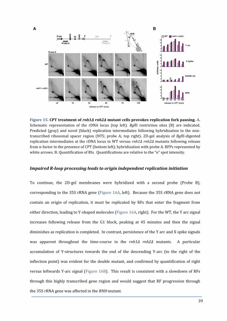
39
Figure 15. CPT treatment of rnh1Δ rnh2Δ mutant cells provokes replication fork pausing. A.
Schematic representation of the rDNA locus (top left). BglII restriction sites (B) are indicated.
Predicted (gray) and novel (black) replication intermediates following hybridization to the non-
transcribed ribosomal spacer region (NTS; probe A; top right). 2D-gel analysis of BglII-digested
replication intermediates at the rDNA locus in WT versus rnh1Δ rnh2Δ mutants following release
from α-factor in the presence of CPT (bottom left); hybridization with probe A. RFPs represented by
white arrows. B. Quantification of RIs. Quantifications are relative to the “n” spot intensity.
Impaired R-loop processing leads to origin independent replication initiation
To continue, the 2D-gel membranes were hybridized with a second probe (Probe B),
corresponding to the 35S rRNA gene (Figure 16A, left). Because the 35S rRNA gene does not
contain an origin of replication, it must be replicated by RFs that enter the fragment from
either direction, leading to Y-shaped molecules (Figure 16A, right). For the WT, the Y arc signal
increases following release from the G1 block, peaking at 45 minutes and then the signal
diminishes as replication is completed. In contrast, persistence of the Y arc and X spike signals
was apparent throughout the time-course in the rnh1∆ rnh2∆ mutants. A particular
accumulation of Y-structures towards the end of the descending Y-arc (to the right of the
inflection point) was evident for the double mutant, and confirmed by quantification of right
versus leftwards Y-arc signal (Figure 16B). This result is consistent with a slowdown of RFs
through this highly transcribed gene region and would suggest that RF progression through
the 35S rRNA gene was affected in the RNH-mutant.

40
Surprisingly, for the rnh1∆ rnh2∆ double mutant we noted the appearance of bubble-shaped
molecules, most clearly seen at 105 minutes (indicated by blue arrow, Figure 16A; quantified
in Figure 16B), but arising 75 minutes after release from CPT treatment.
Figure 16. CPT treatment of rnh1Δ rnh2Δ mutant cells provokes rARS-independent
replication initiation at late S-phase. A. Predicted (gray) and novel (black) replication
intermediates (right) following hybridization to the transcribed 35S gene (probe B). 2D-gels as for
Figure 15. but hybridized with probe B (left). Blue arrows represent bubble arcs; red arrows
represent sub-Y arcs. B. Quantification of RIs for CPT treated samples. Quantifications are relative
to the “n” spot intensity.
A bubble arc is indicative of active replication from an origin within this fragment, since only
active replication within a fragment leads to the formation of replication bubbles in 2D-gel.
Additional signals resembling Y-arcs were observed in the 2D-gels of the RNH double mutants,
which were not observed in the WT and were dependent upon CPT treatment (Figure 17).
These signals, apparent from 45 minutes, have been denominated sub-Y arcs (150), as they
migrate below the standard Y-arc, (indicated by red arrows, Figure 16A).
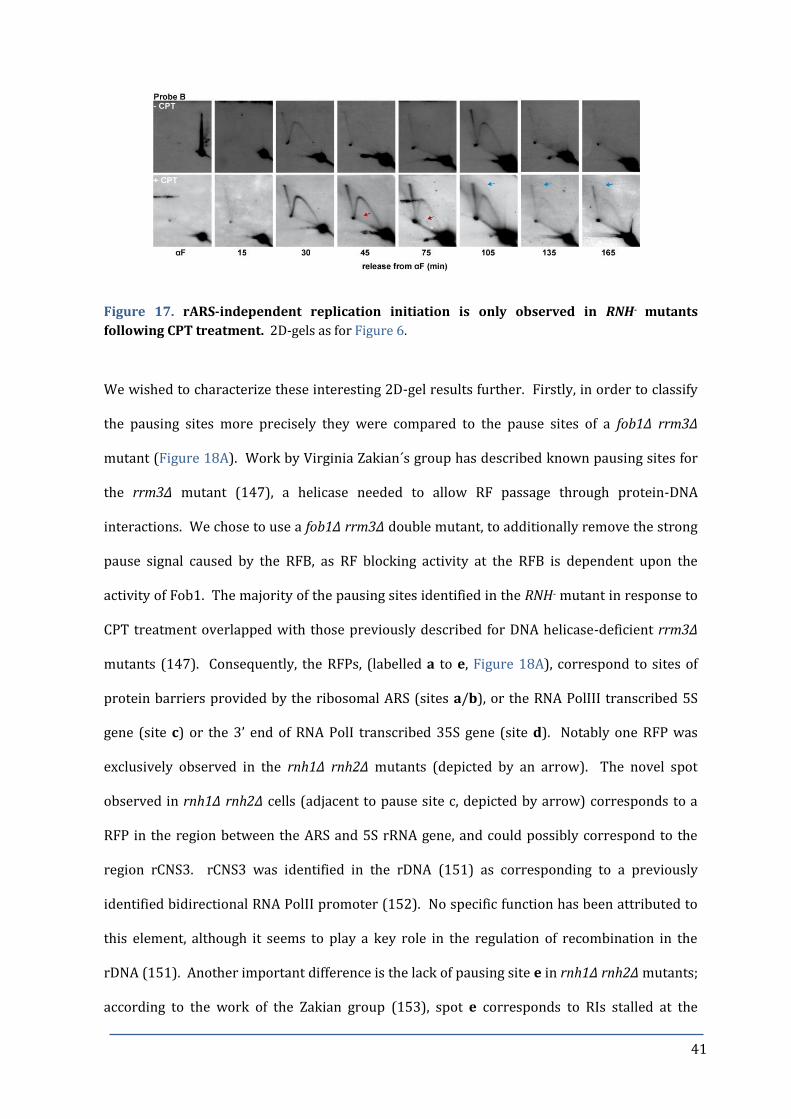
41
Figure 17. rARS-independent replication initiation is only observed in RNH- mutants
following CPT treatment. 2D-gels as for Figure 6.
We wished to characterize these interesting 2D-gel results further. Firstly, in order to classify
the pausing sites more precisely they were compared to the pause sites of a fob1∆ rrm3∆
mutant (Figure 18A). Work by Virginia Zakian´s group has described known pausing sites for
the rrm3∆ mutant (147), a helicase needed to allow RF passage through protein-DNA
interactions. We chose to use a fob1∆ rrm3∆ double mutant, to additionally remove the strong
pause signal caused by the RFB, as RF blocking activity at the RFB is dependent upon the
activity of Fob1. The majority of the pausing sites identified in the RNH- mutant in response to
CPT treatment overlapped with those previously described for DNA helicase-deficient rrm3∆
mutants (147). Consequently, the RFPs, (labelled a to e, Figure 18A), correspond to sites of
protein barriers provided by the ribosomal ARS (sites a/b), or the RNA PolIII transcribed 5S
gene (site c) or the 3’ end of RNA PolI transcribed 35S gene (site d). Notably one RFP was
exclusively observed in the rnh1∆ rnh2∆ mutants (depicted by an arrow). The novel spot
observed in rnh1∆ rnh2∆ cells (adjacent to pause site c, depicted by arrow) corresponds to a
RFP in the region between the ARS and 5S rRNA gene, and could possibly correspond to the
region rCNS3. rCNS3 was identified in the rDNA (151) as corresponding to a previously
identified bidirectional RNA PolII promoter (152). No specific function has been attributed to
this element, although it seems to play a key role in the regulation of recombination in the
rDNA (151). Another important difference is the lack of pausing site e in rnh1∆ rnh2∆ mutants;
according to the work of the Zakian group (153), spot e corresponds to RIs stalled at the

42
ribosomal ARS. The lack of this pausing site might be explained by the temporal appearance of
pausing sites in the RNH- mutants. In rrm3∆ mutants, replication pausing sites coincide with
ongoing DNA synthesis, while rnh1∆ rnh2∆ mutant-dependent pausing sites are restricted to
late S/G2, at which time the ribosomal ARS might be devoid of origin interacting proteins that
are needed to impair fork progression.
To address whether these bubble-shaped intermediates consist of extensive R-loops, after
restriction digestion we performed in vitro RNaseH digestion of the genomic DNA from rnh1∆
rnh2∆ yeast cells at the 105 min time point prior to 2D-gel (Figure 18B). RNaseH would digest
regions of RNA hybridized to DNA, and therefore if the bubble-shaped molecules simply
corresponded to extended RNA:DNA hybrid regions the bubble arcs should collapse upon RNA
degradation. This, however, was not the case, as the bubble-shaped molecules were resistant
to in vitro RNaseH treatment (Figure 18B, blue arrows). Furthermore, the sub-Y arcs,
consistent with the presence of segments of ssDNA, were equally unaffected by RNaseH
treatment (Figure 18B, red arrows). Secondly, we questioned whether such molecules
contained a 3’ extendable DNA polymerase substrate. Prior to 2D-gel, RIs were subjected to in
vitro treatment with the exo- Klenow Polymerase, gp32 single-stranded DNA binding protein
Figure 18. A. Characterization of
RFP sites in the NTS region in rnh1∆
rnh2∆ as compared to fob1Δ rrm3Δ
mutant strain. Pausing sites are
labelled a-e; black arrow represents
unique RFP site. B. Prior to 2D-gel,
RIs isolated from CPT-treated rnh1∆
rnh2∆ cells at 105 minutes were
subjected to heat or enzymatic
treatments as indicated. Blue arrows
represent bubble arcs; red arrows
represent sub-Y arcs. Schematic
representation of species that would
migrate as sub-Y arcs (right). 2D-gel
analyses shown were performed by
Nestor García Rodríguez.

43
and nucleotides (154) (Figure 18B). The disappearance of bubble-shaped molecules suggests
that these intermediates are a substrate for the DNA polymerase and indicates that they
contain an extendable 3´ hydroxyl group. Together our results open the possibility that the
bubble signals were not made up of an RNA:DNA hybrid, but rather have all of the features of
replicating molecules, and thus represent origin-independent replication initiation events.
Lack of Top1 is crucial for origin-independent replication initiation events
The bubble arcs detected in the RNH double mutant were only evident following growth in the
presence of CPT. CPT stabilizes a Top1-DNA complex and results in nicked DNA by a so-called
Top1 cleavage complex (Top1cc). This kind of DNA damage or the sequestering of Top1 could
be critical for bubble arc formation. To distinguish between these two possibilities, we decided
to employ the conditional protein degradation system, or “Degron”, as a tool to rapidly deplete
Top1 proteins from yeast cells. Consequently, we constructed an auxin-inducible Top1 degron
(Top1AID-9Myc) in rnh1∆rnh2∆ yeast expressing the SCFTIR1. The auxin-inducible degron (AID)
system acts to proteolytically remove the target protein upon addition of the plant auxin-like
pheromone, indole acetic acid (IAA) to the growth media (see Materials & Methods for details)
(155).
Firstly, we confirmed that Top1AID-9Myc protein levels were reduced in response to auxin.
G1-synchronized cells were incubated for 30 minutes in the presence of 1mM IAA to induce
degradation of the aid-tagged protein, prior to release into fresh YPAD containing IAA, and
samples were collected at the indicated timepoints for western blot analysis (Figure 19A).
After 30 minutes, Top1AID-9Myc protein levels were significantly down-regulated. In line with
the rapid degradation of Top1AID-9Myc, growth of the rnh1∆ rnh2∆ Top1AID-9Myc strain was
severely impaired in the presence of IAA (Figures 19B and 19C), thus resembling synthetic
lethality observed for the top1∆ rnh1∆ rnh2∆ triple mutant (60).

44
Figure 19. Confirmation of functionality of the 9Myc-Top1AID degron in rnh1∆ rnh2∆ TIR1-
expressing yeast. A. Western blot analysis against the Myc tag to confirm degradation of Top1AID
protein in response to 1mM IAA. Ponceau stained membrane (right panel); Western with α-Myc
and α-PGK loading control (left panels). B. Halo assay with increasing concentrations of IAA. C.
Drop test analysis on YPAD plates containing 1mM IAA in the presence or absence of 5µg/ml CPT.
D. Immunofluorescence analysis of RNA:DNA hybrids with S9.6 antibody in the Top1AID degron in
rnh1∆ rnh2∆ yeast in the absence or presence of 1mM IAA for 30 minutes. Representative images
(left) of RNA:DNA hybrids (S9.6; red) and co-localisation with DAPI nuclear strain (blue); photos
taken with the same settings for comparison of signal intensity. Quantification of cells with
RNA:DNA hybrid and DAPI co-localising signals.
Furthermore, formation of RNA:DNA hybrids was significantly increased (Figure 19D), as
determined by immunofluorescence using the S9.6 antibody that recognizes RNA:DNA hybrids
(156). Importantly, the signal for RNA:DNA hybrids co-localized in its majority with DAPI-
stained nuclear material, although some additional foci could be observed, indicative of
mitochondrial RNA:DNA hybrid detection.
Next, we examined the fate of DNA replication of the auxin-inducible rnh1∆ rnh2∆ Top1AID-
9Myc strain. Monitoring cell cycle progression following release from α-factor in the presence
of IAA (Figure 20A) the conditional triple mutant was able to pass through and complete S
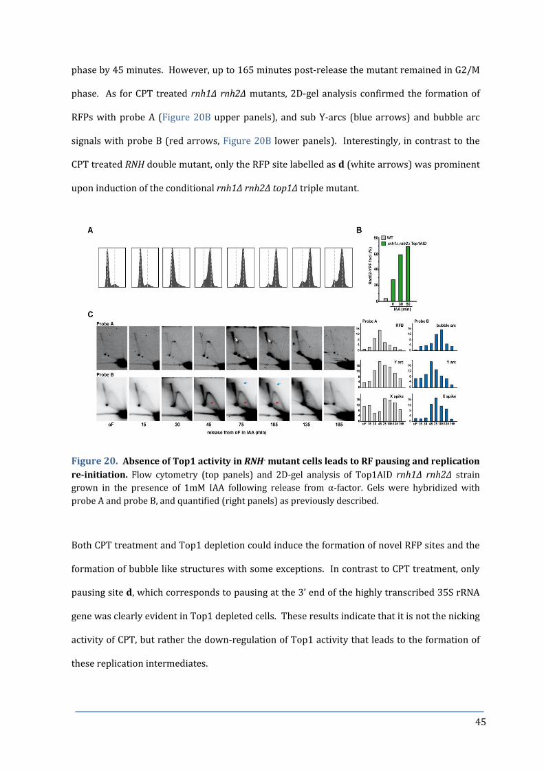
45
phase by 45 minutes. However, up to 165 minutes post-release the mutant remained in G2/M
phase. As for CPT treated rnh1∆ rnh2∆ mutants, 2D-gel analysis confirmed the formation of
RFPs with probe A (Figure 20B upper panels), and sub Y-arcs (blue arrows) and bubble arc
signals with probe B (red arrows, Figure 20B lower panels). Interestingly, in contrast to the
CPT treated RNH double mutant, only the RFP site labelled as d (white arrows) was prominent
upon induction of the conditional rnh1∆ rnh2∆ top1∆ triple mutant.
Figure 20. Absence of Top1 activity in RNH- mutant cells leads to RF pausing and replication
re-initiation. Flow cytometry (top panels) and 2D-gel analysis of Top1AID rnh1∆ rnh2∆ strain
grown in the presence of 1mM IAA following release from α-factor. Gels were hybridized with
probe A and probe B, and quantified (right panels) as previously described.
Both CPT treatment and Top1 depletion could induce the formation of novel RFP sites and the
formation of bubble like structures with some exceptions. In contrast to CPT treatment, only
pausing site d, which corresponds to pausing at the 3’ end of the highly transcribed 35S rRNA
gene was clearly evident in Top1 depleted cells. These results indicate that it is not the nicking
activity of CPT, but rather the down-regulation of Top1 activity that leads to the formation of
these replication intermediates.
46
Unscheduled replication initiation events at rDNA are RNA PolI transcription-
dependent
Because the bubble-shaped replication intermediates were restricted to the RNA PolI-
transcribed 35S rRNA fragment, we asked whether their appearance was dependent on
transcription. To specifically reduce transcription of the rDNA locus we took advantage of
temperature sensitive RNA PolI mutants for alleles of Rpa190 (rpa190-3), which is the largest
and catalytic core component of RNA PolI, and Rrn3 (rrn3-8), which recruits RNA PolI to the
35S rRNA gene promoter. Transcription of the rDNA array occurs normally when the
temperature sensitive RNA PolI mutants are grown at permissive temperatures (23ºC), but
growth of these mutants at higher restrictive temperatures (34-37ºC) impairs transcription
and leads to cell death due to impaired ribosome biogenesis (157). These mutants are viable at
semi-permissive temperatures (30ºC), although rDNA transcription by RNA PolI is reduced.
Interestingly, growth of the rpa190-3 (and to a lesser extent, rrn3-8) triple mutant at semi-
permissive temperatures suppressed the CPT-sensitivity of the RNH- mutants (Figure 21A),
suggesting a link between RNA PolI transcription and CPT toxicity.
It´s tempting to speculate that highly transcribed genes are more prone to DNA damage
formation, perhaps caused by increased RF stalling at RNA:DNA hybrids or by collision with
the transcription machinery. To test for this possibility we assayed the RNA PolI-dependent
formation of Rad52-YFP foci formation. Interestingly, a down-regulation of RNA PolI
transcription reduced the formation of nucleolar DNA repair centres (Figure 21B). Raising the
temperature from 23º to 30ºC (permissive to semi-permissive conditions), caused a general
increase in the number of Rad52-YFP foci in the rpa190-3 rnh1∆ rnh2∆ triple mutant, however,
the number of foci that localize to the nucleolar periphery was significantly decreased in the
triple mutant at the semi-permissive temperature of 30ºC. These experiments suggest that
rDNA transcription by RNA PolI has a major impact on the formation of CPT induced rDNA
lesions in the RNH double mutant. To gain insight into this possibility, we determined the

47
recombination frequency within the rDNA repeats (Figure 21C). Indeed, and in accordance
with a previous study (143), down-regulation of RNA PolI transcription at the semi-permissive
temperature reduced the recombination frequency within the rDNA repeats.
Figure 21. CPT sensitivity of rnh1∆ rnh2∆ is related to rDNA transcription by RNA PolI. A. 10-
fold serial dilutions of the temperature-sensitive conditional RNA PolI subunit mutant rpa190-3
grown on YPAD or YPAD-containing 5µg/ml CPT for 3 days at permissive (23ºC) or semi-
permissive (30ºC) temperature. B. Reduced RNA PolI transcription in rpa190-3 rnh1∆ rnh2∆ triple
mutants correlates with a reduced amount of nucleolar DNA damage. Percentage of nuclear versus
nucleolar Rad52-YFP foci (determined by co-localization with the Nop1-mRFP) at permissive
(23ºC) or semi-permissive (30ºC) temperature with or without the presence of 10µg/ml CPT (3hr
treatment). Data represent mean ± SD, from at least three independent experiments. The p-value
for 23°C rDNA versus 30°C rDNA was 0.0053 without and 0.001 with CPT. C. Recombination
frequencies of strains containing the leu2-k::ADE2-URA3::leu2-k recombination system. Data
represent the mean ± SD of three independent fluctuation tests, each fluctuation test representing
the median value of 6 independent colonies. Fold change relative to WT is indicated. The p-value
for rpa190-3 rnh1∆ rnh2∆ at 30°C versus 23°C was 0.0361. Differences were examined by Student’s
t-test and were considered statistically significant for p-values<0.05 (asterisks).
Subsequently we examined the fate of replication in rpa190-3 rnh1∆ rnh2∆ mutants by 2D-gels,
to test if the previously observed origin-independent replication re-initiation requires active
transcription of the rDNA unit by RNA PolI (Figure 22A). Triple mutant rpa190-3 rnh1∆ rnh2∆
yeast were grown at 26°C, prior to synchronization with α-factor and release into CPT
containing media at permissive (23°C) or semi-permissive (30°C) temperatures. As expected,
upon hybridization with probe B against the transcribed 35S rRNA gene, we observed bubble
arcs in the rpa190-3 triple mutant at the permissive temperature when RNA PolI transcription
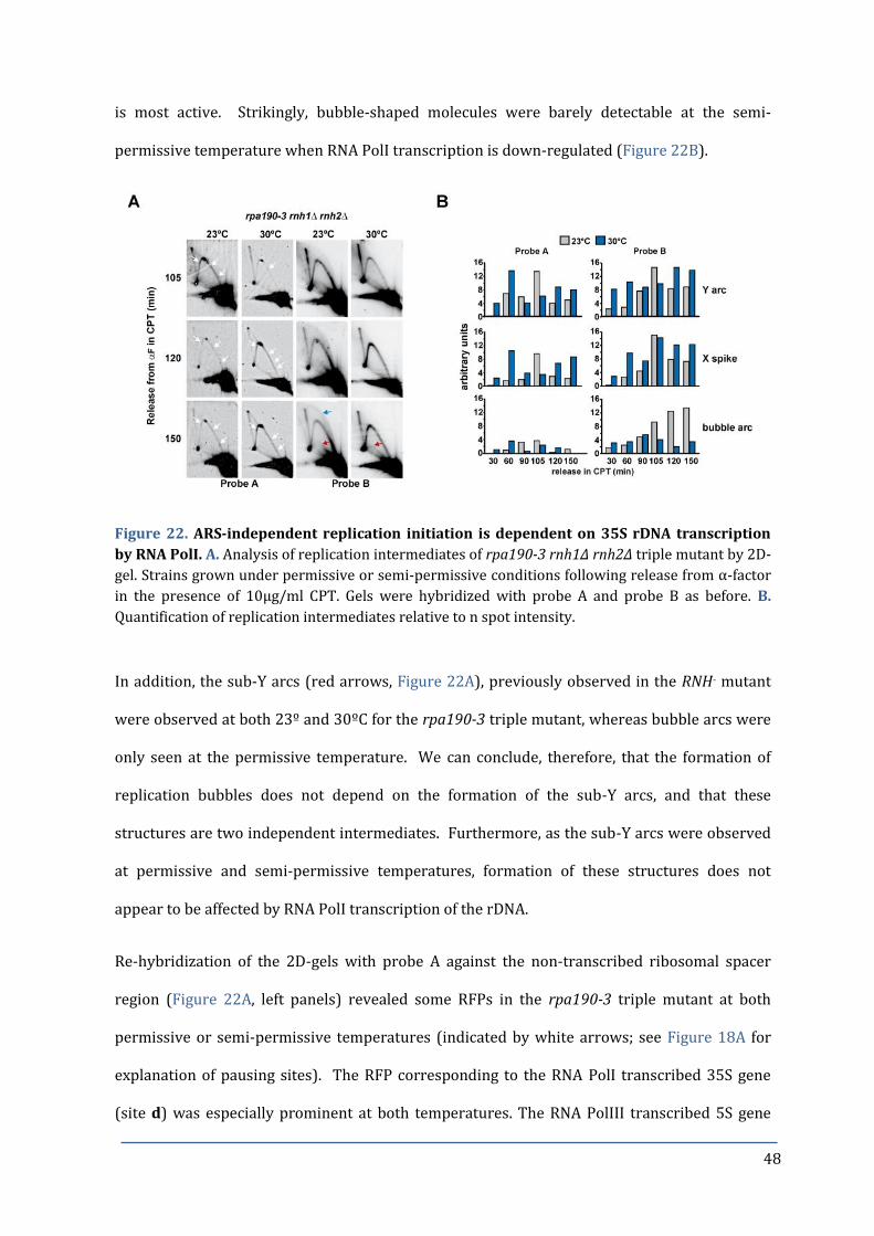
48
is most active. Strikingly, bubble-shaped molecules were barely detectable at the semi-
permissive temperature when RNA PolI transcription is down-regulated (Figure 22B).
Figure 22. ARS-independent replication initiation is dependent on 35S rDNA transcription
by RNA PolI. A. Analysis of replication intermediates of rpa190-3 rnh1Δ rnh2Δ triple mutant by 2D-
gel. Strains grown under permissive or semi-permissive conditions following release from α-factor
in the presence of 10µg/ml CPT. Gels were hybridized with probe A and probe B as before. B.
Quantification of replication intermediates relative to n spot intensity.
In addition, the sub-Y arcs (red arrows, Figure 22A), previously observed in the RNH- mutant
were observed at both 23º and 30ºC for the rpa190-3 triple mutant, whereas bubble arcs were
only seen at the permissive temperature. We can conclude, therefore, that the formation of
replication bubbles does not depend on the formation of the sub-Y arcs, and that these
structures are two independent intermediates. Furthermore, as the sub-Y arcs were observed
at permissive and semi-permissive temperatures, formation of these structures does not
appear to be affected by RNA PolI transcription of the rDNA.
Re-hybridization of the 2D-gels with probe A against the non-transcribed ribosomal spacer
region (Figure 22A, left panels) revealed some RFPs in the rpa190-3 triple mutant at both
permissive or semi-permissive temperatures (indicated by white arrows; see Figure 18A for
explanation of pausing sites). The RFP corresponding to the RNA PolI transcribed 35S gene
(site d) was especially prominent at both temperatures. The RNA PolIII transcribed 5S gene

49
pause site (site c) was also observed in both conditions as would be expected, since the down-
regulation of RNA PolI would not necessarily affect the transcription of RNA PolIII. These
results indicate that even with the down-regulation of RNA PolI activity, replication forks have
problems to progress through the rDNA locus, and in particular the 35S rRNA gene.
Taken together, our results reveal that the CPT sensitivity and instability of the rDNA array of
rnh1∆ rnh2∆ yeast are linked to the transcriptional activity of rDNA. We propose that the
absence of RNaseH activity, in combination with a reduction in Top1 activity (either by
reducing protein levels or by CPT sequestering) leads to an accumulation of RNA:DNA hybrids,
particularly across highly transcribed regions such as the rDNA locus. These hybrids can
interfere with ongoing replication, lead to replication fork blockage or even initiate
unscheduled replication (see Figure 23 for hypothetical model). We infer from our results that
RNA:DNA hybrids provide the nucleation point for the assembly of a non-canonical replication
machinery and are able to initiate origin-independent DNA replication in a eukaryotic genome.
Figure 23. Model for Transcription Initiated Replication in Yeast Ribosomal DNA.

50
DISCUSSION for CHAPTER 1
Consequences of Persistent R-loops
Persistent R-loops have previously been linked to genomic instability ((16) and references
within), including mutations and hyper-recombination. This thesis confirms the critical role of
the RNaseH enzymes in removing R-loops for the maintenance of genome integrity, and
extends these findings, highlighting in particular the ribosomal DNA array as a hot-spot for R-
loop mediated genome instability. Furthermore, we reveal a novel impact of R-loops as sites of
replication initiation in a eukaryotic genome, which we refer to as “transcription-initiated
replication” or TIR.
R-loops Promote Origin-Independent Replication
DNA replication is normally initiated at determined origins of replication throughout the
genome and requires the function of DNA Polα-primase to initiate DNA synthesis.
Furthermore, origin firing follows a strict temporal control, with replication initiated
specifically during S phase of the cell cycle (reviewed in (158)). However, we show that the
origin-independent replication initiation observed in the highly transcribed 35S rRNA occurs
outside of the normal program of origin firing, both spatially (not at a defined ori) and
temporally (the replication initiation occurs in late S/G2 phase of the cell cycle, when
replication would normally be completed).
The function of RNA:DNA hybrids as “origins of replication” has been well documented in
prokaryotic plasmids, bacteriophages and mitochondrial DNA. In these cases, transcription
produces an RNA:DNA hybrid that can subsequently prime replication initiation. In fact, an
origin-independent replication initiation was observed in dnaA mutants of E.coli over 40 years
ago (44). DnaA is a replication initiation factor that promotes the unwinding and strand
separation of DNA at the bacterial origin (oriC), essential for the assembly of the replisome
51
(159). Mutants capable of initiating DNA replication in the absence of DnaA were mapped to
the RNH locus (rnhA) (45) encoding the bacterial RNaseH. RNA:DNA hybrids are
thermodynamically stable structures, where the two strands of the DNA duplex are open.
Therefore, the persistence of such structures could by-pass the need for DnaA or strand
separation activity. However, mutants in rnhA are unable to suppress the need for DnaG, the
bacterial primase (46), suggesting that this mode of replication initiation is not sufficient for
replication of the complete genome.
Various alternatives could account for the R-loop-mediated replication observed in the yeast
genome. These could include the direct assembly of the replisome at the open and stable R-
loop, with the RNA moiety acting as a primer for extension by a DNA Pol (with or without
processing to generate a 3´end), analogous to the replication initiation of the multi-copy
plasmid ColE1 (160). Another possibility may be that an R-loop could be a suitable substrate
for a specialized polymerase such as the recently characterized human PrimPol (161). PrimPol
could re-prime with dNTPs to re-initiate DNA synthesis downstream of hybrid-stalled RFs
(162), although such an activity has not been identified in yeast to date. On the other hand, the
R-loop structure could facilitate strand invasion for a recombination-dependent assembly of
the replisome. An ongoing canonical RF may stall at the R-loop creating a break, and the
processed 3' single stranded end could invade a homologous template, promoting RF assembly.
The repetitive nature of the rDNA array, and subsequent ease of homology search, might make
this mechanism of replication initiation particularly favourable. This mode of origin-
independent replication would be similar to the recombination-dependent replication
mechanism of E. coli and bacteriophage. This possible mechanism of TIR will be investigated in
more detail in Chapter 2 of this thesis. An alternative hypothesis, not explored further in this
thesis, would be the possibility that TIR events are restricted to ERCs. Yeast lacking RNaseH
activities exhibited copy number heterogeneity and concomitant increase in the number of
ERCs (see Figure 13) and interestingly, top1 mutants were also reported to have more ERCs
(163). ERCs are maintained in the cell as plasmid-like circular DNA (145), and can replicate
52
autonomously (164). One could presume that an rDNA repeat-containing ERC could replicate
by an R-loop mediated mechanism analogous to ColE1 plasmid replication. However,
investigating this possibility is hampered by the fact that the DNA sequences that constitute
ERCs and the genomic rDNA are identical. Therefore, ERCs would have to be separated from
genomic DNA to distinguish between an rDNA 9.1kb repeat unit located on chromosome XII or
found on an ERC by 2D-gel and subsequent probe hybridization.
Persistent R-loops Particularly Affect the Stability of rDNA
The accumulation of ERCs has been shown to be toxic and has been linked to a decreased
lifespan (senescence) in yeast (145). However, in the case of the hpr1 mutant, shown to
accumulate R-loops (19), no increase in ERCs was observed despite an increased rate of
recombination within the rDNA array (165). In this case the reduced life span was associated
with increased genomic instability, and over time a revised “rDNA instability” theory of aging
has been proposed (142). In fact, in aging mammalian cells changes in nucleolar morphology
have been detected (166), and the tumour suppressor protein RB, an important regulator of
senescence (167), has been shown to accumulate in the nucleolus and repress PolI
transcription (168,169). Due to the increased production of ERCs in yeast lacking RNaseH
activities, and the increased genomic instability of the rDNA locus, it would be interesting to
investigate whether the lifespan of the RNH- mutants is affected.
DNA damaging events were found to be particularly frequent in the rDNA locus in yeast lacking
RNaseH activities (see Figure 12). We found that the down-regulation of RNA PolI could
suppress the CPT sensitivity of the RNH- mutants (see Figure 21), suggesting that the majority
of the DNA lesions were mediated by rDNA transcription. A recent study demonstrated that
Top1ccs are specifically enriched and stabilized at the RFB of the yeast rDNA (170), and the
formation of these targeted Top1cc complexes would explain the previously reported
occurrence of DSBs near the RFB (63,64). Consequently, we were very interested to note that
there is a Top1 binding site near the 35S promoter, at approximately 180bp upstream of the

53
35S rRNA transcriptional start (171). This specifically located Top1 binding site may reflect
the importance of the action of Top1 to prevent RNA:DNA hybrid formation across this highly
transcribed active gene, and thus repress R-loop assisted RF assembly. An alternative
explanation may be that programmed RF pausing at the 35S promoter, due to the presence of a
Top1 cleavage complex, is necessary for subsequent DSB formation and recombination-
mediated replication fork re-start. Interestingly, TIR was only observed upon the down-
regulation of Top1 activity (by protein depletion or CPT treatment). This would indicate that
the action of Top1 for the avoidance of the formation of RNA:DNA hybrids is normally
sufficient to suppress TIR in the absence of RNaseH activity.
Origin-Independent Replication Outside of S-phase
As seen by our immunofluorescence timecourse, RNA:DNA hybrids were present in yeast from
G1 synchronization and throughout S phase (see Figure 19), and therefore replication
initiation could have occurred at any stage during the cell cycle. However, the appearance of
the RF pause sites and the transcription dependent replication bubbles were only observed
when cells were held in late S/G2 phases of the cell cycle. Perhaps TIR can only occur in late
S/G2 phase, since this is when an enzymatic activity needed for the formation of a replication
bubble is available. For example, an enzymatic activity that could liberate a free 3´OH end to
allow strand invasion, such as the nucleases Mus81, Yen1, or Slx1-4, may be necessary for R-
loop assisted RF assembly. The highly redundant activities of the 3´end resection factors make
it difficult to study the contributions of each individual factor but it would be interesting to
investigate the impact of these nucleases on TIR.
R-loops Provoke Replication Fork Pausing
The progression of both transcription (24) and replication machineries (25,172) can be
blocked by R-loops and our results confirm that yeast cells lacking RNaseH activity have an
impaired ability in replication progression. The majority of the pausing sites identified in this
study corresponded to sites of protein barriers provided by the ribosomal ARS, or the RNA

54
PolIII transcribed 5S- or RNA PolI transcribed 35S genes (see Figure 18). However, we
suggested that one novel pause site corresponded to rCNS3 (151), a bidirectional RNA PolII
promoter (152) of dubious function. It´s interesting to note that the RDN1 (rDNA) locus
contains some RNA PolII transcribed units, including the gene TAR1, encoding a mitochondrial
protein, found on the opposite strand to the 25S rRNA gene (173). Furthermore, as we
observed common pausing sites between yeast lacking RNaseH activity and those published
for the rrm3∆ mutant, we can reason that the replisome assembled for TIR missed some factors
necessary to move through protein-DNA interaction sites. Our findings predict a non-canonical
replication machinery is involved in TIR. A future challenge will be to determine the molecular
composition of such a replication machinery e.g. by selective immunoprecipitation against
factors that may interact with TIR intermediates.
Removal of R-loops Protects Genome Integrity
Our report of a transcription-dependent mechanism of replication initiation reveals a critical
interplay between replication and transcription. Both processes must be highly regulated and
coordinated in order to occur simultaneously without risking genome integrity. Maintaining
the stability of rDNA is vital since cell growth is directly dependent on the rate of protein
synthesis and thus on the transcription of rDNA. Interestingly, a common feature of rapidly
proliferating cancer cells is an increase in rDNA transcription (reviewed in (174)) and the
human prostate cancer line 15PC3 was shown to express RNaseH2 throughout the cell, when
it´s expression is usually confined to the nucleus (175). As such, determining how cells activate
rDNA transcription and how this can influence DNA replication is important for understanding
mechanisms that can lead to tumourigenesis. We reveal TIR at the highly transcribed 35S rRNA
gene and one may presume that such a mechanism of R-loop primed replication initiation
could occur at other highly-transcribed regions of the genome.
Our work highlights the essential roles of the RNaseH enzymes, and redundant activities, to
eliminate RNA:DNA hybrids, and thus repress an inefficient and unregulated origin-

55
independent mechanism of DNA replication in eukaryotic cells. Unscheduled replication,
particularly in regions of repetitive sequences such as the rDNA array, could lead to re-
replication, loss of heterozygosity, and the deletion of repetitive sequences or gene
amplifications. Such copy number changes are relevant to carcinogenesis, as gene
amplification can lead to tumourigenesis, i.e. through the enhanced levels of an oncogene
(176,177), and DNA amplification is also one of the key mechanisms by which cells acquire
resistance to many cytotoxic compounds, i.e. the overexpression of multidrug transporter
proteins has been observed frequently in many types of human tumors (178). However, gene
amplification is also an important condition for beneficial adaptation to environmental changes
(reviewed in (179)). In some organisms gene amplification is developmentally regulated and
is an essential feature of the life cycle, for example for the amplification of chorion gene
clusters in Drosophila melanogaster follicle cells during oogenesis (180), where gene
amplification occurs through repeated initiation of the chorion gene cluster (for a review see
(181)). We believe that highly transcribed genes might be more prone to gene amplification
events induced by RNA:DNA hybrid-mediated replication. Such events might be more frequent
than anticipated and may contribute significantly to genetic alterations in eukaryotic
chromosomes, and open the possibility that TIR could present a driving force in evolution.
The RNaseHs have an essential role in the removal of R-loops, whose formation has been
linked to genome instability and cancer, neurodegenerative diseases and possibly senescence.
Our work adds a new perspective to the critical role of the RNaseH enzymes and Top1 enzymes
in eukaryotic genomes to prevent unscheduled TIR mediated by R-loops.

56
RESULTS
CHAPTER 2 – RNH coding genes: genetic interactions reveal a link to
genome stability and nucleolar function
Genetic interactions of RNH enzymes with DNA replication and repair factors
To understand how the RNaseH activities are linked to rDNA replication, stability and
organisation, we first conducted a genetic analysis by crossing a selection of non-essential
mutants covering representative steps in DNA replication and/or DNA repair with rnh1Δ rnh2Δ
lacking yeast, then analysing the growth and viability of meiotic segregants (examples are
shown in Figure 24A and summarized in Table 1). Triple mutants were classified as synthetic
sick if the triple mutants showed a considerably smaller spore size or a slow growth phenotype
on rich medium, when compared to the rnh1Δ rnh2Δ double mutant. Synthetic sick
interactions were obtained as soon as cells were impaired in checkpoint function (e.g. DUN1
and RAD24), DNA repair (e.g. EXO1, RAD18 and MUS81), replication fork stability (e.g. CSM3
and RRM3) or rDNA organisation (e.g. FOB1 and HMO1).
Besides the well-documented synthetic lethal interactions between RNH- mutants with TOP1
and RAD27 (60,182), our genetic analyses revealed previously unreported synthetic lethal
interactions between rnh1Δ rnh2Δ mutants and mutants in RAD52, SRS2, POL32, and the MRX
complex (MRE11, RAD50, and XRS2). We confirmed that Rad52 activity is essential in RNH-
mutants by expressing RAD52 under control of a galactose-inducible promoter (from plasmid
pMDL5 (183)) in the double mutant, and crossed with a rad52 mutant (Figure 24A). The
rad52∆ rnh1∆ rnh2∆ (GAL1p-RAD52-URA3) triple mutant was subsequently streaked onto S-
Ura galactose (plasmid inducing conditions) or SC-Ura glucose-containing plates (plasmid
expression repressed). The rad52∆ rnh1∆ rnh2∆ (GAL1p-RAD52-URA3) triple mutants were
unable to grow on medium containing glucose, indicating that the RNH double mutant cannot

57
survive without RAD52 expression. Therefore, the action of Rad52 and/or HR is essential for
rnh1∆ rnh2∆ viability.
Figure 24. Synthetic lethal and synthetic sick interactions with the RNH mutants. A. Tetrad
analysis of rnh1∆ rnh2∆ in combination with null mutations of rad52, rad51, srs2, rad59, and the
MRX complex members mre11, rad50 and xrs2 (top panel). Rad52 activity is essential in RNH
double mutants (lower panel). Yeast transformed with GAL1p-RAD52-URA3 were streaked onto SC-
Ura and SC supplemented with 500mg/ml of 5-Fluorotic acid (5-FOA). B. Diagram of synthetic
lethal and synthetic sick interactions with the rnh1Δ rnh2Δ double mutant. Filled circles represent
synthetic lethal, and open circles represent synthetic sick interactions. The mutants were broadly
grouped according to gene function, as indicated by the different coloured connecting lines. PRR,
post-replicative repair; NER, nucleotide excision repair. Note that the synthetic lethal interactions
between the rnh1Δ rnh2Δ double mutant, SIC1, MRC1 and other checkpoint genes will be further
explored in Chapter 3 of this thesis.

58
TIR is Rad51 independent
Our genetic analyses revealed that homologous recombination is a critical pathway in yeast
lacking RNaseH activity, because Rad52 activity became essential even in the absence of
exogenous DNA damage. Rad52 is a key factor for the repair of single-strand and double-
strand DNA breaks, and is required for the homology search and strand invasion steps of all
homology-dependent repair pathways (reviewed in (184)). For example, Rad52 acts in both
the Rad51-independent pathways: single-strand annealing (SSA) and break-induced
replication (BIR), (185) and the Rad51-dependent DSBR and synthesis-dependent strand
annealing (SDSA) pathways (as summarized in Figure 9). In contrast to the synthetic lethal
interaction with RAD52 and the synthetic sick genetic interaction with RAD59, the rad51∆
rnh1∆ rnh2∆ triple mutants were viable and did not manifest a significant growth defect when
compared to the RNH double mutant. The rad51∆ simple mutant itself is very CPT sensitive
(drop tests carried out with 0.1µg/ml CPT), and the rad51∆ rnh1∆ rnh2∆ triple mutant is even
more sensitive to CPT (Figure 25A). This result indicates different constrains mediate CPT
sensitivity in the absence of Rad51 or RNaseH activities.
HR may drive the formation of bubble shaped replication intermediates in CPT-treated rnh1∆
rnh2∆ mutants. Given that rad51∆ rnh1∆ rnh2∆ triple mutants are viable we intended to test
for this possibility by 2D agarose gel analysis of replication intermediates (Figure 25B).
Interestingly, bubble shaped replication intermediates were present in CPT-treated rad51∆
rnh1∆ rnh2∆ triple mutants. However, we cannot rule out that Rad51, but not HR, is
dispensable for the formation of bubble shaped RI because melting of the DNA double helix by
RNA:DNA hybrids may provide sufficient single-stranded DNA in order to bypass the need for
Rad51 activity during HR. As HR is stimulated in the absence of Srs2 (186,187), we questioned
whether we would see more TIR events in the absence of Srs2 activity. We were interested to
find that the srs2∆ rnh2∆ double mutant is synthetic lethal, and only 31% of srs2∆ rnh1∆ rnh2∆
triple mutants were viable (Figure 24A). Viable triple mutants were very small and sensitive to

59
CPT, suggesting that Srs2 is an important factor when RNaseH activity is absent, particularly in
the case of Rnh2. However, we were still able to observe bubble arcs in the srs2 triple mutant
(Figure 25B). Therefore, although the activity of Srs2 is important in response to CPT and for
survival of the RNaseH- yeast, it does not contribute to the R-loop dependent replication
initiation. In this manner, we demonstrate that neither Rad51 nor antagonistic Srs2 activity is
required for TIR.
Figure 25. Rad51 is not needed for the formation of replication bubbles by TIR. A. Drop test
analysis of CPT sensitivity of rad51. B. 2D-gel analysis of rad51, rad18, srs2 and mrc1 triple mutants
following release from α-factor in the presence of CPT, as previously described. C. Tetrad analysis
of rnh1∆ rnh2∆ in combination with null mutations of the PRR factors rad18 and pol32. D. Drop test
analysis of CPT sensitivity of rad18 triple mutants.
PRR, NER but not NHEJ is required for the repair of CPT mediated DNA damage
For viable triple mutants, we performed a more extensive analysis of how the formation of
CPT-mediated DNA damage or Top1-depletion would affect the viability by drop test analysis.

60
Classification was carried out according to the following categories; as additive – the triple
mutant was more sensitive than the double mutant; epistatic – the triple mutant has the same
sensitivity as the double mutant; or suppressive – the triple mutant is less sensitive than the
double mutant, or the triple mutant has an intermediate CPT sensitivity, between the double
mutant and the single mutant (in the case of an exceptionally sensitive simple mutant). The
results are summarized in Table 1.
The NHEJ-member triple mutants showed no synthetic sick interaction with the RNH- mutant
and behaved differently upon treatment with CPT, e.g. the triple mutant with ku70 showed an
additive CPT sensitivity whilst the triple mutant with lig4 suppressed to some extent the CPT
sensitivity of the RNH double mutant (data not shown). This difference may be due to the
additional role of Ku70 in telomere maintenance (for a review see (188)). However, we can
conclude that NHEJ, in contrast to HR, is not an essential process in yeast lacking RNaseH
activity.
Genetic analyses revealed that homologous recombination is a critical pathway in yeast that
lack RNaseH activity, even in the absence of exogenous damage. The structure-specific
endonuclease Mre11 plays an important role in the repair of DSBs by HR. As a component of
the MRX complex (Mre11-Rad50-Xrs2), it is responsible for processing of the ends of the break
to produce a free 3´ ssDNA end, which is essential for strand invasion and for subsequent
extension by a DNA Pol. Mre11 has also been reported to be a specialized nuclease capable of
removing bulky adducts from DNA ends (67,189), and may be able to cleave the Top1 tyrosyl-
DNA bond (190). Therefore, Mre11 could play a role in the repair of CPT-induced damage in
yeast (67). Therefore, we were particularly interested to identify a synthetic lethal interaction
of the rnh1Δ rnh2Δ double mutant with mre11Δ (Figure 24A). Subsequently, we crossed rnh1Δ
rnh2Δ cells with rad50Δ and xrs2Δ, and identified synthetic lethal interactions with all the
members of the MRX complex (Figure 24A).

61
YPAD CPT Checkpoints
WT +++++ +++++ rad9Δ +++++ +++++
rnh1Δ rnh2Δ +++++ +++ rad9Δ rnh1Δ rnh2Δ +++++ ++
chk1Δ +++++ +++++
Replication chk1Δ rnh1Δ rnh2Δ +++++ ++
sic1Δ +++++ ND tel1Δ +++++ +++++
sic1Δ rnh1Δ rnh2Δ SL ND tel1Δ rnh1Δ rnh2Δ +++++ ++
clb5Δ +++++ +++++ rad24Δ +++++ +++
clb5Δ rnh1Δ rnh2Δ +++ ++++ rad24Δ rnh1Δ rnh2Δ ++++ ++
rad27Δ +++++ +++++ dun1Δ +++++ +++++
rad27Δ rnh1Δ rnh2Δ SL ND dun1Δ rnh1Δ rnh2Δ ++++ +++
mrc1Δ +++++ +++++ mec1Δ sml1Δ +++++ +++
mrc1Δ rnh1Δ rnh2Δ +++ ++ mec1Δ sml1Δ rnh1Δ rnh2Δ ++++ ++
csm3Δ +++++ +++++ sml1Δ +++++ ++++
csm3Δ rnh1Δ rnh2Δ ++++ ++ sml1Δ rnh1Δ rnh2Δ +++++ +++
tof1Δ +++++ +++++ swe1Δ +++++ +++++
tof1Δ rnh1Δ rnh2Δ ++++ +++ swe1Δ rnh1Δ rnh2Δ +++++ ++++
top1Δ +++++ ++++ mad1Δ +++++ +++++ top1Δ rnh1Δ rnh2Δ SL ND mad1 Δrnh1Δ rnh2Δ +++++ +++
rrm3Δ +++++ +++++
rrm3Δ rnh1Δ rnh2Δ ++++ ++ PRR
pol32Δ +++++ +++++ rDNA related functions pol32Δ rnh1Δ rnh2Δ SL ND
fob1Δ +++++ ++++ rad18Δ +++++ ++++
fob1Δ rnh1Δ rnh2Δ ++++ ++ rad18Δ rnh1Δ rnh2Δ +++ ++
hmo1Δ +++++ ++++ shu1Δ +++++ +++++ hmo1Δ rnh1Δ rnh2Δ ++++ ++ shu1Δ rnh1Δ rnh2Δ +++++ ++
rpa190-3 +++++ ++++
rpa190-3 rnh1Δ rnh2Δ +++++ ++++ NHEJ
tof2Δ +++++ +++++ ku70Δ +++++ +++
tof2Δ rnh1Δ rnh2Δ +++++ +++ ku70Δ rnh1Δ rnh2Δ +++++ ++
rrn3-8 +++++ ++++ lig4Δ +++++ +++++
rrn3-8 rnh1Δ rnh2Δ +++++ ++++ lig4Δ rnh1Δ rnh2Δ +++++ ++++
tdp1Δ +++++ ++++ tdp1Δ rnh1Δ rnh2Δ +++++ ++ Recombination
nsr1Δ +++++ +++++ rad52Δ +++++ +++
nsr1Δ rnh1Δ rnh2Δ +++++ +++ rad52Δ rnh1Δ rnh2Δ SL ND
rpa12Δ +++++ ++++ rad51Δ +++++ ++
rpa12Δ rnh1Δ rnh2Δ +++++ ++++ rad51Δ rnh1Δ rnh2Δ ++++ +
rpa49Δ +++++ ++++ exo1Δ +++++ ++++
rpa49Δ rnh1Δ rnh2Δ +++++ ++++ exo1Δ rnh1Δ rnh2Δ ++++ +
uaf30Δ ++++ ++ rad59Δ +++++ ++
uaf30Δ rnh1Δ rnh2Δ ++++ +++ rad59Δ rnh1Δ rnh2Δ ++++ +
mus81Δ +++++ ++
Telomere related functions mus81Δ rnh1Δ rnh2Δ ++++ +
rif2Δ +++++ +++++ mre11Δ +++++ + rif2Δ rnh1Δ rnh2Δ +++++ ++++ mre11Δ rnh1Δ rnh2Δ SL ND
pif1-m2 +++++ +++++ srs2Δ +++++ +++ pif1-m2 rnh1Δ rnh2Δ +++++ +++++ srs2Δ rnh1Δ rnh2Δ +++ +
sgs1Δ +++++ +++++
NER sgs1Δ rnh1Δ rnh2Δ +++++ ++
rad16Δ +++++ +++++ top3Δ +++++ ++ rad16Δ rnh1Δ rnh2Δ +++++ ++ top3Δ rnh1Δ rnh2Δ ++++ ++
rad3-2 +++++ +++++ sae2Δ +++++ ++ rad3-2 rnh1Δ rnh2Δ +++++ ++ sae2Δ rnh1Δ rnh2Δ ++++ +
rad1Δ +++++ +++++ siz1Δ +++++ +++++
rad1Δ rnh1Δ rnh2Δ ++++ ++ siz1Δ rnh1Δ rnh2Δ +++++ ++

62
Table 1. Analysis of the CPT sensitivity of rnh1Δ rnh2Δ triple mutants. Triple mutants were
obtained by genetic crosses and tested for sensitivity to CPT by drop test assay. Sensitivity was
scored as SL synthetic lethal/unviable; + severe growth defect/ very CPT sensitive; +++ moderate
CPT sensitivity; +++++ normal growth, as WT; ND not determined. Growth of the triple mutants
was compared to growth of the rnh1Δ rnh2Δ double mutant, whose CPT sensitivity was scored as
+++. Light gray shading represents an additive effect and dark gray shading represents suppression
of CPT sensitivity. PRR, post-replicative repair; NER, nucleotide excision repair, NHEJ, non-
homologous end joining.
100% of mre11Δ and xrs2 triple mutants were lethal, and 86% of rad50 triple mutants. In
addition, 39% of mre11Δ rnh2Δ, 50% of rnh2Δ rad50Δ and 33% of rnh2Δ xrs2Δ were synthetic
lethal, revealing important interactions of the RNaseH enzymes with the MRX end-processing
complex under normal conditions. Mre11 is believed to be recruited to DSBs by the
endonuclease Sae2. Sae2 itself possesses endonuclease activity and is thought to be involved
in processing of 3´ends for ssDNA tail generation (191). In a yeast deletion screen, Deng et al.
identified deletion of sae2 as one of the most CPT sensitive (ranked 6th out of the 4728 deletion
strains tested) and suggest that Sae2 acts in a pathway redundant to Tdp1 or Rad1 to repair
CPT-induced lesions. In fact, it has been reported that Sae2 is able to cleave off by
endonucleolytic action the Top1 enzyme covalently bound at the 3´ end of the SSB, to create an
appropriate end for repair and/or strand invasion (192). The structure-specific endonuclease
Mus81 (acting with Mms4) has also been implicated in repairing the Top1-DNA complex by
resecting the DNA ends where Top1 is covalently attached (66). Additionally, Exo1 has been
shown to perform 3´end resection roles (193-195). We identified interesting interactions with
the end-processing factors Mus81, Exo1, and Sae2 (Figure 24B), with roles in recombinational
repair. The single mutants were already extremely sensitive to CPT, suggesting that these
factors play a role in the processing of CPT damage, compatible with the published data for
these mutants. However, the triple mutants were even more CPT sensitive in combination
with the RNH double mutant and showed a synthetic sick interaction with the RNH enzymes
(data not shown). Together these results highlight a key role for end-processing factors, and
the recombinational repair machinery in general, in yeast cells lacking RNaseH activity under
normal growth conditions as well as for the repair of CPT-induced lesions.

63
Additionally, we observed that mutants affected in PRR (rad18∆ and shu1∆) became highly
sensitive to CPT treatment. The PRR pathway allows the bypass of DNA lesions so that DNA
replication can resume, through an error-prone translesion synthesis (TLS) or error-free
template switch mechanism (196,197). Both pathways are controlled by Rad6 and Rad18
(95,96) and mediated by the mono- or poly-ubiquitination of PCNA, respectively (198). We
found a major interaction with the E3 ubiquitin ligase Rad18. The rad18Δ rnh1∆ rnh2∆ triple
mutant had a significant synthetic growth defect, including a slow growth phenotype on rich
medium and an increased sensitivity to CPT (Figures 25C and 25D). However, we decided not
to investigate interactions with other members of the RAD6 epistasis group further in this
study, since contributions of the template switch and TLS pathways to the genomic stability of
RNH- mutants had been published recently by another group (99). Nevertheless, we did ensure
that the function of Rad18 was not contributing to the origin-independent replication events
observed in this study. We demonstrated by 2D-gel that replication bubbles still appeared
when the rad18 triple mutant was released from G1 block in the presence of CPT (Figure 25B,
blue arrows). In fact, the replication bubbles increased in intensity during the timecourse,
persevering until 180 minutes and possibly longer (data not shown). We conclude that RAD18-
RAD6 mediated PRR is important for the fitness of RNaseH lacking yeast (this study, and (99)),
and PRR pathways may play a role in the repair of single-stranded gaps and replication-
blocking Top1-DNA lesions introduced by CPT. However, the origin-independent replication
initiation observed in the RNH- mutants is not dependent on Rad18, and the higher intensity
bubble arcs suggest that absence of template switching activity may even channel lesions into
an alternative pathway that encourages TIR events.
Break induced replication (BIR) is a PRR pathway capable of repairing collapsed replication
forks, where homology only exists on one side of the DSB, or where only one of the two free
DNA ends can find homology for strand invasion (for a review see (199)). The non-essential
subunit of DNA Polδ, Pol32, has been shown to play an important role in BIR (200), although it
is dispensable for normal DNA replication and DSB repair (200). We were unable to recover

64
any viable pol32∆ rnh1∆ rnh2∆ triple mutants (Figure 25C). This result indicates that Pol32 is
essential for the survival of yeast cells lacking RNaseH activity, and also implies that BIR may
be an important mechanism in the RNH- mutants.
It is interesting to note that RNaseH lacking cells became more sensitive to CPT in the absence
of the NER factors rad1, rad3-2 and rad16. Rad16 codes for a protein that binds to damaged
DNA and is involved in transcription-coupled repair (TCR), a subpathway of NER mediated
DNA repair. Rad16 acts together with Rad7 and TCR-defects can only be detected in the
absence of either repair factor (see (89) for a recent review on TCR). TCR has also been shown
to be active on ribosomal DNA lesions (201), questioning if NER may facilitate the repair of
CPT-mediated DNA lesions in the presence of RNA:DNA hybrids. There are various reports
that NER factors can erroneously recognize and bind abnormal DNA structures, including R-
loops (202), mistaking these for the intermediate bubble structures formed during NER. For
example, the NER nucleases XPF-ERCC1 and XPG can cleave bubble structures at the duplex-
single strand junctions, at the 5´side, and 3´side of the junction, respectively. Both XPF-ERCC1
and XPG have been shown to cleave RNA:DNA hybrids formed in the transcribed S regions of
the class switch sequences (203).
Nucleolar activity and integrity is linked to CPT sensitivity
We found a rather striking correlation between CPT sensitivity and nucleolar function.
Specifically, the lack of factors involved in rDNA replication and organisation (fob1∆, rrm3∆,
hmo1∆ and siz1∆) enhanced CPT sensitivity, while mutants impaired in rDNA/35S
transcription (uaf30∆, rpa190-3, rrn3-8, rpa12Δ and rpa49Δ) alleviated CPT hypersensitivity.
The link between impaired RNA PolI transcriptional activity and CPT resistance is given by the
fact that: Uaf30 is needed to assist RNA PolI in the initiation of rDNA transcription together
with the core factor, the TATA-binding protein and Rrn3, (for a review see (204)); proficient
recruitment of the Rrn3-RNA PolI complex to the 35S rRNA gene promoter requires Rpas12
and 49 (205); and mutants affected in Rpa190, the largest and catalytic core component of RNA

65
PolI, are also more CPT resistant at semi-permissive temperatures, as previously discussed in
Chapter 1 (Chapter 1, Figure 21A).
Although the rDNA array on cXII contains approximately 150 repeats of the rDNA locus, under
normal conditions only about 50% of the 35S rRNA genes are active (206). Hmo1 is a high-
mobility group protein found to bind across the 35S rDNA sequence that has a structural role
in the formation of rDNA-specific chromatin (207). In fact, actively transcribed rRNA genes are
largely devoid of histone molecules (206,208,209), and instead associate with Hmo1 (210),
although not all studies concur on this matter (211). Work by Takehiko Kobayashi´s group has
shown that inactive ribosomal genes are necessary since transcription by RNA Pol I blocks
binding of cohesin and condensin (75), important for chromosome segregation and
chromosome condensation, respectively (212). In combination with the rnh1Δ rnh2Δ, the
hmo1 triple mutant showed a synthetic sick phenotype and an additive CPT sensitivity.
Therefore, as previously described for Fob1 and Rrm3 (Chapter 1, Figure 13), the role of Hmo1
in maintaining the structural integrity of the rDNA locus seems to be an important contributory
factor to the sensitivity to CPT of the rnh1Δ rnh2Δ mutants.
In chapter 1 we described that the regulation of rDNA repeats was significantly affected in
yeast lacking RNaseH activity (Chapter 1, Figure 13). The lack of factors involved in rDNA
repeat stability (mus81Δ, tof2Δ, sgs1Δ and top3Δ) had an additive effect such that the
corresponding triple mutants were further sensitized to CPT. Mus81 has key roles both in the
quality control of replication forks at the rDNA and in the maintenance of rDNA repeat number
(213), and from our genetic analyses we observed that the mus81 triple mutant was synthetic
sick and extremely CPT sensitive. Furthermore, interactions with tof2, required for rDNA
silencing, and sgs1-top3, needed to maintain the integrity of rDNA repeats (214) and prevent
rDNA recombination (215), both led to an increased CPT sensitivity.
The synthetic growth defect of fob1∆ rnh1∆ rnh2∆ mutants is especially interesting, because it
suggests that collision of the replication and transcription machineries could increase torsional
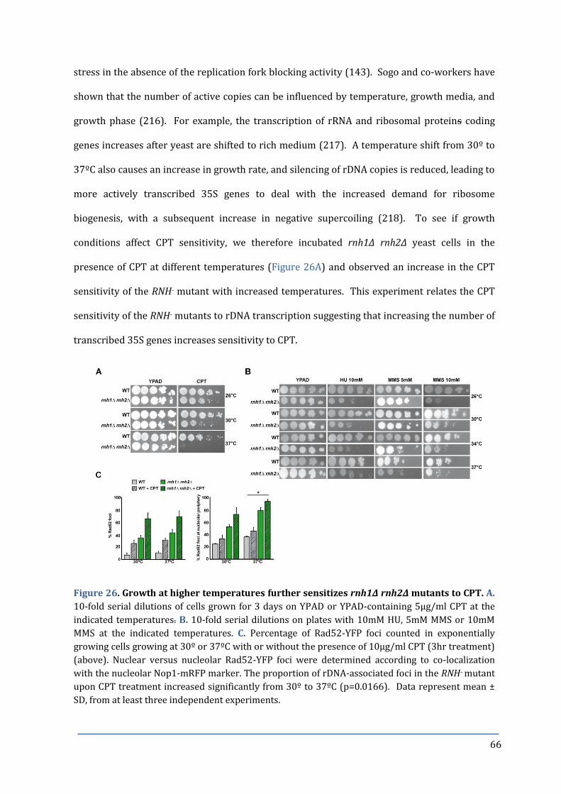
66
stress in the absence of the replication fork blocking activity (143). Sogo and co-workers have
shown that the number of active copies can be influenced by temperature, growth media, and
growth phase (216). For example, the transcription of rRNA and ribosomal proteins coding
genes increases after yeast are shifted to rich medium (217). A temperature shift from 30º to
37ºC also causes an increase in growth rate, and silencing of rDNA copies is reduced, leading to
more actively transcribed 35S genes to deal with the increased demand for ribosome
biogenesis, with a subsequent increase in negative supercoiling (218). To see if growth
conditions affect CPT sensitivity, we therefore incubated rnh1∆ rnh2∆ yeast cells in the
presence of CPT at different temperatures (Figure 26A) and observed an increase in the CPT
sensitivity of the RNH- mutant with increased temperatures. This experiment relates the CPT
sensitivity of the RNH- mutants to rDNA transcription suggesting that increasing the number of
transcribed 35S genes increases sensitivity to CPT.
Figure 26. Growth at higher temperatures further sensitizes rnh1∆ rnh2∆ mutants to CPT. A.
10-fold serial dilutions of cells grown for 3 days on YPAD or YPAD-containing 5µg/ml CPT at the
indicated temperatures. B. 10-fold serial dilutions on plates with 10mM HU, 5mM MMS or 10mM
MMS at the indicated temperatures. C. Percentage of Rad52-YFP foci counted in exponentially
growing cells growing at 30º or 37ºC with or without the presence of 10µg/ml CPT (3hr treatment)
(above). Nuclear versus nucleolar Rad52-YFP foci were determined according to co-localization
with the nucleolar Nop1-mRFP marker. The proportion of rDNA-associated foci in the RNH- mutant
upon CPT treatment increased significantly from 30º to 37ºC (p=0.0166). Data represent mean ±
SD, from at least three independent experiments.

67
To study whether the increase in CPT sensitivity with higher temperature was specific to CPT
or a general effect, similar drop test analyses were performed with the genotoxic agents MMS
and HU. Raising the temperature from 30º to 37ºC sensitized WT and rnh1∆ rnh2∆ mutants to
the DNA alkylating agent MMS but not to HU (Figure 26B).
Ide et al. previously demonstrated that sensitivity to MMS depends on transcription by RNA
Pol I and state that increased rDNA transcription may be toxic due to greater sensitivity to DNA
damaging agents (75). Therefore, we can speculate that higher RNA PolI transcription rates
are responsible for the increased sensitivity to MMS observed at 37ºC in an RNA:DNA hybrid
independent manner. Notably, growth at 30°C or 37°C did not change the number of cells with
damaged DNA as determined by Rad52-YFP foci formation in both the WT and rnh1∆ rnh2∆
cells (Figure 26C). However, growth at 37ºC caused a marked increase in the proportion of
Rad52-YFP foci that co-localized with the Nop1 nucleolar marker protein, suggesting an
increase in the proportion of rDNA-associated DNA damage. Thus, it is possible that increased
rDNA transcription exacerbates genomic instability of the rDNA locus at the higher
temperature.

68
DISCUSSION for CHAPTER 2
HR and PRR are Critical Pathways in Yeast Lacking RNaseH Activity
In Chapter 1 of this thesis we described how unprocessed R-loops could act as sites of
replication initiation, demonstrating a critical role for the RNaseH enzymes and Top1 in
preventing unscheduled TIR events and thus maintaining genomic stability. We then sought in
this Chapter, by genetic analysis, to identify activities needed for survival in the absence of
RNH. RNH double mutants display negative genetic interactions or increase the CPT sensitivity
of mutants affected in genes coding for proteins involved in NER, HR, and PRR pathways. HR
was identified as a critical pathway in yeast lacking RNaseH activity, since both Rad52 and
MRX-complex activity became essential even in the absence of exogenous DNA damage. These
observations suggest RNH- cells accumulate toxic DNA intermediates that are a substrate for
HR. R-loops could also be channeled into repair pathways that convert them into toxic DNA
intermediates. Yet, it remains to be determined if TIR-intermediates are HR dependent. HR
could contribute to the formation or resolution of TIR-intermediates and it would be
interesting to test for this possibility, for example by the use of Rad52- or Rnh2-degron
constructs. Surprisingly, RNH- cells were still viable in the absence of Rad51 (see Figure 25),
the bacterial RecA homologue, which binds to ssDNA and catalyzes the search for and strand
invasion of homologous sequences (219). Importantly, we detected bubble-shaped replication
intermediates in rad51∆ rnh1∆ rnh2∆ mutants suggesting that TIR is Rad51-independent.
However, Rad51 seems to be essential in the repair of Top1-mediated DNA lesions, seen by the
acute CPT sensitivity of rad51∆ mutants (Figure 25 and (66)). It is therefore perhaps
surprising that we did not detect more TIR molecules in the rad51∆ background. This result
would suggest that TIR is not initiated by DNA repair of a Top1cc but rather due to the lack of
Top1 activity, perhaps due to an increase in torsional stress.

69
As described in Chapter 1 (Figure 23) two alternative mechanisms could account for TIR,
including a direct assembly of the replication machinery at the R-loop, or by recombination-
dependent replication. TIR appears to be a post-replicative event, as bubble arcs were only
observed in cells held in late S/G2 phase of the cell cycle, and apart from RAD52, genetic
analyses revealed that POL32 is essential for the viability of RNH- yeast. Rad52 and Pol32 are
required for break-induced replication (BIR), a mechanism of replication-dependent repair
(220). BIR was originally studied in prokaryotes (where it is referred to as recombination-
dependent repair (RDR)) as a mode of oriC-independent chromosomal replication, where the
processing of arrested replication forks into a DSB led to replication restart (reviewed in
(221)). In the BIR described in yeast, a DSB with only one free end initiates repair by strand
invasion of the 3´end into an intact homologous duplex DNA, priming DNA replication to the
chromosome end (reviewed in (199)). The complex lesion of a Top1cc bound to the 3´ end of a
DSB could potentially explain why there is only one free end available for strand invasion.
Rad52-mediated strand invasion could occur by annealing between the single-stranded strand
and the open and stable R-loop structure, in the absence of Rad51 (222), perhaps assisted by
Rad59 activity (223). According to Lydeard et al.: “Two of the major questions regarding the
DNA synthesis steps of BIR are: 1. What DNA helicases are responsible for unwinding the template
DNA? 2. How is the replication fork established in the absence of an origin?” (220). In our
opinion, R-loops could open up the DNA, particularly in combination with a loss of Top1
activity and associated increase in torsional stress, and provide a starting point for replication,
as described for prokaryotes, thereby offering a solution to both of these questions.
Mrc1 is important for viability in the absence of RNaseH activities
The observation that RNH- mutants require a functional MRC1-complex to combat CPT toxicity
suggests that replication fork stability may be important for the fitness of yeast lacking RNaseH
activity. A functional MRC1-complex is important for both cell viability and CPT resistance in
the absence of RNaseH activities. This was a rather surprising observation given that single

70
mutants of the MRC1 complex do not display sensitivity to CPT. Mrc1, together with Csm3 and
Tof1, exists as an integral member of the replisome (224), maintaining the stability of stalled
RFs and promoting subsequent DNA repair events by activation of Rad53-dependent intra S
replication checkpoint (109). These observations made it likely that RNA:DNA hybrids not
only constrain genome stability but also interfere with cell cycle progression (for further
results and discussion on the MRC1-complex see Chapter 3). Importantly, TIR-mediated
replication intermediates were evident in CPT treated mrc1 triple mutants, suggesting that TIR
events do not constrain the viability of these cells. R-loops could hinder replication fork
progression leading to replication fork break-down. The Sgs1 and RecQ-like helicases have
been suggested to play roles in maintaining lagging strand polymerases at stalled forks (225)
via interactions with the ssDNA binding complex, RPA (226). The differences observed in the
CPT sensitivity between sgs1 and top3 triple mutants, may suggest that the Top3-independent
RF stability function of Sgs1 may be important in response to CPT treatment in the RNH-
mutants, however, we have not investigated further the roles of Sgs1 in yeast lacking RNaseH
activity. Rrm3 belongs to the Pif1-class of helicases and is needed for the bypass of protein-
DNA interaction sites, and whose absence leads to the accumulation of paused replication forks
in the rDNA (147). In vitro studies showed that Pif1 is able to resolve RNA:DNA hybrids (57),
but notably, pif1-m2 triple mutants are viable and CPT resistant suggesting that, at least for
ribosomal DNA, Pif1 is dispensable for RNA:DNA hybrid processing in vivo. Interestingly,
mutations in the E.coli mutation Pif1 homologue recG are synthetic lethal in combination with
mutation to rnhA (46). Recent reports suggest a role for Pif1 in BIR (227), and therefore the
difference in the requirement for Pif1 and Pol32 activity may reflect the need for BIR for R-loop
tolerance. Mutations to Pol32 specifically affected the rDNA array with no detectable
alterations to other chromosomes (228), suggesting that Pif1-independent but Pol32-
dependent BIR may be of particular relevance for stability of the rDNA region of the genome.

71
Loss of Rnh2 Activity is more detrimental than Loss of Rnh1
We find that the absence of Rnh2 activity generally had a greater contribution to viability and
CPT sensitivity in combination with DNA replication and/or DNA repair mutants, than the
absence of Rnh1. For example 39% of mre11Δ rnh2Δ, 50% of rnh2Δ rad50Δ and 33% of rnh2Δ
xrs2Δ mutants were inviable, while 100% of the corresponding rnh1 double mutants were
viable and only affected in growth. The same was true for cells lacking Srs2, where 96% of the
srs2Δ rnh2Δ double mutants were inviable, but 100% of the srs2Δ rnh1Δ double mutants were
found to be viable. These results emphasize that although Rnh1 and Rnh2 have overlapping
specificities in removing transcription-associated R-loops, Rnh2 has additional functions,
including the removal of misincorporated rNMPs that contribute to genomic stability (14).
Interestingly, deletion of RNH1 was reported to be more deleterious than the deletion of
RNH201 in combination with defects on RNA biogenesis factors (21). It may be possible to take
advantage of the Rnh201 P45D-Y219A separation-of-function mutant that can remove R-loops
but not rNMPs to see if impaired rNMP repair would still constrain viability in the absence of
Top1 (229). The lack of Rnh201 may have additive effects, weakening protein interactions of
its accessory subunits, Rnh202 and Rnh203, with other factors. For example, Rnh202 has been
reported to interact with the cyclin-dependent kinase Cak1, that has roles in passage through
the G2/M stages of the cell cycle; and the replication fork clamp, proliferating cell nuclear
antigen complex (PCNA) (12), the sliding clamp for DNA Polδ and an interaction site for other
replication and repair proteins (for a review see (230)). PCNA itself is responsible for the
recruitment of PRR (231), NER (232), and cohesion (233) factors. In addition, PCNA interacts
recruits Srs2 (234), playing a role in the repair of replicative damage by directing the repair of
stalled RFs away from HR and into translesion synthesis (TLS) and template switching
pathways, both shown to be important repair pathways for yeast lacking RNaseH activities
(99).

72
Persistent R-loops may impede TCR
Rad16, together with Rad7, has a role in both global genomic repair (GGR) and TCR. TCR-
defects can only be detected in the absence of either repair factor (see (89) for a recent review
on TCR). Interestingly the rad16 triple mutant was found to be more sensitive to CPT than the
RNH double mutant. This result would suggest that yeast have defective TCR in the absence of
RNaseH activity, and TCR may be particularly impeded at the rDNA locus. We would like to
confirm this observation in a rad7 triple mutant, as it would suggest that persistent R-loops
impede RNA Pol progression through transcribed regions and could lead to more lesions than
TCR can deal with.
Nucleolar Function Affects CPT Sensitivity and Viability
Ribosomal DNA function was shown to have a major influence on CPT sensitivity in Chapter 1,
and in this chapter we analysed many different factors linked to rDNA organisation and
transcription. We observed the reproducible pattern that a disruption of rDNA organisation
enhanced CPT sensitivity of RNH- mutants, whereas the down-regulation of RNA PolI
transcription alleviated CPT sensitivity. Together these results confirm that proficient
transcription of the rDNA locus is crucial for CPT-mediated genotoxicity and reveal the rDNA
as a major contributory factor of CPT-mediated DNA damage.
As the rDNA constitutes such a large part of the eukaryotic genome, it is perhaps not surprising
that RNA PolI-mediated R-loops in the rDNA can have such a major impact on cell viability. In
Chapter 3 of this thesis we will investigate in more detail by what means viability is
constrained in the RNH- mutants.

73
RESULTS
CHAPTER 3 – Yeast lacking RNaseH activity exhibit altered cell cycle
progression
RNH lacking cells suffer from premature S-phase entry
Cell cycle analysis of rnh1∆ rnh2∆ yeast revealed that the RNH double mutant consistently
showed a much faster S-phase transition than the WT following release from α-factor
synchronization (Figure 27A, best observed at the 30 minute time point). This highly
reproducible, premature progression through S-phase was observed in both BAR1+ and bar1∆
strains, and was CPT independent.
Figure 27. Yeast lacking RNase H activity show a premature S-phase transition. A. Flow cytometry analysis of strains following release from α-factor in YPAD. B. Representation of the S.cerevisiae cell cycle with approximate time of activity for different cyclins and checkpoint factors. C. Genetic analysis of sic1∆ rnh1∆ rnh2∆ and clb5∆ rnh1∆ rnh2∆ tetrads.
An earlier activation of standard origins of replication, leading to a precipitated G1/S
transition, would make the S-phase appear “shorter”. CDC28 is an essential gene, encoding the
catalytic subunit of the main CDK factor, which acts as the major coordinator of the yeast cell
cycle (reviewed in (235)). The activity of Cdc28 is controlled post-translationally via
associations with many different regulators throughout the cell cycle. During G1, Sic1 binds to
and inhibits the CDK Cdc28 (Figure 27B). Cells are able to enter S-phase and begin DNA
74
replication by phosphorylating Sic1, which targets it for ubiquitin-mediated degradation (236),
thus permitting Cdc28 activity and the G1 to S-phase transition. Mutants in yeast sic1 exhibit
premature entry into S-phase due to unusually high Cdc28 activity (237). We studied the
interaction of RNH- mutants with the cyclin-dependent kinase inhibitor (CKI) SIC1 to examine
the possibility that yeast lacking RNaseH activity present a faster transition through S phase
due to an accelerated entry into S phase. Interestingly, we revealed synthetic lethal
interactions between the RNH double mutant and sic1Δ by genetic analysis (Figure 27C) and
furthermore, deletion of sic1 was lethal in combination with the rnh1∆ or rnh2∆ simple
mutants in 33% and 25% of the cases, respectively. Also of interest was the observation that
the RNH double mutant is synthetically sick with the B-type cyclin Clb5 (Figure 27C). Like Sic1,
Clb5 also acts to regulate Cdc28 levels and promotes the initiation of DNA synthesis, however,
contrary to mutation of sic1, loss of clb5 results in an extension of the S-phase due to the
inability to activate late origins of replication (238). Collectively, these results suggest that
rnh1∆ rnh2∆ mutants enter early into S-phase, and the G1 to S-phase transition activity of Sic1,
and to a lesser extent Clb5, is essential to maintain the viability of cells in the absence of
RNaseH activity.
An earlier activation of standard origins of replication, leading to a precipitated G1/S
transition, would make the S-phase appear “shorter”. CDC28 is an essential gene, encoding the
catalytic subunit of the main CDK factor, which acts as the major coordinator of the yeast cell
cycle (reviewed in (235)). The activity of Cdc28 is controlled post-translationally via
associations with many different regulators throughout the cell cycle. During G1, Sic1 binds to
and inhibits the CDK Cdc28. Cells are able to enter S-phase and begin DNA replication by
phosphorylating Sic1, which targets it for ubiquitin-mediated degradation (236), thus
permitting Cdc28 activity and the G1 to S-phase transition. Mutants in yeast sic1 exhibit
premature entry into S-phase due to unusually high Cdc28 activity (237). We studied the
interaction of RNH- mutants with the cyclin-dependent kinase inhibitor (CKI) SIC1 to examine
the possibility that yeast lacking RNaseH activity present a faster transition through S phase

75
due to an accelerated entry into S phase. Interestingly, we revealed synthetic lethal
interactions between the RNH double mutant and sic1Δ by genetic analysis (Figure 27C).
Deletion of sic1 was also lethal in combination with the rnh1∆ or rnh2∆ simple mutants in 33%
and 25% of the cases, respectively, indicating a significant interaction between RNaseH activity
and Sic1. Also of interest was the observation that the RNH double mutant is synthetically sick
with the B-type cyclin Clb5 (Figure 27C). Like Sic1, Clb5 also acts to regulate Cdc28 levels and
promotes the initiation of DNA synthesis, however, contrary to mutation of sic1, loss of clb5
results in an extension of the S-phase due to the inability to activate late origins of replication
(238). Collectively, these results demonstrate that rnh1∆ rnh2∆ mutants enter early into S-
phase, and the G1 to S-phase transition activity of Sic1, and to a lesser extent Clb5, is essential
to maintain the viability of cells in the absence of RNaseH activity.
R-loop formation partially overcomes cdc7-4 temperature sensitivity
The initiation of DNA replication in eukaryotic cells during S phase is regulated by 'origin
licensing', and requires the sequential assembly of pre-RC proteins, such as Cdc7 and Cdc6, at
the ARS. Cdc6 is an essential component of the pre-RC and acts prior to Cdc7 kinase activation,
loading the Mcm2-7 proteins onto the ORC (239,240). Firing from normally inactive origins, or
the formation of entirely new sites of replication initiation could account for the completion of
DNA synthesis more quickly in the absence of RNaseH activity. We questioned whether the
origin-independent, R-loop-mediated replication initiation (see Chapter 1), could by-pass the
need for canonical origin firing. To address this question we created a cdc7-4 rnh1∆ rnh2∆
triple mutant (Figure 28A). The cdc7-4 allele permits growth at 23ºC (permissive
temperature) but not at 30ºC (4) because the Cdc7 kinase is essential for the opening up and
firing of replication origins by phosphorylation of Mcm2-7 proteins (4,241). Notably,
restrictive temperature was shifted from 30°C to 37ºC in the cdc7-4 rnh1∆ rnh2∆ triple mutant,
indicating that R-loops could help to make replication origins more accessible to replication
factors. Additionally, the CPT sensitivity of cdc7-4 rnh1∆ rnh2∆ mutants was comparable to

76
rnh1∆ rnh2∆ mutants at 23ºC, but this sensitivity was increased dramatically at 30ºC (Figure
28B). Interestingly, transcription through origins of replication has been shown to inactivate
replication firing (242). It is conceivable, that R-loop stabilization within replication origins by
CPT could have the same effect.
Figure 28. R-loop formation partially overcomes cdc7-4 temperature sensitivity. A. Viability
of cdc7-4 simple and triple mutants grown at 23º, 30º, or 37ºC. B. Drop test analysis of cdc7-4
rnh1∆ rnh2∆ at 23º or 30º on YPAD or YPAD-containing CPT (5µg/ml). C. Tetrads (top) and drop
test analysis for cdc7∆ mcm5-bob1 quadruple mutant. Plates contained 0.5 or 1µg/ml CPT.
Besides its well-reported roles in replication initiation, Cdc7 also operates in post-replicative
repair (PRR). Cdc7 is a member of the DNA damage tolerance RAD6 epistasis group, associated
with the TLS branch (243). It has been proposed that different cdc7 alleles can result in hyper-
or hypo-mutagenic phenotypes (244). To exclude the possibility that the PRR function of Cdc7
was interfering in the analysis of viability of the cdc7-4 rnh1∆ rnh2∆ mutant we opted to use a
cdc7∆ mcm5-bob1 strain. The function of Cdc7 in replication initiation is no longer essential in
a mcm5-bob1 (P83L) mutant (245), since presence of the mcm5-bob1 allele bypasses the need
for the phosphorylation and activation of the MCM helicase by Cdc7 kinase (246). Analysing
the viable spores from genetic crosses between the cdc7∆ mcm5-bob1 and rnh1∆ rnh2∆ yeast
strains, we attained cdc7∆ mcm5-bob1 rnh1∆ rnh2∆ quadruple mutant spores (Figure 28B).

77
We confirmed the mcm5-bob1 genotype by back-crossing the quadruple spore with a WT yeast
strain: the ability to recover viable cdc7∆ spores from the back-cross meant that the yeast also
carried the mcm5-bob1 allele, originating from the quadruple spore from the first cross (results
not shown). These analyses tell us that cdc7∆ rnh1∆ rnh2∆ yeast are inviable without the
compensatory mcm5-bob1 allele, and that Cdc7 kinase activity is essential in the RNH- mutant,
as for the WT. Therefore, the origin-independent transcription-initiated replication events that
we observe in the RNH double mutant do not bypass the need for normal replication initiation
from origins in S-phase.
RNH- mutants are not held in G2/M in the absence of Mrc1 activity
The synthetic lethal interaction of RNH- mutants and SIC1 suggested that cells may suffer from
constrains that may be generated during replication and still be present when cells are in the
next G1 phase. To assess for replication-associated DNA damage, we inactivated different S-
phase specific checkpoint pathways including rad9, chk1, tel1, and rad24. No significant
differences were observed in the cell cycle progression of the triple mutants tested; all the
triple mutants exhibited a similar holding of cells in G2/M phase as per the RNH double mutant
(data not shown), although they showed an additive CPT sensitivity (see Chapter 2, Table 1).
Interestingly, we detected a synthetic lethal interaction of RNH- mutants with MRC1 (see
Chapter 2; Figure 24). Mrc1, mediator of the replication checkpoint, the homologue of human
Claspin, associates with replication forks shortly after replication initiation, remaining during
elongation as an integral part of the replication machinery (224) and has been shown to have a
dual role, acting both as a component of the RF and as a mediator of the S/G2 checkpoint (247).
Viable mrc∆ rnh1∆ rnh2∆ triple mutants notably manifested a different cell cycle progression
profile to that of the RNH double mutant. The mrc1Δ rnh1Δ rnh2Δ triple mutant was able to re-
enter G1-phase of the following cell cycle whilst the RNH double mutant remained held in late
S/G2 (Figure 29A). Moreover, the mrc1Δ rnh1Δ rnh2Δ yeast exhibited an even more
accelerated entry into and passage through the S-phase, compared to the RNH double mutant.

78
Furthermore, of significant interest was the finding that Mrc1 is important for cell survival in
the absence of RNaseH enzymes. From tetrad analysis we detected that only 33% of triple
spores were viable (Figure 29B). Remaining viable triple mutant spores have a growth defect,
and interestingly, whilst the mrc1Δ itself is not CPT sensitive, elimination of MRC1 in the RNH
double mutant resulted in a highly CPT sensitive triple mutant (Figure 29C). These results
suggest that Mrc1 activity is crucial in cells lacking RNaseH activity and can protect them from
CPT-induced replication stress.
Figure 29. Mrc1 protects cells from CPT-induced replication stress. A. Flow cytometry analysis of mrc1 triple mutant cells grown in the presence or absence of CPT following release from α-factor. B. Tetrad analyses of Mrc1 separation-of-function alleles (top) and Mrc1 mediator complex members (bottom). C. Drop test analysis of Mrc1 separation-of-function alleles (top) and Mrc1 mediator complex members (bottom). D. Flow cytometry analysis of Mrc1 mediator complex members.
Since mrc1∆ rnh1∆ rnh2∆ triple mutants did not arrest in late S/G2-phase in response to CPT
(Figure 29A), their increased CPT sensitivity could reflect a problem in S/G2-dependent
damage repair or RF stability. To discriminate between these possibilities, we took advantage
of the Mrc1 separation of function mutants. Combination of the checkpoint-defective allele
mrc1AQ, which cannot be phosphorylated by the checkpoint kinases (248), and the RF stability
mutant allele, mrc1-c14 (249), with the RNH double mutant, resulted in an increased CPT

79
sensitivity. These results suggest that both S/G2-checkpoint and RF stability functions of Mrc1
contribute to CPT tolerance in the rnh1Δ rnh2Δ background. In contrast, only the triple mutant
with the RF stability mutant allele, mrc1-c14, closely resembled the growth, CPT sensitivity and
spore viability phenotypes of the mrc1∆ rnh1∆ rnh2∆ mutant (Figures 29B and 29C). The
mrc1-c14 triple mutant spores were affected negatively in growth with some lethality observed
in meiotic segregants (87% viability of spores corresponding to triple mutants). Collectively,
these results demonstrate that Mrc1 plays an important role in the stabilization of RFs in the
RNH- mutant, and may be particularly important for the survival of CPT-induced lesions, and
that both functions of Mrc1 are indispensable for the viability of yeast lacking RNaseH activity.
Mrc1 exists as an integral member of the replisome (224), with Csm3 and Tof1, as part of the
Mrc1-mediator complex. Csm3 and Tof1 are specifically required for the association of Mrc1
with the RF, interacting directly with the MCM helicase, and thus play a central role in RF
progression. The complex is also important in maintaining the stability of stalled RFs and
promoting subsequent DNA repair events (224,247). Genetic interaction analysis revealed
novel functional relationships between the RNaseH enzymes and all members of the Mrc1
mediator complex (Figure 29B). Triple mutants of rnh1Δ rnh2Δ with csm3∆ and tof1∆ were
synthetic sick; triple mutants also exhibited extreme sensitivity to CPT, in contrast to the CPT
resistant csm3∆ and tof1∆ single mutants (Figure 29C). Cell cycle progression of the tof1∆
triple mutant, upon release from G1 synchronization into media in the presence of CPT, was
very similar to that previously observed for the mrc1∆ triple mutant (Figure 29D). In contrast,
the csm3∆ triple mutant remained held in late S/G2, as for the RNH double mutant, suggesting a
possible difference in functions for individual members of the complex in the absence of
RNaseH activity. These results indicate that the replication fork stabilization function of Mrc1
(together with Tof1) is important in protecting cells from R-loop mediated replication
constrains and, similar to its role in the response to osmostress (250), support a role for Mrc1
in the co-ordination of transcription and replication events in CPT treated RNH- mutants.
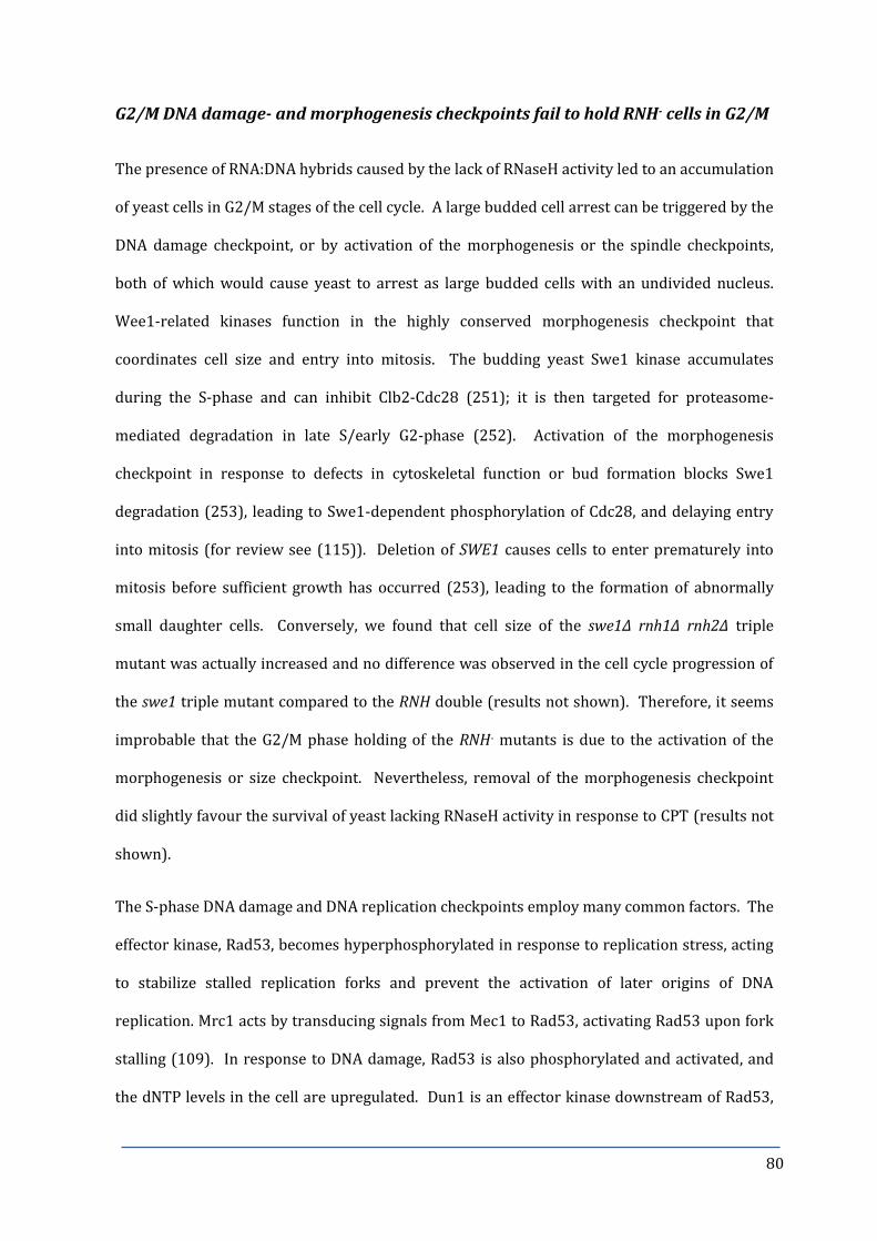
80
G2/M DNA damage- and morphogenesis checkpoints fail to hold RNH- cells in G2/M
The presence of RNA:DNA hybrids caused by the lack of RNaseH activity led to an accumulation
of yeast cells in G2/M stages of the cell cycle. A large budded cell arrest can be triggered by the
DNA damage checkpoint, or by activation of the morphogenesis or the spindle checkpoints,
both of which would cause yeast to arrest as large budded cells with an undivided nucleus.
Wee1-related kinases function in the highly conserved morphogenesis checkpoint that
coordinates cell size and entry into mitosis. The budding yeast Swe1 kinase accumulates
during the S-phase and can inhibit Clb2-Cdc28 (251); it is then targeted for proteasome-
mediated degradation in late S/early G2-phase (252). Activation of the morphogenesis
checkpoint in response to defects in cytoskeletal function or bud formation blocks Swe1
degradation (253), leading to Swe1-dependent phosphorylation of Cdc28, and delaying entry
into mitosis (for review see (115)). Deletion of SWE1 causes cells to enter prematurely into
mitosis before sufficient growth has occurred (253), leading to the formation of abnormally
small daughter cells. Conversely, we found that cell size of the swe1∆ rnh1∆ rnh2∆ triple
mutant was actually increased and no difference was observed in the cell cycle progression of
the swe1 triple mutant compared to the RNH double (results not shown). Therefore, it seems
improbable that the G2/M phase holding of the RNH- mutants is due to the activation of the
morphogenesis or size checkpoint. Nevertheless, removal of the morphogenesis checkpoint
did slightly favour the survival of yeast lacking RNaseH activity in response to CPT (results not
shown).
The S-phase DNA damage and DNA replication checkpoints employ many common factors. The
effector kinase, Rad53, becomes hyperphosphorylated in response to replication stress, acting
to stabilize stalled replication forks and prevent the activation of later origins of DNA
replication. Mrc1 acts by transducing signals from Mec1 to Rad53, activating Rad53 upon fork
stalling (109). In response to DNA damage, Rad53 is also phosphorylated and activated, and
the dNTP levels in the cell are upregulated. Dun1 is an effector kinase downstream of Rad53,

81
whose activation leads to an up-regulation in the transcription of the ribonucleotide reductase
(RNR) genes. The RNR complex catalyzes the rate-limiting step in the synthesis of dNTPs from
NTPs, a process essential for both DNA repair and for normal replication. In addition, Sml1, an
inhibitor of RNR, is degraded in response to DNA damage (254). Sml1 is the substrate for
Dun1, making Dun1 directly responsible for Sml1 phosphorylation and its subsequent
degradation (255). Analysis of genetic interactions revealed that the triple mutant with dun1
resulted in a synthetic sick interaction (see Chapter 2; Table 1), yet no difference was observed
in the CPT sensitivity of the triple mutant as compared to the RNH double mutant; the sml1
triple was also as sensitive to CPT as the RNH double (results not shown). The observation that
triple mutants are not more sensitive to CPT than the RNH double mutant could indicate an
epistatic interaction between the RNaseH enzymes and control of cellular dNTP levels.
Detection of the “active” phosphorylated form of Rad53 by Western blot is widely used to
follow S-phase checkpoint activation. Hyper-phosphorylated Rad53 can be detected as a shift
in the mobility of the 92 kDa band corresponding to Rad53 on a protein gel. We examined
whether Rad53 is activated in RNH- mutants by performing a Western blot against the
phosphorylated form of Rad53 (Figure 30, upper panel). We observed that HU treatment of
WT yeast cells caused a positive shift in Rad53 mobility related to phosphorylation and thus
checkpoint activation. In contrast to cellular treatment with other DNA damaging agents, CPT-
induced DNA lesions are not sensed by the intra-S-phase checkpoint protein Rad53 (256) and
accordingly, we see no activation of Rad53 for the WT upon CPT treatment. However, we did
note a minimal shift in the mobility of the Rad53 bands for the simple mutants rnh1Δ and
rnh2Δ in response to CPT treatment, and a slight shift for the CPT treated rnh1Δ rnh2Δ double
mutant.

82
Figure 30. Western analysis against Rad53 (top) and Rad53 in situ kinase assay (ISA; below). HU
treatment was used as positive and hexokinase (Hxk1) as loading control. Experiments performed
by Nestor García Rodríguez.
To confirm whether CPT treatment of the RNH- mutants is able to activate the replication
checkpoint we monitored Rad53 kinase activation directly using the Rad53 in situ kinase assay
(ISA; (257)) (Figure 30, lower panel). We saw a weak Rad53 autophosphorylation in response
to CPT-treated WT, rnh1Δ, rnh2Δ and rnh1Δ rnh2Δ yeast, albeit to lower levels than following
HU treatment of the WT. These results support the previously described minor
phosphorylation of Rad53 for the rnh1Δ rnh2Δ double mutant following HU or MMS treatment
(99) and suggest that CPT leads to an increased replicative stress in the RNH- mutants.
However, the threshold of hyper-phosphorylation of Rad53 necessary to trigger the S-phase
DNA damage checkpoint is not reached, as the RNH double mutant, even following CPT
treatment, shows no delay in S-phase progression.
The degradation of cyclin Clb2 is delayed in RNH- mutants
The protein levels of Sic1 following release from α-factor were monitored in yeast lacking
RNaseH activity. Sic1 multiple phosphorylation and subsequent degradation is essential for
the G1/S transition and the initiation of DNA replication. The timing of Sic1 degradation for
the double mutant seemed similar to the WT (Figure 31A), and thus, would not account for the
accelerated S-phase progression in our RNH- mutants. However, the RNH double mutant
appeared to have lower levels of Sic1 throughout the cell cycle.

83
Figure 31. Western blot analysis of Sic1 and Clb2. A. Sic1 and Clb2 levels following release from α-
factor. α-factor was re-added 60 minutes after release. B. Release of yeast cells in the presence of
10µg/ml CPT. Western analyses performed by Marta Muñoz Barrera.
The B-type cyclins Clb1-4 are required for mitotic events, such as spindle morphogenesis, but
must be down-regulated for cytokinesis to take place (reviewed in (258)). Clb2 is
proteolytically degraded in early anaphase, necessary for the exit from mitosis and re-entry
into G1 of a new cell cycle (259). In response to microtubule destabilizing drugs, mitotic arrest
of WT cells is maintained by high levels of Clb2 and Cdc28 kinase activities (260). We
monitored the levels of Clb2 by Western blot during the cell cycle to investigate whether a
disregulation of Clb2 levels could account for the prolongation of cells in late G2/M-phase in
the RNH- mutant. Western blot analysis (Figure 31A) shows that Clb2 protein appears at 45
minutes post-release and is degraded from 135 minutes in the WT. However, in the double
mutant, Clb2 protein was detectable even in the G1 synchronized sample and remains present
until 210 minutes. The degradation of Clb2 is further delayed by treatment with CPT in both
the WT and the rnh1∆ rnh2∆, but in the case of the RNH- mutant, considerable levels of Clb2
protein can be observed throughout the complete time course (Figure 31B). These results may
help explain why rnh1∆ rnh2∆ cells are held in G2/M-phase, since they cannot exit mitosis due
to the high activity of Clb2. Additionally, this experiment reveals that RNaseH activity is
required for the normal fluctuation of Clb2 protein since the cyclin is observed during G1 and S
phases, when it is not normally active, and the levels of Clb2 remain elevated in the RNH-
mutant. These results are very similar to those published for yeast mutants in hct1 (CDH1), an
activator of the APC (261).

84
The CDK Cdc28 is the key cell cycle regulator, whose activity declines as mitosis is completed
due to the degradation of mitotic Clbs such as Clb2 and the accumulation of the G1-phase-
specific Cdc28 inhibitor Sic1 (reviewed in (262)). Prevailing levels of Clb2 at G2/M and low
levels of Sic1 throughout the cell cycle would explain why yeast cells lacking RNaseH activity
manifest abnormal cell cycle transitions at G2/M and G1/S respectively, and may be the reason
why RNH- mutants re-initiate DNA replication without having finished mitosis.
Nucleolar Cdc14 is constrained in RNH- mutants
The nucleolus is known to segregate after the nucleus, with the segregation of rDNA occurring
in late anaphase (263). During anaphase, yeast cells must condense and compact the long
rDNA array to ensure that segregation takes place before cytokinesis (264). We followed the
progression of RNH- mutants though meta- and anaphase monitoring spindle morphology with
tubulin staining. The percentage of cells in metaphase and anaphase was determined for the
RNH double mutant and found to be similar to the WT (Figure 32A). However, the rnh1∆ rnh2∆
cells entered slightly earlier into metaphase, and metaphase itself seemed slightly longer (by
15 minutes approximately), consistent with earlier observations by flow cytometry of RNH-
cells rushing through the S phase into G2/M.
The action of Cdc14 is required for rDNA segregation as it is necessary for the localization of
the condensin complex to rDNA (265). Cdc14 is regulated by the inhibitory protein Net1, both
being members of the RENT complex (for regulator of nucleolar silencing and telophase),
which also contains Sir2 with important roles in transcriptional silencing of the rDNA locus.
Net1 binding of Cdc14 maintains Cdc14 inactive in the nucleolus during the cell cycle. Net1
only releases Cdc14 during anaphase, promoted by the FEAR (CDC Fourteen Early Anaphase
Release) and MEN (Mitotic Exit Network). Net1 release allows active Cdc14 to
dephosphorylate its downstream targets and stimulate the exit from mitosis and cytokinesis

85
(266). Of particular interest to this study, Cdc14 activity in anaphase also leads to the
dephosphorylation and thus stabilization of Sic1 (267).
Figure 32. Reduced nucleolar Cdc14 is released in RNH- mutants. A. Percentages of metaphase
and anaphase cells as determined by spindle (tubulin) morphology are shown for each timepoint.
B. Representative images of 3HA-Cdc14 subcellular localization. Cdc14 is shown in red,
microtubules (tubulin) are shown in green and DNA (DAPI) is shown in blue. C. Percentage of cells
with 3HA-Cdc14 sequestered, partially released, or fully released for the WT and rnh1∆ rnh2∆ cells
(left panel). At least 200 cells were counted at each timepoint. Size bar represents 10µm.
To address if the timing of Cdc14 release from the nucleolus was affected in RNH- mutants we
investigated the localization of Cdc14 during the cell cycle following release from G1
synchronization. We first crossed rnh1∆ rnh2∆ yeast with a 3HA-tagged Cdc14 bearing strain
and confirmed that the HA tag did not affect either CPT sensitivity or cell cycle progression of
the RNH double mutant (data not shown). Next, cells were released from α-factor block in G1
and samples were fixed every 15 minutes (α-factor was re- added after 45 min to prevent re-
entry into the next cell cycle) and the localization of Cdc14 was monitored by
immunofluorescence in relation to spindle length. Cdc14 localization was recorded as
sequestered in the nucleolus, partially released into the nucleus, or fully released and
cytoplasmic (see Figure 32B for an example of each). Cdc14 was released from the nucleolus of
rnh1∆ rnh2∆ cells with approximately the same kinetics as the WT (Figure 32C). However,

86
fewer cells were counted with partially- and fully-released Cdc14 in the RNH- mutant. This
result could indicate that the FEAR (or MEN) pathway was not fully activated in the absence of
RNaseH activity. Perhaps a critical level of Cdc14 release is necessary to trigger mitotic exit
and this level is not reached in yeast lacking RNaseH activity.
RNH- mutants do not respond to the spindle assembly checkpoint (SAC)
The apparent delay in G2/M exit of the RNH double mutant could be a consequence of
activation of the spindle assembly checkpoint (SAC) which, through Mad1 and Mad2 activity,
delays the onset of anaphase in cells with defects in mitotic spindle assembly (for a recent
review see (268)). To investigate whether activation of the SAC was responsible for holding of
the RNH- mutants in late S/G2 we deleted the mitotic spindle checkpoint gene MAD1 in rnh1∆
rnh2∆. The triple mutant was viable, and CPT sensitivity at the level of viability and cell cycle
progression (results not shown) were very similar to the double mutant. Spindle checkpoint
mutants, such as MAD (Mitotic Arrest Deficient) mutants, are sensitive to the microtubule
destabilizing drug benomyl. Consequently, we decided to analyse the benomyl sensitivity of
the rnh1∆ rnh2∆ double and mad1∆ triple mutant (Figure 33A). The RNH double and the mad1
triple mutant were as sensitive to benomyl as mad1∆. Such epistasis would imply that the
RNaseH enzymes and Mad1 are found in the same pathway, implying that the SAC is defective
in these mutants. Nevertheless, the benomyl sensitivity phenotype alone is not sufficient to
classify a mutant as checkpoint defective.
Treatment with either benomyl or nocodazole (Noc), another microtubule destabilizing drug
that activates the SAC, causes an interference of microtubule polymerization and attachment to
kinetochores, such that cells are unable to progress into mitosis (269,270). Cells treated with
Noc arrest with a 2N DNA content at the G2/M phase of the cell cycle, indicating that
replication is complete but chromosome separation has not yet taken place. SAC mutants,
such as the mad mutants, re-enter G1 of the next cell cycle with concomitant initiation of

87
replication. Given that the mutants are unable to complete mitosis correctly, further rounds of
replication can lead to a higher than 2N DNA content that can be easily monitored by flow
cytometry. In addition, the re-initiation of DNA replication without appropriate mitosis of the
previously replicated genetic material can be observed by microscopy as the generation of new
buds or “re-budding”. To elucidate whether yeast lacking RNaseH activity bypass the SAC we
analysed the RNH double mutant by FACS and microscopy following a prolonged incubation
with Noc (Figure 33B). Cells were first synchronized with α-factor before release into Noc-
containing medium and growth for 2 or 4 hours in continual presence of the drug (with re-
addition of Noc every 90 minutes). WT cells treated with Noc exhibit a dumbbell phenotype of
large budded cells, characteristic of G2/M arrest, and following 4h incubation just 10% of G2-
arrested cells had more than one bud. In contrast 29% of large budded rnh1∆ rnh2∆ cells
manifested the re-budded phenotype after 2h, and after 4h of continual Noc incubation 37% of
rnh1∆ rnh2∆ cells were re-budded (Figure 33B), with more than half of these G2/M cells having
more than one re-bud. The peak of cells corresponding to a 2N DNA content (Figure 33B, black
arrow), was shifted slightly to the right for the RNH- mutant, which would infer that the
majority of cells lacking RNaseH activity had a DNA content between 2- and 3N. Furthermore,
a small group of cells with a cellular DNA content of 3-4N was observed in the RNH double
mutant using flow cytometry (Figure 33B, blue arrow), further evidence of the re-initiation of
DNA replication in these mutants. The percentage of re-budded cells was not as high for the
RNH double mutant as for the mad1 simple mutant, and the mad1 triple mutant showed an
additive effect in the proportion of re-budded cells and loss of viability.

88
Figure 33. RNH- mutants do not respond to the spindle assembly checkpoint. A. Benomyl sensitivity drops at 5 and 10µg/ml. B. Representative microscope images of “re-budding observed in rnh1∆ rnh2∆ cells in G2/M following 4h Noc treatment (top). Size bar represents 10µm. Analysis of replication by flow cytometry following 4h Noc treatment (bottom). Black arrow showing population of cells with 2N DNA content; 2N content indicated by dotted line. Red arrow represents < 1N DNA content; blue arrow represents > 2N DNA content. Percentage of G2 cells re-budded (right).
To confirm whether the SAC was indeed activated in yeast lacking RNaseH activity we
monitored the phosphorylation of Mad1, since Mad1 is hyperphosphorylated when the SAC is
activated. Utilizing a GFP-tagged Mad1 strain we were unable to observe
hyperphosphorylation of the Mad1 protein in the RNH double mutant by Western analysis
(results not shown). Collectively, these results imply that yeast lacking RNaseH do not respond
to the SAC and continue to replicate their DNA although chromosome separation has not yet
been completed.
R-loops are responsible for chromosome segregation defects
To further monitor chromosome segregation in the RNH- mutants, we employed clamped
homogeneous electrical field (CHEF) analysis, to separate replicating chromosomes. The yeast

89
genome contains 16 linear chromosomes, ranging from approximately 200kb to 2.2Mb.
Chromosomes with branched DNA structures, such as replication forks and recombination
intermediates, cannot migrate into the gel and remain trapped in the wells. Following alpha-
factor synchronization, and release with or without CPT, we collected and embedded cells in
low-melting agarose plugs at various timepoints. Replicating chromosomes were separated
using CHEF analysis and then probes against two different chromosomes were applied (cV and
cXII) to look at the replication and segregation of these chromosomes. We quantified linear
molecules that are able to migrate into the gel (signal b, Figure 34A) and include fully
replicated DNA, versus non-linear chromosomes that cannot migrate due to their branched
structures and remain stuck in the well (signal a) and would include replication and
recombination intermediates, (271,272). For the quantifications, the signal corresponding to
time 0 (cells held in G1) was set as 100%, as one would expect no replication of DNA in G1
synchronized cells (nor recombination). A high bar in the quantifications corresponds to more
signal in the gel and, therefore, more fully replicated molecules (Figure 34B).
Following release from G1, a signal can be detected in the well for WT at 30 minutes for
chromosome V, as chromosomes are beginning replication; this signal would include
replication bubbles, and replication forks. Through 60 to 90 minutes the signal in the gel is
increasing, until the 120 minute timepoint where we observe the majority of the signal in the
gel rather than the well, as chromosomes V and XII are fully replicated and resolved. At 150
minutes, for cV of the WT we can observe a second round of replication, with some signal once
again in the well. The pattern for cXII of the WT is similar, although the appearance of the
signal in the gel is slightly detailed. This result is compatible with the observation that the
rDNA locus, located on cXII, is one of the last chromosome regions to be fully replicated and
resolved (263). However in the presence of CPT, less signal is observed in the gel for cXII in
the WT indicating that CPT leads to cXII-specific problems.

90
Figure 34. Yeast lacking RNase H activity show chromosome segregation defects. A. Analysis of replication status by CHEF of DNA from cells synchronized in G1 prior to release in the presence or absence of CPT 10µg/ml. Plugs were prepared with cells taken at the indicated time-points following release. Hybridization with cV-specific or cXII-specific probes. Nonlinear chromosomes (a), which include replication intermediates, correspond to the signal coming from the gel well. B. Quantification of the results (below). The intensity of chromosomal signal from the Southern blot that migrates into the gel (b) is plotted relative to total intensity of chromosome bands in the gel and well (b+a). 100% implies that 100% of DNA has replicated and entered the gel.
For cV of the rnh1∆ rnh2∆ mutant we can observe the signal in the gel appearing earlier than
for the WT. This can be best seen by comparing the 60 minute timepoints for rnh1∆ rnh2∆
versus the WT. This result supports the earlier observation by flow cytometry analysis that
double mutants show a faster S-phase transition than the WT. As for cV, we note that cXII
molecules enter into the gel earlier for the double mutant. However, upon comparison of
signals for cXII, we note a much lower signal corresponding to fully replicated cXII molecules
for the rnh1∆ rnh2∆ mutant than for the WT and more perseverance of the well signal. Upon
CPT treatment, we see the gel signal earlier for cV and cXII of the WT, with a higher signal at 60
minutes upon CPT treatment compared to non-treated cells. This difference is not observed in
the CPT-treated rnh1∆ rnh2∆ cells, presumably because the S-phase transition is already
quicker. CPT treatment may cause more nicking of the DNA, which could potentially lead to
more strand breaks, and therefore lead to more linear molecules, explaining the apparent
earlier entry of molecules into the gel. Chromosomes V and XII from CPT-treated rnh1∆ rnh2∆

91
cells also seems to be more slightly more smeared than for the WT. Smearing observed in a
CHEF analysis is analogous with more breakage of DNA, and would suggest more DSBs in
genomic DNA from rnh1∆ rnh2∆ cells. It´s also interesting to note that a large fraction of the
cXII-probed DNA remained trapped in the wells for the RNH- mutant with or without CPT
treatment. This would suggest that cXII of the RNH- mutant contains structures blocking DNA
migration, such as unresolved replication or recombination intermediates, and such structures
could also impede the segregation of chromosomes. Furthermore, CPT treatment led to
problems in the resolution of cV only for the RNH- mutant.
To verify these observations we performed a visual analysis of fixed cells to determine the
percentage of large-budded cells, and we assessed the nuclear morphology of large budded
cells using the DNA stain DAPI (Figure 35A, bottom left). Exponentially growing cells were
classified according to the cell cycle stage by cellular morphology, and scored as: unbudded
(G1), small budded (S), or large budded (G2/M), in the presence or absence of CPT (Figure 35A,
top left). We considered nuclear material that remained solely with the mother, or was
observed in the bud neck, as indications of problems in chromosomal segregation, in contrast
to nuclear material observed in both sides of the large budded cell as would be expected for
normal chromosome segregation. The WT showed a distribution of G1, S and G2/M phase cells
typical of an exponentially growing culture (273). Significantly more large budded cells were
observed for the rnh1∆ rnh2∆ double mutant, indicating the accumulation of cells that had
replicated their DNA but had not yet undergone mitosis, and this difference was even more
pronounced following a 3 hour CPT treatment. By this visual analysis, we noticed that CPT
treatment caused an increase in the number of cells in G2 and in cell size (Figures 35A and
35B) for both the double mutant and the WT. Interestingly, RNH- yeast cells were already
somewhat larger in size than the WT. Yeast cells typically measure 5-8 µm, however, a larger
proportion of the cell population was observed to have a size above this threshold in the rnh1∆
rnh2∆ double mutant. CPT treatment caused an increase in cell size for the rpa190-3 RNA PolI
triple mutant at the permissive temperature of 23ºC, but this effect was suppressed at the
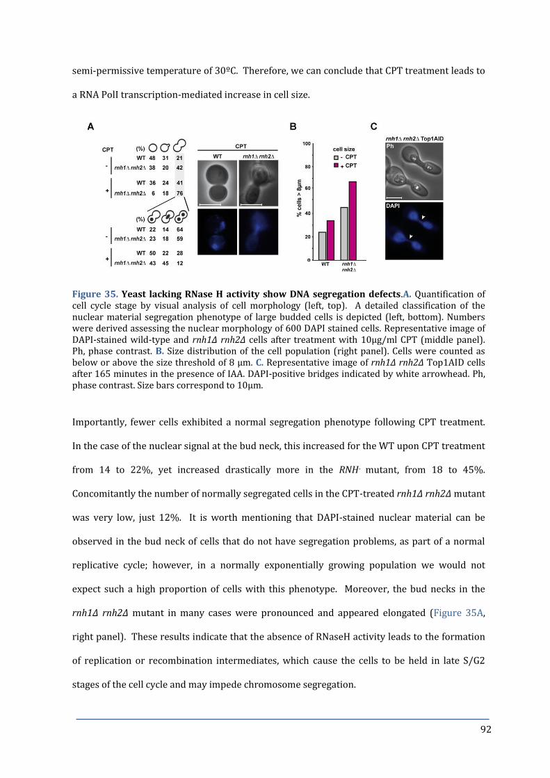
92
semi-permissive temperature of 30ºC. Therefore, we can conclude that CPT treatment leads to
a RNA PolI transcription-mediated increase in cell size.
Figure 35. Yeast lacking RNase H activity show DNA segregation defects.A. Quantification of cell cycle stage by visual analysis of cell morphology (left, top). A detailed classification of the nuclear material segregation phenotype of large budded cells is depicted (left, bottom). Numbers were derived assessing the nuclear morphology of 600 DAPI stained cells. Representative image of DAPI-stained wild-type and rnh1∆ rnh2∆ cells after treatment with 10µg/ml CPT (middle panel). Ph, phase contrast. B. Size distribution of the cell population (right panel). Cells were counted as below or above the size threshold of 8 µm. C. Representative image of rnh1∆ rnh2∆ Top1AID cells after 165 minutes in the presence of IAA. DAPI-positive bridges indicated by white arrowhead. Ph, phase contrast. Size bars correspond to 10µm.
Importantly, fewer cells exhibited a normal segregation phenotype following CPT treatment.
In the case of the nuclear signal at the bud neck, this increased for the WT upon CPT treatment
from 14 to 22%, yet increased drastically more in the RNH- mutant, from 18 to 45%.
Concomitantly the number of normally segregated cells in the CPT-treated rnh1∆ rnh2∆ mutant
was very low, just 12%. It is worth mentioning that DAPI-stained nuclear material can be
observed in the bud neck of cells that do not have segregation problems, as part of a normal
replicative cycle; however, in a normally exponentially growing population we would not
expect such a high proportion of cells with this phenotype. Moreover, the bud necks in the
rnh1∆ rnh2∆ mutant in many cases were pronounced and appeared elongated (Figure 35A,
right panel). These results indicate that the absence of RNaseH activity leads to the formation
of replication or recombination intermediates, which cause the cells to be held in late S/G2
stages of the cell cycle and may impede chromosome segregation.
93
When Top1 protein was depleted from G1 synchronized rnh1∆ rnh2∆ yeast, cells were able to
progress into the G2 stage but unable to complete mitosis and re-enter the next cell cycle (see
Figure 20A), suggesting that the persistence of RNA:DNA hybrids was responsible for the
holding of cells in the G2 phase. Furthermore, the majority of Top1 depleted RNH- cells were
large budded and had extremely long necks when observed under the microscope (Figure
35C). Strikingly the DAPI stained nuclear material could be seen in the bud neck as a DAPI
bridge (shown by arrowhead) connecting the mother and daughter nuclei. Such DAPI-positive
bridges are representative of incompletely segregated chromosomal DNA, such as hemi-
catenated sister chromatids (274) and such structures have been associated with increased to
chromosome breakage or nondisjunction during mitosis. Taken together our results reveal
that persistent RNA:DNA hybrids can lead to the accumulation of replication intermediates,
culminating in difficulties in chromosome segregation and failure to complete mitosis.
RNH- mutants are prone to premature re-budding and apoptosis
Cells respond to impaired chromosome segregation by a delay in cytokinesis (reviewed in
(275)). The fact that RNH- mutants don’t show genetic interactions with mutants affected in
NHEJ (e.g. ku70∆; see Chapter 2, Table 1) indicates that RNaseH-dependent chromosome
segregation defects do not lead to DNA breaks after sister chromatide disjunction. Indeed, cells
lacking RNaseH activity rely on HR, but NHEJ would be required to process such DNA breaks.
In contrast to WT cells, where nocodazole treatment arrests cells at the G2/M phase of the cell
cycle, rnh1∆ rnh2∆ cells re-initiated DNA replication without having undergone mitosis. We
therefore asked whether the premature DNA replication could induce apoptosis in RNH-
mutant cells.
Indeed, we observed a substantial loss of viability of Noc treated rnh1∆ rnh2∆ cells (Figure
33B), which was exaggerated by prolonged treatment of cells with nocodazole (results not
shown). In addition measurement of cellular DNA content by flow cytometry revealed a sub-
population of rnh1∆ rnh2∆ cells with a less than 1N cellular DNA content (Figure 33B, red

94
arrow), which may be due to cells undergoing apoptotic DNA degradation (276). We
determined the percentage of apoptosing cells by microscopic analysis of cells using methylene
blue staining, which stains dying or dead cells with defective cell wall integrity (Figure 36A). A
considerable increase in positively stained cells was observed in the RNH double mutant after
2 hours of Noc treatment; this difference was even greater after 4 hour treatment, reaching
56% of rnh1∆ rnh2∆ cells compared to less than 5% for the WT. In parallel we monitored DNA
fragmentation, a typical marker of apoptosis, using DAPI staining of nuclear DNA (Figure 36B).
The RNaseH lacking yeast had almost twice as many cells with the DAPI nuclear material stain
disrupted into several signals as the WT, with the number of cells containing fragmented
nuclear material increasing with the length of nocodazole treatment.
Figure 36. Nocodazole treatment of RNH- mutants causes re-budding and apoptosis. A. Methylene blue stained cells (indicated by black arrowheads) in the representative image of the RNH double mutant after 2h Noc treatment (top). Size bar represents 10µm. Percentage of methylene blue stained G2 cells (bottom). B. Representative microscope image of rnh1∆ rnh2∆ cells with fragmented nuclei (indicated by white arrowheads). Percentage of cells with fragmented nuclei, as determined by visual inspection of DAPI stained cells after 2h NOC treatment. C. Detection of FITC-VAD-FMK positive cells by flow cytometry for the analysis of metacaspase activation. Representative flow cytometry profiles (top) and quantification of fluorescent intensity (bottom).
Consequently, we analysed other features of apoptosis in S. cerevisiae. To do so, we detected
the amount of activated metacaspase by flow cytometry with the caspase activity indicator

95
FITC-VAD-FMK (277). Significantly more cells with activated metacaspase were detected in
the RNH double mutant than in WT cells (Figure 36C). Furthermore, the production of reactive
oxygen species (ROS), monitored by flow cytometry with the detection of H2DCFA-FITC, was
increased 5.7 fold in rnh1∆ rnh2∆ yeast with respect to the WT (results not shown), and
apoptosis has been reported to be preceded by an increased generation of ROS (reviewed in
(278). CPT treatment itself led to an increase in both the production of ROS and the activation
of metacaspases, consistent with the reported effects of CPT in inducing apoptosis in other
organisms (279-281). Noc treatment held WT cells at the G2/M phase of the cell cycle,
however, rnh1∆ rnh2∆ cells re-initiated DNA replication without having undergone mitosis,
with an associated loss of viability caused by the activation of apoptosis.

96
DISCUSSION for CHAPTER 3
Lack of RNaseH Activity Leads to Abnormal Cell Cycle Transitions at G2/M and G1/S
Given the connection between RNaseH activity and the rDNA described in the first two
chapters and based on the observation that S-phase transition is altered in cells lacking
RNaseH activity, we strove to dissect how R-loops may interfere with cell cycle regulation. We
first observed that Sic1, a stoichiometric inhibitor of Cdk1-Clb (B-type cyclins) (282), became
essential for viability in these mutants (see Figure 27). Sic1 prevents cells from a premature
entry into S-phase and its phosphorylation-induced degradation allows cells to initiate DNA
replication (236). Sic1 levels accumulate during mitosis and are controlled by the activity of
the phosphatase Cdc14. Cdc14 is constrained in the nucleolus during the cell cycle and only
becomes activated upon nucleolar liberation in anaphase. Cdc14 phosphatase activity causes
the accumulation of Sic1, by dephosphorylating and activating the SIC1 transcription factor
Swi5 (283), and by dephosphorylation of Sic1 itself, thereby stabilizing the protein (267).
Furthermore, Cdc14 activity triggers mitotic exit inducing the degradation of mitotic cyclins
(266,284). Yeast lacking RNaseH activity had lower levels of Sic1 protein throughout the cell
cycle, and the degradation of cyclin Clb2 was significantly delayed (Figure 31), indications that
Cdc14 may not have been fully active in RNH- mutants.
RNH- Mutants are partially defective in nucleolar Cdc14 release
Yeast uses an elaborate mechanism to control Cdc14 activity. At S phase entry, Cdc14 is ‘caged’
in the nucleolus by its competitive inhibitor Net1 (285). Net1, a core subunit of the RENT
complex, is a Sir2-associated nucleolar protein that stimulates transcription by RNA PolI and
regulates nucleolar structure (286). Indeed, the timing of Cdc14 release from the nucleolus
occurred normally in the RNH- mutants, but fewer cells released Cdc14 (see Figure 32). This
finding points to the possibility that the lack of RNaseH enhances nucleolar Cdc14 retention.
The role of Net1 in RNA PolI transcription may link R-loop formation and Cdc14 retention,

97
although such a connection still needs to be explored. Interestingly, Net1 has been shown to
stimulate RNA PolI transcription and to regulate nucleolar structure independently of
controlling mitotic exit (287). It should be noted that deletion of the NET1 paralogue TOF2 has
been shown to have synthetic genetic interactions with TOP1 and HPR1 (288,289).
Interestingly, R-loop formation is stimulated in cells lacking Top1 or Hpr1 (19,60) but we could
not detect a genetic interactions between RNH and TOF2 suggesting that Tof2 may not be
involved in rDNA-dependent R-loop processing.
Apart from its role in the cyclin B proteolysis, Cdc14 is required for the localization of the
condensin complex to rDNA to resolve the sister chromatids held together at the rDNA loci
(265). When released by the FEAR pathway, active Cdc14 was required for the proper
targeting of condensin to rDNA during anaphase (290). Perhaps, a critical level of active Cdc14
is required to complete sister chromatid separation. It would be very interesting to address
whether the localization of the condensin complex to rDNA is affected in the RNH- cells, and if
mis-localized condensin could be related to a defect in sister chromatid decatenation. Also of
interest to this study are reports that the polo-like kinase Cdc5 promotes the nucleolar release
of Cdc14 by stimulating the degradation of Swe1, and high levels of Swe1 were shown to
impair FEAR activation (291). In Chapter 2 of this thesis, we found the triple mutant with swe1
to be viable and have large sized spores; furthermore, the triple mutant suppressed the CPT
sensitivity of the RNH- cells (see Table 1). Therefore, it might be worthwhile monitoring the
timing of Cdc14 nucleolar release in the swe1 triple mutant, to see whether Cdc14 release is
normalized in the absence of Swe1 activity.
A further role of Cdc5 in yeast is to regulate sister chromatid separation by phosphorylation of
the cohesin subunit Scc1 (292). For successful chromosome segregation, cohesion complexes
must be removed by separase (293), and as sister chromatids are frequently catenated or
connected by hemicatenates (274), these structures must also be resolved before migration of
duplicated genetic material into the daughter cell. The Structural Maintenance of

98
Chromosomes complex Smc5-Smc6 plays a key role in cohesin functions (294), and the rDNA
has been shown as a major binding site for the Smc5-Smc6 complex (295). Furthermore,
Smc5-Smc6 may contribute to the accurate restart of RFs after pausing (296) and we observe
frequent RF pausing across the rDNA locus in yeast cells lacking RNaseH activity. Therefore, it
would be interesting to investigate whether there is a defect in cohesin binding at the rDNA
locus and to elucidate the impact of deletion of Smc complex members on the segregation of
chromosomes in RNH- yeast. Moreover, as smc5-smc6 mutants could enter anaphase before a
complete DNA replication of the rDNA array (296), it would be particularly interesting to see
how deletion of Smc complex members would affect the cell cycle progression of yeast lacking
RNaseH activity.
Separase is activated when securin is targeted for degradation by a ubiquitin ligase, the
anaphase promoting complex/cyclosome (APC/C) (for a review see (297)). Notably, Cdc14 is
required to dephosphorylate Cdh1, and this event is needed to trigger APC activation. An
alternative possibility for the defective chromosome segregation observed in yeast lacking
RNaseH activity, may be that the lower levels of nucleolar released Cdc14 are not sufficient to
activate the APC/C. A read-out to test for this possibility would be to look at Cdc20, an
activator of the APC (117) and see whether the APC is effectively activated in yeast lacking
RNaseH activity.
Persistent R-loops Impede Chromosome Segregation
Christman et al. found that cXII, and specifically the presence of the rDNA locus impeded the
complete segregation of replicating chromosomes of top1Δ yeast (298). Moreover, it had been
found that rDNA segregation in the absence of Cdc14 takes place if rRNA genes were not
transcribed (299) and high rates of RNA PolI transcription have been reported to impede rDNA
segregation by promoting the establishment of rDNA linkages (300). In this chapter we
describe that persistent RNA:DNA hybrids can lead to unresolved replication or recombination
intermediates, which prevent cells from exiting mitosis. Intrinsic segregation problems were

99
specifically observed for the rDNA-harbouring chromosome XII of yeast lacking RNaseH
activity (Figure 34). Furthermore, the incompletely segregated chromosomal DNA of the RNH-
mutant cells was evident as DAPI-positive bridges in pronounced and elongated bud necks
connecting the mother and daughter nuclei (see Figure 35). A consequence of such events may
be increased chromosome breakage or non-disjunction (the failure of the normal separation of
chromosomes to opposite poles during nuclear division), and indeed RNH- mutants suffer from
an elevated loss of heterozygosity (LOH), and a significant increase in spontaneous DNA
damage (see Figure 12). It remains to be determined whether the DNA in these bridges is in
fact rDNA. However, the LOH observed in yeast lacking RNaseH activity corresponded to
chromosome III, at a non-rDNA locus, and such LOH could be attributed to the loss of
chromosome III due to defective chromosome segregation. Likewise, we show that CPT
hinders the segregation of chromosome V (see Figure 34), further evidence that persistent R-
loops impede chromosome segregation. Similar anaphase chromosome bridges have been
observed in yeast mutants lacking condensin or Topoisomerase II (Top2) (301), and are also a
feature of solid tumours in humans (302,303). The yeast Rnh203 accessory subunit physically
interacts with Top2 (304), raising the possibility that the Rnh2 complex might direct Top2 to
R-loop associated anaphase chromosome bridges. The ‘so-called’ NoCut checkpoint has been
suggested to delay cytokinesis in response to impaired chromosome segregation (305). NoCut
inhibits completion of cytokinesis in the budding yeast top2 mutant (reviewed in (306)). Thus,
if the correct targeting of Top2 activity is constrained in RNH- mutants it is conceivable that the
NoCut checkpoint is activated in order to delay cytokinesis.
RNH- mutants are prone to premature re-budding and apoptosis
We noted that RNH- cells re-bud and initiated DNA replication without having completed
chromosome separation (Figure 33). These events were associated with a loss of viability
caused by the activation of apoptosis. Interestingly, fewer cells exhibited rebudding when RNA
PolI transcription was down-regulated (Figure 27), suggesting that a high rate of transcription

100
at the rDNA locus leads to segregation defects in the RNH- mutant, that cause mitotic arrest.
CPT-mediated DNA damage has been reported to lead to an increased ROS production and
increased apoptosis (280,281,307), however, there are no reports to our knowledge that such
increases are related to R-loops. The uncoupling of DNA replication from the cell cycle, such as
an inappropriate activation of S phase, can contribute to apoptosis (277). On the other hand,
R-loops could induce apoptosis by causing chromosome segregation constrains, and at the
same time by interfering with checkpoints that would normally restrain subsequent cell cycle
events such as M/G1 or G1/S phase transition. It is worth noting that the Polα-primase itself
undergoes cell cycle-dependent phosphorylation and dephosphorylation, becoming
phosphorylated and activated early in S phase and dephosphorylated while cells exit from
mitosis (308). As such events depend upon a functional Cdc28 complex (309), the mis-
regulation of cyclins observed in yeast lacking RNaseH activity could allow the primase
complex to still be active during G2/M stages of the cell cycle, thus permitting TIR and/or re-
replication events.
R-loop-Mediated Replication Cannot By-Pass the Need for Canonical Origin Firing
Low Sic1 levels are associated with premature S phase entry and problems in M/G1 transition,
both phenotypes that we identified in yeast lacking RNaseH activity. Premature S-phase entry
could also be attributed to genomic, R-loop dependent DNA replication initiation. To gain more
insight into this possibility, we tested if the need for Cdc6 and Cdc7 is bypassed in RNH- cells.
CDC6 codes for an essential component of the pre-replicative complex (pre-RC) needed to load
Mcm2-7 at origins of replication (239), while CDC7 codes for the kinase, whose
phosphorylation of Mcm2-7 is required for origin firing (4). Both genes are essential for
replication initiation and cell viability, and their absence caused lethality in a RNH- strain
background. Surprisingly, temperature sensitivity at 30°C of the cdc7-4 allele was alleviated in
cells lacking RNaseH activity (Figure 28). In contrast to the CPT-resistant cdc7-4 allele, cdc7∆

101
cells carrying a mcm5-bob1 mutation were CPT-sensitive, which may be related to the dual
function of Cdc7 in replication initiation and post-replicative repair (243).
Critical Role of the MRC1-Complex in Yeast Lacking RNaseH Activity
A key finding from this study was the observation that the MRC1-complex is required for
viability and CPT-resistance in yeast lacking RNaseH activity (see Figure 29). We find that
RNH- mutants were sensitive to the microtubule destabilizing drug benomyl; however the
spindle assembly checkpoint was not activated. Interestingly, mutants in mrc1, csm3 or tof1
are described as benomyl sensitive (256) and it was shown that cells with chromatid cohesion
defects, such as Ctf8-RFC complex mutants are also sensitive to benomyl (310). The mrc1
simple mutant was not CPT sensitive, suggesting that CPT itself does not affect cohesion,
however in combination with the lack of RNaseH activity, the triple mutant was in many cases
synthetic lethal, and extremely sensitive to CPT. Although Mrc1 function is mainly related to
replication fork stabilization in response to replicative constrains (224,247), these
observations suggest that the critical role of the MRC1-complex in yeast lacking RNaseH
activity may be related to cohesion functions. Indeed, the human Timeless-Tipin (Tof1-Csm3)
were shown to co-purify with cohesion subunits (311), and the Tof1 orthologue in C. elegans,
Tim1, is required for proper chromosome cohesion and segregation (312). Consequently, it
will be interesting to examine the contributions of the cohesin subunits to the viability and
chromosome segregation phenotypes of yeast lacking RNaseH function.
The MRC1-complex may also contribute to stabilize rDNA replication (313). Mrc1 itself can be
phosphorylated by Hog1 in response to osmotic stress (250). This osmotic stress induced S-
phase checkpoint acts to prevent conflicts between DNA replication and transcription (250).
In a similar fashion, Mrc1 could be required to co-ordinate replication through the rDNA in
RNH- mutants. Mrc1 has been shown to induce a replicative checkpoint, leading to
phosphorylation by Rad53 in response to replicative stress (109). However, we find that
neither the loss of RNaseH activity, nor CPT treatment is sufficient to activate Rad53

102
phosphorylation. CPT does not affect S phase progression in WT or RNH- cells, although, it
does provoke a delay in G2/M phases of the cell cycle, suggesting that CPT lesions are not
sensed as DNA lesions during S-phase progression. Mec1 is a master protein in the checkpoint,
acting upstream of Mrc1. It was therefore interesting, given the important genetic interaction
of the RNaseH enzymes with Mrc1, to observe in Chapter 2 that the mec1sml1 quadruple
mutant was viable, although spores were smaller in size and more sensitive to CPT than the
RNH double mutant (see Table 1). This result supports that the important role of Mrc1 in
maintaining the viability of yeast cells lacking RNaseH activity is not related to the Rad53-
dependent replicative checkpoint.
Loss of RNaseH Activity Does Not Activate the Rad53-Dependent S-phase Checkpoint
Finally, it is interesting to note that yeast lacking RNaseH activity suffer from increased DNA
damage (see Chapters 1 and 2), yet RNH- cells did not activate the Rad53-dependent S phase
checkpoint. This finding is in contrast to yeast hpr1 mutants, which also have been shown to
accumulate R-loops, leading to a constitutive activation of the Rad53-signalling pathway (314).
On the other hand, hpr1 mutants required a functional S-phase checkpoint but not DSB repair
for survival (314), while DSB repair is critical for the viability of RNH- mutants even in the
absence of exogenous stress. Both, hpr1∆ and rnh1∆ rnh2∆ show a genetic growth defect with
top1∆ (60,288,289). However, Hpr1 and RNaseH may have a more specialized role in the
avoidance of R-loop formation in genomic or rDNA, respectively.
RNaseH Enzymes Play A Critical Role in Preventing Aneuploidy
As detailed in Chapter 1, the RNaseH and Top1 enzymes play key roles in protecting the
eukaryotic genome by preventing unscheduled TIR events mediated by R-loops, with
particularly important repercussions on genome instability and cancer. We also highlight the
criticality of RNaseH activity in preventing non-disjunction, a major causative factor for
aneuploidy, one of the most common characteristics of human tumours (315). It remains to be

103
determined whether the aberrant segregation of chromosomes in the RNH- mutants observed
in this study are the cause of defects in chromosome cohesion or condensin activities.

104
6. CONCLUSIONS
Chapter 1
1. Chemical (CPT) or degron-mediated depletion of Top1 activity promotes R-loop
formation and induces cell death in RNaseH lacking yeast.
2. R-loops promote the formation of unscheduled origin-independent replication
intermediates indicative of transcription-induced replication (TIR) in ribosomal DNA
at late S/G2 of the cell cycle.
3. Replication by TIR paused at sites of protein-DNA interaction, which points to the
possibility that TIR is driven by non-canonical replication machinery.
4. TIR is RNA PolI transcription-dependent.
Chapter 2
5. The homologous recombination (HR), post-replicative repair (PRR), break-induced
repair (BIR) and nucleotide excision repair (NER) pathways are required for viability
or are essential to combat the genotoxic constrains in CPT-treated RNaseH lacking
cells.
6. Transcription-coupled repair (TCR), a NER sub-pathway may play a prominent role in
the repair of CPT damage in RNaseH mutants.
7. The genotoxic effects of CPT-treatment can be enhanced or alleviated by alterations in
the structural integrity of rDNA or by down-regulation of RNA PolI transcription,
respectively.
8. TIR is not dependent on Rad18, Rad51 or Srs2 activities.

105
Chapter 3
9. RNaseH H- cells enter prematurely into S-phase, making Sic1 essential to maintain
viability in the absence of RNaseH activity.
10. R-loops cannot bypass the need for Cdc6 and Cdc7 in genome replication but partially
alleviate the temperature sensitive phenotype of the cdc7-4 mutation.
11. The MRC1-complex is required to combat the R-loop induced constrains in a Rad53-
independent manner.
12. R-loop dependent alterations in nucleolar organisation constrain nucleolar Cdc14
release.
13. Constrained Cdc14 activity precedes the mis-regulation of Clb2 (up) and Sic1 (down)
protein levels.
14. R-loops constrain chromosome segregation and lead to the formation of ‘chromosome
bridges’.
15. R-loop-mediated chromosome segregation defects are not monitored by the spindle
assembly checkpoint (SAC).
16. R-loop dependent replication and chromosome segregation constrains induce
apoptotic events.

106
7. MATERIALS & METHODS
1. MEDIA
1.1 Bacterial media
LB: (Lysogeny broth): 0.5% yeast extract, 1% tryptone and 1% NaCl, adjusted to pH 7.0.
LB+Amp: LB media supplemented with 80 g/ml of ampicillin (Sigma).
For solid media 2% agar is added.
1.2 Yeast media
YPAD: 1% yeast extract, 2% peptone, 2% glucose supplemented with 20mg/l of adenine.
YPAG: 1% yeast extract, 2% peptone, 3% glycerol supplemented with 20mg/l of adenine.
S: synthetic minimal medium, 0.17% YNB, 0.5% ammonium sulphate, that can be
supplemented with different carbon sources, and with amino acids (leucine, tryptophan,
histidine, methionine, lysine, aspartate, threonine, adenine and uracil) added to the final
concentration as described by (316).
SC: S media supplemented with 2% glucose and amino acids.
SD: S media supplemented with 2% glucose (no amino acids).
SGL: S media supplemented with 3% glycerol, 2% lactate and amino acids.
SGAL: S media supplemented with 2% galactose and amino acids.
SC+FOA: S media supplemented with 500mg/ml of 5-Fluorotic acid (5-FOA) and amino acids.
Uracil was added at half the usual concentration.
Sporulation media (SPO): 1% potassium acetate, 0.1% yeast extract, 0.005% glucose,
supplemented with half the usual amount of each amino acid as used for SC media.

107
2. STRAINS AND GROWTH CONDITIONS
2.1 Escherichia coli strains
DH5 : F´ ∆lacU169 Φ80 lacZ∆M15 supE44 gyrA96 recA1 relA1 endA1 thi-1 hsdR17 (317).
2.2 Saccharomyces cerevisiae strains
The genotypes of all yeast strains used in this thesis are indicated in Table 2. Yeast strains
used in this study were derived from the YKL83 strain (124), based on W303-1a, unless
otherwise stated. If not generated by a direct knock-out in the YKL background, mutant strains
were backcrossed at least twice to the YKL83 strain background. Gene deletions were
constructed by PCR-based methods using various template plasmids: pFA6a-kanMX6 (318),
pAG32-hphMX4 (Euroscarf), pAG25-natMX4 (Euroscarf) and pFA6a-klLEU2MX6 (kindly
provided by B. Pardo). Deletion mutants were verified by Southern blot, or by PCR analysis
from genomic DNA preparations with primers complementary to sequences within the gene
cassette and upstream of the disrupted gene locus. All experiments were performed with
several meiotic segregants of the same genotype to ensure they behaved similarly.
Meiotic segregants from crosses with the pif1-m2 strain were confirmed by PCR from genomic
DNA using the forward primer with sequence: TGTAATATTATCCATTGAGC, and reverse
primer: TATCATTTCCAAACTTCTCT. The resulting 620bp band was digested with XhoI as the
mutation creates a novel XhoI site. To detect whether crossed strains were wild-type RAD5 or
rad5-535, PCR was performed from genomic DNA with the forward primer:
GCAGCAGGACCATGTAAACG, and reverse primer: AAACTCGTTACTCCACTGCG. The resulting
PCR fragment was digested with MnlI, as the mutation creates an additional MnlI site.
Digestion of WT RAD5 PCR products leads to two fragments of 155 and 182bp, or three
fragments of 155, 120, and 62 bp for digested rad5-535 PCR products, separated on a 3%
agarose gel, respectively.

108
2.3 Genetic analyses
Some strains were generated by genetic crosses, using standard methods. Parental strains
were crossed on solid YPAD, diploids were selected and sporulated on SPO media for 4-5 days.
Tetrads were dissected in a Singer MK1 micromanipulator (Singer LTD) after treatment with
2mg/ml zymolyase 20T (USBiological) for 3 min. The genotype was determined by replica
plating on different selective media and the sex was determined by the ability to mate with the
reporter strains F4 (a) and F15 (α).
2.4 Growth conditions
The rpa190-3 and rrn3-8 RNA PolI temperature-sensitive mutants, used in Chapter 1, are
unable to grow at 37ºC. Experiments were performed at the permissive or semi-permissive
temperatures of 23ºC and 30ºC, respectively. Growth of the cdc7-4 strain is blocked in G1
phase at 37ºC. Experiments were performed at the permissive or semi-permissive
temperatures of 23ºC and 30ºC, respectively. All other experiments were performed at 30ºC
unless otherwise stated.
2.5 Degron strains
An auxin-inducible degron (AID) strain was created to control the protein levels of Top1, in
order to produce a conditional triple mutant with rnh1∆ rnh2∆. The AID system, developed by
Kohei Nishimura et al., involves a plant-specific mechanism that responds to the plant hormone
auxin (155) (Figure 37). Auxin binds to the transport inhibitor response 1 (TIR1) protein and
promotes the binding of the SCF (Skp1, Cullin and F-box protein) complex. The SCF in turn
recruits the E3 ubiquitin ligase and polyubiquitinates the SCFTIR1, targeting it for degradation
by the 26S proteasome. Expressing the TIR1 protein in yeast cells, which can assemble a
functional SCFTIR1 complex due to the conserved nature of the Skp1 protein (319), permits the
degradation of the protein of interest tagged with the auxin-binding domain, upon the addition
of auxin.

109
The AID degron system was crossed into the rnh1∆ rnh2∆ strain, to create strain RS315
expressing TIR1 under control of the constitutive ADH promoter. The Top1 protein was tagged
at the C-terminus with the IAA17 auxin-binding domain, which was amplified by PCR from the
plasmid pKan–AID*–9myc (320) (see Table 3 for a list of plasmids used in this thesis) using
primers specific for the protein of interest, and included the gene for GEN resistance.
Subsequently, the rnh1∆ rnh2∆ degron-containing strain RS315 was transformed with the
respective PCR products, and positive candidates were selected by growth on plates containing
HYG, NAT and GEN. The functionality of the aid strain was confirmed by halo assays, drop tests
and Western blot analysis against the 9Myc tag.
Figure 37. Schematic illustration of the auxin inducible degron (AID) system. Adapted from (155).
3. TRANSFORMATIONS
3.1 Transformation of bacteria
Competent bacterial cells were prepared using the method of Hanahan et al. (317), modified by
Inoue et al. (321). 100μl of competent cells were mixed with 50-100ng of DNA and incubated
on ice for 30 min, then cells were subjected to heat-shock at 42ºC for 1 min followed by 1 min

110
on ice. 600µl of LB media was added and cells were incubated at 37ºC for 45 min. Finally, cells
were collected and plated on LB+Amp.
3.2 Transformation of yeast
Yeast cells were transformed according to the method described by Ito et al. (322) and
modified by Gietz et al. (323). 30ml of cells growing in YPAD to an OD600 0.4-0.6 were collected
and washed with sterile water. Cells were then washed with 100mM LiAc and a
“transformation mix” added, consisting of 240μl 50% PEG3350, 36μl LiAc 10M, 5μl SS-DNA
(10mg/ml) and 0.1-10μg DNA. Cells were vortexed vigorously to mix and incubated at 30ºC
for 30 min, followed by a heat-shock at 42ºC for 20 min. Finally, cells were washed with sterile
water and plated on selective medium.
3.3 Plasmid isolation from E.coli cells
Plasmid DNA was extracted from 1.5 ml of an overnight culture growing in LB+Amp at 37ºC.
The isolation was performed with the boiling method described by Holmes and Quigley (324).
3.4 Yeast DNA extraction
Yeast genomic DNA was extracted according to the yeast DNA miniprep protocol described by
Amberg et al. (325).
4. VIABILITY ASSAYS
4.1 Growth rate determination
The optical density is proportional to the number of cells, and therefore the growth rate of
yeast strains (represented as the doubling time) can be quantified by monitoring the change in
optical density at 600nm. Yeast strains were grown overnight to stationary phase. Cells were
diluted in fresh YPAD to an OD600 of 0.1, and the OD was taken every hour. Optical density was
plotted as a function of time on a semi-log scale.

111
4.2 Viability assays
Viability and sensitivity to genotoxic agents were determined by drop test assays. Yeast cells
were adjusted to an initial OD600 of 0.4, then serially diluted 1:10 and spotted onto plates with
or without different genotoxic agents at the indicated concentrations. Plates were incubated at
30ºC for 3 days, except for temperature-sensitive mutants that were incubated at the
corresponding permissive or semi-permissive temperatures.
For UV irradiation, cells were serially diluted and spotted onto YPAD plates, irradiated with 50
or 100 J/m2 UVC light in a UV-Mat (Dr. Gröbel UV-Elektronik GmbH) irradiation chamber, and
incubated in the dark at 30°C for 3 days.
4.3 Survival assays
A sample of exponentially growing cells was taken from a liquid YPAD culture and plated on a
YPAD plate – this point was considered as time 0. At time point 0, 10µg/ml CPT was added to
the medium and cells were incubated for 24 hours, with appropriate dilutions to maintain cells
in the exponential phase of growth. At determined time points, cells were taken from the
population and plated onto YPAD plates. Cells were counted after 3 days growth, and
presented as the percentage of surviving cells, with respect to time 0, set as 100% survival.
4.4 Halo assays
To confirm whether a yeast strain is bar1Δ, or to confirm the functionality of an aid degron
strain, 100µl of cells at OD600 0.2 were streaked over the surface of a YPAD plate to create a
lawn. Once dry, a sterile filter paper disc was laid onto the plate surface, and 2 µl of either α-
factor or IAA was dropped onto it. A halo, or clearing in the lawn of cells around the paper
circle containing α-factor or IAA was observed if cells were bar1Δ or if cells contained the
functional aid degron system, respectively (Figure 38).

112
Figure 38. An α-factor halo assay to test for bar1Δ phenotype of a yeast strain.
4.5 Cell size distribution
The size distributions of various yeast strains were analyzed using the COULTER COUNTER®
Cell and Particle Counter (Beckman Coulter). The proportion of cells above or below the size
threshold of 8 µm was determined.
5. RECOMBINATION AND MUTATION ASSAYS
5.1 A-like faker assay (ALF)
The formation of a-mating cells from MAT strains was scored as described (132) with some
modifications. Briefly, MAT strains were grown on YPAD plates for three days to obtain
single colonies. A-like faker cells were selected by growing on YPAD plates overnight at 30ºC.
Cells were then transferred onto a mating tester lawn of MAT (F15 strain) by replica plating
followed by incubation at 30º overnight. The mated lawn was then replica plated to SD
medium and mated products were counted. Total cells were grown on YPAD plates. ALF
frequency was calculated as mated products/total cells. Each ALF frequency value was
obtained by the mean of at least two different fluctuation tests of four independent colonies
each. Data are shown as the mean ± standard deviation. Differences between groups were
examined by Student’s t-test.
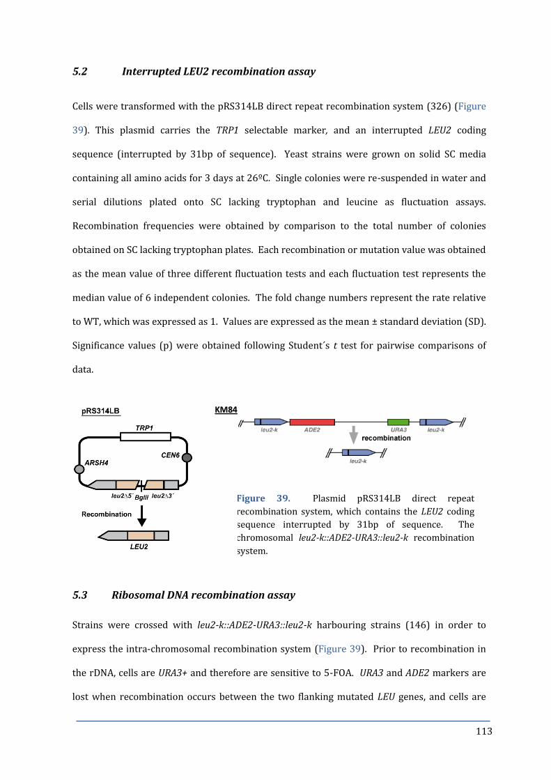
113
5.2 Interrupted LEU2 recombination assay
Cells were transformed with the pRS314LB direct repeat recombination system (326) (Figure
39). This plasmid carries the TRP1 selectable marker, and an interrupted LEU2 coding
sequence (interrupted by 31bp of sequence). Yeast strains were grown on solid SC media
containing all amino acids for 3 days at 26ºC. Single colonies were re-suspended in water and
serial dilutions plated onto SC lacking tryptophan and leucine as fluctuation assays.
Recombination frequencies were obtained by comparison to the total number of colonies
obtained on SC lacking tryptophan plates. Each recombination or mutation value was obtained
as the mean value of three different fluctuation tests and each fluctuation test represents the
median value of 6 independent colonies. The fold change numbers represent the rate relative
to WT, which was expressed as 1. Values are expressed as the mean ± standard deviation (SD).
Significance values (p) were obtained following Student´s t test for pairwise comparisons of
data.
Figure 39. Plasmid pRS314LB direct repeat
recombination system, which contains the LEU2 coding
sequence interrupted by 31bp of sequence. The
chromosomal leu2-k::ADE2-URA3::leu2-k recombination
system.
5.3 Ribosomal DNA recombination assay
Strains were crossed with leu2-k::ADE2-URA3::leu2-k harbouring strains (146) in order to
express the intra-chromosomal recombination system (Figure 39). Prior to recombination in
the rDNA, cells are URA3+ and therefore are sensitive to 5-FOA. URA3 and ADE2 markers are
lost when recombination occurs between the two flanking mutated LEU genes, and cells are
KKMM8844

114
now resistant to 5-FOA. Strains containing the chromosomal system were first streaked onto
SC plates lacking adenine and uracil for 2 days, and then streaked onto SC plates supplemented
with all amino acids for a further 2 days. Fluctuation tests were performed as previously
described, with serial dilutions plated onto SC lacking uracil to score totals, and onto SC+FOA
plates to score recombinants.
5.4 Laur mutation assay
Mutation frequencies were determined from cells transformed with the pCM184-LAUR
plasmid (134). Fluctuation tests were performed as previously described, with serial dilutions
plated onto SC lacking uracil to score totals, and onto SC+FOA plates to score mutators.
5.5 Canavanine mutation assay
Forward mutation frequencies of nuclear DNA were obtained by fluctuation test as previously
described, comparing the number of colonies growing on SC-Arg plates (SC media lacking
arginine) containing 60mg/ml canavanine to the total number of colonies obtained on SC.
6. CELL CYCLE SYNCHRONIZATION AND PROGRESSION ANALYSIS
6.1 Alpha factor synchronization
Cell cycle progression was determined by measuring DNA content using flow cytometry. MATa
yeast recognize the presence of alpha factor (α-factor), and respond by growing protrusions
known as schmoos, and halt cell growth in the G1 phase of the cell cycle. Following release
from α-factor, the synchronized population of cells continues to grow.
For G1 synchronization, cells were grown to OD600 0.4 in YPAD medium and synchronized by
incubation with 1μg/ml α-factor (Biomedal) for 90 minutes. An additional 1μg/ml α-factor was
added for a further 90 minutes. Cells were released from α-factor treatment by washing three
times in pre-warmed, fresh YPAD medium. For the G1 synchronization of bar1Δ cells, which
are more sensitive to α-factor than BAR1 strains, a lower concentration of 0.3μg/ml was used.

115
Since bar1Δ yeast lack expression of the aspartyl protease that cleaves and inactivates α-factor,
cells were released by washing three times in pre-warmed, fresh YPAD medium containing
0.1mg/ml Pronase E (Sigma).
6.2 Flow cytometry analysis of cell cycle progression
Following α-factor synchronization and release, samples of 1ml were taken at indicated times,
fixed in 70% ethanol and stored at -20ºC for analysis by flow cytometry. Samples were washed
in 1x PBS (see Appendices for buffer compositions) and then resuspended in 1x PBS containing
5μg/ml propidium iodide, following RNase A (1mg/ml) overnight treatment. Cell doublets
were separated by brief sonication and cell cycle progression was analyzed by flow cytometry
using a FACSCalibur (Becton Dickinson) with CellQuest software.
6.3 Nocodazole synchronization
For flow cytometry analysis of the release from nocodazole synchronization, cells were grown
to OD600 0.4 in YPAD medium, and synchronized in G2 phase of the cell cycle by incubation with
15µg/ml nocodazole for 90 minutes, with 1% DMSO added to aid dissolution. Cells were
washed and released in pre-warmed, fresh YPAD medium. 1ml samples were taken at specified
time points following release and fixed in 70% ethanol at -20ºC for later analysis by flow
cytometry.
For analysis of rebudded cells, yeast strains were grown to OD600 0.4 in YPAD medium and
synchronized, as previously described, with α-factor. Cells were released in pre-warmed, fresh
YPAD medium in the presence of 15µg/ml nocodazole and 1% DMSO. 1ml samples were taken
at specified time points and fixed for flow cytometry analysis as previously described.
Nocodazole was added every 60 minutes during the incubation.

116
6.4 Induction of AID degron strains
Cells were grown to OD600 0.4 in YPAD medium and synchronized in G1 as before. In the last
30 minutes of α-factor incubation, 5µM IAA was added to the medium to induce degradation of
the aid-tagged protein. Cells were released into fresh YPAD medium and samples were
collected for FACS cell cycle progression analysis, protein extraction for Western analysis, and
genomic DNA extraction for 2D-gel.
7. SOUTHERN BLOT ANALYSIS OF DNA FRAGMENTS
7.1 Genomic DNA extraction
10ml of cells growing in YPAD to OD600 1 were collected and washed. Cells were spheroplasted
by incubation in 320µl spheroplasting buffer (see Appendices for buffer compositions)
containing 150μg/ml zymolyase and 1% (v/v) β-mercaptoethanol at 37°C with occasional
mixing for 60 min. Following centrifugation at 13,000rpm for 30 seconds, the pellet was
resuspended in 370µl cocktail gently buffer. A volume of 16μl 10% SDS was added, the mix
gently agitated and incubated at 65°C for 30 min, followed by the addition of 85μl 5M
potassium acetate with incubation with on ice for 1h. After centrifugation for 15 min at 13,000
rpm, the supernatant was transferred to a new tube and DNA was precipitated with 1.2ml 96%
ethanol for 30 minutes at -20ºC. The pellet, following 10 min centrifugation at 13,000rpm, was
resuspended in 300µl TE containing 0.1mg/ml RNase A and incubated at 37°C for 30 min.
200µl of phenol and chloroform were added and well mixed, and the upper phase was carefully
transferred into a new Eppendorf tube following 5 min centrifugation at 13,000rpm. DNA was
precipitated with 1ml 96% ethanol and 120μl NaCl 2.5M followed by centrifugation at
13,000rpm for 10 minutes, and a 500μl 70% ethanol wash of the pellet. The dried pellet was
finally resuspended in 30μl TE. The purified DNA was digested for 5 hours with the
determined restriction enzyme and loaded onto a 0.8% TAE-buffered agarose gel.
Electrophoresis was carried out without ethidium bromide at 2 V/cm for 30h. .

117
7.2 Alkaline DNA transfer
Following separation of DNA fragments by electrophoresis, gels were treated with 0.25M HCl
for 10 min, washed with water and then incubated with denaturation buffer for 30 min. DNA
was transferred to nylon membrane Hybond XL (Amersham) by capillary action in
denaturation buffer for 12-16 h. After UV-crosslinking (70 mJ/cm2), membranes were washed
briefly with 2X SSC buffer and allowed to air-dry.
7.3 DNA hybridization
Membranes were hybridized with various radioactive DNA probes. For α32P-dCTP labelling,
100ng of denatured DNA was mixed with 1mM random hexanucleotides, 0.5mM dATP, dGTP
and dTTP, 25μCi α32P-dCTP and 2U of Klenow DNA polymerase. The mix was incubated at
37ºC for 1h and non-incorporated radioactive nucleotides were then removed by using a G50-
Sephadex column. Membranes were pre-incubated at 65ºC for 30 min in hybridization
solution with constant rotation. Hybridization was performed at 65ºC for 12-16 h in 10-15ml
hybridization solution containing the radioactive probe, previously denatured at 100ºC for 5
min. Following hybridization, unbound probe was washed off the membrane during two
washes of 30 min at 65ºC using 50ml wash solution. For re-hybridization, probes were
stripped off the membranes by washing three times with 500ml boiled stripping solution for
15 min.
7.4 Signal quantification
Radioactive signals were detected by exposure of the membranes on PhosphorImager screens
(Fuji) that were scanned by a PhosphorImager FujiFilm FLA 5100. Quantification of the signal
was performed with the Image Gauge software (Fuji).
7.5 Analysis of extrachromosomal rDNA circles
For separation of ERCs, about 1µg CTAB-extracted genomic DNA from exponentially growing
cells was digested with 15U BamHI in 20 μl of 1x buffer for 6hrs at 37°C and separated by gel

118
electrophoresis in 0.6% TBE-buffered agarose gel at 2 V/cm for 30hrs. ERCs were detected by
hybridization with ribosomal probe A, following alkaline transfer to Hybond XL (Amersham)
membrane. ERC levels were normalized against the rDNA repeat and expressed as fold change
relative to the amount of ERCs in WT cells.
8. BI-DIMENSIONAL AGAROSE GEL ELECTROPHORESIS (2D-GEL)
2D-gel first separates restriction-digested RIs by mass (1st dimension), and the 2nd dimension,
performed in the presence of ethidium bromide (EtBr), separates molecules by their three-
dimensional shape. When a linear DNA molecule (n) is replicated, it progressively doubles in
mass until reaching a fully replicated linear molecule (2n), and will adopt various bubbled or
branched structures in the process, depending on where the closest site of replication initiation
is located (Figure 40). Following transfer onto a membrane, replication across the DNA
fragment of interest can be detected using specific probes.
Figure 40. Schematic representation of two-dimensional gel analysis (2D-gel). The types of
replication intermediates (RIs) that can be detected, include: bubble arcs, corresponding to an
active origin within the fragment; simple Y arcs, which represent passive replication from an
outside origin by a single replication fork entering from one end of a restriction fragment; and
spikes above the 2n spot, which correspond to X-shaped molecules such as almost fully replicated
molecules, converged forks and recombination hemicatenates. The n spot represents non-
replicating linear molecules. Information adapted from (327).
In order to monitor RI formation by 2D-gel, cells were released from α-factor synchronization
into minimal medium lacking adenine. Work by Aiko Matsui et al. demonstrated that removal

119
of essential nutrients can slow progression through the yeast cell cycle (328). We observed
that entry into S-phase was specifically delayed in the RNH double mutant when released into
minimal medium lacking adenine, but not into medium lacking other amino acids, such as
leucine or tryptophan (Figure 41). This delay helped to ensure that CPT was efficiently taken
up by the cells upon α-factor release. It is interesting to note that only rnh1∆ rnh2∆ mutants
cannot proceed through S-phase in the absence of Trp, because Trp has been shown to act as a
Top1-DNA anchor (329).
Figure 41. FACS showing synchronization in SC and release into SC, SD-Leu, SD-Trp or SD-Ade.
Cells were synchronized in G1 with α-factor and released into minimal medium lacking
adenine and containing 10µg/ml CPT. 100ml samples were retrieved at the specified time
points following release (in minutes) and cooled down on ice. To each sample, 0.1% sodium
azide (final concentration) was added, since sodium azide immediately stops DNA replication
(327).
Following centrifugation, cells were washed with cold water and resuspended in 2.4ml
spheroplasting buffer and incubated for 20 min at 30ºC, followed by a 30 min incubation at
37ºC. Spheroplasts were collected by centrifugation and the cell pellets were carefully
resuspended in 1.125 ml of G2 solution. After solubilization, 25μl of RNase A (10mg/ml) and
75μl of freshly prepared proteinase K (20mg/ml) were added and the sample was incubated at

120
50ºC for 45-60 min. The solution was centrifuged at 8500 rpm for 10 min and the supernatant
was transferred to a new tube. Then, 750μl of chloroform/isoamyl alcohol 24:1 at RT was
added and the solution was mixed repeatedly. After 10 min centrifugation at 8500 rpm, the
upper aqueous phase was recovered and DNA was precipitated by adding 2 volumes of CTAB
Solution II. The sample was centrifuged for 10 min at 8500 rpm and the pellet was
resuspended in 0.8ml of CTAB Solution III and 0.2ml of 2.5M NaCl. DNA was precipitated with
1 volume of isopropanol and centrifuged at full speed for 10 min. The pellet was briefly rinsed
with 96% ethanol, air-dried and resuspended in 250μl of 2mM Tris-HCl pH 8.
In a total volume of 100µl, about 5µg of CTAB-extracted genomic DNA was digested with 40
units BglII for 6 hrs. DNA was precipitated with 1 volume of isopropanol and 20μl 2.5M NaCl,
followed by centrifugation at full speed for 15 min. The pellet was washed in 96% ethanol, air-
dried and resuspended in 20μl of 2mM Tris-HCl pH8 with loading buffer.
Replication intermediates were resolved by 2D gel electrophoresis using a 20 cm x 25 cm gel
tray. The first dimension was carried out at room temperature in 0.4% agarose gels in TBE 1X
at 45 V (28 mA) for 18 hours. Gels were stained with 0.33μg/ml EtBr in water for 30 min and
DNA fragments between 3-12 kb were cut off and rotated 90º for the second dimension
electrophoresis in a 20 cm x 25 cm gel tray. These fragments were resolved in the second
dimension in 1% agarose gels in TBE 1X containing 0.33μg/ml ethidium bromide at 140 V (75
mA) for 16 hours. This electrophoresis was performed at 4ºC and with buffer re-
circularization. After electrophoresis, gels were treated and transferred to a Hybond XL
(Amersham) membrane by alkaline transfer. Replication intermediates were detected by
hybridization with specific 32P-labeled DNA probes: probe A of 4.6kb that hybridizes within the
non-transcribed intergenic region (NTS1) and probe B of 4.5kb that hybridizes within the 35S
gene. Probe A was generated from the PCR product obtained using the primers: forward:
GTGGTATTTCACTGGCGCCGA, and reverse: ATAACCGCAGCAGGTCTCA. Probe B was generated

121
from the PCR product obtained using the primers: forward: CAACCCTGACGGTAGAGTAT, and
reverse: ATTTCACTGGGCCAGCATCA.
Signals were quantified using a PhosphorImager with ImageGauge software (FUJI). The
relative intensity of replication intermediates was normalized to the signal intensity obtained
in the 1n-spot (non-saturating exposure). Gel images represented in the figures are optimal
exposures to show the replication intermediates. Scanned images were optimized, in some
cases, for brightness and contrast, but no gamma corrections or any other manipulations were
carried out.
8.1 Characterization of replication intermediates
Following BglII digestion and isopropanol precipitation, DNA was resuspended in 2mM Tris-
HCl pH8.0 and subjected to heat or enzymatic treatments as previously described (154). In
brief, to induce branch migration, DNA was incubated at 56°C for 1h in the presence of 10mM
EDTA pH8. For the strand displacement reaction, Klenow polymerase was used. Stepwise, 1 μl
of nucleotide mix (5mM each of dATP, dCTP, dGTP, dTTP; Pharmacia), 1μl of gp32 protein
(Biolabs, 4μg/μl), a single-stranded DNA binding protein that binds at 3´ hydroxyl group, and
1μl of Klenow (Takara; 5U/μl) were added to 17 μl of restriction enzyme-digested DNA in 1×
restriction buffer and were incubated for 1h at 37° C. For R-loop removal, 1µl RNase H
(Biolabs; 5U/µl) was added to 19 μl of restriction enzyme-digested DNA in 1× restriction buffer
and incubated for 1h at 37°C.
8.2 Characterization of RF pausing sites
.For characterization of RF pausing sites, replication intermediates from rnh1Δ rnh2Δ cells
were compared to fob1Δrrm3Δ after growth with 10µg/ml CPT for 105 minutes or 60 minutes,
respectively, following release from α-factor synchronization.

122
9. CLAMPED HOMOGENEOUS ELECTRIC FIELD (CHEF) GEL
ELECTROPHORESIS
9.1 Agarose plug preparation
DNA was extracted from approximately 108 cells per low melting agarose (Pronadisa) plug as
previously described (330). Briefly, yeast cells were washed in 50mM EDTA pH8 and then
resuspended in 600µl CPES with 1.2M D-sorbitol and freshly added zymolyase 20T at 1mg/ml
to hydrolyze the cell walls. The spheroplasts formed are embedded in 1ml 2% low-melting-
point agarose prepared in CPE. This mixture was pipetted into moulds and allowed to cool and
solidify during 10 minutes at 4ºC. Plugs were subsequently transferred to 10ml tubes and
incubated for 4 hours at 30ºC with 5ml CPE. The solution was poured off and the plugs were
incubated overnight at 50ºC in 5ml TESP. Plugs were washed three times at 50 ºC for 30 min,
then twice at room temperature with TE. Finally the plugs were covered with 5ml 50mM
EDTA pH8 and stored at 4ºC.
9.2 Analysis of replicating chromosomes
To analyze the replication status of chromosome cXII in exponentially growing cells, samples
were retrieved at the specified time points following release (in minutes) from α-factor
synchronization in the presence or absence of 10µg/ml CPT. Genomic DNA was extracted in
low-melting agarose plugs as described previously. Replication intermediates were separated
using a CHEF Mapper (Bio-Rad), with a linear voltage gradient of 6.0V/cm, pulse time of 0.22 s
to 266 s, switch angle of 120º, in 0.5x TBE buffered-1% agarose gel for 15.2 hrs at 14°C, as
previously described (327). For hybridization, 32P-labelled probes for cV or cXII were used. cV
probe was generated from PCR with forward primer: GTACGGTGGCCATTCTATGT, and reverse
primer: AGATCCAGTAATGTGTTGGA. The previously described ribosomal probe A was used to
probe cXII.

123
9.3 rDNA array repeat length determination
rDNA repeat length was determined by CHEF analysis of approximately 108 cells per LM-
agarose plug. Cell lysis and chromosome separation conditions were carried out as previously
described (75). In brief, electrophoresis was performed in a Bio-Rad CHEF Mapper, using a
voltage gradient of 3.4V/cm, pulse time of 300-900s, switch angle of 120º, in 0.8% agarose gel
in 0.5x TBE for 68 hrs at 14ºC. Chr. XII was detected by hybridization with ribosomal probe A.
10. MICROSCOPY
For wide-field fluorescence microscopy, a Leica DM-6000B equipped with a 100×/1.40 NA oil
immersion objective lens was used, with the appropriate light excitation and emission
conditions (Table 4). Images were taken using a digital charge-coupled device camera
(DFC350; Leica) and LAS AF software (Leica).
Leica filter λ excitation λ emission
DAPI A4 360nm 470nm
Mitotracker orange N3 546nm 600nm
GFP L5 395nm 509nm
YFP L5 514nm 527nm
mRFP N3 584nm 607nm
Table 4. Light excitation and emission conditions for fluorescence microscopy.
10.1 Fixation of cells
Cells were fixed in 2.5% formaldehyde and 0.1M potassium phosphate buffer pH6.4 for 10 min.
Cells were then washed twice with 0.1M potassium phosphate buffer pH6.6 and resuspended
in 0.1M potassium phosphate buffer pH7.4. Fixed cells were stored at 4ºC. For DAPI staining,
cells were fixed in 3.7% formaldehyde and 0.1M potassium phosphate buffer pH6.4 for 10 min.
Cells were then washed twice with 0.1M potassium phosphate buffer pH6.4 and resuspended
in 0.1M potassium phosphate buffer pH6.4. Cells were permeabilized using 1ml 80% ethanol
124
for 10 min, washed in 0.1M potassium phosphate buffer pH6.4 and incubated in 1μg/ml DAPI
for 5 min in the dark.
10.2 Classification of nuclear phenotypes
For each sample, 600 previously fixed cells were counted from three independent experiments
using Phase contrast (Ph) and the fraction of cells with no bud (G1), a small bud (smaller than
one half of the yeast cell; S phase) or a large bud (equal or larger than one half of the yeast cell;
G2) was noted. Nuclear segregation phenotypes for G2 cells were determined using DAPI
fluorescent nuclear DNA stain.
10.3 Quantification of Rad52-YFP foci
Rad52-YFP foci formation was analysed by transforming cells with the plasmid pWJ1213
(LEU2, Rodney Rothstein) or p316-RAD52-YFP (URA3, Marina Murillo/Félix Prado).
Transformants were grown to exponential phase in SC-Leucine or SC-Uracil liquid cultures and
foci formation was detected by fluorescence microscopy using a L5 filter. Approximately 600
cells derived from three independent transformation experiments were analysed for each
strain.
For time-course analysis of Rad52-YFP foci, cells expressing RAD52-YFP were synchronized
with α-factor and released in the presence or absence of 10µg/ml CPT. Samples were retrieved
at the specified time points following release (in minutes) and fixed with 2.5% formaldehyde.
Approximately 600 cells derived from three independent colonies were analyzed for each
condition. Data are shown as the mean ± standard deviation. Differences between groups were
examined by Student’s t-test.
10.4 Determination of nucleolar Rad52-YFP foci co-localization
For co-localization of Rad52-YFP foci with the nucleolar marker Nop1, cells were co-
transformed with the Rad52-YFP expressing plasmid and a plasmid expressing Nop1-mRFP

125
(331). Transformants were grown to exponential phase in selective SC medium to maintain
the plasmids, in the presence or absence of 10µg/ml CPT for 3 hrs, and fixed using 2.5%
formaldehyde. YFP and RFP fluorescence were detected by wide-field fluorescence
microscopy using L5 and N3 filters. The number of Rad52-YFP foci were counted and noted as
co-localizing or not with the Nop1-mRFP signal. Approximately 600 cells derived from three
independent transformants were analyzed for each strain. Data are shown as the mean ±
standard deviation. Differences between groups were examined by Student’s t-test and were
considered statistically significant for p-values<0.05. Images were assembled in Photoshop
(Adobe) with only linear adjustments.
10.5 Analysis of Rebudding
Cells were synchronized with α-factor and then released in the presence of 15µg/ml
nocodazole with 1% DMSO. Nocodazole was repeatedly added every 60 min. 1ml samples of
cells were retrieved at the indicated time points and fixed with formaldehyde. The percentage
of G2 cells showing more than one bud was scored. Images were assembled in Photoshop
(Adobe) with only linear adjustments.
10.6 Methylene blue staining of dead cells
1ul of 10mM methylene blue was added per 500µl of fixed cells and incubated for 5 min. Cells
were analysed using bright field microscopy and the number of blue-stained cells was scored.
11. IMMUNOFLUORESCENCE
11.1 Cdc14 and α-tubulin staining
Immunofluorescence analysis of 3HA-Cdc14 and tubulin was performed as described in (266).
Briefly, cells were fixed overnight in 3.7% formaldehyde and 0.1 M potassium phosphate
buffer, pH 6.4. Cells were then washed twice with 0.1 M potassium phosphate buffer, pH 6.4
and resuspended in sorbitol citrate (see Appendices for buffer compositions). Fixed cells were

126
digested with 0.1 mg/ml zymolyase-100T (US Biological) and 1/10 volume of glusulase
(PerkinElmer) at 30°C for 10 min. Cells were washed then resuspended in sorbitol citrate.
3HA-Cdc14 was detected using anti-HA antibody (HA.11; Covance) at 1:150 and anti–mouse
Cy3 antibody (Jackson ImmunoResearch Laboratories, Inc.) at 1:1000. Anti-tubulin (Abcam)
and anti–rat FITC (Jackson ImmunoResearch Laboratories, Inc.) antibodies were used at 1:200.
All antibodies were diluted in PBS with 1% BSA. Microscope preparations were analyzed at
25°C using a Leica DM-6000B equipped with a 100×/1.40 NA oil immersion objective lens.
Images were taken with a digital charge-coupled device camera (DFC350; Leica) and LAS AF
(Leica) software.
11.2 RNA:DNA hybrid detection
In order to analyse the formation of RNA:DNA hybrids, strains were first synchronized with α-
factor, before incubation with 1mM IAA for 30 minutes and release into fresh medium
containing IAA. Cells were fixed and immunofluorescence was performed as for 12.1 using the
S9.6 antibody (ATCC) at 1:200 diluted in PBS with 1% BSA, and anti-mouse Alexa 546
(Invitrogen) antibody at 1:500.
12. ANALYSIS OF ROS AND APOPTOSIS
Intracellular reactive oxygen species (ROS) were detected and measured by flow cytometry
using the ROS indicator H2DCF-DA (Life Technologies). Upon cleavage of the acetate groups by
oxidation, the non-fluorescent H2DCFCA is converted to the highly fluorescent 2´-7´-DCF. Cells
were grown to exponential phase in YPAD. 1ml of cells were resuspended in 1 x PBS containing
5µM H2DCF-DA (prepared directly before use) and incubated in the dark for 30 minutes before
sonication. Fluorescence intensity was measured in the fluorescein range (FL1 channel, 525-
550nm), and adjusted for autofluorescence from non-stained cells.
Caspase activity was detected using FITC-VAD-FMK (Promega), which irreversibly binds to
activated metacaspases. 1ml of exponentially growing cells were resuspended in 1 x PBS

127
containing 1µM FITC-VAD-FMK. Samples were incubated in the dark for 10 minutes before
sonication. Emission signals were detected in the fluorescein range (FL1 channel, 525-
550nm), and fluorescent intensity was measured, with adjustment for autofluorescence from
unstained cells.
13. PROTEIN ANALYSIS
13.1 Protein extraction
Yeast protein extracts were prepared from approximately 108 cells by trichloroacetic acid
precipitation as described (332) with some modifications. Cells were washed with water and
resuspended in 200μl of 20% trichloroacetic acid at room temperature. After addition of the
same volume of glass beads (Sigma), cells were disrupted by vortexing 7-10 times for 30
seconds at 4ºC with intervals of 1 min on ice. After recovering the supernatant in a new tube,
glass beads were washed with 600μl of 5% trichloroacetic acid, and the resulting extract was
spun for 5 min at 13000 rpm. The pellet was resuspended in 100μl of Laemmli buffer,
neutralized by adding 50μl of 1M Tris base, boiled for 5 min, and finally clarified by
centrifugation.
13.2 Western blot analysis
Protein extracts were separated by sodium dodecyl sulphate-polyacrylamide gel
electrophoresis (SDS-PAGE) at 200V for 1-3 h in running buffer using 8% polyacrylamide
(37.5:1) gel, unless otherwise specified. ColorBurstTM Electrophoresis Marker (Sigma) was
used as protein molecular weight marker. Gels were transferred to a previously methanol-
activated PVDF membrane (Immobilon-P, Millipore) applying 30V for 12-16 hours at 4ºC and
submerged in transfer buffer using the Mini Trans-Blot (BioRad). After the transfer,
membranes were washed with methanol and air-dried. Transfer efficiency was checked by
staining with Ponceau S.
128
Membranes were blocked at room temperature during 1h with gentle shaking in 5ml of TBS-
Tween-Milk (1% Tween-20, 5% milk powder), unless otherwise stated. Membranes were then
incubated with the corresponding primary and secondary antibodies as indicated. Finally,
membranes were washed three times with TBS-Tween and chemiluminescence was detected
using the HRP substrate (Millipore) and exposition to photographic films (Kodak X-Omat LS
film).
13.3 Analysis of Clb2 levels
The levels of Clb2 protein were followed during one cell cycle following release from a G1
block with α-factor. Cells were released into pheromone-free medium with or without CPT for
60 minutes, when small buds were observed, at which point α-factor was re-added. TCA-
extracted protein was separated on a 10% polyacrylamide (37.5:1) gel. PVDF membrane was
blocked with TBS-milk (5%) The rabbit α-Clb2 primary antibody (Santa Cruz Biotechnology)
was incubated at 4ºC overnight at 1:2000 dilution, and the secondary donkey anti-rabbit ECL
HRP-labelled antibody (Amersham) was incubated at 4ºC overnight at 1:10,000 dilution, both
in TBS-Tween-Milk (0.03% Tween, 5% milk).
Mouse α-PGK1 was used as loading control (Invitrogen), following block with PBS-Tween-Milk
(0.1% Tween, 5% milk). The primary antibody at 1:20,000 dilution and the secondary (α-HRP
ECL anti-mouse (GE Healthcare) at 1:10,000 dilution, were both incubated at 4ºC overnight in
PBS-Milk-BSA (1% milk, 1% BSA).
13.4 Analysis of Sic1 levels
The levels of Sic1 protein were followed during one cell cycle, with synchronization as
described in 14.3. TCA-extracted protein was separated on a 12% polyacrylamide (37.5:1) gel.
PVDF membrane was blocked with TBS-Milk (5% milk). The rabbit α-Sic1 primary antibody
(Santa Cruz Biotechnology) was incubated at 4ºC overnight at 1:500 dilution, and the
secondary donkey anti-rabbit ECL HRP-labelled antibody (Amersham) was incubated at 4ºC

129
overnight at 1:10,000 dilution, both in TBS-Tween-Milk (0.05% Tween, 5% milk). Mouse α-
PGK1 was used as loading control as for 13.3.
13.5 Analysis of Rad53 phosphorylation
Phosphorylation of Rad53 protein was used as an indicator of checkpoint activation. Yeast
cells were grown in the presence or absence of 10ug/ml CPT. WT cells were grown in the
presence of 200mM hydroxyurea (HU) as a positive control for Rad53 hyper-phosphorylation.
Phosphorylated forms of Rad53 were detected by incubation with anti-Rad53 (λC-19) primary
antibody (Santa Cruz Biotechnology) at 1:500 dilution for 1-2 hrs.
Protein extracts were run for the Rad53 in situ kinase assay and the auto-phosphorylation
reaction was performed as described (257).
13.6 Confirmation of AID-tagged protein depletion
Yeast cells were grown in YPAD at 30ºC to exponential phase. Protein degradation was induced
by the addition of 1.5mM IAA to the media, with incubation for 30 or 60 minutes. Top1-9Myc
protein levels were detected by incubation with the mouse anti-c-Myc primary antibody
(Abcam) at 1:1000 dilution at 4ºC overnight, and goat anti-mouse IgG-HRP (Sigma) at
1:10,000. The truncated IAA17 peptide (AID*) (320) adds an extra 4.7kDa, approximately, to
the size of the protein. Mouse α-PGK1 was used as loading control as for 13.3.

130
Table 2. Yeast strains used in this study.
Strain Genotype Source
YKL83 MATa ubr1Δ::GAL-UBR1::HIS3 ade2-1 trp1-1 ura3-1 his3-11,15 leu2-3,112 can1-
100
(124)
RS310 YKL83; rnh1Δ::hphMX4 This study
RW027.17 YKL83; rnh201Δ::natMX4 This study
RW025.C YKL83; rnh1Δ:: hphMX4 rnh201Δ::natMX4 This study
RS072 MATα YKL83 This study
RS075 MATα YKL83; rnh1Δ::HYG rnh201Δ::NAT This study
RS153 YKL83; rad1Δ::HIS This study
RS154 YKL83; tdp1Δ::KAN This study
RS287 YKL83; rad1Δ::HIS tdp1Δ::KAN This study
RS130 YKL83; rad1Δ::HIS rnh1Δ::hphMX rnh201Δ::natMX4 This study
RS129 YKL83; tdp1Δ::KAN rnh1Δ::hphMX rnh201Δ::natMX4 This study
RS128 YKL83; rad1Δ::HIS tdp1Δ::KAN rnh1Δ::hphMX rnh201Δ::natMX4 This study
BY4741 MATa trp1-1 ura3 Δ0 leu2Δ0 his3Δ0 met15Δ0 Euroscarf
YMR234W BY; rnh1Δ:: KAN Euroscarf
YNL072W BY; rnh201Δ:: KAN Euroscarf
RS171 BY; rnh1Δ:: KAN rnh201Δ::natMX4 This study
RS103 YKL83; leu2-k::URA3-ADE2::leu2-k This study
RS105 rnh1Δ::hphMX4 leu2-k::URA3-ADE2::leu2-k This study
RS108 rnh201Δ::natMX4 leu2-k::URA3-ADE2::leu2-k This study
RS111 rnh1Δ:: hphMX4 rnh201Δ::natMX4 leu2-k::URA3-ADE2::leu2-k This study
NGY203 YKL83; fob1Δ::kanMX4 This study
RS179 YKL83; rrm3Δ::kanMX4 This study
RS165 YKL83; fob1Δ::kanMX4 rnh1Δ::hphMX rnh201Δ::natMX4 This study
RS204 YKL83; rrm3Δ::kanMX4 rnh1Δ::hphMX rnh201Δ::natMX4 This study
RS182 YKL83; fob1Δ::kanMX4 rrm3Δ::kanMX4 This study
RS031 W303; bar1Δ RAD5 This study
RS149 YKL83; rnh1Δ::hphMX rnh201Δ::natMX4 bar1Δ This study
RS364 TOP1AID*-9myc
::KanMX rnh1Δ::hphMX rnh201Δ::natMX4 bar1Δ URA3:: ADH1-
AtTIR19myc
This study
RS284 YKL83; rpa190-3 This study
RS230 YKL83; rpa190-3 rnh1Δ::hphMX rnh201Δ::natMX4 This study
RS266 YKL83; rrn3-8 This study
RS267 YKL83; rrn3-8 rnh1Δ::hphMX rnh201Δ::natMX4 This study

131
RWY020.2 YKL83; rad27Δ::natMX4 This study
TOI-1Bu W303; top1Δ::hisG A.Aguilera
RS375 YKL83; hmo1Δ:: KAN This study
RS376 YKL83; hmo1Δ:: KAN rnh1Δ::hphMX rnh201Δ::natMX4 This study
RS377 YKL83; tof2Δ:: KAN This study
RS378 YKL83; tof2Δ:: KAN rnh1Δ::hphMX rnh201Δ::natMX4 This study
RS392 W303; nsr1Δ:: HIS This study
RS221 YKL83; nsr1Δ:: HIS rnh1Δ::hphMX rnh201Δ::natMX4 This study
RS393 YKL83; rpa12Δ::LEU This study
RS215 YKL83; rpa12Δ::LEU rnh1Δ::hphMX rnh201Δ::natMX4 This study
RS394 YKL83; rpa49Δ::KAN This study
RS216 YKL83; rpa49Δ::KAN rnh1Δ::hphMX rnh201Δ::natMX4 This study
RS307 YKL83; uaf30Δ:: KAN This study
RS308 YKL83; uaf30Δ:: KAN rnh1Δ::hphMX rnh201Δ::natMX4 This study
RS379 YKL83; rif2Δ:: KAN This study
RS380 YKL83; rif2Δ:: KAN rnh1Δ::hphMX rnh201Δ::natMX4 This study
Pif1-m2 W303; pif1-m2 (6)
RS395 pif1-m2 rnh1Δ::hphMX rnh201Δ::natMX4
r 1Δr2Δ
This study
RS381 YKL83; rad16Δ:: KAN This study
RS382 YKL83; rad16Δ:: KAN rnh1Δ::hphMX rnh201Δ::natMX4 This study
RM132-BBL rad3-2 ade2-1 Canr his7 lys2-2 tyr1 leu1 trp5 met13-c cyh2r ura3 R.Malone
RS383 YKL83; rad3-2 rnh1Δ::hphMX rnh201Δ::natMX4 This study
RS018 W303; rad9Δ::hphMX This study
RS214 YKL83; rad9Δ::hphMX rnh1Δ::hphMX rnh201Δ::natMX4 This study
RS396 YKL83; chk1Δ:: KAN This study
RS213 YKL83; chk1Δ:: KAN rnh1Δ::hphMX rnh201Δ::natMX4 This study
RS397 YKL83; tel1Δ:: KAN This study
RS398 YKL83; tel1Δ:: KAN rnh1Δ::hphMX rnh201Δ::natMX4 This study
DD379 W303; rad24Δ::TRP1 (333)
RS166 YKL83; rad24Δ::TRP1 rnh1Δ::hphMX rnh201Δ::natMX4 This study
RS384 YKL83; dunl1Δ::KAN This study
RS385 YKL83; dun1Δ:: KAN rnh1Δ::hphMX rnh201Δ::natMX4 This study
YJT74 W303; mec1Δ::LEU2 sml1Δ::URA3 This study
RS399 YKL83; mec1Δ::LEU2 sml1Δ::URA3 rnh1Δ::hphMX rnh201Δ::natMX4 This study
MCY012 YKL83; sml1Δ::URA3 This study

132
RS400 YKL83; sml1Δ::URA3 rnh1Δ::hphMX rnh201Δ::natMX4 This study
RS286 YKL83; pol32Δ::hphMX This study
YH6120-8B W303; rad18Δ::KAN TRP1 A. Aguilera
RS236 YKL83; rad18Δ::KAN rnh1Δ::hphMX rnh201Δ::natMX4 This study
RS386 YKL83; shu1Δ:: KAN This study
RS387 YKL83; shu1Δ:: KAN rnh1Δ::hphMX rnh201Δ::natMX4 This study
WKU70-1B YKL83; yku70Δ::hphMX A. Aguilera
RS184 YKL83; yku70Δ:: hphMX rnh1Δ::hphMX4 rnh201Δ::natMX4 This study
RS028 W303; lig4Δ::KAN This study
RS167 YKL83; lig4Δ::KAN rnh1Δ::hphMX4 rnh201Δ::natMX4 This study
RS285 YKL83; rad52Δ::klLEU This study
RS253 YKL83; rad51Δ:: KAN This study
RS254 YKL83; rad51Δ:: KAN rnh1Δ::hphMX4 rnh201Δ::natMX4 This study
RS205 YKL83; exo1Δ:: KAN This study
NGY195 YKL83; exo1Δ:: KAN rnh1Δ::hphMX4 rnh201Δ::natMX4 This study
RS272 YKL83; rad59Δ::KAN This study
RS244 YKL83; rad59Δ:: KAN rnh1Δ::hphMX4 rnh201Δ::natMX4 This study
RS292 YKL83; mus81Δ::KAN This study
RS124 YKL83; mus81Δ:: KAN rnh1Δ::hphMX4 rnh201Δ::natMX4 This study
RS021 W303; mre11Δ:: hphMX4 This study
YNL250W BY; rad50Δ:: KAN Euroscarf
YDR369C BY; xrs2Δ:: KAN Euroscarf
RS152 YKL83; srs2Δ:: natMX4 This study
RS237 YKL83; srs2Δ::natMX4 rnh1Δ::hphMX4 rnh201Δ::natMX4 This study
RS151 YKL83; sgs1Δ::natMX4 This study
RS238 YKL83; sgs1Δ::natMX4 rnh1Δ::hphMX4 rnh201Δ::natMX4 This study
RS388 YKL83; top3Δ::KAN This study
RS389 YKL83; top3Δ:: KAN rnh1Δ::hphMX4 rnh201Δ::natMX4 This study
RS160 YKL83; sae2Δ::KAN This study
RS125 YKL83; sae2Δ:: KAN rnh1Δ::hphMX4 rnh201Δ::natMX4 This study
RS390 YKL83; siz1Δ::KAN This study
RS391 YKL83; siz1Δ:: KAN rnh1Δ::hphMX4 rnh201Δ::natMX4 This study
RS401 YKL83; sic1Δ::KAN This study
NGY157 YKL83; clb5Δ: hphMX4 This study
RS358 YKL83; clb5Δ::hphMX4 rnh1Δ::hphMX4 rnh201Δ::natMX4 This study

133
PP544 W303-1A; cdc7-4 RAD5 (334)
RS321 YKL83;cdc7-4 rnh1Δ::hphMX4 rnh201Δ::natMX4 This study
RSY727 cdc7Δ::HIS3 mcm5-bob1 his6 leu2 ura3 lys2 cyh2
R. Sclafani
RS346 YKL83; cdc7Δ::HIS3 mcm5-bob1 rnh1Δ::hphMX4 rnh201Δ::natMX4 This study
NGY132 W303; swe1Δ::KAN This study
RS122 YKL83; swe1Δ::KAN rnh1Δ::hphMX4 rnh201Δ::natMX4 This study
RS161 YKL83; mad1Δ::HIS This study
RS159 YKL83; mad1Δ::HIS rnh1Δ::hphMX4 rnh201Δ::natMX4 This study
P350 W303 mad1Δ::MAD1-YEGFP F. Monje
RS264 YKL83; mrc1Δ::kanMX4 This study
RS265 YKL83; mrc1Δ::kanMX4 rnh1Δ::hphMX4 rnh201Δ::natMX4 This study
RS279 YKL83; mrc1AQ
::HIS This study
RS255 YKL83; mrc1-c14::KAN This study
RS280 YKL83; mrc1AQ:: HIS rnh1Δ::hphMX rnh201Δ::natMX4 This study
RS256 YKL83; mrc1-c14:: KAN rnh1Δ::hphMX rnh201Δ:natMX4 This study
RS258 YKL83; csm3Δ::kanMX4 This study
RS261 YKL83; tof1Δ::kanMX4 This study
RS259 YKL83; csm3Δ::kanMX4 rnh1Δ::hphMX4 rnh201Δ::natMX4 This study
RS262 YKL83; tof1Δ::kanMX4 rnh1Δ::hphMX4rnh201Δ::natMX4 This study
F665 W303-1A; cdc14::3HA-CDC14 (335)
RS325 cdc14::3HA-CDC14 rnh1Δ::hphMX4 rnh201Δ::natMX4 This study
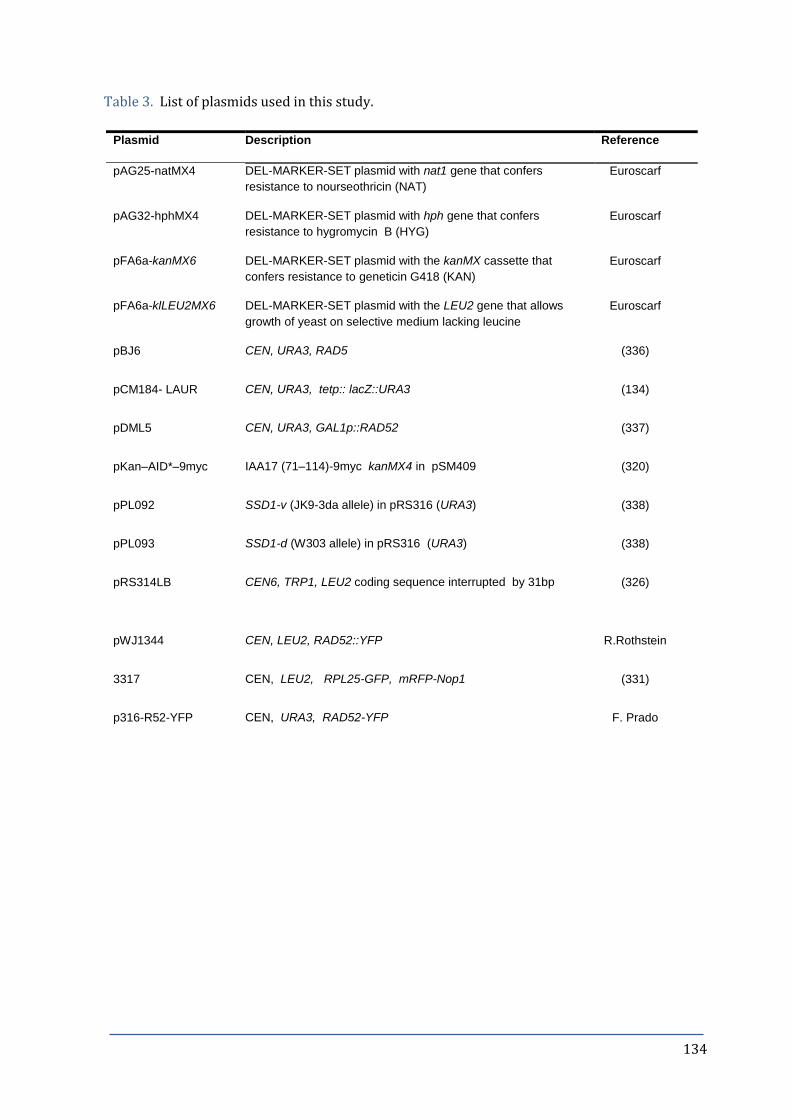
134
Table 3. List of plasmids used in this study.
Plasmid Description Reference
pAG25-natMX4 DEL-MARKER-SET plasmid with nat1 gene that confers
resistance to nourseothricin (NAT)
Euroscarf
pAG32-hphMX4 DEL-MARKER-SET plasmid with hph gene that confers
resistance to hygromycin B (HYG)
Euroscarf
pFA6a-kanMX6 DEL-MARKER-SET plasmid with the kanMX cassette that
confers resistance to geneticin G418 (KAN)
Euroscarf
pFA6a-klLEU2MX6 DEL-MARKER-SET plasmid with the LEU2 gene that allows
growth of yeast on selective medium lacking leucine
Euroscarf
pBJ6 CEN, URA3, RAD5 (336)
pCM184- LAUR CEN, URA3, tetp:: lacZ::URA3 (134)
pDML5 CEN, URA3, GAL1p::RAD52 (337)
pKan–AID*–9myc IAA17 (71–114)-9myc kanMX4 in pSM409 (320)
pPL092 SSD1-v (JK9-3da allele) in pRS316 (URA3) (338)
pPL093 SSD1-d (W303 allele) in pRS316 (URA3) (338)
pRS314LB CEN6, TRP1, LEU2 coding sequence interrupted by 31bp
(326)
pWJ1344 CEN, LEU2, RAD52::YFP R.Rothstein
3317 CEN, LEU2, RPL25-GFP, mRFP-Nop1 (331)
p316-R52-YFP CEN, URA3, RAD52-YFP F. Prado

135
8. ANNEXES
ANNEX I - Drugs and Reagents
Alpha factor (1mg/ml stock solution, dissolved in DMSO, Biomedal) was added to a final
concentration of 1µg/ml for BAR1+ or 0.3µg/ml for bar1Δ strains. The alpha factor pheromone
arrests yeast in the G1 phase of the cell cycle.
Ampicillin (Amp) (80mg/ml stock solution, dissolved in water, Sigma) was added to solid or
liquid LB at a final concentration of 80 g/ml. The Beta-lactame antibiotic was used to select
bacterial strains transformed with a plasmid of interest.
Benomyl (10mg/ml stock solution, dissolved in DMSO, Sigma) was added to a final
concentration of 5, 10 or 20µg/ml. The appropriate amount of stock solution was added to
warm agar with constant stirring and gentle heating to aid dissolution. Benomyl binds to
microtubules, acting as a spindle-depolymerizing drug.
Calcofluor white (CFW) (1mg/ml stock solution, dissolved in water, Sigma; CFW sold as
“Fluorescent Brightener 28”) was added to a final concentration of 5µg/ml. CFW binds to chitin
in the yeast cell wall and negatively affects normal cell wall assembly.
Camptothecin (CPT) (5mg/ml stock solution in 1% DMSO, Santa Cruz) was added to a final
concentration of 5µg/ml for plates, or 10µg/ml for liquid cultures, unless otherwise stated.
Canavanine sulphate (Can) (Sigma) was added to a final concentration of 60µg/ml, directly to
warm agar. Used to determine nuclear mutation rates.
Ethidium bromide (EtBr) (10mg/ml stock solution dissolved in water, Sigma) was added to a
final concentration of 0.4mg per 1 litre of agarose.
FITC-VAD-FMK (5mM stock solution dissolved in DMSO, Promega) was used at a final
concentration of 1µM. Used as an indicator to detect activated caspases.
5-fluororotic acid (FOA) (Melford) was added to a final concentration of 500mg/ml.
G418 disulphate (GEN) (200mg/ml stock solution, Melford) was added to a final
concentration of 200 g/ml. Used to select yeast that carry gene for resistance to
kanamycin/geneticin.

136
H2DCFDA (5mM stock solution dissolved in DMSO, Life Technologies) was used at a final
concentration of 5µM. Used as an indicator to detect the generation of ROS in cells.
Hydroxyurea (HU) (USBiological) was added to a final concentration of 100mM, directly to
warm agar.
Hygromycin (Hyg) (Applichem, 46KU/ml) was added to a final concentration of 0.3mg/ml.
Used to select yeast that carry gene for resistance to hygromycin.
Indole-3-acetic acid (IAA) (500mM stock solution dissolved in ethanol, Sigma) was added at a
final concentration of 500µM.
Methylene blue (10mM stock solution, diluted in water, Sigma) was added to a final
concentration of 20µM. Used to determine the viability of yeast cells.
Methyl methanesulphonate (MMS) (250M stock solution, Fluka) was added to a final
concentration of 25mM or as specified, directly to warm agar.
Nocodazole (Noc) (10mg/ml stock solution, dissolved in DMSO, Sigma) was added to a final
concentration of 15 g/ml.
Nourseotricine (NAT) (Werner BioAgents, Germany; NAT sold as “clonNAT”) was added to a
final concentration of 100µg/ml. Used to select yeast that carry gene for resistance to NAT.
Pronase (50mg/ml stock solution, dissolved in 50% glycerol, Sigma; pronase sold as “protease
from Streptococcus griseus”) was added to a final concentration of 1 g/ml. Used to digest alpha
factor, following G1 synchronization of bar1Δ yeast strains.
Propidium iodide (5mg/ml stock solution, dissolved in water, Sigma) was added to a final
concentration of 5 g/ml.

137
ANNEX II – Composition of buffers and solutions
Cocktail gently buffer 50mM Tris-HCl pH8, 20mM EDTA pH8
Coomassie stain 0.1% Coomassie Blue (w/w), 50% methanol, 1% acetic
acid
Coomassie destain 5% methanol, 7.5% acetic acid
CPE 40mM citric acid pH6, 120mM sodium phosphate pH6,
20mM EDTA-Na pH8
CPES 40mM citric acid pH6, 120mM sodium phosphate pH6,
20mM EDTA-Na pH8, 1.2M D-sorbitol, 1mg/ml
zymolyase 20T
CTAB II 1% w/v CTAB (cetyl-trimethyl-ammonium-bromide),
50mM Tris-HCl pH 7.6, 10mM EDTA pH8
CTAB III 1.4M NaCl, 10mM Tris-HCl pH7.6, 1mM EDTA pH8
Denaturation buffer 1.5M NaCl, 0.5M NaOH
G2 solution 800mM guanidine HCl, 30mM Tris-HCl pH8, 30mM
EDTA pH8, 5% Tween-20, 0.5% Triton X-100
Hybridization solution 0.25M Na2HPO4, 7% SDS, 1mM EDTA pH7.2
Laemmli buffer 2x 4% SDS, 20% glycerol, 10% β-mercaptoethanol, 0.004% bromophenol blue, 0.125 M Tris HCl
Neutralization buffer 1M Tris-HCl, 1.5M NaCl, adjusted to pH7.5.
PBS 10x 1,37 M NaCl, 27mM KCl, 81mM Na2HPO4, 14.7mM
KH2PO4
Ponceau Ponceau S 0.1% w/v, 5% acetic acid
Running buffer (Western) 50mM Tris Base, 192mM glycine pH8.3, 0.1% SDS
Sorbitol-citrate 1.2 M sorbitol in 0.12 M K2HPO4, 0.033 M citric
acid, pH 5.9
Spheroplasting buffer 1M sorbitol, 100mM EDTA pH8, 2mM Tris-HCl pH8,
0.1% v/v β-mercaptoethanol, 15U zymolyase/ml
Stripping solution (hybridization)0.1% SDS, 0.1X SSPE
SSC 20x 3M NaCl, 0.3M trisodium citrate, adjusted to pH7

138
TAE 50x 2M Tris, 1M acetic acid, and 10mM EDTA.
TBE 5x 0.45M Tris borate, 10mM EDTA-Na, adjusted to pH8
TBS 20 mM Tris-HCl pH8, 0.13M NaCl
TBS-Tween 20 mM Tris-HCl pH 8, 0.13M NaCl, 1% Tween-20, 5%
milk powder
TESP 10mM Tris-HCl pH8, 450mM EDTA-Na pH8, 1% SDS,
with 1mg/ml freshly prepared proteinase K
Transfer buffer (Western) 50mM Tris Base, 192mM glycine pH8.3, 0.01% SDS,
20% methanol
Wash solution (hybridization) 0.1X SSPE, 0.5mM EDTA, 0.5% SDS

139
ANNEX III – Published articles
STUCKEY R, Díaz de la Loza MC and Wellinger RE.
Cellular responses to mitochondrial DNA damage in yeast.
In: Mitochondria: Structure, Functions and Dysfunctions; Pp: 709-729, Ed. Ed. O. Svensson,
Nova Science Publishers NY; ISBN 978-1-61761-490-3 (2010).

140
9. BIBLIOGRAPHY
1. Mechali, M. (2010) Eukaryotic DNA replication origins: many choices for appropriate answers. Nat Rev Mol Cell Biol, 11, 728-738.
2. Lundblad, L. and Struhl, K. (eds.) (2010) Yeast. Current Protocols in Molecular Biology. 13.0.1-13.0.4.
3. Diffley, J.F., Cocker, J.H., Dowell, S.J. and Rowley, A. (1994) Two steps in the assembly of complexes at yeast replication origins in vivo. Cell, 78, 303-316.
4. Bousset, K. and Diffley, J.F. (1998) The Cdc7 protein kinase is required for origin firing during S phase. Genes Dev, 12, 480-490.
5. Kearsey, S.E. and Cotterill, S. (2003) Enigmatic variations: divergent modes of regulating eukaryotic DNA replication. Mol Cell, 12, 1067-1075.
6. Pursell, Z.F., Isoz, I., Lundstrom, E.B., Johansson, E. and Kunkel, T.A. (2007) Yeast DNA polymerase epsilon participates in leading-strand DNA replication. Science, 317, 127-130.
7. Crouse, G.F. (2010) An end for mismatch repair. Proc Natl Acad Sci U S A, 107, 20851-20852.
8. Reaban, M.E., Lebowitz, J. and Griffin, J.A. (1994) Transcription induces the formation of a stable RNA.DNA hybrid in the immunoglobulin alpha switch region. J Biol Chem, 269, 21850-21857.
9. Drolet, M., Bi, X. and Liu, L.F. (1994) Hypernegative supercoiling of the DNA template during transcription elongation in vitro. J Biol Chem, 269, 2068-2074.
10. Roy, D. and Lieber, M.R. (2009) G clustering is important for the initiation of transcription-induced R-loops in vitro, whereas high G density without clustering is sufficient thereafter. Mol Cell Biol, 29, 3124-3133.
11. Reddy, K., Tam, M., Bowater, R.P., Barber, M., Tomlinson, M., Nichol Edamura, K., Wang, Y.H. and Pearson, C.E. (2011) Determinants of R-loop formation at convergent bidirectionally transcribed trinucleotide repeats. Nucleic Acids Res, 39, 1749-1762.
12. Cerritelli, S.M. and Crouch, R.J. (2009) Ribonuclease H: the enzymes in eukaryotes. FEBS J, 276, 1494-1505.
13. Jeong, H.S., Backlund, P.S., Chen, H.C., Karavanov, A.A. and Crouch, R.J. (2004) RNase H2 of Saccharomyces cerevisiae is a complex of three proteins. Nucleic Acids Res, 32, 407-414.
14. Kim, N., Huang, S.N., Williams, J.S., Li, Y.C., Clark, A.B., Cho, J.E., Kunkel, T.A., Pommier, Y. and Jinks-Robertson, S. (2011) Mutagenic processing of ribonucleotides in DNA by yeast topoisomerase I. Science, 332, 1561-1564.
15. Nick McElhinny, S.A., Watts, B.E., Kumar, D., Watt, D.L., Lundstrom, E.B., Burgers, P.M., Johansson, E., Chabes, A. and Kunkel, T.A. (2010) Abundant ribonucleotide incorporation into DNA by yeast replicative polymerases. Proc Natl Acad Sci U S A, 107, 4949-4954.
16. Aguilera, A. and Garcia-Muse, T. (2012) R loops: from transcription byproducts to threats to genome stability. Mol Cell, 46, 115-124.
17. Beletskii, A. and Bhagwat, A.S. (1996) Transcription-induced mutations: increase in C to T mutations in the nontranscribed strand during transcription in Escherichia coli. Proc Natl Acad Sci U S A, 93, 13919-13924.
18. Helmrich, A., Ballarino, M., Nudler, E. and Tora, L. (2013) Transcription-replication encounters, consequences and genomic instability. Nat Struct Mol Biol, 20, 412-418.
19. Huertas, P. and Aguilera, A. (2003) Cotranscriptionally formed DNA:RNA hybrids mediate transcription elongation impairment and transcription-associated recombination. Mol Cell, 12, 711-721.
20. Li, X. and Manley, J.L. (2005) Inactivation of the SR protein splicing factor ASF/SF2 results in genomic instability. Cell, 122, 365-378.

141
21. Wahba, L., Amon, J.D., Koshland, D. and Vuica-Ross, M. (2011) RNase H and multiple RNA biogenesis factors cooperate to prevent RNA:DNA hybrids from generating genome instability. Mol Cell, 44, 978-988.
22. Stirling, P.C., Chan, Y.A., Minaker, S.W., Aristizabal, M.J., Barrett, I., Sipahimalani, P., Kobor, M.S. and Hieter, P. (2012) R-loop-mediated genome instability in mRNA cleavage and polyadenylation mutants. Genes Dev, 26, 163-175.
23. Luna, R., Jimeno, S., Marin, M., Huertas, P., Garcia-Rubio, M. and Aguilera, A. (2005) Interdependence between transcription and mRNP processing and export, and its impact on genetic stability. Mol Cell, 18, 711-722.
24. Baaklini, I., Hraiky, C., Rallu, F., Tse-Dinh, Y.C. and Drolet, M. (2004) RNase HI overproduction is required for efficient full-length RNA synthesis in the absence of topoisomerase I in Escherichia coli. Mol Microbiol, 54, 198-211.
25. Wellinger, R.E., Prado, F. and Aguilera, A. (2006) Replication fork progression is impaired by transcription in hyperrecombinant yeast cells lacking a functional THO complex. Mol Cell Biol, 26, 3327-3334.
26. Gan, W., Guan, Z., Liu, J., Gui, T., Shen, K., Manley, J.L. and Li, X. (2011) R-loop-mediated genomic instability is caused by impairment of replication fork progression. Genes Dev, 25, 2041-2056.
27. McIvor, E.I., Polak, U. and Napierala, M. (2010) New insights into repeat instability: role of RNA*DNA hybrids. RNA Biol, 7, 551-558.
28. Lin, Y., Dent, S.Y., Wilson, J.H., Wells, R.D. and Napierala, M. (2010) R loops stimulate genetic instability of CTG.CAG repeats. Proc Natl Acad Sci U S A, 107, 692-697.
29. Skourti-Stathaki, K., Proudfoot, N.J. and Gromak, N. (2011) Human senataxin resolves RNA/DNA hybrids formed at transcriptional pause sites to promote Xrn2-dependent termination. Mol Cell, 42, 794-805.
30. Yu, K., Chedin, F., Hsieh, C.L., Wilson, T.E. and Lieber, M.R. (2003) R-loops at immunoglobulin class switch regions in the chromosomes of stimulated B cells. Nat Immunol, 4, 442-451.
31. Muramatsu, M., Kinoshita, K., Fagarasan, S., Yamada, S., Shinkai, Y. and Honjo, T. (2000) Class switch recombination and hypermutation require activation-induced cytidine deaminase (AID), a potential RNA editing enzyme. Cell, 102, 553-563.
32. Zarrin, A.A., Del Vecchio, C., Tseng, E., Gleason, M., Zarin, P., Tian, M. and Alt, F.W. (2007) Antibody class switching mediated by yeast endonuclease-generated DNA breaks. Science, 315, 377-381.
33. Ginno, P.A., Lott, P.L., Christensen, H.C., Korf, I. and Chedin, F. (2012) R-loop formation is a distinctive characteristic of unmethylated human CpG island promoters. Mol Cell, 45, 814-825.
34. Deng, W. (2010) AID in reprogramming: quick and efficient: identification of a key enzyme called AID, and its activity in DNA demethylation, may help to overcome a pivotal epigenetic barrier in reprogramming somatic cells toward pluripotency. Bioessays, 32, 385-387.
35. Bhutani, N., Brady, J.J., Damian, M., Sacco, A., Corbel, S.Y. and Blau, H.M. (2010) Reprogramming towards pluripotency requires AID-dependent DNA demethylation. Nature, 463, 1042-1047.
36. Dunn, K. and Griffith, J.D. (1980) The presence of RNA in a double helix inhibits its interaction with histone protein. Nucleic Acids Res, 8, 555-566.
37. Castellano-Pozo, M., Santos-Pereira, J.M., Rondon, A.G., Barroso, S., Andujar, E., Perez-Alegre, M., Garcia-Muse, T. and Aguilera, A. (2013) R loops are linked to histone H3 S10 phosphorylation and chromatin condensation. Mol Cell, 52, 583-590.
38. Fuller, C.W. and Richardson, C.C. (1985) Initiation of DNA replication at the primary origin of bacteriophage T7 by purified proteins. Site and direction of initial DNA synthesis. J Biol Chem, 260, 3185-3196.
39. Itoh, T. and Tomizawa, J. (1980) Formation of an RNA primer for initiation of replication of ColE1 DNA by ribonuclease H PNAS, 77, 2450-2454

142
40. Kasiviswanathan, R., Collins, T.R. and Copeland, W.C. (2012) The interface of transcription and DNA replication in the mitochondria. Biochim Biophys Acta, 1819, 970-978.
41. Chang, D.D., Hauswirth, W.W. and Clayton, D.A. (1985) Replication priming and transcription initiate from precisely the same site in mouse mitochondrial DNA. EMBO J, 4, 1559-1567.
42. Xu, B. and Clayton, D.A. (1996) RNA-DNA hybrid formation at the human mitochondrial heavy-strand origin ceases at replication start sites: an implication for RNA-DNA hybrids serving as primers. EMBO J, 15, 3135–3143.
43. Lee, D.Y. and Clayton, D.A. (1997) RNase mitochondrial RNA processing correctly cleaves a novel R loop at the mitochondrial DNA leading-strand origin of replication. Genes Dev, 11, 582-592.
44. Kogoma, T. and Lark, K.G. (1970) DNA replication in Escherihia coli: replication in absence of protein synthesis after replication inhibition. J Mol Biol, 52, 143-164.
45. Ogawa, T., Pickett, G., Kogoma, T. and Kornberg, A. (1984) RNase H confers specificity in the dnaA-dependent initiation of replication at the unique origin of the Escherichia coli chromosome in vivo and in vitro. Proc Natl Acad Sci U S A, 81, 1040-1044.
46. Sandler, S.J. (2005) Requirements for replication restart proteins during constitutive stable DNA replication in Escherichia coli K-12. Genetics, 169, 1799-1806.
47. Lindahl, T. (1993) Instability and decay of the primary structure of DNA. Nature, 362, 709-715.
48. Vengrova, S. and Dalgaard, J.Z. (2006) The wild-type Schizosaccharomyces pombe mat1 imprint consists of two ribonucleotides. EMBO Rep, 7, 59-65.
49. Nick McElhinny, S.A., Kumar, D., Clark, A.B., Watt, D.L., Watts, B.E., Lundstrom, E.B., Johansson, E., Chabes, A. and Kunkel, T.A. (2010) Genome instability due to ribonucleotide incorporation into DNA. Nat Chem Biol, 6, 774-781.
50. Cerritelli, S.M., Frolova, E.G., Feng, C., Grinberg, A., Love, P.E. and Crouch, R.J. (2003) Failure to produce mitochondrial DNA results in embryonic lethality in Rnaseh1 null mice. Mol Cell, 11, 807-815.
51. Reijns, M.A., Rabe, B., Rigby, R.E., Mill, P., Astell, K.R., Lettice, L.A., Boyle, S., Leitch, A., Keighren, M., Kilanowski, F. et al. (2012) Enzymatic removal of ribonucleotides from DNA is essential for mammalian genome integrity and development. Cell, 149, 1008-1022.
52. Crow, Y.J., Leitch, A., Hayward, B.E., Garner, A., Parmar, R., Griffith, E., Ali, M., Semple, C., Aicardi, J., Babul-Hirji, R. et al. (2006) Mutations in genes encoding ribonuclease H2 subunits cause Aicardi-Goutieres syndrome and mimic congenital viral brain infection. Nat Genet, 38, 910-916.
53. Crow, Y.J., Hayward, B.E., Parmar, R., Robins, P., Leitch, A., Ali, M., Black, D.N., van Bokhoven, H., Brunner, H.G., Hamel, B.C. et al. (2006) Mutations in the gene encoding the 3'-5' DNA exonuclease TREX1 cause Aicardi-Goutieres syndrome at the AGS1 locus. Nat Genet, 38, 917-920.
54. Rigby, R.E., Leitch, A. and Jackson, A.P. (2008) Nucleic acid-mediated inflammatory diseases. Bioessays, 30, 833-842.
55. Prinz, M. and Knobeloch, K.P. (2012) Type I interferons as ambiguous modulators of chronic inflammation in the central nervous system. Front Immunol, 3, 67.
56. Crow, Y.J. and Rehwinkel, J. (2009) Aicardi-Goutieres syndrome and related phenotypes: linking nucleic acid metabolism with autoimmunity. Hum Mol Genet, 18, R130-136.
57. Boule, J.B. and Zakian, V.A. (2007) The yeast Pif1p DNA helicase preferentially unwinds RNA DNA substrates. Nucleic Acids Res, 35, 5809-5818.
58. Mischo, H.E., Gomez-Gonzalez, B., Grzechnik, P., Rondon, A.G., Wei, W., Steinmetz, L., Aguilera, A. and Proudfoot, N.J. (2011) Yeast Sen1 helicase protects the genome from transcription-associated instability. Mol Cell, 41, 21-32.

143
59. Drolet, M. (2006) Growth inhibition mediated by excess negative supercoiling: the interplay between transcription elongation, R-loop formation and DNA topology. Mol Microbiol, 59, 723-730.
60. El Hage, A., French, S.L., Beyer, A.L. and Tollervey, D. (2010) Loss of Topoisomerase I leads to R-loop-mediated transcriptional blocks during ribosomal RNA synthesis. Genes Dev, 24, 1546-1558.
61. Wang, J.C. (2002) Cellular roles of DNA topoisomerases: a molecular perspective. Nat Rev Mol Cell Biol, 3, 430-440.
62. Pourquier, P., Pilon, A.A., Kohlhagen, G., Mazumder, A., Sharma, A. and Pommier, Y. (1997) Trapping of mammalian topoisomerase I and recombinations induced by damaged DNA containing nicks or gaps. Importance of DNA end phosphorylation and camptothecin effects. J Biol Chem, 272, 26441-26447.
63. Staker, B.L., Hjerrild, K., Feese, M.D., Behnke, C.A., Burgin, A.B., Jr. and Stewart, L. (2002) The mechanism of topoisomerase I poisoning by a camptothecin analog. Proc Natl Acad Sci U S A, 99, 15387-15392.
64. Pommier, Y. (2006) Topoisomerase I inhibitors: camptothecins and beyond. Nat Rev Cancer, 6, 789-802.
65. Pouliot, J.J., Yao, K.C., Robertson, C.A. and Nash, H.A. (1999) Yeast gene for a Tyr-DNA phosphodiesterase that repairs topoisomerase I complexes. Science, 286, 552-555.
66. Vance, J.R. and Wilson, T.E. (2002) Yeast Tdp1 and Rad1-Rad10 function as redundant pathways for repairing Top1 replicative damage. Proc Natl Acad Sci U S A, 99, 13669-13674.
67. Deng, C., Brown, J.A., You, D. and Brown, J.M. (2005) Multiple endonucleases function to repair covalent topoisomerase I complexes in Saccharomyces cerevisiae. Genetics, 170, 591-600.
68. Warner, J.R. (1999) The economics of ribosome biosynthesis in yeast. Trends Biochem Sci, 24, 437-440.
69. Staub, E., Mackowiak, S. and Vingron, M. (2006) An inventory of yeast proteins associated with nucleolar and ribosomal components. Genome Biol, 7, R98.
70. Mandal, R.K. (1984) The organization and transcription of eukaryotic ribosomal RNA genes. Prog Nucleic Acid Res Mol Biol, 31, 115-160.
71. Brewer, B.J. and Fangman, W.L. (1988) A replication fork barrier at the 3' end of yeast ribosomal RNA genes. Cell, 55, 637-643.
72. Little, R.D., Platt, T.H. and Schildkraut, C.L. (1993) Initiation and termination of DNA replication in human rRNA genes. Mol Cell Biol, 13, 6600-6613.
73. Laloraya, S., Guacci, V. and Koshland, D. (2000) Chromosomal addresses of the cohesin component Mcd1p. J Cell Biol, 151, 1047-1056.
74. Nicklas, R.B. (1997) How cells get the right chromosomes. Science, 275, 632-637. 75. Ide, S., Miyazaki, T., Maki, H. and Kobayashi, T. (2010) Abundance of ribosomal RNA
gene copies maintains genome integrity. Science, 327, 693-696. 76. Liu, B. and Alberts, B.M. (1995) Head-on collision between a DNA replication apparatus
and RNA polymerase transcription complex. Science, 267, 1131-1137. 77. Prado, F. and Aguilera, A. (2005) Impairment of replication fork progression mediates
RNA polII transcription-associated recombination. EMBO J, 24, 1267-1276. 78. Brewer, B.J. (1988) When polymerases collide: replication and the transcriptional
organization of the E. coli chromosome. Cell, 53, 679-686. 79. Pomerantz, R.T. and O'Donnell, M. (2010) What happens when replication and
transcription complexes collide? Cell Cycle, 9, 2537-2543. 80. Pomerantz, R.T. and O'Donnell, M. (2008) The replisome uses mRNA as a primer after
colliding with RNA polymerase. Nature, 456, 762-766. 81. Linskens, M.H. and Huberman, J.A. (1988) Organization of replication of ribosomal DNA
in Saccharomyces cerevisiae. Mol Cell Biol, 8, 4927-4935. 82. Deshpande, A.M. and Newlon, C.S. (1996) DNA replication fork pause sites dependent
on transcription. Science, 272, 1030-1033.

144
83. Bermejo, R., Lai, M.S. and Foiani, M. (2012) Preventing replication stress to maintain genome stability: resolving conflicts between replication and transcription. Mol Cell, 45, 710-718.
84. Branzei, D. and Foiani, M. (2008) Regulation of DNA repair throughout the cell cycle. Nat Rev Mol Cell Biol, 9, 297-308.
85. Clark, J.M. and Beardsley, G.P. (1986) Thymine glycol lesions terminate chain elongation by DNA polymerase I in vitro. Nucleic Acids Res, 14, 737-749.
86. Tornaletti, S., Maeda, L.S., Lloyd, D.R., Reines, D. and Hanawalt, P.C. (2001) Effect of thymine glycol on transcription elongation by T7 RNA polymerase and mammalian RNA polymerase II. J Biol Chem, 276, 45367-45371.
87. Lu, A.L., Li, X., Gu, Y., Wright, P.M. and Chang, D.Y. (2001) Repair of oxidative DNA damage: mechanisms and functions. Cell Biochem Biophys, 35, 141-170.
88. Shuck, S.C., Short, E.A. and Turchi, J.J. (2008) Eukaryotic nucleotide excision repair: from understanding mechanisms to influencing biology. Cell Res, 18, 64-72.
89. Gaillard, H. and Aguilera, A. (2012) Transcription coupled repair at the interface between transcription elongation and mRNP biogenesis. Biochim Biophys Acta, 1829, 141-150.
90. Lieber, M.R. (2008) The mechanism of human nonhomologous DNA end joining. J Biol Chem, 283, 1-5.
91. Barlow, J.H., Lisby, M. and Rothstein, R. (2008) Differential regulation of the cellular response to DNA double-strand breaks in G1. Mol Cell, 30, 73-85.
92. Symington, L.S. and Gautier, J. (2011) Double-strand break end resection and repair pathway choice. Annu Rev Genet, 45, 247-271.
93. Wiltzius, J.J., Hohl, M., Fleming, J.C. and Petrini, J.H. (2005) The Rad50 hook domain is a critical determinant of Mre11 complex functions. Nat Struct Mol Biol, 12, 403-407.
94. Paques, F. and Haber, J.E. (1999) Multiple pathways of recombination induced by double-strand breaks in Saccharomyces cerevisiae. Microbiol Mol Biol Rev, 63, 349-404.
95. Prakash, L. (1981) Characterization of postreplication repair in Saccharomyces cerevisiae and effects of rad6, rad18, rev3 and rad52 mutations. Mol Gen Genet, 184, 471-478.
96. Bailly, V., Lauder, S., Prakash, S. and Prakash, L. (1997) Yeast DNA repair proteins Rad6 and Rad18 form a heterodimer that has ubiquitin conjugating, DNA binding, and ATP hydrolytic activities. J Biol Chem, 272, 23360-23365.
97. Woodgate, R. (1999) A plethora of lesion-replicating DNA polymerases. Genes Dev, 13, 2191-2195.
98. Zhang, H. and Lawrence, C.W. (2005) The error-free component of the RAD6/RAD18 DNA damage tolerance pathway of budding yeast employs sister-strand recombination. Proc Natl Acad Sci U S A, 102, 15954-15959.
99. Lazzaro, F., Novarina, D., Amara, F., Watt, D.L., Stone, J.E., Costanzo, V., Burgers, P.M., Kunkel, T.A., Plevani, P. and Muzi-Falconi, M. (2012) RNase H and postreplication repair protect cells from ribonucleotides incorporated in DNA. Mol Cell, 45, 99-110.
100. Hartwell, L.H. and Weinert, T.A. (1989) Checkpoints: controls that ensure the order of cell cycle events. Science, 246, 629-634.
101. Zhou, B.B. and Elledge, S.J. (2000) The DNA damage response: putting checkpoints in perspective. Nature, 408, 433-439.
102. Hartwell, L.H. and Kastan, M.B. (1994) Cell cycle control and cancer. Science, 266, 1821-1828.
103. Elledge, S.J. (1996) Cell cycle checkpoints: preventing an identity crisis. Science, 274, 1664-1672.
104. Hanahan, D. and Weinberg, R.A. (2011) Hallmarks of cancer: the next generation. Cell, 144, 646-674.
105. Sherlock, G. and Rosamond, J. (1993) Starting to cycle: G1 controls regulating cell division in budding yeast. J Gen Microbiol, 139, 2531-2541.

145
106. Gerald, J.N., Benjamin, J.M. and Kron, S.J. (2002) Robust G1 checkpoint arrest in budding yeast: dependence on DNA damage signaling and repair. J Cell Sci, 115, 1749-1757.
107. Branzei, D. and Foiani, M. (2007) Interplay of replication checkpoints and repair proteins at stalled replication forks. DNA Repair (Amst), 6, 994-1003.
108. Weinert, T.A. and Hartwell, L.H. (1988) The RAD9 gene controls the cell cycle response to DNA damage in Saccharomyces cerevisiae. Science, 241, 317-322.
109. Alcasabas, A.A., Osborn, A.J., Bachant, J., Hu, F., Werler, P.J., Bousset, K., Furuya, K., Diffley, J.F., Carr, A.M. and Elledge, S.J. (2001) Mrc1 transduces signals of DNA replication stress to activate Rad53. Nat Cell Biol, 3, 958-965.
110. Sanchez, Y., Desany, B.A., Jones, W.J., Liu, Q., Wang, B. and Elledge, S.J. (1996) Regulation of RAD53 by the ATM-like kinases MEC1 and TEL1 in yeast cell cycle checkpoint pathways. Science, 271, 357-360.
111. Tercero, J.A. and Diffley, J.F. (2001) Regulation of DNA replication fork progression through damaged DNA by the Mec1/Rad53 checkpoint. Nature, 412, 553-557.
112. Zhou, Z. and Elledge, S.J. (1993) DUN1 encodes a protein kinase that controls the DNA damage response in yeast. Cell, 75, 1119-1127.
113. Santocanale, C. and Diffley, J.F. (1998) A Mec1- and Rad53-dependent checkpoint controls late-firing origins of DNA replication. Nature, 395, 615-618.
114. Weinert, T.A., Kiser, G.L. and Hartwell, L.H. (1994) Mitotic checkpoint genes in budding yeast and the dependence of mitosis on DNA replication and repair. Genes Dev, 8, 652-665.
115. Lew, D.J. (2003) The morphogenesis checkpoint: how yeast cells watch their figures. Curr Opin Cell Biol, 15, 648-653.
116. Musacchio, A. and Salmon, E.D. (2007) The spindle-assembly checkpoint in space and time. Nat Rev Mol Cell Biol, 8, 379-393.
117. Visintin, R., Prinz, S. and Amon, A. (1997) CDC20 and CDH1: a family of substrate-specific activators of APC-dependent proteolysis. Science, 278, 460-463.
118. Cohen-Fix, O., Peters, J.M., Kirschner, M.W. and Koshland, D. (1996) Anaphase initiation in Saccharomyces cerevisiae is controlled by the APC-dependent degradation of the anaphase inhibitor Pds1p. Genes Dev, 10, 3081-3093.
119. Storchova, Z. and Pellman, D. (2004) From polyploidy to aneuploidy, genome instability and cancer. Nat Rev Mol Cell Biol, 5, 45-54.
120. Arudchandran, A., Cerritelli, S., Narimatsu, S., Itaya, M., Shin, D.Y., Shimada, Y. and Crouch, R.J. (2000) The absence of ribonuclease H1 or H2 alters the sensitivity of Saccharomyces cerevisiae to hydroxyurea, caffeine and ethyl methanesulphonate: implications for roles of RNases H in DNA replication and repair. Genes Cells, 5, 789-802.
121. Saavedra, R.A. and Huberman, J.A. (1986) Both DNA topoisomerases I and II relax 2 micron plasmid DNA in living yeast cells. Cell, 45, 65-70.
122. Pommier, Y. (2009) DNA topoisomerase I inhibitors: chemistry, biology, and interfacial inhibition. Chem Rev, 109, 2894-2902.
123. Marinello, J., Chillemi, G., Bueno, S., Manzo, S.G. and Capranico, G. (2013) Antisense transcripts enhanced by camptothecin at divergent CpG-island promoters associated with bursts of topoisomerase I-DNA cleavage complex and R-loop formation. Nucleic Acids Res, 41, 10110-10123.
124. Labib, K., Tercero, J.A. and Diffley, J.F. (2000) Uninterrupted MCM2-7 function required for DNA replication fork progression. Science, 288, 1643-1647.
125. Fan, H.Y., Cheng, K.K. and Klein, H.L. (1996) Mutations in the RNA polymerase II transcription machinery suppress the hyperrecombination mutant hpr1 delta of Saccharomyces cerevisiae. Genetics, 142, 749-759.
126. Kaeberlein, M. and Guarente, L. (2002) Saccharomyces cerevisiae MPT5 and SSD1 function in parallel pathways to promote cell wall integrity. Genetics, 160, 83-95.
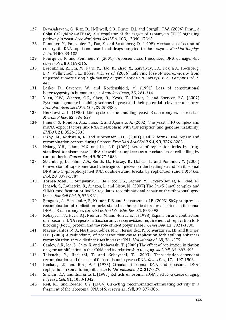
146
127. Devasahayam, G., Ritz, D., Helliwell, S.B., Burke, D.J. and Sturgill, T.W. (2006) Pmr1, a Golgi Ca2+/Mn2+-ATPase, is a regulator of the target of rapamycin (TOR) signaling pathway in yeast. Proc Natl Acad Sci U S A, 103, 17840-17845.
128. Pommier, Y., Pourquier, P., Fan, Y. and Strumberg, D. (1998) Mechanism of action of eukaryotic DNA topoisomerase I and drugs targeted to the enzyme. Biochim Biophys Acta, 1400, 83-105.
129. Pourquier, P. and Pommier, Y. (2001) Topoisomerase I-mediated DNA damage. Adv Cancer Res, 80, 189-216.
130. Beroukhim, R., Lin, M., Park, Y., Hao, K., Zhao, X., Garraway, L.A., Fox, E.A., Hochberg, E.P., Mellinghoff, I.K., Hofer, M.D. et al. (2006) Inferring loss-of-heterozygosity from unpaired tumors using high-density oligonucleotide SNP arrays. PLoS Comput Biol, 2, e41.
131. Lasko, D., Cavenee, W. and Nordenskjold, M. (1991) Loss of constitutional heterozygosity in human cancer. Annu Rev Genet, 25, 281-314.
132. Yuen, K.W., Warren, C.D., Chen, O., Kwok, T., Hieter, P. and Spencer, F.A. (2007) Systematic genome instability screens in yeast and their potential relevance to cancer. Proc Natl Acad Sci U S A, 104, 3925-3930.
133. Herskowitz, I. (1988) Life cycle of the budding yeast Saccharomyces cerevisiae. Microbiol Rev, 52, 536-553.
134. Jimeno, S., Rondon, A.G., Luna, R. and Aguilera, A. (2002) The yeast THO complex and mRNA export factors link RNA metabolism with transcription and genome instability. EMBO J, 21, 3526-3535.
135. Lisby, M., Rothstein, R. and Mortensen, U.H. (2001) Rad52 forms DNA repair and recombination centers during S phase. Proc Natl Acad Sci U S A, 98, 8276-8282.
136. Hsiang, Y.H., Lihou, M.G. and Liu, L.F. (1989) Arrest of replication forks by drug-stabilized topoisomerase I-DNA cleavable complexes as a mechanism of cell killing by camptothecin. Cancer Res, 49, 5077-5082.
137. Strumberg, D., Pilon, A.A., Smith, M., Hickey, R., Malkas, L. and Pommier, Y. (2000) Conversion of topoisomerase I cleavage complexes on the leading strand of ribosomal DNA into 5'-phosphorylated DNA double-strand breaks by replication runoff. Mol Cell Biol, 20, 3977-3987.
138. Torres-Rosell, J., Sunjevaric, I., De Piccoli, G., Sacher, M., Eckert-Boulet, N., Reid, R., Jentsch, S., Rothstein, R., Aragon, L. and Lisby, M. (2007) The Smc5-Smc6 complex and SUMO modification of Rad52 regulates recombinational repair at the ribosomal gene locus. Nat Cell Biol, 9, 923-931.
139. Benguria, A., Hernandez, P., Krimer, D.B. and Schvartzman, J.B. (2003) Sir2p suppresses recombination of replication forks stalled at the replication fork barrier of ribosomal DNA in Saccharomyces cerevisiae. Nucleic Acids Res, 31, 893-898.
140. Kobayashi, T., Heck, D.J., Nomura, M. and Horiuchi, T. (1998) Expansion and contraction of ribosomal DNA repeats in Saccharomyces cerevisiae: requirement of replication fork blocking (Fob1) protein and the role of RNA polymerase I. Genes Dev, 12, 3821-3830.
141. Mayan-Santos, M.D., Martinez-Robles, M.L., Hernandez, P., Schvartzman, J.B. and Krimer, D.B. (2008) A redundancy of processes that cause replication fork stalling enhances recombination at two distinct sites in yeast rDNA. Mol Microbiol, 69, 361-375.
142. Ganley, A.R., Ide, S., Saka, K. and Kobayashi, T. (2009) The effect of replication initiation on gene amplification in the rDNA and its relationship to aging. Mol Cell, 35, 683-693.
143. Takeuchi, Y., Horiuchi, T. and Kobayashi, T. (2003) Transcription-dependent recombination and the role of fork collision in yeast rDNA. Genes Dev, 17, 1497-1506.
144. Rochaix, J.D. and Bird, A.P. (1975) Circular ribosomal DNA and ribosomal DNA: replication in somatic amphibian cells. Chromosoma, 52, 317-327.
145. Sinclair, D.A. and Guarente, L. (1997) Extrachromosomal rDNA circles--a cause of aging in yeast. Cell, 91, 1033-1042.
146. Keil, R.L. and Roeder, G.S. (1984) Cis-acting, recombination-stimulating activity in a fragment of the ribosomal DNA of S. cerevisiae. Cell, 39, 377-386.

147
147. Ivessa, A.S., Zhou, J.Q. and Zakian, V.A. (2000) The Saccharomyces Pif1p DNA helicase and the highly related Rrm3p have opposite effects on replication fork progression in ribosomal DNA. Cell, 100, 479-489.
148. Vengrova, S. and Dalgaard, J.Z. (2004) RNase-sensitive DNA modification(s) initiates S. pombe mating-type switching. Genes Dev, 18, 794-804.
149. Dalgaard, J.Z. (2012) Causes and consequences of ribonucleotide incorporation into nuclear DNA. Trends Genet, 28, 592-597.
150. Pohjoismaki, J.L., Holmes, J.B., Wood, S.R., Yang, M.Y., Yasukawa, T., Reyes, A., Bailey, L.J., Cluett, T.J., Goffart, S., Willcox, S. et al. (2010) Mammalian mitochondrial DNA replication intermediates are essentially duplex but contain extensive tracts of RNA/DNA hybrid. J Mol Biol, 397, 1144-1155.
151. Ganley, A.R., Hayashi, K., Horiuchi, T. and Kobayashi, T. (2005) Identifying gene-independent noncoding functional elements in the yeast ribosomal DNA by phylogenetic footprinting. Proc Natl Acad Sci U S A, 102, 11787-11792.
152. Santangelo, G.M., Tornow, J., McLaughlin, C.S. and Moldave, K. (1988) Properties of promoters cloned randomly from the Saccharomyces cerevisiae genome. Mol Cell Biol, 8, 4217-4224.
153. Torres, J.Z., Bessler, J.B. and Zakian, V.A. (2004) Local chromatin structure at the ribosomal DNA causes replication fork pausing and genome instability in the absence of the S. cerevisiae DNA helicase Rrm3p. Genes Dev, 18, 498-503.
154. Wellinger, R.E., Schar, P. and Sogo, J.M. (2003) Rad52-independent accumulation of joint circular minichromosomes during S phase in Saccharomyces cerevisiae. Mol Cell Biol, 23, 6363-6372.
155. Nishimura, K., Fukagawa, T., Takisawa, H., Kakimoto, T. and Kanemaki, M. (2009) An auxin-based degron system for the rapid depletion of proteins in nonplant cells. Nat Methods, 6, 917-922.
156. Boguslawski, S.J., Smith, D.E., Michalak, M.A., Mickelson, K.E., Yehle, C.O., Patterson, W.L. and Carrico, R.J. (1986) Characterization of monoclonal antibody to DNA.RNA and its application to immunodetection of hybrids. J Immunol Methods, 89, 123-130.
157. Wittekind, M., Dodd, J., Vu, L., Kolb, J.M., Buhler, J.M., Sentenac, A. and Nomura, M. (1988) Isolation and characterization of temperature-sensitive mutations in RPA190, the gene encoding the largest subunit of RNA polymerase I from Saccharomyces cerevisiae. Mol Cell Biol, 8, 3997-4008.
158. Bell, S.P. and Dutta, A. (2002) DNA replication in eukaryotic cells. Annu Rev Biochem, 71, 333-374.
159. Bramhill, D. and Kornberg, A. (1988) Duplex opening by dnaA protein at novel sequences in initiation of replication at the origin of the E. coli chromosome. Cell, 52, 743-755.
160. Itoh, T. and Tomizawa, J. (1980) Formation of an RNA primer for initiation of replication of ColE1 DNA by ribonuclease H. Proc Natl Acad Sci U S A, 77, 2450-2454.
161. Mouron, S., Rodriguez-Acebes, S., Martinez-Jimenez, M.I., Garcia-Gomez, S., Chocron, S., Blanco, L. and Mendez, J. (2013) Repriming of DNA synthesis at stalled replication forks by human PrimPol. Nat Struct Mol Biol, 20, 1383-1389.
162. Garcia-Gomez, S., Reyes, A., Martinez-Jimenez, M.I., Chocron, E.S., Mouron, S., Terrados, G., Powell, C., Salido, E., Mendez, J., Holt, I.J. et al. (2013) PrimPol, an archaic primase/polymerase operating in human cells. Mol Cell, 52, 541-553.
163. Kim, R.A. and Wang, J.C. (1989) A subthreshold level of DNA topoisomerases leads to the excision of yeast rDNA as extrachromosomal rings. Cell, 57, 975-985.
164. Kobayashi, T. (2006) Strategies to maintain the stability of the ribosomal RNA gene repeats--collaboration of recombination, cohesion, and condensation. Genes Genet Syst, 81, 155-161.
165. Merker, R.J. and Klein, H.L. (2002) hpr1Delta affects ribosomal DNA recombination and cell life span in Saccharomyces cerevisiae. Mol Cell Biol, 22, 421-429.

148
166. Mehta, I.S., Figgitt, M., Clements, C.S., Kill, I.R. and Bridger, J.M. (2007) Alterations to nuclear architecture and genome behavior in senescent cells. Ann N Y Acad Sci, 1100, 250-263.
167. Campisi, J. and d'Adda di Fagagna, F. (2007) Cellular senescence: when bad things happen to good cells. Nat Rev Mol Cell Biol, 8, 729-740.
168. Cavanaugh, A.H., Hempel, W.M., Taylor, L.J., Rogalsky, V., Todorov, G. and Rothblum, L.I. (1995) Activity of RNA polymerase I transcription factor UBF blocked by Rb gene product. Nature, 374, 177-180.
169. Hannan, K.M., Hannan, R.D., Smith, S.D., Jefferson, L.S., Lun, M. and Rothblum, L.I. (2000) Rb and p130 regulate RNA polymerase I transcription: Rb disrupts the interaction between UBF and SL-1. Oncogene, 19, 4988-4999.
170. Krawczyk, C., Dion, V., Schar, P. and Fritsch, O. (2014) Reversible Top1 cleavage complexes are stabilized strand-specifically at the ribosomal replication fork barrier and contribute to ribosomal DNA stability. Nucleic Acids Res.
171. Vogelauer, M. and Camilloni, G. (1999) Site-specific in vivo cleavages by DNA topoisomerase I in the regulatory regions of the 35 S rRNA in Saccharomyces cerevisiae are transcription independent. J Mol Biol, 293, 19-28.
172. Gan, W., Guan, Z., Liu, J., Gui, T., Shen, K., Manley, J.L. and Li, X. R-loop-mediated genomic instability is caused by impairment of replication fork progression. Genes Dev, 25, 2041-2056.
173. Coelho, P.S., Bryan, A.C., Kumar, A., Shadel, G.S. and Snyder, M. (2002) A novel mitochondrial protein, Tar1p, is encoded on the antisense strand of the nuclear 25S rDNA. Genes Dev, 16, 2755-2760.
174. White, R.J. (2005) RNA polymerases I and III, growth control and cancer. Nat Rev Mol Cell Biol, 6, 69-78.
175. ten Asbroek, A.L., van Groenigen, M., Nooij, M. and Baas, F. (2002) The involvement of human ribonucleases H1 and H2 in the variation of response of cells to antisense phosphorothioate oligonucleotides. Eur J Biochem, 269, 583-592.
176. Teeter, L.D., Atsumi, S., Sen, S. and Kuo, T. (1986) DNA amplification in multidrug, cross-resistant Chinese hamster ovary cells: molecular characterization and cytogenetic localization of the amplified DNA. J Cell Biol, 103, 1159-1166.
177. Schwab, M. (1998) Amplification of oncogenes in human cancer cells. Bioessays, 20, 473-479.
178. Kuo, M.T., Sen, S., Hittelman, W.N. and Hsu, T.C. (1998) Chromosomal fragile sites and DNA amplification in drug-resistant cells. Biochem Pharmacol, 56, 7-13.
179. Elliott, K.T., Cuff, L.E. and Neidle, E.L. (2013) Copy number change: evolving views on gene amplification. Future Microbiol, 8, 887-899.
180. Spradling, A.C. (1981) The organization and amplification of two chromosomal domains containing Drosophila chorion genes. Cell, 27, 193-201.
181. Spradling, A.C. (ed.) (1993) Developmental genetics of oogenesis. . Cold Spring Harbor Laboratory Press, Cold Spring Harbor, NY.
182. Qiu, J., Qian, Y., Frank, P., Wintersberger, U. and Shen, B. (1999) Saccharomyces cerevisiae RNase H(35) functions in RNA primer removal during lagging-strand DNA synthesis, most efficiently in cooperation with Rad27 nuclease. Mol Cell Biol, 19, 8361-8371.
183. Dornfeld, K.J. and Livingston, D.M. (1991) Effects of controlled RAD52 expression on repair and recombination in Saccharomyces cerevisiae. Mol Cell Biol, 11, 2013-2017.
184. Symington, L.S. (2002) Role of RAD52 epistasis group genes in homologous recombination and double-strand break repair. Microbiol Mol Biol Rev, 66, 630-670, table of contents.
185. Malkova, A., Ivanov, E.L. and Haber, J.E. (1996) Double-strand break repair in the absence of RAD51 in yeast: a possible role for break-induced DNA replication. Proc Natl Acad Sci U S A, 93, 7131-7136.
149
186. Krejci, L., Van Komen, S., Li, Y., Villemain, J., Reddy, M.S., Klein, H., Ellenberger, T. and Sung, P. (2003) DNA helicase Srs2 disrupts the Rad51 presynaptic filament. Nature, 423, 305-309.
187. Veaute, X., Jeusset, J., Soustelle, C., Kowalczykowski, S.C., Le Cam, E. and Fabre, F. (2003) The Srs2 helicase prevents recombination by disrupting Rad51 nucleoprotein filaments. Nature, 423, 309-312.
188. Bertuch, A.A. and Lundblad, V. (2003) Which end: dissecting Ku's function at telomeres and double-strand breaks. Genes Dev, 17, 2347-2350.
189. Llorente, B. and Symington, L.S. (2004) The Mre11 nuclease is not required for 5' to 3' resection at multiple HO-induced double-strand breaks. Mol Cell Biol, 24, 9682-9694.
190. Hamilton, N.K. and Maizels, N. (2010) MRE11 function in response to topoisomerase poisons is independent of its function in double-strand break repair in Saccharomyces cerevisiae. PLoS One, 5, e15387.
191. Lengsfeld, B.M., Rattray, A.J., Bhaskara, V., Ghirlando, R. and Paull, T.T. (2007) Sae2 is an endonuclease that processes hairpin DNA cooperatively with the Mre11/Rad50/Xrs2 complex. Mol Cell, 28, 638-651.
192. Huertas, P., Cortes-Ledesma, F., Sartori, A.A., Aguilera, A. and Jackson, S.P. (2008) CDK targets Sae2 to control DNA-end resection and homologous recombination. Nature, 455, 689-692.
193. Zhu, Z., Chung, W.H., Shim, E.Y., Lee, S.E. and Ira, G. (2008) Sgs1 helicase and two nucleases Dna2 and Exo1 resect DNA double-strand break ends. Cell, 134, 981-994.
194. Mimitou, E.P. and Symington, L.S. (2008) Sae2, Exo1 and Sgs1 collaborate in DNA double-strand break processing. Nature, 455, 770-774.
195. Nicolette, M.L., Lee, K., Guo, Z., Rani, M., Chow, J.M., Lee, S.E. and Paull, T.T. (2010) Mre11-Rad50-Xrs2 and Sae2 promote 5' strand resection of DNA double-strand breaks. Nat Struct Mol Biol, 17, 1478-1485.
196. Torres-Ramos, C.A., Yoder, B.L., Burgers, P.M., Prakash, S. and Prakash, L. (1996) Requirement of proliferating cell nuclear antigen in RAD6-dependent postreplicational DNA repair. Proc Natl Acad Sci U S A, 93, 9676-9681.
197. Cassier-Chauvat, C. and Fabre, F. (1991) A similar defect in UV-induced mutagenesis conferred by the rad6 and rad18 mutations of Saccharomyces cerevisiae. Mutat Res, 254, 247-253.
198. Andersen, P.L., Xu, F. and Xiao, W. (2008) Eukaryotic DNA damage tolerance and translesion synthesis through covalent modifications of PCNA. Cell Res, 18, 162-173.
199. Kraus, E., Leung, W.Y. and Haber, J.E. (2001) Break-induced replication: a review and an example in budding yeast. Proc Natl Acad Sci U S A, 98, 8255-8262.
200. Lydeard, J.R., Jain, S., Yamaguchi, M. and Haber, J.E. (2007) Break-induced replication and telomerase-independent telomere maintenance require Pol32. Nature, 448, 820-823.
201. Conconi, A., Bespalov, V.A. and Smerdon, M.J. (2002) Transcription-coupled repair in RNA polymerase I-transcribed genes of yeast. Proc Natl Acad Sci U S A, 99, 649-654.
202. Lin, Y. and Wilson, J.H. (2007) Transcription-induced CAG repeat contraction in human cells is mediated in part by transcription-coupled nucleotide excision repair. Mol Cell Biol, 27, 6209-6217.
203. Tian, M. and Alt, F.W. (2000) Transcription-induced cleavage of immunoglobulin switch regions by nucleotide excision repair nucleases in vitro. J Biol Chem, 275, 24163-24172.
204. Nomura, M. (ed.) (1998) Transcriptional factors used by Saccharomyces cerevisiae RNA polymerase I and the mechanism of initiation. Springer-Verlag and R.G. Landes Co., Austin, TX.
205. Beckouet, F., Labarre-Mariotte, S., Albert, B., Imazawa, Y., Werner, M., Gadal, O., Nogi, Y. and Thuriaux, P. (2008) Two RNA polymerase I subunits control the binding and release of Rrn3 during transcription. Mol Cell Biol, 28, 1596-1605.

150
206. Dammann, R., Lucchini, R., Koller, T. and Sogo, J.M. (1993) Chromatin structures and transcription of rDNA in yeast Saccharomyces cerevisiae. Nucleic Acids Res, 21, 2331-2338.
207. Hall, D.B., Wade, J.T. and Struhl, K. (2006) An HMG protein, Hmo1, associates with promoters of many ribosomal protein genes and throughout the rRNA gene locus in Saccharomyces cerevisiae. Mol Cell Biol, 26, 3672-3679.
208. Conconi, A., Widmer, R.M., Koller, T. and Sogo, J.M. (1989) Two different chromatin structures coexist in ribosomal RNA genes throughout the cell cycle. Cell, 57, 753-761.
209. French, S.L., Osheim, Y.N., Cioci, F., Nomura, M. and Beyer, A.L. (2003) In exponentially growing Saccharomyces cerevisiae cells, rRNA synthesis is determined by the summed RNA polymerase I loading rate rather than by the number of active genes. Mol Cell Biol, 23, 1558-1568.
210. Merz, K., Hondele, M., Goetze, H., Gmelch, K., Stoeckl, U. and Griesenbeck, J. (2008) Actively transcribed rRNA genes in S. cerevisiae are organized in a specialized chromatin associated with the high-mobility group protein Hmo1 and are largely devoid of histone molecules. Genes Dev, 22, 1190-1204.
211. Jones, H.S., Kawauchi, J., Braglia, P., Alen, C.M., Kent, N.A. and Proudfoot, N.J. (2007) RNA polymerase I in yeast transcribes dynamic nucleosomal rDNA. Nat Struct Mol Biol, 14, 123-130.
212. Hagstrom, K.A. and Meyer, B.J. (2003) Condensin and cohesin: more than chromosome compactor and glue. Nat Rev Genet, 4, 520-534.
213. Ii, M., Ii, T. and Brill, S.J. (2007) Mus81 functions in the quality control of replication forks at the rDNA and is involved in the maintenance of rDNA repeat number in Saccharomyces cerevisiae. Mutat Res, 625, 1-19.
214. Sun, H., Bennett, R.J. and Maizels, N. (1999) The Saccharomyces cerevisiae Sgs1 helicase efficiently unwinds G-G paired DNAs. Nucleic Acids Res, 27, 1978-1984.
215. Gangloff, S., McDonald, J.P., Bendixen, C., Arthur, L. and Rothstein, R. (1994) The yeast type I topoisomerase Top3 interacts with Sgs1, a DNA helicase homolog: a potential eukaryotic reverse gyrase. Mol Cell Biol, 14, 8391-8398.
216. Dammann, R., Lucchini, R., Koller, T. and Sogo, J.M. (1995) Transcription in the yeast rRNA gene locus: distribution of the active gene copies and chromatin structure of their flanking regulatory sequences. Mol Cell Biol, 15, 5294-5303.
217. Woolford, J.L., Jr. (ed.) (1991) The ribosome and its synthesis. Cold Spring Harbour Laboratory Press, Cold Spring Harbour, N.Y.
218. Bi, X., Yu, Q., Sandmeier, J.J. and Elizondo, S. (2004) Regulation of transcriptional silencing in yeast by growth temperature. J Mol Biol, 344, 893-905.
219. Sung, P. (1994) Catalysis of ATP-dependent homologous DNA pairing and strand exchange by yeast RAD51 protein. Science, 265, 1241-1243.
220. Lydeard, J.R., Lipkin-Moore, Z., Sheu, Y.J., Stillman, B., Burgers, P.M. and Haber, J.E. (2010) Break-induced replication requires all essential DNA replication factors except those specific for pre-RC assembly. Genes Dev, 24, 1133-1144.
221. Kogoma, T. (1997) Stable DNA replication: interplay between DNA replication, homologous recombination, and transcription. Microbiol Mol Biol Rev, 61, 212-238.
222. Aguilera, A. (2001) Double-strand break repair: are Rad51/RecA--DNA joints barriers to DNA replication? Trends Genet, 17, 318-321.
223. Davis, A.P. and Symington, L.S. (2004) RAD51-dependent break-induced replication in yeast. Mol Cell Biol, 24, 2344-2351.
224. Katou, Y., Kanoh, Y., Bando, M., Noguchi, H., Tanaka, H., Ashikari, T., Sugimoto, K. and Shirahige, K. (2003) S-phase checkpoint proteins Tof1 and Mrc1 form a stable replication-pausing complex. Nature, 424, 1078-1083.
225. Lebel, M., Spillare, E.A., Harris, C.C. and Leder, P. (1999) The Werner syndrome gene product co-purifies with the DNA replication complex and interacts with PCNA and topoisomerase I. J Biol Chem, 274, 37795-37799.

151
226. Hegnauer, A.M., Hustedt, N., Shimada, K., Pike, B.L., Vogel, M., Amsler, P., Rubin, S.M., van Leeuwen, F., Guenole, A., van Attikum, H. et al. (2012) An N-terminal acidic region of Sgs1 interacts with Rpa70 and recruits Rad53 kinase to stalled forks. EMBO J, 31, 3768-3783.
227. Wilson, M.A., Kwon, Y., Xu, Y., Chung, W.H., Chi, P., Niu, H., Mayle, R., Chen, X., Malkova, A., Sung, P. et al. (2013) Pif1 helicase and Poldelta promote recombination-coupled DNA synthesis via bubble migration. Nature, 502, 393-396.
228. Houseley, J. and Tollervey, D. (2011) Repeat expansion in the budding yeast ribosomal DNA can occur independently of the canonical homologous recombination machinery. Nucleic Acids Res, 39, 8778-8791.
229. Chon, H., Sparks, J.L., Rychlik, M., Nowotny, M., Burgers, P.M., Crouch, R.J. and Cerritelli, S.M. (2013) RNase H2 roles in genome integrity revealed by unlinking its activities. Nucleic Acids Res, 41, 3130-3143.
230. Maga, G. and Hubscher, U. (2003) Proliferating cell nuclear antigen (PCNA): a dancer with many partners. J Cell Sci, 116, 3051-3060.
231. Hoege, C., Pfander, B., Moldovan, G.L., Pyrowolakis, G. and Jentsch, S. (2002) RAD6-dependent DNA repair is linked to modification of PCNA by ubiquitin and SUMO. Nature, 419, 135-141.
232. Shivji, K.K., Kenny, M.K. and Wood, R.D. (1992) Proliferating cell nuclear antigen is required for DNA excision repair. Cell, 69, 367-374.
233. Moldovan, G.L., Pfander, B. and Jentsch, S. (2006) PCNA controls establishment of sister chromatid cohesion during S phase. Mol Cell, 23, 723-732.
234. Pfander, B., Moldovan, G.L., Sacher, M., Hoege, C. and Jentsch, S. (2005) SUMO-modified PCNA recruits Srs2 to prevent recombination during S phase. Nature, 436, 428-433.
235. Mendenhall, M.D. and Hodge, A.E. (1998) Regulation of Cdc28 cyclin-dependent protein kinase activity during the cell cycle of the yeast Saccharomyces cerevisiae. Microbiol Mol Biol Rev, 62, 1191-1243.
236. Verma, R., Annan, R.S., Huddleston, M.J., Carr, S.A., Reynard, G. and Deshaies, R.J. (1997) Phosphorylation of Sic1p by G1 Cdk required for its degradation and entry into S phase. Science, 278, 455-460.
237. Mendenhall, M.D. (1993) An inhibitor of p34CDC28 protein kinase activity from Saccharomyces cerevisiae. Science, 259, 216-219.
238. Schwob, E. and Nasmyth, K. (1993) CLB5 and CLB6, a new pair of B cyclins involved in DNA replication in Saccharomyces cerevisiae. Genes Dev, 7, 1160-1175.
239. Cocker, J.H., Piatti, S., Santocanale, C., Nasmyth, K. and Diffley, J.F. (1996) An essential role for the Cdc6 protein in forming the pre-replicative complexes of budding yeast. Nature, 379, 180-182.
240. Tanaka, T., Knapp, D. and Nasmyth, K. (1997) Loading of an Mcm protein onto DNA replication origins is regulated by Cdc6p and CDKs. Cell, 90, 649-660.
241. Sato, N., Arai, K. and Masai, H. (1997) Human and Xenopus cDNAs encoding budding yeast Cdc7-related kinases: in vitro phosphorylation of MCM subunits by a putative human homologue of Cdc7. EMBO J, 16, 4340-4351.
242. Tanaka, S., Halter, D., Livingstone-Zatchej, M., Reszel, B. and Thoma, F. (1994) Transcription through the yeast origin of replication ARS1 ends at the ABFI binding site and affects extrachromosomal maintenance of minichromosomes. Nucleic Acids Res, 22, 3904-3910.
243. Pessoa-Brandao, L. and Sclafani, R.A. (2004) CDC7/DBF4 functions in the translesion synthesis branch of the RAD6 epistasis group in Saccharomyces cerevisiae. Genetics, 167, 1597-1610.
244. Hollingsworth, R.E., Jr., Ostroff, R.M., Klein, M.B., Niswander, L.A. and Sclafani, R.A. (1992) Molecular genetic studies of the Cdc7 protein kinase and induced mutagenesis in yeast. Genetics, 132, 53-62.

152
245. Hardy, C.F., Dryga, O., Seematter, S., Pahl, P.M. and Sclafani, R.A. (1997) mcm5/cdc46-bob1 bypasses the requirement for the S phase activator Cdc7p. Proc Natl Acad Sci U S A, 94, 3151-3155.
246. Sclafani, R.A., Tecklenburg, M. and Pierce, A. (2002) The mcm5-bob1 bypass of Cdc7p/Dbf4p in DNA replication depends on both Cdk1-independent and Cdk1-dependent steps in Saccharomyces cerevisiae. Genetics, 161, 47-57.
247. Tourriere, H., Versini, G., Cordon-Preciado, V., Alabert, C. and Pasero, P. (2005) Mrc1 and Tof1 promote replication fork progression and recovery independently of Rad53. Mol Cell, 19, 699-706.
248. Osborn, A.J. and Elledge, S.J. (2003) Mrc1 is a replication fork component whose phosphorylation in response to DNA replication stress activates Rad53. Genes Dev, 17, 1755-1767.
249. Naylor, M.L., Li, J.M., Osborn, A.J. and Elledge, S.J. (2009) Mrc1 phosphorylation in response to DNA replication stress is required for Mec1 accumulation at the stalled fork. Proc Natl Acad Sci U S A, 106, 12765-12770.
250. Duch, A., Felipe-Abrio, I., Barroso, S., Yaakov, G., Garcia-Rubio, M., Aguilera, A., de Nadal, E. and Posas, F. (2012) Coordinated control of replication and transcription by a SAPK protects genomic integrity. Nature, 493, 116-119.
251. Booher, R.N., Deshaies, R.J. and Kirschner, M.W. (1993) Properties of Saccharomyces cerevisiae wee1 and its differential regulation of p34CDC28 in response to G1 and G2 cyclins. EMBO J, 12, 3417-3426.
252. Sia, R.A., Bardes, E.S. and Lew, D.J. (1998) Control of Swe1p degradation by the morphogenesis checkpoint. EMBO J, 17, 6678-6688.
253. Harvey, S.L. and Kellogg, D.R. (2003) Conservation of mechanisms controlling entry into mitosis: budding yeast wee1 delays entry into mitosis and is required for cell size control. Curr Biol, 13, 264-275.
254. Zhao, X., Muller, E.G. and Rothstein, R. (1998) A suppressor of two essential checkpoint genes identifies a novel protein that negatively affects dNTP pools. Mol Cell, 2, 329-340.
255. Zhao, X. and Rothstein, R. (2002) The Dun1 checkpoint kinase phosphorylates and regulates the ribonucleotide reductase inhibitor Sml1. Proc Natl Acad Sci U S A, 99, 3746-3751.
256. Redon, C., Pilch, D.R., Rogakou, E.P., Orr, A.H., Lowndes, N.F. and Bonner, W.M. (2003) Yeast histone 2A serine 129 is essential for the efficient repair of checkpoint-blind DNA damage. EMBO Rep, 4, 678-684.
257. Pellicioli, A., Lucca, C., Liberi, G., Marini, F., Lopes, M., Plevani, P., Romano, A., Di Fiore, P.P. and Foiani, M. (1999) Activation of Rad53 kinase in response to DNA damage and its effect in modulating phosphorylation of the lagging strand DNA polymerase. EMBO J, 18, 6561-6572.
258. Bloom, J. and Cross, F.R. (2007) Multiple levels of cyclin specificity in cell-cycle control. Nat Rev Mol Cell Biol, 8, 149-160.
259. Wasch, R. and Cross, F.R. (2002) APC-dependent proteolysis of the mitotic cyclin Clb2 is essential for mitotic exit. Nature, 418, 556-562.
260. Straight, A.F. and Murray, A.W. (1997) The spindle assembly checkpoint in budding yeast. Methods Enzymol, 283, 425-440.
261. Schwab, M., Lutum, A.S. and Seufert, W. (1997) Yeast Hct1 is a regulator of Clb2 cyclin proteolysis. Cell, 90, 683-693.
262. King, R.W., Deshaies, R.J., Peters, J.M. and Kirschner, M.W. (1996) How proteolysis drives the cell cycle. Science, 274, 1652-1659.
263. Torres-Rosell, J., Machin, F., Jarmuz, A. and Aragon, L. (2004) Nucleolar segregation lags behind the rest of the genome and requires Cdc14p activation by the FEAR network. Cell Cycle, 3, 496-502.
264. Machin, F., Torres-Rosell, J., Jarmuz, A. and Aragon, L. (2005) Spindle-independent condensation-mediated segregation of yeast ribosomal DNA in late anaphase. J Cell Biol, 168, 209-219.

153
265. D'Amours, D., Stegmeier, F. and Amon, A. (2004) Cdc14 and condensin control the dissolution of cohesin-independent chromosome linkages at repeated DNA. Cell, 117, 455-469.
266. Visintin, R., Hwang, E.S. and Amon, A. (1999) Cfi1 prevents premature exit from mitosis by anchoring Cdc14 phosphatase in the nucleolus. Nature, 398, 818-823.
267. Bloom, J. and Cross, F.R. (2007) Novel role for Cdc14 sequestration: Cdc14 dephosphorylates factors that promote DNA replication. Mol Cell Biol, 27, 842-853.
268. Foley, E.A. and Kapoor, T.M. (2013) Microtubule attachment and spindle assembly checkpoint signalling at the kinetochore. Nat Rev Mol Cell Biol, 14, 25-37.
269. Taylor, S.S. (1999) Chromosome segregation: dual control ensures fidelity. Curr Biol, 9, R562-564.
270. Hoyt, M.A. (2000) Exit from mitosis: spindle pole power. Cell, 102, 267-270. 271. Hennessy, K.M., Lee, A., Chen, E. and Botstein, D. (1991) A group of interacting yeast
DNA replication genes. Genes Dev, 5, 958-969. 272. Ide, S. and Kobayashi, T. (2010) Analysis of DNA replication in Saccharomyces
cerevisiae by two-dimensional and pulsed-field gel electrophoresis. Curr Protoc Cell Biol, Chapter 22, Unit 22 14.
273. Calvert, M.E., Lannigan, J.A. and Pemberton, L.F. (2008) Optimization of yeast cell cycle analysis and morphological characterization by multispectral imaging flow cytometry. Cytometry A, 73, 825-833.
274. Germann, S.M., Schramke, V., Pedersen, R.T., Gallina, I., Eckert-Boulet, N., Oestergaard, V.H. and Lisby, M. (2014) TopBP1/Dpb11 binds DNA anaphase bridges to prevent genome instability. J Cell Biol, 204, 45-59.
275. Mendoza, M. and Barral, Y. (2008) Co-ordination of cytokinesis with chromosome segregation. Biochem Soc Trans, 36, 387-390.
276. Nicoletti, I., Migliorati, G., Pagliacci, M.C., Grignani, F. and Riccardi, C. (1991) A rapid and simple method for measuring thymocyte apoptosis by propidium iodide staining and flow cytometry. J Immunol Methods, 139, 271-279.
277. Weinberger, M., Ramachandran, L., Feng, L., Sharma, K., Sun, X., Marchetti, M., Huberman, J.A. and Burhans, W.C. (2005) Apoptosis in budding yeast caused by defects in initiation of DNA replication. J Cell Sci, 118, 3543-3553.
278. Carmody, R.J. and Cotter, T.G. (2001) Signalling apoptosis: a radical approach. Redox Rep, 6, 77-90.
279. Sen, N., Das, B.B., Ganguly, A., Mukherjee, T., Tripathi, G., Bandyopadhyay, S., Rakshit, S., Sen, T. and Majumder, H.K. (2004) Camptothecin induced mitochondrial dysfunction leading to programmed cell death in unicellular hemoflagellate Leishmania donovani. Cell Death Differ, 11, 924-936.
280. Ramirez, R., Carracedo, J., Jimenez, R., Canela, A., Herrera, E., Aljama, P. and Blasco, M.A. (2003) Massive telomere loss is an early event of DNA damage-induced apoptosis. J Biol Chem, 278, 836-842.
281. Li, Y., Rory Goodwin, C., Sang, Y., Rosen, E.M., Laterra, J. and Xia, S. (2009) Camptothecin and Fas receptor agonists synergistically induce medulloblastoma cell death: ROS-dependent mechanisms. Anticancer Drugs, 20, 770-778.
282. Schwob, E., Bohm, T., Mendenhall, M.D. and Nasmyth, K. (1994) The B-type cyclin kinase inhibitor p40SIC1 controls the G1 to S transition in S. cerevisiae. Cell, 79, 233-244.
283. Knapp, D., Bhoite, L., Stillman, D.J. and Nasmyth, K. (1996) The transcription factor Swi5 regulates expression of the cyclin kinase inhibitor p40SIC1. Mol Cell Biol, 16, 5701-5707.
284. Visintin, R., Craig, K., Hwang, E.S., Prinz, S., Tyers, M. and Amon, A. (1998) The phosphatase Cdc14 triggers mitotic exit by reversal of Cdk-dependent phosphorylation. Mol Cell, 2, 709-718.
285. Shou, W., Seol, J.H., Shevchenko, A., Baskerville, C., Moazed, D., Chen, Z.W., Jang, J., Charbonneau, H. and Deshaies, R.J. (1999) Exit from mitosis is triggered by Tem1-

154
dependent release of the protein phosphatase Cdc14 from nucleolar RENT complex. Cell, 97, 233-244.
286. Straight, A.F., Shou, W., Dowd, G.J., Turck, C.W., Deshaies, R.J., Johnson, A.D. and Moazed, D. (1999) Net1, a Sir2-associated nucleolar protein required for rDNA silencing and nucleolar integrity. Cell, 97, 245-256.
287. Shou, W., Sakamoto, K.M., Keener, J., Morimoto, K.W., Traverso, E.E., Azzam, R., Hoppe, G.J., Feldman, R.M., DeModena, J., Moazed, D. et al. (2001) Net1 stimulates RNA polymerase I transcription and regulates nucleolar structure independently of controlling mitotic exit. Mol Cell, 8, 45-55.
288. Park, H. and Sternglanz, R. (1999) Identification and characterization of the genes for two topoisomerase I-interacting proteins from Saccharomyces cerevisiae. Yeast, 15, 35-41.
289. Sadoff, B.U., Heath-Pagliuso, S., Castano, I.B., Zhu, Y., Kieff, F.S. and Christman, M.F. (1995) Isolation of mutants of Saccharomyces cerevisiae requiring DNA topoisomerase I. Genetics, 141, 465-479.
290. Wang, B.D., Yong-Gonzalez, V. and Strunnikov, A.V. (2004) Cdc14p/FEAR pathway controls segregation of nucleolus in S. cerevisiae by facilitating condensin targeting to rDNA chromatin in anaphase. Cell Cycle, 3, 960-967.
291. Liang, F., Jin, F., Liu, H. and Wang, Y. (2009) The molecular function of the yeast polo-like kinase Cdc5 in Cdc14 release during early anaphase. Mol Biol Cell, 20, 3671-3679.
292. Alexandru, G., Uhlmann, F., Mechtler, K., Poupart, M.A. and Nasmyth, K. (2001) Phosphorylation of the cohesin subunit Scc1 by Polo/Cdc5 kinase regulates sister chromatid separation in yeast. Cell, 105, 459-472.
293. Uhlmann, F., Lottspeich, F. and Nasmyth, K. (1999) Sister-chromatid separation at anaphase onset is promoted by cleavage of the cohesin subunit Scc1. Nature, 400, 37-42.
294. Bermudez-Lopez, M., Ceschia, A., de Piccoli, G., Colomina, N., Pasero, P., Aragon, L. and Torres-Rosell, J. The Smc5/6 complex is required for dissolution of DNA-mediated sister chromatid linkages. Nucleic Acids Res, 38, 6502-6512.
295. Torres-Rosell, J., Machin, F., Farmer, S., Jarmuz, A., Eydmann, T., Dalgaard, J.Z. and Aragon, L. (2005) SMC5 and SMC6 genes are required for the segregation of repetitive chromosome regions. Nat Cell Biol, 7, 412-419.
296. Torres-Rosell, J., De Piccoli, G., Cordon-Preciado, V., Farmer, S., Jarmuz, A., Machin, F., Pasero, P., Lisby, M., Haber, J.E. and Aragon, L. (2007) Anaphase onset before complete DNA replication with intact checkpoint responses. Science, 315, 1411-1415.
297. Peters, J.M. (2006) The anaphase promoting complex/cyclosome: a machine designed to destroy. Nat Rev Mol Cell Biol, 7, 644-656.
298. Christman, M.F., Dietrich, F.S., Levin, N.A., Sadoff, B.U. and Fink, G.R. (1993) The rRNA-encoding DNA array has an altered structure in topoisomerase I mutants of Saccharomyces cerevisiae. Proc Natl Acad Sci U S A, 90, 7637-7641.
299. Machin, F., Torres-Rosell, J., De Piccoli, G., Carballo, J.A., Cha, R.S., Jarmuz, A. and Aragon, L. (2006) Transcription of ribosomal genes can cause nondisjunction. J Cell Biol, 173, 893-903.
300. Tomson, B.N., D'Amours, D., Adamson, B.S., Aragon, L. and Amon, A. (2006) Ribosomal DNA transcription-dependent processes interfere with chromosome segregation. Mol Cell Biol, 26, 6239-6247.
301. Uemura, T. and Tanagida, M. (1986) Mitotic spindle pulls but fails to separate chromosomes in type II DNA topoisomerase mutants: uncoordinated mitosis. EMBO J, 5, 1003-1010.
302. Gisselsson, D., Pettersson, L., Hoglund, M., Heidenblad, M., Gorunova, L., Wiegant, J., Mertens, F., Dal Cin, P., Mitelman, F. and Mandahl, N. (2000) Chromosomal breakage-fusion-bridge events cause genetic intratumor heterogeneity. Proc Natl Acad Sci U S A, 97, 5357-5362.
155
303. Hoffelder, D.R., Luo, L., Burke, N.A., Watkins, S.C., Gollin, S.M. and Saunders, W.S. (2004) Resolution of anaphase bridges in cancer cells. Chromosoma, 112, 389-397.
304. Ho, Y., Gruhler, A., Heilbut, A., Bader, G.D., Moore, L., Adams, S.L., Millar, A., Taylor, P., Bennett, K., Boutilier, K. et al. (2002) Systematic identification of protein complexes in Saccharomyces cerevisiae by mass spectrometry. Nature, 415, 180-183.
305. Norden, C., Mendoza, M., Dobbelaere, J., Kotwaliwale, C.V., Biggins, S. and Barral, Y. (2006) The NoCut pathway links completion of cytokinesis to spindle midzone function to prevent chromosome breakage. Cell, 125, 85-98.
306. Funabiki, H. (2011) To Cut or NoCut in mitosis. Nat Rev Mol Cell Biol, 12, 463. 307. Sordet, O., Khan, Q.A., Kohn, K.W. and Pommier, Y. (2003) Apoptosis induced by
topoisomerase inhibitors. Curr Med Chem Anticancer Agents, 3, 271-290. 308. Foiani, M., Liberi, G., Lucchini, G. and Plevani, P. (1995) Cell cycle-dependent
phosphorylation and dephosphorylation of the yeast DNA polymerase alpha-primase B subunit. Mol Cell Biol, 15, 883-891.
309. Marini, F., Pellicioli, A., Paciotti, V., Lucchini, G., Plevani, P., Stern, D.F. and Foiani, M. (1997) A role for DNA primase in coupling DNA replication to DNA damage response. EMBO J, 16, 639-650.
310. Mayer, M.L., Gygi, S.P., Aebersold, R. and Hieter, P. (2001) Identification of RFC(Ctf18p, Ctf8p, Dcc1p): an alternative RFC complex required for sister chromatid cohesion in S. cerevisiae. Mol Cell, 7, 959-970.
311. Leman, A.R., Noguchi, C., Lee, C.Y. and Noguchi, E. (2010) Human Timeless and Tipin stabilize replication forks and facilitate sister-chromatid cohesion. J Cell Sci, 123, 660-670.
312. Chan, R.C., Chan, A., Jeon, M., Wu, T.F., Pasqualone, D., Rougvie, A.E. and Meyer, B.J. (2003) Chromosome cohesion is regulated by a clock gene paralogue TIM-1. Nature, 423, 1002-1009.
313. Krings, G. and Bastia, D. (2004) swi1- and swi3-dependent and independent replication fork arrest at the ribosomal DNA of Schizosaccharomyces pombe. Proc Natl Acad Sci U S A, 101, 14085-14090.
314. Gomez-Gonzalez, B., Felipe-Abrio, I. and Aguilera, A. (2009) The S-phase checkpoint is required to respond to R-loops accumulated in THO mutants. Mol Cell Biol, 29, 5203-5213.
315. Kops, G.J., Weaver, B.A. and Cleveland, D.W. (2005) On the road to cancer: aneuploidy and the mitotic checkpoint. Nat Rev Cancer, 5, 773-785.
316. Sherman, F., Fink, G.R. and Hicks, J.B. (1986) Methods in yeast genetics : a Cold Spring Harbor Laboratory course manual. Cold Spring Harbor, N.Y.
317. Hanahan, D. (1983) Studies on transformation of Escherichia coli with plasmids. J Mol Biol, 166, 557-580.
318. Longtine, M.S., McKenzie, A., 3rd, Demarini, D.J., Shah, N.G., Wach, A., Brachat, A., Philippsen, P. and Pringle, J.R. (1998) Additional modules for versatile and economical PCR-based gene deletion and modification in Saccharomyces cerevisiae. Yeast, 14, 953-961.
319. Deshaies, R.J. (1999) SCF and Cullin/Ring H2-based ubiquitin ligases. Annu Rev Cell Dev Biol, 15, 435-467.
320. Morawska, M. and Ulrich, H.D. (2013) An expanded tool kit for the auxin-inducible degron system in budding yeast. Yeast, 30, 341-351.
321. Inoue, H., Nojima, H. and Okayama, H. (1990) High efficiency transformation of Escherichia coli with plasmids. Gene, 96, 23-28.
322. Ito, H., Fukuda, Y., Murata, K. and Kimura, A. (1983) Transformation of intact yeast cells treated with alkali cations. J Bacteriol, 153, 163-168.
323. Gietz, R.D., Schiestl, R.H., Willems, A.R. and Woods, R.A. (1995) Studies on the transformation of intact yeast cells by the LiAc/SS-DNA/PEG procedure. Yeast, 11, 355-360.
156
324. Holmes, D.S. and Quigley, M. (1981) A rapid boiling method for the preparation of bacterial plasmids. Anal Biochem, 114, 193-197.
325. Amberg, D.C., Burke, D. and Strathern, J.N. (2005) Methods in yeast genetics : a Cold Spring Harbor Laboratory course manual. Cold Spring Harbor, N.Y.
326. Prado, F. and Aguilera, A. (1995) Role of reciprocal exchange, one-ended invasion crossover and single-strand annealing on inverted and direct repeat recombination in yeast: different requirements for the RAD1, RAD10, and RAD52 genes. Genetics, 139, 109-123.
327. Ide, S. and Kobayashi, T. Analysis of DNA replication in Saccharomyces cerevisiae by two-dimensional and pulsed-field gel electrophoresis. Curr Protoc Cell Biol, Chapter 22, Unit 22 14.
328. Matsui, A., Kamada, Y. and Matsuura, A. (2013) The role of autophagy in genome stability through suppression of abnormal mitosis under starvation. PLoS Genet, 9, e1003245.
329. Laco, G.S. and Pommier, Y. (2008) Role of a tryptophan anchor in human topoisomerase I structure, function and inhibition. Biochem J, 411, 523-530.
330. Naumov, G.I., Naumova, E.S., Lantto, R.A., Louis, E.J. and Korhola, M. (1992) Genetic homology between Saccharomyces cerevisiae and its sibling species S. paradoxus and S. bayanus: electrophoretic karyotypes. Yeast, 8, 599-612.
331. Ulbrich, C., Diepholz, M., Bassler, J., Kressler, D., Pertschy, B., Galani, K., Bottcher, B. and Hurt, E. (2009) Mechanochemical removal of ribosome biogenesis factors from nascent 60S ribosomal subunits. Cell, 138, 911-922.
332. Foiani, M., Marini, F., Gamba, D., Lucchini, G. and Plevani, P. (1994) The B subunit of the DNA polymerase alpha-primase complex in Saccharomyces cerevisiae executes an essential function at the initial stage of DNA replication. Mol Cell Biol, 14, 923-933.
333. Kanellis, P., Agyei, R. and Durocher, D. (2003) Elg1 forms an alternative PCNA-interacting RFC complex required to maintain genome stability. Curr Biol, 13, 1583-1595.
334. Alabert, C., Bianco, J.N. and Pasero, P. (2009) Differential regulation of homologous recombination at DNA breaks and replication forks by the Mrc1 branch of the S-phase checkpoint. EMBO J, 28, 1131-1141.
335. Valerio-Santiago, M. and Monje-Casas, F. (2011) Tem1 localization to the spindle pole bodies is essential for mitotic exit and impairs spindle checkpoint function. J Cell Biol, 192, 599-614.
336. Johnson, R.E., Henderson, S.T., Petes, T.D., Prakash, S., Bankmann, M. and Prakash, L. (1992) Saccharomyces cerevisiae RAD5-encoded DNA repair protein contains DNA helicase and zinc-binding sequence motifs and affects the stability of simple repetitive sequences in the genome. Mol Cell Biol, 12, 3807-3818.
337. Domfield, K.J. and Livingstone, D.M. (1991) Effects of Controlled RAD52 Expression on Repair and Recombination in Saccharomyces cerevisiae. Mol Cell Biol, 11, 2013-2017.
338. Reinke, A., Anderson, S., McCaffery, J.M., Yates, J., 3rd, Aronova, S., Chu, S., Fairclough, S., Iverson, C., Wedaman, K.P. and Powers, T. (2004) TOR complex 1 includes a novel component, Tco89p (YPL180w), and cooperates with Ssd1p to maintain cellular integrity in Saccharomyces cerevisiae. J Biol Chem, 279, 14752-14762.