toxicity of an antitumor ribonuclease to purkinje · pdf filetoxicity of an antitumor...
TRANSCRIPT
The Journal of Neuroscience, February 1994, 14(2): 538-544
Toxicity of an Antitumor Ribonuclease to Purkinje Neurons
Dianne L. Newton,’ Stuart Walbridge,’ Stan&law M. Mikulski,* Wojciech Ardelt,2 Kuslima Shogen,* Steven J. Ackerman,3 Susanna M. Rybak,’ and Richard J. Youlel
‘Biochemistry Section, Surgical Neurology Branch, National Institute of Neurological Diseases and Stroke, National Institutes of Health, Bethesda, Maryland 20892, 2Alfacell Corporation, Bloomfield, New Jersey 07003, and 3Department of Medicine, Infectious Disease Division, Beth Israel Hospital, Harvard Medical School, Boston, Massachusetts 02215
Purkinje cell toxicity is one of the characteristic features of the Gordon phenomenon, a syndrome manifested by ataxia, muscular rigidity, paralysis, and tremor that may lead to death (Gordon, 1933). Two members of the RNase superfamily found in humans, EDN (eosinophil-derived neurotoxin) and ECP (eosinophil cationic protein), cause the Gordon phe- nomenon when injected intraventricularly into guinea pigs or rabbits. We have found that another member of the RNase superfamily, an antitumor protein called onconase, isolated from Rana pipiens oocytes and early embryos, will also cause the Gordon phenomenon when injected into the cerebro- spinal fluid of guinea pigs at a dose similar to that of EDN (LD,,, 3-4 rg). Neurologic abnormalities of onconase-treated animals were indistinguishable from those of EDN-treated animals, and histology showed dramatic Purkinje cell loss in the brains of onconase-treated animals. The neurotoxic activity of onconase correlates with ribonuclease activity. Onconase modified by iodoacetic acid to eliminate 70% and 98% of the ribonuclease activity of the native enzyme dis- plays a similar decrease in ability to cause the Gordon phe- nomenon. In contrast, the homologous bovine pancreatic RNase A injected intraventricularly at a dose 5000 times greater than the LD,, dose of EDN or onconase is not toxic and does not cause the Gordon phenomenon. A comparison of the RNase activities of EDN, onconase, and bovine pan- creatic RNase A using three pancreatic RNA substrates dem- onstrates that onconase is orders of magnitude less active enzymatically than EDN and RNase A. Thus, another member of the RNase superfamily in addition to EDN and ECP can cause the Gordon phenomenon. Ribonuclease activity of on- conase appears essential for onconase-mediated neurotox- icity. However, substantial differences in neurotoxicity ob- served among some homologous ribonucleases cannot be due to their different enzymatic activities; other features of the enzymes are considered.
[Key words: eosinophil-derived neurotoxin (EDN), oncon- ase, ribonucleases, RNase A, Purkinje cells, Gordon phe- nomenon]
Received Dec. 2 I, 1992; revised June 25, 1993; accepted June 30, 1993. We thank Drs. Bruno Dipasquale and Zvi Ram for their comments on the
histology of the cerebella of the onconase-treated animals. The work of S.J.A. was supported by grants from the National Institutes of Health (AI22660 and A125230).
Correspondence should be addressed to Dianne L. Newton, Biochemistry Sec- tion, Surgical Neurology Branch, National Institute of Neurological Diseases and Stroke, National Institutes of Health, Building 10, Room 5D37, Bethesda, MD 20892. Copyright 0 1994 Society for Neuroscience 0270.6474/94/140538-07$05.00/O
In 1933, Gordon reported that the intracerebral injection of suspensions from human lymphadenomas into rabbits or guinea pigs caused muscular rigidity, incoordination, ataxia, spastic paralysis, and weight loss usually leading to death within 3 d to 1 month. This syndrome became known as the Gordon phe- nomenon. The pathogenic agent (Turner et al., 1938) in the lymphadenoma extract responsible for the destruction of cere- bellar Purkinje cells (Kelser and King, 1936; King, 1939) was identified to be the eosinophil. Two proteins isolated from eo- sinophils, EDN (eosinophil-derived neurotoxin) and ECP (eo- sinophil cationic protein), have since been shown to be the active components (Durack et al., 1981; Fredens et al., 1982). EDN and ECP are 67% homologous to one another (Barker et al., 1989; Hamann et al., 1989; Rosenberg et al., 1989a,b) and both are members of the ribonuclease A superfamily (Gleich et al., 1986; Gullberg et al., 1986; Slifman et al., 1986) with approx- imately 35% identity to the human pancreatic RNase (Barker et al., 1989; Hamann et al., 1989; Rosenberg et al., 1989a,b). The ribonuclease activity of EDN is similar to that of RNase A (Slifman et al., 1986) whereas ECP is approximately 100 times less enzymatically active than EDN (Gullberg et al., 1986). How EDN and ECP kill Purkinje cells is not clear but ribonu- clease activity appears to be involved (Sorrentino et al., 1992).
Onconase, a protein currently in clinical trials for cancer ther- apy, was recently sequenced (Ardelt et al., 1991) and found to be a member of the RNase superfamily 30% identical to RNase A and approximately 33% identical to EDN and ECP. Onconase was originally isolated from extracts of Rana pipiens early em- bryos based on its antiproliferative/cytotoxic activity against cancer cell lines in vitro (Darzynkiewicz et al., 1988) and in vivo (Mikulski et al., 1990). The mechanism of onconase anticancer activity is unknown.
We demonstrate here that onconase causes the Gordon phe- nomenon when injected intraventricularly into guinea pigs. In contrast, bovine pancreatic RNase A has no effect on the guinea pigs at doses 5000 times greater than the LD,,, of EDN or on- conase. Although onconase is orders of magnitude less active enzymatically than EDN or RNase A at physiological pH, its ability to cause the Gordon phenomenon is equal to that of EDN. Chemical modification of onconase leading to 70% and 98% elimination of RNase activity leads to a commensurate decrease in its ability to cause the Gordon phenomenon, indi- cating that ribonuclease activity of onconase is required for Purkinje cell toxicity and the Gordon phenomenon. In addition, ribonuclease activity also appears to be involved in the cytotoxic activity of onconase against tumor cell lines in vitro, as shown by the correlation between the loss of the ability of onconase to
The Journal of Neuroscience, February 1994, 14(2) 539
inhibit protein synthesis of cells and the loss of ribonuclease activity (Ardelt et al., 199 1; Wu et al., 1993). Thus, a new mechanism for neurotoxicity and cellular toxicity is suggested: the degradation of cellular RNA.
Materials and Methods Onconase and EDN were purified as described (Ackerman et al., 1983b; Ardelt et al., 199 1). Bovine pancreatic RNase A and highly polymerized RNA were purchased from Calbiochem or Worthington.
Chemical modification ofonconase. Alkylation of onconase with io- doacetate was performed according to a modified method of Crestfield et al. (1963). Onconase (1 mM) was incubated with a SO- or 9.6-fold molar excess of sodium iodoacetate (Aldrich) in 0.1 M sodium acetate buffer (pH 5.5) for 18 hr at 23°C. The samples were desalted on a Bio- Gel P-2 (Bio-Rad) column in 5% (v/v) formic acid and lyophilized.
Intrathecal injections of RNases. RNases were injected intraventri- cularly into female Hartley guinea pigs (350-400 gm) obtained from Charles River Breeding Laboratories and anesthetized with intraperi- toneal injections of ketamine and xylazine (SO-85 mg/kg and 10 mg/ kg, respectively). A midline sagittal incision from the inion to the lam- inal arch of Cl was made in guinea pigs in a stereotaxic frame. The fascia and underlying muscle were dissected away, exposing the mem- brane and underlying cistema magna. With the use of an operating microscope, the dura overlying the cistema magna was carefully pierced with a 30 gauge needle and the RNase (20-100 ~1 in PBS) was slowly injected into the subarachnoid space. The overlying dissected muscle was sutured and the skin incision closed with autoclips. The incision was treated with Furazolidone aerosol powder (Veterinary Products Laboratories). Animals were observed daily for 2 weeks, then period- ically for 1-2 months, and were treated according to the National In- stitutes of Health guidelines. Several animals were anesthetized and Derfused with 10% neutral buffered formalin fixative. The whole brains were removed and embedded in paraffin. Sagittal step sections were taken and stained with hematoxylin/eosin.
Neurological sytnptomology. The Gordon phenomenon was deter- mined as described (King, 1939) by placing the animals on their backs and observing their ability to obtain an upright position. As the Gordon phenomenon advances, it takes the animal longer and longer to resume the upright position. In the more severe cases, one of the hind limbs may become paralyzed and the animal must be killed within 1-3 d after the onset of this symptom. Some animals also demonstrated a slight tremor when they tried to walk. Animals with mild neurological deficit recovered after several days of the early symptoms, whereas those with a moderate Gordon phenomenon exhibited symptoms for several weeks.
RNase assay. RNase activity was determined at pH 6.0 and 7.5. The assay at pH 7.5 contained, in a final volume of 0.3 ml, 0.33 mg/ml highly polymerized yeast RNA, the appropriate concentration of ri- bonuclease (dilutions made in 0.5 mg/ml human serum albumin, Cal- biochem), and 0.2 M Tris-HCI, pH 7.5, 0.2 mM EDTA, 0.2 mg/ml human serum albumin. The incubation mixtures (0.3 ml) at pH 6.0 contained 0.56 mg/ml highly polymerized yeast RNA, 20 fig/ml human serum albumin, 0.2 M MES (2[N-morpholino]ethanesulfonic acid) buff- er, pH 6.0, and the appropriate concentrations of ribonuclease diluted as above. The mixtures were incubated for 15 min at 37°C and the reaction was terminated with 700 ~1 of 3.4% ice-cold perchloric acid. The remaining steps were as described (Rybak et al., 1991).
Protein synthesis assay. The effect of the various RNases on protein synthesis of KS62 cells, a human erythroleukemia cell line, was mea- sured as described (Newton et al., 1992).
Results and Discussion When EDN or ECP is injected intraventricularly into rabbits or guinea pigs, they cause the Gordon phenomenon, character- ized by muscular rigidity, ataxia, paralysis, and tremor that can result in death (Durack et al., 198 1; Fredens et al., 1982). EDN injected into the cerebral spinal fluid via the cistema magna resulted in the Gordon phenomenon with all the characteristic neurological symptoms. As shown in Table 1, EDN caused the Gordon phenomenon in guinea pigs with an IC,, of 2 pg. In- jection of 10 pg resulted in severe neurotoxicity in all guinea pigs, which led to death in 4 d. Similar neurotoxicity leading to
i8 40
20
0
0.01 1 100 10000
pg RNase Injected per Animal
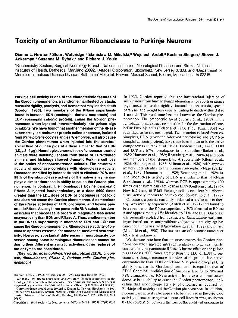
Figure I. Intrathecal injection of RNases into guinea pigs. Guinea pigs were injected into the CSF with the indicated doses of EDN (solid circles), onconase (open circles), bovine pancreatic RNase A (open di- amonds), modified onconase retaining 30% RNase activity (open squares), and modified onconase retaining 2% RNase activity (solid square) as described in Materials and Methods. Neurological symptoms and sur- vival of animals were monitored. Plotted is the percentage survival of animals. Data points represent three to six animals per group except for RNase A, in which there were two animals per group.
death was observed by Fredens et al. (1982). The LD,, for EDN was 4 pg per animal (Fig. 1). We examined the neurotoxicity of RNase A and onconase, two homologs of EDN and ECP. As shown in Table 1 and Figure 1, the intraventricular injection of as much as 20 mg of bovine pancreatic RNase A did not result in neurotoxicity as measured both by the neurologic symptoms of weakness and slight incoordination, shown best by the re- duced ability to gain the upright position when the animal is placed on its back, as well as by the survival of animals. The animals demonstrated no ataxia, tremor, or other neurologic symptoms and appeared healthy. This is a dose 5000 times greater than that of EDN needed to give an LD,, (4 wg/animal) and 10,000 times greater than the amount of EDN (2.0 ~g/ animal) where 50% of the animals developed neurologic symp- toms of the Gordon phenomenon. Sorrentino et al. (1992) also report that RNase A does not cause the Gordon phenomenon in rabbits at a dose of 200 pg per rabbit.
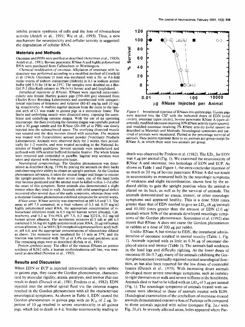
Unlike RNase A but similar to EDN, the intrathecal admin- istration of onconase resulted in animal toxicity (Table 1, Fig. 1). Animals injected with as little as 0.36 wg of onconase dis- played ataxia and tremor (Table 1). The animals had weakness in the hind legs and difficulty righting. At the lower doses of onconase (0.36-3.7 pg), many ofthe animals exhibiting the Gor- don phenomenon eventually regained normal neurological func- tion, as has also been reported for the low doses of eosinophil lysates (Durack et al., 1979). With increasing doses animals developed more severe neurologic symptoms, such as inability to right themselves or walk and severe stiffness in the hind limbs. Animals died or had to be killed with an LD,, of 5 Fg per animal (Fig. 1). The neurologic symptoms of animals treated with on- conase were identical to those of animals treated with EDN. Histological examination of the cerebellum of onconase-treated animals demonstrated extensive loss of Purkinje cells compared to those animals injected with saline (compare Fig. 2a,c with Fig. 2b,d). In severely affected areas, holes appeared where Pur-
540 Newton et al l EDN, Onconase, and RNase A Cytotoxicity to Purkinje Cells
Table 1. Neurotoxicity of RNases
RNase Dose (PLP)
Number Number days with Number Days to ob- symptoms” survived death served
EDN 0.02 O/4 44 17 0.2 o/4 4/4 17 2.0 2/4 3/4 9 (I)* 17
10.0 3/3 o/3 4 (3) 4
Onconase 0.36 2/3 3/3 12 1.0 2/6 6/6 14 3.7 a/9 6/9 7 (1X 16 (2) 16 8.0 213 l/3 5 (2) 23
16.0 3/3 o/3 3 (1),4 (2) 4 20.0 313 o/3 3 (2), 4 (1) 4 36.0 3/3 o/3 2 (2), 7 (1) 7
Alkylated onconase< 4.0 3/4 4/4 11 (30% active) 8.0 o/3 3/3 15
17.0 o/3 3/3 24 26.0 2/3 l/3 5 (2) 23 40.0 3/3 o/3 4 (1),5 (2) 5
100.0 3/3 o/3 3 (3) 3
Alkylated onconase= (2% active) 99.9 o/3 3/3 18
RNase A 10,000.0 o/3 3/3 18 20,000.0 o/2 2/2 18
” RNases were injected intrathecally into guinea pigs and their neurological symptoms measured as described in Materials and Methods. A Numbers in parentheses indicate the number of animals that died on that day.
( Onconase was chemically modified as described in Materials and Methods.
kinje cells were previously located. EDN also causes a selective matter of the cerebellum in guinea pigs treated with onconase. loss of Purkinje cells (Fredens et al., 1982). Furthermore, like Therefore, based upon neurologic symptoms and the loss of the results reported by Durack et al. (1979, 198 1) for rabbits Purkinje cells, onconase appears to cause the Gordon phenom- treated with EDN, there was a spongy vacuolation of the white enon.
Figure 2. Photomicrography of cere- bellar folia from a control animal (a and c) or an onconase-treated animal (band d): low (100 x ) (a, b) and high (400 x ) (c, d) magnification of animals injected intraventricularly with either PBS or onconase (3.6 pg) as described in Ma- terials and Methods. Purkinje cells (a) or holes previously occupied by Pur- kinje cells (b) are indicated by the ar- rows.
The Journal of Neuroscience, February 1994, M(2) 541
RNase A
EDN
ECP
Onconase
RNase A
EDN
ECP
Onconase
RNase A
EDN
ECP
Onconase
----E-DWLT
HESL#&@
-~I+JLKKST
* DSST
NMTS pR------
RDVD----
---S-KNVLT
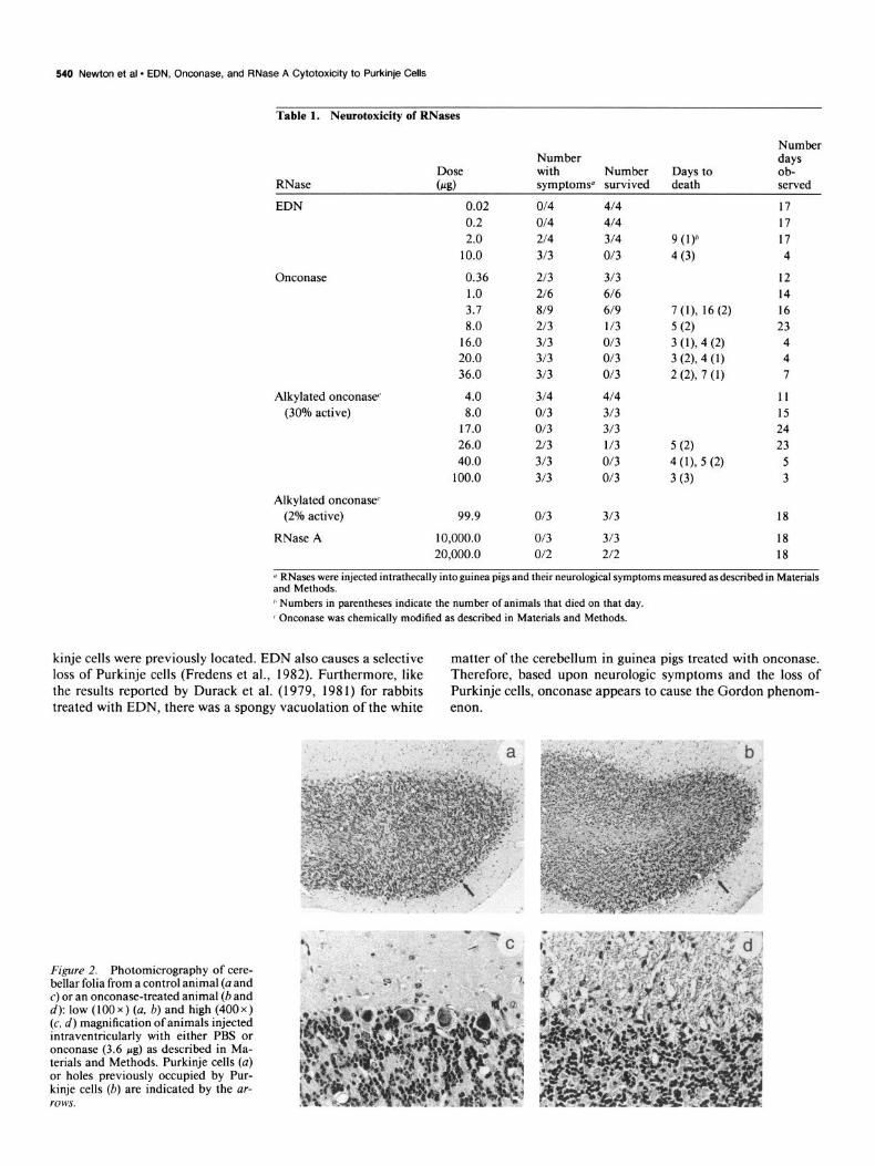
Comparing the sequences of onconase, RNase A, EDN, and ECP reveals that there is a close homology in primary structure among the four proteins (Fig. 3). Three of the four disulfide bonds of RNase A are conserved in onconase and all four di- sulfide bonds of RNase A are conserved in EDN and ECP. Onconase has an extra disulfide loop at the C-terminus not found in the other three proteins. The three key active site residues involved in enzymatic activity of RNase A, H 12, K4 1, and Hl 19, are all conserved in the four proteins. Close exam- ination of the amino acid sequence of the four proteins does not reveal any region common to EDN, ECP, and onconase that is lacking in RNase A that might explain the different abil- ities to cause the neurotoxicity. The extra nine amino acid region found in EDN and ECP that is lacking in RNase A is also lacking in onconase (between residues 113 and 114, Fig. 3) and thus cannot account for the neurotoxicity. The basicity of the ribo- nucleases has been suggested to predict neurotoxic activity (Sor- rentino et al., 1992). As shown in Table 3 and in Sorrentino et al. (1992) bovine pancreatic RNase A has the lowest net positive charge (four excess basic amino acids) and lacks the ability to cause the Gordon phenomenon. EDN is both more positively charged (seven excess basic amino acids) and more neurotoxic than RNase A. Onconase, on the other hand, is as neurotoxic as EDN but has only five basic amino acids in excess, inter- mediate between RNase A (lacking neurotoxicity) and human pancreatic RNase (approximately 14-20% as neurotoxic as EDN), which contains six excess basic amino acids (Sorrentino et al., 1992). Therefore, the results with onconase show that basicity does not correlate with RNase cytotoxicity to Purkinje cells. The determination of the number of excess basic amino acid residues does not define the charge distribution on the surface of the molecules, which must be quite varied since EDN has the lowest isoelectric point (~18.9) (Rosenberg et al., 1989a) compared to RNase A (p1 9.4) (Richards and Wyckoff, 1971) and onconase (p1 >9.5) (W. Ardelt, unpublished observation). Our results also show that glycosylation, an important structural difference between EDN, ECP, and RNase A, does not mediate neurotoxicity because onconase completely lacks carbohydrate (see Table 3) (Beintema et al., 1988; Ardelt, unpublished ob- servation), whereas EDN and ECP are glycosylated to varying degrees depending on the tissue oforigin (Beintema et al., 1988).
VG
Figure 3. Sequence alignment of the amino acid sequences of EDN (Rosen- berg et al., 1989b), ECP (Rosenberg et al., 1989a), and onconase (Ardelt et al., I99 I; Mosimann et al., 1992) with those of bovine pancreatic RNase A (Bein- tema et al., 1988). Regions of identical sequence are enclosed in boxes; cyste- ine residues are in lightly shaded boxes and the putative catalytic histidine and lysine residues are in heavily shaded boxes and marked by an asterisk. Dash- es represent gaps introduced to align cysteine residues and the catalytic res- idues. Residues are numbered accord- ing to bovine pancreatic RNase A.
0
,o.l, ,o’” ,o-9 .7 ,o.I
Ribonuclease Concentra:io”n CM)
0.7
0.6 s
0.5
r : 0.4
f 0.3 ~
0.2
0.1
po.ll 10’” 1o.a 10” 10.5 Ribonuch,. Concenlr.llon CM,
0.3
E 0.2 c :
4.
a 0.1
0 1 0 10 20 30 40 50 60 70
Ribonuclease Concentration (nM)
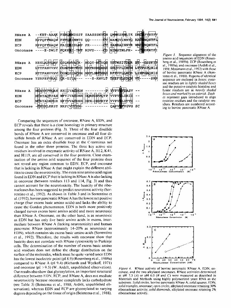
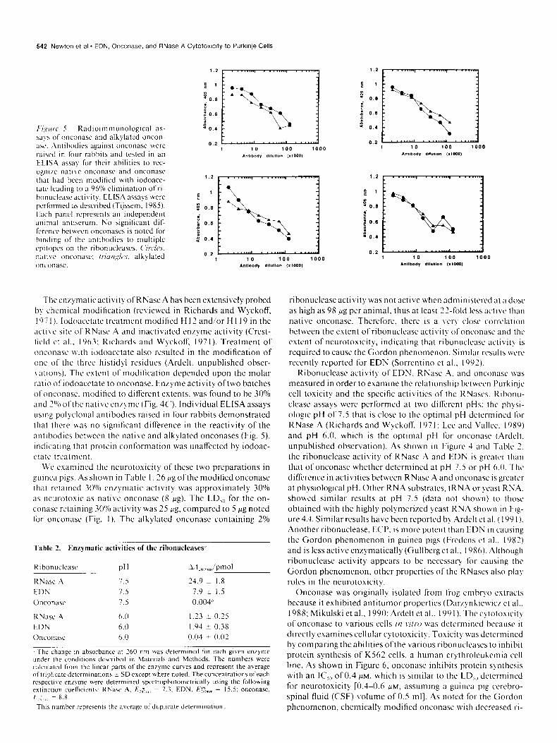
Figure 4. RNase activity of bovine pancreatic RNase A, EDN, on- conase, and the two alkylated onconases. RNase activities determined at pH 7.5 (A) or pH 6.0 (B and C) were measured as described in Materials and Methods using highly polymerized yeast RNA as the substrate. Solid circles, bovine pancreatic RNase A; solid squares, EDN; solid triangles, onconase; open circles, alkylated onconase retaining 30% ribonuclease activity; solid diamonds, alkylated onconase retaining 2% ribonuclease activity.
542 Newton et al * EDN, Onconase. and RNase A Cytotoxlcity to Purkinje Cells
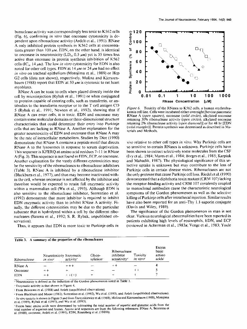
F‘/,ylrrc’ r. Kad~o~mmunolog~cal as- w!s ofonconnse and alkylated oncon- xc. Antihodlcs against onconase were raised in four rabbits and tested in an
ELlSA assal for their abilities to rcc- ognl/e native onconasc and onconasc that had been modified with iodoace- tatc leadIng 10 a 96% etlmlnatlon ofri- honuctcnseactlvity. EL-ISA assays were performed as described (Tijssem, 1985). Each panct represents an Independent antmal antiserum. No significant dif-
fcrcncc between onconascs is noted for handing of the antibodies to multiple
cpltopcs on the ribonuclcases. C‘rrc/~s, natlve onconasc: /~ur~,~$cs, alkylated onconasc.
1.2
e ’ i- 0.8 i e ; 0.6 . 90,
1.z
E 1
3 0.6
.j:\;;:
: : 0.6 P : 9 0.4
0.2 1 10 100 1000
AntIbody dllutlon ,r1ow
The en/) matic activity of RNase A has been extensively probed b) chemical modification (reviewed in Richards and Wyckoff, I97 I). lodoacetate treatment modified H 12 and/or H 1 19 in the active site of RNase A and inactivated enlyme activity (C‘rcst- ticld ct al.. 1963; Richards and Wyckoff. 197 I). Treatment of onconasc with iodoacetate also resulted in the modification of one of the three histidyl residues (Ardelt. unpublished obser- I atlons). The extent of modification depended upon the molar ratlo ofiodoacctate to onconase. Enzyme activity oftwo batches ofonconasc. modified to difTcrcnt extents, was found to be 30% and 2O’n ofthe native cn/yme (Fig. 4C’). Individual ELISA assays using polyclonal antibodies raised in four rabbits demonstrated that there was no significant difference in the reactivity of the antibodies between the native and alkylated onconases (Fig. S), indicating that protein conformation was unaffected by iodoac- ctatc treatment.
We examined the neurotoxicity of these two preparations in guinea pigs. .As shown in Table 1.26 pg ofthe modified onconase that retained 30(/o enzymatic activity was approximately 3O’Yo as neurotoxlc as native onconase (8 18). The LDI,, for the on- conase retaining 30% activity was 25 pg. compared to 5 Kg noted for onconasc (Fig. 1). The alkylated onconase containing 2”/0
Table 2. Enqmatic activities of the ribonucleases4
Rlbonuctease PH Ail ,,,,,,,,,hmol
RNasc .A 7.5 24.9 k I.8 EDN 7.5 7.9 i I.5 Onconasc 7.5 0.004”
KNasc .A 6.0 1.23 t 0.25
EDN 6.0 1.94 i 0.38
Onconasc 6.0 0.04 -t 0.02
The change In absorbance at 260 nm was determined for each given enryme under the condluons dcscrtbed m Materials and Methods. The numbers were calculated from the hncar parts of the enzyme curves and represent the average of tnphcate determinatmns t SD except where noted. The concentrations ofeach respccu\c etqme wrrc determlned spectrophotometrically using the following extmctlon coefficwnts: KNase A. Ej$ ,,,,,, = 7.3. EDN, fii>; ,““, = 15.5: onconase,
E.: ,,, , = 8.8. This number represents the average of duplicate determinatmn:.
s - 0.6
I + 0.6 B 9 0.4
0.2
ribonuclease activity was not active when administered at a dose as high as 98 fig per animal, thus at least 22-fold los active than native onconase. Therefore. thcrc is a \cr-y close correlation between the extent of ribonuclcase acti\ it! of onconasc and the extent of ncurotoxicity. indicating that ribonuclcasc activity is required to cause the Gordon phenomenon. Similar results wcrc rcccntly reported for EDN (Sorrentino et al.. 1992).
Ribonuclease activity of EDN, RNase A. and onconasc was measured in order to examine the relationship bctwcen Purkinjc cell toxicity and the specific activities of the KNascs. Ribonu- clease assays were performed at two different pHs: the physi- ologic pH of 7.5 that is close to the optimal pH detcrmincd f’or RNase A (Richards and Wyckoff, I97 1: Lee and Vallcc. I989) and pH 6.0, which is the optimal pH for onconase (Ardeh. unpublished observation). As shown in Figure 4 and Table 2. the ribonuclease activity of RNasc A and EDN is greater than that of onconase whether determined at pH 7.5 or pH 6.0. The difference in activities between RNase A and onconase is greater at physiological pH. Other RNA substrates. tKNA or qcast RNA. showed similar results at pH 7.5 (data not shown) to those obtained with the highly polymerired yeast RNA shown in Fig- ure 4.,1. Similar results have been reported by Ardclt et al. (199 1). Another ribonuclease, ECP. is more potent than EDN in causing the Gordon phenomenon in guinea pigs (Frcdens ct al.. 1983) and is less active enzymatically (Gullberget al.. 1986). Although ribonuclcasc activity appears to be necessary for causing the Gordon phenomenon, other properties of the RNascs also plal roles in the neurotoxicity.
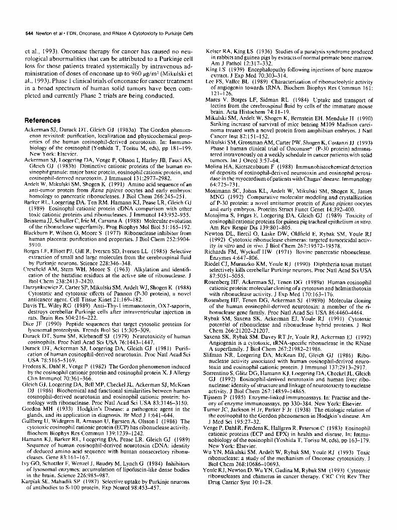
Onconase was originally isolated from frog embryo extracts because it exhibited antitumor properties (Dar/ynkicwic/ et al.. 1988; Mikulski et al., 1990; Ardelt et al., I99 I). The cytotoxicit! of onconasc to various cells irl ~ilro was determined bccausc it directly examines cellular cytotoxicity. Toxicit) was determined by comparing the abilities ofthe various ribonuclcascs to Inhibit protein synthesis of K562 cells, a human crythrolcukemia cell line. As shown in Figure 6, onconase inhibits protein synthesis with an IC,,, of 0.4 MM, which is similar to the LD,,, determined for ncurotoxicity [0.4-0.6 PM, assuming a guinea pig cerebro- spinal fluid (CSF) volume of 0.5 ml]. As noted for the Gordon phenomenon, chemically modified onconasc with decreased ri-
The Journal of Neuroscience, February 1994, 74(2) 543
bonuclease activity was correspondingly less toxic to KS62 cells (Fig. 6) confirming in vitro that onconase cytotoxicity is de- pendent upon ribonuclease activity (Ardelt et al., 199 1). RNase A only inhibited protein synthesis in K562 cells at concentra- tions greater than 100 FM. EDN, on the other hand, is identical to onconase in neurotoxicity (LD,, 0.5 PM) yet is 35 times less active than onconase in protein synthesis inhibition of K562 cells (IC,, 14 PM). The low in vitro cytotoxicity for EDN is also noted for other cell types. EDN at 14 FM or 24 WM had no effect in vitro on tracheal epithelium (Motojima et al., 1989) or Hep G2 cells (data not shown), respectively. Molina and Kierszen- baum (1988) report that EDN at 50 PM is cytotoxic to rat heart myoblasts.
RNase A can be toxic to cells when placed directly inside the cell by microinjection (Rybak et al., 199 1) or when conjugated to proteins capable of entering cells, such as transferrin, or an- tibodies to the transferrin receptor or to the T cell antigen CD 5 (Rybak et al., 1991; Newton et al., 1992) showing that if RNase A can enter cells, it is toxic. EDN and onconase may contain some molecular domains or three-dimensional structure characteristics that could determine their entry into Purkinje cells that are lacking in RNase A. Another explanation for the greater neurotoxicity of EDN and onconase than RNase A may be the rate of intracellular metabolism. Studies by Dice (1990) demonstrate that RNase A contains a peptide motif that directs RNase A to the lysosomes in response to serum deprivation. This sequence is KFERQ at amino acid residues 7-l 1 in RNase A (Fig. 3). This sequence is not found in EDN, ECP, or onconase. Another explanation for the vastly different cytotoxicities may be the sensitivity of the ribonucleases to ribonuclease inhibitors (Table 3). RNase A is inhibited by a ribonuclease inhibitor (Blackbum et al., 1977) and thus may become inactivated with- in the cell, whereas onconase is not affected by the inhibitor and therefore would be expected to retain full enzymatic activity within a mammalian cell (Wu et al., 1993). Although EDN is also sensitive to the ribonuclease inhibitor, Sorrentino et al. (1992) demonstrate that more inhibitor is required to inhibit EDN enzymatic activity than to inhibit RNase A activity. Fi- nally, the different cytotoxicities may be due to the particular substrate that is hydrolyzed within a cell by the different ribo- nucleases (Saxena et al., 1992; S. R. Rybak, unpublished ob- servation).
Thus, it appears that EDN is more toxic to Purkinje cells in
Table 3. A summary of the properties of the ribonucleases -
.z 60 3 g 40 f
.c 20 al ti L 0
0 .Ol 0.1 1 10 100 1000 RNase Concentration [PM]
Figure 6. Toxicity of the RNases to K562 cells, a human erythroleu- kemia cell line. Cells were incubated either overnight [bovine pancreatic RNase A (open squares), onconase (solid circles), alkylated onconase retaining 30% ribonuclease activity (open circles), alkylated onconase retaining 2% ribonuclease activity (open diamond)] or for 48 hr [EDN (solid triangles)]. Protein synthesis was determined as described in Ma- terials and Methods.
vivo relative to other cell types in vitro. Why Purkinje cells are so sensitive to certain RNases is unknown. Purkinje cells have been shown to extract selectively some molecules from the CSF (Ivy et al., 1984; Mares et al., 1984; Borges et al., 1985; Karpiak and Mahadik, 1987). The physiological significance of this se- lective uptake is unknown but may contribute to the loss of Purkinje cells in certain disease states. Ribonucleases are not the only proteins that cause Purkinje cell loss. Riedel et al. (1990) demonstrated that a diphtheria toxin mutant (CRM 107) lacking the receptor-binding activity and CRM 107 covalently coupled to monoclonal antibodies cause the characteristic neurological symptoms of the Gordon phenomenon as well as the selective killing of Purkinje cells after intrathecal injection. Similar results have also been reported for an anti-Thy 1.1 -saporin conjugate (Davis and Wiley, 1989).
The significance of the Gordon phenomenon in man is not clear. Various neurological abnormalities have been reported in patients exhibiting high levels of eosinophils, EDN, and ECP (reviewed in Ackerman et al., 1983a; Venge et al., 1983; Youle
Excess Ribonuclease basic
Neurotoxicity Enzymatic Glyco- inhibitor Toxicity amino Ribonuclease in viva” activity” sylation sensitivityd in vitro acids’
RNase A - +++ - ++ - 4
Onconase ++ + - - ++ 5 EDN ++ ++(+I + + + 7
” Neurotoxicity is defined as the induction of the Gordon phenomenon noted in Table 1. I’ Enzymatic activity is that shown in Figure 4.
’ From Beintema et al. (1988) and Ardelt (unpublished observations). d From Blackbum and Moore (1982), Sorrentino et al. (1992), Wu et al. (1993), and Ardelt (unpublished observations).
L In wtro toxicity is shown in Figure 5 and from Darzynkiewicz et al. (1988), Molina and Kierszenbaum (1988), Motojima et al. (1989), Rybak et al. (1991), and Wu et al. (1993).
’ Excess basic amino acids were determined by subtracting the total number of aspartic and glutamic acids from the total number of arginines and lysines. Amino acid sequences are from the following references: RNase A, Beintema et al. (1988), onconase, Ardelt et al. (199 l), EDN, Rosenberg et al. (1989b).
544 Newton et al * EDN. Onconase, and RNase A Cytotoxicity to Purkinje Cells
et al., 1993). Onconase therapy for cancer has caused no neu- rological abnormalities that can be attributed to a Purkinje cell loss for those patients treated systemically by intravenous ad- ministration of doses of onconase up to 960 fig/m* (Mikulski et al., 1993). Phase 1 clinical trials ofonconase for cancer treatment in a broad spectrum of human solid tumors have been com- pleted and currently Phase 2 trials are being conducted.
References Ackerman SJ, Durack DT, Gleich GJ (1983a) The Gordon phenom-
enon revisited: purification, localization and physicochemical prop- erties of the human eosinophil-derived neurotoxin. In: Immuno- biology of the eosinophil (Yoshida T, Torisu M, eds), pp 18 l-1 99. New York: Elsevier.
Ackerman SJ, Loegering DA, Venge P, Olsson I, Harley JB, Fauci AS, Gleich GJ (1983b) Distinctive cationic oroteins of the human eo- sinophil granule: major basic protein, eosinophil cationic protein, and eosinophil-derived neurotoxin. J Immunol 13 1:2977-2982.
Ardeh W, Mikulski SM, Shogen K (1991) Amino acid sequence of an anti-tumor protein from Rana pipiens oocytes and early embryos: homology to pancreatic ribonucleases. J Biol Chem 266:245-25 1.
Barker RL, Loegering DA, Ten RM, Hamann KJ, Pease LR, Gleich GJ (1989) Eosinophil cationic protein cDNA comparison with other toxic cationic proteins and ribonucleases. J Immunol 143:952-955.
Beintema JJ, Schuller C, Irie M, Carsana A (1988) Molecular evolution of the ribonuclease superfamily. Prog Biophys Mol Biol 5 1: 165-I 92.
Blackbum P, Wilson G, Moore S (1977) Ribonuclease inhibitor from human placenta: purification and properties. J Biol Chem 252:5904 5910.
Borges LF, Elliott PJ, Gill R, Iversen SD, Iversen LL (1985) Selective extraction of small and large molecules from the cerebrospinal fluid by Purkinje neurons. Science 228:346-348.
Crestfield AM, Stem WH, Moore S (1963) Alkylation and identifi- cation of the histidine residues at the active site of ribonuclease. J Biol Chem 238:2413-2420.
DarzynkiewiczZ, Carter SP, Mikulski SM, Ardelt WJ, Shogen K (1988) Cytostatic and cytotoxic effects of Pannon (P-30 protein), a novel anticancer agent. Cell Tissue Kinet 2 1: 169-I 82.
Davis TL, Wiley RG (1989) Anti-Thy- 1 immunotoxin, 0X7-saporin, destroys cerebellar Purkinje cells after intraventricular injection in rats. Brain Res 504:216-222.
Dice JF (1990) Peptide sequences that target cytosolic proteins for lysosomal proteolysis. Trends Biol Sci I5:3b5-i09. -
Durack DT. Sumi SM. Klebanoff SJ (1979) Neurotoxicitv of human eosinophils. Proc Nat1 Acad Sci USA 76:i443-1447. ’
Durack DT, Ackerman SJ, Loegering DA, Gleich GJ (1981) Purifi- cation of human eosinophil-derived neurotoxin. Proc Nat1 Acad Sci USA 78:5165-5169.
Fredens K, Dahl R, Venge P (1982) The Gordon phenomenon induced by the eosinophil cationic protein and eosinophil protein X. J Allergy Clin Immunol 70:36 I-366.
Gleich GJ, Loegering DA, Bell MP, Checkel JL, Ackerman SJ, McKean DJ (1986) Biochemical and functional similarities between human eosinophil-derived neurotoxin and eosinophil cationic protein: ho- mology with ribonuclease. Proc Nat1 Acad Sci USA 83:3 146-3 150.
Gordon MH (1933) Hodgkin’s Disease: a pathogenic agent in the glands, and its application in diagnosis. Br Med J I:64 l-644.
Gullberg U, Widegren B, Amason U, Egesten A, Olsson I (1986) The cytotoxic eosinophil cationic protein (ECP) has ribonuclease activity. Biochem Bioohvs Res Commun 139: 1239-1242.
Hamann KJ, B&ker RL, Loegering DA, Pease LR, Gleich GJ (1989) Sequence of human eosinophil-derived neurotoxin cDNA: identity of deduced amino acid sequence with human nonsecretory ribonu- cleases. Gene 83: 161-167.
Ivy GO, Schottler F, Wenzel J, Baudry M, Lynch G (1984) Inhibitors of lysosomal enzymes: accumulation of lipofuscin-like dense bodies in the brain. Science 226:985-987.
Karpiak SE, Mahadik SP (1987) Selective uptake by Purkinje neurons of antibodies to S-100 protein. Exp Neural 98:453-457.
Kelser RA, King LS (1936) Studies of a paralysis syndrome produced in rabbits and guinea pigs by extracts ofnormal primate bone marrow. Am J Path01 12:317-332.
King LS (1939) Encephalopathy following injections of bone marrow extract. J Exp Med 70:303-3 14.
Lee FS, Vallee BL (1989) Characterization of ribonucleolytic activity of angiogenin towards tRNA. Biochem Biophys Res Commun 16 1: 121-126.
Mares V, Barges LF, Sidman RL (1984) Uptake and transport of lectins from the cerebrospinal fluid by cells of the immature mouse brain. Acta Histochem 74:11-19.
Mikulski SM, Ardelt W, Shogen K, Bernstein EH, Menduke H (1990) Striking increase of survival of mice bearing Ml09 Madison carci- noma treated with a novel protein from amphibian embryos. J Nat1 Cancer Inst 82:151-152.
Mikulski SM, Grossman AM, Carter PW, Shogen K, Costanzi JJ (1993) Phase I human clinical trial of OnconaseR1 (P-30 protein) adminis- tered intravenously on a weekly schedule in cancer patients with solid tumors. Int J Oncol 3:57-64.
Molina HA, Kierszenbaum F (1988) Immunohistochemical detection of deposits of eosinophil-derived neurotoxin and eosinophil peroxi- dase in the myocardium ofpatients with Chagas’disease. Immunology 64~725-73 I.
Mosimann SC, Johns KL, Ardelt W, Mikulski SM, Shogen K, James MNG (1992) Comparative molecular modeling and crystallization of P-30 protein: a novel antitumor protein of Rana piplens oocytes and early embryos. Proteins Struct Funct Genet 14:392-400.
Motojima S, Frigas E, Loegering DA, Gleich GJ (1989) Toxicity of eosinophil cationic proteins for guinea pig tracheal epithelium in vitro. Am Rev Respir Dis 139:801-805.
Newton DL, Ilercil 0, Laske DW, Oldfield E, Rybak SM, Youle RJ (1992) Cytotoxic ribonuclease chimeras: targeted tumoricidal activ- ity in vitro and in vivo. J Biol Chem 267:19<72-19578.
Richards FM. Wvckoff HW (197 1) Bovine oancreatic ribonuclease. Enzymes 4:b47-806. ~ ’
Riedel CJ, Muraszko KM, Youle RJ (I 990) Diphtheria toxin mutant selectively kills cerebellar Purkinje neurons. Proc Nat1 Acad Sci USA 87:505 l-5055.
Rosenberg HF, Ackerman SJ, Tenen DG (I 989a) Human eosinophil cationic protein: molecular cloning ofa cytotoxin and helminthotoxin with ribonuclease activity. J Exp Med 170: 163-l 76.
Rosenberg HF, Tenen DG, Ackerman SJ (1989b) Molecular cloning of the human eosinophil-derived neurotoxin: a member of the ri- bonuclease gene family. Proc Nat1 Acad Sci USA 86:4460-4464.
Rybak SM, Saxena SK, Ackerman EJ, Youle RJ (1991) Cytotoxic potential of ribonuclease and ribonuclease hybrid proteins. J Biol Chem 26612 1202-2 1207.
Saxena SK, Rybak SM, Davey RT Jr, Youle RJ, Ackerman EJ (1992) Angiogenin is a cytotoxic, iRNA-specific ribonuclease in the RNase A suoerfamilv. J Biol Chem 267:2 1982-2 1986.
Slifman NR, Lbegering DA, McKean DJ, Gleich GJ (1986) Ribo- nuclease activity associated with human eosinophil-derived neuro- toxin and eosinophil cationic protein. J Immunol 137:29 13-29 17.
Sorrentino S, Glitz DG, Hamann KJ, Loegering DA, Checkel JL, Gleich GJ (1992) Eosinophil-derived neurotoxin and human liver ribo- nuclease: identity of structure and linkage of neurotoxicity to nuclease activity. J Biol Chem 267:14859-14865.
Tijssem P (1985) Enzyme-linked immunoassays. In: Practise and the- ory of enzyme immunoassays, pp 330-384. New York: Elsevier.
Turner JC, Jackson H Jr, Parker F Jr (1938) The etiologic relation of the eosinophil to the Gordon phenomenon in Hodgkin’s disease. Am J Med Sci 195:27-32.
Venge P, Dahl R, Fredens K, Hallgren R, Peterson C (I 983) Eosinophil cationic proteins (ECP and EPX) in health and disease. In: Immu- nobiology of the eosinophil (Yoshida T, Torisu M, eds), pp 163-l 79. New York: Elsevier.
Wu YN, Mikulski SM, Ardelt W, Rybak SM, Youle RJ (I 993) Toxic ribonuclease: a study of the mechanism of Onconase cytotoxicity. J Biol Chem 268:10686-10693.
Youle RJ, Newton D, Wu YN, Gadina M, Rybak SM (1993) Cytotoxic ribonucleases and chimeras in cancer therapy. CRC Crit Rev Ther Drug Carrier Syst 10: I-28.