“twisted tales: insights into genome diversity of ciliates ... · downloaded from by guest on 26...
TRANSCRIPT
“Twisted Tales: Insights into Genome Diversity of Ciliates Using Single-Cell ‘omics”
Xyrus X. Maurer-Alcalá a,b,*,#, Ying Yan b,*, Olivia Pilling b, Rob Knight c,d,e, Laura A. Katz a,b,†
a University of Massachusetts Amherst, Program in Organismic and Evolutionary Biology,
Amherst, Massachusetts, USA
b Smith College, Department of Biological Sciences, Northampton, Massachusetts, USA,
c University of California San Diego, Department of Pediatrics, San Diego, California, USA
d University of California San Diego, Department of Computer Science and Engineering, San Diego, California, USA e University of California San Diego, Center for Microbiome Innovation, San Diego, California, USA
* These authors contributed equally
# Current address: University of Bern, Institute of Cell Biology, Bern, Switzerland
† Corresponding author: E-mail: [email protected]
© The Author(s) 2018. . Published by Oxford University Press on behalf of the Society for Molecular Biology and Evolution.
This is an Open Access article distributed under the terms of the Creative Commons Attribution Non-Commercial License
(http://creativecommons.org/licenses/by-nc/4.0/), which permits non-commercial re-use, distribution, and reproduction in any medium,
provided the original work is properly cited. For commercial re-use, please contact [email protected]
Downloaded from https://academic.oup.com/gbe/advance-article-abstract/doi/10.1093/gbe/evy133/5045407by gueston 13 July 2018
Abstract
The emergence of robust single-cell ‘omics techniques enables studies of uncultivable
species, allowing for the (re)discovery of diverse genomic features. In this study, we combine
single-cell genomics and transcriptomics to explore genome evolution in ciliates, a >1 billion
year old clade. Analysis of the data resulting from these single-cell ‘omics approaches show: 1)
the description of the ciliates in the class Karyorelictea as ‘primitive’ is inaccurate because their
somatic macronuclei contain loci of varying copy number (i.e. they have been processed by
genome rearrangements from the zygotic nucleus); 2) gene-sized somatic chromosomes exist
in the class Litostomatea, consistent with Balbiani’s 1890 observation of giant chromosomes in
this lineage; and 3) gene scrambling exists in the underexplored Postciliodesmatophora (the
classes Heterotrichea and Karyorelictea, abbreviated here as the Po-clade), one of two major
clades of ciliates. Together these data highlight the complex evolutionary patterns underlying
germline genome architectures in ciliates and provide a basis for further exploration of principles
of genome evolution in diverse microbial lineages.
Downloaded from https://academic.oup.com/gbe/advance-article-abstract/doi/10.1093/gbe/evy133/5045407by gueston 13 July 2018
Introduction
Although genomes are often described as being conserved within species, a plethora of
data demonstrate their inherently dynamic nature (e.g. Maurer-Alcalá and Katz 2015; Oliverio
and Katz 2014; Parfrey, et al. 2008). In eukaryotes, examples of dynamic genomes include the
separation of germline and somatic genetic material and variation throughout life cycles (such
as changes in ploidy or DNA content during development; Maurer-Alcalá and Katz 2015;
Parfrey, et al. 2008). These changes are often regulated by epigenetic mechanisms that are
involved in analogous (and perhaps homologous) processes among anciently diverged lineages
of eukaryotes (Maurer-Alcalá and Katz 2015; Matzke and Mosher 2014; Rogato, et al. 2014),
which has led to the hypothesis that such mechanisms existed in the last common ancestor of
extant eukaryotes (Maurer-Alcalá and Katz 2015; Oliverio and Katz, 2014; Parfrey 2008; Zufall,
Robinson and Katz 2005).
Dynamic genomes, including the separation of germline and somatic DNA into distinct
nuclei, are present in ciliates, an ancient clade of predominantly single-celled eukaryotic
microorganisms. Unlike multicellular organisms, where germline (i.e. gametic) and somatic (i.e.
leaves, hyphae, muscle) genomes are in distinct cell types, the germline micronucleus (MIC)
and somatic macronucleus (MAC) share a common cytoplasm in ciliates (Prescott 1994; Raikov
1982). As in other eukaryotes, both the germline and somatic genomes differentiate from a
zygotic nucleus after sex (conjugation in ciliates). However, the development of a new somatic
genome includes complex epigenetically-guided processes (i.e. large-scale genome
rearrangements, DNA elimination, chromosome fragmentation, de novo telomere addition, and
chromosome amplification; Bellec and Katz 2012; Chalker and Yao 2011; Chen, et al. 2013;
Cheng, et al. 2016; Fuhrmann, et al. 2017; Guerin, et al. 2017; Hamilton, et al. 2016; Heyse, et
al. 2011; Huang and Katz 2014; Maurer-Alcalá, et al. 2018; Wang and Blackburn 1997; Xu, et
al. 2012).
Downloaded from https://academic.oup.com/gbe/advance-article-abstract/doi/10.1093/gbe/evy133/5045407by gueston 13 July 2018
Molecular studies of germline-soma differentiation in ciliates are largely limited to a few
cultivable ‘model’ ciliates – Oxytricha trifallax (Chen, et al. 2014; Swart, et al. 2013),
Paramecium tetraurelia (Arnaiz, et al. 2012; Aury, et al. 2006; Guerin, et al. 2017), and
Tetrahymena thermophila (Esien, et al. 2006; Hamilton, et al. 2016). All of these models fall
within the ‘Intramacronucleata’ (referred to as the Im-clade for this study), which is one of the
two major clades of ciliates (Fig. 1). The other major clade, the ‘Postciliodesmatophora’
(referred to as the Po-clade), shares a common ancestor with the model ciliates over 1.0 GYA
(see Fig. 2 in Parfrey, et al. 2011; Eme et al. 2014; V’dačný 2015). Yet the Po-clade remain
largely under-sampled for genome features.
Arguably, one of the most notable differences among the model ciliates is the dramatic
variation in somatic genomic architecture: ciliates can be grouped into those with ‘long’ vs ‘nano’
sized somatic chromosomes. In the models T. thermophila and P. tetraurelia (Class
Oligohymenophorea), the somatic chromosomes are ‘large’ (by ciliate standards) being on
average 100’s of kilobases to ~ 1-2 megabases in length, lack centromeres (e.g. Aury, et al.
2006; Eisen et al. 2006) and are substantially gene-rich (~60-80% of their length composed of
open reading frames). By contrast, the somatic genome of O. trifallax (Cl: Spirotrichea) is
predominantly composed of ~16,000 unique ‘nano-chromosomes’, most of which contain a
single ORF (ranging from < 1Kbp to ~66Kbp; Swart, et al. 2013). Prior to our work, evidence for
the phylogenetic distribution of somatic nano-chromosomes was limited to only three ciliate
classes: Spirotrichea, Armophorea and Phyllopharyngea (Riley and Katz 2001; Fig. 1.).
In addition to variable chromosome size among ciliate somatic genomes, there are
differences in patterns of chromosome copy number. For example, in Tetrahymena thermophila,
each of its 225 unique somatic chromosomes are maintained at ~45 copies each in the somatic
nucleus (Doerder, et al. 1992; Eisen et al. 2006). In the ciliates Chilodonella uncinata and
Oxytricha trifallax, both with nano-sized somatic chromosomes, the macronuclei contain millions
of somatic chromosomes maintained at variable but heritable copy numbers (Bellec and Katz
Downloaded from https://academic.oup.com/gbe/advance-article-abstract/doi/10.1093/gbe/evy133/5045407by gueston 13 July 2018
2012; Heyse, et al. 2011; Huang and Katz 2014; Xu, et al. 2012). The range of copy numbers of
these chromosomes can span multiple orders of magnitude from several hundred to >50,000
(Bellec and Katz 2012; Huang and Katz 2014; Xu, et al. 2012). Current data suggest that
differential chromosome amplification is limited to those ciliates with macronuclear nano-
chromosomes (Fig. 1; Bellec and Katz 2012; Heyse, et al. 2011; Huang and Katz 2014; Xu, et
al. 2012).
Ciliates in the Po-clade represent two presumed extremes in genome architecture.
Ciliates in the Heterotrichea are often very large (some species are > 1 mm in length) with
correspondingly large somatic nuclei that contain from ~ 1,000 to > 13,000 times more DNA
than their germline nuclei (Ovchinnikova, et al. 1965; Wancura, et al. 2017). The other class,
Karyorelictea, can be of similar sizes yet often have numerous clusters of somatic nuclei with
relatively low DNA content (~1.1 to 12 times more DNA in their somatic nuclei; reviewed in Yan,
et al. 2017); based on this observation, Karyorelictea are the only group of ciliates to be
described as paradiploid (i.e. nearly diploid) and their name (karyo = nucleus; relictea – relict)
suggests a primitive state (Bobyleva, et al. 1980; Kovaleva and Raikov 1978; Raikov 1982,
1985; Raikov and Karadzhan 1985; Yan, et al. 2017). Karyorelictean ciliates have been
described as primitive based on three features: 1) relatively simply ciliature, 2) unusually low
DNA content (‘paradiploid’, or nearly diploid; Kovaleva and Raikov 1978; Raikov and Karadzhan
1985; Yan et al. 2017) and 3) the inability to divide their macronuclei during asexual divisions,
which has only been described in members of this class(Raikov 1985; Raikov 1994; Yan et al.
2017). Molecular phylogenies place the Karyorelictea sister to the Heterotrichea (i.e. the Po-
clade), inconsistent with the idea that this lineage represents the ancestral state for ciliates (Gao
and Katz 2014; Gao et al. 2016). We use qPCR to estimate chromosome copy number to
address the putative paradiploidy of Karyorelictea.
Downloaded from https://academic.oup.com/gbe/advance-article-abstract/doi/10.1093/gbe/evy133/5045407by gueston 13 July 2018
The complex processing underlying development of somatic nuclei from zygotic nucleus
in ciliates relies on the elimination of germline-limited DNA (i.e. internally eliminated sequences;
IESs) and the accurate ‘assembly’ of functional somatic regions (i.e. macronuclear-destined
sequences; MDS). The removal of IESs during the development of the somatic genome is
analogous to intron-splicing during mRNA maturation, though IES excision occurs within the
DNA (Allen and Nowacki 2017; Jönsson, et al. 2009; Wahl, et al. 2009). The organization of
MDS/IES in germline genomes fall into two major categories: scrambled and non-scrambled
(Ardell, et al. 2003; Chen, et al. 2014; Maurer-Alcalá, et al. 2018; Möllenbeck, et al. 2006;
Prescott 1994; Wong and Landweber 2006). We define non-scrambled germline loci as those
with MDSs that are on the same DNA strand and joined in ‘order’ during DNA elimination in
ciliates (Fig. 5A). By contrast, scrambled germline loci are characterized by MDSs being found
on opposing DNA strands and/or in non-consecutive order (Fig. 5B). Germline scrambling has
only been documented in the Phyllopharyngea and Spirotrichea clades (Fig. 1; Ardell, et al.
2003; Chen, et al. 2014; Gao, et al. 2015; Katz and Kovner 2010; Maurer-Alcalá, et al. 2018;
Wong and Landweber 2006).
The details on germline genome architecture and the transformations that underlie the
development of the somatic genome have largely been explored in only three classes of ciliates
(Oligohymenophorea, Phyllopharyngea and Spirotrichea; Fig. 1). Taking advantage of single-
cell genomics and transcriptomics technologies, we explore the genomes of Blepharisma
americanum (Heterotrichea, Po-clade), several Loxodes species. (Karyorelictea; Po-clade) the
large Bursaria truncatalla (Colpodea, Im-clade) and voracious predatory ciliate Didinium
nasutum (Litostomatea, Im-clade), capturing a deep (> 1.0 GYA) divide between the Im and Po
clades (Fig. 1; see Fig. 2 in Parfrey, et al. 2011). We present insights into genome evolution
from these non-traditional models, which demonstrate a greater diversity of genomic
architectures than we expected from the literature.
Downloaded from https://academic.oup.com/gbe/advance-article-abstract/doi/10.1093/gbe/evy133/5045407by gueston 13 July 2018
Results and Discussion
Differential chromosome amplification in the Po-clade
We explore patterns of somatic chromosome copy numbers in the Po-Clade, focusing on
the genera Blepharisma (Heterotrichea) and Loxodes (Karyorelictea) to test whether either of
these ciliates differentially amplifies somatic chromosomes. In fact, many eukaryotes
extensively amplify extrachromosomal copies of their ribosomal RNA genes (Cohen, et al. 2008;
Sinclair and Guarente 1997; Zufall et al. 2005), so we compare the nuclear small subunit
ribosomal RNA gene(nSSU-rRNA) to several protein coding genes. We analyze chromosome
copy number using DNA isolated from a population of ~1300 Blepharisma americanum
individuals (pop-DNA) and compare this to copy number estimates from three individual B.
americanum following whole genome amplification (WGA). Given that we do not find any
significant bias produced by the WGA reactions, we then use this single-cell ‘omic approach on
the uncultivable genus Loxodes (Karyorelictea).
In the analyses of both total genomic DNA (total gDNA) and single-cells WGA (sc-WGA)
of B. americanum, the nSSU-rRNA gene is characteristically high, with an estimated 2.55×10⁷ ±
8.42x10⁶ copies/ng per cell and 7.90×10⁷ ±1.02×10⁷ copies/ng per cell, respectively. Copy
numbers were estimated from DNA contents of quantitative PCR (qPCR) results and were
adjusted to per ng per cell. Estimates of copy numbers for protein coding genes between the
different preparations of Blepharisma (total gDNA and sc-WGA) are similarly consistent, ranging
from 1.18x10⁶ ± 4.38x10⁴ copies/ng per cell and 8.45x10⁵
± 1.14x10⁵ copies/ng per cell (for one
α-tubulin paralog). The least abundant of the protein coding genes from the total gDNA and
single-cells are 9.77x10⁴ ± 2.41x10⁴ copies/ng per cell and 6.06x10² ± 2.87x10² copies/ng per
cell for EF-1α and an α-tubulin paralog (Table S1).
To compare relative abundances across taxa, we set the nSSU-rRNA copy number to
106 (a value based on evidence from diverse ciliates; Gong et al. 2013; Heyse, et al. 2010;
Downloaded from https://academic.oup.com/gbe/advance-article-abstract/doi/10.1093/gbe/evy133/5045407by gueston 13 July 2018
Huang and Katz 2014). We find that the relative copy numbers for chromosomes containing
protein coding genes (two paralogs of Actin and α-Tubulin, and EF-1α) in B. americanum span
~2 orders of magnitude (Fig. 2B) with the exception of actin paralog 2, which is consistently at
low copy number across all samples (p = 0.093 from an ANOVA). Despite greater variability in
absolute copy numbers from the population of cells (pop-DNA) compared to the individual cells,
we observe no significant biases between methods (total pop-DNA versus single-cell WGA; p =
0.474 from a Kruskal-Wallis test; Fig. 2B). In other words, the sc-WGA method can be used to
assess patterns of individual chromosome copy numbers because this method yields the same
results as studies of a population of cells.
We then deployed the same methods (qPCR after single-cell WGA) to study the
uncultivable genus Loxodes in the ‘paradiploid’ class Karyorelictea, which is predicted to have
~2 copies of every protein coding gene (Raikov 1982; Yan, et al. 2017). We performed a similar
qPCR experiment using five genes (nSSU-rRNA, EF-1α, Actin, Rs11 and α-Tubulin) from sc-
WGAs of wild-caught individuals of Loxodes, representing two distinct species based on rRNA
gene diversity (Table S2). As we only have relative numbers here, we again set the nSSU-rRNA
gene to 106 copies to allow comparison of patterns of chromosome copy numbers. By contrast
to the stochastic patterns of chromosome copy number in B. americanum, the differences in
copy number among protein coding genes in Loxodes spp. consistently spanned a far greater
range with the highest being Actin at ~2.87x10⁴ copies relative to the rRNA gene and the lowest
being EF-1α at ~4.39x10⁻¹ copies/ng per cell (~4 orders of magnitude; Table S2; Fig. 2D). We
observe significant differences in gene copy number within each cell of Loxodes spp. (p <<
0.05), implicating the differential amplification of chromosomes. For both of the Loxodes
species, gene copy numbers are maintained in a mostly-conserved order: nSSU-rRNA >> Actin
> Rs11 > α-Tubulin > EF-1α (Figure 2D), which contrasts with the stochastic pattern in
Heterotrichea.
Downloaded from https://academic.oup.com/gbe/advance-article-abstract/doi/10.1093/gbe/evy133/5045407by gueston 13 July 2018

The contrasting pattern of stochasticity in chromosome copy number in B. americanum
and the predictability in chromosome number in Loxodes spp. likely reflects differences genome
architecture of their somatic nuclei. The macronuclei of Blepharisma house large quantities of
DNA and possess the ability to divide, while Loxodes spp.’ macronuclei are DNA poor and do
not divide with cell division (Katz 2001; Raikov 1982; Yan, et al. 2017). The stochasticity in
chromosome copy number for Blepharisma may be a byproduct of the massive genome
amplification that occurs during development (Santangelo and Barone 1987), as the somatic
nucleus is estimated to have > 1000x more DNA than the germline nucleus (Ovchinnikova, et al.
1965; Wancura, et al. 2017). Variable chromosome copy number among individuals is likely an
inherent feature of Blepharisma and its relatives (in the class Heterotrichea; Fig. 1), exemplified
by Stentor coeruleus, whose chromosome copy numbers of the nSSU-rDNA are clearly
correlated to cell size (Slabodnick, et al. 2017) and likely nuclear volume (Cavalier-Smith 1978).
This suggests that the observed stochasticity from our measurements is likely the result of
biological differences (e.g. cell volume or life-cycle stages; Fig. 2A&B).
Although Loxodes spp. are found in the sister class to B. americanum (both in the Po-
clade), Loxodes and its relatives have long been considered as ‘primitive’ ciliates (Orias 1991;
Raikov 1985, 1994; Raikov and Karadzhan 1985). This presumption arose from early studies
that found that the somatic macronucleus is unable to divide (instead needing to be
differentiated from a germline nucleus with each cell division; reviewed in Yan, et al. 2017) as
well as from estimates of DNA content based on autoradiographic measurements from the
somatic and germline nuclei of Loxodes and its relatives (Bobyleva, et al. 1980; Kovaleva and
Raikov 1978). From these early measurements, where the somatic nuclei typically harbor only
~1.1 to ~12 times the amount of DNA compared to the germline nuclei, karyorelictid lineages
were labeled as paradiploid (‘nearly-diploid’). This has led to the expectation that the relative
copy number among protein-coding genes would be approximately equal in this class of ciliate
(Fig. 2C). Such low ploidy is unusual among ciliates. For example, ploidy is species-dependent
Downloaded from https://academic.oup.com/gbe/advance-article-abstract/doi/10.1093/gbe/evy133/5045407by gueston 13 July 2018
and ranges from ~45N in Tetrahymena thermophila (Woodard, et al. 1972) to ~800N in
Paramecium tetraurelia (Duret, et al. 2008) and an average of ~2,000N in the differentially
amplified nano-chromosomes in Oxytricha trifallax (Swart et al. 2013).
Surprisingly, our data demonstrate that Loxodes spp. is neither paradiploid nor are all
chromosomes equally amplified. Our estimates of relative chromosome copy number show that
instead of being present in roughly equal abundance, chromosomes containing our target genes
differ by several orders of magnitude (Fig. 2C & D). Though non-dividing macronuclei in
Loxodes spp. (and other members of the class Karyorelictea) age over time (at most 7
generations; Raikov 1982, 1985, 1994; Yan, et al. 2017), we do not believe aging alone is
sufficient to explain our data as the replicability of estimates across cells suggests heritable
differences in copy number (from 2 to > 1000 copies). These copy number data suggest that the
long-held description of Loxodes spp. as ‘primitive’, based upon DNA content estimates and the
inability to divide their macronuclei, is inaccurate.
Unexpected extensive fragmentation of somatic genomes from the Im-clade
Extensive fragmentation of chromosomes into gene-sized ‘nano-chromosomes’ during
the development of somatic macronuclei is well documented in only three ciliate classes (e.g. in
Chilodonella uncinata (cl: Phyllopharyngea; McGrath, et al. 2007), Oxytricha trifallax (cl:
Spirotrichea; Swart, et al. 2013), and Nycotherus ovalis (cl: Armophorea; McGrath, et al. 2007;
Ricard et al. 2008; Fig. 1). We searched for evidence of extensive fragmentation in the class
Litosomatea (Im-clade; Fig. 1), analyzing a single-cell WGA assembly for Didinium nasutum and
the recently released genome assembly of Entodinium caudatum (a distantly-related member of
the same class). We evaluated the ends of scaffolds for both D. nasutum and E. caudatum to
look for telomeres as no record of telomeres has been reported for members in this class. This
approach resulted in a common repetitive motif in both taxa, C4A2T. As telomeric sequences
seem well conserved over broad phylogenetic scales in ciliates (Aeschlimann, et al. 2014; Aury,
Downloaded from https://academic.oup.com/gbe/advance-article-abstract/doi/10.1093/gbe/evy133/5045407by gueston 13 July 2018
et al. 2006; Eisen, et al. 2006; McGrath, et al. 2007; Swart, et al. 2013), this simple repeat may
be specific to Litostomatea.
To assess the size distributions of somatic chromosomes, we use the telomeric motif to
identify scaffolds bounded by repeats at both ends (e.g. complete assembled chromosomes) for
both D. nasutum and E. caudatum. To our surprise, we identified 328 complete nano-
chromosomes in D. nasutum’s telomere-bound scaffolds and 7,560 complete chromosomes
from the released E. caudatum genome assembly (Figs. 3-4, Fig. S1, and Supplementary Data
1 and 2). Although the nano-chromosome estimates are very disparate among D. nasutum and
E. caudatum, previous work has demonstrated the bias in the genome amplification reaction
against ciliate nano-chromosomes, which may be present in genome assemblies as “by-catch”
(Maurer-Alcalá, Knight and Katz 2018) and which account for the order of magnitude difference
between Didinium and Entodinium. To further check that these were not simply assembly
artefacts, we mapped transcripts from single D. nasutum individuals to the pool of 328 putatively
complete nano-chromosomes (i.e. those with telomeres on each end). Of these 328
chromosomes, 254 (77.4%) harbor a single ORF and overall 316 (96.3%) of the chromosomes
are actively transcribed as evidenced in the single-cell transcriptomes. As no transcriptome data
are publicly available for E. caudatum, we used more relaxed conditions (e.g. chromosomes
with ≥ 50% identity and align to ≥ 50% of the translated ORF) to map 5,692 translated ORFs
from our D. nasutum transcriptome to 5,293 (70.0%) of E. caudatum’s complete chromosomes.
Having demonstrated the presence of nano-chromosomes in the D. nasutum and E.
caudatum genome assemblies, we determined that the size range of these complete
chromosomes are nearly identical for both, ranging from ~0.4 Kbp to ~ 26 Kbp, despite
differences in the methods used to obtain the genomic data (e.g. use of sc-WGA techniques for
D. nasutum and more traditional DNA isolation approaches used for E. caudatum; Fig. S1).
However, previous work using pulsed-field gel electrophoresis of total gDNA from D. nasutum
did not report chromosomes below 50 kbp (Popenko, et al. 2015), which suggests that the
Downloaded from https://academic.oup.com/gbe/advance-article-abstract/doi/10.1093/gbe/evy133/5045407by gueston 13 July 2018
nano-chromsomes may be present at relatively low copy numbers and/or that the retention of
these chromosomes is strongly dependent on the DNA isolation approaches. However,
comparisons of the size distribution of these complete chromosomes for D. nasutum and E.
caudatum to genomic data from diverse taxa, demonstrate that these chromosomes’ sizes are
consistent with the ‘gene-sized’ chromosomes found in divergent ciliate taxa (e.g. Chilodonella
uncinata and Oxytricha trifallax; McGrath, et al. 2007; Swart, et al. 2013; Figs. 3-4, and Fig. S1).
The data on nano-sized chromosomes in the class Litostomatea are consistent with the
1890 description of giant germline chromosomes, which are presumably generated through
endoreplication during development of a new macronucleus (Balbiani 1890). The
correspondence between the appearance of giant chromosomes during development and the
presence of nano-sized chromosomes in somatic genomes has been extensively documented
(most notably Chilodonella and Stylonychia, classes Phyllopharygnea and Spirotrichea
respectively; Ammermann 1986; Juranek, et al. 2005; Katz 2001; Katz and Kovner 2010;
Postberg, et al. 2008; Pyne 1978; Riley and Katz 2001; Figs. 1 and 4). In these ciliate classes,
polytenization occurs just prior to the extensive genome remodeling that ultimately leads to the
formation of the thousands of unique nano-chromosomes through epigenetically guided DNA
elimination, large-scale genome rearrangements and de novo telomere addition (Ammermann
1986; Chen, et al. 2014; Fuhrmann, et al. 2016; Postberg, et al. 2008; Pyne 1978; Spear and
Lauth 1976). The absence of polytenization of germline chromosomes from the model ciliates
Paramecium tetraurelia and Tetrahymena thermophila, which possess ‘large’ macronuclear
chromosomes (ranging from ~0.2Mbp to several Mbp in size; Aury, et al. 2006; Eisen, et al.
2006; Fig. 3 and Fig. S1), further implicates generation of giant chromosomes as being limited
to nano-chromosome formation.
Well over 100 years ago, Éduoard-Gérard Balbiani, who provided the original description
of polytene chromosomes in the dipteran Chironomus (Balbiani 1881), described the presence
of polytene chromosomes in the ciliate Loxophyllum meleagris (also in the class Litostomatea;
Downloaded from https://academic.oup.com/gbe/advance-article-abstract/doi/10.1093/gbe/evy133/5045407by gueston 13 July 2018
Balbiani 1890). Unfortunately, there had been little work able to corroborate the observations of
Balbiani (1890). However, given the robust phylogenetic relationships between the classes
Litostomatea, Spirotrichea and Armophorea (“SAL” clade; Gao et al. 2016; Gentekaki et al.
2014), all of which possess both nano-chromosomes (Ricard, et al. 2008; Riley and Katz 2001;
Swart, et al. 2013) and giant chromosomes (Golikova 1965; Wichterman 1937), these unusual
genome architectural features could be a synapomorphy that further unites these classes within
the Im-clade (Fig. 1).
Germline genome architecture from diverse ciliates
Previous studies of germline genome architecture in ciliates are from even more
sparsely-sampled lineages than studies of somatic genomes, with data available from only a
few model species in the classes Oligohymenophorea, Phyllopharyngea and Spirotrichea
(Arnaiz, et al. 2012; Chen et al. 2014; Gao, et al. 2015; Guerin, et al. 2017; Hamilton, et al.
2016; Landweber, et al. 2000; Maurer-Alcalá, et al. 2018; Nowacki, et al. 2008). This has largely
been a result of the lack of robust methods for the efficient extraction of high-quality germline
DNA from uncultivable lineages. To overcome these limitations, we use a combination of single-
cell genomics and transcriptomics to gain insights into the germline genome organization of
three ciliate taxa, representing members of both the Im (Bursaria truncatella; cl: Colpodea) and
Po clades (B. americanum; cl: Heterotrichea and Loxodes sp.; cl: Karyorelictea; Fig. 1), building
on our previous work in C. uncinata (Maurer-Alcalá, et al. 2018).
To explore the germline genome architecture of three ciliates, Blepharisma, Loxodes
and Bursaria, we map transcripts from single-cell transcriptome assemblies to germline
scaffolds generated by WGA. By following established methods for characterizing germline
scaffolds (Maurer-Alcalá, et al. 2018), we identify numerous putative germline scaffolds for all
three taxa. We also find several scrambled germline loci in both B. americanum and Loxodes
sp. (24 and 23 respectively; Fig. 5B; Table S4) as well as non-scrambled germline loci (15 and
Downloaded from https://academic.oup.com/gbe/advance-article-abstract/doi/10.1093/gbe/evy133/5045407by gueston 13 July 2018
11 respectively; Table S4). By contrast, we find no evidence of scrambling among the 162
transcripts mapped in Bursaria (Table S4). We define non-scrambled loci as those where
macronuclear destined sequences are maintained in consecutive order (e.g. “MDS 1 – MDS 2 –
MDS 3”; Fig. 5A) while scrambled loci meet at least one of two criteria: 1) MDSs are present in a
non-consecutive order (e.g. “MDS 2 – MDS 3 – MDS 1”) and/or 2) MDSs can be found on both
strands of the germline scaffolds (i.e. some are inverted; Fig. 5B).
Our pilot study also reveals that scrambled germline loci in members of the Po-clade
vary from patterns in C. uncinata and O. trifallax, both of which are members of the
Intramacronucleata. For example, the data on gene scrambling in the classes Spirotrichea and
Phyllopharyngea (Im-clade) reveal small MDSs separated by relatively large distances in the
germline genome (Chen, et al. 2014; Maurer-Alcalá, et al. 2018). This is not the case in the
germline scaffolds of B. americanum and Loxodes sp. (Po-clade), where differences in the
distances between MDSs for both scrambled and non-scrambled germline loci were insignificant
(p = 0.301). Similarly, in both C. uncinata and O. trifallax, scrambled germline loci are composed
of a greater number of MDSs than non-scrambled loci (Chen, et al. 2014; Maurer-Alcalá, et al.
2018), yet for both B. americanum and Loxodes sp. nearly all germline loci (i.e. scrambled and
non-scrambled) are composed of only 2 large MDSs and are most often found on opposing
DNA strands (i.e. inverted; Fig. 5B; Table S4).
The observations from the members of the Po-clade contrast with those from Bursaria
truncatella (Im clade), whose last common ancestor with the model ciliates Paramecium and
Tetrahymena was more recent (~800-1,000 MYA; Fig. 2 of Parfrey, et al. 2011). We did not find
any evidence of scrambled germline loci from the mapping of transcriptomic data back to the
putative germline scaffolds for B. truncatella, with all 162 identifiable germline loci being non-
scrambled (Fig. 5A). This suggests that B. truncatella’s germline genome lacks substantial
amounts of gene-scrambling and that the single-cell genomic methods used here do not
introduce false evidence of scrambling.
Downloaded from https://academic.oup.com/gbe/advance-article-abstract/doi/10.1093/gbe/evy133/5045407by gueston 13 July 2018
Given the absence of germline gene-scrambling in B. truncatella, we evaluated the
similarity its non-scrambled germline genome architecture might have by comparison to the
model ciliates Paramecium and Tetrahymena. The germline-limited IESs present in the B.
truncatella germline scaffolds do interrupt the protein-coding domains (Fig. 5A), as is the case in
P. tetraurelia (Arnaiz, et al. 2012; Guérin, et al. 2017) but not its close relative, T. thermophila
where the majority of IESs occur in the intergenic regions (Hamilton, et al. 2016). The pointer
sequences for Paramecium tetraurelia and Tetrahymena thermophila, which are involved in
aiding the guided genome rearrangements during development, are redundant and are often
delineated by a terminal ‘TA’ di-nucleotide in Paramecium and Tetrahymena (Arnaiz, et al.
2012; Guérin, et al. 2017; Hamilton, et al. 2016). Unlike these model ciliates, the identified
pointer sequences from the germlines of B. americanum, B. truncatella, and Loxodes sp. were
unique at each germline locus. This suggests that the observable “simple” consensus
sequences found in Paramecium and Tetrahymena are likely to be unique to the
Oligohymenophorea and not a general feature of across the ciliate phylogeny.
Synthesis
In this study we use single-cell ‘omics to explore the somatic and germline genome
architectures of diverse ciliates. Our data demonstrate the presence of differential chromosome
amplification and some scrambled germline loci in what has long been considered as a
“primitive” class of ciliates, the Karyorelictea (Loxodes). These data, and those from their sister
class (Heterotrichea, Blepharisma), suggest that the last common ancestor of ciliates was fairly
complex, with a polyploid somatic nucleus and a complex developmental life-cycle that may
have included gene unscrambling. Our analyses support the >100-year-old observations of
Balbiani, who used light microscopy to identify unusual genome features in ciliates in the class
Litostomatea. The inter-related insights presented here highlight how the power of single-cell
Downloaded from https://academic.oup.com/gbe/advance-article-abstract/doi/10.1093/gbe/evy133/5045407by gueston 13 July 2018
genomics techniques can be harnessed to critically evaluate long-standing questions in genome
biology, especially uncultivable lineages.
Acknowledgements
The work in this study was supported by an NIH award (1R15GM113177) and NSF Go-LIFE
(DEB-1541511) to LAK and a Blakeslee award to Smith College. We thank Kelsie Maurer-Alcalá
for contributing artistic renderings of the organisms used in the figures of this manuscript. We
also thank members of the Katz Lab for frequent and insightful discussion and James Gaffney
of the Knight Lab for his invaluable technical guidance.
Downloaded from https://academic.oup.com/gbe/advance-article-abstract/doi/10.1093/gbe/evy133/5045407by gueston 13 July 2018
Methods
Ciliate culturing and isolation
Blepharisma americanum, Bursaria truncatella, and Didinium nasutum cultures were ordered
from Carolina Biological whereas Loxodes spp. were collected from a small pond in Hawley Bog
(Hawley, MA; 42°35’N, 72°53’W) by collecting water at the sediment-water column interface.
From these wild-caught Loxodes spp., we observed two dominant morphospecies which we
used for our analyses in this study. Cultures of B. americanum were maintained in filtered pond
water with a sterilized rice grain to support bacterial growth. For isolation, individual cells were
picked from cultures and then washed through a series of dilutions with filtered pond or bog
water to dilute any contaminating bacteria and micro-eukaryotes that may have been carried
over with the cell.
Total DNA extraction
For Blepharisma americanum, approximately 1,300 cells were collected on a 10 µm filter and
rinsed thoroughly with filtered pond water. DNA extraction from the filter was done using the ZR
Soil Microbe DNA MiniPrep kit (Zymo Research, catalog number D6001) following the
manufacturer’s instructions. The eluted gDNA was stored at -20ºC prior to the qPCR analyses
performed, described below.
Single-cell whole genome amplification
For whole genome amplification (WGA), each washed cell was placed into a minimal volume of
media in an individual sterile 0.2 mL tube containing 1 µL of molecular grade water. For each
morphospecies this was done in triplicate. Cells lysis and genome amplification were then
carried out following the manufacturer’s instructions (Qiagen; Repli-g Single Cell Kit, catalog
number 150343). Of the resulting WGA products, we selected the most robust products (e.g.
with the best amplification plots over time) for high-throughput sequencing and subsequently
Downloaded from https://academic.oup.com/gbe/advance-article-abstract/doi/10.1093/gbe/evy133/5045407by gueston 13 July 2018
used in our analyses. In the end, we used a single WGA product for B. americanum, B.
truncatella and D. nasutum. For the 2 distinct Loxodes spp. morphospecies, several WGAs
were produces, although only 2 WGA products for each of the morphospecies were used in our
study. Of Loxodes WGAs, only a portion of a single WGA product for each morphospecies was
used for high-throughput sequencing, but all four products were used for the qPCR analyses in
this study (detailed below).
Single-cell whole transcriptome amplification
For the morphospecies with successful whole genome amplifications, freshly isolated (and
washed) individual cells of the same morphospecies were placed in a minimal volume of their
media in individual sterile 0.2 mL centrifuge tubes containing 1 µL of molecular grade water. The
WTA reactions for each of the cells, followed the manufacturer’s protocols (Clontech; SMART-
Seq v4 Ultra Low Input RNA Kit, catalog number 634888) adjusting all volumes to ¼ reaction
volumes. For B. americanum, five WTA products were prepared, three of which were from
‘typical’ individuals from a log-phase culture and the remaining two from ‘giant’ individuals with
obvious signs of predation on other B. americanum (e.g. bright red vacuoles). For B. truncatella,
D. nasutum, and each of the two morhospecies of Loxodes, two WTA products from ‘vegetative’
individuals (e.g. no apparent signs of conjugation, division or gigantism) were used for
downstream analyses. Overall 13 WTA products were sequenced and used in this study.
Library preparation, genome and transcriptome sequencing
Libraries of the amplified WGAs and WTAs were constructed using the Nextera XT DNA Library
Preparation kit, following the manufacturer’s instructions (Illumina). The prepared libraries were
sequenced at the IGM Genome Center at University of California at San Diego on a portion of a
single channel of a HiSeq4000. For Loxodes spp., WGA and WTAs were also later sequenced
Downloaded from https://academic.oup.com/gbe/advance-article-abstract/doi/10.1093/gbe/evy133/5045407by gueston 13 July 2018
at the IGS Genome Resource Center at the University of Maryland on a portion of a single
channel of a HiSeq4000.
Genome and transcriptome assembly
The raw reads from all data sources were processed using BBDuK (Bushnell 2015) with a
minimum quality score of 24 and minimum length 120 bp. Single-cell genomes were assembled
with SPAdes (v3.10.0; Bankevich, et al. 2012) using the single-cell and careful parameters. For
Loxodes spp. WGAs, we pooled the raw reads by morphospecies prior to assembly as they had
been re-sequenced at a later date. All single-cell transcriptomes were assembled individually
using rnaSPAdes, which is part of the SPAdes package, using default parameters. Prior to the
single-cell genome assembly of Bursaria truncatella, trimmed read pairs were mapped against
the P. tetraurelia genome (CAAL00000000), the dominant food source in the culture, using
BBDuK. Those pairs that remained unalignable to the P. tetraurelia genome were then used for
the B. truncatella genome assembly.
Post-assembly preparation of transcriptome data
A suite of custom python scripts was used to process the transcriptomic data generated from
our single-cell WTAs (github.com/maurerax/KatzLab/tree/HTS-Processing-PhyloGenPipeline).
In brief the processing includes: 1) the removal of contaminating rRNAs and bacterial
transcripts; 2) the identification of putative ORFs from the transcripts; 3) the removal of
transcripts of near identity ( > 98% nucleotide identity) across ≥70% of their length to larger
transcripts. For all of our taxa, the pooling of ‘redundant’ transcripts were performed after we
concatenated the assemblies by taxon, resulting in a single ‘core’ transcriptome for each.
Identification of telomeric repeats
Downloaded from https://academic.oup.com/gbe/advance-article-abstract/doi/10.1093/gbe/evy133/5045407by gueston 13 July 2018
Prior to the identification of potential telomeric repeats from the taxa whose genomes we
partially sequenced, we also downloaded the genomes of Entodinium caudatum, Stentor
coeruleus and Condylostoma magnus (NBJL00000000, MPUH00000000, and CVLX00000000
respectively) from GenBank. These additional taxa were downloaded as they represent the only
currently available large-scale genomic data from the same classes of ciliates to those in our
studies (with the exception of B. truncatella and Loxodes spp. as no genomic data for members
of the Colpodea and Karyorelictea, respectively, was publicly released). For all of the genome
assemblies, we isolated the first and last 30bp of every scaffold. These scaffold ends were run
through MEME (v4.11.4; Bailey, et al. 2009) twice to evaluate the presence (or absence) of
repetitive motifs, once without shuffling the sequences of the scaffolds’ ends and the second
that did randomly shuffle the sequences. Putative telomeric ends (e.g. significant motifs that
were not found in the ‘shuffled’ run of MEME) were only found for Stentor coeruleus, Didinium
nasutum, and Entodinium caudatum. Afterwards, we used custom python scripts using these
potential telomeric repeats to identify and extract scaffolds that were capped on both ends with
telomeric repeats (allowing for a single mismatch;
github.com/maurerax/KatzLab/tree/SingleCellGermSoma).
Evaluation of putative germline genome scaffolds
Genomic scaffolds of the taxa we sequenced in this study that were not capped by telomeric
repeats were used to identify putative germline loci that may have been amplified by the WGA
reaction (given its previously demonstrated ability to amplify portions of the germline genome in
ciliates; Maurer-Alcalá, et al. 2018). For the Identification of putative germline genome scaffolds
and identification of germline-soma architecture, we used previously outlined protocols (Maurer-
Alcalá, et al. 2018). Briefly, this includes identification of ORF-poor genomic scaffolds, alignment
of transcripts to those scaffolds and evaluation of common signatures of germline-soma
architectures found in other ciliates.
Downloaded from https://academic.oup.com/gbe/advance-article-abstract/doi/10.1093/gbe/evy133/5045407by gueston 13 July 2018
Evaluation of germline genome architecture
After identifying a set of putative germline (micronuclear) scaffolds from Blepharisma
amercianum, Bursaria truncatella, and a single Loxodes sp. (due to poor assembly of the
second morphospecies; fragmented and strong signatures of contamination), we used BLAST
(v2.4.0; Camacho, et al. 2009), with parameters of “-ungapped -perc_identity 97 -outfmt 6 -
word_size 25”, to map each taxon’s transcriptomic data to its germline scaffolds. Custom python
scripts (github.com/maurerax/KatzLab/tree/SingleCellGermSoma) analyzed the output from
BLAST and categorized the loci and transcriptome data into three broad categories: non-
scrambled, scrambled, and unmapped. Based on data from a previous study exploiting single-
cell genomics and transcriptomics for analyses of germline architecture, we also only used
germline loci where ≥ 60% of the length of a transcript was successfully mapped for subsequent
analyses.
As a precaution to ensure that these loci were more likely germline than soma (which
often comprised a substantial proportion of the overall initial genome assembly), we explored
the portions of the mapped transcripts that represented alignment boundaries with the genome
assembly (e.g. genome assembly limited DNA). To be considered a true putative germline
sequence these boundaries must not be nearly identical to the canonical GT-YAG intron-exon
boundaries. Similarly, to characterize the genomic-loci as being germline (e.g. harboring an
IES), the genome-limited DNA must be flanked by identical pointer sequences that are present
at these mapped-unmapped boundaries.
Quantitative PCR
Quantitiave real-time PCR (qPCR) was used to estimate patterns of gene copy number in
Loxodes spp. and Blepharisma americanum. Ten-fold serially diluted plasmids (1ng/μL to 10-
7ng/μL) containing gene fragments of interest were prepared and used to generate the standard
Downloaded from https://academic.oup.com/gbe/advance-article-abstract/doi/10.1093/gbe/evy133/5045407by gueston 13 July 2018
curve for each gene. Primers were designed using sequences obtained from both the WGA and
WTA products (Table S3) of B. americanum and Loxodes spp. The DyNAmo Flash SYBR
Green qPCR kit (Fisher Scientific, USA) was used for all quantitative PCR experiments in 96-
well plates on an ABI StepOnePlus thermal-cycler. Reactions were conducted in a final volume
of 20 μL, containing 10 μL 2×master mix, 150nM of each primer, 1 μL of template DNA (at
1ng/μL), and 8 μL of water. qPCR of each targeted gene fragment and WGA sample was
performed in triplicate for each experiment. Each experiment was replicated 2 times. To
estimate the copy number of chromosomes (copies/ ng per cell), we divided the copy numbers
of each cell (copies/µL per cell) by the concentration of the WGA (X ng/µL) . To mitigate the
potential impact of genome amplification on absolute copy number (and allow comparisons
between species), we estimated the relative copy number for each gene of interest by setting
the nSSU-rDNA copy number to 1x10⁶ while maintaining the genes’ copy number ratio to the
nSSU-rDNA locus.
Statistical analyses
All statistical analyses were performed using R (Team 2017). For qPCR data, we used a mixed
effects ANOVA evaluating patterns of copy number abundance between and within cells for
both B. americanum and Loxodes spp.
Code availability
All custom python scripts used in this study are available from:
github.com/maurerax/KatzLab/tree/SingleCellGermSoma and
github.com/maurerax/KatzLab/tree/HTS-Processing-PhyloGenPipeline.
Data availability
Downloaded from https://academic.oup.com/gbe/advance-article-abstract/doi/10.1093/gbe/evy133/5045407by gueston 13 July 2018

The raw reads generated in this study are available in GenBank’s Short Read Archive (SRA)
under BioProject number PRJNA427655.
Downloaded from https://academic.oup.com/gbe/advance-article-abstract/doi/10.1093/gbe/evy133/5045407by gueston 13 July 2018
References:
1. Aeschlimann SH, Jönsson F, Postberg J, Stover NA, Petera RL, Lipps HJ, Nowacki M, Swart
EC. 2014. The Draft Assembly of the Radically Organized Stylonychia lemnae Macronuclear Genome. Genome Biology and Evolution 6: 1707-1723.
2. Allen SE, Nowacki M. 2017. Necessity Is the Mother of Invention: Ciliates, Transposons, and Transgenerational Inheritance. Trends in Genetics 33: 197-207.
3. Ammermann D. 1986. Giant Chromosomes in Ciliates. Biological Chemistry Hoppe-Seyler 367: 1102-1102.
4. Ardell DH, Lozupone CA, Landweber LF. 2003. Polymorphism, recombination and alternative unscrambling in the DNA polymerase alpha gene of the ciliate Stylonychia lemnae (Alveolata; class Spirotrichea). Genetics 165: 1761-1777.
5. Arnaiz O, Mathy N, Baudry C, Malinsky S, Aury JM, Wilkes CD, Garnier O, Labadie K, Lauderdale BE, Le Mouël A, et al. 2012. The Paramecium germline genome provides a niche for intragenic parasitic DNA: evolutionary dynamics of internal eliminated sequences. Plos Genetics 8.
6. Aury JM, Jaillon O, Duret L, Noel B, Jubin C, Porcel BM, Ségurens B, Daubin V, Anthouard V, Aiach N, et al. 2006. Global trends of whole-genome duplications revealed by the ciliate Paramecium tetraurelia. Nature 444: 171-178.
7. Bailey TL, Boden M, Buske FA, Frith M, Grant CE, Clementi L, Ren J, Li WW, Nobles WS. 2009. MEME SUITE: tools for motif discovery and searching. Nucleic Acids Research 37: W202-W208.
8. Baird SE, Fino GM, Tausta SL, Klobutcher LA. 1989. Micronuclear Genome Organization in Euplotes-Crassus - a Transposon-Like Element Is Removed during Macronuclear Development. Molecular and Cellular Biology 9: 3793-3807.
9. Balbiani E. 1881. Sur la structure du noyau des cellules salivaires chez les larves de Chironomus. Zoologischer Anzeiger 4: 637-641.
10. Balbiani EG. 1890. Sur la structure intime du noyau du Loxophyllum meleagris. Zool. Anz 13: 110-115, 132-136.
11. Bankevich A, Nurk S, Antipov D, Gurevich AA, Dvorkin M, Kulikov AS, Lesin VM, Niolenko SI, Pham S, Prjibelski AD, et al. 2012. SPAdes: A New Genome Assembly Algorithm and Its Applications to Single-Cell Sequencing. Journal of Computational Biology 19: 455-477.
12. Bellec L, Katz LA. 2012. Analyses of chromosome copy number and expression level of four genes in the ciliate Chilodonella uncinata reveal a complex pattern that suggests epigenetic regulation. Gene 504: 303-308.
13. Bobyleva NN, Kudrjavtsev BN, Raikov IB. 1980. Changes of the DNA Content of Differentiating and Adult Macronuclei of the Ciliate Loxodes-Magnus (Karyorelictida). Journal of Cell Science 44: 375-394.
14. Bushnell B. 2015. BBMap Short-Read Aligner, and Other Bioinformatics Tools. 15. Camacho C, Coulouris G, Avagyan V, Ma N, Papadopoulos J, Bealer K, Madden TL.
2009. BLAST plus: architecture and applications. Bmc Bioinformatics 10:421. 16. Cavalier-Smith T. 1978. Nuclear volume control by nucleoskeletal DNA, selection for
cell-volume and cell-growth rate, and solution of DNA C-value paradox. Journal of Cell Science 34: 247-278.
17. Chalker DL, Yao MC. 2011. DNA Elimination in Ciliates: Transposon Domestication and
Genome Surveillance. Annual Review of Genetics 45(1): 227–46.
18. Cheng CY, Young JM, Lin CYG, Chao JL, Malik HS, Yao MC. 2016. The PiggyBac
Transposon-Derived Genes TPB1 and TPB6 Mediate Essential Transposon-like Excision
Downloaded from https://academic.oup.com/gbe/advance-article-abstract/doi/10.1093/gbe/evy133/5045407by gueston 13 July 2018
during the Developmental Rearrangement of Key Genes in Tetrahymena Thermophila.
Genes & Development 30(24): 2724–36.
19. Chen X, Bracht JR, Goldman AD, Dolzhenko E, Clay DM, Swart EC, Perlman DH, Doak TG, Stuart A, Amemiya CT, Sebra RP, Landweber LF. 2014. The architecture of a scrambled genome reveals massive levels of genomic rearrangement during development. Cell 158: 1187-1198.
20. Cohen S, Houben A, Segal D. 2008. Extrachromosomal circular DNA derived from tandemly repeated genomic sequences in plants. Plant Journal 53: 1027-1034.
21. Doerder FP, Deak JC, Lief JH. 1992. Rate of Phenotypic Assortment in Tetrahymena-Thermophila. Developmental Genetics 13: 126-132. doi: DOI 10.1002/dvg.1020130206
22. Duret L, Cohen J, Jubin C, Dessen P, Goût JF, Mousset S, Aury JM, Jaillon O, Noël B, Arnaiz O, et al. 2008. Analysis of sequence variability in the macronuclear DNA of Paramecium tetraurelia: A somatic view of the germline. Genome Research 18: 585-596.
23. Eisen JA, Coyne RS, Wu M, Wu D, Thiagarajan M, Wortman JR, Badger JH, Ren Q, Amedeo P, Jones KM, et al. 2006. Macronuclear genome sequence of the ciliate Tetrahymena thermophila, a model eukaryote. Plos Biology 4: e286.
24. Eme L, Sharpe SC, Brown MW, Roger AJ. 2014. On the age of eukaryotes: evaluating
evidence from fossils and molecular clocks. Cold Spring Harb Perspect Biol. 6. 25. Fuhrmann G, Jönsson F, Weil PP, Postberg J, Lipps HJ. 2016. RNA-template
dependent de novo telomere addition. Rna Biology 13: 733-739. 26. Gao F, Katz LA. 2014. Phylogenomic analyses support the bifurcation of ciliates into two
major clades that differ in properties of nuclear division. Molecular Phylogenetics and Evolution 70:240-243.
27. Gao F, Roy SW, Katz LA. 2015. Analyses of alternatively processed genes in ciliates provide insights into the origins of scrambled genomes and may provide a mechanism for speciation. Mbio 6.
28. Gao F, Warren A, Zhang Q, Gong J, Miao M, Sun P, Xu D, Huang J, Yi Z, Song W. 2016. The All-Data-Based Evolutionary Hypothesis of Ciliated Protists with a Revised Classification of the Phylum Ciliophora (Eukaryota, Alveolata). Scientific Reports 6: 24874. Molecular Phylogenetics and Evolution 78: 36-42.
29. Gentekaki E, Kolisko M, Boscaro V, Bright KJ, Dini F, Di Giuseppe G, Gong Y, Miceli C, Modeo L, Molestina RE, Petroni G, Pucciarelli S, Roger AJ, Strom SL, Lynn DH. 2014. Large-scale phylogenomic analysis reveals the phylogenetic position of the problematic taxon Protocruzia and unravels the deep phylogenetic affinities of the ciliate lineages.
30. Golikova M. 1965. Der Aufbau des Kernapparates und die Verteilung der Nukleinsäuren und Proteine bei Nyctotherus cordiformis stein. Archiv für Protistenkunde: 191-216.
31. Gong J, Dong J, Liu XH, Massana R. 2013. Extremely High Copy Numbers and Polymorphisms of the rDNA Operon Estimated from Single Cell Analysis of Oligotrich and Peritrich Ciliates. Protist 164: 369-379.
32. Guérin F, Arnaiz O, Boggetto N, Denby Wilkes C, Meyer E, Sperling L, Duharcourt S. 2017. Flow cytometry sorting of nuclei enables the first global characterization of Paramecium germline DNA and transposable elements. Bmc Genomics 18.
33. Hale CA, Jacobs ME, Estes HG, Ghosh S, Klobutcher LA. 1996. Micronuclear and Macronuclear Sequences of a Euplotes crassus gene Encoding a Putative Nuclear Protein Kinase. J. Euk. Microbio 43: 389-392.
34. Hamilton EP, Kapusta A, Huvos PE, Bidwell SL, Zafar N, Tang H, Hadjithomas M, Krishnakumar V, Badger JH, Caler EV, et al. 2016. Structure of the germline genome of Tetrahymena thermophila and relationship to the massively rearranged somatic genome. Elife 5.
Downloaded from https://academic.oup.com/gbe/advance-article-abstract/doi/10.1093/gbe/evy133/5045407by gueston 13 July 2018
35. Heyse G, Jönsson F, Chang WJ, Lipps HJ. 2010. RNA-dependent control of gene amplification. Proceedings of the National Academy of Sciences of the United States of America 107: 22134-22139.
36. Huang J, Katz LA. 2014. Nanochromosome Copy Number Does Not Correlate with RNA Levels Though Patterns are Conserved between Strains of the Ciliate Morphospecies Chilodonella uncinata. Protist 165: 445-451.
37. Jönsson F, Postberg J, Lipps HJ. 2009. The unusual way to make a genetically active nucleus. DNA and Cell Biology 28: 71-78.
38. Juranek SA, Rupprecht S, Postberg J, Lipps HJ. 2005. snRNA and heterochromatin formation are involved in DNA excision during macronuclear development in stichotrichous ciliates. Eukaryotic Cell 4: 1934-1941.
39. Katz LA. 2001. Evolution of nuclear dualism in ciliates: a reanalysis in light of recent molecular data. Int. J. Syst. Evol. Microbiol. 51: 1587-1592.
40. Katz LA, Kovner AM. 2010. Alternative processing of scrambled genes generates protein diversity in the ciliate Chilodonella uncinata. J. Exper. Zool. B 314: 480-488.
41. Klobutcher LA, Gygax SE, Podoloff JD, Vermeesch JR, Price CM, Tebeau CM, Jan CL. 1998. Conserved DNA sequences adjacent to chromosome fragmentation and telomere addition sites in Euplotes crassus. Nucleic Acids Research 26: 4230-4240.
42. Kovaleva VG, Raikov IB. 1978. Diminution and Re-Synthesis of DNA during Development and Senescence of Diploid Macronuclei of Ciliate Trachelonema-Sulcata (Gymnostomata-Karyorelictida). Chromosoma 67: 177-192.
43. Landweber LF, Kuo TC, Curtis EA. 2000. Evolution and assembly of an extremely scrambled gene. Proceedings of the National Academy of Sciences of the United States of America 97: 3298-3303.
44. Matzke MA, Mosher RA 2014. RNA-directed DNA methylation: an epigenetic pathway of increasing complexity. Nature Reviews Genetics 15: 394-408.
45. Maurer-Alcalá XX, Katz LA 2015. An epigenetic toolkit allows for diverse genome architectures in eukaryotes. Current Opinion in Genetics & Development 35: 93-99.
46. Maurer-Alcalá XX, Knight R, Katz LA. 2018. Exploring the Germline Genome of the Ciliate Chilodonella uncinata Through Single-cell Omics (Transcriptomics and Genomics). mBio 9(1).
47. McGrath CL, Zufall RA, Katz LA. 2007. Variation in macronuclear genome content of three ciliates with extensive chromosomal fragmentation: a preliminary analysis. Journal of Eukaryotic Microbiology 54: 242-246.
48. Möllenbeck M, Cavalcanti ARO, Jönsson F, Lipps HJ, Landweber LF. 2006.
Interconversion of Germline-Limited and Somatic DNA in a Scrambled Gene. Journal of
Molecular Evolution 63(1): 69–73.
49. Nowacki M, Vijayan V, Zhou Y, Schotanus K, Doak TG, Landweber LF. 2008. RNA-mediated epigenetic programming of a genome-rearrangement pathway. Nature 451: 153-158.
50. Oliverio AM, Katz LA. 2014. The dynamic nature of genomes across the tree of life. Genome Biology and Evolution 6: 482-488.
51. Orias E. 1991. Evolution of amitosis of the ciliate macronucleus: gain of the capacity to divide. J. Protozool 38: 217-221.
52. Ovchinnikova L, Cheissin E, Selivanova G. 1965. Photometric study of the DNA content in the nuclei of Spirostomum ambiguum (Ciliata, Heterotricha).
53. Parfrey LW, Lahr DJG, Katz LA. 2008. The dynamic nature of eukaryotic genomes. Molecular Biology and Evolution 25: 787-794.
Downloaded from https://academic.oup.com/gbe/advance-article-abstract/doi/10.1093/gbe/evy133/5045407by gueston 13 July 2018
54. Parfrey LW, Lahr DJG, Knoll AH, Katz LA. 2011. Estimating the timing of early eukaryotic diversification with multigene molecular clocks. Proceedings of the National Academy of Sciences of the United States of America 108: 13624-13629.
55. Popenko VI, Potekhin AA, Karajan BP, Skarlato SO, Leonova OG. 2015. The Size of DNA Molecules and Chromatin Organization in the Macronucleus of the Ciliate Didinium nasutum (Ciliophora). Journal of Eukaryotic Microbiology 62: 260-264.
56. Postberg J, Heyse K, Cremer M, Cremer T, Lipps HJ. 2008. Spatial and temporal plasticity of chromatin during programmed DNA-reorganization in Stylonychia macronuclear development. Epigenetics & Chromatin 1.
57. Prescott DM. 1994. The DNA of Ciliated Protozoa. Microbiological Reviews 58: 233-267. 58. Pyne CK. 1978. Electron-microscopic studies on macronuclear development in ciliate
Chilodonella uncinata. Cytobiologie 18: 145-160. 59. R Core Team. 2017. R: A language and environment for statistical computing. R
Foundation for Statistical Computing, Vienna, Austria. 60. Raikov IB. 1982. The Protozoan Nucleus: Morphology and Evolution. Wien: Springer-
Verlag. 61. Raikov IB. 1985. Primitive Never-Dividing Macronuclei of Some Lower Ciliates.
International Review of Cytology-a Survey of Cell Biology 95: 267-325. 62. Raikov IB. 1994. The Nuclear-Apparatus of Some Primitive Ciliates, the Karyorelictids -
Structure and Divisional Reorganization. Bollettino Di Zoologia 61: 19-28. 63. Raikov IB, Karadzhan BP. 1985. Fine-Structure and Cyto-Chemistry of the Nuclei of the
Primitive Ciliate Tracheloraphis-Crassus (Karyorelictida). Protoplasma 126: 114-129. 64. Ricard G, de Graaf RM, Dutilh BE, Duarte I, van Alen TA, van Hoek AH, Boxma B, van
der Staay GW, Moon-van der Staay SY, Chang WJ, et al. 2008. Macronuclear genome structure of the ciliate Nyctotherus ovalis: Single-gene chromosomes and tiny introns. BMC Genomics 9:587.
65. Riley JL, Katz LA. 2001. Widespread distribution of extensive chromosomal fragmentation in ciliates. Molecular Biology and Evolution 18: 1372-1377.
66. Rogato A, Richard H1, Sarazin A, Voss B, Cheminant Navarro S, Champeimont R, Navarro L, Carbone A, Hess WR, Falciatore A. 2014. The diversity of small non-coding RNAs in the diatom Phaeodactylum tricornutum. BMC Genomics 15.
67. Santangelo G, Barone E. 1987. Experimental Results on Cell-Volume, Growth-Rate, and Macronuclear DNA Variation in a Ciliated Protozoan. Journal of Experimental Zoology 243: 401-407.
68. Sinclair DA, Guarente L. 1997. Extrachromosomal rDNA circles - A cause of aging in yeast. Cell 91: 1033-1042.
69. Slabodnick MM, Ruby JG, Reiff SB, Swart EC, Gosai S, Prabakaran S, Witkowska E, Larue GE, Fisher S, Freeman RM Jr, et al. 2017. The Macronuclear Genome of Stentor coeruleus Reveals Tiny Introns in a Giant Cell. Current Biology 27: 569-575
70. Spear BB, Lauth MR. 1976. Polytene Chromosomes of Oxytricha - Biochemical and Morphological Changes during Macronuclear Development in a Ciliated Protozoan. Chromosoma 54: 1-13.
71. Swart EC, Bracht JR, Magrini V, Minx P, Chen X, Zhou Y, Khurana JS, Goldman AD, Nowacki M, Schotanus K, et al. 2013. The Oxytricha trifallax Macronuclear Genome: A Complex Eukaryotic Genome with 16,000 Tiny Chromosomes. Plos Biology 11.
72. V’dačný P. 2015. Estimation of divergence times in litostomatean ciliates (Ciliophora:
Intramacronucleata), with Bayesian relaxed clock and 18S rRNA gene. Eur J Protistol.
51:321–334. 73. Wahl MC, Will CL, Luhrmann R. 2009. The Spliceosome: Design Principles of a
Dynamic RNP Machine. Cell 136: 701-718.
Downloaded from https://academic.oup.com/gbe/advance-article-abstract/doi/10.1093/gbe/evy133/5045407by gueston 13 July 2018
74. Wancura MW, Yan Y, Katz LA, Maurer-Alcalá XX. 2017. Nuclear features of the heterotrich Ciliate Blepharisma americanum: genome amplification, life cycle, and nuclear inclusion. Journal of Eukaryotic Microbiology 0: 1-8.
75. Wang H, Blackburn EH. 1997. De Novo Telomere Addition by Tetrahymena Telomerase
in Vitro. The EMBO Journal 16(4): 866–79. 76. Wichterman R. 1937. Division and conjugation in Nyctotherus cordiformis (Ehr.) Stein
(Protozoa, Ciliata) with special reference to the nuclear phenomena. Journal of Morphology: 563-611.
77. Wong LC, Landweber LF. 2006. Evolution of programmed DNA rearrangements in a scrambled gene. Molecular Biology and Evolution 23: 756-763.
78. Woodard J, Gorovsky MA, Kaneshiro E. 1972. Cytochemical Studies on Problem of Macronuclear Subnuclei in Tetrahymena. Genetics 70.
79. Xu K, Doak TG, Lipps HJ, Wang J, Swart EC, Chang WJ. 2012. Copy number variations of 11 macronuclear chromosomes and their gene expression in Oxytricha trifallax. Gene 505: 75-80.
80. Yan Y, Rogers AJ, Gao F, Katz LA. 2017. Unusual features of non-dividing somatic macronuclei in the ciliate class Karyorelictea. European Journal of Protistology 61.
81. Zufall RA, Robinson T, Katz LA. 2005. Evolution of developmentally regulated genome rearrangements in eukaryotes. Journal of Experimental Zoology Part B-Molecular and Developmental Evolution 304B: 448-455.
Downloaded from https://academic.oup.com/gbe/advance-article-abstract/doi/10.1093/gbe/evy133/5045407by gueston 13 July 2018
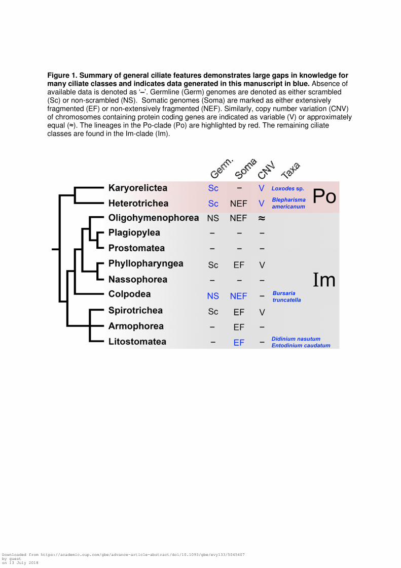
Figure 1. Summary of general ciliate features demonstrates large gaps in knowledge for many ciliate classes and indicates data generated in this manuscript in blue. Absence of available data is denoted as ‘–’. Germline (Germ) genomes are denoted as either scrambled (Sc) or non-scrambled (NS). Somatic genomes (Soma) are marked as either extensively fragmented (EF) or non-extensively fragmented (NEF). Similarly, copy number variation (CNV) of chromosomes containing protein coding genes are indicated as variable (V) or approximately equal (≈). The lineages in the Po-clade (Po) are highlighted by red. The remaining ciliate classes are found in the Im-clade (Im).
Downloaded from https://academic.oup.com/gbe/advance-article-abstract/doi/10.1093/gbe/evy133/5045407by gueston 13 July 2018
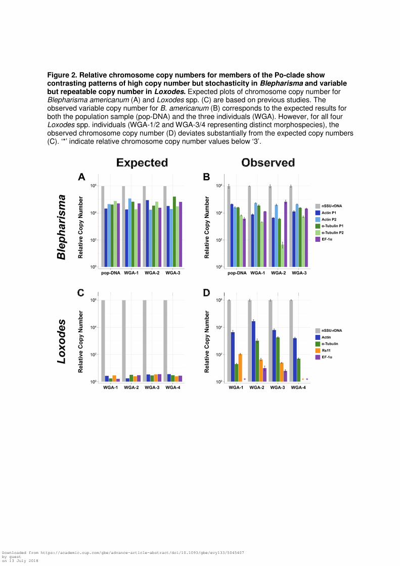
Figure 2. Relative chromosome copy numbers for members of the Po-clade show contrasting patterns of high copy number but stochasticity in Blepharisma and variable but repeatable copy number in Loxodes. Expected plots of chromosome copy number for Blepharisma americanum (A) and Loxodes spp. (C) are based on previous studies. The observed variable copy number for B. americanum (B) corresponds to the expected results for both the population sample (pop-DNA) and the three individuals (WGA). However, for all four Loxodes spp. individuals (WGA-1/2 and WGA-3/4 representing distinct morphospecies), the observed chromosome copy number (D) deviates substantially from the expected copy numbers (C). ‘*’ indicate relative chromosome copy number values below ‘3’.
Downloaded from https://academic.oup.com/gbe/advance-article-abstract/doi/10.1093/gbe/evy133/5045407by gueston 13 July 2018
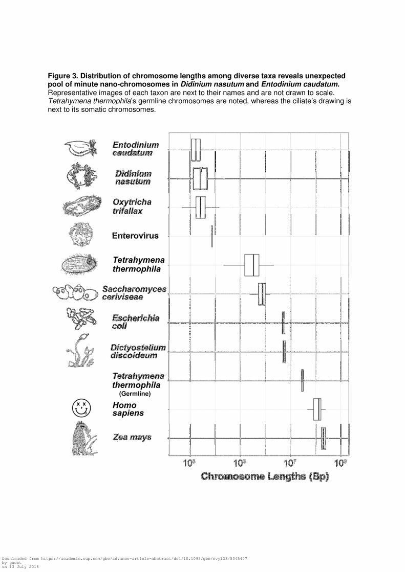
Figure 3. Distribution of chromosome lengths among diverse taxa reveals unexpected pool of minute nano-chromosomes in Didinium nasutum and Entodinium caudatum. Representative images of each taxon are next to their names and are not drawn to scale. Tetrahymena thermophila’s germline chromosomes are noted, whereas the ciliate’s drawing is next to its somatic chromosomes.
Downloaded from https://academic.oup.com/gbe/advance-article-abstract/doi/10.1093/gbe/evy133/5045407by gueston 13 July 2018

Figure 4. Exemplar chromosomes harboring α-tubulin from diverse ciliates highlights the unusual ciliate genome architecture. Telomere sequences bound complete ciliate chromosomes, with non-protein coding regions found in gray, and α-tubulin in blue (orientation of the coding sequence is noted by arrows). For Entodinium caudatum, a single nanochromosome was found with two divergent α-tubulin genes which are noted by differences in the shade of blue. Black scale bar represents 1Kbp.
Downloaded from https://academic.oup.com/gbe/advance-article-abstract/doi/10.1093/gbe/evy133/5045407by gueston 13 July 2018
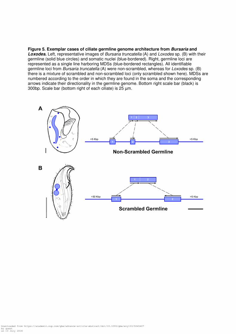
Figure 5. Exemplar cases of ciliate germline genome architecture from Bursaria and Loxodes. Left, representative images of Bursaria truncatella (A) and Loxodes sp. (B) with their germline (solid blue circles) and somatic nuclei (blue-bordered). Right, germline loci are represented as a single line harboring MDSs (blue-bordered rectangles). All identifiable germline loci from Bursaria truncatella (A) were non-scrambled, whereas for Loxodes sp. (B) there is a mixture of scrambled and non-scrambled loci (only scrambled shown here). MDSs are numbered according to the order in which they are found in the soma and the corresponding arrows indicate their directionality in the germline genome. Bottom right scale bar (black) is 300bp. Scale bar (bottom right of each ciliate) is 25 µm.
Downloaded from https://academic.oup.com/gbe/advance-article-abstract/doi/10.1093/gbe/evy133/5045407by gueston 13 July 2018