ultrasonographic diagnosis and monitoring of …cvaslibrary.com/journals/1_44_1.pdf · to its...
TRANSCRIPT
1
REVIEW ARTICLE
J. V
et. A
nim
.Sci
. 201
3. 4
4 : 1
- 7
P. Sridevi
Received - 20.08.13Accepted - 29.09.13
ULTRASONOGRAPHIC DIAGNOSIS ANDMONITORING OF PREGNANCY IN THEBITCH - A REVIEW
The use of ultrasound as a tool in smallanimal reproduction has expanded from its initialrole in the evaluation of pregnancy in the femaleto its current use in monitoring fetal development,timing gestation and predicting parturition,diagnosis and management of reproductive tractdisease and in supplementing breedingsoundness examinations. In fact, B-modeultrasonography has become an indispensabletool in veterinary practice. When ultrasonographicdiagnosis of pregnancy is being performed it isimportant to remember that all events in caninepregnancy are related to day of the pre-ovulatoryLH surge (day 0) or to the time of ovulation (whichoccurs 2 days after the LH surge) and not to themating dates. Parturition typically occurs 64 to66 days after the LH surge. This article aims toreview the use of ultrasonography in monitoringpregnancy in the bitch.
A 5-7.5 MHz transducer is adequatefor most dogs while for large and giant breedsof dogs a 3.5 MHz transducer is ideal. Fordiagnosis of mid to late-term pregnancy a 5.0-MHz transducer may be sufficient but may notprovide adequate resolution for study of moresubtle pathologic changes of the reproductivetract (Yeager and Concannon, 1990, 1995,1996 and Yeager et al., 1992). Ultrasonographyis usually performed with the dog in dorsal orlateral recumbency. Preparation of theabdomen includes clipping the hair andapplying acoustic coupling gel on the skin. Theability to ultrasonographically detect ordiscriminate a developmental feature at one dayversus another during gestation is highlydependent on the equipment used (England
et al., 2003). Forced respiration or panting canseriously affect the stillness of the image andthus make its interpretation much more difficult.Temporarily closing either the mouth or thenostrils of the dog can reduce the disturbingeffect of respiratory movements, or momentarilyremove them (Wolfgang Kahn, 2004).
DIAGNOSIS OF PREGNANCY
Real time ultrasonography has provento be a valuable tool for diagnosing caninepregnancy and assessing fetal viability (Inabaet al., 1984). The ultrasonographic appearanceof a gravid uterus in Beagle bitches at knowntime of gestation was studied in detail byYeager and Concannon (1990) and Yeageret al. (1992). They detected cardiac activityand fetal movements as early as 25 and 34days respectively after LH surge. Mattoon andNyland (1995) reported detection of gestationalsac at 20 days post breeding as the first signof confirming pregnancy using ultrasonography but preferred to wait until day 30 asgestational sac with viable embryo could beidentified with high level of confidence at thattime. Yeager et al. (1992) reported that thegestational sacs or embryonic yolk sacs canbe first imaged approximately 18 to 19 daysafter the LH surge and appear as sphericalanechoic structures 1 to 2 mm in diameterwithin the lumen of the uterus. The embryonicheartbeat has been detected as a brightechogenic flicker as early as 23 days after theLH surge. The embryo has a bipolar shape byday 28 of pregnancy. The head region isidentifiable as containing an anechoic area by
P. Sridevi*Department of Clinics,Madras Veterinary College, Chennai.e-mail: [email protected]
* Professor,
2
REVIEW ARTICLEJ.
Vet
. Ani
m.S
ci. 2
013.
44
: 1 -
7
Ultrasonographic diagnosis and monitoring of pregnancy in the bitch - a review
day 30; limb buds are usually identifiable fromday 32 to 34 onwards. The fetal skeleton isevident by day 34 of pregnancy. The bones ofthe head appear first, followed by those of thelower body. At this stage the hyperechoic heartvalves can be imaged and are seen to bemoving and the great vessels can be tracedcranially and caudally.
The zonary, circumferential placentawraps around the central portion of theconceptus like a waistband, and is observedultrasonographically between the fetus and theuterine walls in all planes. When imaged inthe longitudinal plane, the placenta appearsas two thick bands one on either side of thefetus, between the fetus and the uterine wall(Concannon et al., 2003).
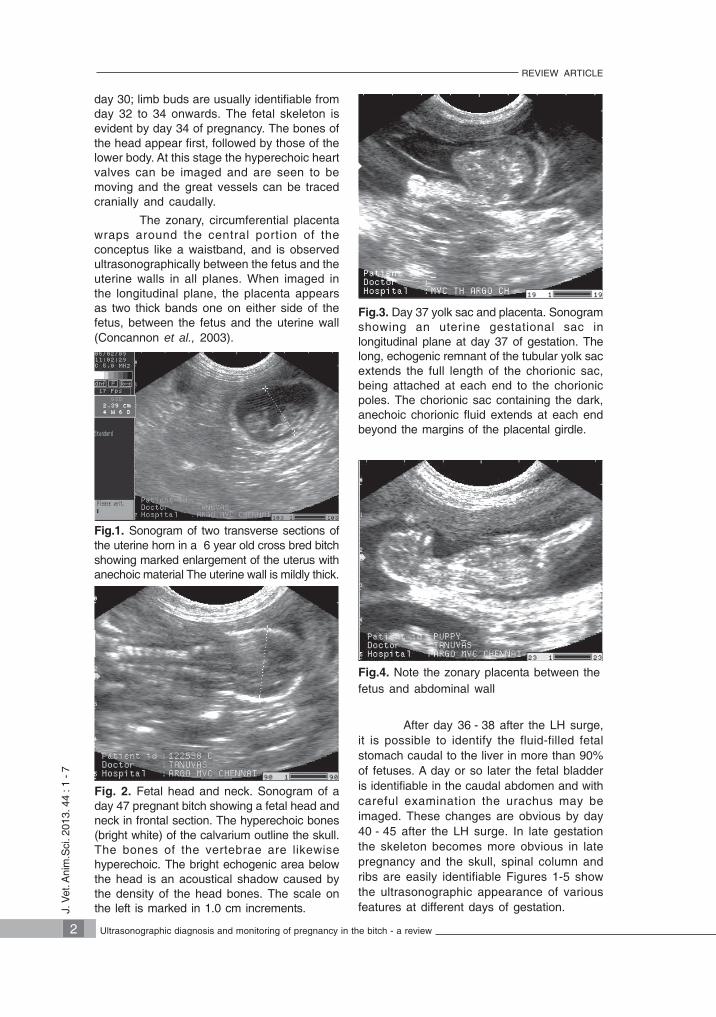
Fig. 2. Fetal head and neck. Sonogram of aday 47 pregnant bitch showing a fetal head andneck in frontal section. The hyperechoic bones(bright white) of the calvarium outline the skull.The bones of the vertebrae are likewisehyperechoic. The bright echogenic area belowthe head is an acoustical shadow caused bythe density of the head bones. The scale onthe left is marked in 1.0 cm increments.
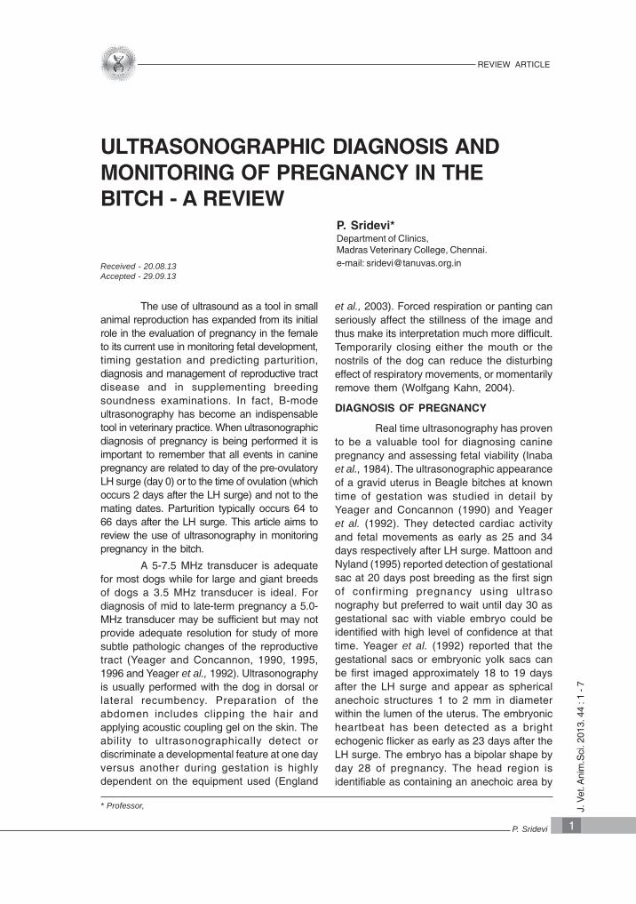
Fig.1. Sonogram of two transverse sections ofthe uterine horn in a 6 year old cross bred bitchshowing marked enlargement of the uterus withanechoic material The uterine wall is mildly thick.
After day 36 - 38 after the LH surge,it is possible to identify the fluid-filled fetalstomach caudal to the liver in more than 90%of fetuses. A day or so later the fetal bladderis identifiable in the caudal abdomen and withcareful examination the urachus may beimaged. These changes are obvious by day40 - 45 after the LH surge. In late gestationthe skeleton becomes more obvious in latepregnancy and the skull, spinal column andribs are easily identifiable Figures 1-5 showthe ultrasonographic appearance of variousfeatures at different days of gestation.
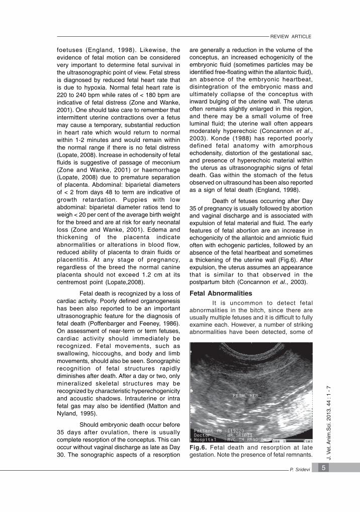
Fig.4. Note the zonary placenta between thefetus and abdominal wall
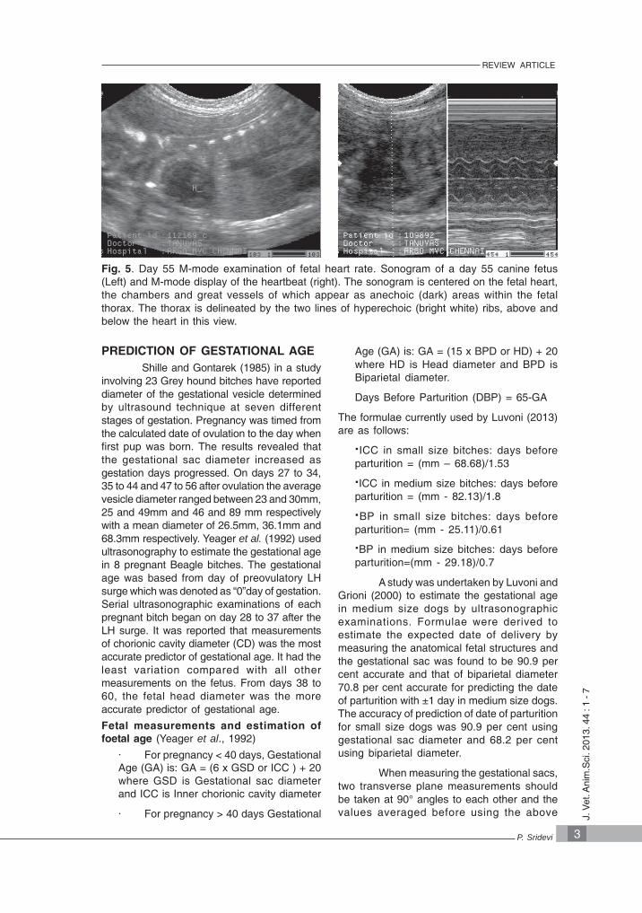
Fig.3. Day 37 yolk sac and placenta. Sonogramshowing an uterine gestational sac inlongitudinal plane at day 37 of gestation. Thelong, echogenic remnant of the tubular yolk sacextends the full length of the chorionic sac,being attached at each end to the chorionicpoles. The chorionic sac containing the dark,anechoic chorionic fluid extends at each endbeyond the margins of the placental girdle.
3
REVIEW ARTICLE
J. V
et. A
nim
.Sci
. 201
3. 4
4 : 1
- 7
P. Sridevi
Fig. 5. Day 55 M-mode examination of fetal heart rate. Sonogram of a day 55 canine fetus(Left) and M-mode display of the heartbeat (right). The sonogram is centered on the fetal heart,the chambers and great vessels of which appear as anechoic (dark) areas within the fetalthorax. The thorax is delineated by the two lines of hyperechoic (bright white) ribs, above andbelow the heart in this view.
PREDICTION OF GESTATIONAL AGEShille and Gontarek (1985) in a study
involving 23 Grey hound bitches have reporteddiameter of the gestational vesicle determinedby ultrasound technique at seven differentstages of gestation. Pregnancy was timed fromthe calculated date of ovulation to the day whenfirst pup was born. The results revealed thatthe gestational sac diameter increased asgestation days progressed. On days 27 to 34,35 to 44 and 47 to 56 after ovulation the averagevesicle diameter ranged between 23 and 30mm,25 and 49mm and 46 and 89 mm respectivelywith a mean diameter of 26.5mm, 36.1mm and68.3mm respectively. Yeager et al. (1992) usedultrasonography to estimate the gestational agein 8 pregnant Beagle bitches. The gestationalage was based from day of preovulatory LHsurge which was denoted as “0”day of gestation.Serial ultrasonographic examinations of eachpregnant bitch began on day 28 to 37 after theLH surge. It was reported that measurementsof chorionic cavity diameter (CD) was the mostaccurate predictor of gestational age. It had theleast variation compared with all othermeasurements on the fetus. From days 38 to60, the fetal head diameter was the moreaccurate predictor of gestational age.
Fetal measurements and estimation offoetal age (Yeager et al., 1992)
· For pregnancy < 40 days, GestationalAge (GA) is: GA = (6 x GSD or ICC ) + 20where GSD is Gestational sac diameterand ICC is Inner chorionic cavity diameter
· For pregnancy > 40 days Gestational
Age (GA) is: GA = (15 x BPD or HD) + 20where HD is Head diameter and BPD isBiparietal diameter.
Days Before Parturition (DBP) = 65-GA
The formulae currently used by Luvoni (2013)are as follows:
·ICC in small size bitches: days beforeparturition = (mm – 68.68)/1.53
·ICC in medium size bitches: days beforeparturition = (mm - 82.13)/1.8
·BP in small size bitches: days beforeparturition= (mm - 25.11)/0.61
·BP in medium size bitches: days beforeparturition=(mm - 29.18)/0.7
A study was undertaken by Luvoni andGrioni (2000) to estimate the gestational agein medium size dogs by ultrasonographicexaminations. Formulae were derived toestimate the expected date of delivery bymeasuring the anatomical fetal structures andthe gestational sac was found to be 90.9 percent accurate and that of biparietal diameter70.8 per cent accurate for predicting the dateof parturition with ±1 day in medium size dogs.The accuracy of prediction of date of parturitionfor small size dogs was 90.9 per cent usinggestational sac diameter and 68.2 per centusing biparietal diameter.
When measuring the gestational sacs,two transverse plane measurements shouldbe taken at 90° angles to each other and thevalues averaged before using the above

4
REVIEW ARTICLEJ.
Vet
. Ani
m.S
ci. 2
013.
44
: 1 -
7
Ultrasonographic diagnosis and monitoring of pregnancy in the bitch - a review
formulas. Head and body diameters aremeasured to the transverse planes. Whentaking measurements of fetal or extra-fetalstructures at least two distinct fetuses orgestational sac should be measured wheneverpossible and the measurements averagedbefore applying them to formulas. Thisbecomes difficult if it is a singleton fetus andmeasurement of multiple features such asGSD, HD, CRL or body diameter may becarried out to increase the accuracy.
Lenard et al. (2007) assessed theaccuracy of estimating the gestational age andlitter size in 76 bitches using one or twotechniques. The first method used thedifferential features of fetal organ developmentthat occur in early and mid-pregnancy, basedon published tables for Beagles. The secondmethod used biparietal head and trunkdiameters to predict gestational age based ontables published for late gestational period forLabrador Retrievers. The accuracy of the twomethods was compared to evaluate the effectof maternal body weight and litter size. Littersize and maternal body weight did not affectthe accuracy of gestational age prediction.Using a combination of both the methods, theoverall accuracy of predicting parturition dateto within 65 ± 1 day and ± 2 days was 70.8per cent and 86.1 per cent, respectively. Thecorrect litter size was predicted in 65 per centof cases and in 89.5 per cent of cases for ± 1pup. It was concluded that the optimum timefor sonographic estimation of fetal age andlitter size was early and mid-pregnancy.
PREDICTION OF PARTURITION
Accurate prediction of the date ofparturition in the bitch is clinically useful toprevent or minimize reproductive losses bytimely intervention. For example, an accuratemethod of predicting parturition date isnecessary to plan an elective caesareansection. Intervening when the pregnancy is fullterm can reduce losses of offspring frombitches having obstructions of the pelvis orvagina, histories of primary or secondaryuterine inertia, or prolonged parturition withresultant puppy mortality. For bitches withhistories of pyometra, abortion, embryonicreabsorption, or insufficient luteal phase,accurate assessment of gestational age canassist in therapeutic decision making. Finally,progress in assisted reproductive techniquesin this species, such as estrous synchroni-zation and embryo transfer, requires accurate
prediction of ovulation, gestational age, andparturition date (Kim et al., 2007).
The duration of canine gestation, astimed from the preovulatory serum LH peak,is 65 ±1d. However full-term gestation,calculated from insemination, is reported torange from 57 to 72 d (Concannon et al.,1993). The difference between thesemeasurements was attributed to the potential6-day viability of sperm in the femalereproductive tract and the long period ofreceptivity in the bitch. Therefore the key totiming the duration of canine gestation wasthe preovulatory LH surge rather than mating/ insemination date or estrus onset (Meyers-Wallen, 1995). Ultrasound provides anaccurate estimate of parturition date and themost accurate prediction was obtained whenthe ultrasound examination was conducted atDay 30 (Kutzler et al., 2003). However,prediction was inaccurate when made fromfetal measurements in late gestation (>Day39). Prediction accuracy was also significantlyaffected by non-pregnant body weight of thedam. Fetal growth was linear from Days 17 to30 and subsequently became exponential(England, 1998). Kutzler et al. (2003) reportedthat after day 30, fetuses of small bitches (<9kg) grew slower, and fetuses of giant bitches(>40 kg) grew faster, than those of medium orlarge bitches. When corrected for body weightof the dam, the overall accuracy for parturitiondate prediction by the ultrasound method was75% for the Day 65 ± 1 prediction, 87% forthe Day 65 ± 2 prediction, and 100% for theDay 65 ± 3 prediction.
MONITORING FETAL WELL BEINGFetal Death
Recognition of fetal death at or nearparturition is of extreme importance to theveterinarian and breeder. As the date ofparturition approaches, the following areindications for fetal monitoring withtransabdominal ultrasonography:
· Failure to initiate parturition asexpected.
· Unusual vaginal discharge
· Vague signs of illness
· Delay in parturition after delivery ofpart of a litter
Fetal heart rate has beenrecommended as a very useful parameter toestimate the survival possibilities of the
5
REVIEW ARTICLE
J. V
et. A
nim
.Sci
. 201
3. 4
4 : 1
- 7
P. Sridevi
foetuses (England, 1998). Likewise, theevidence of fetal motion can be consideredvery important to determine fetal survival inthe ultrasonographic point of view. Fetal stressis diagnosed by reduced fetal heart rate thatis due to hypoxia. Normal fetal heart rate is220 to 240 bpm while rates of < 180 bpm areindicative of fetal distress (Zone and Wanke,2001). One should take care to remember thatintermittent uterine contractions over a fetusmay cause a temporary, substantial reductionin heart rate which would return to normalwithin 1-2 minutes and would remain withinthe normal range if there is no fetal distress(Lopate, 2008). Increase in echodensity of fetalfluids is suggestive of passage of meconium(Zone and Wanke, 2001) or haemorrhage(Lopate, 2008) due to premature separationof placenta. Abdominal: biparietal diametersof < 2 from days 48 to term are indicative ofgrowth retardation. Puppies with lowabdominal: biparietal diameter ratios tend toweigh < 20 per cent of the average birth weightfor the breed and are at risk for early neonatalloss (Zone and Wanke, 2001). Edema andthickening of the placenta indicateabnormalities or alterations in blood flow,reduced ability of placenta to drain fluids orplacentitis. At any stage of pregnancy,regardless of the breed the normal canineplacenta should not exceed 1.2 cm at itscentremost point (Lopate,2008).
Fetal death is recognized by a loss ofcardiac activity. Poorly defined organogenesishas been also reported to be an importantultrasonographic feature for the diagnosis offetal death (Poffenbarger and Feeney, 1986).On assessment of near-term or term fetuses,cardiac activity should immediately berecognized. Fetal movements, such asswallowing, hiccoughs, and body and limbmovements, should also be seen. Sonographicrecognition of fetal structures rapidlydiminishes after death. After a day or two, onlymineralized skeletal structures may berecognized by characteristic hyperechogenicityand acoustic shadows. Intrauterine or intrafetal gas may also be identified (Matton andNyland, 1995).
Should embryonic death occur before35 days after ovulation, there is usuallycomplete resorption of the conceptus. This canoccur without vaginal discharge as late as Day30. The sonographic aspects of a resorption
are generally a reduction in the volume of theconceptus, an increased echogenicity of theembryonic fluid (sometimes particles may beidentified free-floating within the allantoic fluid),an absence of the embryonic heartbeat,disintegration of the embryonic mass andultimately collapse of the conceptus withinward bulging of the uterine wall. The uterusoften remains slightly enlarged in this region,and there may be a small volume of freeluminal fluid; the uterine wall often appearsmoderately hyperechoic (Concannon et al.,2003). Konde (1988) has reported poorlydefined fetal anatomy with amorphousechodensity, distortion of the gestational sac,and presence of hyperechoic material withinthe uterus as ultrasonographic signs of fetaldeath. Gas within the stomach of the fetusobserved on ultrasound has been also reportedas a sign of fetal death (England, 1998).
Death of fetuses occurring after Day35 of pregnancy is usually followed by abortionand vaginal discharge and is associated withexpulsion of fetal material and fluid. The earlyfeatures of fetal abortion are an increase inechogenicity of the allantoic and amniotic fluidoften with echogenic particles, followed by anabsence of the fetal heartbeat and sometimesa thickening of the uterine wall (Fig.6). Afterexpulsion, the uterus assumes an appearancethat is similar to that observed in thepostpartum bitch (Concannon et al., 2003).
Fetal AbnormalitiesIt is uncommon to detect fetal
abnormalities in the bitch, since there areusually multiple fetuses and it is difficult to fullyexamine each. However, a number of strikingabnormalities have been detected, some of
Fig.6. Fetal death and resorption at lategestation. Note the presence of fetal remnants.

6
REVIEW ARTICLEJ.
Vet
. Ani
m.S
ci. 2
013.
44
: 1 -
7
Ultrasonographic diagnosis and monitoring of pregnancy in the bitch - a review
which have necessitated delivery of the litter bycaesarean operation. Such abnormalities includehydrocephalus, fetal anasarca (Fig.7), herniationof the ventral abdominal wall and fetal monsters(Poffenbarger and Feeney, 1986).
APPLICATION OF DOPPLERULTRASONOGRAPHY IN CANINEPREGNANCY
Miranda and Domingues (2010)evaluated blood flow in the uterine (UA) andumbilical (Uma) arteries in the pregnant bitch,by measuring the resistive index (RI) andpulsatility index (PI); and performed conceptusecobiometry for fetal growth assessmentduring pregnancy.Triplex Doppler and B-modeultrasonography were used to assess bloodflow and conceptus ecobiometry. Allpregnancies ended with a normal whelping andbirth of live puppies. Prior to whelping, allconceptus dimensions increased significantly,whereas RI and PI of both the Uma and UAdecreased significantly. For the UA, RI and PIwere (mean +/- SEM) 0.95 +/- 0.02 and 2.75+/- 0.41, respectively, on Day -44, and were0.60 +/- 0.01 and 0.99 +/- 0.03 on Day -4. Forthe Uma, RI and PI were 0.99 +/- 0.01 and2.42 +/- 0.03 on Day -31, and were 0.62 +/-0.01 and 1.15 +/- 0.02 on Day -4. The completedisappearance of the early diastolic notch inthe UA, and the appearance of diastolic flowin the Uma occurred on Days -16 +/- 5 and -21 +/- 1. The authors concluded that UA andUma perfusion were important end points toassess fetal vitality in bitches. Furthermore,the current reference values provided abaseline for monitoring normal and abnormalpregnancies in bitches
Blanco et al. (2011) described thechanges of uterine artery, umbilical artery andfetal abdominal aorta, renal and internalcarotid arteries blood flow in abnormal caninepregnancy. Color and pulsed-wave Dopplerexaminations of uterine artery were conductedevery 10 days from Day 20 to 50 fromestimated luteinizing hormone peak. Dopplerultrasonography was also conducted in thefetuses to assess umbilical artery, abdominalaorta, renal and internal carotid arteries fromDay 40 to 60 of gestation. Throughout thestudy, resistance index (RI) of uterine,umbilical and fetal renal arteries decreased upto -15% compared to -36% (P<0.01), -11%compared to -23% (P<0.05) and 2% comparedto -13% (P<0.05), respectively in the abnormaland normal bitches. Fetal abdominal aorta andinternal carotid did not differ between groups(P>0.05). They concluded that in dogs, uterineartery, umbilical artery and fetal renal arteryRI differ between normal and abnormalgestation and therefore were useful for theprediction of adverse obstetric outcome.
Thus, to conclude ultrasonographicexamination is an indispensable tool forpracticing veterinarians for monitoring fetalgrowth, for assessing gestational age and forprediction of parturition which is particularlyvaluable for providing clinical assistance duringwhelping or elective caesarean sections.
ReferencesBlanco, P. G., Rodríguez, R., Rube, A., Arias,
D.O., Tórtora, M., Díaz, J. D.andGobello. C. 2011. Dopplerultrasonographic assessment ofmaternal and fetal blood flow inabnormal canine pregnancy. Anim.Reprod. Sci. 126 (1-2) :130-135.
Concannon, P. W., England, G., Verstegen J.and Linde-Forsberg C. 2003.Ultrasound imaging of reproductivetract of the bitch. InternationalVeterinary Information Service(www.ivis.org), Ithaca, New York, USA.
Concannon, P. W., Whaley, S., Lein, D.,Wissler. R. 1993. Canine gestationlength: variation related to time ofmating and fertile life of sperm. Am.J. Vet. Res. 44 : 1819–1821.
England, G. 1998. Ultrasonographicassessment of abnormal pregnancy.Vet. Clin. North Am. Small Anim.Pract., 28: 1233-1256
Fig.7. Ultrasonographic image of fetal anas-arca. Note the increased subcutaneousoedema (arrow) and serous effusions (astrix).
7
REVIEW ARTICLE
J. V
et. A
nim
.Sci
. 201
3. 4
4 : 1
- 7
P. Sridevi
�
England, G., Yeager, A. and Concannon, P. W.2003. Ultrasound Imaging of theReproductive Tract of the Bitch. In: Recentadvances in small animal reproduction.IVIS, Ithaca. New York, USA.
Inaba, T., Matsu, N., Shimizu, R. and Imori, T.1984. The use of echography inbitches for detection of ovulation andpregnancy. Vet. Rec. 115 : 267-277.
Kim, B. S. and Son. C. H. 2007. Time of initialdetection of fetal and extra-fetalstructures by ultrasonographicexamination in Miniature Schnauzerbitches. J. Vet. Sci. 8 : 289-93
Kim Se Ra, Kang Hyun Gu, Oh KiSeok, Park InChul, Park SangGuk, Kim Sung Ho, SonChang Ho. 2000. Establishment ofprediction table of parturition day byultrasonography in Korean Jindo bitches.Korean J. Vet. Res. 40 : 373-381.
Konde, L.1988. Diagnostic ultrasound incanine pregnancy and uterinedisease. Proc. Ann. Meet Soc. forTheriogenology, Orlando, Florida,September 16–17, 247-249.
Kutzler, M. A., Yeager, A. E., Mohammed H.O. and Meyers. W. V. N. 2003.Accuracy of canine parturition dateprediction using fetal measurementsobtained by ultrasonography.Theriogenology. 60:1309-1317.
Lenard, Z., Hopper, B., Lester, N., RichardsonJ. and Robertson. I. 2007. Accuracyof prediction of canine litter size andgestational age with ultrasound. Aust.Vet. J. 85:222-225.
Lopate, C. 2008. Estimation of gestational ageand assessment of canine fetalmeasurements using radiology andultrasonography.: A review.Theriogenology, 70 (3): 397-402.
Luvoni, G.C. 2013. Ultrasonographic study ofgestation in dogs and cats. Rev. Bras.Reprod. Anim., Belo Horizonte, 37:172-173.
Luvoni, G. C. and Grioni . A. 2000.Determination of gestational age inmedium and small size bitches usingultrasonographic fetal measurements.J. Small. Anim. Pract. 41:294-296.
Mattoon, J. S. and Nyland T. G. 1995.Ultrasonography of the genital system:In Veterinary Diagnostic Ultrasound.
Eds. Nyland, T.G. and Mattoon, J.S.,W.B. Saunders, USA. pp146-148.
Meyers-Wallen, V. N.1995. The electivecesarean section. In: Bonagura J.D.,Kirk R.W., (Eds.) Current veterinarytherapy XII. Philadelphia, WBSaunders Co., pp. 1085–1089.
Miranda, S. A. and Domingues, S. F. S. 2010.Conceptus ecobiometry and triplexDoppler ultrasonography of uterineand umbilical arteries for assessmentof fetal viability in dogs. Theriogenology. 74: 608–617.
Poffenbarger, E. and Feeney, D. 1986. Use ofgray-scale ultrasonography in thediagnosis of reproductive disease inthe bitch: 18 cases (1981-1984). J.Am. Vet. Med. Assoc. 189 : 90-95.
Shille, V. M. and J. Gontarek.1985. The useof ultrasonography for pregnancydiagnosis in the bitch. J.Am.Vet.Med.Assoc. 187: 1021-1025.
Yeager, A. E. and Concannon, P. W. 1990.Association between the preovulatoryLH surge and the early ultrasonographic detection of pregnancy andfetal heartbeats in beagle dogs.Theriogenology. 34 : 655-665.
Yeager, A. E. and P. W. Concannon .1995.Ultrasonography of the reproductive tractof the female dog and cat. In: BonaguraJ. D., Kirk R.W., (Eds.) Kirk’s CurrentVeterinary Therapy XII. Philadelphia:W.B. Saunders Co.,1040-1052.
Yeager, A.E. and Concannon, P. W. 1996.Uterus. In: Green R.W., (Ed.) SmallAnimal Ultrasound. Philadelphia:Lippincott-Raven, 265-292.
Yeager, A. E., Mohammed, H. O., Meyers-Wallen,V., Vannerson L. and Concannon, P. W.1992. Ultrasono graphic appearance ofthe uterus, placenta, fetus, and fetalmembranes throughout accurately timedpregnancy in beagles. Am. J. Vet. Res.53:342-351.
Wolfgang Kahn. 2004. Veterinary Reproductive Ultrasonography. SchlûterscheVerlagsgesellschaft mbH & Co. KG,Hans·BOc:klcr·Allee 7, 30173 Hannover, pp.233-245.
Zone, M. A. and Wanke M. M. 2001. Diagnosisof canine fetal health by ultrasonography. J. Reprod. Fertil. 57: 215-219.