universidade de coimbra imperial college london: 2012/2013s... · universidade de coimbra imperial...
TRANSCRIPT

Universidade de Coimbra Imperial College London: 2012/2013
Natalia C.Couto Francisco Master Degree: Biomedical Research 2
Acknowledgments
I am very honoured to have been given this opportunity by Professor Stephen Durham to
work in such a world class research facility, the knowledge and experience I have gained will
prove invaluable for me in my future science career. I am also indebted to Dr Mohamed
Shamji for all his supervision and experience given to me from the fascinating world of
clinical immunology. I acknowledge and extend my sincere warmest gratitude to Dr.
Henrique Girão for his unwavering support. I would like to thank Dr. Tomokatzu Matsouka
for his support both in and out of the lab, without his patient teachings I would never have
mastered the techniques implemented in this thesis. I would also like to thank Rachel Yan
and Andrea Goldstone for the hard work at the Royal Brompton hospital, for providing all the
necessary patients for my research. For some of the technical support that I received in the
lab, I would like to show my appreciation to Amy Switzer, Orla McMahon and Miss Rebecca
Parkin. To Janice Layhadi, James Charlesworth and Alan Perera for their friendship and
support given me throughout my project. Finally, I would like to extend my gratitude to Jinjin
Zhan, Clara Wo, Esther Steveling, Gilda Varrichi, Liliana Fuentes, Mimi Poon, Natalia-
Klimowska, Guy Scadding, Mongkol Lao-aray, Faris Mustafa and for those hugs, chocolates,
lunch and chill out sessions.

Universidade de Coimbra Imperial College London: 2012/2013
Natalia C.Couto Francisco Master Degree: Biomedical Research 3
I dedicate all my work, achievements and success to my mother Maria.
All this would not be possible without her support.
Universidade de Coimbra Imperial College London: 2012/2013
Natalia C.Couto Francisco Master Degree: Biomedical Research 4
Abstract
Introduction: Allergic Rhinitis affects ¼ of the population in developed countries.
Sublingual grass pollen-specific immunotherapy (SLIT) involves immunomodulation of Th2
responses and the induction of IL-10+ Tregs (Tr1). IL-35-producing regulatory T cells
(iTR35) have been recently reported as a novel subset of regulatory T cells with modulatory
properties. We hypothesized that IL-35 suppresses grass pollen-driven Th2 responses
following ex-vivo allergen stimulation, and induces iTR35 cells following grass pollen-SLIT.
We further hypothesized that IL-35 suppresses an in vitro Th2 inflammatory response,
induced by epithelial derived cytokines.
Methods: T effector cells (CD4+CD25-) obtained from grass pollen allergics (n=12) were
purified and enriched from peripheral blood mononuclear cells by magnetic separation.
CD4+CD25- T cells were co-cultured with irradiated antigen-presenting cells using 5ug/mL
of Phleum pratense in the presence/absence 10ng/mL of recombinant human IL-35: Fc. T cell
proliferative responses were measured by 3H-thymidine incorporation. Cytokine protein
levels were assessed by Luminex MagPix assay. Proportion of FoxP3, IL-10 and iTR35 was
determined in non-atopics (NA, n=12), untreated allergics (SAR, n=12) and SLIT-treated
patients (SLIT, n=7). DCs obtained from SAR patients with seasonal allergic rhinitis (n=14)
were primed with TSLP, IL-25, IL-33 or all three cytokines in the presence of 5ug/mL of
Phleum pratense for 24 hour. Primed DCs were co-cultured with naive T cells for 6 days at
1:10 ratio. Proliferative responses of naive T cells were measured by 3H tritiated thymidine
incorporation and cytokine analysis by Luminex MagPix assay.
Results: IL-35 significantly suppressed Phleum pratense-driven CD4+CD25- T cell
proliferative responses (n=12; p<0.0009). This suppression was associated with reduced IL-4
Universidade de Coimbra Imperial College London: 2012/2013
Natalia C.Couto Francisco Master Degree: Biomedical Research 5
(p=0.0001), IL-5 (p=0.0001), IL-9 (p=0.0001), IL-13 (p=0.0009) and an increase in IFN-γ
(p<0.0001) and IL-10 (p<0.0001) was also demonstrated. Furthermore, iTR35 cells, IL-10+
and FoxP3+ T regs were decreased in SAR compared to NA (p=0.0003; 0.0005; p=0.001).
SLIT resulted in recovery of iTR35 (p=0.016) and IL-10+ Treg (p=0.016) cells. FoxP3+
Tregs did not increased in SLIT group. TSLP to allergen-primed DCs resulted in a 97-fold
increase (p=0.003) in naive T cell proliferation when compared to allergen-only primed DCs.
IL-25- (p=0.426) and IL-33- (p=0.502) primed DCs did not augment this allergen-stimulated
proliferative response. In the same system, IL-35 suppressed naive T cell proliferative
responses when allergen+TSLP (p=0.0001) were cultured with DCs. In addition, results have
shown an increase in Th2 cytokines when TSLP primed DCs are added to naive Tcells and
their suppression when treated with IL-35.
Conclusion: Our findings suggest that iTR35 cells suppress grass pollen-driven Th2
responses and are induced following grass SLIT. IL-35 not only inhibits grass-pollen and
TSLP-primed DC activation of naive T cells as it also suppressed Th2 cytokines induced by
TSLP. The mechanism of this suppression suggests that IL-35 is a potential target for
seasonal allergic rhinitis immunotherapy.
Key words: IL-35, Seasonal Allergic Rhinitis, Th2 Allergic Response, Epithelial derived
cytokines, TSLP, IL-25, IL-33, Sublingual Immunotherapy.
Universidade de Coimbra Imperial College London: 2012/2013
Natalia C.Couto Francisco Master Degree: Biomedical Research 6
Resumo
Introdução: A Rinite alérgica afecta um quarto da população em países desenvolvidos. A
imunoterapia sublingual involve a imunoregulação de respostas Th2 e a indução de células T
reguladoras, produtoras de IL-10 (Tr1). As células T reguladoras induzidas pela IL-35
(iTR35) são um novo subtipo de células T reguladoras com funcionalidade modeladora.
Neste projecto, conjecturamos que a IL-35 suprime respostas do tipo Th2 induzidas pelo
alergenio em causa (phlp) e induz o aumento de células iTR35 após imunoterapia sublingual.
Em acréscimo, também conjecturamos que a IL-35 tem um papel supressor num modelo de
resposta inflamatória Th2 induzido por citocinas proinflamatórias derivadas de células
epiteliais.
Métodos: Linfócitos T efectores (CD4+CD25-) foram coletados de pacientes alérgicos a
polén de gramíneas (n=12), por isolamento magnético de céluals mononucleares de sangue
periférico. Células apresentadoras de antigénios foram irradiadas e cultivadas com as células
T efectoras na presença de 5ug/mL de Phleum pratense e de 10ng/mL de IL-35: Fc. A
proliferação celular foi medida pela incorporação de 3H-timidina e os níveis de citocinas
produzidos por multiplex ELISA. Em seguida, a porpoção de FoxP3, IL-10 e iTR35 foi
avaliada em pacientes não atópicos (NA, n=12), alérgicos (SAR, n=12) e pacientes
submetidos a imunoterapia sublingual (SLIT, n=7). Finalmente, células dendriticas obtidas de
pacientes alérgicos a gramíneas (n=14) foram co-estimulados na presença de TSLP, IL-25,
IL-33 ou as três proteínas em simultâneo, e de 5ug/mL de Phleum pratense durante 24h.
Estas células dendríticas foram posteriormente cultivadas com células T naive durante 6 dias
a um rácio de 1:10. Ensaios de proliferação foram conseguidos por incorporação de 3H-
timidina e análise de citocinas por multiplex ELISA.
Universidade de Coimbra Imperial College London: 2012/2013
Natalia C.Couto Francisco Master Degree: Biomedical Research 7
Results: A IL-35 inibe significativamente a proliferação de células T efectoras, induzida pelo
Phleum pratense (n=12; p<0.0009). Esta supressão foi associada à redução de IL-4
(p=0.0001), IL-5 (p=0.0001), IL-9 (p=0.0001), IL-13 (p=0.0009) e ao aumento de IFN-γ
(p<0.0001) e IL-10 (p<0.0001). Em seguida, as células T reguladores que expressão IL-35,
IL-10 e FoxP3+ foram avaliadas e observou-se um decréscimo das mesmas em SAR
comparadamente a NA (p=0.0003; p=0.0005; p=0.001). Pacientes submetidos a
imunoterapia sublingual demonstram um aumento em células reguladoras que expressão IL-
35 (p=0.016) e IL-10 (p=0.016), não mostrando variação em T reguladores que expressão
FoxP3+. As células dendriticas expostas a TSLP induziram um aumento de 97% (p=0.003)
na proliferação de células T naive. No entanto, células dendríticas estimuladas com a IL-25
(p=0.426) e a IL-33 (p=0.502) não tiveram qualquer efeito na proliferação das células T
naive. O papel modelador da IL-35 foi testado nas mesmas condições, mostrando supressão
da proliferação de células T naive induzida por células dendríticas expostas a TSLP
(p=0.0001). Por fim, a proliferação de células T naive foi associado ao aumento de citocinas
envolvidas em respostas do tipo Th2, e a supressão das mesmas conseguida pela presença da
IL-35.
Conclusão: Os nossos resultado sugerem um papel importante da iTR35 na supressão de
respostas Th2 induzidas por phlp. Também se observou a indução destas células em pacientes
que receberam tratatment sublingual. Adicionalmente, a IL-35 não só suprime proliferação de
células T naive induzidas pelo efeito da TSLP nas células dendríticas, como também reduz
citocinas caracteristicas the uma resposta alérgica inflamatória Th2. A IL-35 demonstra ser
um potencial alvo para terapias futuras.
Palavras-chave: IL-35, Rinite alérgica, Resposta alérgica do tipo Th2, Citocinas derivadas
Universidade de Coimbra Imperial College London: 2012/2013
Natalia C.Couto Francisco Master Degree: Biomedical Research 8
de células epiteliais, TSLP, IL-25, IL-33, Imunoterapia sublingual.
Index
Page
Hypersensitivity Diseases 16
Immediate (Type I) Hypersensitivity: Allergy 17-18
Respiratory Allergy - Allergic Rhinitis 19
Immunological Mechanisms of Allergic Rhinitis 20-22
Chapter I
Introduction
Potential Inflammatory mediators in Seasonal Allergic Rhinitis
Potential Immunotherapy targets in Seasonal Allergic Rhinitis
23-25
25-27
Ethical Statement 30
Skin prick test and ImmunoCap 30
PBMC isolation 30
Thawing PBMCs from Liquid Nitrogen 30-31
Cell staining of FoxP3+Treg, IL10+CD4+Treg and IL-35+CD4+Treg 31
CD4+CD25- T effector cells antigen-specific stimulation and IL-35
neutralisation
31
T cell polarisation using epithelial derived cytokines and IL-35
suppression
31-32
TSLP receptor expression on DCs 32
T cell polarisation towards Th1 or Th2 cells 32-34
Surface staining 34
Intracellular and nuclear staining by Flow cytometry 35
Chapter II
Methodology
Gene expression assay by qPCR 35-36 Cytokine Analysis by Multiplex ELISA 36

Universidade de Coimbra Imperial College London: 2012/2013
Natalia C.Couto Francisco Master Degree: Biomedical Research 9
Statistical Analysis 36
Rhinitis symptom scores declines after sublingual immunotherapy 38
IL-10 + and IL-35 +, but not, FoxP3 + Treg is increased following
SLIT 38
IL-35 suppresses allergen driven Teff cell proliferation and induces
Th2 differentiation 40
TSLP, but not Il-25 and IL-33 primed DCs induce T cell proliferation
in an allergen driven response and Th2 differentiation 42
TSLP Receptor is expressed in higher amounts in SAR compared to
NA patients 44
IL-35 suppresses TSLP primed DCs driven naive T cell proliferation
and induces Th2 response deviation 45
Naive T cells fully polarised into Th1 cells after 2 weeks of treatment 47
Naive T cells fully polarised into Th2 cells after 3 weeks of treatment 47
Chapter III
Results
Polarisation of Th1 and Th2 cells were confirmed by Multiplex and
q-PCR 49
Universidade de Coimbra Imperial College London: 2012/2013
Natalia C.Couto Francisco Master Degree: Biomedical Research 10
Chapter IV
Discussion
50-54
Chapter V
Future
Studies
55-56
Chapter VI
Conclusion
57
References 58-60
Glossary 62
Key to illustrations found in the introduction 63
Relevant Training 64
Awards 64
Attachments
Publications 64

Universidade de Coimbra Imperial College London: 2012/2013
Natalia C.Couto Francisco Master Degree: Biomedical Research 11

Universidade de Coimbra Imperial College London: 2012/2013
Natalia C.Couto Francisco Master Degree: Biomedical Research 12
Abbreviation List
APC Antigen Presenting Cell pDCs Plamacytoid DCs
BFA Brefeldin A PHA Phytohaemagglutinin
CD Cluster of differentiation PMA Phorbol 12-Myristate 13-Acetate
DC Dendritic cell PT Portugal
EPR Early Phase response RAST Radioallergosorbent test
Foxp3
iTregs
Forkhead box P3
Induced T regulatory cells
RPMI Roswell Park Memorial Institute
(RPMI) medium
IL- Interleukin rpms Revolutions per minute
IgE Immunoglobulin E SAR Seasonal Allergic Rhinitis
LFR Late phase response SIT Specific Immunotherapy
Mɸ Macrophage SLIT Sublingual Immunotherapy
MHC Major Histocompatibility Complex TCM Tissue culture medium
mDC Myeloid DCs TG Timothy Grass
nTcell Naive T cell Th1 T helper 1
N/A Not applicable Th2 T helper 2
NA Non Atopic Tregs T regulatory cells
PBMCs Peripheral Blood Mononuclear Cells TSLP Thymic Stromal Lymphopoietin
Phlp Phleum pratense UK United Kingdom
Symbols
* The definition can be found in the “Glossary” section of the attachments (Page 62)
# Product catalogue number

Universidade de Coimbra Imperial College London: 2012/2013
Natalia C.Couto Francisco Master Degree: Biomedical Research 13
Figure List
I. Introduction
1.1 Immediate Hypersensitivity. Kinetics of the early-phase response (EPR) and
late-phase response (LPR) after allergen challenge to a sensitised individual. 22
1.2 Mechanisms of seasonal allergic rhinitis. 24
1.3 Mechanisms of allergen specific immunotherapy. 27
II. Methodology
2.1 Tcell Polarisation Protocol. 33
2.2 Naive T cell purification check. 34
III. Results
3.1 RA symptom score and PBMC surface and intracellular staining of different
subtypes of Tregs in SAR and SLIT patients 39
3.2 Teff cells treated with or without IL-35 were submitted to a proliferation assay
as to cytokine analysis 41
3.3 Epithelial-derived pro-allergic mediator TSLP but not IL-25 and IL-33 induces
grass pollen-specific naive T cell proliferation and Th2 cytokines 43
3.4 Effect of TSLP on TSLPR expression on mDCs and pDCs obtained from SAR
and NA patients 44
3.5 Effect of IL-35 on naive T cells exposed to Phlp and Phlp+TSLP primed DCs 46
3.6 Surface, intracellular and nuclear staining of Th1 and Th2 markers, of fully
polarised cells 48
3.7 Cytokine analysis and gene expression of polarised T cells 49
Universidade de Coimbra Imperial College London: 2012/2013
Natalia C.Couto Francisco Master Degree: Biomedical Research 14
Table List
I. Introduction
1.1 Causative agents of allergic diseases. 17
1.2 Highest seasonal allergen counts. 19
II. Methodology
2.1 Forward and reverse primer sequences for Th1 and Th2 related targets. 36
Attachments
- Glossary 62
- Key to illustrations found in the introduction 63
Universidade de Coimbra Imperial College London: 2012/2013
Natalia C.Couto Francisco Master Degree: Biomedical Research 15
Portugal, Tão Diferente de seu Ser Primeiro
Os reinos e os impérios poderosos,
Que em grandeza no mundo mais cresceram, Ou por valor de esforço floresceram, Ou por varões nas letras espantosos.
Teve Grécia Temístocles; famosos,
Os Cipiões a Roma engrandeceram; Doze Pares a França glória deram; Cides a Espanha, e Laras belicosos.
Ao nosso Portugal, que agora vemos
Tão diferente de seu ser primeiro, Os vossos deram honra e liberdade.
E em vós, grão sucessor e novo herdeiro
Do Braganção estado, há mil extremos Iguais ao sangue e mores que a idade.
Luís Vaz de Camões in “Sonetos”

Universidade de Coimbra Imperial College London: 2012/2013
Natalia C.Couto Francisco Master Degree: Biomedical Research 16
Chapter I: Introduction
Hypersensitivity Diseases 16
Immediate (Type I) Hypersensitivity: Allergy 17-18
Respiratory Allergy - Allergic Rhinitis 19
Immunological Mechanisms of Allergic Rhinitis 20-22
Potential Inflammatory mediators in SAR
Potential Immunotherapy targets in SAR
23-25
25-27
Universidade de Coimbra Imperial College London: 2012/2013
Natalia C.Couto Francisco Master Degree: Biomedical Research 17
1.1) Hypersensitivity Diseases
Individuals are said to be “sensitised” when they build up an immune response against a
commonly harmless antigen. Therefore, the excessive reactions towards the specific antigen
result in manifestations of hypersensitivity.
An exquisite immune system is capable of maintaining balance while eradicating infecting
organisms and preventing serious cell tissue injury to the host. In contrast, a hyper-reactive
individual shows signs of serious injuries to its own tissue, indicating inadequate control or
inappropriate tissue targeting by their immune system.
There are four sub-types of hypersensitivity diseases named Immediate (type I)
hypersensitivity, Antibody-mediated (type II) hypersensitivity, Immune complex-mediated
(Type III) hypersensitivity and T-cell-mediated (type IV) hypersensitivity. Types I, II and III
hypersensitivity disorders are characterised by antibody mediated injuries while type IV is
defined as a hypersensitivity disorder of cell mediated injury1–4.
It is important to emphasise that cell injury is caused by the same basic mechanisms that
eradicate infectious pathogens, in which we find involved the Innate, Adaptive and Humoral
immune responses. The uncontrolled balance of this cascade and the persistence of the
stimuli at the site of inflammation create positive immune feedbacks, becoming difficult to
terminate this pathologic cycle of immune reactions.
Universidade de Coimbra Imperial College London: 2012/2013
Natalia C.Couto Francisco Master Degree: Biomedical Research 18
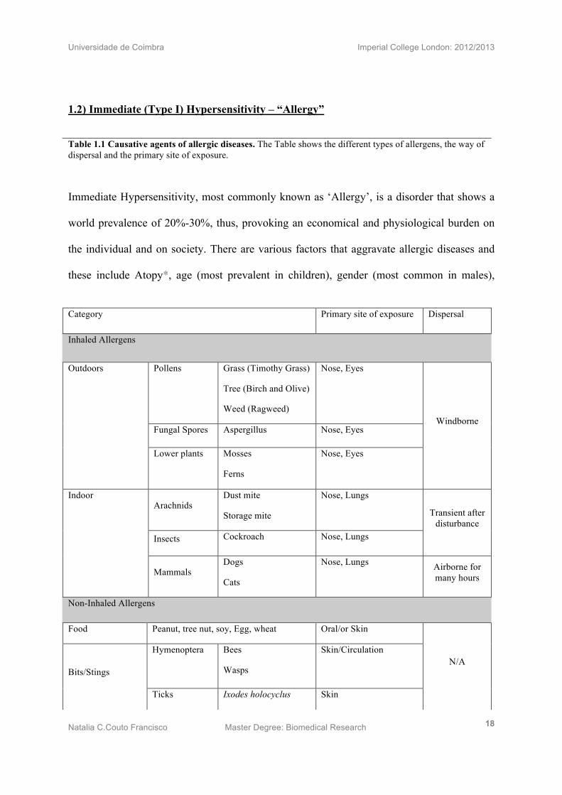
1.2) Immediate (Type I) Hypersensitivity – “Allergy”
Immediate Hypersensitivity, most commonly known as ‘Allergy’, is a disorder that shows a
world prevalence of 20%-30%, thus, provoking an economical and physiological burden on
the individual and on society. There are various factors that aggravate allergic diseases and
these include Atopy*, age (most prevalent in children), gender (most common in males),
Category Primary site of exposure Dispersal
Inhaled Allergens
Pollens Grass (Timothy Grass)
Tree (Birch and Olive)
Weed (Ragweed)
Nose, Eyes
Fungal Spores Aspergillus Nose, Eyes
Outdoors
Lower plants Mosses
Ferns
Nose, Eyes
Windborne
Arachnids Dust mite
Storage mite
Nose, Lungs
Insects Cockroach Nose, Lungs
Transient after disturbance
Indoor
Mammals Dogs
Cats
Nose, Lungs Airborne for many hours
Non-Inhaled Allergens
Food Peanut, tree nut, soy, Egg, wheat Oral/or Skin
Hymenoptera Bees
Wasps
Skin/Circulation
Bits/Stings
Ticks Ixodes holocyclus Skin
N/A
Table 1.1 Causative agents of allergic diseases. The Table shows the different types of allergens, the way of dispersal and the primary site of exposure.
Universidade de Coimbra Imperial College London: 2012/2013
Natalia C.Couto Francisco Master Degree: Biomedical Research 19
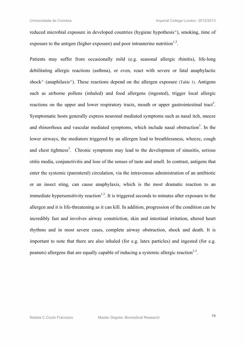
reduced microbial exposure in developed countries (hygiene hypothesis*), smoking, time of
exposure to the antigen (higher exposure) and poor intrauterine nutrition1,3.
Patients may suffer from occasionally mild (e.g. seasonal allergic rhinitis), life-long
debilitating allergic reactions (asthma), or even, react with severe or fatal anaphylactic
shock* (anaphilaxis*). These reactions depend on the allergen exposure (Table 1). Antigens
such as airborne pollens (inhaled) and food allergens (ingested), trigger local allergic
reactions on the upper and lower respiratory tracts, mouth or upper gastrointestinal tract3.
Symptomatic hosts generally express neuronal mediated symptoms such as nasal itch, sneeze
and rhinorrhoea and vascular mediated symptoms, which include nasal obstruction3. In the
lower airways, the mediators triggered by an allergen lead to breathlessness, wheeze, cough
and chest tightness3. Chronic symptoms may lead to the development of sinusitis, serious
otitis media, conjunctivitis and lose of the senses of taste and smell. In contrast, antigens that
enter the systemic (parenteral) circulation, via the intravenous administration of an antibiotic
or an insect sting, can cause anaphylaxis, which is the most dramatic reaction to an
immediate hypersensitivity reaction1,3. It is triggered seconds to minutes after exposure to the
allergen and it is life-threatening as it can kill. In addition, progression of the condition can be
incredibly fast and involves airway constriction, skin and intestinal irritation, altered heart
rhythms and in most severe cases, complete airway obstruction, shock and death. It is
important to note that there are also inhaled (for e.g. latex particles) and ingested (for e.g.
peanuts) allergens that are equally capable of inducing a systemic allergic reaction1,3.
Universidade de Coimbra Imperial College London: 2012/2013
Natalia C.Couto Francisco Master Degree: Biomedical Research 20
1.3) Respiratory allergy: Allergic Rhinitis
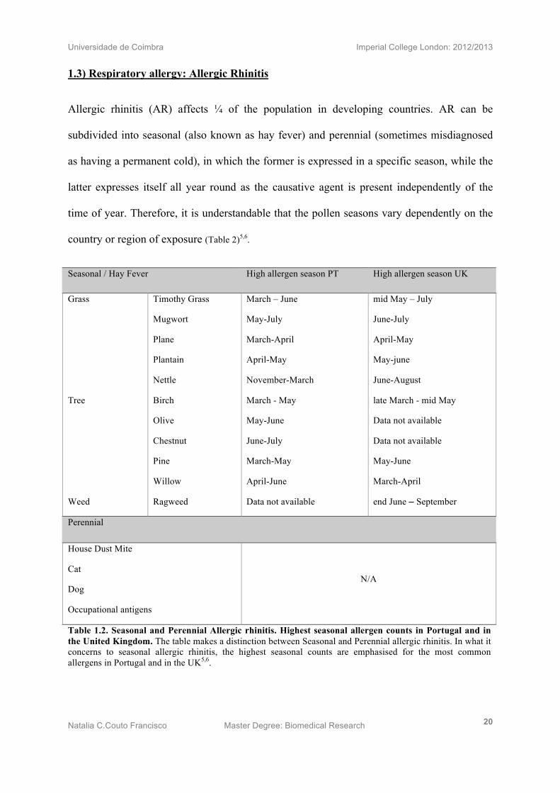
Allergic rhinitis (AR) affects ¼ of the population in developing countries. AR can be
subdivided into seasonal (also known as hay fever) and perennial (sometimes misdiagnosed
as having a permanent cold), in which the former is expressed in a specific season, while the
latter expresses itself all year round as the causative agent is present independently of the
time of year. Therefore, it is understandable that the pollen seasons vary dependently on the
country or region of exposure (Table 2)5,6.
Seasonal / Hay Fever High allergen season PT High allergen season UK
Grass
Tree
Weed
Timothy Grass
Mugwort
Plane
Plantain
Nettle
Birch
Olive
Chestnut
Pine
Willow
Ragweed
March – June
May-July
March-April
April-May
November-March
March - May
May-June
June-July
March-May
April-June
Data not available
mid May – July
June-July
April-May
May-june
June-August
late March - mid May
Data not available
Data not available
May-June
March-April
end June – September
Perennial
House Dust Mite
Cat
Dog
Occupational antigens
N/A
Table 1.2. Seasonal and Perennial Allergic rhinitis. Highest seasonal allergen counts in Portugal and in the United Kingdom. The table makes a distinction between Seasonal and Perennial allergic rhinitis. In what it concerns to seasonal allergic rhinitis, the highest seasonal counts are emphasised for the most common allergens in Portugal and in the UK5,6.

Universidade de Coimbra Imperial College London: 2012/2013
Natalia C.Couto Francisco Master Degree: Biomedical Research 21
1.4) Immunological Mechanisms of Allergic Rhinitis
The clinical symptoms expressed by individuals suffering from AR can be explained by a
sequence of immune events, triggered upon the entry of an allergen to the lining of the nasal
mucosa. The two main characteristics of an allergic response, is the presence of Th2 (Type 2
helper) cells and allergen specific-Immunoglobulin E (IgE) antibodies.
It is believed that in AR patients, the detection of the allergen at the site of inflammation is
achieved by innate immunity cells called Dendritic cells (DCs), more specifically CD11c+
myeloid Dendritic cells (mDCs). These cells are antigen presenting cells (APCs), which
capture and cleave the allergen into small peptides, while they mature and migrate to lymph
nodes in order to present the processed antigenic peptide to naive T cells (nTcells), also
known as Th0 cells. At this stage, the cross-talk between innate and adaptive immune
responses is achieved. For full activation of nTcells, it is necessary the involvement of
surface receptors such as the Major Histocompatibility Complex two (MHC-II) and B7-2 on
DCs and the T cell receptor (TCR) and CD28 on nTcells7. With the presentation of the
allergen to Th0 cells, these suffer a molecular changes, which allows them to produce IL-4
and therefore called IL-4 competent CD4+ T cells, becoming Th2 cells8. These adaptive
immune cells activate T follicular helper (TFH) cells, which also produce IL-4, the agent able
to trigger B cell to undergo heavy-chain class switching to allergen specific IgE9,10.
Th2 migrate to the site of inflammation, where they secrete the well known IL-4, IL-13 and
IL-5 cytokines, in which the two former cytokines recruit mast cells and basophils and the
latter cytokine guide the local Eosinophils to infiltrate the site of inflammation7,11–13.
In addition, at the site of inflammation, the allergen specific IgE is known to activate Masts
Universidade de Coimbra Imperial College London: 2012/2013
Natalia C.Couto Francisco Master Degree: Biomedical Research 22
cells, Basophils and Eosinophils as they, commonly, express FcεRI, which is the high-affinity
receptor for the Fc portion of the ε heavy chain of IgE. Binding of IgE to the FcεRI on Masts
cells, trigger degranulation of its granule stores. Mast cells granules contain chemotactic
factors that recruit neutrophils and Eosinophils1.
The secretion of histamines from Masts cells cause vasodilation, increased vascular
permeability, smooth muscle contraction, and increased mucus secretion. In addition,
adenosine is also secreted and it is the causative agent of bronchoconstrictions and
suppression of platelet aggregation. In addition, neutral proteases such as tryptase are also
released and are capable of cleaving complement components, which induce chemotactic and
inflammatory mediators that may end up provoking cell damage. Furthermore, newly
synthesised lipid mediators such as Prostaglandins (PGD) and Leukotrienes (LTC) are stored
in Mast cells and are involved in mucus secretion (PGD2) and, finally, several thousand times
more active than histamines, LTC4 and LTD4 that cause vasodilation and bronchospasms.
Similarly, when IgE activates Eosinophils, they also secrete LTC4 and promote inflammation
by releasing platelet activating factors. Additionally, Eosinophils produce eosinophil cationic
protein, which is a toxic agent known to cause cell damage. The role of IgE activated
Basophils is still unclear in the allergy field and requires further analysis1,2.
Clinically speaking, these events of inflammatory reactions cause sensitised individuals to
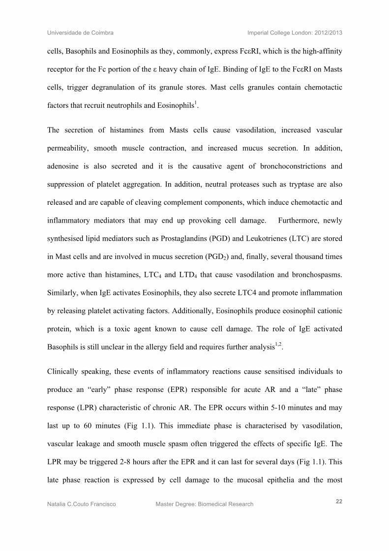
produce an “early” phase response (EPR) responsible for acute AR and a “late” phase
response (LPR) characteristic of chronic AR. The EPR occurs within 5-10 minutes and may
last up to 60 minutes (Fig 1.1). This immediate phase is characterised by vasodilation,
vascular leakage and smooth muscle spasm often triggered the effects of specific IgE. The
LPR may be triggered 2-8 hours after the EPR and it can last for several days (Fig 1.1). This
late phase reaction is expressed by cell damage to the mucosal epithelia and the most
Universidade de Coimbra Imperial College London: 2012/2013
Natalia C.Couto Francisco Master Degree: Biomedical Research 23
abundant cells are neutrophils, eosinophils and T lymphocytes (in particular Th2
lymphocytes).
It is important to note that the cell events at the inflammatory site do not translate the changes
occurring in circulation. Therefore, clinicians measure the inflammatory mediators in blood
and at sites of allergen exposure. Diagnostic assays include a skin prick test and a RAST test.
A Positive skin prick test to causative aeroallergens selects allergic rhinitis from non-allergic
rhinitis and the RAST test consists in detecting the amount of specific IgE in blood serum.
Normally, the RAST test is only requested in case the skin prick test shows unclear results. In
addition, performing a histopathological exam may be also necessary as it shows
oversecretion of nasal fluid containing basophils and eosinophils. Furthermore, in nasal
secretions, it is also possible to measure mast cell mediators and detectable IgE, IgG and
IgA1,3,14.
Figure 1.1 Immediate Hypersensitivity. Kinetics of the early-phase response (EPR) and late-phase response
(LPR) after allergen challenge to a sensitised individual1.
Allergen exposure
Clin
ical
Man
ifest
atio
ns
Hours after allergen challenge exposure
0 1 4 8 12 16 20
EPR LPR
Universidade de Coimbra Imperial College London: 2012/2013
Natalia C.Couto Francisco Master Degree: Biomedical Research 24
1.5) Potential inflammatory mediators in SAR
In order to understand which molecules are involved in the enhancement of an allergen
driven Th2 response, allergists collected cells from nasal mucosa of AR patients.
Previous results reported high expression of epithelial-derived cytokines named thymic
stromal lymphopoietin (TSLP) 15, IL-25 (also known as IL-17E) 16 and IL-3317. Since these
discoveries, research in both mice and human samples; have directed their focus towards the
understanding of these non-haematopoietic-cell-derived cytokines in allergic immunity (Fig
1.2).
TSLP is an IL-7-like cytokine and its heterodimeric receptor is composed by IL-7Rα and
TSLP receptor (TSLPR). Studies have identified higher affinity upon ligand binding when
both receptor subunits are activated simultaneously. Reports to date have shown that TSLP is
mainly secreted by epithelial cells and it is detected by mDCs by its [IL-7Rα-TSLPR]
receptor. TSLP primed DCs result in the upregulation of OX40L, which is the ligand for
OX40, expressed on DCs, resulting in prolonged DC survival. Such interaction is critical for
the development of a Th2 driven response11,12,18. Primed mDCs then migrate from the site of
inflammation to lymph nodes, where they present the antigen to nT cells through the major
histocompatibility complex class II (MHC-II), inducing nT cell differentiation into Th2
cells11–13,18.
IL-25 belongs to the IL-17 family and activates the IL-25 receptor (IL-25R) expressed on
Basophils, Mast cells and CD25R Natural helper cells. Studies associated to asthma
demonstrated that CD25R Natural helper cells secrete TNF-α, IL-1β, IL-6 and IL-8 in which
IL-8 is the key cytokine for Eosinophil recruitment; and therefore IL-25 is necessary to
maintain eosinophilia at the site of inflammation10. In addition, it has been also shown the
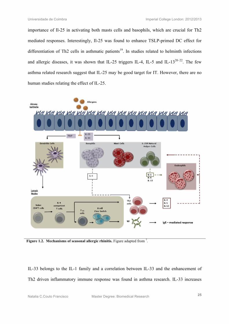
Universidade de Coimbra Imperial College London: 2012/2013
Natalia C.Couto Francisco Master Degree: Biomedical Research 25
importance of Il-25 in activating both masts cells and basophils, which are crucial for Th2
mediated responses. Interestingly, Il-25 was found to enhance TSLP-primed DC effect for
differentiation of Th2 cells in asthmatic patients19. In studies related to helminth infections
and allergic diseases, it was shown that IL-25 triggers IL-4, IL-5 and IL-1320–22. The few
asthma related research suggest that IL-25 may be good target for IT. However, there are no
human studies relating the effect of IL-25.
IL-33 belongs to the IL-1 family and a correlation between IL-33 and the enhancement of
Th2 driven inflammatory immune response was found in asthma research. IL-33 increases
Figure 1.2. Mechanisms of seasonal allergic rhinitis. Figure adapted from 7.
Universidade de Coimbra Imperial College London: 2012/2013
Natalia C.Couto Francisco Master Degree: Biomedical Research 26
IgE mediated response since it seems to activate both Mast cells and Basophils through its
receptor, the ST2 receptor. IL-33 is only able to activate nTcells directly and not indirectly by
priming mDCs10,23. Research on AR showed that IL-33 increases the production of IL-8,
resulting in eosinophil recruitment24.
In summary, these epithelial-derived cytokines have shown to, synergistically, maintain a
Th2 response. However, in AR patients, the way they work independently from each other to
achieve such end, has not been tested and therefore remains unclear.
1.6) Potential Immunotherapy targets in SAR
The main aim of research is to increase knowledge on the immune mechanisms underlying
AR, in order to crack the puzzle of inflammatory modulation for a better diagnosis and
reduced symptom score. Immunotherapy targets the site of inflammation in order to regulate
and/or redirect the course of response.
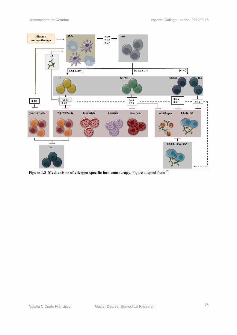
Studies have indicated the involvement of specific subsets of T cells with the role of
regulating allergen specific Th2 mediated allergic immune responses (Fig 1.3). These cells
are called regulatory CD4+T cells (Treg) and they can be subdivided into the naturally
occurring Treg (nTreg) cells found in the thymus, which are characterised by
CD4+CD25+FOXP3+ Treg cells and the induced CD4+ CD25+ FOXP3- Tregs (iTregs), found in
the periphery, which can be IL-10 producing Tr1 or IFN-γ producing Th1 cells25.
Interestingly, there are cells that contain both Tr1 and Th1 phenotype. In other words, these
Tr1/Th1 cells are activated by IL-10 and IL-12 derived APCs and secret both IL-10 and IFN-
γ. This may indicate that they are suffering a phenotype change from a Th1 to a Tr1 cell or
the way around. These cells, however, are able to both inhibit Th2 cells and impair B cell IgE
class switch, redirecting it towards IgG1, IgG4 or IgA Abs26. It has been shown in a couple of
Universidade de Coimbra Imperial College London: 2012/2013
Natalia C.Couto Francisco Master Degree: Biomedical Research 27
studies the increase of IL-10 and IFN-γ after specific immunotherapy (SIT) 27,28. However, a
study involving sublingual immunotherapy (SLIT), in which patients obtained increased IL-
10 and IFN-γ, did not translate its effect in a clinical point of view, since the patients’
symptoms were not alleviated despite the immunological changes29.
The question remaining is if either these cells can regulate inflammation by suppressing Th2
responses or change their course from a Th2 to a Th1 phenotype.
Therefore, in this project, we focus on a novel target named IL-3530, found by Collison et al.
in 2007. IL-35 belongs to the IL-12 family, which include the pro-inflammatory cytokines
IL-12 and IL-23, and the anti-inflammatory cytokines IL-27 and the Il-35. Its structure
consists of the EBV-induced gene 3 (EBI3) subunit of IL-27 and the p35 subunit of IL-1231.
IL-35 demonstrated to inhibit T cell proliferation.
In addition, Collison’s work also demonstrated that IL-35 has the effect of suppressing T cell
proliferation, resulting in reduced inflammation32. One year earlier, mouse studies were
showing that IL-35 reduces IL-17, an inflammatory cytokine, thus inhibiting proliferation of
Th17 by inducing CD4+CD25+Foxp3+ Treg 33. Untill this point, it was suspected the anti-
inflammmatory role of IL-35, however its mechanism was still unclear. It wasn’t until later,
in 2010, that Collison was able to show IL-35 inducible FoxP3- Tregs34. Soon after, in 2011,
transwell plate experiments revealed that iTr35 cells implement contact free suppression
through IL-35 and partially IL-103536.
Universidade de Coimbra Imperial College London: 2012/2013
Natalia C.Couto Francisco Master Degree: Biomedical Research 28
Figure 1.3 Mechanisms of allergen specific immunotherapy. Figure adapted from 37.

Universidade de Coimbra Imperial College London: 2012/2013
Natalia C.Couto Francisco Master Degree: Biomedical Research 29
Hypotheses
• Grass pollen-SLIT treated patients have higher proportions of Foxp3+, inducible IL-
10 (TR1) and IL-35+Tregs (iTR35) compared to untreated allergics.
• IL-35 suppresses grass pollen-specific effector Th2 responses following ex-vivo grass
pollen stimulation.
• Epithelial derived mediators/cytokines such as thymic stromal lymphopoetin,
interleukin -25 and 33 primed DCs when stimulated with grass pollen allergen drives
naïve T to diferenciate into allergen-specific Th2 cells that produce IL-5 and IL-13.
• IL-35 supresses TSLP, IL-25 and IL-33 responses on naïve T cells.
• IL-35 supresses Th2 polarisation from Th0 cells.
This project will attempt to accomplish the underlying aims:
• To assess the proportion of Foxp3, IL-10+ and IL-35+ Tregs in SAR, SLIT and NA.
• To develop an in vitro model of allergen-driven Th2 response induced by TSLP, IL-
25 and IL-33 primed DCs.
• To analyse the behaviour of IL-35 on allergen-driven Th2 responses.
• To optimise a T cell polarisation protocol towards Th1 and Th2 cells, to run in the lab,
daily for future IL-35 studies.
Universidade de Coimbra Imperial College London: 2012/2013
Natalia C.Couto Francisco Master Degree: Biomedical Research 30
Chapter II: Methodology
Ethical Statement 30
Skin prick test and ImmunoCap 30
PBMC isolation 30
Thawing PBMCs from Liquid Nitrogen 30-31
Cell staining of FoxP3+Treg, IL10+CD4+Treg and IL-35+CD4+Treg 31
CD4+CD25- T effector cells antigen-specific stimulation and IL-35 neutralisation 31
T cell polarisation using epithelial derived cytokines and IL-35 suppression 31-32
TSLP receptor expression on DCs 32
T cell polarisation towards Th1 or Th2 cells 32-34
Surface staining 34
Intracellular and nuclear staining by Flow cytometry 35
Gene expression assay by qPCR 35-36
Cytokine Analysis by Multiplex ELISA 36
Statistical Analysis 36

Universidade de Coimbra Imperial College London: 2012/2013
Natalia C.Couto Francisco Master Degree: Biomedical Research 31
2.1 Ethical Statement
The clinical practice implemented in this project was in accordance with the guidelines of the
Ethics Committee of the Royal Brompton and Harefield Hospital NHS trust. All tissue and
blood samples were collected with prior written approval from all patients involved. Samples
were collected according to good clinical practice.
2.2 Skin prick test and ImmunoCap
ImmunoCap was used to verify the allergic sensitisation of patients quantifying specific and
total and specific IgE in blood serum (Phadia). The protocol was followed as instructed by
the manufacture.
2.3 PBMC isolation
Venous blood was collected into Lithium-heparin tubes and kept sterile. Blood was then
centrifuged for 10 minutes at 20°C at 1500rpms. Plasma was removed and stored for future
serology work. Following serum extraction and storage, RPMI-1640 without glutamine
(Gibco, Invitrogen, UK) was added to equal the volume of serum removed. 35ml of the
diluted sample was layered carefully over 15ml of Ficol (VWR International Ltd., UK)
within a 50ml tube. After a 25 minute centrifugation at 2200 rpms at 20°C (without
centrifuge brake), the layer of peripheral blood mononuclear cells (PBMCs) which is isolated
and kept in tissue culture medium.
2.4 Thawing PBMCs from Liquid Nitrogen
PBMCs initially stored in cryovials were removed from liquid nitrogen and 70% thawed with
agitation in a 37°C waterbath. Cells were then transferred from cryovials to 10mL tubes,
immersed in RPMI-1640 and centrifuged at 1500RPM for 10min. Any supernatant was then
Universidade de Coimbra Imperial College London: 2012/2013
Natalia C.Couto Francisco Master Degree: Biomedical Research 32
discarded and samples resuspended in 5mL RPMI-1640. A 20µL cell suspension in trypan
blue at a 1:5 dilution was then loaded into a haemocytometer chamber and a live cell number
calculated.
2.5 Cell staining of FoxP3+Treg, IL10+CD4+Treg and IL-35+CD4+Treg
Cells were stained with the surface staining protocol indicated below (2.10) for APC CD4+
(clone: RPA-T4, BD Bioscences, USA) and APC/Cy7 CD25+ (clone: M-A251, BD
Bioscience). Intracellular and nuclear stainings were performed accordingly to the protocol
below (2.11) of PerCP IL12p35 (clone: 27537, R&D), AlexaFluor488 EBI3(clone 607201 ,
R&D) and V450 FOXP3 (clone: 236A/E7, BD Bioscience) and Pe/Cy7 IL-10 (JE53-9D7,
Biolegend).
2.6 CD4+CD25- T effector cells antigen-specific stimulation and IL-35 neutralisation
The separated CD4+CD25- Teff cells were incubated in 1:1 ratio with irradiated Ag-presenting
cells (APC) in concentration 300x106 cells/well. In each well 3ug/ml PhlP (Alk abello,
Denmark) was added along with 10 ng/ml IL-35 (Enzo life Science,UK). When determining
the best concentration of Phl P to be used experiments were carried out with 0,1, 3, 5 and 10
ug/ml. The plates were cultured for 9 days at 370C in 5% CO2 and pulsed for the last 18 hours
with [3H] - thymidine. Proliferation was measured using the described above method.
2.7 T cell polarisation using epithelial derived cytokines and IL-35 suppression
PBMCs were isolated from 200 ml of whole blood from allergic rhinitis patients. DCs and
naive T cells (nT cells) were purified from isolated PBMCs, using Pan-DC Pre-enrichment
Kit and naïve CD4+ T enrichment kits respectively (Stem Cell technologies, UK). DCs and
nT cells were counted, using a haemocytometer and dead cell excluded, Trypan blue (Sigma
Universidade de Coimbra Imperial College London: 2012/2013
Natalia C.Couto Francisco Master Degree: Biomedical Research 33
Aldrich Company Ltd., UK). 5x10³ cells of Dcs were added in 50µl of TCM per well.
15ng/ml TSLP (eBioscience,UK), 10ng/ml IL-25 (R&D,UK) and 10ng/ml IL-33 (R&D) were
added as described for 24h, where used timothy grass pollen, Phleum Partenses allergen
(Phlp) was added at 5µg/ml (ALK-Abelló, Denmark). At 24h primed, DCs were co-cultured
with naive T cells, at 5x10⁴ cells per well. In some experiments, IL-35 was added IL-35
(Enzo life Science) at 10ng/ml. Phytohaemagglutanin (PHA) (10µg/ml) was added as
positive control for T cell proliferation. At day six of co-culture, some wells were pulsed with
1µl of 3H-Thymidine to determine proliferation. Following a further 16h, cells were
harvested onto filtermats (Harvester 96Tomtec) and beta counts detected by 1450 Micro Beta
Trilux. Non-pulsed cell supernatants were stored at -20°C for cytokine analysis by Multiplex
ELISA.
2.8 TSLP receptor expression on DCs
DCs were purified from isolated PBMCs and stained for flow cytometric analysis at 0 and
24h following stimulation with phlp and TSLP. PE TSLPR+ (clone:147036, R&D) and APC+
IL-7Rα (clone:40131, R&D) were used to check TSLP receptor heterodimer, APC-Cy7
CD1c+ (clone: L161, eBioscience) and V450 CD11c+ (clone:B-ly6, BD, UK) for myeloid
DCs, PerCpCy5.5 CD123+ (clone:6H6, Biolegend,UK) and FITC CD303+ (clone:AC144,
Miltenyi Biotec,UK) for plasmacytoid DCs.
2.9 T cell polarisation towards Th1 or Th2 cells
After PBMC and nT cell isolation, nT cells were counted and equally divided into three,
1µ/ml αCD3 (R&D) and 2µ/ml αCD28 (R&D) pre-coated, 24well plates (Previously
incubated for 4h in the incubator at 37°C and 5% CO2). Non-polarised nT cells were
stimulated with 2ng/ml IL-2 (clone: R&D) alone. Th1 polarised cells were polarised with

Universidade de Coimbra Imperial College London: 2012/2013
Natalia C.Couto Francisco Master Degree: Biomedical Research 34
2ng/ml IL-2 (clone: R&D), 2.5ng/ml rhIL-12 (R&D,UK) and 5µ/ml αhIL-4 (clone:MP4-
25D2, BD). Th2-polarised cells were stimulated with 2ng/ml rhIL-2 (R&D), 12.5ng/ml rhIL-
4 (R&D), 5µ/ml αhIL-10 (clone: JES3-19F1, BD) and 5µ/ml αhIFN-γ (clone:B27, BD). This
experiment was carried out in three atopic patients (Fig 2.1).
Figure 2.1 T cell Polarisation Protocol
Weekly, prior to re-stimulation cells were stained for flow cytometry with the following
fluorochrome-coupled antibodies:
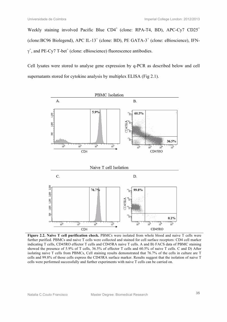
To check purification of naïve T cells from isolated PBMCs on day zero, the staining
antibodies used were: PE-Cy7 CD4+ (clone: SK3, BD), APC-Cy7 CD25+ (clone:BC96,
Biolegend), PE CD45RO+ (clone: BD) and FITC CD45RA+ (clone: BD). Purification was
achieved at 99.8% nTcell isolated from PBMCs. (Fig 2.2)
Thursday Friday Sat/Sun Friday Thursday Wednesday Tuesday Monday
Day 0
1. PBMC Isolation
2. naive T cell Isolation
3. Culture: 3.1) CD3-CD28 Stimulation 3.3) Cell Polarisation 4. FACS Staining: 4.1) PBMCs 4.2) Naive T cell Purification
Day 4
1. Cell Rest
2. Cell Polarisation
Day 6
1. CD3-CD28 Stimulation
2. Cell Polarisation
Collect (18h): Supernatant Cell Lysate
Day 8
1. Cell Polarisation
Day 7
Perform: FACS
Staining
Universidade de Coimbra Imperial College London: 2012/2013
Natalia C.Couto Francisco Master Degree: Biomedical Research 35
Weekly staining involved Pacific Blue CD4+ (clone: RPA-T4, BD), APC-Cy7 CD25+
(clone:BC96 Biolegend), APC IL-13+ (clone: BD), PE GATA-3+ (clone: eBioscience), IFN-
γ+, and PE-Cy7 T-bet+ (clone: eBioscience) fluorescence antibodies.
Cell lysates were stored to analyse gene expression by q-PCR as described below and cell
supernatants stored for cytokine analysis by multiplex ELISA (Fig 2.1).
Figure 2.2. Naive T cell purification check. PBMCs were isolated from whole blood and naive T cells were further purified. PBMCs and naive T cells were collected and stained for cell surface receptors: CD4 cell marker indicating T cells, CD45RO effector T cells and CD45RA naive T cells. A and B) FACS data of PBMC staining showed the presence of 5.9% of T cells, 36.5% of effector T cells and 60.5% of naive T cells. C and D) After isolating naive T cells from PBMCs, Cell staining results demonstrated that 76.7% of the cells in culture are T cells and 99.8% of those cells express the CD45RA surface marker. Results suggest that the isolation of naive T cells were performed successfully and further experiments with naive T cells can be carried on.

Universidade de Coimbra Imperial College London: 2012/2013
Natalia C.Couto Francisco Master Degree: Biomedical Research 36
2.10) Surface staining
Followed the addition of 10µl of FC Blocker for 15 minutes, surface staining was added
accordingly for 40 minutes. Cells were then washed with BD Cell staining buffer and run
through flow cytometry.
2.11) Intracellular and nuclear staining by Flow cytometry
For cytokine intracellular staining, cells were stimulated with PMA (Sigma-Aldrich),
Ionomycin (Sigma-Aldrich) for one hour and BFA (BD #347688) added for the remaining
4h. The protocol used for the intracellular and nuclear staining was the BD transcription
factor Buffer set staining (BD # 562574). Very briefly, 500x10^6 per sample were stimulated
as indicated above and 10µl of FC Blocker added for 15 minutes. Surface staining was then
added for 50 minutes at 4°C. Cells were washed twice with BD cell staining buffer. 1 ml of
1x Fix/Perm solution (BD) was added to the cells prior to a 50 minute incubation at 4°C.
Cells were submitted to three washes with the BD 1xPerm/wash solution and incubated for
50 minutes with the corresponding intracellular staining. After a two time wash, cells were
run through flow cytometry.
2.12) Gene expression assay by qPCR
Cells were collected and lysed with RLT Buffer (Qiagen) containing 1% β-Metacaptoethenol
(Sigma-Aldrich, UK), 16h after polyclonal stimulatiom with αCD3 (R&D) and αCD28
(R&D) antibodies and kept at -20°C. Cell lysates were added to the QIA Cube (Qiagen),
which uses the RNeasy Mini Isolation Kit (Qiagen) and QIA shredder (Qiagen), for RNA
extraction from cell lysates. Nanodrop was performed in order to check the concentration and
purity of RNA isolated. Once confirmed, it was added per mix 4µl of 5xTaqman RT Buffer
(Thermo Scientific), 2µl dNTPs Thermo Scientific), 1µl Random Hexomers (Thermo

Universidade de Coimbra Imperial College London: 2012/2013
Natalia C.Couto Francisco Master Degree: Biomedical Research 37
Scientific), 1µl Reverse Transcriptase (Thermo Scientific), 0.5µl RNase inhibitor and the
11.5µl RNA sample, which are then run in Sense Quest Labcycler for cDNA production. The
following program wasused: 25°C for 10 minutes, 47°C for 1h and 70°C for10miutes. cDNA
is analysed by Nanodrop and diluted 1:10 in molecular biology water, in epMotion machine
together with the corresponding primers (1.6µl) mixed with Syber Green (5µl) (AB Applied
Biosystems). All the primers used for this experiment were ordered from Sigma-Aldrich. The
housekeeping gene more appropriate for the experiment was EF2A and the primers of interest
IL-4, IL-5, GATA 3 IFN-µ and T-Bet (Table 2.1).
Forward primer 5’-3’ Reverse primer 5’-3’
EF2A CTG AAC ATC CAG GCC AAT GCCGTGTGGCAATCCAAT
IL-4 AAA CGG CTC GAC ACG AAC CT ACTCTGCTTGGCTTCCTTCACA
IL5 AGC TGC CTA CGT GTA TGC CA GCAGTGCCAAGGTCTCTTTCA
GATA-3 GCG GGC TCT ATC ACA AAA TGA GCTCTCCTGGCTGCAGACAGC
IFN-γ TCT GGA AAC GAT GAA ATA TAC AAG TT GTA ACA GCC AAG AGA ACC CAA
AA
T-Bet GAT GCG CCA GGA AGT TTC AT GCA CAA TCA TCT GGG TCA CAT T
Table 2.1: Forward and reverse primer sequences for Th1 and Th2 related targets.
2.1) Cytokine Analysis by Multiplex ELISA
Milliplex MAP Kits (Luminex Millipore, Germany) were used to preform multiplex ELISA.
Both Th1/Th2 and Th17 human magnetic bead panel kits were used to quantify cytokine
concentrations in supernatant (#HCYTOMAG-60K, #HTH17MAG-14K). Manufacture’s
protocol was followed and MAGPIX Luminex XMap Technology was used to run the
analysis.
Universidade de Coimbra Imperial College London: 2012/2013
Natalia C.Couto Francisco Master Degree: Biomedical Research 38
2.14) Statistical Analysis
Analysis of all data was conducted with GraphPad Prism 5.01 software (San Diego, USA).
Non-parametric FACS data was processed with a wilcoxon test, with statistical significance
represented as (p<0.05).
Chapter III: Results
Rhinitis symptom scores declines after sublingual immunotherapy 38
IL-10 + and IL-35 +, but not, FoxP3 + Treg is increased following SLIT 38
IL-35 suppresses allergen driven Teff cell proliferation and induces Th2 differentiation 40
TSLP, but not Il-25 and IL-33 primed DCs induce T cell proliferation in an allergen
driven response and Th2 differentiation
42
TSLP Receptor is expressed in higher amounts in SAR compared to NA patients 44
IL-35 suppresses TSLP primed DCs driven naive T cell proliferation and induces Th2
response deviation
45
Naive T cells fully polarised into Th1 cells after 2 weeks of treatment 47
Naive T cells fully polarised into Th2 cells after 3 weeks of treatment 47
Polarisation of Th1 and Th2 cells were confirmed by Multiplex and q-PCR 49

Universidade de Coimbra Imperial College London: 2012/2013
Natalia C.Couto Francisco Master Degree: Biomedical Research 39
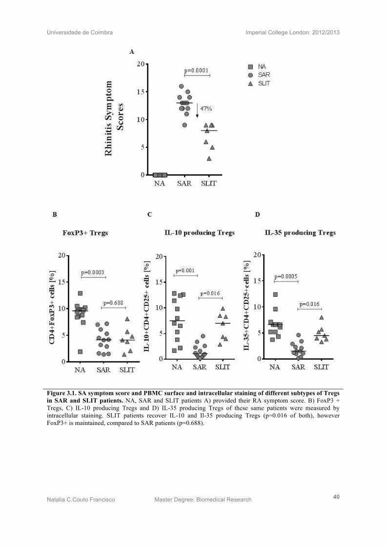
3.1) Rhinitis symptom scores declines after sublingual immunotherapy
Subjects treated with sublingual immunotherapy (n=7), were asked to grade the severity of
their allergic rhinitis. Untreated SAR patients (n=12) and NA patients (n=12) were recruited
for the same evaluation. SLIT patients demonstrated a significant improvement in AR
symptom scores of 47% (p=0.006) compared to SAR patients.
3.2) IL-10 + and IL-35 +, but not, FoxP3 + Treg is increased following SLIT
SLIT (n=7), SAR (n=12) and NA (n=12) provided whole blood from which PBMC were
isolation for FoxP3+, IL-10 and IL35+ cell staining. By analysing the CD4+ T cell population
for Foxp3 expression, no statistically discernible difference between SAR and SLIT patients
was noted (p=0.688) (Fig 3.1B), suggesting that the Treg numbers remain unchanged between
treated and untreated patients. Intracellular fluorescent Ab FACS staining for IL-10 revealed
a noticeable increase in IL-10+CD4+CD25+Foxp3+ Treg cells from SLIT PBMCs as opposed to
those from SAR (p=0.016) patients (Fig 3.7C). In contrast, NA patients show the highest
percentage of IL-10 producing Tregs (p=0.001). Intracellular fluorescent Ab FACS staining
for the subunits of IL-35 (Ebi3 & IL-12P35) demonstrated a clear increase in induced IL-35+
CD4+CD25+FoxP3+ Treg population of PBMCs for SLIT-treated compared to SAR (p=0.016)
patients (Fig 3.1D). SAR has significantly decreased proportions of IL-35+ Tregs on
compared to NA (p=0.0005).
Universidade de Coimbra Imperial College London: 2012/2013
Natalia C.Couto Francisco Master Degree: Biomedical Research 40
Figure 3.1. SA symptom score and PBMC surface and intracellular staining of different subtypes of Tregs in SAR and SLIT patients. NA, SAR and SLIT patients A) provided their RA symptom score. B) FoxP3 + Tregs, C) IL-10 producing Tregs and D) IL-35 producing Tregs of these same patients were measured by intracellular staining. SLIT patients recover IL-10 and Il-35 producing Tregs (p=0.016 of both), however FoxP3+ is maintained, compared to SAR patients (p=0.688).
Universidade de Coimbra Imperial College London: 2012/2013
Natalia C.Couto Francisco Master Degree: Biomedical Research 41
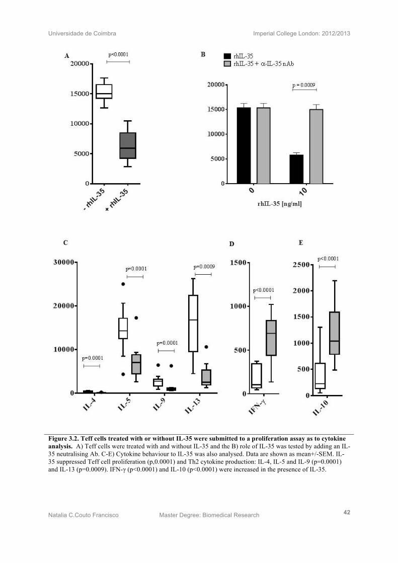
3.3) IL-35 suppresses allergen driven Teff cell proliferation and induces Th2
differentiation
Teff cells isolated from PBMCs of SAR (n=12) patients, were treated with or without IL-35.
Proliferation assay results showed a statistically significant suppression of Teff cell
proliferation upon addition of IL-35 (p<0.0001) (Fig. 2A). In addition, IL-35 suppressing
effect was tested by adding an IL-35 neutralising antibody, which successfully recovered the
inhibitory effect of the anti-inflammatory cytokine (p=0.0009) (Fig. 2B).
Cytokine levels were measured, in order to depict the role of IL-35 in Th2 responses. Th2
suffered a statistically significant decrease (IL-4 p=0.0001, IL-5 p=0.0001, IL-9 p=0.0001
and IL-13p=0.0009) (Fig 2C), while inducing IFN-γ (p<0.0001) (Fig 2D) and IL-10
(p<0.0001) (Fig 2E), suggesting not only that IL-35 suppresses Th2 responses but also
deviated the response to what it seems to be a T regulatory response.
Universidade de Coimbra Imperial College London: 2012/2013
Natalia C.Couto Francisco Master Degree: Biomedical Research 42
Figure 3.2. Teff cells treated with or without IL-35 were submitted to a proliferation assay as to cytokine analysis. A) Teff cells were treated with and without IL-35 and the B) role of IL-35 was tested by adding an IL-35 neutralising Ab. C-E) Cytokine behaviour to IL-35 was also analysed. Data are shown as mean+/-SEM. IL-35 suppressed Teff cell proliferation (p,0.0001) and Th2 cytokine production: IL-4, IL-5 and IL-9 (p=0.0001) and IL-13 (p=0.0009). IFN-γ (p<0.0001) and IL-10 (p<0.0001) were increased in the presence of IL-35.

Universidade de Coimbra Imperial College London: 2012/2013
Natalia C.Couto Francisco Master Degree: Biomedical Research 43
3.4) TSLP, but not Il-25 and IL-33 primed DCs induce T cell proliferation in an allergen
driven response and Th2 differentiation
A proliferation assay (n=14) of nT cell co-cultured with TSLP primed DCs was performed
and demonstrated a statistically significant induction in nT cell proliferation (p=0.003).
Equally, dendritic cells primed with TSLP, IL-25 and IL-33 showed statistically significant
proliferative effect on nT cells (p=0.0001). In contrast, there are no alterations in the
proliferation of nTcells when exposed to DCs that had been previously exposed to IL-25
(p=0.426) or IL-33 (p=0.502) (Fig 3.3A).
In addition, Supernatants were collected (n=7) and analysed in multiplex for IL-5 and IL-13
(Fig 3.3B) as to IFN-γ (Fig 3.3C). TSLP+Phlp primed DCs clearly trigger T cells to secrete
IL-5 and IL-13 production. Unexpectedly, a modest increase in IFN-γ was also seen.
Cytokine results were not statistically significant.

Universidade de Coimbra Imperial College London: 2012/2013
Natalia C.Couto Francisco Master Degree: Biomedical Research 44
Figure 3.3. Epithelial-derived pro-allergic mediator TSLP but not IL-25 and IL-33 induces grass pollen-specific naive T cell proliferation and Th2 cytokines. A) TSLP, IL-25 and IL-33 primed DCs stimulated with/without Phlp were co-culture with naive T cells and proliferative responses were measured. B) IL-5, IL-13 and C) IFN-γ were measured from cell culture supernatants. Data are shown as mean+/-SEM. TSLP+Phlp primed DCs induce nTcell proliferation (p=0.003) as well as the three epithelial-derived cytokines+Phlp (p=0.0001).
Universidade de Coimbra Imperial College London: 2012/2013
Natalia C.Couto Francisco Master Degree: Biomedical Research 45
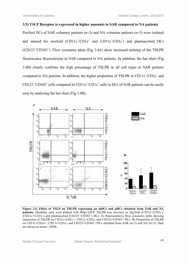
3.5) TSLP Receptor is expressed in higher amounts in SAR compared to NA patients
Purified DCs of SAR volunteer patients (n=3) and NA volunteer patients (n=3) were isolated
and stained for myeloid (CD11c+/CD1c+ and CD11c+/CD1c-) and plasmacytoid DCs
(CD123+/CD303+). Flow cytometry plots (Fig 3.4A) show increased staining of the TSLPR
fluorescence fluorochrome in SAR compared to NA patients. In addition, the bar chart (Fig
3.4B) clearly confirms the high percentage of TSLPR in all cell types in SAR patients
compared to NA patients. In addition, the higher proportion of TSLPR in CD11c+/CD1c- and
CD123+/CD303+ cells compared to CD11c+/CD1c+ cells in DCs of SAR patients can be easily
seen by analysing the bar chart (Fig 3.4B).
Figure 3.4. Effect of TSLP on TSLPR expression on mDCs and pDCs obtained from SAR and NA patients. Dendritic cells were primed with Phlp+TSLP TSLPR was assessed on Myeloid (CD11c+/CD1c+), (CD11c+/CD1c-) and plasmacytoid (CD123+/CD303+) DCs. A) Representative flow cytometry plots showing expression of TSLPR on CD11c+CD1c+, CD11c+CD1c- and CD123+CD303+ DCs. B) Proportion of TSLPR on CD11c+CD1c+, CD11c+CD1c- and CD123+CD303+ DCs obtained from SAR (n=3) and NA (n=3). Data are shown as mean+/-SEM.
Universidade de Coimbra Imperial College London: 2012/2013
Natalia C.Couto Francisco Master Degree: Biomedical Research 46
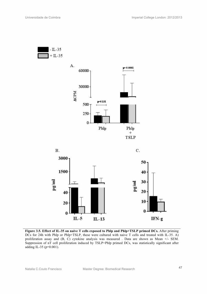
3.6) IL-35 suppresses TSLP primed DCs driven naive T cell proliferation and induces
Th2 response deviation
TSLP+Phlp primed DCs were co-cultured with n T cells with or without IL-35. Proliferation
data (n=14) (Fig 3.5A) demonstrated the inhibitory effect of IL-35 on nT cell proliferation
induced by TSLP primed DCs (p=0.0001).
Cytokine measurements (n=7) of Th2 related cytokines, IL-5 and IL-13, show a clear
decrease in IL-35 treated nT cells (Fig 3.5B). Interestingly, IFN-γ also suffered a modest
decrease in IL-35 treated cells (Fig 2.5C). However, cytokine suppression values of the three
measurements are not statistically significant.
Universidade de Coimbra Imperial College London: 2012/2013
Natalia C.Couto Francisco Master Degree: Biomedical Research 47
Figure 3.5. Effect of IL-35 on naive T cells exposed to Phlp and Phlp+TSLP primed DCs. After priming DCs for 24h with Phlp or Phlp+TSLP, these were cultured with naive T cells and treated with IL-35. A) proliferation assay and (B, C) cytokine analysis was measured . Data are shown as Mean +/- SEM. Suppression of nT cell proliferation induced by TSLP+Phlp primed DCs, was statistically significant after adding IL-35 (p=0.001).

Universidade de Coimbra Imperial College London: 2012/2013
Natalia C.Couto Francisco Master Degree: Biomedical Research 48
Universidade de Coimbra Imperial College London: 2012/2013
Natalia C.Couto Francisco Master Degree: Biomedical Research 49
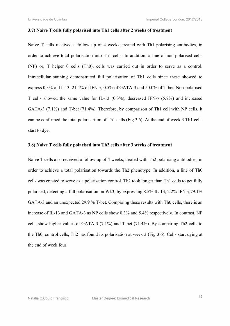
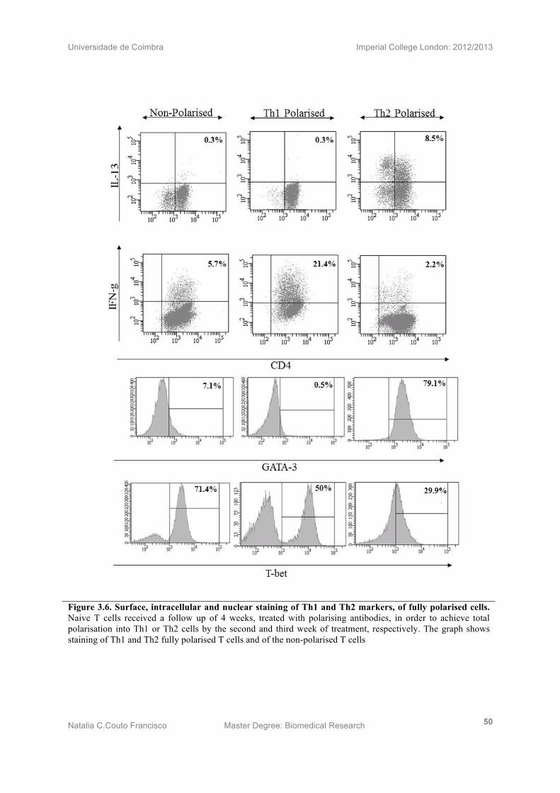
3.7) Naive T cells fully polarised into Th1 cells after 2 weeks of treatment
Naive T cells received a follow up of 4 weeks, treated with Th1 polarising antibodies, in
order to achieve total polarisation into Th1 cells. In addition, a line of non-polarised cells
(NP) or, T helper 0 cells (Th0), cells was carried out in order to serve as a control.
Intracellular staining demonstrated full polarisation of Th1 cells since these showed to
express 0.3% of IL-13, 21.4% of IFN-γ, 0.5% of GATA-3 and 50.0% of T-bet. Non-polarised
T cells showed the same value for IL-13 (0.3%), decreased IFN-γ (5.7%) and increased
GATA-3 (7.1%) and T-bet (71.4%). Therefore, by comparison of Th1 cell with NP cells, it
can be confirmed the total polarisartion of Th1 cells (Fig 3.6). At the end of week 3 Th1 cells
start to dye.
3.8) Naive T cells fully polarised into Th2 cells after 3 weeks of treatment
Naive T cells also received a follow up of 4 weeks, treated with Th2 polarising antibodies, in
order to achieve a total polarisation towards the Th2 phenotype. In addition, a line of Th0
cells was created to serve as a polarisation control. Th2 took longer than Th1 cells to get fully
polarised, detecting a full polarisation on Wk3, by expressing 8.5% IL-13, 2.2% IFN-γ,79.1%
GATA-3 and an unexpected 29.9 % T-bet. Comparing these results with Th0 cells, there is an
increase of IL-13 and GATA-3 as NP cells show 0.3% and 5.4% respectively. In contrast, NP
cells show higher values of GATA-3 (7.1%) and T-bet (71.4%). By comparing Th2 cells to
the Th0, control cells, Th2 has found its polarisation at week 3 (Fig 3.6). Cells start dying at
the end of week four.
Universidade de Coimbra Imperial College London: 2012/2013
Natalia C.Couto Francisco Master Degree: Biomedical Research 50
Figure 3.6. Surface, intracellular and nuclear staining of Th1 and Th2 markers, of fully polarised cells. Naive T cells received a follow up of 4 weeks, treated with polarising antibodies, in order to achieve total polarisation into Th1 or Th2 cells by the second and third week of treatment, respectively. The graph shows staining of Th1 and Th2 fully polarised T cells and of the non-polarised T cells

Universidade de Coimbra Imperial College London: 2012/2013
Natalia C.Couto Francisco Master Degree: Biomedical Research 51
3.9) Polarisation of Th1 and Th2 cells were confirmed by Multiplex and q-PCR
Weekly supernatants were collected and the cytokine analysis of the supernatants in culture
(Fig 3.7A) showed absence of IL-4, low amounts of IL-5 and IL-9 and high number of IL-9
cytokine in Th1 polarised cells. In contrast, Th2 T cells showed high levels of IL-4, increased
IL-5 and IL-9 compared to Th0 and Th1 polarised T cells.
Together with the multiplex data, mRNA gene expression was also performed. Weekly cell
lysates were collected from the three polarising cells. Gene expression (Fig 3.7B) of Th1 and
Th2 polarised cells compared to Th0 T cells was carried out. Th1 polarised cells showed
increased T-bet and high expression of IFN-γ compared to the control. In contrast, Th2
polarised cells exhibited an increased in the amount of IL-4 and GATA-3.
Figure 3.7. Cytokine and gene expression analysis of polarised T cells. Th1and Th2 polarised cell lysates and supernatants were collected. This graph represents data from week 2 for Th0 and Th1 cells and week 3 for Th2 cells. A) demonstrates cytokine analysis while B) shows the relative expression mRNA for Th1 and Th2 gene targets.
Rel
ativ
e E
xpre
ssio
n m
RN
A
pg/m
l

Universidade de Coimbra Imperial College London: 2012/2013
Natalia C.Couto Francisco Master Degree: Biomedical Research 52
Universidade de Coimbra Imperial College London: 2012/2013
Natalia C.Couto Francisco Master Degree: Biomedical Research 53
Chapter IV: Discussion
Universidade de Coimbra Imperial College London: 2012/2013
Natalia C.Couto Francisco Master Degree: Biomedical Research 54
Due to the increased prevalence of allergic rhinitis world wild, SCIT was developed.
However, reports have shown occurrences of patients with severe or rarer fatal events, due to
this therapy. Therefore, the prescription of SCIT has been restricted and alternative methods,
such as SLIT are being improved in order to offer a solution to patients that react badly to
SCIT.
Inspired by the cases of unsuccessful SCIT treatment, this investigation has attempted to
illustrate the important immunosuppressive properties of IL-35 iTregs and their characteristic
cytokine IL-35 in grass pollen sublingual immunotherapy. This novel subset of Treg cells,
were first identified in humans in 201038.
Results conducted in this project identified in PBMCs of NA (n=12), SAR (n=12) and SLIT-
treated (n=7) patients, a recovery in IL-10+CD4+CD25+ Treg (Tr1) (Fig 3.1C) and IL-
35+CD4+CD25+ Treg (Fig 3.1D) in SLIT patients that received treatment for 2 years. These
results were in accordance with a RA symptom score enquiry answered by these same SLIT
patients, who scored an average reduction of 47% (p=0.0001) in their symptoms of allergic
rhinitis compared to untreated SAR patients (Fig 3.1A). This revival in the population of Tr1
cells in SLIT–treated patients has been noted in several grass pollen SCIT studies39, while
also being alluded to in grass pollen SLIT findings, via an increase in IgG4 serum levels40. In
contrast, on analysis of Foxp3+CD4+ T cells it was interesting to note that natural
Foxp3+Tregs remained unchanged between SAR and SLIT-treated patients (Fig 3.1B).
However, some aeroallergen immunotherapies have expressed an initial peak in Foxp3
following treatment, only to subside back to atopic levels after prolonged treatment.41
So far this data is as expected and is a reproducibility of results shown in previous studies,
consequently validating the efficacy of the SLIT treatment under investigation.
Universidade de Coimbra Imperial College London: 2012/2013
Natalia C.Couto Francisco Master Degree: Biomedical Research 55
Additionally, in order to discover the behaviour of IL-35 in different in vitro inflammatory
reactions, an in vitro model of Th2 allergic inflammation was developed by simultaneously
testing the role of epithelial-derived TSLP, IL-25 and IL-33 cytokines in SAR. Finally, Th1
and Th2 cells were polarised in vitro, from original nT isolated cells. This protocol was
optimised over time in order to be used for further IL-35 analysis.
IL-25 and IL-33 together with TSLP are believed to be the initial power at site of allergen
contact, for the induction of an allergic response. Following this understanding, the three
cytokines were tested for their role on DCs. The proliferation assay concerning DCs primed
with the epithelial derived cytokines (Fig 3.3A), demonstrated an impressive effect of TSLP
primed DCs (in the presence of phlp) in inducing proliferation of nT cells (p=0.003).
Interestingly, when priming DCs with TSLP, IL-25 and IL-33, their exposure to nT cells also
showed a bust in nT cell proliferation (p=0.0001). However, the difference between the
proliferation triggered by TSLP primed DCs and the three epithelial cytokines primed DCs
does not show significance (p=0.583). Therefore, the proliferation achieved in the second
experiment is believed to happen due to the presence of TSLP, but not IL-25 or IL-33, as
these alone had no indirect effect on nT cells (p=0.426 and p=0.502; respectively).
In fact, while previous studies proved that TSLP acts on OX40 receptor located on the
surface of DCs13, IL-25 and IL-33 receptors have not been shown to have any effect on these
innate cells. There is however, one report42 suggesting the expression of the IL-33 receptor,
ST2, on naive T cells only, therefore, explaining the failure of IL-33 primed DCs in
triggering nT cell proliferation (Fig 3.3A). In addition, eventhough there is no significance
between TSLP primed DCs and [TSLP+IL-25+IL-33] primed DCs, a previous experiment
hypothesised the enhancement of TSLP effect in the presence of IL-2519.

Universidade de Coimbra Imperial College London: 2012/2013
Natalia C.Couto Francisco Master Degree: Biomedical Research 56
As the effect of TSLP on DCs indicated aggressiveness towards nT cells, resulting in its
proliferation (Fig. 3.3A), cytokine analysis were measured in culture supernatants (Fig 3.3B
and Fig 3.3C). Interestingly, TSLP primed DCs induced mainly IL-5 and IL-13 production by
T cells, suggesting that these have suffered a deviation towards a Th2 like phenotype.
Unexpectedly, naive T cells also showed an increase in the levels of IFN-γ, which still
remains to be explained. According to mouse studies TSLP was shown to induce Th2 allergic
immune responses. Thus, and most importantly, recognised as a potential target for AR
immunotherapy.
As our results show a clear involvement of TSLP in Timothy grass AR, the next question was
if either the expression of TSLPR varies on DCs of Timothy grass (TG) sensitised patients
compared to healthy controls. There are different subtypes of DCs such as the myeloid
(CD11c+/CD11c+) and plasmacytoid DCs (CD123+/CD303+). The role of these two subtypes
is still unclear. Therefore, while staining for the TSLPR and IL-7α, plasmacytoid and
myeloid DCs were also stained, in order to check the expression of these two chains in
different DC cell subsets. However, the number of patients used were reduced (n=3 SAR and
n=3 NA) the surface staining of TSLPR chain successfully showed an increased expression in
SAR compared to NA patients (Fig 3.4). Interestingly, CD11c+CD11c- DCs cells showed
increased TSLPR compared to CD11c+/CD11c+ myeloid DCs. In addition, similarly to
CD11c+CD11c- DCs, increased TSLPR chain was seen in CD123+/CD303+ plasmacytoid
DCs. Furthermore, it was previously published that if TSLP binds to both chains of its
heterodimer receptor, TSLPR and IL-7αR, the downstream signal will be stronger than if it
binds to TSLPR chain alone. Our results demonstrate much lower % of both chains than
TSLPR alone (Fig 3.4A). Further experiments must be done to increase the number of
patients, in order to prove this preliminary data as a true, statistically significant result.
Universidade de Coimbra Imperial College London: 2012/2013
Natalia C.Couto Francisco Master Degree: Biomedical Research 57
Furthermore, it would be interesting to use immunohistochemistry to colocalise the two
chains of the heterodimeric TSLP receptor. This could proof that the TSLPR and IL-7αR
present are found together, as an heterodimeric receptor, rather than just spread individually
throughout the surface of pDCs and mDCs.
Once the in vitro model of a Th2 response had been developed successfully, the IL-35 effect
was analysed. In the presence of IL-35 (10ng/ml), T cell proliferation induced by TSLP
primed DCs, showed a clear suppression by achieving a significant value of p=0.0001 (Fig
3.5 A). Following the results obtained for the suppression in proliferation, the cytokine
phenotype secreted by nT cells, were looked at. Interestingly, not only the Th2 related
cytokines (IL-5 and IL-13) were suppressed (Fig 3.5B) as the Th1 cytokine, IFN-γ suffered a
modest reduction (Fig 3.5B), in the presence of IL-35. The question that arises is if either
these changes are a result of reduced proliferation or deviation in response. Independently of
the answer, both would be a result of the IL-35 treatment.
At last but not least, nT cells were polarised into Th1 and Th2 cells by a follow up of 4weeks.
Weekly, intracellular staining by flow cytometry, cytokine analysis by multiplex ELISA and
gene expression by q-PCR were performed in order to check for polarisation. In Fig 3.6,
FACS plots represent Th1 at Week2 and Th2 cells at week 3. As obtained higher amounts of
IFN-γ in Th1 than Th0 cells, shows indication of polarisation. In contrast eventhough there is
more T-bet in Th0 than Th1, it doesn’t rule out the opinion that these cells are fully polarised,
as it can be observed a high number of dying cells in Th1 culture. Additionally, Th2 showed
very cleary, increased in both IL-13 and GATA-3, leaving no doubt of it’s polarising
phenotype. We can now use Th1 cells at week 2 and Th2 cells at week 3, for further IL-35
testing.
Universidade de Coimbra Imperial College London: 2012/2013
Natalia C.Couto Francisco Master Degree: Biomedical Research 58
Chapter VI: Future Studies
The characterisation of iTr35 cells is very recent; as a consequence little is understood with
regards to its mechanisms of suppression and its clinical relevance towards allergy and
immunotherapy. I believe that this project would benefit from the following assays and
avenues of research concerning iTr35 cells:
• As an additional validation towards IL-35 as the primary suppressive mechanism of
IL-35 in the Th2 model developed involving primed DCs and TSLP could be repeated
in a transwell plate. Providing the pore size of the membrane between the wells is
smaller than the width of a T cell (0.4µM in diameter would suffice), this assay could
identify if the mode of iTr35 cell suppression is cell-cell mediated. So far only
Collison et al., 2009 has implemented this assay to suggest that the suppressive
capacity of iTr35 cells is only IL-35 and partially IL-10 dependent.
• Additional in vivo assays could be attempted involving transgenic mice with a
conditional knockout genes coding for the subunits of IL-35. The mice could be
exposed to a number of aeroallergens (grass pollen, birch pollen, dust mite) and their
peripheral Th2 response quantified with and without the induction of the genes coding
for EBi3 and IL-12p35. Furthermore, allergic mouse models could be treated with
recombinant IL-35 intravenously and any impact on the Th2 response scrutinised.
• The impact of IL-35 on B cells is still poorly understood. B cells could be isolated
from SAR patient PBMCS, stimulated with grass pollen allergen (Phl P) and cultured
alongside incremental doses of IL-35. Any resulting Ig isotypes could then be
identified with an enzyme-linked immunosorbent assay (ELISA), thus determining if
IL-35 instigates B cell class switching. If an Ig isotype is recognised its quantity can
Universidade de Coimbra Imperial College London: 2012/2013
Natalia C.Couto Francisco Master Degree: Biomedical Research 59
then be analysed in the serum of SLIT patients to gauge the impact of IL-35 on the
treatment.
• As this project has demonstrated a recovery in peripheral quantity of iTr35 cells
following grass pollen SLIT, it may be interesting to attempt similar findings in other
allergen derived SLIT treatments, such as birch pollen and house dust mite.
• It would be useful to repeat the T cell polarisation to check the behaviour of IL-35 on
these cells. Other cell type such as Th17 could also be polarised and IL-35 effect
tested.
• Finally, not forgetting the pro-inflammatory effect of TSLP, I would increase the
number of peitents to check the TSLPR on the surface of different DC subtypes not
only in NA, SAR bur also in SLIT patients. I belive that te produced preliminary data
suggests that TSLPR can potentially become an immunotherapy target.
Universidade de Coimbra Imperial College London: 2012/2013
Natalia C.Couto Francisco Master Degree: Biomedical Research 60
Chapter VII: Conclusion
This project has been the first to identify a clear recovery in the quantity of iTr35 cells within
the peripheral blood of SAR patients treated with SLIT. In addition, IL-35 was shown to have
a suppressor role in allergen-driven Teff cells and TSLP-primed DC induced Th2 responses.
Cell lines Th1 and Th2 polarised T cells were optimised for further analysis of IL-35.

Universidade de Coimbra Imperial College London: 2012/2013
Natalia C.Couto Francisco Master Degree: Biomedical Research 61
References
1. Robbins, Kumar, Abbas, Fausto, M. Basic Pathology. (Saunders Elsevier: 2007).
2. Abul. Abbas, A. H. L. Basic Immunology. (Saunders Elsevier: 2006).
3. Helen Chapel, Mansel Haeney, Siraj Misbah, N. S. Clinical Immunology. (Blackwell Publishing: 2007).
4. Kenneth Murph Janeway’s Immunobiology. (Garland Science: 2012).
5. Sociedade Portuguesa de Alergologia e Imunologia Clinica The pollen dictionary. at <http://www.spaic.pt/?stl=EN>
6. Met Office Pollen callender. at <http://www.metoffice.gov.uk/health/public/pollen-forecast>
7. Paul, W. E. & Zhu, J. How are T(H)2-type immune responses initiated and amplified? Nature reviews. Immunology 10, 225–35 (2010).
8. Sin, B. & Togias, A. Pathophysiology of allergic and nonallergic rhinitis. Proceedings of the American Thoracic Society 8, 106–14 (2011).
9. Reinhardt, R. L., Liang, H.-E. & Locksley, R. M. Cytokine-secreting follicular T cells shape the antibody repertoire. Nature immunology 10, 385–93 (2009).
10. Locksley, R. M. Asthma and allergic inflammation. Cell 140, 777–83 (2010).
11. Ziegler, S. & Artis, D. Sensing the outside world: TSLP regulates barrier immunity. Nature immunology 11, 289–293 (2010).
12. Soumelis, V. et al. Human epithelial cells trigger dendritic cell mediated allergic inflammation by producing TSLP. Nature immunology 3, 673–80 (2002).
13. Li, D. et al. Short Ragweed Pollen Triggers Allergic Inflammation via TLR4-dependent TSLP/OX40L/OX40 Signaling Pathways. The Journal of allergy and clinical immunology 128, 1318–1325 (2011).
14. Durham, S. R. ABC of Allergies. (BMJ: 1998).
15. Mou, Z. et al. Overexpression of thymic stromal lymphopoietin in allergic rhinitis. Acta oto-laryngologica 129, 297–301 (2009).
16. Xu, G. et al. Opposing roles of IL-17A and IL-25 in the regulation of TSLP production in human nasal epithelial cells. Allergy 65, 581–9 (2010).

Universidade de Coimbra Imperial College London: 2012/2013
Natalia C.Couto Francisco Master Degree: Biomedical Research 62
17. Asaka, D. et al. Elevated levels of interleukin-33 in the nasal secretions of patients with allergic rhinitis. International archives of allergy and immunology 158 Suppl , 47–50 (2012).
18. Perrigoue, J. G. et al. MHC class II-dependent basophil-CD4+ T cell interactions promote T(H)2 cytokine-dependent immunity. Nature immunology 10, 697–705 (2009).
19. Wang, Y.-H. et al. IL-25 augments type 2 immune responses by enhancing the expansion and functions of TSLP-DC-activated Th2 memory cells. The Journal of experimental medicine 204, 1837–47 (2007).
20. Fallon, P. G. et al. Identification of an interleukin (IL)-25-dependent cell population that provides IL-4, IL-5, and IL-13 at the onset of helminth expulsion. The Journal of experimental medicine 203, 1105–16 (2006).
21. Angkasekwinai, P. et al. Interleukin 25 promotes the initiation of proallergic type 2 responses. The Journal of experimental medicine 204, 1509–17 (2007).
22. Saenz, S. a et al. IL25 elicits a multipotent progenitor cell population that promotes T(H)2 cytokine responses. Nature 464, 1362–6 (2010).
23. Silver, M. R. et al. IL-33 synergizes with IgE-dependent and IgE-independent agents to promote mast cell and basophil activation. Inflammation research : official journal of the European Histamine Research Society ... [et al.] 59, 207–18 (2010).
24. Kamekura, R. et al. The role of IL-33 and its receptor ST2 in human nasal epithelium with allergic rhinitis. Clinical and experimental allergy : journal of the British Society for Allergy and Clinical Immunology 42, 218–28 (2012).
25. Yamazaki, S. & Steinman, R. M. Dendritic cells as controllers of antigen-specific Foxp3+ regulatory T cells. Journal of dermatological science 54, 69–75 (2009).
26. Soyer, O. U. et al. Mechanisms of peripheral tolerance to allergens. Allergy 68, 161–70 (2013).
27. Radulovic, S., Jacobson, M. R., Durham, S. R. & Nouri-Aria, K. T. Grass pollen immunotherapy induces Foxp3-expressing CD4+ CD25+ cells in the nasal mucosa. The Journal of allergy and clinical immunology 121, 1467–72, 1472.e1 (2008).
28. Lou, W., Wang, C., Wang, Y., Han, D. & Zhang, L. Enhancement of the frequency and function of IL-10-secreting type I T regulatory cells after 1 year of cluster allergen-specific immunotherapy. International archives of allergy and immunology 159, 391–8 (2012).
29. Bonvalet, M. et al. Allergen-specific CD4+ T cell responses in peripheral blood do not predict the early onset of clinical efficacy during grass pollen sublingual immunotherapy. Clinical and experimental allergy : journal of the British Society for Allergy and Clinical Immunology 42, 1745–55 (2012).
Universidade de Coimbra Imperial College London: 2012/2013
Natalia C.Couto Francisco Master Degree: Biomedical Research 63
30. Collison, L. W. et al. The inhibitory cytokine IL-35 contributes to regulatory T-cell function. Nature 450, 566–9 (2007).
31. Fujita, H., Soyka, M. B., Akdis, M. & Akdis, C. a Mechanisms of allergen-specific immunotherapy. Clinical and translational allergy 2, 2 (2012).
32. Collison, L. W. & Vignali, D. a a Interleukin-35: odd one out or part of the family? Immunological reviews 226, 248–62 (2008).
33. Niedbala, W. et al. IL-35 is a novel cytokine with therapeutic effects against collagen-induced arthritis through the expansion of regulatory T cells and suppression of Th17 cells. European journal of immunology 37, 3021–9 (2007).
34. Collison, L. W. et al. Interleukin-35-mediated induction of a novel regulatory T cell population. 11, 1093–1101 (2011).
35. Vignali, A. Human Regulatory T Cells Require Interleukin-35 to Mediate Suppression and Infectious Tolerance. 186, 6661–6666 (2011).
36. Collison, L. W., Pillai, M. R., Chaturvedi, V. & Vignali, D. a a Regulatory T cell suppression is potentiated by target T cells in a cell contact, IL-35- and IL-10-dependent manner. Journal of immunology (Baltimore, Md. : 1950) 182, 6121–8 (2009).
37. Maggi, E. T-cell responses induced by allergen-specific immunotherapy. Clinical and experimental immunology 161, 10–8 (2010).
38. Collison, L. W. et al. IL-35-mediated induction of a potent regulatory T cell population. Nature immunology 11, 1093–101 (2010).
39. Nouri-Aria, K. T. et al. Grass pollen immunotherapy induces mucosal and peripheral IL-10 responses and blocking IgG activity. Journal of immunology (Baltimore, Md. : 1950) 172, 3252–9 (2004).
40. Scadding, G. W. et al. Sublingual grass pollen immunotherapy is associated with increases in sublingual Foxp3-expressing cells and elevated allergen-specific immunoglobulin G4, immunoglobulin A and serum inhibitory activity for immunoglobulin E-facilitated allergen binding to B. Clinical and experimental allergy : journal of the British Society for Allergy and Clinical Immunology 40, 598–606 (2010).
41. Bohle, B. et al. Sublingual immunotherapy induces IL-10-producing T regulatory cells, allergen-specific T-cell tolerance, and immune deviation. The Journal of allergy and clinical immunology 120, 707–13 (2007).
42. Kamekura, R. et al. The role of IL-33 and its receptor ST2 in human nasal epithelium with allergic rhinitis. Clinical and experimental allergy : journal of the British Society for Allergy and Clinical Immunology 42, 218–28 (2012).
Universidade de Coimbra Imperial College London: 2012/2013
Natalia C.Couto Francisco Master Degree: Biomedical Research 64
Attachments
Glossary 62
Key to illustrations found in the introduction 63
Relevant Courses 64
Awards 64
Publications 64
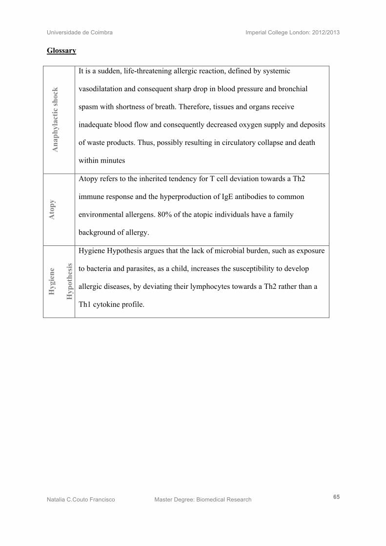
Universidade de Coimbra Imperial College London: 2012/2013
Natalia C.Couto Francisco Master Degree: Biomedical Research 65
Glossary A
naph
ylac
tic sh
ock
It is a sudden, life-threatening allergic reaction, defined by systemic
vasodilatation and consequent sharp drop in blood pressure and bronchial
spasm with shortness of breath. Therefore, tissues and organs receive
inadequate blood flow and consequently decreased oxygen supply and deposits
of waste products. Thus, possibly resulting in circulatory collapse and death
within minutes
Ato
py
Atopy refers to the inherited tendency for T cell deviation towards a Th2
immune response and the hyperproduction of IgE antibodies to common
environmental allergens. 80% of the atopic individuals have a family
background of allergy.
Hyg
iene
Hyp
othe
sis
Hygiene Hypothesis argues that the lack of microbial burden, such as exposure
to bacteria and parasites, as a child, increases the susceptibility to develop
allergic diseases, by deviating their lymphocytes towards a Th2 rather than a
Th1 cytokine profile.

Universidade de Coimbra Imperial College London: 2012/2013
Natalia C.Couto Francisco Master Degree: Biomedical Research 66
Key to illustrations in the introduction
Airway Epithelium
Naïve CD4+ Tcells
Allergen
Natural killer cells
Antibodies
Specific allergen antibody
Basophiles
T Follicular Helper Cells
B cells
T helper 0 cells
Plasma cells secreting antibodies
T helper 1
Eosinophils
T helper 1 like T regulatory 1 cells
Immunogenic Dendritic cells
T helper 2
Interleukin 4 competent Tcells
T helper 2 and T helper17 cells
IL-25R Natural Helper Cells
Type 1 regulatory Tcells
Macrophages
Tolerogenic Dendritic cells
Mast Cells
Universidade de Coimbra Imperial College London: 2012/2013
Natalia C.Couto Francisco Master Degree: Biomedical Research 67
Relevant Courses
• Statistical analysis course
• Radiation course
Awards
• Travel Grant from the European academy of allergy and Clinical immunology
(EAACI).
• Spot Award by the Department of allergy and clinical Immunology – Imperial
College London.
Publications
• Abstract submission for EAACI winter school , Austria 2013: Oral Presentation