universiti teknologi malaysia -...
TRANSCRIPT

PHYTOCHEMICAL AND BIOLOGICAL STUDIES OF TIBOUCHINA
SEMIDECANDRA L.
MOHD FAZLIN BIN REZALI
UNIVERSITI TEKNOLOGI MALAYSIA

PHYTOCHEMICAL AND BIOLOGICAL STUDIES OF TIBOUCHINA
SEMIDECANDRA L.
MOHD FAZLIN BIN REZALI
A thesis submitted in fulfillment of the
requirements for the award of the degree of
Master of Science (Chemistry)
Faculty of Science
Universiti Teknologi Malaysia
JUN 2008

Dedicated to
My beloved parents
My brothers and sisters
My teachers and
My friends

ACKNOWLEDGEMENTS
The first person I wish to express my sincere gratitude and appreciation is my
research supervisor, Professor Dr. Hasnah Mohd Sirat. Her guidance, continual
support, constant encouragement and patient throughout the completion of this
research are deeply appreciated.
Special thanks also go to Assoc. Prof. Dr. Farediah Ahmad, Dr. Shajarahtunnur
Jamil and Dr. Deny Susanti who kept an eye on the progress of my work and always
available when I needed their advices, valuable suggestions and moral support.
I would also like to thank Dr. Ling of FRIM for giving me the opportunity of
using her lab for the isolation work and also to Dr. Zanariah Ujang of SIRIM BHD.
for the guidance of the antityrosinase assay.
I would like to extend my gratitude to Universiti Teknologi Malaysia for
granting me scholarship, MOSTI for financial support and Ibnu Sina Institute for the
NMR and ESR facilities.
To members of the Natural Products Research Group (2005-2007): Mdm.
Norazah, Mr. Ngai, Mr. Emrizal, Mr. Haffiz, Mr. Fariz, Ms. Adiana and Ms. Mala,
many thanks for your help and friendship.
My deepest appreciation goes to my parents, brothers, sisters and Ms Faizah
who never fail to pray for my success.
PREFACE
This thesis is the result of my work carried out in the Department of Chemistry,
Universiti Teknologi Malaysia between December 2005 and November 2007 under
the supervision of Prof. Dr. Hasnah Mohd. Sirat. Part of my work described in this
thesis has been reported in the following publications:
1. Mohd Fazlin Rezali and Hasnah Mohd Sirat (2006). Phytochemical Studies of the
Leaves of Tibouchina semidecandra L. Paper presented at the Asian Symposium
on Medicinal Plants, Spices and Other Natural Products (ASOMPS) XII at
Padang, West Sumatra, Indonesia. 13-18 November 2006
2. Mohd Fazlin Rezali and Hasnah Mohd Sirat (2006). Flavonoids from the Leaves
of Tibouchina semidecandra L. Paper presented at the 22nd Annual Seminar of the
Malaysian Natural Products Society 2006 at Cititel, Midvalley City, Kuala
Lumpur. 8-10 November 2006.

ABSTRACT
Phytochemical studies on Tibouchina semidecandra L., previously known as
T. urvilleana have resulted in the isolation of nine pure compounds comprising of one
flavonol, three flavonol glycosides, two plant sterols, one fatty acid, an ester and one
ellagic acid glycoside. Five compounds have been successfully isolated from the
leaves, i.e quercetin, β-sitosterol-O-β-D-glucopyranoside, quercetin 3-O-α-L-(2′′-O-
acetyl) arabinofuranoside, avicularin and quercitrin. Three compounds identified as
oleic acid, eicosanyl trans-p-coumarate and 23-ethyl-cholest-5-en-3-ol have been
isolated from the roots. The stem barks gave one ellagitannin, identified as 3,3′-O-
dimethyl ellagic acid 4-O-α-L-rhamnopyranoside. Methylation of quercetin gave
quercetin tetramethyl ether, while acetylation of quercetin yielded quercetin
tetraacetoxyl acetate. The structures of all compounds were established based on
spectral studies using nuclear magnetic resonance, infrared and ultraviolet
spectroscopies as well as mass spectrometry. Evaluation of the antioxidative activity
on the crude extracts and pure compounds by electron spin resonance (ESR) and
ultraviolet-visible (UV-vis) spectrophotometric assays showed that the pure isolated
flavonoids and the EtOAc extract of the leaves possessed strong antioxidative
capabilities. Quercetin was found to be the most active as radical scavenger in DPPH-
UV and ESR methods with SC50 of 0.7 μM ± 1.4 and 0.7 μM ± 0.6, respectively in the
antioxidant assay. A combination of quercetin and quercitrin was tested for synergistic
anti-oxidative capacity. However, there was no significant improvement observed.
The antimicrobial assay on the crude extracts and pure compounds were carried out
against the Gram-positive bacteria, Bacillus subtilis and Staphylococcus aureus and
the Gram-negative bacteria, Pseudomonas aeruginosa and Escherichia coli. However,
no significant activity was observed. Quercetin and quercetin tetraacetoxyl acetate
exhibited strong antityrosinase agent with percent inhibition of 95.0% and 93.4%
respectively, equivalent to the positive control, kojic acid in the tyrosinase enzyme
assay.

ABSTRAK
Kajian fitokimia ke atas Tibouchina semidecandra L., dahulunya dikenali
sebagai T. urvilleana telah berjaya mengasingkan sembilan sebatian tulen yang terdiri
daripada satu flavonol, tiga flavonol glikosida, dua sterol tumbuhan, satu asid lemak,
satu ester dan satu asid ellagik glikosida. Lima sebatian berjaya dipisahkan daripada
bahagian daun iaitu kuersetin, β-sitosterol-O-β-D-glukopironosida, kuersetin 3-O-α-L-
(2′′-O-asetil) arabinofuranosida, avikularin dan kuersitrin. Tiga sebatian dikenalpasti
sebagai asid oleik, eikosanil trans-p-koumarat dan 23-etil-kolest-5-en-3-ol berjaya
diasingkan daripada bahagian akar. Kulit batang menghasilkan satu sebatian
ellagitanin dikenalpasti sebagai 3,3′-O-dimetil asid ellagik 4-O-α-L-rhamnopiranosida.
Pemetilan kuersetin memberikan kuersetin tetrametil eter, manakala pengasetilan
kuersetin menghasilkan kuersetin tetraasetoksil asetat. Struktur kesemua sebatian
dikenalpasti berdasarkan kepada kajian spektroskopi resonans magnet nukleus,
inframerah dan ultralembayung serta kajian spektrometri jisim. Penilaian ujian
antioksidan ke atas ekstrak mentah dan sebatian tulen secara RSE dan UL
menunjukkan flavonoid dan ekstrak EtOAc mempunyai aktiviti antioksidan yang
tinggi. Kuersetin didapati paling aktif sebagai perencat radikal bebas bagi kaedah
DPPH-UL dan RSE dengan masing-masing SC50 0.7 μM ± 1.4 dan 0.7 μM ± 0.6
dalam ujian antioksidan. Kajian sinergi antioksidan terhadap gabungan kuersetin dan
kuersitrin didapati tidak menunjukkan kesan yang signifikan. Biocerakinan antimikrob
ekstrak mentah dan sebatian tulen diuji terhadap bakteria Gram-positif, Bacillus
subtilis dan Staphylococcus aureus dan bakteria Gram-negatif, Pseudomonas
aeruginosa dan Escherichia coli. Walau bagaimanapun, tiada kesan signifikan dapat
diperhatikan. Kuersetin dan kuersetin tetraasetoksil asetat menunjukkan aktiviti
antitirosinase yang tinggi dengan peratus perencatan masing-masing, 95.0% dan
93.4%, iaitu setara dengan kawalan positif, asid kojik dalam cerakinan enzim
tirosinase.

TABLE OF CONTENTS
CHAPTER TITLE PAGE
DECLARATION OF THE STATUS OF THESIS
SUPERVISOR’S DECLARATION
CERTIFICATION OF EXAMINATION
TITLE PAGE i
DECLARATION OF ORIGINALITY AND ii
EXCLUSIVENESS
DEDICATION iii
ACKNOWLEDGEMENTS iv
PREFACE v
ABSTRACT vi
ABSTRAK vii
TABLE OF CONTENTS viii
LIST OF TABLES xii
LIST OF SCHEMES xiv
LIST OF FIGURES xv
LIST OF ABBREVIATIONS xvi
LIST OF APPENDICES xix
1 INTRODUCTION
1.1 Medicinal Plants for Drugs Discovery 1
1.2 Melastomataceae Family 2
1.2.1 A Review of Phytochemicals and Biological 3
Properties of Melastomataceae Family
1.3 Tibouchina Genus 14
1.3.1 Tibouchina semidecandra L. 14
1.3.2 Chemical Investigation of Tibouchina 15
1.4 Biosynthesis of Flavonoids 22
1.5 Research Objectives 25
2 PHYTOCHEMICAL STUDIES OF TIBOUCHINA
SEMIDECANDRA L.
2.1 Chemical Components of the Leaves of Tibouchina 26
semidecandra L.
2.1.1 Quercetin (2) 27
2.1.2 β-Sitosterol-O-β-D-glucopyranoside (70) 31
2.1.3 Quercetin 3-O-α -L-(2″-O-acetyl)- 34
arabinofuranoside (71)
2.1.4 Avicularin (65) 38
2.1.5 Quercitrin (15) 41
2.2 Chemical Components of the Roots of 45
Tibouchina semidecandra L.
2.2.1 Oleic acid (77) 45
2.2.2 Eicosanyl trans-p-coumarate (78) 46
2.2.3 23-Ethyl-cholest-5-en-3-ol (79) 48
2.3 Chemical Components of the Stem Barks of 51
Tibouchina semidecandra L.
2.3.1 3,3′-O-Dimethyl ellagic acid 4-O-α-L- 51
rhamnopyranoside (80)
3 BIOACTIVITY STUDIES OF TIBOUCHINA
SEMIDECANDRA L.
3.1 Antioxidant, Free Radicals and Reactive 55
Oxygen Species
3.1.1 Plant-Derived Antioxidants 56
3.1.2 Scavenging Activity on 57
2,2-diphenyl-1-picrylhydrazyl (DPPH) Radical
3.1.3 UV Spectrophotometry Method 58
3.1.4 Electron Spin Resonance (ESR) 60
Spectrometry Method
3.1.5 Structure-Activity Relationship 62
3.2 Antimicrobials 66
3.2.1 Spectrum of Antimicrobial Activity 66
3.2.2 Disc-diffusion Methods 67
3.3 Enzyme Tyrosinase 68
3.3.1 Antityrosinase Assay 68
4 EXPERIMENTAL
4.1 Phytochemical Studies 71
4.1.1 General Experimental Procedures 71
4.1.2 Chromatographic Methods 72
4.1.3 Plant Materials 72
4.1.4 Extraction of the Leaves of 72
Tibouchina semidecandra L.
4.1.4.1 Quercetin (2) 73
4.1.4.2 Quercetin tetramethyl ether (72) 73
4.1.4.3 Quercetin tetraacetoxyl acetate (73) 74
4.1.4.4 β-Sitosterol-3-O-β-D-glucopyranoside (70) 74
4.1.4.5 Quercetin 3-O-α-L-(2″-O-acetyl)- 75
arabinofuranoside (71)
4.1.4.6 Avicularin (65) 76
4.1.4.7 Quercitrin (15) 76
4.1.5 Extraction of the Roots of Tibouchina 77
semidecandra L.
4.1.5.1 Oleic acid (77) 77
4.1.5.2 Eicosanyl trans-p-coumarate (78) 78
4.1.5.3 23-Ethyl-cholest-5-en-3-ol (79) 78
4.1.6 Extraction of the Stem Barks of Tibouchina 79
semidecandra L.
4.1.6.1 3,3′-O-Dimethyl ellagic acid 4-O-α-L- 79
rhamnopyranoside (80)
4.2 Antioxidant Assays 80
4.2.1 UV Spectrophotometric Assay 80
4.2.2 DPPH Electron Spin Resonance (ESR) Assay 81
4.3 Antibacterial Assays 82
4.3.1 Microorganisms 82
4.3.2 Disc-Diffusion Method 82
4.4 Antityrosinase Assay 83
5 CONCLUSION AND SUGGESTIONS 84
REFERENCES 86
APPENDICES 98

LIST OF TABLES
TABLE NO. TITLE PAGE
2.1 The Crude Extracts of the Leaves of 26
T. semidecandra L.
2.2 The NMR data of quercetin (2) 29
2.3 The NMR data of β-sitosterol-O-β-D-glucopyranoside (70) 33
2.4 The NMR data of quercetin 3-O-α-L-(2″-O-acetyl)- 37
arabinofuranoside (15)
2.5 The NMR data of avicularin (65) 40
2.6 The NMR data of quercitrin (15) 44
2.7 Comparison of 1H and 13C NMR of oleic acid (77) with 46
authentic sample
2.8 The NMR data of eicosanyl trans-p-coumarate (78) 48
2.9 The NMR data of 23-ethylcholest-5-en-3-ol (79) 50
2.10 The NMR data of 3,3′-O-dimethyl ellagic acid 4-O-α-L- 54
rhamnopyranoside (80)
3.1 The scavenging capacity of the crude extracts from T. 59
semidecandra L. by UV spectroscopy method as measured by
SC50 values
3.2 The scavenging capacity of the isolated polyphenols from 59
T. semidecandra L. by UV spectroscopy method as measured
by SC50 values
3.3 The scavenging capacity of the crude extracts from T. 60
semidecandra L. by ESR spectrometry method as measured by
SC50 values
3.4 The scavenging capacity of the isolated polyphenols from 62
T. semidecandra L. by ESR spectrometry method as measured

by SC50 values
3.5 Antimicrobial activity of T. semidecandra 67
3.6 Tyrosinase Inhibitory Activity of Pure Compounds and 70
Crude Extract of T. semidecandra L.

LIST OF SCHEMES
SCHEME NO. TITLE PAGE
2.1 Methylation of quercetin 30
2.2 Acetylation of quercetin 31
2.3 Acid hydrolysis of β-sitosterol-O-β-D-glucopyranoside (70) 34
2.4 Acid hydrolysis of quercetin 3-O-α-L-(2″-O-acetyl)- 37
arabinofuranoside (15)
2.5 Acid hydrolysis of avicularin (65) 41
2.6 Acid hydrolysis of quercitrin (15) 43
2.7 Mc Lafferty rearrangement of eicosanyl trans-p-coumarate (78) 47
2.8 Acid hydrolysis of 3,3′-O-dimethyl ellagic acid 4-O-α-L- 54
rhamnopyranoside (80)
3.1 Reduction of DPPH by antioxidant compound 58
3.2 Unpaired electron delocalization by the intermediate structures 64
of quercetin (2) from ring-B to ring-A
3.3 Unpaired electron delocalization by the intermediate structures 65
of quercetin 3-O-glycoside from ring-B to ring-C

LIST OF FIGURES
FIGURES NO. TITLE PAGE
1.1 Tibouchina semidecandra L. 14
1.2 Scheme of general flavonoid pathway 24
3.1 ESR Spectrum of 25 mM ethanolic DPPH solution 61
3.2 ESR Spectra of Scavenging Effects of (a) Vitamin C and (b) 61
Quercetin on 25 mM of DPPH radical at various concentrations
3.3 Basic skeleton of flavonoid 63
3.4 Chemical structures of quercetin (1a) and kojic acid (1b) 69
showing 3-hydroxy-4-keto moiety and copper chelation of
quercetin (2a) and kojic acid (2b)

LIST OF ABBREVIATIONS
Ac2O Acetic anhydride
ADP Adenosine diphosphate
AlCl3 Aluminium trichloride 13C Carbon-13
CC Column chromatography
cm-1 Per centimetre
cm Centimeter
ºC Degree Celcius
CDCl3 Deuterated chloroform
CD3OD Deuterated methanol
CHCl3 Chloroform
CH2Cl2 Dichloromethane
COSY Correlation Spectroscopy
d Doublet
dd Doublet of doublets
DEPT Distortionless Enhancement of Polarisation Transfer
DMSO Dimethyl sulphoxide
DMSO-d6 Deuterated dimethyl sulphoxide
DPPH Diphenylpicrylhydrazyl
Et2O Diethyl ether
EtOAc Ethyl acetate
EtOH Ethanol
EIMS Electron Impact Mass Spectrometry
ESR Electron Spin Resonance
FABMS Fast Atom Bombardment Mass Spectrometry
GC-MS Gas Chromatography-Mass Spectrometry
GHz Gigahertz

1H Proton
H3BO3 Boric acid
HMBC Heteronuclear Multiple Bond Correlation
HMQC Heteronuclear Multiple Quantum Coherence
HCl Hydrochloric acid
Hz Hertz
IC Inhibition concentration
i.p. Intra peritoneal
IR Infrared
J Coupling constant
K2HPO4 Potassium hydrogen phosphate
KBr Potassium bromide
Lit. Literature
m Multiplet
M+ Molecular ion
mg Milligram
mM Millimolar
MeOH Methanol
MHz Megahertz
mp Melting point
m/z Mass-to-charge ratio
NaOAc Sodium acetate
NaOMe Sodium methoxide
nm Nanometer
NMR Nuclear Magnetic Resonance
PAF Platelet Activating Factor
pet. ether Petroleum ether
ppm Parts per million
py Pyridine
Rf Retention factor
RP-C18 Reverse phase C18 silica gel
rt Room temperature
s Singlet

SC50 Scavenging concentration to obtain 50% of the maximum
scavenging capacity
t Triplet
TLC Thin-layer chromatography
VLC Vacuum liquid chromatography
δ Chemical shift
UV Ultraviolet
μM Micromolar
γ Gamma
λ Lambda
ν Wave number

LIST OF APPENDICES
APPENDIX TITLE PAGE
1 IR Spectrum of Quercetin (2) 98
2 1H NMR Spectrum of Quercetin (2) 99
3 COSY Spectrum of Quercetin (2) 100
4 13C NMR Spectrum of Quercetin (2) 101
5 MS Spectrum of Quercetin (2) 102
6 HMQC Spectrum of Quercetin (2) 103
7 UV Spectrum of Quercetin (2) 104
8 IR Spectrum of Quercetin tetramethyl ether (72) 105
9 1H NMR Spectrum of Quercetin tetramethyl ether (72) 106
10 13C NMR Spectrum of Quercetin tetramethyl ether (72) 107
11 IR Spectrum of Quercetin tetraacetate (73) 108
12 1H NMR Spectrum of Quercetin tetraacetate (73) 109
13 13C NMR Spectrum of Quercetin tetraacetate (73) 110
14 IR Spectrum of β-sitosterol-O-β-D-glucopyranoside (70) 111
15 1H NMR Spectrum of β-sitosterol-O-β-D-glucopyranoside (70) 112
16 COSY Spectrum of β-sitosterol-O-β-D-glucopyranoside (70) 113
17 HMQC Spectrum of β-sitosterol-O-β-D-glucopyranoside (70) 114
18 13C NMR Spectrum of β-sitosterol-O-β-D-glucopyranoside (70) 115
19 1H NMR Spectrum of β-sitosterol (52) 116
20 COSY Spectrum of β-sitosterol (52) 117
21 13C NMR and DEPT Spectra of β-sitosterol (52) 118
22 GC-MS Spectrum of β-sitosterol (52) 119
23 IR Spectrum of β-sitosterol (52) 120
24 IR Spectrum of Quercetin 3-O-α-L-(2″-O-acetyl)- 121
arabinofuranoside (71)
25 1H NMR Spectrum of Quercetin 3-O-α-L-(2″-O-acetyl)- 122

arabinofuranoside (71)
26 COSY Spectrum of Quercetin 3-O-α-L-(2″-O-acetyl)- 123
arabinofuranoside (71)
27 13C NMR and DEPT Spectra of Quercetin 3-O-α-L- 124
(2″-O-acetyl)-arabinofuranoside (71)
28 MS Spectrum of Quercetin 3-O-α-L-(2″-O-acetyl)- 125
arabinofuranoside (71)
29 HMQC Spectrum of Quercetin 3-O-α-L-(2″-O-acetyl)- 126
arabinofuranoside (71)
30 HMBC Spectrum of Quercetin 3-O-α-L-(2″-O-acetyl)- 127
arabinofuranoside (71)
31 UV Spectrum of Quercetin 3-O-α-L-(2″-O-acetyl)- 128
arabinofuranoside (71)
32 IR Spectrum of Avicularin (65) 129
33 UV Spectrum of Avicularin (65) 130
34 1H NMR Spectrum of Avicularin (65) 131
35 COSY Spectrum of Avicularin (65) 132
36 HMQC Spectrum of Avicularin (65) 133
37 MS Spectrum of Avicularin (65) 134
38 13C NMR and DEPT Spectra of Avicularin (65) 135
39 HMBC Spectrum of Avicularin (65) 136
40 IR Spectrum of Quercitrin (15) 137
41 1H NMR Spectrum of Quercitrin (15) 138
42 COSY Spectrum of Quercitrin (15) 139
43 HMBC Spectrum of Quercitrin (15) 140
44 13C NMR and DEPT Spectra of Quercitrin (15) 141
45 HMQC Spectrum of Quercitrin (15) 142
46 UV Spectrum of Quercitrin (15) 143
47 GC-MS Spectrum of Oleic acid (77) 144
48 13C NMR Spectrum of Oleic acid (77) 145
49 IR Spectrum of Oleic acid (77) 146
50 1H NMR Spectrum of Oleic acid (77) 147
51 1H NMR Spectrum of Authentic Oleic acid (77) 148
52 13C NMR Spectrum of Authentic Oleic acid (77) 149

53 IR Spectrum of Eicosanyl trans-p-coumarate (78) 150
54 1H NMR Spectrum of Eicosanyl trans-p-coumarate (78) 151
55 13C NMR and DEPT Spectra of Eicosanyl trans-p-coumarate (78) 152
56 MS Spectrum of Eicosanyl trans-p-coumarate (78) 153
57 1H NMR Spectrum of 23-ethylcholest-5-en-3-ol (79) 154
58 COSY Spectrum of 23-ethylcholest-5-en-3-ol (79) 155
59 HMQC Spectrum of 23-ethylcholest-5-en-3-ol (79) 156
60 13C NMR and DEPT Spectra of 23-ethylcholest-5-en-3-ol (79) 157
61 GC-MS Spectrum of 23-ethylcholest-5-en-3-ol (79) 158
62 IR Spectrum of 23-ethylcholest-5-en-3-ol (79) 159
63 IR Spectrum of 3,3′-O-dimethyl ellagic acid 160
4-O-α-L-rhamnopyranoside (80)
64 1H NMR Spectrum of 3,3′-O-dimethyl ellagic 161
acid 4-O-α-L-rhamnopyranoside (80)
65 HMBC Spectrum of 3,3′-O-dimethyl 162
ellagic acid 4-O-α-L-rhamnopyranoside (80)
66 COSY Spectrum of 3,3′-O-dimethyl ellagic 163
acid 4-O-α-L-rhamnopyranoside (80)
67 HMQC Spectrum of 3,3′-O-dimethyl ellagic 164
acid 4-O-α-L-rhamnopyranoside (80)
68 13C NMR and DEPT Spectra of 3,3′-O-dimethyl ellagic 165
acid 4-O-α-L-rhamnopyranoside (80)
69 MS Spectrum of 3,3′-O-dimethyl ellagic acid 166
4-O-α-L-rhamnopyranoside (80)
70 1H NMR Spectrum of 3,3′-O-dimethyl ellagic acid (81) 167
71 13C NMR Spectrum of 3,3′-O-dimethyl ellagic acid (81) 168
CHAPTER I
INTRODUCTION
1.1 Medicinal Plants for Drug Discovery
Malaysia is bestowed with diverse floristic resources with nearly 20% of seed
plants and 15% of ferns in the Malay Peninsular, which has been claimed to have
therapeutic significances. However, the quest for unleashing the biomolecules from
unexplored niches of the Malaysian rainforest is a difficult task that needs a
systematic approach to relegate a discovery process. In an extraordinary move that
recognizes the inevitable consequences of biotechnology, the research and
development in Malaysian medicinal plants has ventured new avenues for broad
screening of medicinal plants and new drugs leads [1].
The use of plants or their extracts for the healing of wounds and the treatment
of diseases are as old as human history. Nearly half of today’s modern medicines
have originated from approximately 100 species of plants. Interests in medicinal
plants throughout the world at all levels of society have grown tremendously over the
past twenty years. This is the result of the use of herbal products as natural
cosmetics, food supplements and self medication by the public which lead to the
detailed investigations of many plant species for their biological activity effects on
animals and human beings. Even pharmaceutical companies are now actively
involved in this scientific investigation and a large amount of funds are allocated for
these purposes.
2
Nowadays, the chemistry of natural products is relatively easy but the
economic translation to drugs, pesticides and other high valued products remains
difficult and demanding. Fortunately, many plants with ethnobotanical or
ethnopharmacological activities are acceptable as supplements or botanicals, while
the development to pharmaceuticals can be placed as a long-term research. The
isolation and structural elucidation of natural products are not the big obstacles,
which lead to compounds being quickly identified. In recent years, a rich harvest of
novel natural products have been made, some of which possess cytotoxic or
insecticidal activities. Some of the diverse classes of natural products including
alkaloids, flavonoids, terpenoids and xanthonoids isolated from Malaysian plant
families such as Annonaceae, Moraceae, Piperaceae, Zingiberaceae and
Melastomataceae have been encountered.
1.2 Melastomataceae Family
The Melastomataceae or locally known as “sendudok” is a dicotyledon
tropical plant family with about 4500 species; 3000 species in South America, 250
species in tropical west and east African harbour, and 250 species in Madagascar.
The remaining 1000 species can be found in Asia Oceania and northern Australia
with a concentration in central Malaysia (Borneo) [2]. In Malaysia, there are 25
genera and 180 species which are usually found in lowlands and mountains. Among
them are: Tibouchina, Melastoma, Huberia, Lavoisiera, Microlicia, Trembleya,
Memycelon, Heterocentron and Osbeckia.
Melastomataceae mainly consists of herbs, shrubs, and climbers. The tree
members are mostly rather small, few reaching as much as 60 feet high [3]. The
botanical characters of this family are opposite leaves, simple, generally with three
prominent longitudinals veins. Flowers are small to large, clustered, regular or
bilaterally symmetrical; four or five sepals, or apparently absent; four or five petals,
separate, pink, purple or blue, rarely white; stamens twice as many as petals, eight or
ten, with rather thick, pink or blue stalks (rarely white) and large yellow, pink or blue
3
anthers; ovary inferior. The fruit is a berry with many small seeds or with one large
seed; in some other cases capsular and opening, with dry or pulpy contents.
In traditional medicines, Melastomataceae has been used for the treatment of
diarrhea, puerperal infection, dysentery, leucorrhea, wound healing, post-partum
treatment and hemorrhoids [4].
1.2.1 A Review of Phytochemicals and Biological Properties of
Melastomataceae Family
There has only been a few phytochemical studies reported from the
Melastomataceae family. The family is characterized by the presence of tannins
which is very common, flavonoids (common) and alkaloids (rare). Acylated
anthocyanins have been isolated from the fruits and flowers [5].
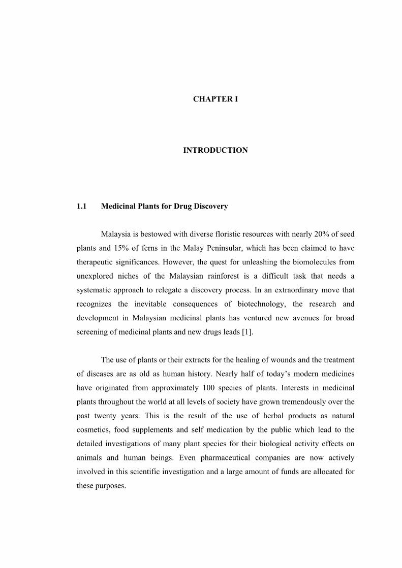
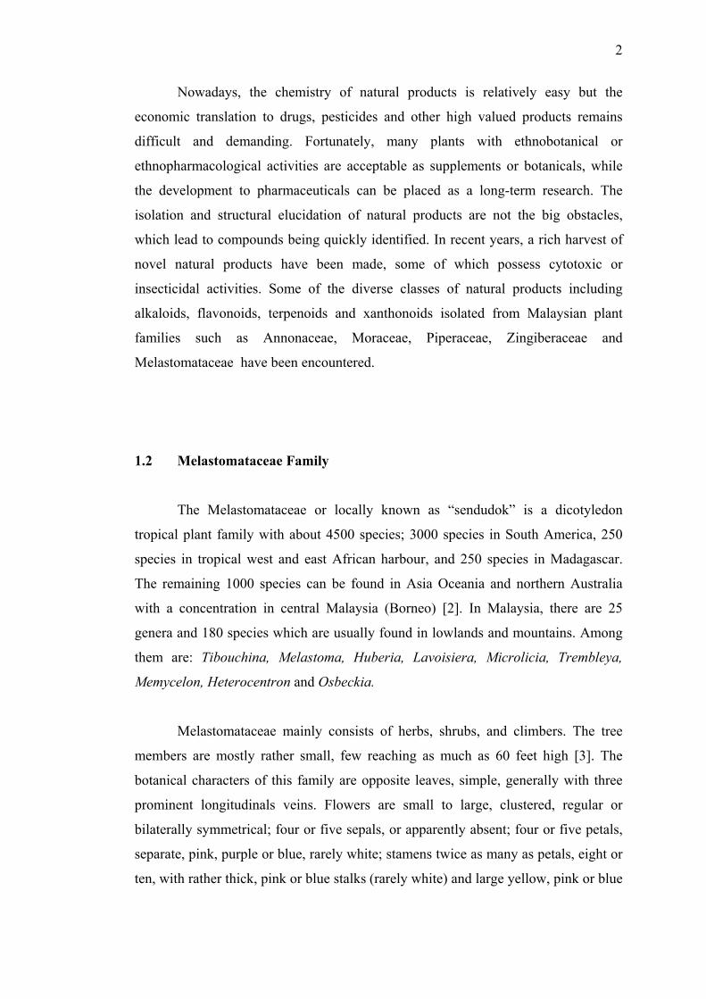
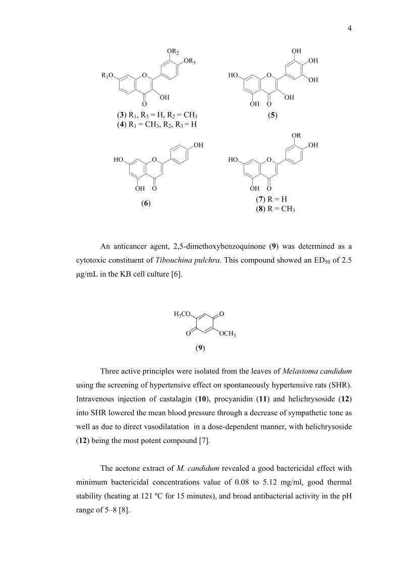
A wide diversity of flavonoid structures has been found in this family, with
the predominance of flavonol glycosides mainly kaempferol (1) and quercetin (2).
Glycosides of isorhamnetin (3), rhamnetin (4) and myricetin (5) were also found,
although less frequently. Derivatives of apigenin (6), luteolin (7) and chrysoeriol (8)
were among the flavones isolated in this family [5].
OOH
HO O
OHOH
OHOOH
HO O
OH
OH
(1) (2)
4
OOH
HO O
OHOH
OH
OH
O
R1O O
OR3
OH
OR2
(3) R1, R3 = H, R2 = CH3(4) R1 = CH3, R2, R3 = H
(5)
OOH
HO O
OROH
OOH
HO O
OH
(7) R = H (8) R = CH3
(6)
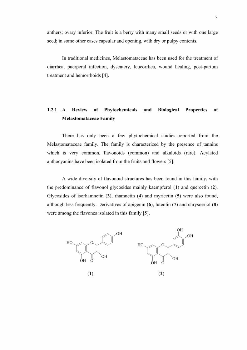
An anticancer agent, 2,5-dimethoxybenzoquinone (9) was determined as a
cytotoxic constituent of Tibouchina pulchra. This compound showed an ED50 of 2.5
µg/mL in the KB cell culture [6].
H3CO
OCH3
O
O
(9)
Three active principles were isolated from the leaves of Melastoma candidum
using the screening of hypertensive effect on spontaneously hypertensive rats (SHR).
Intravenous injection of castalagin (10), procyanidin (11) and helichrysoside (12)
into SHR lowered the mean blood pressure through a decrease of sympathetic tone as
well as due to direct vasodilatation in a dose-dependent manner, with helichrysoside
(12) being the most potent compound [7].
The acetone extract of M. candidum revealed a good bactericidal effect with
minimum bactericidal concentrations value of 0.08 to 5.12 mg/ml, good thermal
stability (heating at 121 ºC for 15 minutes), and broad antibacterial activity in the pH
range of 5–8 [8].
5
O
OHHO
OH
O
OOHO
HO
OH
OHOH
OH
OH
HOOH
O
HO
OHOH HO OH
HO OH
OHO
(10)
OHO
OH
OHOH
OH
OHO
OH
OHOH
OH
OHO
OH
OHOH
OO
O
O
HO
O
OHOH
OH
(12)
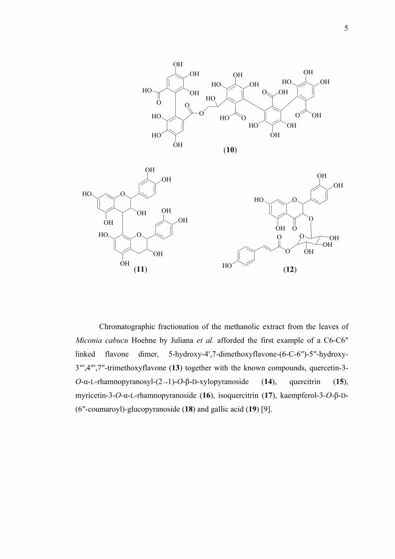
Chromato
Miconia cabucu
linked flavone
3″′,4″′,7″-trimeth
O-α-L-rhamnopy
myricetin-3-O-α-
(6″-coumaroyl)-g
(11)
graphic fractionation of the methanolic extract from the leaves of
Hoehne by Juliana et al. afforded the first example of a C6-C6″
dimer, 5-hydroxy-4′,7-dimethoxyflavone-(6-C-6″)-5″-hydroxy-
oxyflavone (13) together with the known compounds, quercetin-3-
ranosyl-(2→1)-O-β-D-xylopyranoside (14), quercitrin (15),
L-rhamnopyranoside (16), isoquercitrin (17), kaempferol-3-O-β-D-
lucopyranoside (18) and gallic acid (19) [9].

6
OH
HO
O
HO
OOH
HO O
OHOH
HO OH
OHO
O O
OH
OH
O
OOH
H3CO O
OCH3
O OH
OCH3O
OCH3
H
3CO
(13)
O
O
OHOH
O OH
OH
OH
O
HO
HO
HOO
OH
OH
O
OHOH
O OH
OH
OH
O
HO
O
HO
HO
HO
(14)
OH
O
OHOH
O OH
OH
OH
O
OH
(15)OH
(16)
O
O
OH
O OH
OH
OH
O
OO
(17)(18)
(19)

7
Phytochemical and bioactivity studies of the flowers of Melastoma
malabathricum L. have been carried out by Deny Susanti et al. [10]. The ethyl
acetate extract yielded three compounds, identified as naringenin (20), kaempferol
(1) and kaempferol-3-O-D-glucopyranoside (21) while the methanol extract
gave kaempferol-3-O-(2″,6″-di-O-p-trans-coumaroyl)-glucopyranoside (22) and
kaempferol-3-O-D-glucopyranoside (21). All these compounds as well as their crude
extracts were found to be active as free radical scavengers in the DPPH radical-
scavenging electron spin resonance spectroscopic method. Naringenin (20) and
kaempferol-3-O-(2″,6″-di-O-p-trans-coumaroyl)-glucopyranoside (22) were also
found to be active in inhibiting cell proliferation of human cell line from breast
carcinoma with IC50 values of 0.28 µM and 1.3 µM, respectively.
OOH
HO O
OH
O OH
OH
OH
O
HO
OHO
OH
OH
O
(20)
OOH
HO O
OH
O O
OH
OH
O
OHO O
O
(21)
OH
(22)

8
In the search for natural compounds useful against anti-inflammatory activity,
α-amyrin (23), betulinic acid (24), quercetin (2) and quercitrin (15) which were also
isolated from M. malabathricum L. had been assessed in vitro by determining their
inhibitory effects on platelet activating factor (PAF) binding to rabbit platelets using 3H-PAF as a ligand. The results indicated that quercetin (2), quercitrin (15), α-amyrin
(23), and betulinic acid (24) showed inhibition of PAF receptor binding with IC50
values of 33.0, 45.4, 20.0 and 22.2 µM, respectively. The IC50 values of these
compounds were comparable to cedrol (13.1 µM), which was a known PAF receptor
antagonist. These results suggested that natural flavonoids and pentacyclic
triterpenes from M. malabathricum L. possess selective antagonistic activity towards
PAF and could be an attractive candidate as natural anti-inflammatory compounds
[11].
HO
HO
COOH
The antinocic
acetic acid-induced
carried out by Sulaim
i.p.) strongly and do
with an ED50 of 1
malabathricum has a
and central level of n
(23)
eptive effect of ethanolic extract of M. m
abdominal writhing test and hot-plate te
an et al. It was demonstrated that the ex
se-dependently inhibited the acetic acid-i
00 mg/kg i.p., suggesting that, the etha
potential as antinociceptive agent that ac
erves [12].
(24)
alabathricum using
st in mice has been
tract (30-300 mg/kg,
nduced writhing test
nolic extract of M.
ts at both peripheral

9
The chemical investigation on Monochaetum multiflorum yielded
trifolin (25), hyperin (26), quercetin 3-(6′-O-caffeoyl)-β-D-galactopyroside (27),
isoquercitrin (17), quercetin 3-(6′-O-caffeoyl)-β-D-glucopyroside (28), 4-O-β-D-
glucopyranosyl-2-O-methylphloroacetophenone (29), 4-O-(6′-O-galloyl-β-gluco
pyranosyl)-cis-p-coumaric acid (30), 6′-O-galloylprunasin (31), benzyl 6′-O-galloyl-
β-glucopyranoside (32) and a novel diester of tetrahydroxy-µ-trunixic acid with 2
mole of hyperin (monochaetin) (33) [13].
OOH
HO O
OH
O OH
OHO
HO OH
OH
OOH
HO O
OH
O OH
OHO
HO OH
OH
HO
HO
HO
(25)
O
O
OH
O OH
OHO
O OH
OH
OOH
HO O
OH
O OH
OHO
O OH
OH
HO
HO
OO
(27) (28)(26)

10
OO
HOHO
O
OH
OH
OH
OOH
COOH
OO
OH
OCH3
O
HOHO
OH
OH
OO
HOHO
O
OH
HO
HO
OHO
CN
OO
HOHO
O
OH
HO
HO
OHO
OOH
HO O
OH
O OH
OHO
O OH
OH
H HH H
O OH
OHO
HO
OHO
HO O
OHO
O
O
OH
O
OH
OH
(32)
(30)
(29)(31)
H
OH
(33)

11
Bioactivity-directed isolation of an EtOAc extract from the leaves of Miconia
lepidota, afforded two benzoquinones, namely 2-methoxy-6-heptyl-1,4-
benzoquinone (34) and 2-methoxy-6-pentyl-1,4-benzoquinone (35) which showed
potential as an anticancer agents [14].
O
O
H3CO R
(34) R = C7H15(35) R = C5H11
The chemical investigation of Henriettella fascicularis has led to the isolation
of 4′,5,7-trihydroxy-6,8-dimethoxylisoflavone (36) and sesterterpenoic acid (37)
[15].
OHO
OH OOH
CH3
H3C
COOH
HO
The ethano
which was identifie
(36)
l extract of Miconia pilgeriana yielded
d as arjunolic acid (38) [16].
COOH
HO
OH
HO
(37)
a triterpene compound
(38)
12
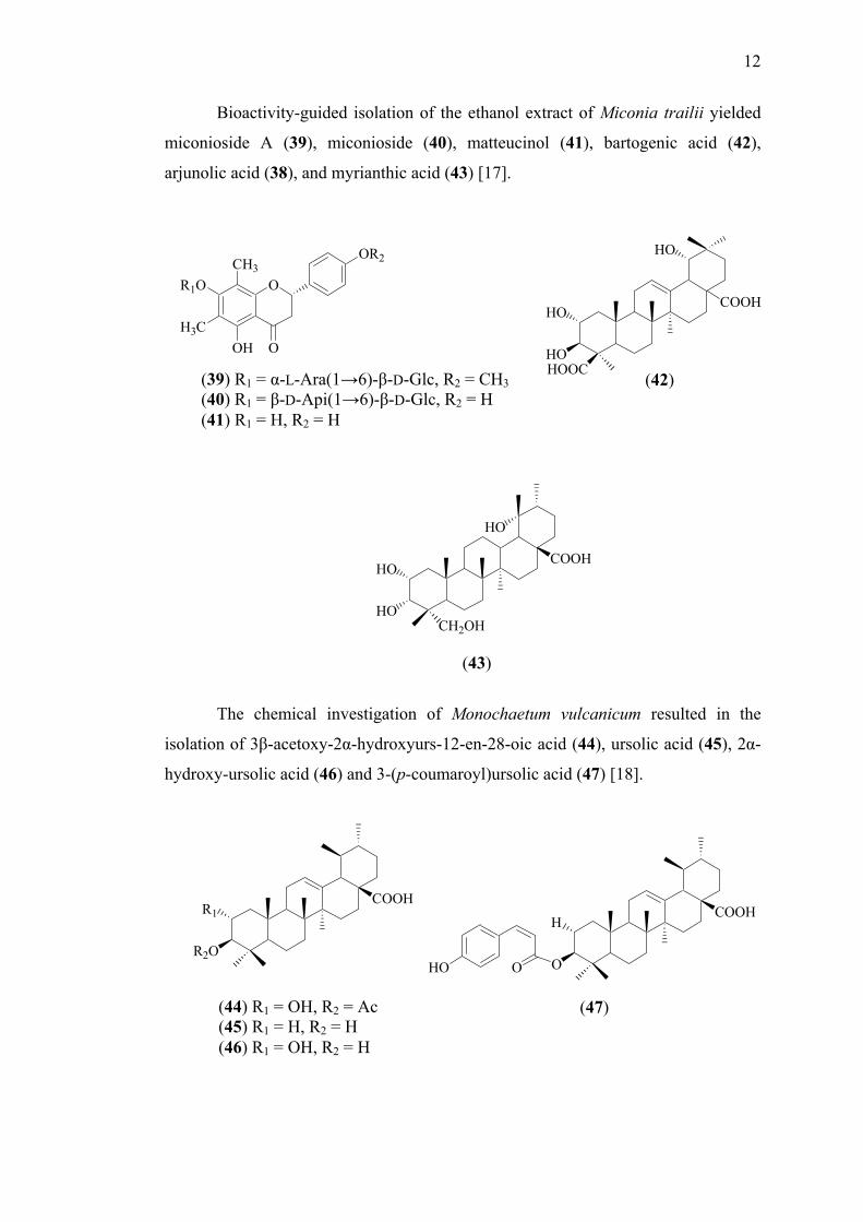
Bioactivity-guided isolation of the ethanol extract of Miconia trailii yielded
miconioside A (39), miconioside (40), matteucinol (41), bartogenic acid (42),
arjunolic acid (38), and myrianthic acid (43) [17].
COOH
HOHOOC
HO
HO
OR1O
OH
OR2
O
CH3
H3C
(39) R1 = α-L-Ara(1→6)-β-D-Glc, R2 = CH3(40) R1 = β-D-Api(1→6)-β-D-Glc, R2 = H (41) R1 = H, R2 = H
(42)
HOCH2OH
HOCOOH
HO
(43)
The chemical investigation of Monochaetum vulcanicum resulted in the
isolation of 3β-acetoxy-2α-hydroxyurs-12-en-28-oic acid (44), ursolic acid (45), 2α-
hydroxy-ursolic acid (46) and 3-(p-coumaroyl)ursolic acid (47) [18].
R2O
R1COOH
O
HCOOH
HO O
(44) R1 = OH, R2 = Ac (45) R1 = H, R2 = H
(47)
(46) R1 = OH, R2 = H

13
Two ellagic acids identified as tri-O-methyl ellagic acid (48) and tri-O-methyl ellagic
acid glucoside (49) were successfully isolated from Melastoma polyanthum [19].
OO
O
O
MeO
OMe
OMe
O
OHOH
HO
OOH
OO
O
O
MeO
OMe
OMe
OH
(48) (49)
The hexane, dichloromethane and ethanol extracts of Miconia rubiginosa
which were evaluated for their analgesic effects showed a significant inhibition in
mice and rats (p < 0.05 and p < 0.01) using the acetic acid-induced writhing and hot
plate tests. These extracts (200 mg/kg body wt.) showed a significant (p < 0.05)
antinociceptive effect, lower than that produced by morphine (4 mg/kg wt.). The
fractionation of the dichloromethane extract yielded ursolic acid (45) and oleanoic
acid (50) as the major compounds. Three triterpenes from the hexane extract was
identified using gas chromatography as α-amyrin (23), β-amyrin (51), and β-
sitosterol (52) [20].
HOH H
HO
H
O
OH
H
(50) (51) (52)

14
Dissotis brazae Cogn. which was traditionally used to treat malaria in Kenya
was tested for in vivo antiplasmodium activity against chloroquin-resistant (ENT-36).
The aqueous extract of the stems showed the strong inhibitory activity with IC50 6.4
µg/mL [21].
1.3 Tibouchina Genus
Tiobouchina is a genus of about 350 species of neotropical plants. Members
are shrubs or subshrubs, and are known as “glory bushes” or “glory trees”. They are
native to rainforest of Mexico, the West Indies, and South America, especially
Brazil. The name comes from an adaptation of the native Guiana term for these
shrubs. Several species are cultivated for their large bright flowers such as T.
multiflora, T. organensis, T. maudhiana and T. semidecandra.
1.3.1 Tibouchina Semidecandra L.
Tibouchina semidecandra L. is a shrub that has been introduced to Malaya
from Brazil [3]. It bears beautiful dark purple flowers throughout the year and grows
well in frost-free areas around the world. The pristine purple flower makes it a
valuable ornamental plant and a potential source for the extraction of natural
colourants. This plant is also used traditionally for both medicinal and food purposes
[22].
Figure 1.1 Tibouchina semidecandra L.

15
1.3.2 Chemical Investigation of Tibouchina
The chromatographic survey of the tannins in this family revealed that
Tibouchina is rich in tannins, particularly in oligomeric hydrolysable tannins.
Repeated chromatography of the n-BuOH extract of the leaves of Tibouchina
multiflora over polystyrene and polyvinyl gel afforded two new oligomeric
hydrolysable tannins named nobotanin O (53) and nobotanin P (54) [23].
Yoshida et al. found by means of a chromatographic survey that T.
semidecandra (collected from Japan) was also rich in tannins, particularly in
oligomeric hydrolysable tannins. Seven new hydrolysable tannins, named as
methylvescalagin (55), nobotanin A (56), nobotanin B (57), nobotanin C (58),
nobotanin D (59), nobotanin E (60) and nobotanin F (61). Several flavonoid
compounds such as quercetin (2), myricetin (5), leucodelphinidin (62), leucocyanidin
(63), quercetin-3-O-(6″-O-galloylgalactoside) (64), avicularin (65) and tibouchinin
(66) have also been isolated from this plant [24-28].
OO
HOO
OH
O
OH
HO
O
HO
OH
OH
OH
HO
OH
OH
O
HO
O
OH
O
OO
OO
O
O
OH
OH
OH
O
O
OH
HO
O
HO
OH
HO
O
OH
OH
OH
OHO
(53)

16
HO
OH
O
OH
HO
OH
O
HO
HO
HO
HOO
OHHO
O
HO
OH
HO
O
OH
O
O
OMe
H
OO
O
(55)
OOO
O
O
O
OH
HO
O
HO
OH
OH
OH
OH
HO
O
OH
O
HO
O
OHO
OH
HOO
HO
HO
OHHO OO
O
O
O
O
OH
HO
O
HO
OH
HO
O
OH
HO
OH
OH
O
R
(56) R = OH (61) R = O
OH
OH
OH
O

17
OOO
O
O
O
OH
HO
O
HO
OH
OH
OH
HO
OH
OH
O
HO
O
OH
O
OO
OO
O
O
OH
OH
OH
O
O
OH
O
O
HO
OH
HO
O
OH
OH
OH
OHO
HO
O
OHO
OH
HOO
HO
HO
OHHO O
OHO
O
O
O
O
OH
HO
O
HO
OH
HO
O
OH
HO
OH
OH
O
OOO
O
O
O
OH
HO
O
HO
OH
OH
OH
OH
HO
O
OH
O
HO
OH
OHO
OH
HOO
HO
(54)

18
OOO
O
O
O
OH
HO
O
HO
OH
OH
OH
OH
HO
O
OH
O
OH
OH
O
OH
O
HO
HO
HO OOO
O
O
O
OH
HO
O
HO
O
HO
O
OH
HO
OHHO
O
O
OH
OH
OH
OHO
OH
OH
(57)
OO
HOO
O
O
OH
OH
OH
O
OH
HO
O
HO
OH
HO
O
OH
HO
OHHO
O
(59)

19
OOO
O
O
O
OH
HO
O
HO
OH
OH
OH
HO
OH
OH
O
HO
O
OH
O
OO
OO
O
O
OH
OH
OH
O
O
OH
HO
O
HO
OH
HO
O
OH
OH
OH
OHO
HO
O
OHO
OH
HOO
HO
HO
OHHO OO
O
O
O
O
OH
HO
O
HO
OH
HO
O
OH
HO
OH
OH
O
R
O
OH
OH
OH
O
(58) R = OH (60) R =

20
OHOHOH
HO OOH
OHOH
OHOHOH
O
OHOH
HO
(62) (63)
OOH
HO O
OH
O OH
OHO
O OH
OH
HO
HOO
HO
OOH
HO O
OH
O
OH
OH
OHO
HO
(64) (65)
OOHO
HO O
OCH3OH
OCH3
OHOH
HO
OOH OH
OH
OH
O
O
OH
O
(66)

21
Acid hydrolysis of nobotanin F (61) gave gallic acid (19), ellagic acid (67),
valoneic acid dilactone (68) and glucose [27].
O
OO
O
HO
OH
HO OH
O
OO
O
HO
OH
HO O
OHHO
OH
HOOC
(67) (68)
The structure of the major pigment in the purple flowers of T. semidecandra
has been identified as maldivin 3-(p-coumaroylglucoside)-5-acetylglucoside (69), a
new anthocyanin by using chromatographic and various NMR techniques [29].
OOHO
HO O
OCH3OH
OCH3
OOH
HO
OOH OH
OH
OH
O
O
OH
OH3C O
(69)

22
1.4 Biosynthesis of Flavonoids
In recent years, flavonoids have attracted the interest of researchers because
they show promise of being powerful antioxidants which can protect the human body
from free radicals [30]. Flavonoids cannot be produced by the human body and thus,
have to be taken in, mainly through the daily diet. The evidence reported in the
chemistry, biochemistry and pharmacy literature supports the view that flavonoids
play a vital biological role, including the function of scavenging reactive oxygen
species.
Chemically, there are three features that confer on flavonoids and their
remarkable antioxidant properties:
• the hydrogen donating substituents (hydroxyl groups), attached to the
aromatic ring structures of flavonoids, enable the flavonoids to undergo
a redox reaction that helps them to scavenge free radicals more easily;
• a stable delocalization system, consisting of aromatic and heterocyclic
rings as well as multiple unsaturated bonds, which helps to delocalize
the resulting free radicals, and
• the presence of certain structural groups which are capable of forming
transition metal-chelating complexes that can regulate the production of
reactive oxygen species such as OH• and O-1•.
Flavonoids represent a highly diverse class of secondary plant metabolites
with about 9000 structures which have been identified up to now. All flavonoids
derive their 15-carbon skeleton from two basic metabolites, malonyl-CoA and p-
coumaroyl-CoA. Basically, flavonoids are derivatives of 1,3-diphenylpropan-1-one
(C6-C3-C6). The crucial biosynthetic reaction is the condensation of three molecules
malonyl-CoA with one molecule p-coumaroyl-CoA to a chalcone intermediate.
Chalcones and dihydrochalcones are classes of flavonoids that consist of two
phenolic groups which are connected by an open three carbon bridge. Derived from
the chalcone structure, a flavonoid-class containing three rings, the flavanones, can
be formed. Here, the three-carbon bridge is part of an additional heterocyclic six-
membered ring that involves one of the phenolic groups on the adjacent ring. Based

23
on these flavanones, all other flavonoid-classes are generated, including isoflavones,
flavanols, anthocyanidins, flavonols and flavones as shown in Figure 1.2. This latter
flavonoid-class is characterized by the presence of a double bond between C2 and C3
in the heterocycle of the flavan skeleton. The B-ring is attached to C2 and usually no
substituents are present at C3. This differentiates them from flavonols where a
hydroxyl group can be found at that C3 position [31].

24
24

25
1.5 Research Objectives
Phytochemical investigations reported in the literature are mostly carried out
on the Tibouchina semidecandra of Japan. A thorough literature search did not reveal
any report on the chemical constituents of T. semidecandra found in Malaysia,
except on the anthocyanin stability in the flower [22] and the chemical manipulation
of growth and flowering of this plant [32]. Furthermore, there has been no report on
bioactivity studies of this species. Therefore, this research will focus on the
phytochemical and biological activity studies of Tibouchina semidecandra L.
The objectives of this research are to extract three parts of the plant (leaves,
roots and stem barks) using organic solvents of different polarity either by soxhlet or
cold extraction, followed by fractionation of the extracts using vacuum liquid
chromatography technique. The next objectives are to isolate the phytochemical
compounds using various chromatographic techniques either on silica gel or
Sephadex LH-20 followed by structural identification using various spectroscopic
methods including high field 1D NMR (1H NMR, 13C NMR, DEPT), 2D NMR
(COSY, HMQC, HMBC), FTIR, UV spectroscopies and Mass spectrometry. The
final objective is to evaluate the biological activities of the crude extracts and the
pure compounds using several bioassays including antioxidant (free radical
scavenging on 2,2-diphenyl-1-picrylhidrazyl (DPPH) method), antibacterial and
antityrosinase assays.

86
REFERENCES
1. Azizul, A. K. (2001). Commercial Prospects of Scientific Discoveries on
Malaysian Bioresources. Kuala Lumpur Vol. 1: 9-10.
2. Whitmore, T. C. (1972). Tree Flora of Malaya. Vol. 1. Longman: Kuala
Lumpur. Vol. 1: 7-18
3. Corner, E. J. H. (1965). Wayside Trees of Malaya. Malayan Nature Society.
Kuala Lumpur. Vol. 1 : 445-453.
4. Burkill, I. H. (1966). A Dictionary of the Economic Products of Malay
Peninsular. Ministry of Agriculture and Co-Operatives: Kuala Lumpur.
1462-1464.
5. Bomfim-Patricio, M. C., Salatino, A., Martins, A. B., Wardack, J. J. and
Salatino M. L. F. (2001). Flavonoids of Lavoisera, Microlia, and Trembleya
(Melastomataceae) and their taxonomic meaning. Biochem. Syst. Ecol. 29:
711-726.
6. Jones, E., Ekandato, O. and Kingston, G. I. (1981). Plant anticancer agents.
XI. 2,5-Dimethoxybenzoquinone as a cytotoxic constituent of Tibouchina
pulchra. J. Nat. Prod. 44: 493-494.
7. Tang Cheng, J., Lin Hsu, F. and Fen Chen, H. (1993). Antihypertensive
Principles from the Leaves of Melastoma candidum. Planta Med. 59: 405-
407.
8. Yuan-Chuen, W., Hsing-Wen, H. and Wen-Ling, Liao (2008). Antibacterial
activity of Melastoma candidum D. Don. Food Sci. Technol.
Doi:10.1016/j.lwt.2008.02.05 Article in press.

87
9. Juliana, R., Daniel, R., Lourdes, C. and Wagner, V. (2007). An unusual C6-
C6” linked flavonoid from Miconia cabucu (Melastomataceae).
Phytochemistry. 68: 1781-1784.
10. Deny, S., Hasnah, M. S., Farediah, A., Rasadah, M. A., Norio, A. and
Mariko, K. (2007). Antioxidant and cytotoxic flavonoids from the flowers of
Melastoma malabathricum L. Food Chem. 103: 710-716.
11. Mazura, M. P., Deny, S. and Rasadah, M. A. (2007). Anti-inflammatory
Action of Components from Melastoma malabathricum. Pharm. Biol. 5 (45):
372-375.
12. Sulaiman, M. R., Somchit, M. N., Israf, D. A., Ahmad, Z. and Moin, S.
(2004). Antinociceptive Effect of Melastoma malabathricum Ethanolic
Extract in Mice. Fitoterapia. 75: 667-672.
13. Jose, S. I., Hideyuki, I. and Takashi, Y. (2001). A flavonol glycoside-lignan
ester and accompanying acylated glucosides from Monochaetum multiflorum.
Phytochemistry. 58: 321-327.
14. Gunatilaka, A. A. L., Berger, J. M., Evans, R., Miller, J. S., Wisse, J. H.,
Neddermann, K. M., Bursuker, I. and Kingston, D. G. I.(2001). Isolation,
Synthesis and Structure-Activity Relationships of Bioactive Benzoquinones
from Miconia lepidota from Suriname Rainforest. J. Nat. Prod. 64: 2-5.
15. Calderon A. I., Terreaux, C., Schenk, K., Pattison, P., Burdette J. E., Pezzuto,
J. M., Gupta, M. P. and Honstettmann, K. (2002). Isolation and Structure
Elucidation of an Isoflavone and a Sesterterpenoic Acid from Henrienttella
fascicularis. J. Nat. Prod. 65: 1749-1753.
16. Cong Li, X., Joshi, A. S., El Sohly, H. N., Khan, S. I. and Jaacob, M. R.
(2002). Fatty Acid Synthase Inhibitors from Plants: Isolation, Structure
Elucidation and SAR Studies. J. Nat. Prod. 65: 1909-1914.
88
17. Zhang, H., El Sohly, H. N., Cong Li, X., Khan, S. I., Broedel, S. E., Raulli,
R., Cihlar, R. L. and Walker, L. A. (2003). Flavanone Glycoside from
Miconia trailii. J. Nat. Prod. 66: 39-41.
18. Chaturvedula , V. S. P., Gao, Z., Jones, S. H., Feng, X., Hecht, S. M. and
Kingston D. G. I. (2004). A New Ursane Triterpene from Monochaetum
vulcanicum that Inhibits DNA Polymerase β Lyase. J. Nat. Prod. 67: 899-
901.
19. Liu, S. R., (1986). Chemical Constituents of Melastoma polyanthum. Zhong
Yao Tong Bao. 11: 42-43.
20. Spessoto, M. A., Ferreira, D. S., Crotti, A. E. M., Silva, M. L. A. and Cunha
W. R. (2003). Evaluation of the Analgesic Activity of Extract of Miconia
rubiginosa (Melastomataceae). Phytomedicine. 10: 606-609.
21. Omulokoli, E., Khan, B. and Chhabra, S. C. (1997). Antiplasmodial Activity
of Four Kenyan Medicinal Plants. J. Ethnopharmacol. 56: 133-137.
22. Janna, O. A., Khairul, A. K., Maziah, M. (2006). Anthocyanin Stability
Studies in Tibouchina semidecandra L. Food Chem. 101: 1640-1646.
23. Yoshida, T., Amakura, Y., Yokura, N., Ito, H., Isaza, J. H., Ramirez, S.,
Palaez D. P. and Renner, S. S. (1999) Oligomeric Hydrolysable Tannins from
Tibouchina multiflora. Phytochemistry. 52: 1661-1666.
24. Yoshida, T., Haba, K., Shingu, T. and Okuda, T. (1987). Revised Structure of
Nobotanin B, a Dimeric Ellagitannin of Tibouchina semidecandra.
Heterocycles. 11: 2845-2848.
25. Yoshida, T., Ikeda, Y., Ohbayashi, H., Ishihara, K., Ohwasyi, W., Shingu T.
and Okuda, T. (1986). Dimeric Ellagitannins in Plants of Melastomataceae.
Chem. Pharm. Bull. 34: 2676-2679.

89
26. Yoshida, T., Okano, Y., Haba, K., Ohbayashi, H., Ishihara, K., Ohwasyi, W.,
Shingu T. and Okuda, T. (1991). Tannins and Related Polyphenols of
Melastomataceous Plants. I. Hydrolyzable Tannins from Tibouchina
semidecandra COGN. Chem. Pharm. Bull. 39: 2233-2240.
27. Yoshida, T., Okano, Y., Haba, K., Ohbayashi, H., Ishihara, K., Ohwasyi, W.,
Shingu T. and Okuda, T. (1991). Tannins and Related Polyphenols of
Melastomataceous Plants. II. Nobotannins B, C and E, Hydrolyzable Tannins
from Tibouchina semidecandra COGN. Chem. Pharm. Bull. 39: 2264-2270.
28. Harborne, J. B. (1964). Plant Polyphenols – XI. The Strucutre of Acylated
Anthocyanins. Phytochemistry. 3: 151-160.
29. Terahara, N. and Suzuki, H. (1993). A Diacylated Anthocyanin from
Tibouchina urvilleana Flowers. J. Nat. Prod. 3: 335-340.
30. Zhao, F. P., Strack, D., Baumert, A., Ramanathan, S., Ngoh, K. G., Tet, F. C.
and Swee, N. T. (2003). Antioxidant Flavonoids from Leaves of Polygonum
hydropiper L. Phytochemistry. 62: 219-228.
31. Martens, S. and Mithofer, A. (2005). Flavones and Flavone Synthases.
Phytochemistry. 66: 2399-2407.
32. Abdullah, Tohirah, Asiah, A. and Siti, H. (1996). Chemical Manipulation of
Growth and Flowering in Potted Melastoma decemfidum and Tibouchina
semidecandra. Third International Symposium on New Floricultural Crops.
297-301.
33. Beck, P. O. D, Dijoux, M. G., Cartier, G. and Mariote, A. M. (1998).
Quercitrin 3’-Sulphate from Leaves of Leea Guinensis. Phytochemistry. 47:
255-258.

90
34. Markham, K. R. (1982). Techniques of Flavonoid Identification. Academic
Press Inc.: London.
35. Masa, A. and Vilanova, M. (2008). Flavonoid and Aromatic Characterisation
of cv. Vitis vinifera L. Food. Chem. 107: 273-281.
36. Long-Ze, L., James, M. H., Marcial, S. P. C. and Devanand L. L. (2008). The
Polyphenolic Profiles of Common Bean (Phaseolus vulgaris L.). Food Chem.
107: 399-410.
37. Zong P. Z., Cheng, W. K., Jianfei, C., Jiajun, W. and Mingfu, W. (2008).
Tyrosinase Inhibitors from Paper Mulberry (Broussonetia papyrifera). Food
Chem. 106: 529–535.
38. Reyes-Caudillo, E., Tecante, A. and Valdivia-Lo, M. A. (2008). Dietary Fibre
Content and Antioxidant Activity of Phenolic Compounds Present in
Mexican chia (Salvia hispanica L.) seeds. Food Chem. 107: 656–663.
39. Mabry, T. J., Markham, K. R. and Thomas, M. B. (1970). The Systematic
Identification of Flavonoids. Springer-Verlag: Berlin.
40. Saha, K., Lajis, N. H., Shaari, K., Hamzah, A. S. and Israf, D. A. (2005).
Chemical Constituents of Leea indica (Burm. f.) Merr. (Leeaceae). Mal. J.
Sci. 24: 75-78.
41. Mizushina, Y., Nakanishi, R., Kuriyama, I., Kamiya, K., Satake, T.,
Shimazaki, N., Koiwai, O., Uchiyama, Y., Yonezawa, Y., Takemura, M.,
Sakaguchi, K. and Yoshida, H. (2006). β-Sitosterol-3-O-β-D-
glucopyranoside: A Eukaryotic DNA Polymerase λ Inhibitor. Journal of
Steroid Biochemistry and Molecular Biology. 99: 100–107.

91
42. Mehrdad, I., Mehdi, M., Hamid, S., Mohammad, Y. H. and Bernd, S. P.
(2007). Secondary Metabolites of Ferula persica roots. Phytochemistry.
Article in Press.
43. Mulabagal, V., Ruby, L. A. L., David, L. D. and Muraleedharan, G. N.
(2008). Functional Food Components of Antigonon leptopus tea. Food Chem.
106: 487–492.
44. Anna, R. B., Jeannette, M. and Ivano, M. (1996). Phytochemical
Investigations of Licania Genus. 1 Flavonoids and Triterpenoids from Licania
carii. Pharmaceutica Acta Helvetiae. 71: 191-197.
45. Godfred, A. A., Pierre, T. and Joseph, D. C. (2004). Aulacocarpin A and B,
Nerolidol and β-Sitosterol glucoside from Aframomum escapum. Biochem.
Syst. Ecol. 32: 1205–1207.
46. Deepak, M., Dipankar, G., Prashanth, D., Asha, M. K., Amit, A.and
Venkataraman, B.V. (2002). Tribulosin and β-Sitosterol-D-glucoside, the
Anthelmintic Principles of Tribulus terrestris. Phytomedicine. 9: 753–756.
47. Yinrong , L. and Yeap Foo, L. (1997). Identification and Quantification of
Major Polyphenols in Apple Pomace. Food Chem. 2: 187-194.
48. Yi-Fen, W., Jian-Xin, C., Thomas, E., Gou-Fang, L. and Shi-De, L. (2006).
Cytotoxic and New Tetralone Derivatives from Berchemia floribunda (Wall.)
Brongn. Chemistry and Biodiversity. 6: 646–653.
49. D'Agostino, M., Dini, I., Ramundo, E. and Senatore, F. (1998). Flavonoid
glycosides of Alchemilla vulgaris L. Phytotherapy Res. 12: 162-163.
92
50. Price, K. R., Prosser, T., Richetin, A. M. F. and Rhodes, M. J. C. (1999). A
Comparison of the Flavonol Content and Composition in Dessert, Cooking
and Cider-making Apples; Distribution within the Fruit and Effect of Juicing.
Food Chem. 66: 489-494.
51. Mino, J., Acevedo, C., Moscatelli, V., Ferraro, G. and Hnatyszyn, O. (2002).
Antinociceptive Effect of the Aqueous Extract of Balbisia calycina. J.
Ethnopharmacol. 79: 179–182.
52. Rosa, M. A., Karine, N., Emerson, F. Q., Jean, R. I., Hostettmannb, K., Luis,
A. B. and Francisca, V. (2004). On-line Characterisation of Apple
Polyphenols by Liquid Chromatography Coupled with Mass Spectrometry
and Ultraviolet Absorbance Detection. J. Chromatogr. A. 1046: 89–100.
53. Ishiguro, K., Nagata, S., Fukumoto, H., Yamaki, M., Takagi, S., and Isoi, K.
(1991). A Flavonol Rhamnoside from Hypericum Japonicum.
Phytochemistry. 30: 3152-3154.
54. Zhang, Y., Shu-fen L. and Xi-wen W. (2008). Pressurized Liquid Extraction
of Flavonoids from Houttuynia cordata Thunb. Separation and Purification
Technology. 58: 305–310.
55. Sonata, T., Jurga, B., Daiva, M.,Valdas, J., Arunas, S. and Adolfas, T. (2007).
Effect of Ginkgo biloba Extract on the Rat Heart Mitochondrial Function. J.
Ethnopharmacol. 111: 512–516.
56. Eiji, Y., Minoru, I., Osamu, K. and Tetsuji, I. (2007). Antioxidant Activity of
Japanese Pepper (Zanthoxylum piperitum DC.) Fruit. Food Chem. 100: 171–
177.
57. Craig, T. M. T., Lutfun, N., Alison, C., Yashodharan, K., Namik, F. M.,
Moira, M., Raymond, G. R. and Satyajit, D. S. (2003). Flavonol Glycosides
from the Seeds of Agrimonia eupatoria (Rosaceae). Biochem. Syst. Ecol. 31:
439–441.
93
58. Mario, J. S., Seiji, A., Satoshi, T., Hui, Y. and Kurt, A. R. (2008) Cytotoxic
Chalcones and Antioxidants from the Fruits of Syzygium samarangense (Wax
Jambu). Food Chem. 107: 813–819.
59. Mahmood, U., Vijay, K. K., Ruchi, A. and Leopold, J. (2003). p-Coumaric
acid esters from Tanacetum longifolium. Phytochemistry. 64: 851–853.
60. Radulovic, N., Stojanovic, G. and Asakawa, Y. (2006). Hydroxycinnamoyl
conjugates from the roots of Achillea holosericea Sibth. et Sm. Biochem.
Syst. Ecol. 34: 83-87.
61. Keriko, Joseph M., Nakajima, Shuhei, B., Naomichi, Iwasa, J. (1997).
Eicosanyl p-coumarates from a Kenyan Plant, Psiadia punctulata: Plant
Growth Inhibitors. Biosci. Biotech. Biochem. 61: 2127-2128.
62. Kisiel, W. and Jakupowic, J. (1995). Long-chain Alkyl hydroxycinnamates
from Crepis taraxacifolia. Planta Med. 61: 87-8.
63. Malhotra, S. and Misra, K. (1981). 3,3′-Di-O-methylellagic acid 4-O-
rhamnoside from the roots of Prosopis juliflora. Phytochemistry. 8: 2043-
2044
64. Khallouki, F., Haubner, R., Hull, W. E., Erben, G., Spiegelhalder, B.,
Bartsch, H. and Owen, R. W. (2007). Isolation, Purification and Identification
of Ellagic acid Derivatives, Catechins, and Procyanidins from the Root Bark
of Anisophyllea dichostyla R. Br. Food Chem. Toxicol. 45: 472–485.
65. Ye, G., Peng, H., Fan, M. and Huang, C. (2007). Ellagic acid Derivatives
from the Stem Bark of Dipentodon sinicus. Chemistry of Natural
Compounds. 43: 125-127.

94
66. Souleman, Ahmed M. A., El-Mousallamy, Amani, M. D. Chemical
Investigation of the Constitutive Phenolics of Rosa Arabica; The Structure of
a New Dimeric Phenolic Glycoside. Nat. Prod. Sci. 6: 82-85.
67. Fazlul, H. S. and Yiwei, L. (2006). Plant-Derived Antioxioxidants. In:
Keshav K. S. Oxidative Stress, Disease and Cancer. Imperial College Press:
London. 995-1005.
68. Burdon, H. R. (1993). Carcinogenesis and Free Radicals. In: Poli, G., Albano,
E. and Dianzani, M. U. Free Radicals: From Basic Science to Medicine.
Molecular and Cell Biology Updates: Basel. 187-197.
69. Paula, F. I., Sussan, K. A. and Ronald, R. W. (1997). Antioxidants and
Immune Function. In: Harinder, S. G. Antioxidants and Disease Prevention.
CRC Press: New York. 19-26.
70. Thomas, W. K., Nancy, E. D. and Kathryn Z. G. (1993). Antioxidants and
Oncogenesis: Roles in Cancer Causation and Prevention. In: Scott, G.
Atmospheric Oxidation and Antioxidants. Elsevier Science: Amsterdam. 333-
347.
71. Paul, J. T. (1993). Electron Spin Resonance and Spin Trapping. In: Scott, G.
Atmospheric Oxidation and Antioxidants. Elsevier Science: Amsterdam. 33-
66.
72. Bektes, T., Munevver, S., Akpulat, H. A. and Atalay, Sokmen. (2006).
Screening of The Antioxidant Potentials of Six Salvia Species from Turkey.
Food Chem. 95: 200-204.
73. Hinneburg, I., Damein, H. J. and Hiltunen, R. (2006). Antioxidant Activities
of Extracts from Selected Culinary Herbs and Spices. Food Chem. 97: 122-
129.

95
74. Larson, R. A. (1988). The Antioxidant of Higher Plants. Phytochemistry. 4:
969-978.
75. Nuchanart, R., Somkid, S., Luksamee, W., Mahidol, C., Mathuros, R. and
Jutamaad, S. (2007). Evaluation of Free Radical Scavenging and
Antityrosinase Activities of Standardized Longan Fruit Extract. Food Chem.
Toxicol. 45: 328-336.
76. Takuya, K., Naoto, I., Yushiro, K., Yoshimitsu, Y., Kuninori, S. and Yosuke,
Y. (2006). Antioxidant Flavonol Glycosides in mulberry (Morus alba L.)
Leaves Isolated Based on LDL Antioxidant Activity. Food Chem. 97: 25-31.
77. Okuda, T., Yoshida, T. and Hatano, T. (1992). Pharmacogically Active
Tannins Isolated from Medicinal Plants. Basic Life Science. 59: 539-569.
78. Zhen, Z., Paula, F. I. and Ronald, R. W. (1997). Antioxidants and AIDS. In:
Harinder, S. G. Antioxidants and Disease Prevention. CRC Press: New York.
31-38.
79. Pobedimskij, D.G. and Burlakova, E. B. (1993). Mechanisms of Antioxidant
Action in Living Organisms. In: Scott, G. Atmospheric Oxidation and
Antioxidants. Elsevier Science: Amsterdam. 223-242.
80. Hall, C. (2001). Sources of Natural Antioxidants; Oilseeds, Nuts, Cereals,
Legumes, Animal Products and Microbial Sources. In: Pokorny, J.
Yanishlieva, N. and Gordon, M. (eds). Antioxidant in Food Practical
Application. CRC Press: Washington. 159-209.
81. Cakir, A., Mavi, A., Yildirim, A., Duru, M. E., Harmandar, M. and Kazaz, C.
(2003). Isolation and Characterization of Antioxidant Phenolic Compounds
from the Aerial Parts of Hypericum hissopifolium L. by Activity Guided
Fractionation. J. Ethnopharmacol. 87: 73-83.

96
82. Heim, K. E., Tagliaferro, A. R. and Bobilya, D. J. (2002). Flavonoid
Antioxidants: Chemistry, Metabolism and Structure-Activity Relationship. J.
Nutr. Biochem. 13: 572-584.
83. Mitchell, W. J. and Slaughter, J. C. (1989). Biology and Biochemistry for
Chemists and Chemical Engineers. Ellis Horwood: New York. 49-50.
84. Edward, A. B. (1992). Modern Microbiology. Wm. C. Brown: Arizona. 263-
265.
85. Tom Betsy, D. C. and Jim, K. (2005). Microbiology Demystified. Mc-Graw
Hill: 233-234, 247-248.
86. Eugene, W. N., Denise, G. A., Evans, R. C., Nancy, N. P. and Martha, T. N.
(2004). Microbiology Human Perspective. Mc-Graw Hill: 210-213.
87. Isao, K. and Ikuyo, K. (1999). Flavonols from Saffron Flower: Tyrosinase
Inhibitory Activity and Inhibition Mechanism. J. Agric. Food Chem. 47:
4121-4125.
88. Isao, K. and Ikuyo, K. (1998). Tyrosinase Inhibitors from Anise Oil. J.
Agric. Food Chem. 46: 1268-1271.
89. Tagashira, M. and Ohtake, Y. (1998). A New Antioxidative 1,3-
Benzodioxole from Melissa officinalis. Planta Med. 64: 555-558.
90. Ohtani, II., Gotoh, N., Tanaka, J., Higa, T., Gyamfi, M.A. and Aniya, Y.
(2000). Thonningianins A and B. New Antioxidants from the African
Medicinal Herb Thonningia sanguinea. J. Nat. Prod. 63: 676-679.

97
91. Zavala, M. A., Perez, G. S. and Perez, G. R. M. (1997). Antimicrobial
Screening of Some Medicinal Plants. Phytother. Res. 11: 368-371.