amino igd - pnas · in its structure has intensified because ofevidence that igd exists in two...
TRANSCRIPT
Proc. Nati Acad. Sc. USAVol. 78, No. 10, pp. 6168-6172, October 1981Biochemistry
Amino acid sequence of the first constant region domain and thehinge region of the 6 heavy chain of human IgD
(antibody structure/IgM/protein evolution/galactosamine oligosaccharides/lymphocyte receptor)
FRANK W. PUTNAM, NOBUHIRo TAKAHASHI, DANIEL TETAERT, BRIGITTE DEBUIRE*, AND LIEN-CHING LINtDepartment of Biology, Indiana University, Bloomington, Indiana 47405
Contributed by Frank W. Putnam, July 13, 1981
ABSTRACT We have determined the amino acid sequence ofthe first constant (C) region domain (C61) and the hinge region ofthe 6 heavy chain of human IgD WAH and also the sequence ofthe adjacent COOH-terminal portion of the variable (V) region,including the JH region. Together with the sequence ofthe Fc frag-ment already reported, this establishes the complete amino acidsequence of the C region of the human 8 chain and confirms thepresence of three C region domains in human IgD. Although theCH1 domains of the five classes of human heavy chains have theexpected degree of homology (='30%), the homology of the C,61domains of the human and mouse chains is less than that exhibitedby the CHI domains of other pairs of human and mouse heavychains. The hinge region of the human 6 chain has an unusualstructure; the NH2-terminal half has four (or five) GalN oligosac-charides attached, whereas the COOH-terminal half lacks car-bohydrate, is dissimilar in sequence, and has a high charge. Acom-puter search verified that the GalN-rich segment has a highdegree of identity in sequence with the middle portion of the hu-man C02 domain and that the high-charge segment is related tothe same sequence. We propose that the two segments of the hu-man 5 hinge have a common evolutionary origin and arose by du-plication and independent mutation of a hinge exon derived fromthe ancestral gene for the C ,A2 domain.
Although the primary structure of the Fc fragment region ofhuman IgD has been determined (1, 2), IgD is the only one ofthe five classes ofhuman immunoglobulins for which the com-plete amino acid sequence has not been reported. Yet, interestin its structure has intensified because of evidence that IgDexists in two forms (3, 4): the IgD secreted into the serum andthe IgD bound to the surface membrane of B lymphocytes,where it functions as a receptor (5). Whereas the 8 heavy chainofhuman IgD has a four-domain structure consisting of a vari-able (VH) region and three constant (C) region domains (C51,C82, and C83) (1, 2, 6), DNA sequence analysis ofthe gene cod-ing for a mouse 8 chain (7) indicates the presence of only twoC region domains, designated C,61 and C&3 with an apparentdeletion of C82. Although-a deletion of C82 has also been pos-tulated in rat IgD on the basis of partial amino acid sequencedata (8), the latter were not placed correctly by homology (9).We report here the tentative amino acid sequence of the JH
region, the C8j domain, and the hinge region of the human 8chain WAH. These results, together with the Fc sequence of226 residues (1), give the complete amino acid sequence of theC region of a human 8 chain and establish the presence ofthreeC region domains and an unusual hinge region structure. Thehinge region is remarkable for its length (--64 residues), thepresence of four or five GalN oligosaccharides, the paucity ofcysteine, the presence of a highly charged region, and the fact
that it appears to be divided into two structurally distinct seg-ments each of -30 residues. We call the NH2-terminal half theGalN-rich segment because it contains all the GalN and theCOOH-terminal half the high-charge segment because of itshigh content of glutamic acid and lysine. The GalN-rich seg-ment has a high degree of identity with a segment from themiddle of the second constant region domain (CH2) of humanand animal , heavy chains (CQ2). A computer search verifiedthe statistical significance of this identity and indicated that thehigh-charge segment of the hinge is related to the same C 2sequence. We propose that the human 8 hinge is coded for 9ytwo separate exons (or a tandem union of two exons), one forthe GalN-rich segment and one for the high-charge segment,and that the two exons arose by duplication and subsequentmutation of a common ancestral exon that originated from thegene for the primordial CQ2 domain.
EXPERIMENTAL PROCEDURESMethods described by Lin and Putnam (6) were used to purifyWAH IgD and to prepare the Fab(t), Fc(t), Fab(p), and Fc(p)fragments and the A light and 8 heavy chains. The Fd(p) frag-ment was prepared from aminoethylated Fab(p). When CNBrcleavage was done as described (1, 6), there was little cleavageofthe Met-Gly (C44 to C45) bond. To cleave this bond the sam-ple was reduced with 6 M thioglycolic acid before treatmentwith CNBr. The CNBr fragments were separated on SephadexG-75 and purified with a high-performance liquid chromatog-raphy system consisting ofa Beckman model 421 controller andmodel 110A pump. The columns (Synchropak RP-P and UL-TRASPHERE ODS) were equilibrated with 0.1% trifluoroace-tic acid or 0.1% hexafluorobutyric acid and eluted at a flow rateof 0.7 ml/min with an isocratic or programmed gradient of n-propanol or acetonitrile.
Peptides were prepared by digestion with trypsin, chymo-trypsin, or Staphylococcus aureus V8 protease (1). The GalN-rich hinge peptide was resistant to these enzymes and so wasalso digested with proline-specific endopeptidase (EC 3.4.21.26)(Seikagaku Kogyo, Tokyo, Japan) (10). Peptide digests were sep-arated by gel filtration and ion-exchange chromatography (1)and purified by high-performance liquid chromatography asdescribed above. The methods for amino acid analysis and se-quence determination of the peptides with a Beckman model890C sequencer have been described (1, 6). To determine the
Abbreviations: Fab(t) and Fc(t), tryptic Fab and Fc fragments of IgD;Fab(p) and Fc(p), papain Fab and Fc fragments of IgD. Abbreviationsfor classes, fragments, regions, and domains ofimmunoglobulins accordwith official World Health Organization recommendations for humanimmunoglobulins published in ref. 35.* Present address: U-124 INSERM, Institut de Recherches sur le Can-cer, BP 311 F-59020 Lille-Cedex, France.
tPresent address: Indiana University School of Medicine, Indianapolis,IN 46223.
6168
The publication costs ofthis article were defrayed in part by page chargepayment. This article must therefore be hereby marked "advertise-ment" in accordance with 18 U. S. C. §1734 solely to indicate this fact.
Proc. Natd Acad. Sci. USA 78 (1981) 6169
amino sugar content, the sample was hydrolyzed in 4 M HCIat 1100C and analyzed with a Beckman model 121M amino acidanalyzer. Several methods were tried in attempts to removesugar from glycopeptides to facilitate enzymatic digestion andsequence analysis. The hinge glycopeptides were treated withHF at 00C for 6 hr or at room temperature for 3 hr or with tri-fluoromethanesulfonic acid for 10 hr at room temperature.Also, Jacques Baenziger of Washington University of St. LouisSchool of Medicine digested the hinge glycopeptides for uswith neuraminidase and endo-a-galactosaminidase.
RESULTS AND DISCUSSIONPrimary Structure of the C,61 Domain and the Hinge Re-
gion. The amino acid sequence ofthe Cj1 domain and the hingeregion of the WAH 8 chain is given in Fig. 1. The C region isassumed to begin at Ala-Cl after the Val-Ser-Ser sequence thatends the JH region (12-14). When these results are added to the226 residues ofthe Fc,(t) sequence that begins at Thr-C158 (1),the C region of the WAH 8 chain has a length of 383 residues.Thus, it is longer than the C regions ofthe yand a chains, whichhave three CH domains (15-18), but shorter than the C regionsof the at and E chains, which have four CH domains (19-21)(Table 1). The 6 chain is similar in length to y3, which has a four-
fold repeating 15-residue sequence (22), but the only repeat inthe 8 hinge is the Ala-Thr-Thr sequence.The NH2-terminal sequence of C561 ofWAH is identical with
the partial sequence given through Val-C38 for human IgD AM;the latter was determined by both protein sequence analysis andprediction from the nucleotide sequence (14). However, bothof these 8 chains lack two amino acids reported in the proteinsequence for the En 8 chain (23). There are other discrepanciesin our sequence for WAH and the sequence of En, which wasreported only through Met-C44. This is a difficult area for se-quence determination because of poor cleavage of the Met-Glybond by CNBr, and this methionine was missed in tentativestructural models of human IgD (1, 6, 9).By homology to the nucleotide sequences of the DNA exons
for the CH1 domain of the mouse yl (24), y2a (25), and y2b (26)chains, the C5j domain is taken to be =98 residues long. Thisestimate is supported by the homology of the CH1 domains ofthe human y and 8 chains (Fig. 2) and agrees with the C8j do-main of 101 residues predicted from the mouse DNA sequence(7). By similar reasoning, the exon of the human Ct,2 domainwas assumed to begin with Ser-C163 of Fig. 1, which corre-sponds to the sixth position in the Fc56(t) sequence (1, 2). Bydifference, this requires the human 8-hinge region to have 64
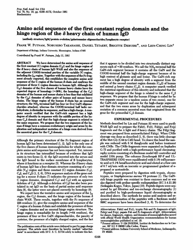
Intra-s vs V10JH VOC T- s r A - oylV95V- SG I Viol V105
Tyr-yr-Cs-Aa-Ar-GlyAsnPro-ro-ro-Tr-Ty-As-IleGlyThr-ly-Sr-Ap-As-GlyIleAsp-aln-pGGyy-ThGlr-h hr-r
V C
vioJH 'a
cI * C61 oC2V110 ~~~C1 C1O C20
Val-His-Val -Ser-Ser Ala-Pro-Thr-Lys-Al-a-Pro-Asp-Val-Phe-Pro-Ile-Ie-Ser-Gly-Cys-Arg-Hs-Pro-Lys-Asp-Asn-Ser-Pro-Val-Val-_% ILight
Chain
I C30 C40 C50Leu-Ala-Cys-Leu-Ile-Thr-Gly-Tyr-His-Pro-Thr-Ser-Val -Thr-Val -Thr-Trp-Tyr-Met-Gly-Thr-Gl n-Ser-Gl n-Pro-Gln-Arg-Thr-Phe-Pro-
a C60 C70 C80Gu-TIVe-Gln-Arg-Arg-Asp-Ser-Tyr-Tyr-Met-Thr-Ser-Ser-n-Leu-Ser-Thr-Pro-Leu-Gln-Gln-Trp-Arg-Gln-Gly-Glu-Tyr-Lys-Cys-Val-
csoCe - | Hinge IC90 C0
Va1-Gln-His-Thr-Ala-Ser-Lys-Ser-Lys-Lys-Glu-Ile-Phe Arg-Trp-Pro-Glu-Ser-Pro-Lys-Ala-Gln4Ala#Ser-Ser-Val -Pro-Thr-Ala-Gln-
P-0Fc(P) C1 Gs*' IP) 9)e3) C130 C140Pro-Gl n-Ala-Gl u:Gly-Ser-Leu-Ala-Lys-Ala-Th r-A a-Pro-Ala-Thr-T r-Arg-Asn-Thr-Gly-Arg-Gly-Gly-Glu-Gl u-Lys-Lys-Lys-Glu-
I Fclt)Hinge C62
C150 C160 C170Lys-Gl u-Lys-Gl u-Gl u-Gl n-Gl u-Glu-Arg-Glu-Thr-Lys Thr-Pro-Gl u-Cys-Pro Ser-H1s-Thr-Gt n-Pro-Leu-Gly-Val -Tyr-Leu-
Heavy__Chain SS
FIG. 1. Amino acid sequence of the CJ1 domain and the hinge region of the 8 heavy chain of IgD WAH and the COOH-terminal portion of theV region and the NH2 terminus of the Fc(t) fragment (1). Because V regions vary in length, several characteristic residues are designated V andto facilitate comparison are numbered according to the adjusted numbering system of Kabat et al. (11). Thus, Cys-V92 is the invariant second half-cystine in the intrachain disulfide bridge of all VH regions and Asp-V101 marks the beginning of the JH region (12-14). The C region residues aredesignated C and are numbered consecutively beginning with Ala-Cl. The hinge region has four (or five) GaiN oligosaccharides attached. Dashedvertical lines indicate multiple sites of cleavage of IgD by papain to yield 8 chain fragments called Fd(p). The tryptic Fc(t) fragment begins at Thr-C158 and has an interchain disulfide bond at Cys-C161 just before the start of the C,2 domain at Ser-C163.
Biochemistry: Putnam et aL
6170 Biochemistry: Putnam et al
Table 1. Number of amino acid residues and carbohydratecontent of the C regions of human heavy chains
CH Oligosaccharides, no.
domains, Residues, no. GalN GlcNChain no. Hinge* C region (hinge) (domains)8 3 64 383 4 or 5 3vl 3 15 329 0 1Y2 3 12 325 0 1y3 3 62 375 0 1al 3 26 353 5 2a2 3 13 340 0 4,5t
4 450 56$ 4 420 6
* Estimates of the length ofthe human hinge regions are approximate.Lengths given for the 'y3 and e chains are estimates because the se-quences are incomplete (21, 22).
t The A2m(1) allotype of the a2 chain has four GlcN oligosaccharides,and the A2m(2) allotype has five (17, 18).
*The ,u and E chains lack a hinge region but have an extra (fourth) CHdomain (19-21).
amino acid residues-i.e., to extend from Arg-C99 through Pro-C162. This needs verification by DNA sequence analysis ofthe8-chain gene.Comparison of the CHI Domains ofHuman Heavy Chains.
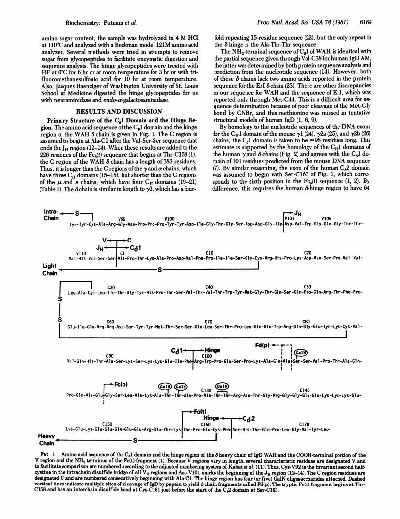
The amino acid sequence ofthe WAH Cd1 domain is comparedwith the sequences of the CHi domains of human AL (19, 20),yl (15), al (16), and e (21) heavy chains in Fig. 2. The CH1domains of the five human heavy chains have about the samedegree of homology when compared pairwise with each other(=z30%). In this alignment, 55 residues of the 8 chain are iden-tical to one or more residues at the same position in the otherfour chains. Only 10 positions are identical in all five chains; 9ofthese are located in the ( strands ofthe 13-pleated sheet struc-ture determined for IgGi, and the 10th is at the end of 1 strand4-2. Hence, these conserved positions probably constitute theframework of the three-dimensional structure of the CH1 do-main, which has the function ofinteraction with the CL domain
4-1 l
(X 1
E~tiElP31 DEDf P I I D NMP5GCLEuSA SP EPLv -D E SsHNur;SASAPTLFP L V S CEN S SST G
A -SV F PhAP-SS[S GGT A G
iLS] [I TS P KUi F iLSL-N S TQE DGCNVI1i!
UP | TrrA- -TI i
ofthe light chain. The degree ofidentity ofthe C81 domain withthe CH1 domain of other chains is A, 29%; yl, 32%; al, 33%;and E, 26%. This exceeds the degree ofidentity ofthe three CHdomains of the 8 chain for each other; in an alignment set tomaximize their similarity (not shown), C,81, C82, and C&3 exhibitonly 17-21% identity. Neither the results for C81 nor those forFc8 support the suggestion (3) that IgD is more closely relatedto IgE. than to the other three classes.
As in IgM, IgA, and IgE (but unlike IgG), the light chain ofIgD is linked by a disulfide bond to the first half-cystine in theCH1 domain (Cys-C15). Although ,., a2, and e have a GlcNcarbohydrate attached at a homologous position in the CH1 do-main, this is absent in 8, al, and v. This carbohydrate and oth-ers present in the CHI domains ofthe a and e chains are locatedin bends ofthe polypeptide chain between the 1 strands; hence,the CH1 domains of all five chains can probably assume similarconformations (the immunoglobulin fold) that enable all ofthemto pair in a combinatorial manner with both K and A light chains.However, some preference for A chains has been noted for IgD(3, 4).Homology of the C1 Domains of Human and Mouse 8
Chains. It is surprising that the C,1 domains of the human andmouse 8 chains have only -25% homology. In an alignment inwhich five gaps were inserted into the mouse 8 chain (7) toachieve maximum homology, only 24 positions of 95 comparedwere identical. This contrasts with the much greater homologyofother pairs ofhuman and mouse CHl1 domains-i.e., yl, 60%;1A, 48%; and al, 40%. Also, the mouse C,61 domain has threepossible sites for attachment ofGlcN to the signal sequence Asn-X-Thr/Ser, whereas none is present in human C8J. Because themouse 8 chain also appears to lack the C82 domain, the primarystructure predicted from the nucleic acid sequence should beverified by sequence analysis of the protein.
Characteristics of the Hinge Region. The hinge region is anunusual structure in the segment of the heavy chain that joinsthe Fd and Fc regions of immunoglobulin classes having onlythree CH domains (IgG, IgA, and IgD) but is absent in IgM andIgE, which have four CH domains (Table 1). The amino acid
[ 42 J
*
I lie H
analL
V iDY
V KJDlFv G 0F
T 11M- aY MHTA Q PQ
D LI T F - W K Y K N N D I S
0Q[0 S-VTl W N SiA T S G V
0 O ML S-V T S ES pEGVTTAHa2Mi i4 - 3 ] L 4 - 4 L//,L X//B / / 5/
H E I R D SM Y xM S-Jo K V H[9RA K VK I F
HSI|T RIPSIFILP|;|DI r rM Ip TA TL V C VD
-P nN G B K1 Q KDH-H-
Y1 H - F P A V L 0 S S - ML L SSV V T V P S S S L G T Y I - - C N V N K LP NN V DE R V
xl H1- - FH P SH N A IS|| L T TSQ A TN C L THKC- H K H 3POQDHTE-~-- ATT- TISG -HLY. ISLiTLJ 5--A( ASKOMLT-CS - ASHFBAi0 5F T NVEJT-
FIG. 2. Comparison of the amino acid sequences of the CHi domains of the five classes of human immunoglobulins. The one-letter notationfor amino acids is given in ref. 27. The boundaries of the CH1 domains are based on structural characteristics and comparison with the knownjunctional sequences of the exon for the CH1 domain of mouse by chains (24-26). Gaps have been inserted to maximize the homology. The threeinvariant cysteine (C) and tryptophan (W) residues in each domainmwere used to place the alignment in register, and they are indicated by arrows.Residues in the IA, y, a, or E sequences that share identity with the corresponding residues in the 8 chain are outlined by shaded boxes; residuesthat are identical in sequences other than the 8 chain are outlined in open boxes. The 13 strands (28) are numbered according to Edmundson et at.(29) with the four-stranded 3-sheet elements in open bars and the three-stranded 1-sheet elements in hatched bars.
-1I1.
I
I
II
Proc. Nad Acad. Sci. USA 78 (1981)
Biochemistry: Putnam et aL
sequence of the hinge region is unique for each class, differsmarkedly even for subclasses, and appears to be unrelated tothe rest of the chain; that is, the hinge region lacks the char-acteristic structural features of the immunoglobulin fold. Thefunction ofsegmented flexibility has been ascribed to the hinge,and this idea is supported by the fact that the hinge region isdisordered in the crystal structure (28). The hinge ofthe 8 chaindiffers from the hinges in 'yand a in four notable characteristics:(i) its length (-64 residues), which is about 4 times the lengthofthe yl, 'y2, and a2 hinges and twice that ofthe al hinge, theonly other one to contain carbohydrate (Table 1); (ii) its divisioninto two distinct segments (the GalN-rich NH2-terminal halfand the high-charge COOH-terminal half); (iii) its dominantcomposition and repetitive pattern (alanine and threonine in thefirst half and glutamic acid and lysine in the second half versushalf-cystine and proline in the yand a chains); and (iv) presenceof only one half-cystine.
Predicted Conformation of the Human IgD Hinge Region.The surprising division ofthe human 8 hinge into two dissimilarhalves led us to attempt to predict the conformation by the pro-cedure of Chou and Fasman (30). No clear result was obtainedfor the GalN-rich segment, which appears to have a randomstructure. The potential for both the P sheet and the a helix arelow because of the frequency of proline and serine. The fre-quency of GalN oligosaccharides probably contributes to theapparent disorder in the first part of the human IgD hinge. Incontrast, the high-charge region from Glu-C140 through Glu-C155 appears to form an ideal a-helical segment. Glutamic acidhas the highest conformational parameter for the a helix andthe lowest for ( sheet of any amino acid (30). Lysine is also fa-vorable for the a helix but not for the P sheet. The rigid highlycharged a helix may be separated from the GalN-rich segmentby a (3 turn induced by glycine in the sequence Gly-Arg-Gly-Gly and would be followed by the first (-sheet strand ofthe C.2domain. Because a-helical segments are rare in immunoglob-ulins, the presence ofone in human IgD is probably associatedwith specific biological function. Thus, its apparent absence inthe mouse IgD hinge is surprising.Number and Location of Oligosaccharides in the Hinge Re-
gion. The presence of four or five GalN-oligosaccharides in thehuman 8 hinge is of interest because of the rarity of GalN inimmunoglobulins, the high local concentration of GalN, theeffect this has on secondary structure and conformation, and itspossible biological significance. The human al hinge, which hasan octapeptide duplication, has five GalN-Gal disaccharides;one is attached in O-glycosidic linkage to each ofthe five serineswithin a sequence of only 17 amino acid residues (16), but thehuman a2 hinge lacks GalN because ofa deletion that includesall the threonine and serine residues (17, 18). In contrast to thehighly charged a-helical section of the 8 hinge, which is easilycleaved by proteolytic enzymes, the GalN-rich segment of 8
Human d Hinge R W P E S P K
Human Cp2 S W L R E G K
Dog CP2 W S L R D G K
Mouse CP2 SHL K D G K
Proc. NatL Acad. Sci. USA 78 (1981) 6171
(like the al hinge) was very resistant to all enzymes we tried.This impeded determination ofthe protein sequence and ofthenumber, location, and structure of the GalN oligosaccharides.Our attempts to remove the carbohydrate from hinge glyco-peptides by chemical or enzymatic treatment also were onlypartly successful. However, the partially deglycosylated gly-copeptides were more amenable to purification, digestion, andsequence determination.The tentative location of four or five GalN oligosaccharides
was assigned by difference in the GalN content of various sub-peptides and by the failure to detect serine or threonine at agiven step in the operation of the protein sequencer. This ap-proach allowed assignment of one GalN to Ser-C109 and oneto each of the threonines in the first Ala-Thr-Thr sequence. Itis uncertain whether the second Ala-Thr-Thr sequence hasGalN on both threonines or only one.
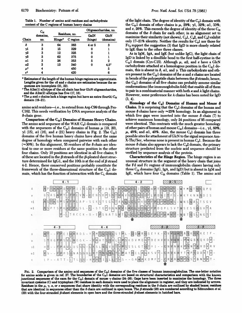
Origin ofthe 8-Chain Hinge. Although the boundaries ofthehinge region were originally based on structural characteristics,it is now known that, in the mouse y-chain subclasses, the hingeregion is precisely defined by a DNAcoding segment (exon) thatis separated by intervening noncodingDNA sequences (introns)from the exons that code for the CH1 and CH2 domains (24-26).The evolutionary origin ofthe hinge is unknown, and the hingeis the most mutable region in immunoglobulin chains, as shownby the frequency of deletions and duplications. These facts andthe distinguishing characteristics of the 8 hinge caused us tosearch for possible regions ofhomology in other classes ofheavychains. Because theA chain is thought to be the prototype heavychain and lacks a hinge but has an extra domain (CM,2), wesearched that domain for possible homology to the 8 hinge, withthe result shown in Fig. 3. In the GalN-rich segment of thehuman 8 hinge, 12 of36 successive residues are identical to thesequence shown from the middle of the CH2 region of the hu-man ti chain except for uncertainty at Gln-C117 and Glu-C119.The same positions are identical in the dog ,u chain; seven ofthem also are identical in the mouse ,u chain. It is unlikely thata 33% identity in sequence occurs by chance in two unrelatedproteins. To ascertain its significance, we asked Winona C.Barker of the National Biomedical Research Foundation tomake a computer search for similarity to the entire collectionofprotein sequences ofthe Atlas ofProtein Sequence and Struc-ture (33). Using the unitary matrix that scores only identities,the search program (34) examined a segment of 30 residues inthe 8 hinge (see Fig. 3) and made 179,748 comparisons. Thehighest score (10 identities) was registered for the 30-residuesegment of the human and dog At chains shown in Fig. 3. (Asegment of the (8 chain of rabbit tropomyosin where the se-quence is proposed but is incomplete also gave a score of 10.)A score of 9 was given for the mouse 8-chain hinge and also forsegments from eight other proteins. However, by insertingsome gaps in the human and mouse 8 hinges to maximize the
r- - --- -.--I
A Q AS S V P T A Q P Q A E G S L A K A T T A P A T T R N T
Q V G S G V T T D E V E A E A K E S G P T T Y K V T S T L T
Q I E S G V T T N E V Z A Z A K Z S G P T T Y K V T S M L T
LVE jG F T DP VTIdJNKGSTPQ jYKVIST L
G R G G E E
I K E S D W
I Q E D A W
I S E I D W
L.-.._ _ _ _ _ _ _ _ _ _ _ _ _ _ _ _ _ _ _ _ _ _
FIG. 3. Identities in amino acid sequence of the GaIN-rich segment of the hinge region of the human 8 chain and the C*2 domain of human(19, 20), dog (31), and mouse (32) ,u chains. Positions having residues identical to those in the 8 chain are enclosed in solid boxes. Sequences alignedas having highest similarity by a computer search using a unitary matrix that scores only identities but does not differentiate Q, E, and Z (33, 34)are enclosed by dashed lines. The human ,z chain sequence begins at position 270 and has been updated.
6172 Biochemistry: Putnam et al
r .-- n
L . - A--_ I
Human R W P E S P K A Q A S - V P T A Q P Q A E G -S L A K A T T A P A T T R N TUG R G
Mouse S - D HT-LJ-jKR L - A K N H SET E A-I-A TT K K D IE u
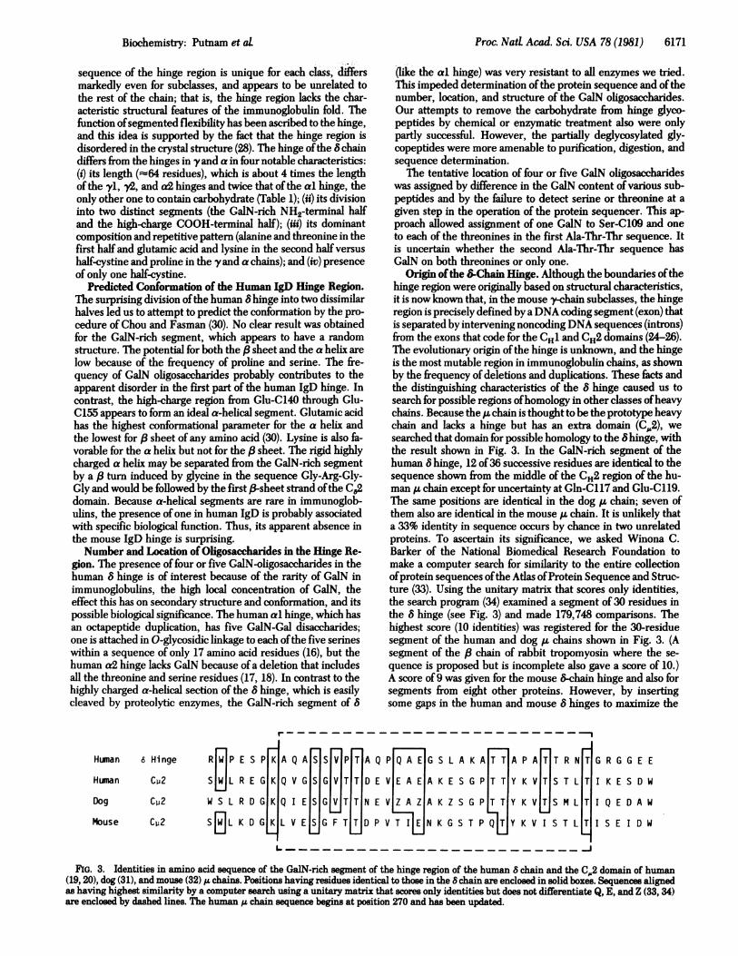
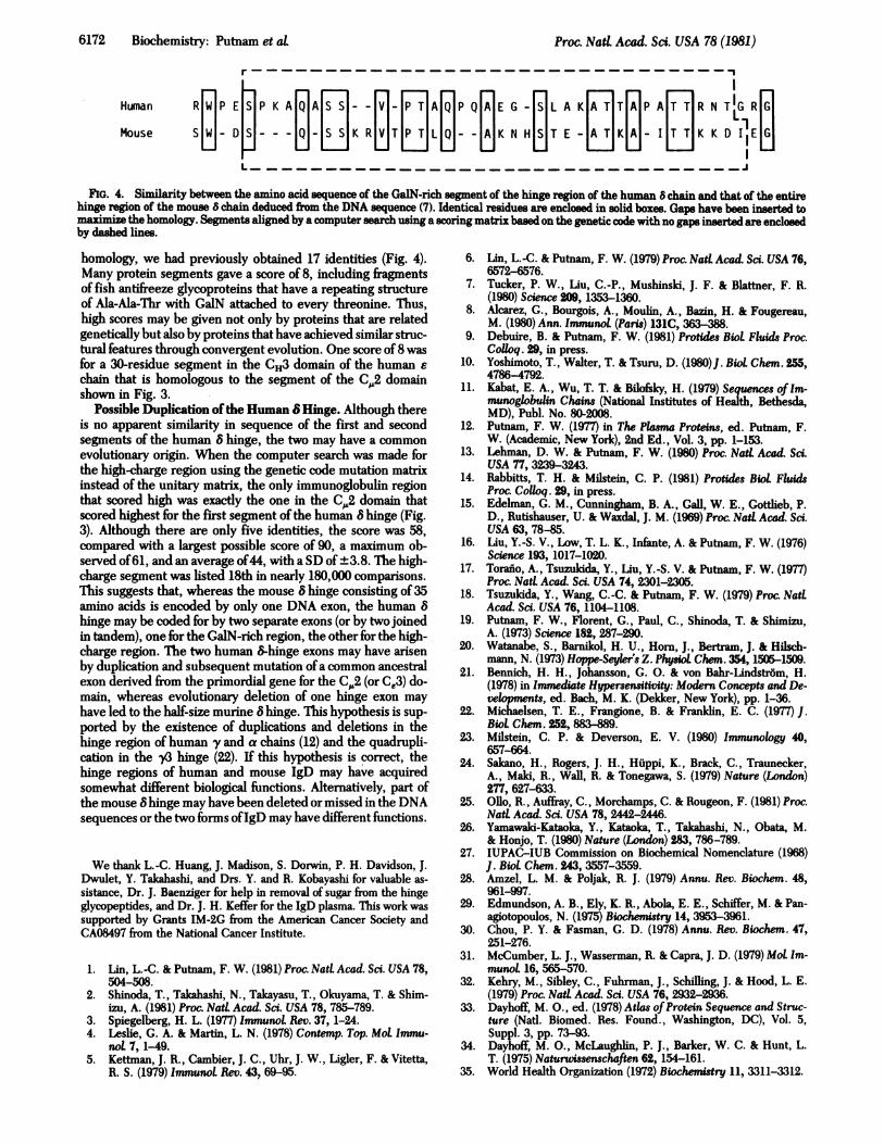
FIG. 4. Similarity between the amino acid sequence of the GaIN-rich segment of the hinge region of the human 8 chain and that of the entirehinge region of the mouse 8 chain deduced from the DNA sequence (7). Identical residues are enclosed in solid boxes. Gaps have been inserted tomaximize the homology. Segments aligned by a computer search using a scoring matrix based on the genetic code with no gaps inserted are enclosedby dashed lines.
homology, we had previously obtained 17 identities (Fig. 4).Many protein segments gave a score of 8, including fragmentsof fish antifreeze glycoproteins that have a repeating structureof Ala-Ala-Thr with GaiN attached to every threonine. Thus,high scores may be given not only by proteins that are relatedgenetically but also by proteins that have achieved similar struc-tural features through convergent evolution. One score of 8 wasfor a 30-residue segment in the CH3 domain of the human echain that is homologous to the segment of the C,,2 domainshown in Fig. 3.
Possible Duplication ofthe Human 6 Hinge. Although thereis no apparent similarity in sequence of the first and secondsegments of the human 8 hinge, the two may have a commonevolutionary origin. When the computer search was made forthe high-charge region using the genetic code mutation matrixinstead of the unitary matrix, the only immunoglobulin regionthat scored high was exactly the one in the CA2 domain thatscored highest for the first segment of the human 8 hinge (Fig.3). Although there are only five identities, the score was 58,compared with a largest possible score of 90, a maximum ob-served of61, and an average of44, with a SD of ±3.8. The high-charge segment was listed 18th in nearly 180,000 comparisons.This suggests that, whereas the mouse 8 hinge consisting of 35amino acids is encoded by only one DNA exon, the human 8hinge may be coded for by two separate exons (or by two joinedin tandem), one for the GalN-rich region, the other for the high-charge region. The two human 8-hinge exons may have arisenby duplication and subsequent mutation ofa common ancestralexon derived from the primordial gene for the C,.2 (or Ce3) do-main, whereas evolutionary deletion of one hinge exon mayhave led to the half-size murine 8 hinge. This hypothesis is sup-ported by the existence of duplications and deletions in thehinge region of human y and a chains (12) and the quadrupli-cation in the y3 hinge (22). If this hypothesis is correct, thehinge regions of human and mouse IgD may have acquiredsomewhat different biological functions. Alternatively, part ofthe mouse 8 hinge may have been deleted or missed in the DNAsequences or the two forms ofIgD may have different functions..
We thank L.-C. Huang, J. Madison, S. Dorwin, P. H. Davidson, J.Dwulet, Y. Takahashi, and Drs. Y. and R. Kobayashi for valuable as-sistance, Dr. J. Baenziger for help in removal of sugar from the hingeglycopeptides, and Dr. J. H. Keffer for the IgD plasma. This work wassupported by Grants IM-2G from the American Cancer Society andCA08497 from the National Cancer Institute.
1. UIn, L.-C. & Putnam, F. W. (1981) Proc. NatL Acad. Sci. USA 78,504-508.
2. Shinoda, T., Takahashi, N., Takayasu, T., Okuyama, T. & Shim-izu, A. (1981) Proc. Natl Acad. Sri. USA 78, 785-789.
3. Spiegelberg, H. L. (1977) ImmunoL Rev. 37, 1-24.4. Leslie, G. A. & Martin, L. N. (1978) Contemp. Top. Mot Immu-
nol. 7, 1-49.5. Kettman, J. R., Cambier, J. C., Uhr, J. W., Ligler, F. & Vitetta,
R. S. (1979) ImmunoL Rev. 43, 69-95.
6. Lin, L.-C. & Putnam, F. W. (1979) Proc. Natl Acad. Sc. USA 76,6572-6576.
7. Tucker, P. W., Liu, C.-P., Mushinski, J. F. & Blattner, F. R.(1980) Science 209, 1353-1360.
8. Alcarez, G., Bourgois, A., Moulin, A., Bazin, H. & Fougereau,M. (1980) Ann. Immunol (Paris) 131C, 363-388.
9. Debuire, B. & Putnam, F. W. (1981) Protides Biol. Fluids Proc.Colloq. 29, in press.
10. Yoshimoto, T., Walter, T. & Tsuru, D. (1980)J. Biol Chem. 255,4786-4792.
11. Kabat, E. A., Wu, T. T. & Bilofsky, H. (1979) Sequences ofIm-munoglobulin Chains (National Institutes of Health, Bethesda,MD), Publ. No. 80-2008.
12. Putnam, F. W. (1977) in The Plasma Proteins, ed. Putnam, F.W. (Academic, New York), 2nd Ed., Vol. 3, pp. 1-153.
13. Lehman, D. W. & Putnam, F. W. (1980) Proc. NatL Acad. Sci.USA 77, 3239-3243.
14. Rabbitts, T. H. & Milstein, C. P. (1981) Protides Biol FluidsProc. Colloq. 29, in press.
15. Edelman, G. M., Cunningham, B. A., Gall, W. E., Gottlieb, P.D., Rutishauser, U. & Waxdal, J. M. (1969) Proc. Natl Acad. Sci.USA 63, 78-85.
16. Liu, Y.-S. V., Low, T. L. K., Infante, A. & Putnam, F. W. (1976)Science 193, 1017-1020.
17. Torafo, A., Tsuzukida, Y., Liu, Y.-S. V. & Putnam, F. W. (1977)Proc. Natl Acad. Sci. USA 74, 2301-2305.
18. Tsuzukida, Y., Wang, C.-C. & Putnam, F. W. (1979) Proc. NatLAcad. Sci. USA 76,1104-1108.
19. Putnam, F. W., Florent, G., Paul, C., Shinoda, T. & Shimizu,A. (1973) Science 182, 287-290.
20. Watanabe, S., Barnikol, H. U., Horn, J., Bertram, J. & Hilsch-mann, N. (1973) Hoppe-Seylers Z. Physiol Chenm. 354, 1505-1509.
21. Bennich, H. H., Johansson, G. 0. & von Bahr-Lindstr6m, H.(1978) in Immediate Hypersensitivity: Modern Concepts and De-velopments, ed. Bach, M. K. (Dekker, New York), pp. 1-36.
22. Michaelsen, T. E., Frangione, B. & Franklin, E. C. (1977) J.Biol. Chem. 252, 883889.
23. Milstein, C. P. & Deverson, E. V. (1980) Immunology 40,657-664.
24. Sakano, H., Rogers, J. H., Htippi, K., Brack, C., Traunecker,A., Maki, R., Wall, R. & Tonegawa, S. (1979) Nature (London)277, 627-633.
25. Ollo, R., Auffray, C., Morchamps, C. & Rougeon, F. (1981) Proc.Natl Acad. Sri. USA 78, 2442-2446.
26. Yamawaki-Kataoka, Y., Kataoka, T., Takahashi, N., Obata, M.& Honjo, T. (1980) Nature (London) 283, 786-789.
27. IUPAC-IUB Commission on Biochemical Nomenclature (1968)J. Bwl. Chem. 243, 3557-3559.
28. Amzel, L. M. & Poijak, R. J. (1979) Annu. Rev. Biochem. 48,961-997.
29. Edmundson, A. B., Ely, K. R., Abola, E. E., Schiffer, M. & Pan-agiotopoulos, N. (1975) Biochemistry 14, 3953-3961.
30. Chou, P. Y. & Fasman, G. D. (1978) Annu. Rev. Bsochem. 47,251-276.
31. McCumber, L. J., Wasserman, R. & Capra, J. D. (1979) Mol. Im-munol 16, 565-570.
32. Kehry, M., Sibley, C., Fuhrman, J., Schilling, J. & Hood, L. E.(1979) Proc. Natl. Acad. Sci. USA 76, 2932-2936.
33. Dayhoff, M. O., ed. (1978) Atlas ofProtein Sequence and Struc-ture (Natl. Biomed. Res. Found., Washington, DC), Vol. 5,Suppl. 3, pp. 73-93.
34. Dayhoff, M. O., McLaughlin, P. J., Barker, W. C. & Hunt, L.T. (1975) Naturwissenschaften 62, 154-161.
35. World Health Organization (1972) Biochemistry 11, 3311-3312.
Proc. Nad Acad. Sci. USA 78 (1981)