cerebellum - univerzita karlova · the cerebellum –relations and structure located in the...
TRANSCRIPT

CEREBELLUM
Institute of Anatomy, Second Faculty of
Medicine
R. Druga


Cerebellar folia
Cortex, subcortical white
matter, nuclei
Excessive folding of the cerebellar surface
(cortex)

The cerebellum – relations and structure
Located in the posterior cranialfossa
Connected with the brain stem-by peduncles (inferior, middle, superior)
Is covered by the cerebellarcortex (3 layers). Cortex isextensively folded (folia-orientedmediolaterally)
In the white matter are thecerebellar nuclei
Fossa cranii posterior
Pedunculi cerebellares (inferior, medius,
superior)
Cortex – 3 vrstvy
V bílé hmotě mozečková jádra

MR
examination
Posterior cranial
fossa
MR vyšetření
Fossa cranii
posterior
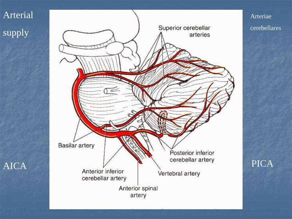
Arterial
supply
AICA PICA
Arteriae
cerebellares

Weigert
staining

Marchi
Staining
Kluver –
Barrera
Marchiho
metoda
Kluver - Barrera

Unfolded surface
of the cerebellum
Dvourozněrná
rekonstrukce
povrchu (kůry)
Mozečku
Two-dimensional
reconstruction of the
cerebellar surface
Length x width


Dvourozměrná
Rekonstrukce
lalůčků mozečku
Dvourozměrní
rekonstrukce
lalůčků mozečku
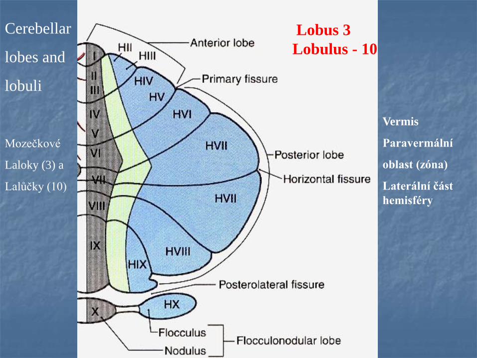
Cerebellar
lobes and
lobuli
Lobus 3
Lobulus - 10
Mozečkové
Laloky (3) a
Lalůčky (10)
Vermis
Paravermální
oblast (zóna)
Laterální část
hemisféry


CEREBELLAR NUCLEI, neurons
glutamatergic, excitatory, high
spontaneous activity
Mozečková jádra
Neurony glutamátergní (excitační), vysoká
spontánní aktivita

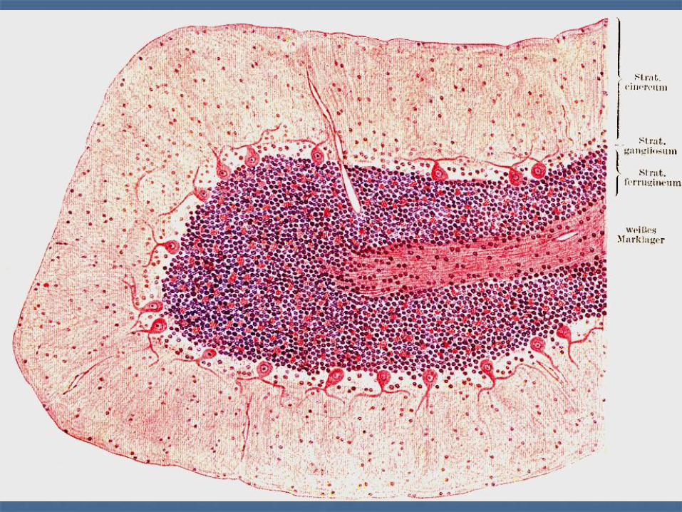
Structure of the cerebellar cortex -3 layers:
I.molecular layer - inhibitory interneuronsI.Molekulární vrstva – inhibiční interneurony
II. Purkyně cell layer – inhibitory projecting neuronsII. Vrstva Purkyňových buněk (stratum purkinjese)– inhibiční, projekční neurony
III. granular layer - prevail excitatory neurons
III. Vrstva granulárních buněk – převaha excitačních neuronů
Mozečková kůra – 3 vrstvy

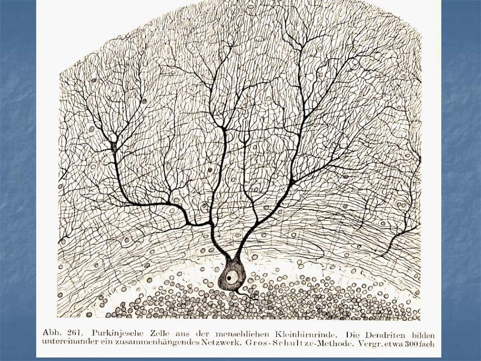
J.E. Purkyně 1837
1832 první mikroskop
Univerzita ve Vratislavi
Prof. Heidenheim :
………“ hatte Purkyně den
charakteristischen,
allgemainen Unterschied
zwischen Tier und
Pflanzenzellen zun erstmale
mit vollkommener Klarheit
erkannt „

R. y Cajal : Histologie du systeme nerveux, 1911.

Santiago Ramon y Cajal 1911


Basket cells, inhibitory interneurons,
GABAergicKošíčkové buňky,
inhibiční interneurony


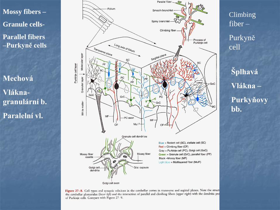
Mossy fibers –
Granule cells-
Parallel fibers
–Purkyně cells
Climbing
fiber –
Purkyně
cell
Mechová
Vlákna-
granulární b.
Paralelní vl.
Šplhavá
Vlákna –
Purkyňovy
bb.
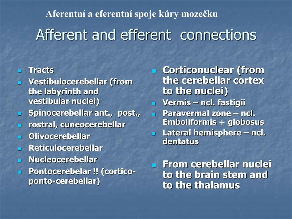
Afferent and efferent connections
Tracts
Vestibulocerebellar (from the labyrinth and vestibular nuclei)
Spinocerebellar ant., post.,
rostral, cuneocerebellar
Olivocerebellar
Reticulocerebellar
Nucleocerebellar
Pontocerebelar !! (cortico-ponto-cerebellar)
Corticonuclear (fromthe cerebellar cortexto the nuclei)
Vermis – ncl. fastigii
Paravermal zone – ncl. Emboliformis + globosus
Lateral hemisphere – ncl. dentatus
From cerebellar nucleito the brain stem and to the thalamus
Aferentní a eferentní spoje kůry mozečku
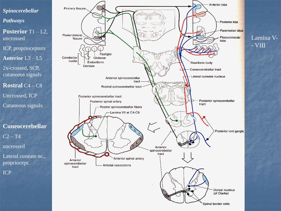
Spinocerebellar
Pathways
Posterior T1 – L2,
uncrossed
ICP, proprioceptors
Anterior L3 – L5
2x-crossed, SCP,
cutaneous signals
Rostral C4 – C8
Uncrossed, ICP
Cutaneous signals
Cuneocerebellar
C2 – T4
uncrossed
Lateral cuneate nc.,
propriocept.
ICP
Lamina V-
- VIII

Dorsal (posterior) spinocerebellar
projection- uncrossed
Ventral (anterior) spinocerebellar
projection - crossed
a b
Origin of the spinocerebellar pathways
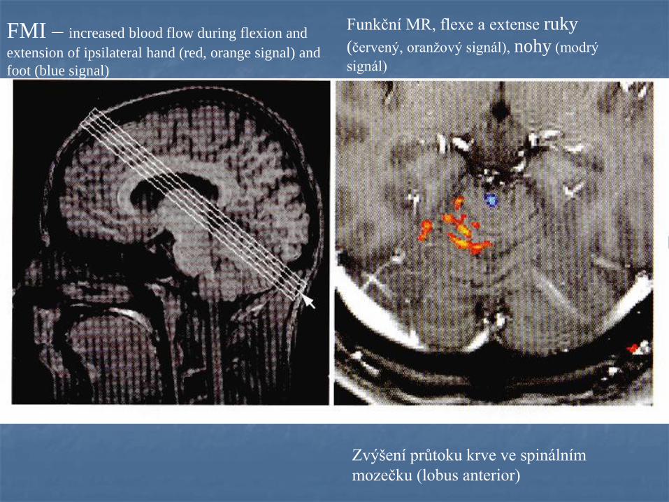
FMI – increased blood flow during flexion and
extension of ipsilateral hand (red, orange signal) and
foot (blue signal)
Funkční MR, flexe a extense ruky
(červený, oranžový signál), nohy (modrý
signál)
Zvýšení průtoku krve ve spinálním
mozečku (lobus anterior)


Afferent
projections
Aferentní
Spoje
Mossy
fibers
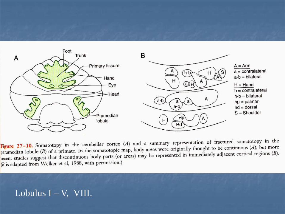
Lobulus I – V, VIII.

AFFERENTS TO THE CEREBELLAR CORTEX I.
Climbing fibers (Šplhavá vlákna)
– inferior olive (each P.cell receives only 1 c.f., many synapses with P.c.), excitatory (glutamate), firing frequencyof the c.f. is very low (1 impulse/sec), c.f. elicit burst ofaction potentials in the P.c.
C.f. inform about errors in the execution of movements –error indicators !!

Climbing
Fibers
Olivo-
cerebellar
projections
(crossed)
Šplhavá vlákna
Olivocerebelární
Projekce
(zkřížená)

Olivocerebellar
projection
Climbing fibers
Šplhavá
vlákna

AFFERENTS TO THE CEREBELLAR CORTEX II
Mossy fibers (Mechová vlákna) - spinal cord, RF, pontine
nuclei, ncll. of cranial nerves.
End in the granular layer and each of which contacts large numberof granular neurons. Granular cell axon contacts large number of P. c. via parallel fibers.
Mossy fibers are excitatory (glutamate).
Each mossy fiber influences many P.c. but the excitatory effect isweak. Many mossy fibers must be active together to providesufficient excitation to fire a P.c.
Mossy fibers provide precisely graded information aboutmovements, skin stimulations, joint position and aboutmotor comands issued from the cerebral cortex.

Cortico-pontine
pathway, 17 millions
fibers
Neocortex –
ipsilat. pontine ncll. -
pontocerebellar
pathway –
contralateral cerebellar
cortex (mossy fibers)
Pontocerebellar fibers =
largest contingent of
mossy fibers
Kortiko-pontinní dráha
17 milionů vláken
Neokortex – ipsilaterální
pontinní jádra
Pontocerebelární dráha –
Kontralaterální
mozečková kůra (mechová
vlákna)

Cortico – ponto – cerebellar pathway
Cortico-ponto-cerebellar pathway
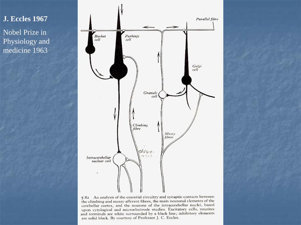
J. Eccles 1967
Nobel Prize in
Physiology and
medicine 1963


Efferent connections of the cerebellar cortexEferentní spoje mozečkové kůry
Cerebellar cortex – cerebellar nucleivermis – ncl. fastigii, ncll. vestibulares
pavermal zone – ncl. emboliformis, globosus. lateral hemisphere – ncl. dentatus
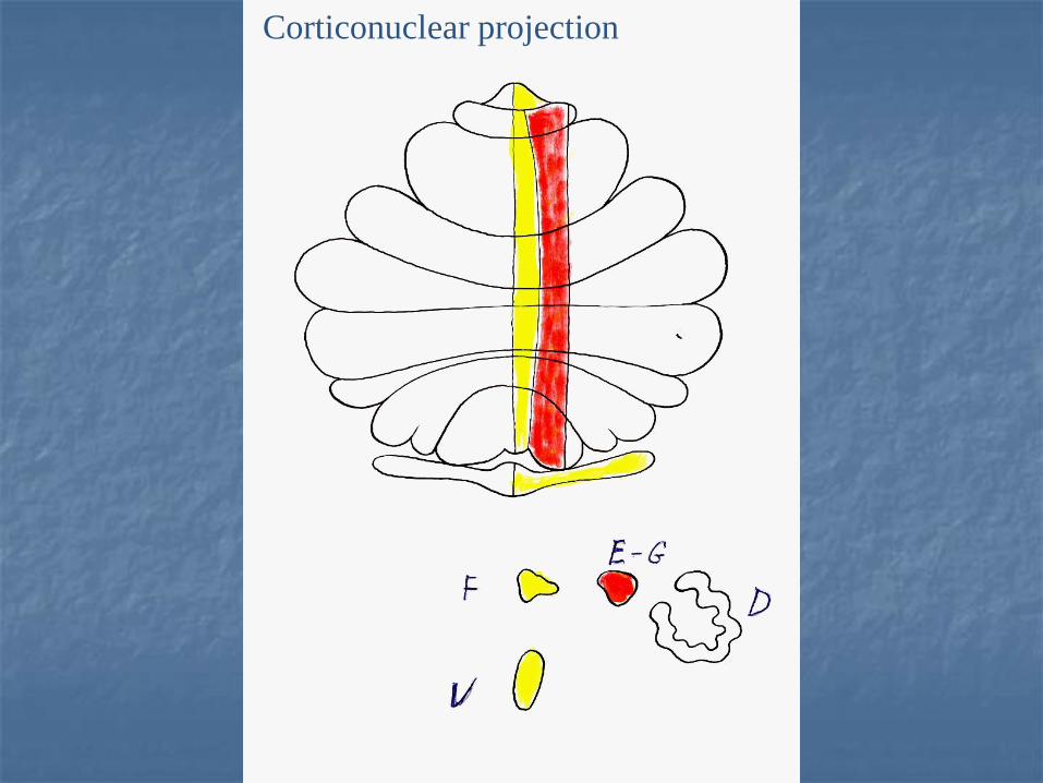
Corticonuclear projection


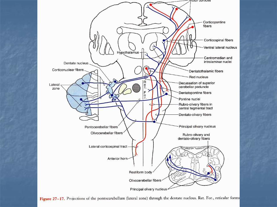
Efferent connections of the cerebellar nucleiEferentní spoje mozečkových jader
Fastigial nucleus – vestibular nuclei, reticular formation
Emboliformis + globosus nucleus - reticular formation, ncl. ruber, thalamus
Nucleus dentatus – ncl. ruber, contralateral thalamus (ventrolateral nucleus, intralaminar thalamic nuclei, ventralanterior nc.,
Ventrolateral nucleus (VL) – primary motor cortex(area 4)


Thalamus
Ncl. Ventralis
Lateralis
Ncl. VL –
Motor cortex

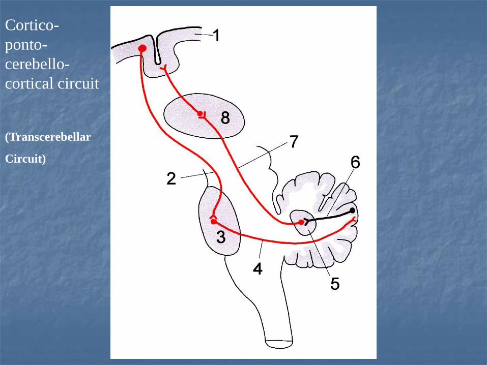
Cortico-
ponto-
cerebello-
cortical circuit
(Transcerebellar
Circuit)
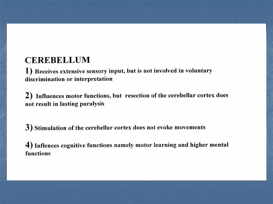
Mozeček
Přijímá sensitivní signály, ale neúčastní se volní diskriminace, nebo interpretace
Ovlivňuje motorické funkce, ale resekce mozečku nevyvolává obrny
Stimulace mozečku nevyvolává pohyb
Ovlivňuje kognitivní funkce, zejména motorické učení a vyšší mentální funkce (uvažování, plánování)

Mozečkové syndromy
Vestibulární mozeček a vermis (zejména v lobus anterior) – poruchy rovnováhy, stoje a chůze, chůze o široké bazi, nystagmus
Spinální mozeček – kontroluje axiální svalstvo a proximální svaly končetin. Při poškození zvýšení tonu extensorů.
Pontinní mozeček (hemisféry) - přestřelování pohybů (hypermetrie, prst – lalůček, prst- špička nosu). Adiadochokinéza, třes(méně než 5 Hz- zhoršuje se na konci zacíleného pohybu), poruchy řeči a výslovnosti (dysartrie, skandovaná řeč), poruchy plánování, paměti, uvažování (kognitivní poruchy).
Cerebellární kognitivně - afektivní syndrom (1998) –vermis, lobus posterior

Paleocerebellar lesions
(syndrome)
equilibrium

Neocerebellar syndrome (lesion of the hemisphere)

Cerebellar - cognitive affective syndrome
Schmahmann and Sherman (1998) in patientswith cerebellar lesions described CCAS
Executive dysfunction (disturbances in planning, abstract reasoning, memory)
Language symptoms (agrammatisms)
Behavior – affective disturbances (blunting ofaffect, disinhibited and inappropriate behavior
Lesions of the cerebellum interruptcommunication with the motor systems, association cortex.
Psychiatric disorders result from midline vermislesions (communication with the limbic system)

THE END
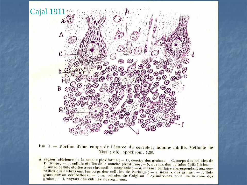
Cajal 1911