characterization of normal and degenerated human …
TRANSCRIPT
University of Szeged
Faculty of Medicine
Department of Orthopedics
Head: Prof. Dr. Habil. Kálmán Tóth
PhD thesis
CHARACTERIZATION OF NORMAL AND
DEGENERATED HUMAN HYALINE CARTILAGE WITH
THERMAL ANALYSIS
By
Dr. Gellért Sohár
Supervisor:
Prof. Dr. Habil. Kálmán Tóth
Szeged
2009
CONTENTS
PUBLICATIONS 1
1) ABBREVIATIONS 3
2) INTRODUCTION 4
3) CONCEPTUAL BACKGROUND 63.1Epidemiology 63.2 Osteoarthritis 63.3 Avascular Necrosis 83.4 Anatomy 93.5 Pathology 103.6 Biomechanics 123.7 Thermal analysis 12
3.7.1 Thermogravimetry 133.7.2 Calorimetry 14
4) AIMS OF THE INVESTIGATIONS 17
5) PATIENTS AND METHODS 185.1 Patients 185.2 Patients grading 185.3 Sample peparation for thermal analysis 195.4 Thermal mesurements 205.5 Statistical analysis 215.6 Ethics 21
6) RESULTS 226.1 Thermogravimetry 226.2 Calorimetry 26
7) DISCUSSION 29
8) SUMMARY AND CONCLUSIONS 34
9) REFERENCES 36
10) ACKNOWLEDGEMENTS 45

1
PUBLICATIONS
List of full papers directly related to the subject of the Thesis
1) Sohár G, Pallagi E, Szabó-Révész P, Tóth K: New Thermogravimetric protocolfor the investigation of normal and damaged human hyaline cartilage. J ThermAnal and Calorimetry 2007; 89(3): 853-56. CIT: 13 IF: 1.483
2) Tóth K, Sohár G, Pallagi E, Szabó-Révész P: Further characterization ofdegenerated human cartilage with differential scanning calorimetry.Thermochimica Acta 2007; 464: 75-77. CIT: 11 IF: 1.562
List of abstracts directly related to the subject of the Thesis
1) Sohár G, Tóth K, Pallagi E, Szabó-Révész P: Thermogravimetric investigation ofnormal and damaged human hyaline cartilage. Osteoarthritis and Cartilage 2006;Vol. 14, Supplement B S109-110. IF: 4.017
2) Sohár G, Aigner Z, Pallagi E, Szabó-Révész P, Tóth K: Complex thermalproperties of human cartilage in grade 4 osteoarthritis. Osteoarthritis andCartilage 2007; 15: Supplement C205-206. IF: 3.793
3) Sohár G, Aigner Z, Szabó-Révész P, Tóth K: Comparing physical properties ofosteoarthritic and necrotic human hyaline cartilage. Osteoarthritis and Cartilage2008; 16(4):196-97. IF: 3.793
4) Sohár G, Mécs L, Wellinger K, Aigner Z, Szabó-Révész P, Tóth K:Physicochemical transformations during thermal degradation of osteoarthritc andrheumatoid hyaline cartilage. 2009; Osteoarthritis and Cartilage Vol 17,Supplement 1. S249. IF: 3.793
List of papers and abstracts not related to the subject of the Thesis
1) Altorjay A, Juhasz A, Kellner V, Sohar G, Fekete M, and Sohar I Metabolicchanges in the lower esophageal sphincter influencing the result of anti-refluxsurgical interventions in chronic gastroesophageal reflux disease. World JGastroenterol 2005; 11(11): 1623-28. CIT: 3 IF: 2.081
2) Sohár G, Anna P, Kopasz N, Tajti L, Meszáros T, Tóth K: Clinical results ofscreening and management of hip dysplasia at our Department. Hip International2006; Vol. 16, 2, pp. 159. IF: 0.19
3) Kopasz N, Sisák K, Sohár G, Mészáros T, Tóth K: McCune-Albright Syndromeand spongious bone plasty of right iliac bone: Case report. Hip International2006; Vol. 16, 2006, pp. 175. IF: 0.19
2
4) Tóth K, Barna I, Nagy G, Kocsis Á, Bender Sr T, Sohár G: Level of paincompared to synoval beta-endorfphin level in osteoarthrotis and femoral headnecrosis. Osteoarthritis and Cartilage 2007; 15: Supplement C204. IF: 3.793
5) Mécs L, Sohár G, Tóth K: Development of spine osteoarthritis after posteriorlumbar spine interbody fusion. Osteoarthritis and Cartilage 2008; 16(4):235-36.
IF: 3.793
6) Tóth K, Sohár G, Aigner Z, Greksa F, Szabó-Révész P: Novel Calorimetricproperties of human cartilagesamples in rheumatoid arthritis. J Thermal Anal andCalorimetry 2009; 95(3): 813–15. IF: 1.483
7) Aigner Z, Mécs L, Sohár G, Wellinger K, Szabó-Révész P, Tóth K: Novelcalorimetric investigation of different degenerative disorders of the human hyalinecartilage. J Thermal Anal and Calorimetry 2009; 95(3):801-804. IF: 1.483
8) Csotye J, Aigner Z, Sohár G, Szabó-Révész P, Tóth K: Calorimetric properties ofdegenerative human shoulder joint hyaline cartilage. J Thermal Anal andCalorimetry 2009; 95(3): 805-808. IF: 1.483
9) Mécs L, Aigner Z, Sohár G, Szabó-Révész P, Tóth K: Characterization of humancartilage in degenerated spine disease with differntial scanning calorimetry. JThermal Anal and Calorimetry 2009; 95(3):809–811. IF: 1.483
10) Greksa F, Szabó A, Wellinger K, Sohár G, Tóth K: The effects of endomedullarimplants on the periosteal vessel structures of the rat tibia Osteoarthritis andCartilage 2009; Vol 17, Supplement 1. S93. IF: 3.793
11) Tóth K, Wellinger K, Sohár G, Bender T, Mécs L: Comparison of synovial -endorphin level in avascular necrosis, rheumatoid arthritis and osteoarthritis of thefemoral head and knee. Osteoarthritis and Cartilage 2009, Vol 17, Supplement 1.S253-254. IF: 3.793
12) Mécs L, Wellinger K, Sohár G, Tóth K: Adjacent segment lumbar facet jointosteoarthritis and intervertebral disc degeneration after posterior lumbar interbodyfusion. Osteoarthritis and Cartilage 2009; Vol 17, Supplement 1. S289. IF: 3.793
13) Tóth K, Wellinger K, Greksa F, Mécs L, Sohár G: New bone preservingtreatment option for young patients with hip osteoarthritis. Osteoarthritis andCartilage 2009 Vol 17, Supplement 1. S290. IF: 3.793
14) Sohár G, Fiszter I, Kovács T, Tóth K: Egy év alatt végzett szelektív ultrahanggalkiegészített csípıficam szőréseink eredményeinek utánkövetése Magyar
Traumatológia, Ortopédia Kézsebészet Plasztikai Sebészet (in press)
3
1. ABBREVIATIONS
AVN avascular necrosis
DMOAD disease-modifying osteoarthritis drug
DSC differential scanning calorimetry
DTA differential thermal analysis
DTG derivative thermogravimetry
∆H enthalpy change
IL-1 interleukin 1
MMP matrix–metalloproteinas
OA osteoarthritis
OARSI Osteoarthritis Research Society International
T Temperature
TG thermogravimetry
TGA thermogravimetric analysis
TNF-α tumor necrosis factor alpha
4
2. INTRODUCTION
Osteoarthritis (OA), the most prevalent joint disease, is characterized by the
progressive loss of articular cartilage that leads to chronic pain and functional restrictions
in affected joints [1-4]. The prior notion of OA as a bland disease related to aging and
‘‘wear and tear’’ of the joint has given way to views of a dynamic system with multiple
pathogenic contributors. Traditional views of articular cartilage failure have centered on a
variety of genetic [5-7], metabolic [2, 8, 9], and biochemical [10-13] factors. Recent
studies has elucidated the importance of local factors [14, 15] as well as crystals [2, 16,
17] and inflammation [2, 18, 19] in contributing to disease progression. The new
paradigm of OA considers it a heterogenous disease with numerous factors leading to its
pathologic hallmark of cartilage loss and the clinical manifestation of joint pain with
movement [3].
Osteoarthritis represents a major therapeutic challenge to medical and health-care
providers. In part, this is because osteoarthritis is a chronic condition in which symptoms
evolve over long periods of time and in which symptomatic episodes are frequently
separated by lengthy asymptomatic periods. It is likely, however, that alterations in joint
structure and function continue during these relative periods of clinical quiescence. In
addition, limited tools are available for the assessment of the progression of structural
changes in joint tissues in association with the progression of osteoarthritis and,
importantly, there is not a good correlation between structural alterations and symptoms.
Another major challenge with respect to definition of the pathogenic mechanisms
associated with the initiation and progression of osteoarthritis is the evidence that
osteoarthritis is not a homogeneous disorder. The underlying pathogenic mechanisms
differ among individuals and, even in the same individual, the pathologic processes and
etiologic mechanisms may differ at specific stages of disease progression. In recent years,
much has been learned regarding the specific risk factors that influence the natural history
of osteoarthritis. In general, these include genetic factors, the influence of aging, a history
of prior injury, abnormal joint mechanics and malalignment, and the presence of
inflammation [2].
Over the past years, the pathophysiology of avascularnecrosis (AVN) of the
femoral head has not been completely elucidated. Whereas some cases of the disease
clearly have a direct cause (trauma, radiation, or Caisson disease), the pathophysiology is
5
uncertain for most cases. Multiple investigators have postulated vascular impairment,
altered bone-cell physiology, and other theories [20-23].
An increasing number of publications have been published with the use of
calorimetric techniques in the examination of human hyaline cartilage. Previously,
thermoanalytical studies were used for the investigation of normal and degenerative
human hyaline cartilage. The first paper from this field was the study of P. Than et al.
[24]. They have concluded that structural manifestation of osteoarthritis appears as a
remarkable change of thermal stability of hyaline cartilage samples. The healthy cartilage
samples used in these studies were of cadaver origin as waste material, pathological
cartilage was derived as intraoperative tissue fragments. The samples were washed in
sterile phosphate-buffered saline and stored in complex solution containing fetal bovine
serum, antibiotic, antimycotic solution, and amino acids. The measurements were
conducted in 48 hours of sample deriving. The reported data on the calorimetric enthalpy
changes proved to be inconsistent. In severely affected osteoarthritis, the ∆H has
increased almost twofold, while in an earlier study, enthalpy changes in the intact hyaline
cartilage altered from higher to lower levels in some cases [25-27].
Prior studies have demonstrated the usefulness of calorimetric examination in the
characterization of cartilage degeneration [24-27]. We have extended the use of thermal
analysis by introducing thermogravimetric investigations. Thereby new information on
the physicochemical properties of normal, OA, and AVN tissues has been acquired.
6
3. CONCEPTUAL BACKGROUND
3.1 Epidemiology
OA is the most common type of joint disease and is one of the 10 most disabling
conditions in developed nations. Although OA does not invariably lead to disability in
those who have clinical signs of joint damage, its impact is enormous. The prevalence of
OA in all joints increases with age [1, 4]. OA is second only to ischemic heart disease as a
cause of work disability in men over age of 50 years [28]. OA disables about 10% of
people who are older than 60 years of age, compromises the quality of life of more than
40 million Americans, and its economic impact in the United States is greater than 60
billion dollars a year [1, 4]. In some populations, including Hungary, more than 75% of
the people over age of 65 years have OA that involves one or more joints [3].
Epidemiologic studies further suggest that there are clear sexspecific differences [3].
Before 50 years of age, the prevalence of OA in most joints is higher in men than in
women [1]. After about age of 50 years, women are more often affected with hand, foot,
and knee OA than men [28]. Of course, the growing elderly population in addition to the
obesity epidemic implies that OA will assume an even greater societal impact in the near
future.
3.2 Osteoarthritis
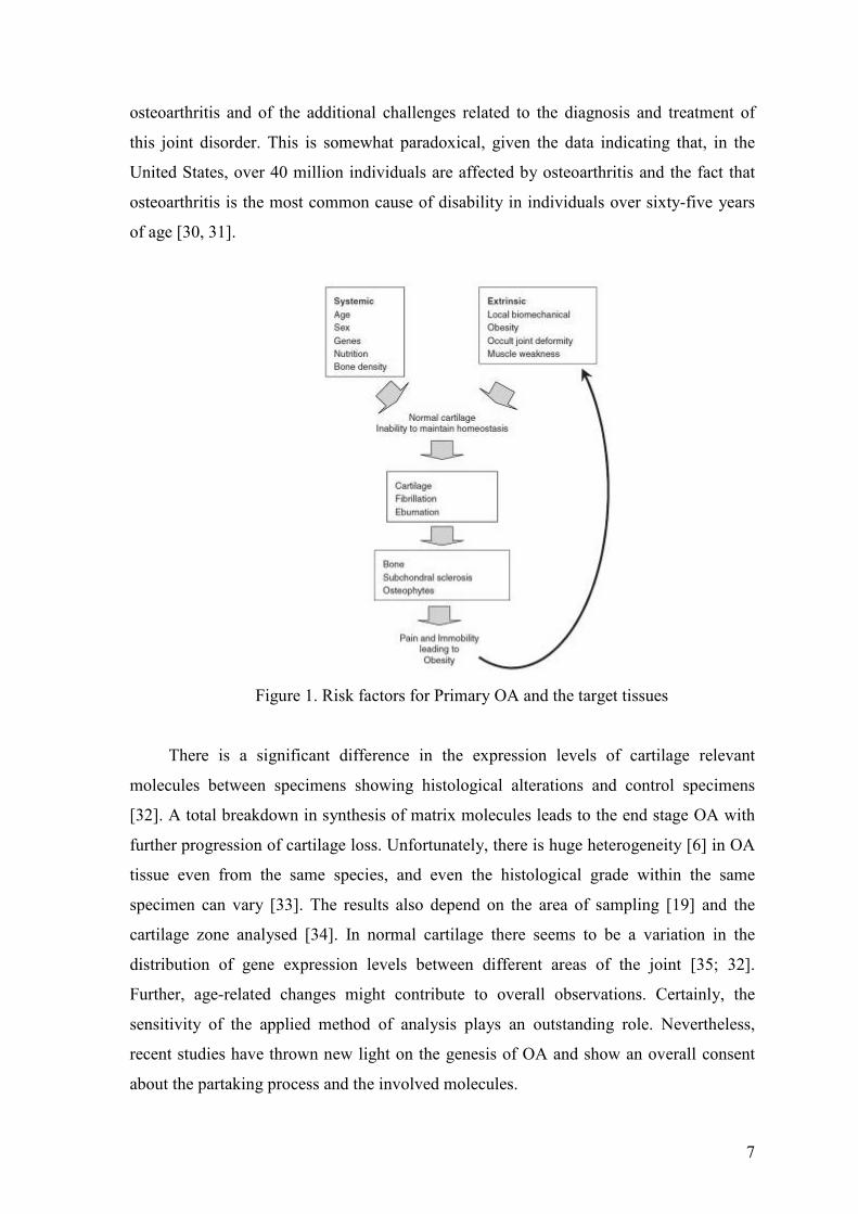
Primary OA is a complex disorder with a largely unknown etiology. Descriptions of
the pathogenesis of OA have undergone many revisions since it was first described but,
despite the name, the focus in recent decades has predominantly been on the articular
cartilage of the synovial joints as the affected tissue, and biomechanics as the causative
agent. Changes in other tissues are believed to be secondary; subchondral bone
responding to abnormal biomechanics and other tissues to secondary inflammation and
enforced inactivity (Fig. 1). Primary OA has to be distinguished from secondary forms of
the disease, which are due to traumatic, metabolic or endocrine abnormalities [29].
Until recently, osteoarthritis has received relatively less attention from the clinical
and research communities within the field of general rheumatology, in part because of the
relative lack of appreciation of the role of inflammation in the pathogenesis of
7
osteoarthritis and of the additional challenges related to the diagnosis and treatment of
this joint disorder. This is somewhat paradoxical, given the data indicating that, in the
United States, over 40 million individuals are affected by osteoarthritis and the fact that
osteoarthritis is the most common cause of disability in individuals over sixty-five years
of age [30, 31].
Figure 1. Risk factors for Primary OA and the target tissues
There is a significant difference in the expression levels of cartilage relevant
molecules between specimens showing histological alterations and control specimens
[32]. A total breakdown in synthesis of matrix molecules leads to the end stage OA with
further progression of cartilage loss. Unfortunately, there is huge heterogeneity [6] in OA
tissue even from the same species, and even the histological grade within the same
specimen can vary [33]. The results also depend on the area of sampling [19] and the
cartilage zone analysed [34]. In normal cartilage there seems to be a variation in the
distribution of gene expression levels between different areas of the joint [35; 32].
Further, age-related changes might contribute to overall observations. Certainly, the
sensitivity of the applied method of analysis plays an outstanding role. Nevertheless,
recent studies have thrown new light on the genesis of OA and show an overall consent
about the partaking process and the involved molecules.
8
Conceptualizing OA as phenotypic subsets related to a primary abnormality allows
more targeted investigation into disease pathophysiology and treatment. Of course, it
must be recognized that this distinction is somewhat artificial, and disease expression is
almost certainly a summation of different, interrelated components.
3.3 Avascular Necrosis
Avascular necrosis (AVN) is still poorly understood. It is believed to be a
multifactorial disease that is associated in some cases with both a genetic predilection and
exposure to certain risk factors. These risk factors include corticosteroid use, alcohol
intake, smoking, and various chronic diseases (renal disease, hematological disease,
inflammatory bowel disease, post-organ transplantation, hypertension, and gout) [21, 22,
36]. The process of this disease has a huge cost impact on the health system due to
surgical treatment. AVN of the femoral head is an increasingly common cause of
musculoskeletal disability as well as a major diagnostic and therapeutic challenge. AVN
of the femoral head is a pathologic process that results from interruption of blood supply
to the bone. AVN of the hip is poorly understood, but this process is the final common
pathway of traumatic or nontraumatic factors that compromise the already precarious
circulation of the femoral head. Femoral head ischemia results in the death of marrow and
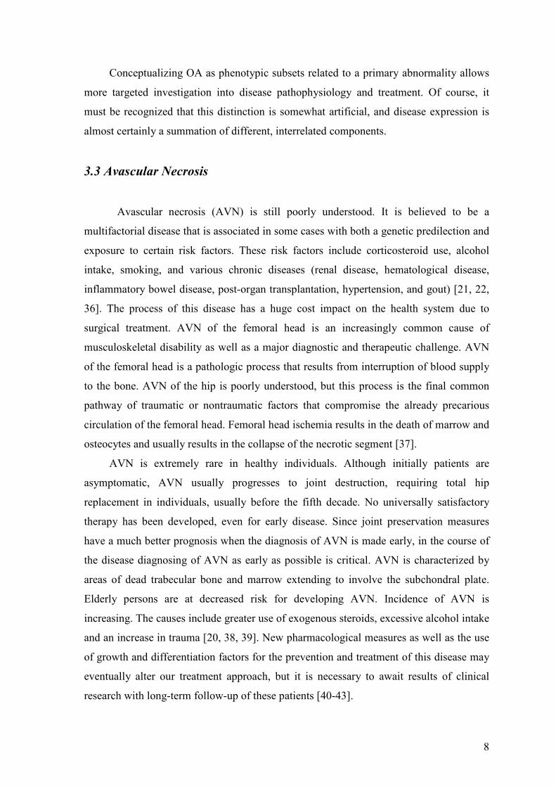
osteocytes and usually results in the collapse of the necrotic segment [37].
AVN is extremely rare in healthy individuals. Although initially patients are
asymptomatic, AVN usually progresses to joint destruction, requiring total hip
replacement in individuals, usually before the fifth decade. No universally satisfactory
therapy has been developed, even for early disease. Since joint preservation measures
have a much better prognosis when the diagnosis of AVN is made early, in the course of
the disease diagnosing of AVN as early as possible is critical. AVN is characterized by
areas of dead trabecular bone and marrow extending to involve the subchondral plate.
Elderly persons are at decreased risk for developing AVN. Incidence of AVN is
increasing. The causes include greater use of exogenous steroids, excessive alcohol intake
and an increase in trauma [20, 38, 39]. New pharmacological measures as well as the use
of growth and differentiation factors for the prevention and treatment of this disease may
eventually alter our treatment approach, but it is necessary to await results of clinical
research with long-term follow-up of these patients [40-43].
9
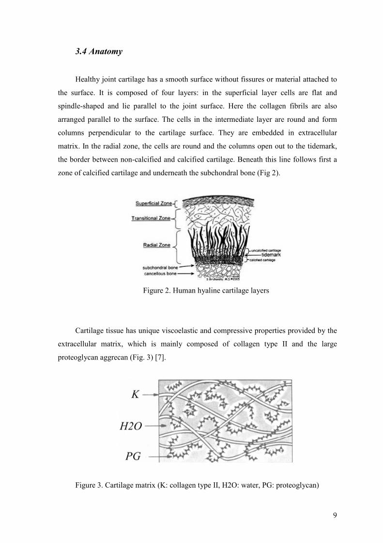
3.4 Anatomy
Healthy joint cartilage has a smooth surface without fissures or material attached to
the surface. It is composed of four layers: in the superficial layer cells are flat and
spindle-shaped and lie parallel to the joint surface. Here the collagen fibrils are also
arranged parallel to the surface. The cells in the intermediate layer are round and form
columns perpendicular to the cartilage surface. They are embedded in extracellular
matrix. In the radial zone, the cells are round and the columns open out to the tidemark,
the border between non-calcified and calcified cartilage. Beneath this line follows first a
zone of calcified cartilage and underneath the subchondral bone (Fig 2).
Figure 2. Human hyaline cartilage layers
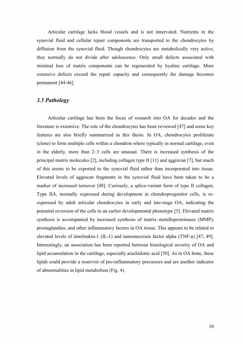
Cartilage tissue has unique viscoelastic and compressive properties provided by the
extracellular matrix, which is mainly composed of collagen type II and the large
proteoglycan aggrecan (Fig. 3) [7].
Figure 3. Cartilage matrix (K: collagen type II, H2O: water, PG: proteoglycan)
10
Articular cartilage lacks blood vessels and is not innervated. Nutrients in the
synovial fluid and cellular repair components are transported to the chondrocytes by
diffusion from the synovial fluid. Though chondrocytes are metabolically very active,
they normally do not divide after adolescence. Only small defects associated with
minimal loss of matrix components can be regenerated by hyaline cartilage. More
extensive defects exceed the repair capacity and consequently the damage becomes
permanent [44-46].
3.5 Pathology
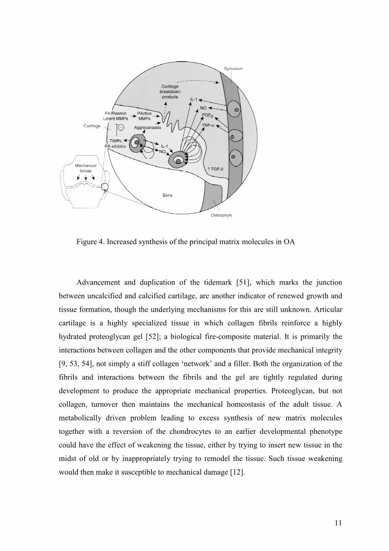
Articular cartilage has been the focus of research into OA for decades and the
literature is extensive. The role of the chondrocytes has been reviewed [47] and some key
features are also briefly summarized in this thesis. In OA, chondrocytes proliferate
(clone) to form multiple cells within a chondron where typically in normal cartilage, even
in the elderly, more than 2–3 cells are unusual. There is increased synthesis of the
principal matrix molecules [2], including collagen type II [11] and aggrecan [7], but much
of this seems to be exported to the synovial fluid rather than incorporated into tissue.
Elevated levels of aggrecan fragments in the synovial fluid have been taken to be a
marker of increased turnover [48]. Curiously, a splice-variant form of type II collagen,
Type IIA, normally expressed during development in chondroprogenitor cells, is re-
expressed by adult articular chondrocytes in early and late-stage OA, indicating the
potential reversion of the cells to an earlier developmental phenotype [5]. Elevated matrix
synthesis is accompanied by increased synthesis of matrix–metalloproteinases (MMP),
prostaglandins, and other inflammatory factors in OA tissue. This appears to be related to
elevated levels of interleukin-1 (IL-1) and tumornecrosis factor alpha (TNF-α) [47, 49].
Interestingly, an association has been reported between histological severity of OA and
lipid accumulation in the cartilage, especially arachidonic acid [50]. As in OA bone, these
lipids could provide a reservoir of pro-inflammatory precursors and are another indicator
of abnormalities in lipid metabolism (Fig. 4).
11
Figure 4. Increased synthesis of the principal matrix molecules in OA
Advancement and duplication of the tidemark [51], which marks the junction
between uncalcified and calcified cartilage, are another indicator of renewed growth and
tissue formation, though the underlying mechanisms for this are still unknown. Articular
cartilage is a highly specialized tissue in which collagen fibrils reinforce a highly
hydrated proteoglycan gel [52]; a biological fire-composite material. It is primarily the
interactions between collagen and the other components that provide mechanical integrity
[9, 53, 54], not simply a stiff collagen ‘network’ and a filler. Both the organization of the
fibrils and interactions between the fibrils and the gel are tightly regulated during
development to produce the appropriate mechanical properties. Proteoglycan, but not
collagen, turnover then maintains the mechanical homeostasis of the adult tissue. A
metabolically driven problem leading to excess synthesis of new matrix molecules
together with a reversion of the chondrocytes to an earlier developmental phenotype
could have the effect of weakening the tissue, either by trying to insert new tissue in the
midst of old or by inappropriately trying to remodel the tissue. Such tissue weakening
would then make it susceptible to mechanical damage [12].
12
3.6 Biomechanics
The mechanical properties of cartilage, including viscoelasticity and high resistance
against load and shear stress, are controlled through the metabolic balance within the
matrix collagen–proteoglycan network. Water is the main composite (60–80%) of the
extracellular matrix [10, 55–58]. Proteoglycans induce a high osmotic pressure and have a
high water binding capacity. Between 5 and 10% of the cartilage mass are proteoglycans,
mostly aggrecan [59]. Collagen (90% collagen type II) is responsible for the high
resistance against tensional forces. Collagen type II contributes to up to 60% of the dry
weight [60]. During inflammation or slow degeneration, the homeostasis within the
collagen–proteoglycan network in the chondral matrix is disordered, mainly due to the
activation of MMPs (stromelysin) after stimulation by cytokines like IL-1 and TNF-alpha
[10 Cartilage defects generally are grouped into four stages: softening with intact surface,
fibrillations within the superficial cartilage layer, clefts down to the subchondral bone,
and complete loss of cartilage [61].
Theoretical and computational analyses of the contact response of cartilage under
various loading conditions have predicted that more than 90% of the load transmitted
across articular layers is supported by the pressurized interstitial fluid, with the remnant
contributed by the collagen-proteoglycan solid matrix. Since this fluid pressure is a
hydrostatic stress, and since cartilage has been shown to be nearly incompressible at
physiological levels of pressures, it has become evident that the interstitial fluid shields
the solid matrix from excessive deformations [62-64].
3.7 Thermal Analysis
Thermal analysis comprises a group of techniques in which a physical property of a
substance measured as a function of temperature, while the substance is subjected to a
controlled temperature programme.
Differential scanning calorimetry (DSC) and thermogravimetric (TG) analysis have
been widely used for determining physicochemical transformations that occur during
thermal degradation [65, 66]. These techniques measure net changes in enthalpy and
weight as a result of many reactions taking place simultaneously and are particularly
useful for indicating the temperature range and the rate of thermal processes as well as
13
giving considerable information on physical and chemical changes [67-70]. Relatively
little has been published on the thermal properties of human hyaline cartilage.
Understanding the response of drugs and their formulations to thermal stresses is an
integral part of the development of stable medicinal products. Thermal analytical methods
have thus become important tools for the development of modern medicines. These are
precise and accurate techniques with low sample requirements, and can provide detailed
information about new chemical entities even at the very earliest stages of drug discovery
and development [70, 71].
3.7.1 Thermogravimetry
Thermogravimetric analysis (TGA) is an analytical technique used to determine a
material’s thermal stability and its fraction of volatile components by monitoring the
weight change that occurs as a specimen is heated [72, 73]. The measurement is normally
carried out in air or in an inert atmosphere, such as Argon, and the mass is recorded as a
function of increasing temperature. Sometimes, the measurement is performed in a lean
oxygen atmosphere (1 to 5% O2 in N2 or He) to slow down oxidation. In addition to mass
changes, some instruments also record the temperature difference between the specimen
and one or more reference pans (differential thermal analysis, or DTA) or the heat flow
into the specimen pan compared to that of the reference pan (differential scanning
calorimetry, or DSC). The latter can be used to monitor the energy released or absorbed
via chemical reactions during the heating process.
In most cases, TG analysis is performed in an oxidative atmosphere (air or oxygen
and inert gas mixtures) with a linear temperature ramp. The maximum temperature is
selected so that the specimen mass is stable at the end of the experiment, implying that all
chemical reactions are completed (i.e., all of the carbon is burnt off leaving behind metal
oxides). This approach provides two important numerical informations: ash content
(residual mass, Mres) and oxidation temperature (To). While the definition of ash content
is unambiguous, oxidation temperature can be defined in many ways, including the
temperature of the maximum in the weight loss rate (dm/dTmax) and the weight loss onset
temperature (Tonset). The former refers to the temperature of the maximum rate of
oxidation, while the latter refers to the temperature when oxidation just begins.
14
The ability of TG to generate fundamental quantitative data from almost any class
of materials, has led to its widespread use in every field of science and technology. Key
application areas [72, 74-79] are listed below:
• Thermal Stability: related materials can be compared at elevated temperatures
under the required atmosphere. The TG curve can help to elucidate decomposition
mechanisms.
• Kinetic Studies: a variety of methods exist for analysing the kinetic features of all
types of weight loss or gain, either with a view to predictive studies, or to
understanding the controlling chemistry.
• Material characterisation: TG and Derivative Thermogravimetry (DTG) curves
can be used to "fingerprint" materials for identification or quality control.
• Corrosion studies: TG provides an excellent means of studying oxidation, or
reaction with other reactive gases or vapours.
• Simulation of industrial processes: the thermobalance furnace may be thought of
as a mini-reactor, with the ability to mimic the conditions in some types of
industrial reactor.
• Compositional analysis: by careful choice of temperature programming and
gaseous environment, many complex materials or mixtures may be analysed by
selectively decomposing or removing their components. This approach is
regularly used to analyse e.g. filler content in polymers; carbon black in oils; ash
and carbon in coals, and the moisture content of many substances.
3.7.2 Calorimetry
Differential scanning calorimetry (DSC) is a thermoanalytical technique for
measuring the energy necessary to establish a nearly zero temperature difference between
a substance and an inert reference material, as the two specimens are subjected to
identical temperature regimes in an environment heated or cooled at a controlled rate. In
heat-flux DSC, the sample and reference are connected by a low-resistance heat-flow path
(a metal disc). The assembly is enclosed in a single furnace (Fig. 5).
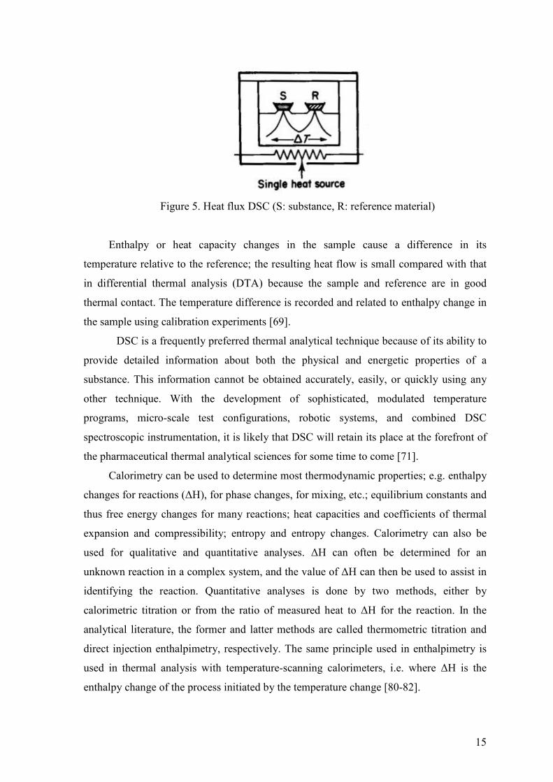
15
Figure 5. Heat flux DSC (S: substance, R: reference material)
Enthalpy or heat capacity changes in the sample cause a difference in its
temperature relative to the reference; the resulting heat flow is small compared with that
in differential thermal analysis (DTA) because the sample and reference are in good
thermal contact. The temperature difference is recorded and related to enthalpy change in
the sample using calibration experiments [69].
DSC is a frequently preferred thermal analytical technique because of its ability to
provide detailed information about both the physical and energetic properties of a
substance. This information cannot be obtained accurately, easily, or quickly using any
other technique. With the development of sophisticated, modulated temperature
programs, micro-scale test configurations, robotic systems, and combined DSC
spectroscopic instrumentation, it is likely that DSC will retain its place at the forefront of
the pharmaceutical thermal analytical sciences for some time to come [71].
Calorimetry can be used to determine most thermodynamic properties; e.g. enthalpy
changes for reactions (∆H), for phase changes, for mixing, etc.; equilibrium constants and
thus free energy changes for many reactions; heat capacities and coefficients of thermal
expansion and compressibility; entropy and entropy changes. Calorimetry can also be
used for qualitative and quantitative analyses. ∆H can often be determined for an
unknown reaction in a complex system, and the value of ∆H can then be used to assist in
identifying the reaction. Quantitative analyses is done by two methods, either by
calorimetric titration or from the ratio of measured heat to ∆H for the reaction. In the
analytical literature, the former and latter methods are called thermometric titration and
direct injection enthalpimetry, respectively. The same principle used in enthalpimetry is
used in thermal analysis with temperature-scanning calorimeters, i.e. where ∆H is the
enthalpy change of the process initiated by the temperature change [80-82].
16
Since calorimetry directly measures the instantaneous rate of the process,
calorimetry is a particularly advantageous method for determination of the kinetics of
slow processes. Analysis of thermodynamic and kinetic data from calorimetry always
involves a model for the system, e.g. a set of chemical reactions, kinetic equations, or a
theoretical model for the property as a function of temperature, pressure, or composition.
Calorimetric data will be fitted to the model to obtain model parameters, therefore
providing a description of the system as a function of the experimental variables.
Calorimetric data can also be used to test the predictive power of such models, and thus to
gain fundamental insight into a process or property of a material, or to predict failure or
hazardous conditions. Such models may require collection of ancillary data
simultaneously with the calorimetric measurements [83].
17
4. AIMS OF THE INVESTIGATIONS
Based on previous studies, we hypothetized that
• thermodynamic findings clearly differentiate normal and degenerated human
hyaline cartilage,
• physicochemical transformations may provide information on the role of water
content in osteoarthritis and avascular necrosis, and
• enthalpy change of the process, initiated by the temperature change, might
represent potential marker of the disease activity.
In order to get answer for the hypotheses above, patients with primary end-stage OA and
AVN were chosen for our investigations.
The aim of this study was:
1. to investigate whether cartilage undergoes complex changes in matrix
composition (water, proteoglycan, and collagen content) during the late stage of
degeneration. These complex deviations develop from the normal matrix
composition during the diseases OA and AVN are hypothesized to correlate with
changes in thermal analysis;
2. to introduce thermogravimetric examination as part of thermal analysis alongside
calorimetry, since water content of the cartilage has not been measured before by
thermogravimetry;
3. to establish the kinetic character of the effect of water loss by heating;
4. to find correlation between the enthalpy changes and the severity of OA and
AVN;
5. to establish a new protocol for sample extraction during live surgery; and
6. the further purpose of this study was to elucidate the importance of water content
in contributing to disease progression.
18
5. PATIENTS AND METHODS
5.1 Patients
In order to conduct the thermoanalytical study, 35 samples were collected from live
surgeries of OA patients between October 2005 and April 2006. During hip arthroplasty
procedures performed at the Orthopedic Department, University of Szeged, 16 OA and 12
AVN human hyaline cartilage samples and normal cartilage from 7 knee were obtained.
There was no clinical meaningful difference in age between OA patients (64 ± 5.2), AVN
patients (59 ± 6.4) and controls (61 ± 4.2). There was no considerable sex differences
between OA patients (75% females), AVN patients (60% females), and controls (70%
females); Chi-square P = 0.54.
Usually, in OA of both medial and lateral knee compartments, total knee
arthroplasty is performed. When only one compartment is affected and ligamental
stability is intact, unicondylar prosthesis is implanted. We were able to obtain normal
cartilage samples from those patients where one compartment of the same knee was
degenerated, and the other one was normal. Therefore, the unaffected femoral condyle
had to be sacrificed for the procedure because ligamental instability was the indication for
total knee arthroplasty.
5.2 Patients grading
Preoperatively, the diagnosis of the patients were established on basis of the patient
history, clinical signs, laboratory tests, and radiological findings. The state of the hyaline
cartilage was determined intraoperatively. All patients in the osteoarthritic group were
considered to be Osteoarthritis Research Society International (OARSI) grade 5-6
articular surface degeneration. OARSI Grade 5-6 OA is characterized by deformation and
change in the contour of the articular surface (Fig. 6) [84]. This results not only from
articular plate fractures, but also from increased metabolic activity of the articular bone
plate, as well as from activation of connective tissue at the lateral and, sometimes, central
cartilage/bone interfaces. All patients in the AVN group were classified as Ficat stage 4
[85]. Samples were considered to be normal when hyaline articular cartilage was

19
uninvolved with OA (OARSI Grade 0). In these results, the cartilage surface is smooth,
no enlargement, distortion, and no proliferative changes are observed.
Figure 6. OA cartilage advanced grading methodology
5.3 Sample preparation for thermal analysis
After the operation, a disc (5mm in diameter) was removed from the unhealthy and
healthy cartilage surfaces. The samples were taken under sterile conditions, excess bone
was removed, and only the remaining full thickness cartilage was used. The disc was first
washed in sterile saline, then stored in 20 ml saline for transportation at room
temperature. Mean storage time was 6 hours (min: 1 hour, max: 26 hour), 29 samples out
of 35 were studied within four hours of preparation. Six samples were stored over-night at
5 °C. Preemptive control examinations did not show any change in the calorimetric and
thermogravimetric properties after storage for 26 hours at 5 °C.
20
5.4 Thermal measurements
The success of the thermal experiments depends on the careful preparation of
samples and the judicious selection of the appropriate experimental conditions (such as
scanning rate and sample size). In general, DSC samples are analyzed in small metal
pans, designed for optimal thermal conductivity and minimum reaction with the samples
(for example, aluminum alloy, platinum, stainless steel, or silver) [71]. For accurate
quantitative work the thermal mass of the sample and reference pans were matched.
All the thermal measuments were conducted at the Department of Pharmaceutical
Technology, University of Szeged. The calorimetric properties of samples were
determined by DSC method (Mettler-Toledo DSC 821e apparatus, Mettler-Toledo
GmbH, Switzerland). Samples were heated from 0 to 80 °C. The heating rate was 0.3
°C/min. Conventional Hastelloy batch vessels were used with 40 µl sample volume. All
the DSC measurements were preceded in Ar atmosphere, and the flow rate was 100
ml/min. From the DSC curves, the decomposition temperature (onset temperature), the
transition temperature range (endset temperature), and the total calorimetric enthalpy
change were calculated. Well-defined standards and calibration procedures are
particularly important, therefore high care was taken in calibrating the instrument as close
to the transition temperatures of interest as possible.
The thermogravimetric analysis was performed with the use of a MOM
Derivatograph (MOM, Budapest, Hungary), and the TG, DTG, and DTA curves were
determined. The temperature (T) curve shows the linear increase of temperature during
the process. DTG curve represents the first derivative of the mass change curve. The DTA
curve shows the same picture as a Differential Scanning Calorimetry examination, in
which the temperature change of a sample is compared with the temperature of a
thermally inert material in order to give information about the endothermic or exothermic
enthalpic transition or other reaction [76-79].
The heating was linear from 25 to 150 °C and the rate of heating was 5 °C/min. Al2O3
was used as reference material. In the first step, the total water loss and kinetic parameters
were calculated. The kinetic parameters calculated by the software are the following: the
reaction order (n), the activation energy (Ea), and the pre-exponential factor (A).
21
The value of n (reaction order) is allocated by the Kissinger method [86], and it is the first
kinetic parameter calculated by the computer:
Sn 2
1
26,1 ⋅= (1)
where S is the form factor which presents the absolute value of the gradients of DTG
curves in the points of min/max. The activation energy (Ea) is determined according to
the natural logarithmic form of the Arrhenius-equation
k(T) = A∗e -Ea/RT (2)
which is widely used in the literature [70, 87-89].
5.5 Statistical analysis
Fisher LSD method by the Statistica for Windows statistical program was used to
compare enthalpy changes in the different groups.
Data are presented as mean±SD. Statistical significance was assessed by the Student
t test and the Kruskal-Wallis one-way ANOVA on ranks. The results were considered
significant, if p < 0.05 (*means significant values on the graphs).
5.6 Ethics
All tissues were yielded in accordance to legal regulation, international ethical
concerns, and patients’ consent. The Human Investigation Review Board of the
University of Szeged has decided (2006.09.18.) that the experiments comply with the
ethics of research and the declaration of the Medical World Federation.
22
6. RESULTS
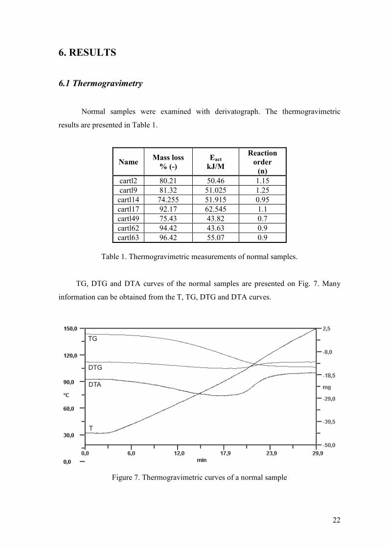
6.1 Thermogravimetry
Normal samples were examined with derivatograph. The thermogravimetric
results are presented in Table 1.
NameMass loss
% (-)
Eact
kJ/M
Reaction
order
(n)
cartl2 80.21 50.46 1.15cartl9 81.32 51.025 1.25
cartl14 74.255 51.915 0.95cartl17 92.17 62.545 1.1cartl49 75.43 43.82 0.7cartl62 94.42 43.63 0.9cartl63 96.42 55.07 0.9
Table 1. Thermogravimetric measurements of normal samples.
TG, DTG and DTA curves of the normal samples are presented on Fig. 7. Many
information can be obtained from the T, TG, DTG and DTA curves.
TG
DTG
DTA
T
Figure 7. Thermogravimetric curves of a normal sample
23
It was found, that the average total water content of intact (normal) cartilage is
81%, which was probably the interstitial water, and the difference was supposedly bound
to the surface. To remove the cartilage extracellular water content, 52 kJ/M energy was
needed.
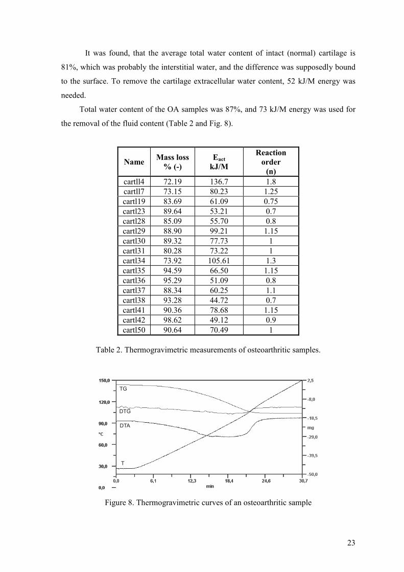
Total water content of the OA samples was 87%, and 73 kJ/M energy was used for
the removal of the fluid content (Table 2 and Fig. 8).
NameMass loss
% (-)
Eact
kJ/M
Reaction
order
(n)
cartll4 72.19 136.7 1.8cartll7 73.15 80.23 1.25cartl19 83.69 61.09 0.75cartl23 89.64 53.21 0.7cartl28 85.09 55.70 0.8cartl29 88.90 99.21 1.15cartl30 89.32 77.73 1cartl31 80.28 73.22 1cartl34 73.92 105.61 1.3cartl35 94.59 66.50 1.15cartl36 95.29 51.09 0.8cartl37 88.34 60.25 1.1cartl38 93.28 44.72 0.7cartl41 90.36 78.68 1.15cartl42 98.62 49.12 0.9cartl50 90.64 70.49 1
Table 2. Thermogravimetric measurements of osteoarthritic samples.
TG
DTG
DTA
T
Figure 8. Thermogravimetric curves of an osteoarthritic sample
24
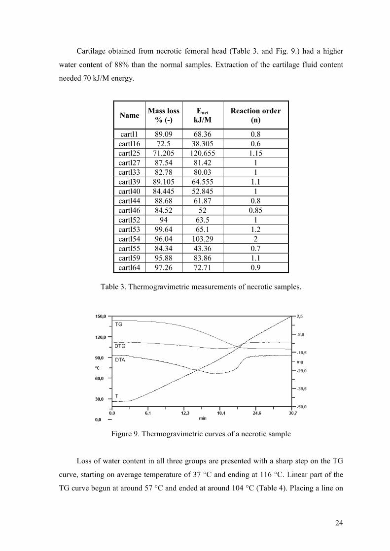
Cartilage obtained from necrotic femoral head (Table 3. and Fig. 9.) had a higher
water content of 88% than the normal samples. Extraction of the cartilage fluid content
needed 70 kJ/M energy.
NameMass loss
% (-)
Eact
kJ/M
Reaction order
(n)
cartl1 89.09 68.36 0.8cartl16 72.5 38.305 0.6cartl25 71.205 120.655 1.15cartl27 87.54 81.42 1cartl33 82.78 80.03 1cartl39 89.105 64.555 1.1cartl40 84.445 52.845 1cartl44 88.68 61.87 0.8cartl46 84.52 52 0.85cartl52 94 63.5 1cartl53 99.64 65.1 1.2cartl54 96.04 103.29 2cartl55 84.34 43.36 0.7cartl59 95.88 83.86 1.1cartl64 97.26 72.71 0.9
Table 3. Thermogravimetric measurements of necrotic samples.
TG
DTG
DTA
T
Figure 9. Thermogravimetric curves of a necrotic sample
Loss of water content in all three groups are presented with a sharp step on the TG
curve, starting on average temperature of 37 °C and ending at 116 °C. Linear part of the
TG curve begun at around 57 °C and ended at around 104 °C (Table 4). Placing a line on
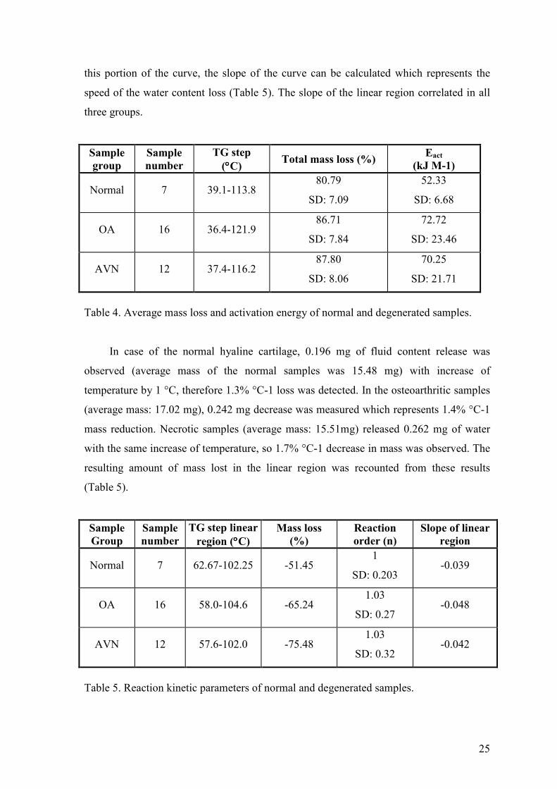
25
this portion of the curve, the slope of the curve can be calculated which represents the
speed of the water content loss (Table 5). The slope of the linear region correlated in all
three groups.
Sample
group
Sample
number
TG step
(°°°°C)Total mass loss (%)
Eact
(kJ M-1)
Normal 7 39.1-113.880.79
SD: 7.09
52.33
SD: 6.68
OA 16 36.4-121.986.71
SD: 7.84
72.72
SD: 23.46
AVN 12 37.4-116.287.80
SD: 8.06
70.25
SD: 21.71
Table 4. Average mass loss and activation energy of normal and degenerated samples.
In case of the normal hyaline cartilage, 0.196 mg of fluid content release was
observed (average mass of the normal samples was 15.48 mg) with increase of
temperature by 1 °C, therefore 1.3% °C-1 loss was detected. In the osteoarthritic samples
(average mass: 17.02 mg), 0.242 mg decrease was measured which represents 1.4% °C-1
mass reduction. Necrotic samples (average mass: 15.51mg) released 0.262 mg of water
with the same increase of temperature, so 1.7% °C-1 decrease in mass was observed. The
resulting amount of mass lost in the linear region was recounted from these results
(Table 5).
Sample
Group
Sample
number
TG step linear
region (°°°°C)
Mass loss
(%)
Reaction
order (n)
Slope of linear
region
Normal 7 62.67-102.25 -51.451
SD: 0.203-0.039
OA 16 58.0-104.6 -65.241.03
SD: 0.27-0.048
AVN 12 57.6-102.0 -75.481.03
SD: 0.32-0.042
Table 5. Reaction kinetic parameters of normal and degenerated samples.
26
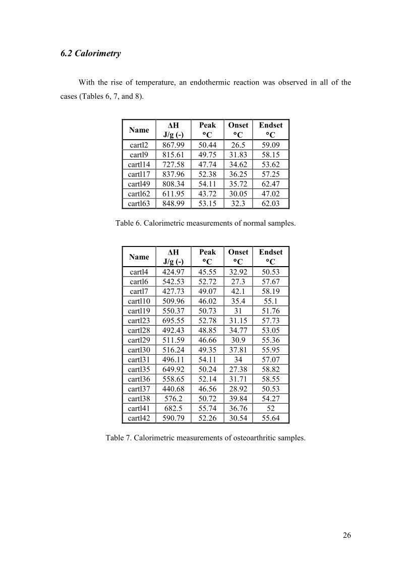
6.2 Calorimetry
With the rise of temperature, an endothermic reaction was observed in all of the
cases (Tables 6, 7, and 8).
Name∆H
J/g (-)
Peak
°°°°C
Onset
°°°°C
Endset
°°°°C
cartl2 867.99 50.44 26.5 59.09cartl9 815.61 49.75 31.83 58.15
cartl14 727.58 47.74 34.62 53.62cartl17 837.96 52.38 36.25 57.25cartl49 808.34 54.11 35.72 62.47cartl62 611.95 43.72 30.05 47.02cartl63 848.99 53.15 32.3 62.03
Table 6. Calorimetric measurements of normal samples.
Name∆H
J/g (-)
Peak
°°°°C
Onset
°°°°C
Endset
°°°°C
cartl4 424.97 45.55 32.92 50.53cartl6 542.53 52.72 27.3 57.67cartl7 427.73 49.07 42.1 58.19
cartl10 509.96 46.02 35.4 55.1cartl19 550.37 50.73 31 51.76cartl23 695.55 52.78 31.15 57.73cartl28 492.43 48.85 34.77 53.05cartl29 511.59 46.66 30.9 55.36cartl30 516.24 49.35 37.81 55.95cartl31 496.11 54.11 34 57.07cartl35 649.92 50.24 27.38 58.82cartl36 558.65 52.14 31.71 58.55cartl37 440.68 46.56 28.92 50.53cartl38 576.2 50.72 39.84 54.27cartl41 682.5 55.74 36.76 52cartl42 590.79 52.26 30.54 55.64
Table 7. Calorimetric measurements of osteoarthritic samples.
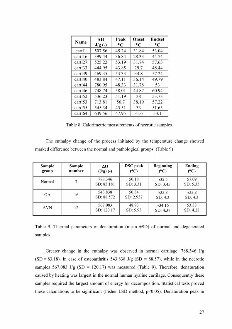
27
Name∆H
J/g (-)
Peak
°°°°C
Onset
°°°°C
Endset
°°°°C
cartl1 507.56 45.24 31.84 53.04cartl16 399.44 36.84 28.33 44.74cartl27 525.22 53.19 31.74 57.63cartl33 444.95 43.85 29.7 48.44cartl39 469.35 53.33 34.8 57.24cartl40 483.84 47.11 36.14 49.79cartl44 780.95 48.33 31.78 53cartl46 748.74 58.01 44.87 60.94cartl52 536.23 51.19 38 53.73cartl53 713.81 56.7 38.19 57.22cartl55 545.34 45.51 33 51.65cartl64 649.56 47.95 31.6 53.1
Table 8. Calorimetric measurements of necrotic samples.
The enthalpy change of the process initiated by the temperature change showed
marked difference between the normal and pathological groups. (Table 9)
Sample
group
Sample
number∆∆∆∆H
(J/g) (-)
DSC peak
(°°°°C)
Beginning
(°°°°C)
Ending
(°°°°C)
Normal 7788.346
SD: 83.18150.18
SD: 3.31≈32.5
SD: 3.4557.09
SD: 5.35
OA 16543.838
SD: 88.57250.34
SD: 2.937≈33.8
SD: 4.3≈33.8
SD: 4.3
AVN 12567.083
SD: 120.1748.93
SD: 5.93≈34.16
SD: 4.3753.38
SD: 4.28
Table 9. Thermal parameters of denaturation (mean ±SD) of normal and degenerated
samples.
Greater change in the enthalpy was observed in normal cartilage: 788.346 J/g
(SD = 83.18). In case of osteoarthritis 543.838 J/g (SD = 88.57), while in the necrotic
samples 567.083 J/g (SD = 120.17) was measured (Table 9). Therefore, denaturation
caused by heating was largest in the normal human hyaline cartilage. Consequently these
samples required the largest amount of energy for decomposition. Statistical tests proved
these calculations to be significant (Fisher LSD method, p<0.05). Denaturation peak in
28
normal cartilage was at 50.18 °C (SD = 3.31), in necrotic samples it was lower at
48.93 °C (SD = 5.93) however in osteoarthritis 50.34 °C (SD = 2.93) was similar to
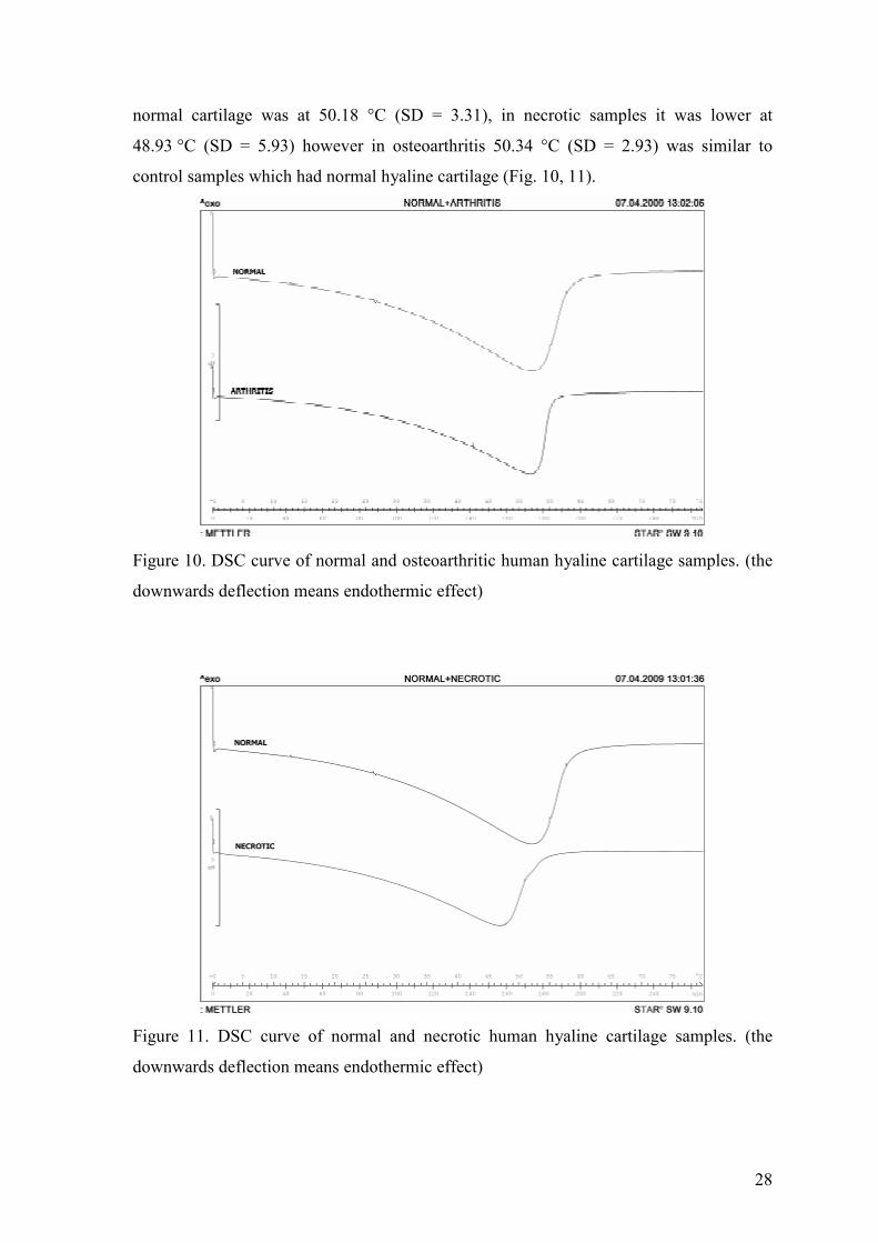
control samples which had normal hyaline cartilage (Fig. 10, 11).
Figure 10. DSC curve of normal and osteoarthritic human hyaline cartilage samples. (the
downwards deflection means endothermic effect)
Figure 11. DSC curve of normal and necrotic human hyaline cartilage samples. (the
downwards deflection means endothermic effect)
29
7. DISCUSSION
OA development takes place in consecutive steps of breakdown and attempted
regeneration. Though the comprehension about OA has grown enormously over the last
years, there is still need to extend our knowledge about the basic context of OA genesis
and development. Several biochemical and biomechanical factors are considered for the
pathogenesis [14, 90]. The data up to date show, however, that OA is a very complex
disease procedure, and it can be speculated, that the context leading to the progressive
process is not finally resolved. There might still be molecules involved in the process,
which molecules have not yet been studied or even identified [91].
OA is widely believed to result from local mechanical factors acting within the
context of systemic susceptibility [8, 11, 15-17, 47, 92]. Molecular pathology of
osteoarthritis is under intense investigation [6, 7, 18, 93-97] since biomechanical factors
result in chemical alteration within the joint [19, 98, 99]. Rearrangements of intra- and
intermolecular bonds in collagen molecule and disaggregation of proteoglycans and their
elimination from OA cartilage found to be responsible for water accumulation [13]. It was
also shown that the most part of water is free water and its quantity is increased in the
osteoarthritis of the hyaline cartilage [100].
We observed increase in water content of the cartilage matrix in all cases of the
investigated degenerative cartilages [101]. Based on our results, it can be stated that water
content is higher in impaired samples, meanwhile water interstitial bonding was stronger
in the OA and AVN cases. Rise in water adherence was well distinguishable since higher
energy was needed for removal. Activation energy correlated considerably with water
content in the samples. Denaturation caused by heating was larger in the normal cartilage
than in the diseased ones, therefore normal samples required the largest amount of energy
for decomposition [102].
The purpose of our study was also to clarify the previously reported studies in the
literature [24-27]. By acquiring normal cartilage from live surgery and by performing the
investigation in a relatively short period of time compared to the earlier reports [24-27,
101, 102], similar sample environment was provided as with the degenerarative samples.
This way, we minimized the extracorpal degeneration. All samples we used showed a
clear denaturation peak on the calorimetric curve, therefore volume of the curve was
easily calculated giving the enthalpy change of the sample. These changes correlated with
30
the water content of the samples. Due to the increased number of samples acquired for
our studies, the results were much better reproducible than results in the literature [24,
102], and the difference between the normal and necrotic samples was significant [101,
102].
The newly established thermogravimetric protocol that we used was sufficient for
compositional thermoanalytical study of normal and degenerative human hyaline
cartilage. Water content elevation contributing to disease progression was observed in
both OA and AVN. Previously, this method has not been used for this type of
investigations. The main goal of the thermogravimetric measurements was to identify the
nature and quantity of water molecules in the investigated samples. Water molecules’
binding mode may have an important consequence in pharmacokinetics. The reaction
order turned out to be approximately 1 in all three cases (normal, OA, and AVN), and the
standard deviation was low (Table 5). The TG curve’s slope of the linear region showed,
that the rate of water loss depends on the water amount remaining in the tissue.
Comparing the data in the presented tables (Tables 4, 5) (Total mass loss: normal:
80.79%, OA: 86.71%, and AVN: 87.80%), it can be concluded that the higher water
content in the degenerative samples bound stronger to the matrix. However, the reaction
order and the slope of the linear region correlated in all three groups. This first order
kinetic means that the rate of water loss depends on the water amount remaining in the
tissue, namely if the amount of water decreases in the tissue, the rate of loss also
decreases.
The pathogenesis of OA progression likely revolves around a complex interplay of
numerous factors. The major contributors include chondrocyte regulation of the
extracellular matrix [11, 12, 95], genetic influences [5-7], local mechanical factors [14,
15, 55], and inflammation [2, 18, 19]. DSC as part of thermal analysis was a reliable
method for differentiating normal hyaline cartilage from degenerated samples. The
available calorimeter proved to be adequate for these measurements.
DSC techniques are still developing and many new variants and applications are
reported each year. Combined techniques [103-105] with microscopic or spectroscopic
instruments are of obvious value to the pharmaceutical scientist, although commercially
available units are not widely used and have limited pharmaceutical applications. With
the rapid development of atomic and molecular scale microscopy, hyphenated micro-
thermal analysis techniques, such as atomic force microscopy-DSC, are also becoming
commercially available. There may be many future applications of micro-DSC
31
measurements to pharmaceutical problems, although these are likely to be limited to basic
research applications in the next few years until the full potential of the technique has
been demonstrated.
AVN of the femoral head is a common cause of painful hip in young adults. The
natural course of this disease has steady progression with eventual collapse of the femoral
head, followed by secondary osteoarthritis in the hip joint. Molecular pathology of AVN
is under intense investigation since biomechanical factors result in chemical alteration
within the joint [23, 106-108]. In AVN of the femoral head, interference with blood
results in infarction of a segment of the head leading to drastic early degeneration of the
surface hyaline cartilage [108, 109]. Patients in our study undergoing arthroplasty
procedure showed clinically and radiologically end-stage degradation of the cartilage,
with very little resemblance to normal cartilage.
Our study has had several limitations, as many other studies on OA and AVN. First,
the sample size was not large enough to arrive at definitive conclusions. Additional
measurements are needed to affirm the results of our study. Secondly, we investigated
those patients for normal cartilage samples of the knee, who underwent surgery for the
other compartment OA. This was the only ethical and technical way of acquiring normal
tissues from living persons for our experiments. Previous thermoanalytical studies used
cadaver samples for the investigation as normal human hyaline cartilage. All samples that
were extracted for our studies were obtained during live surgeries. and were
macroscopically intact [101, 102]. There is no previous report in the literature of
examining normal cartilage from live surgery. Only full thickness cartilage was used for
the normal analysis. Prior results indicate that early OA is primarily characterized by the
changes in collagen orientation and proteoglycan content only in the superficial zone,
while collagen content does not change until OA has progressed to its late stage [110]. A
new protocol had to be established before the detailed investigation of human tissues
could be performed. Most of the known changes in the extracellular matrix in OA come
from animal models in the literature since human samples for investigation are not widely
available for experiments.
Characterization of the altered metabolism in cartilage that promotes disease
progression should lead to future treatment options that can prevent structural damage.
Since damaged articular cartilage has a very limited potential for healing, prevention is
fundamental in treatment. However, prevention is not possible without the knowledge of
the basic pathomorphological mechanism leading to cartilage degeneration. With better
32
understanding the exact amount of matrix water content and its binding characteristics,
preventive measures can be developed. These therapeutic steps can be adequately tested
and monitored with thermogravimetric measurements. The use of this method can also
determine the effectiveness of currently used medications (Glucosamin, Chondroitin) for
resolving cartilage matrix degeneration.
Further understanding of the initiating events in cartilage destruction, the
relationship between the different pathologic influences, and the role of the chondrocyte
in maintaining extracellular matrix homeostasis will be necessary to reveal potential
targets of therapy. Clinical trials are currently underway for a number of potential disease
modifying agents that may significantly change the treatment approach for OA [31, 99].
With the possibility of disease-modifying OA drugs (DMOADs), the necessity for
instruments that are sensitive to changes has become very apparent in clinical trials [111].
With regard to the clinical management of patients with OA, efforts should be made
wherever possible to influence modifiable risk factors. At present, there are a number of
modifying therapeutic options that may be used to alter the rate of disease progression.
While these therapies are currently underutilized, they might play a greater role if
modification of disease progression has to be demonstrated. However, the role of these
therapies is unclear, given the paucity of long-term, well-designed controlled trials [112-
114].
Finally, we have to mention that common histopathologic assessment methods
(Grade) under both clinical and experimental conditions poorly reflect mild phases of the
disease, and are very non-linear over the range from mild to advanced stages of the
disease. In the OARSI (OsteoArthritis Research Society International) Scoring System
grade is defined as OA depth progression into cartilage irrespective of its horizontal
extent. Stage is defined as the horizontal extent of cartilage involvement within one side
of a joint compartment irrespective of the underlying grade. Therefore, a detailed thermal
examination is needed on the same joint surface with samples taken from different grades
of degeneration within the same joint.
The promise of biomarkers has yet to be fulfilled in OA. Type II collagen, cartilage
oligomeric matrix protein, hyaluronan, and aggrecan have been some of the many
biomarkers investigated so far [45, 115-117]. Although numerous clinical studies have
suggested that specific biomarkers or their combinations can have predictive value in
terms of the presence and severity of the disease [118]. The wide variability in these
values limits their use for individual patients. Whereas, the use of thermal analysis could
33
be a simple and effective method for controlling the relationship between these markers
and disease progression. The revised protocol for sample taking during live surgeries
eliminates the presence of disturbing substances during the examination. A detailed
thermal examination is also needed on the same joint surface with samples taken from
different grades of degeneration within the same joint.
34
8. SUMMARY AND CONCLUSIONS
In summary, we examined the water content of human hyaline cartilage of normal
origin and in patients with OA and AVN. We were the first ones who used normal
samples that were extracted from live surgeries for the investigations.
A newly established thermogravimetric protocol was used for our experiments. This
method proved to be suitable for compositional thermoanalytical study of normal and
degenerative human hyaline cartilage.
Our results showed clear evidence that:
• complex deviations from the normal matrix composition during the late
stage of degeneration correlated significantly with changes in thermal
properties;
• patients with primary OA and AVN had significantly higher levels of
water content in the degenerative samples and water bound stronger to the
matrix than in controls;
• the kinetic character of the effect of water loss by heating was established,
the reaction order was approximately 1 in all cases;
• correlation was found between the enthalpy changes and the severity of
OA and AVN;
• a new protocol was established for sample extraction during live surgery
using simple saline solution instead of the previously used phosphate
buffer;
• this new method proved to be suitable for the thermoanalytical
investigations; and
• the introduction of thermogravimetric examination as part of thermal
analysis alongside calorimetry might be a useful method for determining
the severity of OA and AVN.
One of the possibilities of getting fast information is the use of thermoanalytical
methods. The simple new protocol we established might also be used for gaining
information about the healthy or sick state of other human tissues.
Additional investigation will be needed to fully understand how water content
affect cartilage degradation. Further studies are in progress to elucidate the contribution
35
of physicochemical properties of water in cartilage to the pathogenesis of the
degenerative process of OA.
We need to strive to develop these methods and make them available in the
clinical settings since without the means to monitor the effectiveness of DMOADs, we
will never know if we can control and perhaps even prevent osteoarthritis.
From the work described in this thesis, a model can be proposed whereby the level of
injury to cartilage within the joint can be monitored by a simple thermoanalytical method.
A deeper knowledge of the pathways in the development of degenerative cartilage
diseases might lead to the development of newer therapies for arthritis in the future.
We hope that our data provide further evidence for the importance of cartilage
physicochemical properties in developing cartilage degeneration.
36
9. REFERENCES
1. Swoboda B: Epidemiological arthrosis research. Orthopade 2001; 30: 834-840.
2. Goldring MB, Goldring SR: Osteoarthritis. J. Cell. Physiol. 2007; 213: 626-634.
3. Wu CW, Kalunian KC: New Developments in osteoarthritis. Clin. Geriatr. Med.2005; 21: 589-601.
4. Buckwalter J, Saltzman C, Brown T: The impact of osteoarthritis. Clin. Orthop.Relat. Res. 2004; 427 Suppl: S6-15.
5. Aigner T, Zhu Y, Chansky HH, Matsen FA III, Maloney WJ, Sandell LJ:Reexpression of type IIA procollagen by adult articular chondrocytes inosteoarthritic cartilage. Arthritis Rheum. 1999; 42: 1443-1450.
6. Aigner T, Zien A, Gehrsitz A, Gebhard PM, McKenna L: Anabolic and catabolicgene expression pattern analysis in normal versus osteoarthritic cartilage usingcomplementary DNA-array technology. Arth. Rheum. 2001; 44: 2777-2789.
7. Matyas JR, Adams ME, Huang D, Sandell LJ: Discoordinate gene expression ofaggrecan and type II collagen in experimental osteoarthritis. Arth. Rheum. 1995;38: 420-425.
8. Aspden RM: Osteoarthritis: a problem of growth not decay? Rheumatology 2008;47: 1452-1460.
9. Lewis RJ, MacFarland AK, Anandavijayan S, Aspden RM: Site variation ofmaterial properties and metabolic activity of articular cartilage from the bovinecarpometacarpal joint. Osteoarthritis and Cartilage 1998; 6: 383-392.
10. Sandell LJ, Hering TM: Biochemistry and molecular and cell biology of articularcartilage in osteoarthritis. In: Moskowitz RW, Howell DS, Altman RD,Buckwalter JA, Goldberg VM eds. Osteoarthritis. Diagnosis and medical/surgicalmanagement. (WB Sauders Company, Philadelphia) 2001; pp. 115-143.
11. Aigner T, Glückert K, von der Mark K: Activation of fibrillar collagen synthesisand phenotypic modulation of chondrocytes in early human osteoarthritic cartilagelesions. Osteoarthritis and Cartilage 1997; 5: 183-189.
12. Sofat N: Analysing the role of endogenous matrix molecules in the developmentof osteoarthritis. Int. J. Exp. Path. 2009; 90: 463-479.
13. Loeuille D, Olivier P, Watrin A, Grossin L, Gonord P, Guillot G, Etienne S, BlumA, Netter P, Gillet P: Some biochemical characteristics and water exchange inhuman articular cartilage in osteoarthrosis. Bull. Exp. Biol. Med. 2002; 133: 484-487.
14. Appleyard RC, Burkhardt D, Ghosh P, Read R, Cake M, Swain MV, Murrell GA:Topographical analysis of the structural, biochemical and dynamic biomechanical
37
properties of cartilage in an ovine model of osteoarthritis. Osteoarthritis andCartilage 2003; 11: 65-77.
15. Brandt KD, Radin EL, Dieppe PA, van de Putte L: Yet more evidence thatosteoarthritis is not a cartilage disease. Ann. Rheum. Dis. 2006; 65: 1261-1264.
16. Felson DT: Clinical practice. Osteoarthritis of the knee. N. Engl. J. Med. 2006;354: 841-848.
17. Squires GR, Okouneff S, Ionescu M, Poole AR: The pathobiology of focal lesiondevelopment in aging human articular cartilage and molecular matrix changescharacteristic of osteoarthritis. Arth. Rheum. 2003; 48: 1261-1270.
18. Pelletier JP, Martel-Pelletier J, Mehraban F, Malemud CJ: Immunological analysisof proteoglycan structural changes in the early stage of experimental osteoarthriticcanine cartilage lesions. J. Orthop. Res. 1992; 10: 511-523.
19. Adams ME: Cartilage hypertrophy following canine anterior cruciate ligamenttransection differs among different areas of the joint. J. Rheumatol. 1989; 16: 818-824.
20. Mont MA, Hungerford DS: Non-traumatic avascular necrosis of the femoral head.J. Bone Joint. Surg. Am. 1995; 77: 459-474.
21. Etienne G, Mont MA, Ragland PS: The diagnosis and treatment of nontraumaticosteonecrosis of the femoral head. Instr. Course Lect. 2004; 53: 67-85.
22. Lieberman JR, Berry DJ, Mont MA, Aaron RK, Callaghan JJ, Rajadhyaksha AD,Urbaniak JR: Osteonecrosis of the hip: management in the 21st century. Instr.Course Lect. 2003; 52: 337-355.
23. Aldridge JM 3rd, Urbaniak JR: Avascular necrosis of the femoral head: etiology,pathophysiology, classification, and current treatment guidelines. Am. J. Orthop.2004; 33: 327-332.
24. Than P, Vermes C, Schäffer B, Lırinczy D: Differential scanning calorimetricexamination of the human hyaline cartilage. A preliminary study. Thermochim.Acta 2000; 346: 147-151.
25. Than P, Kereskai L: Thermal analysis of the osteoarthritic human hyalinecartilage. J. Therm. Anal. Cal. 2005; 82: 213-216.
26. Than P, Lırinczy D: Differential scanning calorimetric examination of theosteoarthritic hyaline cartilage in rabbits. Thermochim. Acta 2003; 404: 149-153.
27. Than P, Domán I, Lırinczy D: Differential scanning calorimetry in the research ofdegenerative musculoskeletal disorders. Thermochim. Acta 2004; 415: 83-87.
28. Lawrence RC, Helmick CG, Arnett FC, Deyo RA, Felson DT, Giannini EH,Heyse SP, Hirsch R, Hochberg MC, Hunder GG, Liang MH, Pillemer SR, Steen
38
VD, Wolfe F: Estimates of the prevalence of arthritis and selected musculoskeletaldisorders in the United States. Arthritis Rheum. 1998; 41: 778-799.
29. Roach HI, Aigner T, Soder S, Haag J, Welkerling H: Pathobiology ofosteoarthritis: pathomechanisms and potential therapeutic targets. Current DrugTargets 2007; 8: 271-282.
30. Felson DT: Clinical practice. Osteoarthritis of the knee. N. Engl. J. Med. 2006;354: 841-848.
31. Goldring SR: Needs and opportunities in the assessment and treatment ofosteoarthritis of the knee and hip: The view of the rheumatologist. J. Bone Joint.Surg. Am. 2009; 91: 4-6.
32. Lorenz H, Wenz W, Ivancic M, Steck E, Richter W: Early and stable upregulationof collagen type II, collagen type I and YKL40 expression levels in cartilageduring early experimental osteoarthritis occurs independent of joint location andhistological grading. Arthritis Res. Ther. 2005; 7(1): R156-165.
33. Rizkalla G, Reiner A, Bogoch E, Poole AR: Studies of the articular cartilageproteoglycan aggrecan in health and osteoarthritis. Evidence for molecularheterogeneity and extensive molecular changes in disease. J. Clin. Invest. 1992;90: 2268-2277.
34. Pfander D, Rahmanzadeh R, Scheller EE: Presence and distribution of collagen II,collagen I, fibronectin, and tenascin in rabbit normal and osteoarthritic cartilage.J. Rheumatol. 1999; 26: 386-394.
35. Veje K, Hyllested-Winge JL, Ostergaard K: Topographic and zonal distribution oftenascin in human articular cartilage from femoral heads: normal versus mild andsevere osteoarthritis. Osteoarthritis and Cartilage 2003; 11: 217-227.
36. Mont MA, Jones LC, Hungerford DS: Nontraumatic osteonecrosis of the femoralhead: Ten years later. J. Bone Joint. Surg. Am. 2006; 88: 1117-1132.
37. Ficat RP, Arlet J: Functional investigation of bone under normal conditions. In:Hungerford DS, ed. Ischemia and necrosis of bone. (Williamsand Wilkins,Baltimore) 1980; pp 29-52.
38. Lavernia CJ, Sierra RJ, Grieco FR: Osteonecrosis of the femoral head. J. Am.Acad. Orthop. Surg. 1999; 7: 250-261.
39. Urbaniak JR, Barnes CJ: Meeting the challenge of osteonecrosis in adults. J.Musculoskel. Med. 2001; 18: 395-403.
40. Hungerford MW, Mont MA: Potential uses of cytokines and growth factors intreatment of osteonecrosis. Orthopade 2000; 29: 442-448.
39
41. Mont MA, Jones LC, Einhorn TA, Hungerford DS, Reddi AH: Osteonecrosis ofthe femoral head. Potential treatment with growth and differentiation factors. Clin.Orthop. Relat. Res. 1998; 355 Suppl:S314-335.
42. Thornhill TS: Alternatives to total hip arthroplasty in osteonecrosis of the femoralhead. Orthopedics 2001; 24: 861-863.
43. Matsusaki H, Noguchi M, Kawakami T, Tani T: Use of vascularized pedicle iliacbone graft combined with transtrochanteric rotational osteotomy in the treatmentof avascular necrosis of the femoral head. Arch. Orthop. Trauma Surg. 2005; 125:95-101.
44. Lorenz H, Richter W: Osteoarthritis: Cellular and molecular changes indegenerating cartilage. Progress in Histochem. and Cytochem. 2006; 40: 135-163.
45. Wollheim FA: Serum markers of articular cartilage damage and repair. Rheum.Dis. Clin. North Am. 1999; 25: 417-432.
46. Wotton SF, Duance VC: Type III collagen in normal human articular cartilage.Histochem. J. 1994; 26: 412-416.
47. Goldring MB: The role of the chondrocyte in osteoarthritis. Arthritis Rheum.2000; 43: 1916-1926.
48. Lohmander LS, Ionescu M, Jugessur H, Poole AR: Changes in joint cartilageaggrecan after knee injury and in osteoarthritis. Arthritis Rheum. 1999; 42: 534-544.
49. Goldring MB: Osteoarthritis and cartilage: the role of cytokines. Curr. Rheumatol.Rep. 2000; 2: 459-465.
50. Lippiello L, Walsh T, Fienhold M: The association of lipid abnormalities withtissue pathology in human osteoarthritic articular cartilage. Metabolism. 1991; 40:571-576.
51. Oettmeier R, Abendroth K, Oettmeier S: Analyses of the tidemark on humanfemoral heads. II. Tidemark changes in osteoarthrosis–a histological andhistomorphometric study in non-decalcified preparations. Acta Morphol. Hung.1989; 37: 169-180.
52. Aspden RM. Fibre reinforcing by collagen in cartilage and soft connective tissues.Proc. R. Soc. Lond. 1994; B-258: 195-200.
53. Hukins DWL, Aspden RM: Composition and properties of connective tissues.Trends Biochem. Sci. 1985; 10: 260-264.
54. Aspden RM: The theory of fibre reinforced composite materials applied tochanges in the mechanical properties of the cervix during pregnancy. J. Theor.Biol. 1988; 130: 213-221.
40
55. Armstrong CG, Mow VC: Variations in the intrinsic mechanical properties ofhuman articular cartilage with age, degeneration, and water content. J. Bone Joint.Surg. Am. 1982; 64: 88-94.
56. Jaffe FF, Mankin HJ,Weiss C, Zarins A: Water binding in the articular cartilage ofrabbits. J. Bone Joint. Surg. Am. 1974; 56: 1031-1039.
57. Mankin HJ, Thrasher AZ: Water content and binding in normal and osteoarthritichuman cartilage. J. Bone Joint. Surg. Am. 1975; 57: 76-80.
58. Torzilli PA, Rose DE, Dethmers DA: Equilibrium water partition in articularcartilage. Biorheology 1982; 19: 519-537.
59. Eyre D: Collagen of articular cartilage. Arthritis Res. 2002; 4: 30-35.
60. Mayne R: Cartilage collagens. What is their function, and are they involved inarticular disease? Arthritis Rheum. 1989; 32: 241-246.
61. Spahn G, Plettenberg H, Nagel H, Kahl E, Klinger HM, Mückley T, Günther M,Hofmann GO, Mollenhauer JA: Evaluation of cartilage defects with near-infraredspectroscopy (NIR): An ex vivo study. Med. Engineering & Physics 2008; 30:285-292.
62. Park S, Krishnan R, Nicoll SB, Ateshian GA: Cartilage interstitial fluid loadsupport in unconfined compression. J. Biomechanics 2003; 36: 1785–1796.
63. Soltz MA, Ateshian GA: Experimental verification and theoretical prediction ofcartilage interstitial fluid pressurization at an impermeable contact interface inconfined compression. J. Biomechanics 1998; 31: 927-934.
64. Wang CC, Deng JM, Ateshian GA, Hung CT: An automated approach for directmeasurement of two-dimensional strain distributions within articular cartilageunder unconfined compression. J. Biomech. Engineering 2002; 124: 557-567.
65. Collett LA, Brown ME: Biochemical and biological applications of thermalanalysis. J. Therm. Anal. 1998; 51: 693-726.
66. Bihari-Varga M: The application of thermoanalytical methods in the investigationof biological substances. J. Therm. Anal. 1982; 23: 7-13.
67. Burroughs P, Paterson E, Pope MI: Purity determination by differential scanningcalorimetry. Anal. Proc. 1980; 17: 231-234.
68. Richardson MJ: Quantitative aspects of differential scanning calorimetry.Thermochim. Acta 1997; 300: 15-28.
69. O'Neill MJ: The analysis of a temperature-controlled scanning calorimeter. Anal.Chem. 1964; 36: 1238-1245.

41
70. Ford JL, Timmins P: Pharmaceutical Thermal Analysis:Techniques andApplications. Halsted Press, New York, NY, USA 1989; pp. 85-107.
71. Clas SD, Dalton CR, Hancock BC: Differential scanning calorimetry: applicationsin drug development. Pharm. Sci. Techn. Today 1999; 2: 311-320.
72. Gill PS, Sauerbrunn SR, Crowe BS: High resolution thermogravimetry. J. Therm.Anal. 1992; 38: 255-266.
73. Chan JH, Balke ST: The thermal degradation kinetics of polypropylene: Part III.Thermogravimetric analyses. Polymer Degradation and Stabiltiy 1997; 57: 135-149.
74. Sørensen OT: Quasi-isothermal methods in thermal analysis. Thermochim. Acta1981; 50: 163-175.
75. Paulik F, Paulik J: Thermoanalytical examination under quasi-isothermal-quasi-isobaric conditions. Thermochim. Acta 1986; 100: 23-59.
76. Rouquerol J: Controlled transformation rate thermal analysis: The hidden face ofthermal analysis. Thermochim. Acta 1989; 144: 209-224.
77. Reading M: Controlled rate thermal analysis and beyond. In: Charsley EL andWarrington SB eds. Thermal Analysis - Techniques & Applications (The RoyalSociety of Chemistry, Cambridge) 1992; pp. 127-155.
78. Riesen R: Adjustment of heating rate for maximum resolution in TG and TMA(MaxRes). J. Therm. Anal. 1998; 53: 365-374.
79. Reading M: Controlled rate thermal analysis and related techniques. In: BrownME ed. Handbook of Thermal Analysis and Calorimetry, Volume 1, Principlesand Practice, Chapter 8 (Elsevier Science BV, Amsterdam) 1998; pp. 423-443.
80. Jones KJ: The origin and interpretation of the signals of MTDSC. Thermochim.Acta 1997; 304/305: 187-199.
81. Schawe, JEK: Modulated temperature DSC measurements: the influence of theexperimental conditions. Thermochim. Acta 1996; 271: 127-140.
82. Boller A, Jin Y, Wunderlich B: Heat capacity measurement by modulated DSC atconstant temperature. J. Therm. Anal. Cal. 1994; 42: 307-330.
83. Hansen LD, Russell DJ: Which calorimeter is best? A guide for choosing the bestcalorimeter for a given task. Thermochim. Acta 2006; 450: 71-72.
84. Pritzker KPH, Gay S, Jimenez SA, Ostergaard K, Pelletier JP, Revell PA, SalterD, van den Berg WB: Osteoarthritis cartilage histopathology: grading and staging.OsteoArthritis and Cartilage 2006; 14: 13-29.
42
85. Steinberg ME, Hayken GD, Steinberg DR: A quantitative system for stagingavascular necrosis. J. Bone Joint. Surg. Br. 1995; 77: 34-41.
86. Kissinger HE: Reaction kinetics in differential thermal analysis. Anal. Chem.1957; 29: 1702-1706.
87. Arnold M, Somogyvári P, Paulik J, Paulik F: Derivatograph-C-a microcomputer-controlled simultaneous TG, DTG, DTA, TD and EGA apparatus. J. Therm. Anal.1987; 32: 679-683.
88. Doyle D: Series approximations to the equation of thermogravimetric data. Nature1965; 207: 290-291.
89. Wesolowski M, Konarski T: General remarks on the thermal decomposition ofsome drugs. J. Therm. Anal. Cal. 1995; 43: 279-289.
90. Mankin HJ, Dorfman H, Lippiello L, Zarins A: Biochemical and metabolicabnormalities in articular cartilage from osteo-arthritic human hips. II. Correlationof morphology with biochemical and metabolic data. J. Bone Joint. Surg. Am.1971; 53: 523-537.
91. Kumar S, Connor JR, Dodds RA, Halsey W, Van Horn M, Mao J, Sathe G, Mui P,Agarwal P, Badger AM, Lee JC, Gowen M, Lark MW: Identification and initialcharacterization of 5000 expressed sequenced tags (ESTs) each from adult humannormal and osteoarthritic cartilage cDNA libraries. Osteoarthritis and Cartilage2001; 9: 641-653.
92. Lane JM, Weiss C: Review of articular cartilage collagen research. Arth. Rheum.1975; 18: 553-562.
93. Dourado G S, Adams ME,Matyas JR, Huang D: Expression of biglycan, decorinand fibromodulin in the hypertrophic phase of experimental osteoarthritis.Osteoarthritis and Cartilage 1996; 4: 187-196.
94. Carney SL, Billingham ME, Muir H, Sandy JD: Demonstration of increasedproteoglycan turnover incartilage explants from dogs with experimentalosteoarthritis. J. Orthop. Res. 1984; 2: 201-206.
95. Goldwasser M, Astley T, van der Rest M, Glorieux FH: Analysis of the type ofcollagen present in osteoarthritic human cartilage. Clin. Orthop. 1982; 167: 296-302.
96. Fernandes JC, Martel-Pelletier J, Lascau-Coman V, Moldovan F, Jovanovic D,Raynauld JP, Pelletier JP: Collagenase-1 and collagenase-3 synthesis in normaland early experimental osteoarthritic canine cartilage: an immunohistochemicalstudy. J. Rheumatol. 1998; 25: 1585-1894.
97. Little CB, Ghosh P, Bellenger CR: Topographic variation in biglycan and decorinsynthesis by articular cartilage in the early stages of osteoarthritis: anexperimental study in sheep. J. Orthop. Res. 1996; 14: 433-444.
43
98. Carrington JL: Aging bone and cartilage: Cross-cutting issues. Biochem. Biophys.Res. Commun. 2005; 328: 700-708.
99. Aigner T, Haag J, Martin J, Buckwalter J: Osteoarthritis: Aging of matrix andcells - going for a remedy. Curr. Drug. Targets 2007; 8: 325-331.
100. Nikolaeva SS, Chkhol KZ, Bykov VA, Roshchina AA, Iakovleva LV, KorolevaOA, Omel'ianenko NP, Rebrov LB: Water-exchange processes in hyaline cartilageand its basic components in a normal state and in osteoarthritis. Vopr. Med. Khim.2000; 6: 81-90.
101. Sohár G, Pallagi E, Szabó-Révész P, Tóth K: New thermogravimetric protocol forthe investigation of normal and damaged human hyaline cartilage. J. Therm. Anal.Cal. 2007; 89: 853-856.
102. Tóth K, Sohár G, Pallagi E, Szabó-Révész P: Further characterization ofdegenerated human cartilage with differential scanning calorimetry. Thermochim.Acta 2007; 464: 75-77.
103. Han, J, Suryanarayanan R: Applications of pressure differential scanningcalorimetry in the study of pharmaceutical hydrates: I. Carbamazepine dihydrate.Int. J. Pharm. 1997; 157: 209-218.
104. Elder JP: Thermophysical characterization studies of pharmaceutical hydrates.Thermochim. Acta 1994; 234: 153-164.
105. Mi Y, Zheng S: A new study of glass transition of polymers by high pressureDSC. Polymer 1998; 39: 3709-3712.
106. Inoue S, Horii M, Asano T, Fujioka M, Ogura T, Shibatani M, Kim WC,Nakagawa M, Tanaka T, Hirota Y, Kubo T: Risk factors for nontraumaticosteonecrosis of the femoral head after renal transplantation. J. Orthop. Sci. 2003;8: 751-756.
107. LaPorte DM, Mont MA, Mohan V, Jones LC, Hungerford DS: Multifocalosteonecrosis. J. Rheum. 1998; 25: 1968-1974.
108. Lavernia CJ, Sirra RJ, Grieco FR: Osteonecrosis of the femoral head. J. Am. Acad.Orthop. Surg. 1999; 7: 250-261.
109. Kerboul M, Thomine J, Postel M, Merle d’Aubigne R: The conservative surgicaltreatment of idiopathic aseptic necrosis of the femoral head. J. Bone Joint. Surg.Br. 1974; 56: 291-296.
110. Saarakkala S, Julkunen P, Kiviranta P, Mäkitalo J, Jurvelin JS, Korhonen RK:Depth-wise progression of osteoarthritis in human articular cartilage: investigationof composition, structure and biomechanics. Osteoarthritis and Cartilage 2009
Aug 29.
44
111. Qvista P, Bay-Jensena AC, Christiansena C, Erik BD, Pastoureaub P, Morten AK:The disease modifying osteoarthritis drug (DMOAD): Is it in the horizon? Pharm.Res. 2008; 58: 1-7.
112. Altman RD: Measurement of structure (disease) modification in osteoarthritis.Osteoarthritis and Cartilage 2004; 12: 69-76.
113. Reginster J, Deroisy R, Rovati L, Lee R, Lejeune E, Bruyere O, Giacovelli G,Henrotin Y, Dacre J, Gossett C: Long-term effects of glucosamine sulphate onosteoarthritis progression: a randomised, placebo-controlled clinical trial. TheLancet 2001; 357: 251-256.
114. Brandt KD: Non-surgical treatment of osteoarthritis: a half century of "advances".Annals of the Rheumatic Diseases 2004; 63: 117-122.
115. Bauer DC, Hunter DJ, Abramson SB, Attur M, Corr M, Felson D, Heinegård D,Jordan JM, Kepler TB, Lane NE, Saxne T, Tyree B, Kraus VB: Classification ofosteoarthritis biomarkers: a proposed approach. Osteoarthritis and Cartilage2006; 14: 723-727.
116. Frisbie DD, Al-Sobayil F, Billinghurs RC, Kawcak CE, McIlwraith CW: Changesin synovial fluid and serum biomarkers with exercise and early osteoarthritis inhorses. Osteoarthritis and Cartilage 2008; 16: 1196-1204.
117. Kraus VB: Do biochemical markers have a role in osteoarthritis diagnosis andtreatment? Best Practice & Research Clinical Rheumatology 2006; 20: 69-80.
118. Poole A: Biochemical/immunochemical biomarkers of osteoarthritis: utility forprediction of incident or progressive osteoarthritis. Rheumatic Disease Clinics ofNorth America 2003; 29: 803-818.
45
10. ACKNOWLEDGEMENTS
I would like to express my sincere gratitude to Professor Kálmán Tóth who was
my supervisor and tutor, for his guidance, enthusiasm, and inspiration to make this work
possible. His extensive knowledge of this field formed the bases of my clinical and
scientific work. He has always guided me with his valuable advice and instructions.
I am thankful for the thermal measurements performed in the laboratory of
Professor Piroska Szabó-Révész at the Department of Pharmaceutical Technology,
University of Szeged. Her inspiring ideas, useful advice, and constructive criticism have
helped me to accomplish my dissertation.
I greatly acknowledge to Professor Tamás Mészáros providing me the possibility
to work at the Department of Orthopedics under his leadership.
I am indebted to Dr. Edina Pallagi and Dr. Zoltán Aigner for their help in
managing the laboratory measurements and analyzing all the data gathered for this work.
I wish to thank Klára Kovács and Piroska Lakatosné for their excellent
technical assistance at the laboratory work of the Department of Pharmaceutical
Technology.
I am grateful for all the members of the Department of Orthopedics for their help
and friendship.
I am grateful to my Family for their endless and valuable help and support in my
work, and efforts.