chem sug · pdf file · 2017-04-17these compounds are called carbohydrates, or...
TRANSCRIPT

Super Models
Caution: Atom centers and vinyl tubing are a choking hazard. Do not eat or chew model parts.
!
Kit Contents:
26 black 4-peg carbon atom centers 26 red 2-peg oxygen atom centers 50 white 1-peg hydrogen atom centers 90 clear, 1.25” tubes (single bonds)
10 clear, 4 cm tubes (double bonds)
Related Kits Available: Amino Acids
Chemistry of Life DNA
Glycerides Hot and Spicy Molecules
Nucleic Acids Nucleotides
Phone: 806-438-6865 E-mail: [email protected] Website: www.rylerenterprises.com
Address: 5701 1st Street, Lubbock, TX 79416
The contents of this instruction manual may be reprinted for personal use only. Any and all of the material in this PDF is the sole property of Ryler Enterprises, Inc. Permission to reprint any or all of the contents of this manual for resale must be submitted to Ryler Enterprises, Inc.
Chemistry of Sugars
Molecular Model Kit © Copyright 2015
Ryler Enterprises, Inc. Recommended for ages 10 - adult.

General Information
Sugars are the simplest kinds of carbo- hydrates. All carbohydrates contain carbon, hydrogen, and oxygen in the ratio of 1:2:1. Another way to look at this ratio is to write C(H2O) which provides a good idea of why these compounds are called carbohydrates, or hydrates (meaning water) of carbon. However, the name is not accurate. Dehydrating true hydrates by heating will drive off the water which can be regained. But when carbohydrates are heated they will, as table sugar does in caramelization, change form, but not lose water.
Many carbohydrates differ slightly from the 1:2:1 ratio. A common example of an exception is sucrose (common table sugar) the chemical formula of which is C12H22O11. Later we will see why the discrepancy exists.
Still other carbohydrates are made of different elements as well as carbon. An example you may be familiar with is chitin, the material that makes the exoskeleton of insects, crustaceans, and the cell walls of fungi. In all organisms, many kinds of carbohydrates become bonded to proteins to become glycoproteins or to lipids (substances that dissolve in oil, but not in water) to form glycolipids. Others exist independently as single or paired molecules or as long chains.
To a biologist or a biochemist, a carbohydrate is a polyhydroxy (see Fig. 1) aldehyde or ketone (see Fig. 2) or a derivative of one.
Poly- means many, and -hydroxy means –OH (a hydroxyl group) is bonded to a carbon atom of the compound. Because sugars are carbohydrates, basic information about the latter applies to the former.
Fig. 1 Diagram of a polyhydroxy molecule
Fig. 2 A comparison of an aldehyde with a ketone.
Please note that carbon atoms should have four bonds (lines) drawn to them, but for simplicity sake, some of the bonds have been omitted in the above diagrams.
This simple, yet versatile, model kit will give you an opportunity to teach or to learn about topics such as stereochemistry, dehydration synthesis and hydrolysis, and basic biochemical terminology, etc., applied to sugars.
We suggest that you investigate these and other related topics in more detail in organic and/or biochemistry texts, as these instructions will present only a rudimentary introduction to the chemistry of sugars.
Sugars are classified as mono-, or di-saccharides (saccharide comes from the Greek meaning “sweet”). Sugars generally have names that end in the suffix –ose.
Monosaccharides are simple sugars composed of one molecule, whereas disaccharides are combinations of two monosaccharides.
Aldehyde groupH
C
O
CKetone group
C
O
CC
A polyhydroxy compound
CHO HO
HO
CC
Classifying monosaccharides depends on three properties of the molecule: 1) The presence of an aldehyde or a ketone carbon, 2) The number of carbons in the sugar, 3) The positions of the hydroxyl groups.
If the sugar is an aldehyde, the prefix aldo- is used. For ketone sugars, we use the prefix keto-.
Greek combining forms -tri- (3), -tetr- (4), -pent- (5), -hex-(6), and -hept- (7) are used to indicate the number of carbon atoms in the molecule.
Classification of Monosaccharides
Aldoses Ketoses
Trioses Trioses Tetroses Tetroses Pentoses Pentoses Etc. Etc. Etc Some important and interesting compounds related to monosaccahrides are the so called sugar alcohols. Later on in this manual, you will be provided instructions for building some of these alcohols.
Instructions for Assembly of Chemistry of Sugars Kit
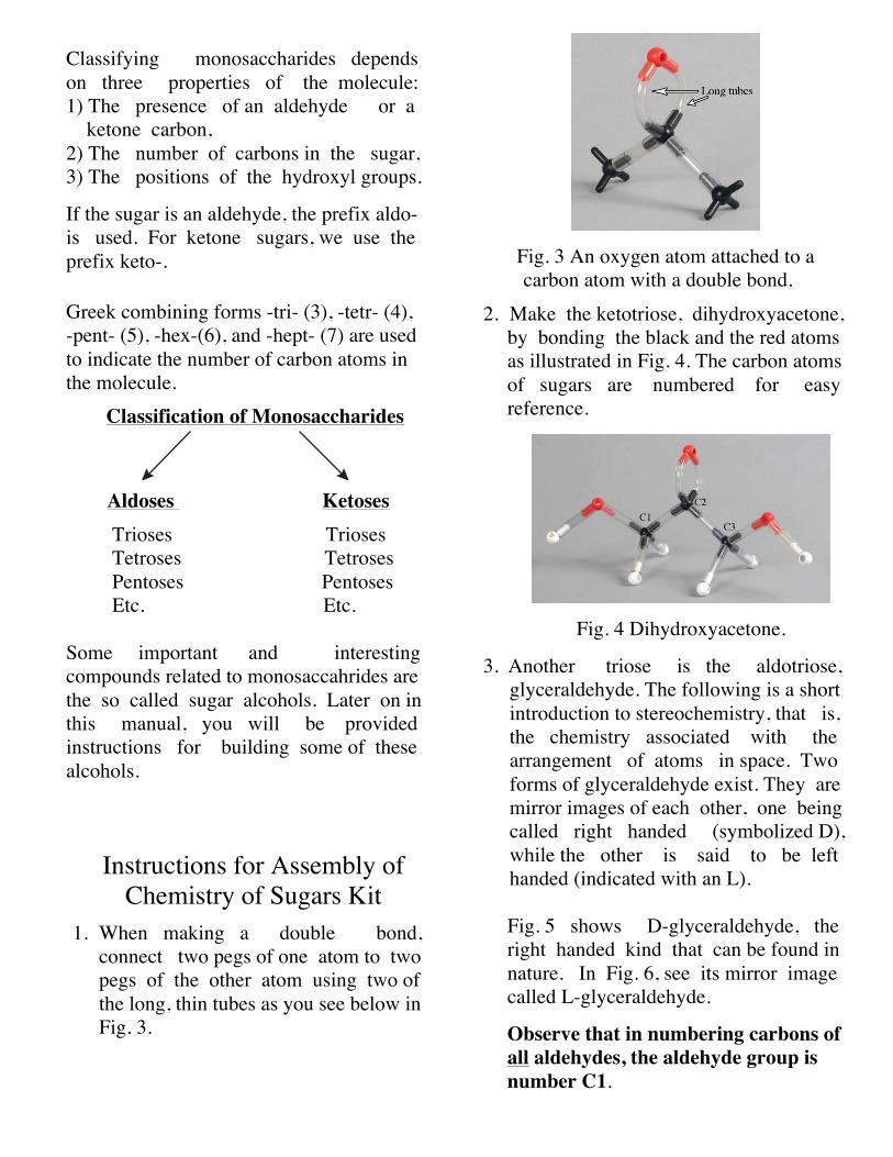
1. When making a double bond, connect two pegs of one atom to two pegs of the other atom using two of the long, thin tubes as you see below in Fig. 3.
Fig. 3 An oxygen atom attached to a carbon atom with a double bond.
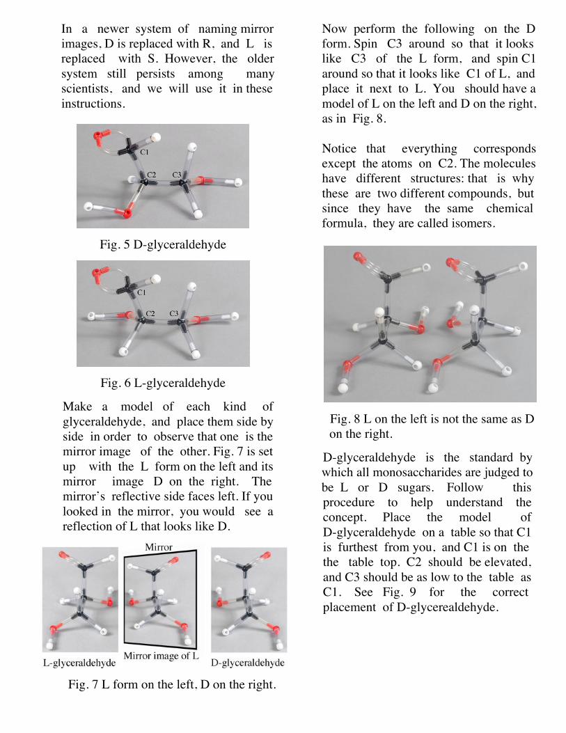
2. Make the ketotriose, dihydroxyacetone, by bonding the black and the red atoms as illustrated in Fig. 4. The carbon atoms of sugars are numbered for easy reference.
Fig. 4 Dihydroxyacetone.
3. Another triose is the aldotriose, glyceraldehyde. The following is a short introduction to stereochemistry, that is, the chemistry associated with the arrangement of atoms in space. Two forms of glyceraldehyde exist. They are mirror images of each other, one being called right handed (symbolized D), while the other is said to be left handed (indicated with an L). Fig. 5 shows D-glyceraldehyde, the right handed kind that can be found in nature. In Fig. 6, see its mirror image called L-glyceraldehyde.
Observe that in numbering carbons of all aldehydes, the aldehyde group is number C1.
In a newer system of naming mirror images, D is replaced with R, and L is replaced with S. However, the older system still persists among many scientists, and we will use it in these instructions.
Fig. 5 D-glyceraldehyde
Fig. 6 L-glyceraldehyde
Make a model of each kind of glyceraldehyde, and place them side by side in order to observe that one is the mirror image of the other. Fig. 7 is set up with the L form on the left and its mirror image D on the right. The mirror’s reflective side faces left. If you looked in the mirror, you would see a reflection of L that looks like D.
Fig. 7 L form on the left, D on the right.
Now perform the following on the D form. Spin C3 around so that it looks like C3 of the L form, and spin C1 around so that it looks like C1 of L, and place it next to L. You should have a model of L on the left and D on the right, as in Fig. 8. Notice that everything corresponds except the atoms on C2. The molecules have different structures: that is why these are two different compounds, but since they have the same chemical formula, they are called isomers.
Fig. 8 L on the left is not the same as D on the right.
D-glyceraldehyde is the standard by which all monosaccharides are judged to be L or D sugars. Follow this
procedure to help understand the concept. Place the model of
D-glyceraldehyde on a table so that C1 is furthest from you, and C1 is on the the table top. C2 should be elevated, and C3 should be as low to the table as C1. See Fig. 9 for the correct placement of D-glycerealdehyde.

Fig. 9 D-glyceraldehyde with C1 and C3 “down” and C2 “up.”
Now observe the following:
1) C2 is the only carbon atom with four different groups of atoms bonded to it. An atom with four different groups attached has the property of existing either in a left handed form (L) or a right handed form (D). We will call this type of atom a stereogenic atom. (There are five other terms in use as well for a stereogenic atom). A molecule with a stereogenic atom can usually exist in a D or L form.
2) C2 is the next to the last carbon atom. It is this stereogenic carbon that determines whether the molecule is D or L. If the hydroxyl group (–OH) is on the right, the molecule is a D sugar. If the –OH is on the left, the sugar is L.
3) The number of stereogenic atoms determines the number of possible forms that can exist. Using the term 2n, where n = the number of stereogenic centers to calculate
the possible number of aldotrioses, we
get 21, which = 2. The possible number of aldohexoses is 24 =16.
In another demonstration of the difference between the two molecules, place one of them on the top of the other to see that they are almost in register. You will again see the different placements of atoms around C2. See Fig. 10.
Fig. 10 Atoms around C2 not in register.
Dihydroxyacetone and (D) glycer- aldehyde are very common and important compounds formed in the metabolism of sugars in all living cells.
Dihydroxyacetone is an ingredient in some over-the-counter tanning agents.
4. Adding another H-C-O-H to the three- carbon sugars brings us to the tetroses, the four-carbon sugars. They have two stereogenic carbons, so there are four aldotetroses, two erythrose sugars (a D and an L) and two threose sugars (a D and an L).
As we build more monosaccharides, we will only consider the D forms.
Fig. 11 shows the structure of D- erythrose.

Fig. 11 The structure of D-erythrose.
If you want to make any D sugar into an L sugar, you must switch
the H– and –OH on every stereogenic carbon atom.
5. See Fig. 12 below for the structure of D-threose.
Fig. 12 The structure of D-threose.
6. As previously stated there are two ketotetroses, a D and an L erythrulose.
Numbering the carbons in ketoses starts with C1.
The following is an illustration of (D) erythrulose (Fig. 13). Observe where the numbering of the carbon atoms begins.
Fig. 13 D-erythrulose.
7. Adding another H-C-O-H to the four- carbon sugars brings us to the pentoses, the five-carbon sugars.
There are four D aldopentoses and four L aldopentoses. They are 1) D and L ribose, 2) D and L arabinose, 3) D and L xylose, and 4) D and L lyxose.
The most important pentose is D-ribose, so we will only build this five-carbon aldose. D-ribose is the sugar that, along with phosphate, makes up the backbone of RNA molecules which are built according to instructions in DNA. See Fig. 14 for the open chain structure of D ribose.
Fig. 14 D-ribose (open chain).
Within living cells, monosaccharides containing four or more carbon atoms actually form ring structures. Ring forms occur because they are more stable than straight chains. The following illustrations

will guide you in understanding how the chains form rings. Fig. 15 shows how D-ribose can become twisted so that C1 is aligned next to the oxygen of C4. The Symbol δ means partial, so δ+ means partial positive charge, and δ− means partial negative charge. Negatives and positives attract each other, so the negative C4 oxygen gets attracted to and bonded with the C1 carbon. See Fig.16, Fig. 17 and Fig. 18.
Fig. 15 Twisted D-ribose.
Fig. 16 Oxygen on C4 loses hydrogen Oxygen on C1 loses one bond to C1.
Fig. 17 D-ribose completed ring.
Fig. 18 Another view of D-ribose ring.
A very important, modified version of ribose is the sugar that, along with phosphate, forms the backbone of DNA molecules. This sugar is called D-2-deoxyribose, the 2 referring to C2, and the deoxy- meaning that an oxygen atom is missing from the ribose. Fig. 19 shows 2-deoxyribose. Compare it to Fig. 18.

Fig. 19 A 2-deoxyribose molecule.
9. There are four ketopentoses possible. They are 1) D and L ribulose, and 2) D and L xylulose. Two phosphate groups bonded to D-ribulose make a compound known as ribulose 1, 3-bisphosphate (RuBP). In photosynthetic organisms such as plants, algae, and blue-green bacteria, CO2 is bonded to RuBP. Then the RuBP-CO2 is made into two molecules of glyceraldehyde which can then be made into glucose and fructose. The process of fusing CO2 with RuBP is known as carbon fixation, and it supplies not only plants and algae with food, but all living animals as well.
Plant chemists estimate that the enzyme ribulose bisphosphate carboxylase/ oxidase, or RUBISCO, which attaches the CO2 to RuBP is the most abundant protein on the planet. Fig. 20 indicates where two phosphates are bonded to D-ribulose.
Fig. 20 D-ribulose showing where the phos- phates replace hydroxyl (-OH) groups.
Bonding a phosphate group to C1 and to C5 of D-ribulose yields 1, 5-ribulose bisphosphate. See Fig. 21.
Fig. 21 Ribulose-1, 5-bisphosphate.
10. There are 16 possible aldohexoses; eight D and eight L forms. However, the most common in nature are D-glucose and D-galactose, so building of those alone will be treated here. The following illustration is called a Fisher projection formula (Fig. 21). The intersection of two lines indicates the presence of a carbon atom. C1 is an aldehyde, and there are six carbons in the molecule, so this must be an aldohexose. Is it a D or an L sugar? The next to the last carbon has a hydroxyl group on the right, so it must be a D sugar. Fig. 21 A fisher projection formula for a D sugar.
C
CH OH
O H
OH
OH
OH
HO
H
H
H
H
2
1
2
3
4
5
6
HO
OO
O
O
O
C1
C2 C5C4C3
HO
HO
HO H
HO
HO
H H
H
H H
PP

It could be glucose, or it might be galactose. Let’s look at a model of glucose, in Fig. 22, first with C1 “up” to see where the hydroxyl groups should be on C3 and on C5.
Fig. 22 A model of D-glucose with C1 “up.”
The C3 hydroxyl is on the left, and on C5 it is on the right. Let’s put that information into the Fisher diagram in Fig. 23.
Fig. 23 A partial Fisher formula.
Now turn the model of D-glucose over so that C1 is “down.” In Fig. 24, the hydroxyl group on C2 is on the right, and the C4 hydroxyl group is also on the right. With the placement of the last two hydroxyl groups, we can complete our Fisher formula as shown in Fig. 25. Fig. 25 is an exact duplicate of Fig. 21, which we have just demonstrated is the Fisher projection formula for D-glucose.
Fig. 24 A model of D-glucose with C1 “down.”
Fig. 25 A completed Fisher formula for D-glucose.
It was stated earlier that monosaccharides with four or more carbons will cyclize to form rings which are more stable than straight chains. We will now take a close look at how D-glucose cyclizes.
11. Rotate the bonds of a model of D-glucose so that it looks like the photograph in Fig. 26. In a water solution in a test tube or in a living cell, the C1 carbon, with the oxygen and hydrogen atoms attached, is free to rotate. About half the time, the oxygen will be “down” as in Fig. 26, and in half of time, the oxygen will be “up,” as in Fig. 27.
C
CH OH
O H
OH
HO
H
H
2
1
2
3
4
5
6
C
CH OH
O H
OH
OH
OH
HO
H
H
H
H
2
1
2
3
4
5
6

Fig. 26 D-glucose with oxygen on C1 “down.”
Fig. 27 D-glucose with oxygen on C1 “up.”
12. Prepare the glucose, with the oxygen “down,” for cyclization by removing the hydrogen atom from the C5 oxygen and taking one of the double bonds from the C1 carbon as indicated in Fig. 28.
Fig. 28 Glucose prepared for cyclization.
Fig. 29 shows the result of D-glucose
forming a ring with the new C1 –OH in a “down,” technically called the
alpha (α), position.
Fig. 29 α-D-glucose. 13. Prepare the glucose, with the oxygen “up,” for cyclization by removing the hydrogen atom from the C5 oxygen and taking one of the double bonds from the C1 carbon as indicated in Fig. 30.
Fig. 30 Glucose prepared for cyclization.
Fig. 31 shows the result of D-glucose forming a ring with the new C1 –OH
in an “up,” technically called the beta (β), position.
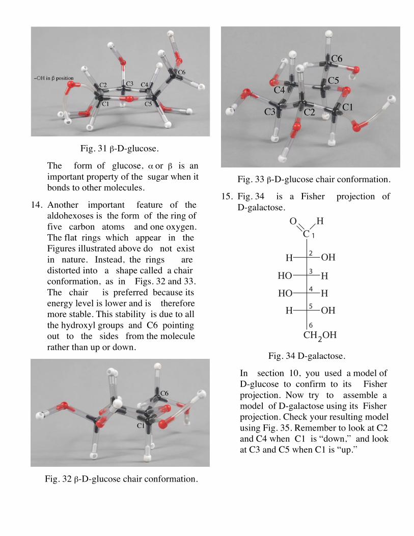
Fig. 31 β-D-glucose.
The form of glucose, α or β is an important property of the sugar when it bonds to other molecules.
14. Another important feature of the aldohexoses is the form of the ring of five carbon atoms and one oxygen. The flat rings which appear in the Figures illustrated above do not exist in nature. Instead, the rings are distorted into a shape called a chair conformation, as in Figs. 32 and 33. The chair is preferred because its energy level is lower and is therefore more stable. This stability is due to all the hydroxyl groups and C6 pointing out to the sides from the molecule rather than up or down.
Fig. 32 β-D-glucose chair conformation.
Fig. 33 β-D-glucose chair conformation.
15. Fig. 34 is a Fisher projection of D-galactose.
Fig. 34 D-galactose.
In section 10, you used a model of D-glucose to confirm to its Fisher projection. Now try to assemble a model of D-galactose using its Fisher projection. Check your resulting model using Fig. 35. Remember to look at C2 and C4 when C1 is “down,” and look at C3 and C5 when C1 is “up.”
C
CH OH
O H
OH
OH
OH
HO
H
H
H
H
2
1
2
3
4
5
6
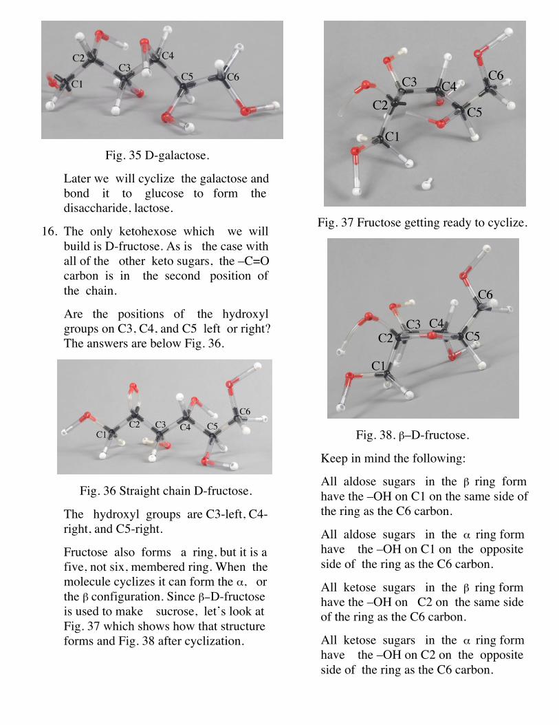
Fig. 35 D-galactose.
Later we will cyclize the galactose and bond it to glucose to form the disaccharide, lactose.
16. The only ketohexose which we will build is D-fructose. As is the case with all of the other keto sugars, the –C=O carbon is in the second position of the chain.
Are the positions of the hydroxyl groups on C3, C4, and C5 left or right? The answers are below Fig. 36.
Fig. 36 Straight chain D-fructose.
The hydroxyl groups are C3-left, C4- right, and C5-right.
Fructose also forms a ring, but it is a five, not six, membered ring. When the molecule cyclizes it can form the α, or the β configuration. Since β−D-fructose is used to make sucrose, let’s look at Fig. 37 which shows how that structure forms and Fig. 38 after cyclization.
Fig. 37 Fructose getting ready to cyclize.
Fig. 38. β–D-fructose.
Keep in mind the following:
All aldose sugars in the β ring form have the –OH on C1 on the same side of the ring as the C6 carbon.
All aldose sugars in the α ring form have the –OH on C1 on the opposite side of the ring as the C6 carbon.
All ketose sugars in the β ring form have the –OH on C2 on the same side of the ring as the C6 carbon.
All ketose sugars in the α ring form have the –OH on C2 on the opposite side of the ring as the C6 carbon.
Our brief survey of monosaccharides is complete, so now we can combine those simple sugars into some important double
sugars, i.e. disaccharides. We will build sucrose, lactose, and maltose. These three most common disaccharides are isomers with the same formula: C12H22O11.
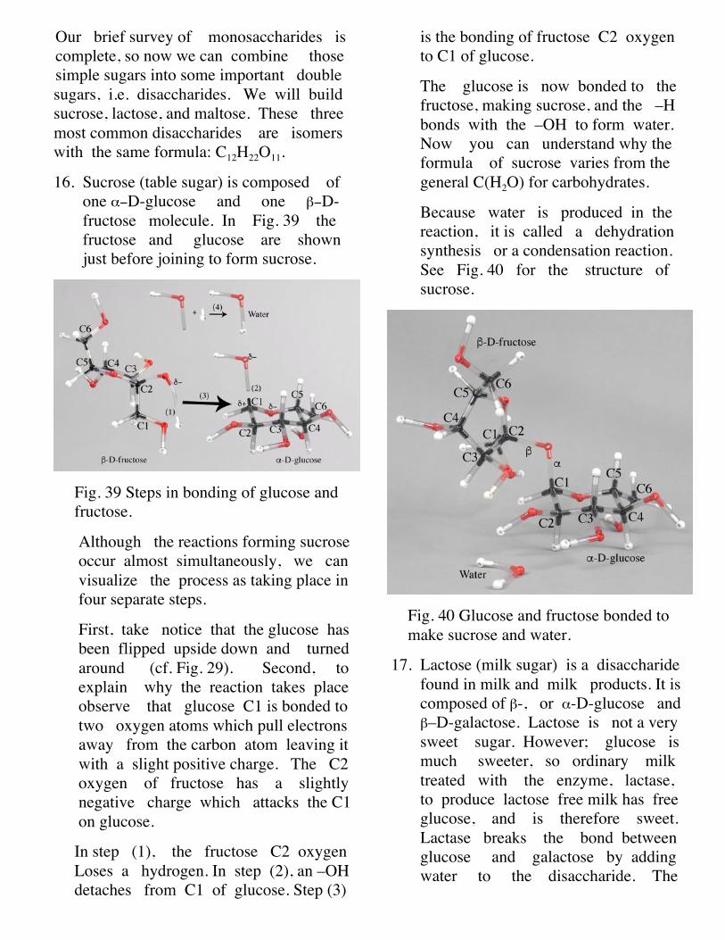
16. Sucrose (table sugar) is composed of one α−D-glucose and one β−D- fructose molecule. In Fig. 39 the fructose and glucose are shown just before joining to form sucrose.
Fig. 39 Steps in bonding of glucose and fructose.
Although the reactions forming sucrose occur almost simultaneously, we can visualize the process as taking place in four separate steps.
First, take notice that the glucose has been flipped upside down and turned around (cf. Fig. 29). Second, to explain why the reaction takes place observe that glucose C1 is bonded to two oxygen atoms which pull electrons away from the carbon atom leaving it with a slight positive charge. The C2 oxygen of fructose has a slightly negative charge which attacks the C1 on glucose.
In step (1), the fructose C2 oxygen Loses a hydrogen. In step (2), an –OH detaches from C1 of glucose. Step (3)
is the bonding of fructose C2 oxygen to C1 of glucose.
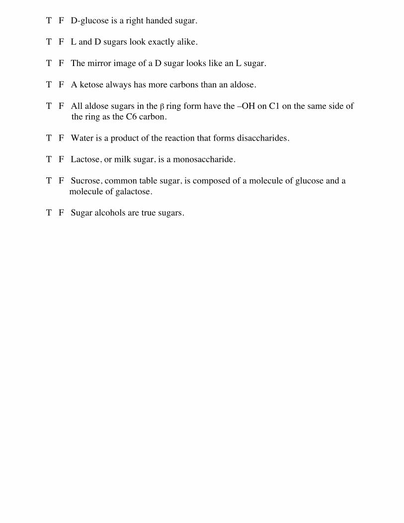
The glucose is now bonded to the fructose, making sucrose, and the –H bonds with the –OH to form water. Now you can understand why the formula of sucrose varies from the general C(H2O) for carbohydrates.
Because water is produced in the reaction, it is called a dehydration synthesis or a condensation reaction. See Fig. 40 for the structure of sucrose.
Fig. 40 Glucose and fructose bonded to make sucrose and water.
17. Lactose (milk sugar) is a disaccharide found in milk and milk products. It is composed of β-, or α-D-glucose and β–D-galactose. Lactose is not a very sweet sugar. However; glucose is much sweeter, so ordinary milk treated with the enzyme, lactase, to produce lactose free milk has free glucose, and is therefore sweet. Lactase breaks the bond between glucose and galactose by adding water to the disaccharide. The

reaction is the opposite of dehydration synthesis, and it is known as a hydrolysis.
Consult Fig. 41 for the structure of β–D-galactose, and then join it to α-D-glucose to make lactose: Fig. 42.
Fig. 41 β–D-galactose
Fig. 42 Lactose with a 1à4-β linkage. The reaction is a dehydration synthesis, so water is an additional product. The term
which describes the bonding between galactose and glucose, β−1à4 linkage, means that C1 β oxygen is bonded to the C4 carbon of glucose.
18. The last disaccharide that is described here is maltose (malt sugar). Maltose is made from one molecule of α-D-glucose and one molecule of either α- or β-D-glucose. The bonding in maltose is an α−1à4 linkage, and again a water molecule is made as one of the products. See Fig. 43, below.
Fig. 43 Maltose with a 1à4-α linkage.
We end our brief study of sugars with structures of some sugar alcohols which are not alcoholic drinks, nor are they sugars. They can be found as natural products in berries, in other fruits, and in some vegetables, and they can be synthesized in industrial facilities.
An alcohol is a carbon compound with at least one –OH (hydroxyl) group bonded to a carbon atom. There are exceptions to this rule, but it is beyond the scope of this instruction booklet to explain them at this time. As you make

models of some sugar alcohols you will immediately see the relationship between sugars and their corresponding sugar alcohols. The names of the sugar alcohols are derived from the parent sugar by dropping –ose ending of the sugar and adding –itol . For example xylose becomes xylitol (–ol is a suffix generally placed on the stem of a name of any alcohol).
Since they are sweet, many food items have sugar alcohols added to them. Additional advantages of the sugar alcohols are they have low caloric value, low glycemic index, low absorbability, and are slow to metabolize. Xylitol in particular is thought to promote good dental health by inhibiting formation of plaque, acting as a bactericide, and causing calcium to be added to the structure of teeth.
There are undesirable side effects to the ingestion of sugar alcohols. These include gas, bloating, abdominal pain, and diarrhea.
A partial list of sugar alcohols include erythritol, glycerol, hydrogenated starch hydrosylates, isomalt, lactitol, mannitol, maltitol, sorbitol, and xylitol.
19. When glucose is hydrogenated (chemically reduced), it is converted into glucitol, also called sorbitol, Fig. 44.
Fig. 44 Reduction of glucose to form glucitol.
Upon the reduction of xylose, xylitol is produced, Fig. 45. Fig. 45 Reduction of xylose to form xylitol.
C
CH OH
O H
OH
OH
OH
HO
H
H
H
H
2
1
2
3
4
5
6
HH H
C
CH OH
HOH
OH
OH
OH
HO
H
H
H
H
2
1
2
3
4
5
6
Glucose Glucitol (sorbitol)
C
CH OH
O H
OH
OH
HO
H
H
H
2
1
2
3
4
5
HH
Xylose
H
CHOH
OH
OH
HO
H
H
H
CH OH2
1
2
3
4
5
Xylitol

ASSESSMENT FOR CHEMISTRY OF SUGARS 1. What are the three main elements of sugars? ________________________,
________________________, and ________________________.
2. Which of the following is not a carbohydrate? a. C6H12O6, b. C6H6O2, c. C3H6O3, d. C5H10O5, e. C4H8O4. _____________ 3. How many carbons are in these sugars? a. a pentose _______, b. a triose _______, c. a hexose _______. 5. Monosaccharides are a. double sugars, b. simple sugars, c. polysaccharides, d. starches, e. amino acids, _______. 6. The following figure is an example of a. a sugar alcohol, b. an amino acid, c. a pentose,
d. a ketose, e. an aldose. _______.
7. Using the letters, match the following diagrams with the correct name. Hydroxyl group _______. Ketone group _______. Aldehyde group _______. A D sugar _______.
a b c d 8. T or F Circle the correct answer. T F If the next to last carbon of a sugar has the hydroxyl group on the right, the sugar is a D sugar. T F The mirror image of D-glyceraldehyde is called D-glucose.
CO H
OHH
CH OH2
1
2
3
HC
O
CC
O
CCOH
CO H
OHH
CH OH2
1
2
3
T F D-glucose is a right handed sugar. T F L and D sugars look exactly alike. T F The mirror image of a D sugar looks like an L sugar. T F A ketose always has more carbons than an aldose. T F All aldose sugars in the β ring form have the –OH on C1 on the same side of the ring as the C6 carbon. T F Water is a product of the reaction that forms disaccharides. T F Lactose, or milk sugar, is a monosaccharide. T F Sucrose, common table sugar, is composed of a molecule of glucose and a molecule of galactose. T F Sugar alcohols are true sugars.