chronology of estrogen receptor expression in testes of...
TRANSCRIPT
526
http://journals.tubitak.gov.tr/medical/
Turkish Journal of Medical Sciences Turk J Med Sci(2015) 45: 526-533© TÜBİTAKdoi:10.3906/sag-1403-34
Chronology of estrogen receptor expression in testes of mouse embryos
Insaf Jasim MAHMOUD1,*, Mohammad Oda SELMAN2, Wameedh Raad Abdulmalik SHEBEB2
1Obesity Research and Therapy Unit, Al-Kindy College of Medicine, Baghdad University, Baghdad, Iraq2Applied Embryology Department, High Institute of Infertility Diagnosis and Assisted Reproductive Technologies, Al-Nahrain
University, Baghdad, Iraq
* Correspondence: [email protected]
1. IntroductionThe testes of mammals are paired organs that essentially perform two functions: production of spermatozoa and hormones, mostly androgens, and also estrogens (1). It is well known that normal testicular development and maintenance of spermatogenesis are controlled by gonadotropins and testosterone, whose effects are modulated by a complex network of factors produced locally. Among these, estrogens are concerned (2). The traditional view of estradiol as the “female” hormone and of testosterone as the “male” hormone has been challenged in recent years (3). The increased interest about the role of estrogens in the male is largely due to the demonstration that male fertility is impaired in mice lacking estrogen receptor-α (ERα) (4), or aromatase (5), along with the discovery of a second estrogen receptor-β (ERβ) (6), which is widely expressed in the male reproductive tract (7).
The androgen/estrogen balance is essential for normal sexual development and reproduction in mammals. In the mammalian testes, maintenance of this balance is under a fine-tuning via endocrine and paracrine factors, but is also related to the aromatase enzyme activity (8).
Prenatal exposure to diethylstilbestrol, a synthetic estrogen, induces abnormalities of the male genital tract of mouse and human (9).
Estrogens are synthesized in the male reproductive system in at least three different cell types of the testis: Leydig cells, Sertoli cells, and germ cells, as well as in the epithelial cells of the epididymis. In the immature testis, the primary source of estrogen is Sertoli cells (10). Currently, a growing body of evidence indicates that germ cells serve as the major source of estrogens in the male reproductive tract (11,12).
That estrogen can influence testis and epididymis function is not unexpected, given the evidence presented that estrogen biosynthesis, via the aromatase enzyme and action on its receptors (α and/or β), occurs in these tissues.
Estrogen is produced by the testes from the fetal period throughout adulthood and, similarly, ERα and β are found in the testes at all ages. While some cells express both ERα and β, such as Leydig cells, the cells in the seminiferous epithelium appear to predominantly contain ERβ.
Background/aim: To localize and determine the time of expression of estrogen receptors (ERs) in testes of mouse embryos by histology and immunohistochemistry.
Materials and methods: Fifty-four mature Swiss-Webster mice (Mus musculus) were used. Group 1 consisted of 34 mature pregnant females, 3 of which were sacrificed every day from 10.5 to 20.5 days postcoitus (dpc). One testis was removed from their embryos and processed for histology and immunohistochemistry. Group 2 consisted of 20 postnatal mice: 5 postpartum (P0) males, 5 males 4 weeks old, 5 males 8 weeks old, and 5 females 4 weeks old.
Results: The first nuclear detection of ERs was observed in embryonic male gonads at 11.5 dpc, and a highly significant increase (P < 0.01) was observed at embryonic days 11.5, 13.5, 15.5, 16.5, 17.5, and 19.5, with a peak at 17.5 dpc and continuing to 20.5 dpc. ER expression increased further after birth.
Conclusion: Expression of ERs occurred at certain days during mouse embryonic development, indicating the need for estrogen for certain metabolic or morphological events occurring at these days. After birth, estrogen played an important role in proliferation and maturation of certain cells in the testes. Another rise in ER expression occurred during puberty in the mature testis.
Key words: Expression of estrogen receptors, testis of mouse embryo
Received: 07.03.2014 Accepted/Published Online: 05.06.2014 Printed: 30.06.2015
Research Article
527
MAHMOUD et al. / Turk J Med Sci
Evidence suggests that estrogens act at multiple levels to control, or interfere with, spermatogenesis (13).
In this study we aimed to localize and determine the time of expression of estrogen receptors (ERs) in the testes of mouse embryos by histological and immunohistochemical evaluation.
2. Materials and methodsFifty-four mature Swiss-Webster mice (Mus musculus) were used. They were obtained from the animal house of the High Institute of Infertility Diagnosis And Assistant Reproductive Technology (HIID & ART)/Al-Nahrain University. The animals were used according to the general guidelines of laboratory animals (Iraqi general health law, experimental protocol section, 1981) and according to the protocol of the Laboratory Animal Center of Baghdad University, Iraq. The experimental protocol was approved by the HIID & ART/Al-Nahrain University, Baghdad, Iraq, on 2/10/2012.
Vaginal smears were performed for all the adult female mice to determine their estrus cycle stage and detect heat stage for mating (14).
Thirty-four mature pregnant females (Group 1) were sacrificed every day from 10.5 to 20.5 days postcoitus (dpc), and one testis was removed from their embryos and processed for histology and immunohistochemistry as follows: an incision was made in the abdomen to remove the live embryos from the uterus. The female embryos were discarded. Each male embryo was washed and fixed in 10% formaldehyde solution for 24 h, then stored in 70% ethanol alcohol for histology tissue processing. Sex of embryos at 10.5 and at 11.5 dpc was determined using polymerase chain reaction (PCR), by detecting the Y-chromosome (SRY gene) in the male mouse embryo.
The embryos were dissected out of their extra embryonic membranes and placed in normal saline and then fixed in 10% formaldehyde solution for 8 h to harden the tissue. The testes and pelvic region were transferred to 10% formaldehyde solution again for a further 16 h to complete fixation.
For immunohistochemical detection of ERs, 4 µm paraffin sections were mounted on Fisher brand positively charged slides (15).
Actual assessment of immunohistochemistry was performed by image analysis of tissue sections using Aperio image scope v 11.1.2.760 software and positive pixel count algorithms were used to quantify the amount of a specific color in a slide image. This algorithm has a set of default input parameters when first selected. These inputs were preconfigured for brown color quantification in the three intensity ranges (weak, positive, and strong).
Immunohistochemistry staining in testis sections of the embryonic and postnatal groups was quantified as the percentage of the positive pixels for ERs color intensity. The data were expressed as mean ± standard deviation and analyzed using Student’s t-test to compare values from the embryonic and postnatal groups at individual time points. Differences between the groups were considered highly significant at P < 0.01, significant at P < 0.05, or nonsignificant at P > 0.05.
Group 2 consisted of 20 postnatal mice, including 5 mice postpartum zero day males (P0), 5 four-week-old males, 5 eight-week-old males, and 5 four-week-old females. One testis was removed from male mice and processed for histology and immunohistochemistry as described above. One ovary was removed from female mice and used as controls for ER detection with immunohistochemistry.
3. Results A cell containing a nucleus with a dense brown color was considered positive for ERs (16). Sections from embryonic and postnatal mice testes showed variable intensity of immunohistochemical stain for ERs.
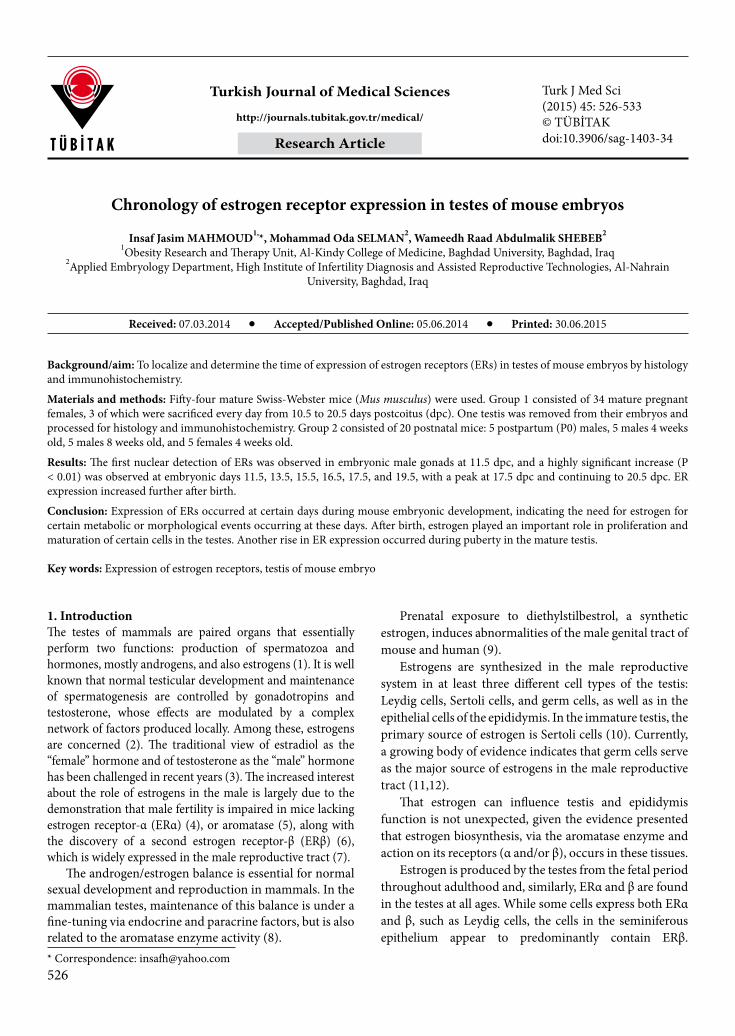
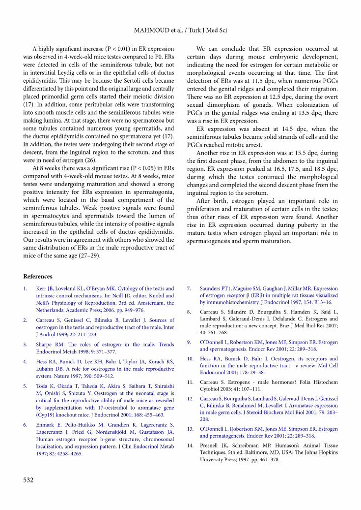
In the testes of mice at P0, few cells were positive for ERs. In the testis of mice at 4 weeks of age, most of the spermatogenic cells were positive (Figure 1), but the ductus epididymidis showed negative results.
Eight-week-old mice testes showed strong positive signals for anti-ER antibodies in spermatogonia, while spermatocyte and spermatid showed weak positive signals (Figure 2). Positive signals increased in intensity in the ductus epididymidis. Gonads of male embryos at 10.5, 12.5, and 14.5 dpc were negatively stained for anti-ER antibodies in primordial germ cells (PGCs), but the nuclear stain was positive in these cells at 11.5, 13.5, and 15.5 dpc. (Figure 3).
Figure 1. Four-week-old mouse testis. Immunohistochemistry for ER identification. Staining by peroxidase/DAB (brown). Positive stain in spermatogenic cells (arrow heads) (400×).

528
MAHMOUD et al. / Turk J Med Sci
Testes of embryos at 15.5 and at 16.5 dpc showed strong positive intensity for anti-ER antibodies in the peritubal cells around sex cords and weak positive intensity in PGCs inside seminiferous tubules. At 17.5 dpc there was strong positive intensity in PGCs of the seminiferous tubules and weak positive intensity in the peritubal cells around sex cords (Figure 4). Embryonic testes at 18.5 dpc showed strong positive intensity for anti-ER antibodies in both peritubal cells and in PGCs. At 19.5 dpc, positive intensity for anti-ER antibodies in PGCs inside seminiferous tubules was seen (Figure 5). Embryonic testes at 20.5 dpc showed strong positive intensity for anti-ER antibodies in PGCs inside seminiferous tubules and weak positive intensity in peritubal cells.
A 2-tailed sample statistics t-test for ER expression in the two sequenced embryonic groups showed a highly significant increase (P < 0.01) between embryonic days 10.5 and 11.5, 18.5 and 19.5, and between 20.5 dpc and P0. There was also a significant increase (P < 0.05) among embryonic days 11.5–12.5, 12.5–13.5, 15.5–16.5, 17.5–
Figure 2. Eight-week-old mouse testis. Immunohistochemistry for ER identification. Positive nuclear stain in spermatogonia (yellow arrow heads) and spermatids (red arrow head). Peroxidase/DAB (brown) (400×).
a b
Figure 3. Section of mouse embryo at 11.5 dpc. Immunohistochemistry for ER identification. a: Whole mount sagittal section (4×). b: Gonads showing positive nuclear staining of ERs in PGCs (arrow heads). Peroxidase/DAB (brown) (400×).
a b
Figure 4. Section of testis of mouse embryo at 17.5 dpc. Immunohistochemistry for ERs identification. a: Sagittal section of testis (4×). b: Strong positive staining of PGCs (arrow heads). Peroxidase/DAB (brown) (400×).

529
MAHMOUD et al. / Turk J Med Sci
18.5, and 19.5–20.5, while there was a nonsignificant decrease (P > 0.05) among embryonic days 13.5–14.5, 14.5–15.5, and 16.5–17.5 (Tables 1 and 2; Figures 6 and 7).
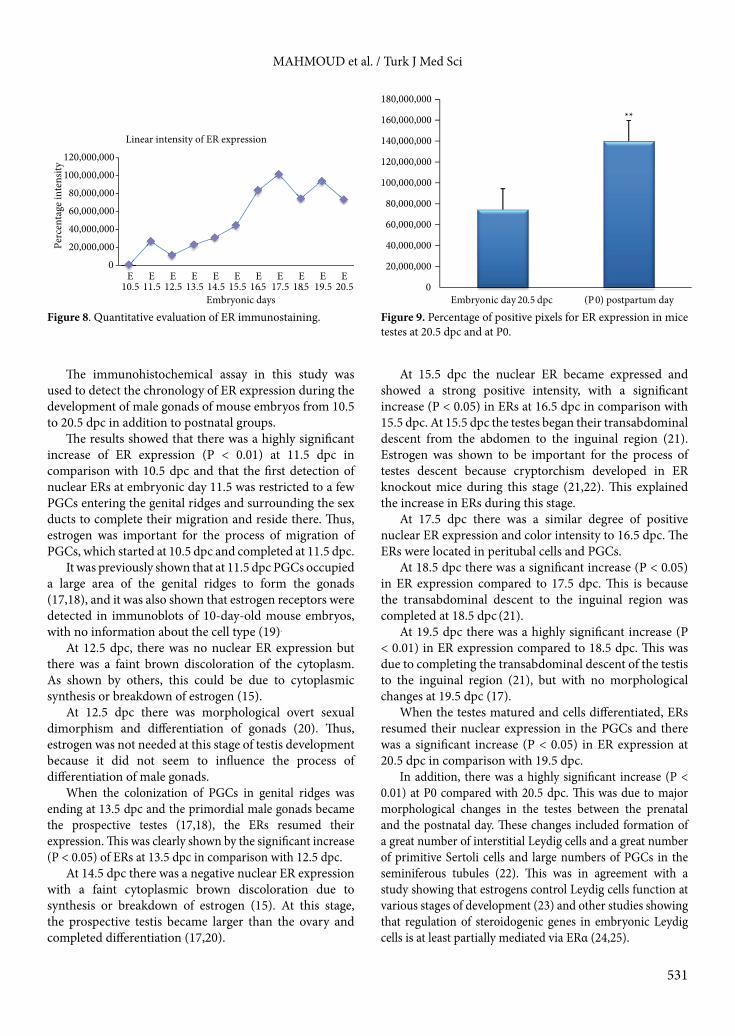
The percentage of positive pixels for ER expression in embryonic male gonads showed distinct sequential elevation during embryonic life from 10.5 to 20.5 dpc (Figure 8). Comparison between percentages of positive pixels for ERs at 20.5 dpc and at P0 showed a highly significant increase at P0 (Figure 9).
4. DiscussionThe sensitivity of the quantitative PCR system was high and specific for amplification of the SRY gene. It was employed on sections of all mice at the indifferent stage, 10.5 dpc, and 11.5 dpc. Positive sections were males, because the specificity was 100% for the SRY gene in male mice, and negative sections were females because the Y-chromosome was absent in female embryos. Thus, paraffin block samples of male embryos were isolated from female samples.
a b
Figure 5. Section of testis of a mouse embryo at 19.5 dpc. Immunohistochemistry for ERs identification. a: Sagittal section of testis (4×). b: Strong positive staining of PGCs (arrows). Peroxidase/DAB (brown) (400×).
Table 1. Percentage of positive pixels for ER expression in gonads of male embryos.
Embryonic groups No. of tissue sections
Percentage of positive pixels for ERs (mean ± SD)
10.5 dpc 13 7639 ± 9214
11.5 dpc 13 266,638 ± 376,548
12.5 dpc 13 112,072 ± 159,189
13.5 dpc 13 228,888 ± 304,751
14.5 dpc 13 311,711 ± 235,154
15.5 dpc 13 444,728 ± 409,341
16.5 dpc 13 831,399 ± 285,918
17.5 dpc 13 1,017,942 ± 203,415
18.5 dpc 13 745,762 ± 223,416
19.5 dpc 13 943,816 ± 142,626
20.5 dpc 13 738,419 ± 264,339

530
MAHMOUD et al. / Turk J Med Sci
Table 2. ER expression in two sequenced embryonic groups. ** (P < 0.01) * (P < 0.05)
ER expressionmean ± SD
95% confidence interval of the difference P-values
10.5 vs 11.5 dpc 2.589995 ± 3.79186 4.88139–2.98595 0.000**
11.5 vs 12.5 dpc 1.54566 ± 4.16832 9.73230–4.06455 0.020*
12.5 vs 13.5 dpc 1.1681± 3.67574 3.38939–1.05306 0.027*
13.5 vs 14.5 dpc 8.28224 ± 3.99677 3.24344–1.58700 0.469
14.5 vs 15.5 dpc 1.33018 ± 4.17897 3.85551–1.19515 0.273
15.5 vs 16.5 dpc 3.86670 ± 4.90208 6.82900–9.04405 0.015*
16.5 vs 17.5 dpc 1.86543 ± 3.70906 4.10680–3.75939 0.095
17.5 vs 18.5 dpc 2.72181 ± 2.80530 1.02658–4.41703 0.004*
18.5 vs 19.5 dpc 1.98055 ± 1.53050 2.90542–1.05568 0.001**
19.5 vs 20.5 dpc 2.05397 ± 2.70190 4.21233–3.68671 0.018*
20.5 vs (P0)day 1.37375 ± 2.56453 1.52873–1.21878 0.000**
E 10.5
E 11.5
E 12.5
E 13.5
E 14.5
E 15.5
E 16.5
E 17.5
E 18.5
E 19.5
E 20.5
Series1 76,386 26,663 11,207 22,888 31,171 44,472 83,139 10,179 74,576 94,381 73841
0
20,000,000
40,000,000
60,000,000
80,000,000
100,000,000
120,000,000
Perc
enta
ge of
ER
inte
nsity
Percentage of the positive pixels for ER expression
** * *
* * ** *
020,000,000 40,000,000 60,000,000 80,000,000
100,000,000 120,000,000 140,000,000
E 10
.5
E 11
.5
E 11
.5
E 12
.5
E 12
.5
E 13
.5
E 13
.5
E 14
.5
E 14
.5
E 15
.5 E
15.5
E
16.5
E
16.5
E
17.5
E
17.5
E
18.5
E
18.5
E
19.5
E
19.5
E
20.5
Tailed t -test of embryonic groups
Figure 6. Percentage of positive pixels for ER expression in gonads of male embryos.
Figure 7. ER expression in two sequenced embryonic groups.** (P < 0.01) * (P < 0.05)
531
MAHMOUD et al. / Turk J Med Sci
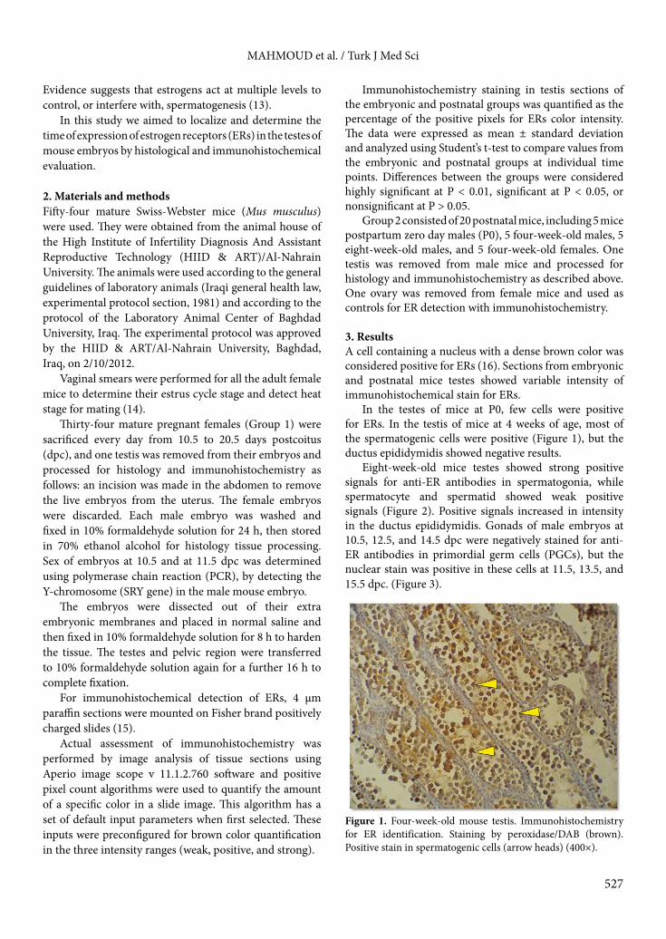
The immunohistochemical assay in this study was used to detect the chronology of ER expression during the development of male gonads of mouse embryos from 10.5 to 20.5 dpc in addition to postnatal groups.
The results showed that there was a highly significant increase of ER expression (P < 0.01) at 11.5 dpc in comparison with 10.5 dpc and that the first detection of nuclear ERs at embryonic day 11.5 was restricted to a few PGCs entering the genital ridges and surrounding the sex ducts to complete their migration and reside there. Thus, estrogen was important for the process of migration of PGCs, which started at 10.5 dpc and completed at 11.5 dpc.
It was previously shown that at 11.5 dpc PGCs occupied a large area of the genital ridges to form the gonads (17,18), and it was also shown that estrogen receptors were detected in immunoblots of 10-day-old mouse embryos, with no information about the cell type (19).
At 12.5 dpc, there was no nuclear ER expression but there was a faint brown discoloration of the cytoplasm. As shown by others, this could be due to cytoplasmic synthesis or breakdown of estrogen (15).
At 12.5 dpc there was morphological overt sexual dimorphism and differentiation of gonads (20). Thus, estrogen was not needed at this stage of testis development because it did not seem to influence the process of differentiation of male gonads.
When the colonization of PGCs in genital ridges was ending at 13.5 dpc and the primordial male gonads became the prospective testes (17,18), the ERs resumed their expression. This was clearly shown by the significant increase (P < 0.05) of ERs at 13.5 dpc in comparison with 12.5 dpc.
At 14.5 dpc there was a negative nuclear ER expression with a faint cytoplasmic brown discoloration due to synthesis or breakdown of estrogen (15). At this stage, the prospective testis became larger than the ovary and completed differentiation (17,20).
At 15.5 dpc the nuclear ER became expressed and showed a strong positive intensity, with a significant increase (P < 0.05) in ERs at 16.5 dpc in comparison with 15.5 dpc. At 15.5 dpc the testes began their transabdominal descent from the abdomen to the inguinal region (21). Estrogen was shown to be important for the process of testes descent because cryptorchism developed in ER knockout mice during this stage (21,22). This explained the increase in ERs during this stage.
At 17.5 dpc there was a similar degree of positive nuclear ER expression and color intensity to 16.5 dpc. The ERs were located in peritubal cells and PGCs.
At 18.5 dpc there was a significant increase (P < 0.05) in ER expression compared to 17.5 dpc. This is because the transabdominal descent to the inguinal region was completed at 18.5 dpc (21).
At 19.5 dpc there was a highly significant increase (P < 0.01) in ER expression compared to 18.5 dpc. This was due to completing the transabdominal descent of the testis to the inguinal region (21), but with no morphological changes at 19.5 dpc (17).
When the testes matured and cells differentiated, ERs resumed their nuclear expression in the PGCs and there was a significant increase (P < 0.05) in ER expression at 20.5 dpc in comparison with 19.5 dpc.
In addition, there was a highly significant increase (P < 0.01) at P0 compared with 20.5 dpc. This was due to major morphological changes in the testes between the prenatal and the postnatal day. These changes included formation of a great number of interstitial Leydig cells and a great number of primitive Sertoli cells and large numbers of PGCs in the seminiferous tubules (22). This was in agreement with a study showing that estrogens control Leydig cells function at various stages of development (23) and other studies showing that regulation of steroidogenic genes in embryonic Leydig cells is at least partially mediated via ERα (24,25).
0
20,000,000
40,000,000
60,000,000
80,000,000
100,000,000
120,000,000
E 10.5
E 11.5
E 12.5
E 13.5
E 14.5
E 15.5
E 16.5
E 17.5
E 18.5
E 19.5
E 20.5
Perc
enta
ge in
tens
ity
Embryonic days
Linear intensity of ER expression
**
0
20,000,000
40,000,000
60,000,000
80,000,000
100,000,000
120,000,000
140,000,000
160,000,000
180,000,000
Embryonic day 20.5 dpc (P 0) postpartum day
Figure 8. Quantitative evaluation of ER immunostaining. Figure 9. Percentage of positive pixels for ER expression in mice testes at 20.5 dpc and at P0.
532
MAHMOUD et al. / Turk J Med Sci
A highly significant increase (P < 0.01) in ER expression was observed in 4-week-old mice testes compared to P0. ERs were detected in cells of the seminiferous tubule, but not in interstitial Leydig cells or in the epithelial cells of ductus epididymidis. This may be because the Sertoli cells became differentiated by this point and the original large and centrally placed primordial germ cells started their meiotic division (17). In addition, some peritubular cells were transforming into smooth muscle cells and the seminiferous tubules were making lumina. At that stage, there were no spermatozoa but some tubules contained numerous young spermatids, and the ductus epididymidis contained no spermatozoa yet (17). In addition, the testes were undergoing their second stage of descent, from the inguinal region to the scrotum, and thus were in need of estrogen (26).
At 8 weeks there was a significant rise (P < 0.05) in ERs compared with 4-week-old mouse testes. At 8 weeks, mice testes were undergoing maturation and showed a strong positive intensity for ERs expression in spermatogonia, which were located in the basal compartment of the seminiferous tubules. Weak positive signals were found in spermatocytes and spermatids toward the lumen of seminiferous tubules, while the intensity of positive signals increased in the epithelial cells of ductus epididymidis. Our results were in agreement with others who showed the same distribution of ERs in the male reproductive tract of mice of the same age (27–29).
We can conclude that ER expression occurred at certain days during mouse embryonic development, indicating the need for estrogen for certain metabolic or morphological events occurring at that time. The first detection of ERs was at 11.5 dpc, when numerous PGCs entered the genital ridges and completed their migration. There was no ER expression at 12.5 dpc, during the overt sexual dimorphism of gonads. When colonization of PGCs in the genital ridges was ending at 13.5 dpc, there was a rise in ER expression.
ER expression was absent at 14.5 dpc, when the seminiferous tubules became solid strands of cells and the PGCs reached mitotic arrest.
Another rise in ER expression was at 15.5 dpc, during the first descent phase, from the abdomen to the inguinal region. ER expression peaked at 16.5, 17.5, and 18.5 dpc, during which the testes continued the morphological changes and completed the second descent phase from the inguinal region to the scrotum.
After birth, estrogen played an important role in proliferation and maturation of certain cells in the testes; thus other rises of ER expression were found. Another rise in ER expression occurred during puberty in the mature testis when estrogen played an important role in spermatogenesis and sperm maturation.
References
1. Kerr JB, Loveland KL, O’Bryan MK. Cytology of the testis and intrinsic control mechanisms. In: Neill JD, editor. Knobil and Neill’s Physiology of Reproduction. 3rd ed. Amsterdam, the Netherlands: Academic Press; 2006. pp. 949–976.
2. Carreau S, Genissel C, Bilinska B, Levallet J. Sources of oestrogen in the testis and reproductive tract of the male. Inter J Androl 1999; 22: 211–223.
3. Sharpe RM. The roles of estrogen in the male. Trends Endocrinol Metab 1998; 9: 371–377.
4. Hess RA, Bunick D, Lee KH, Bahr J, Taylor JA, Korach KS, Lubahn DB. A role for oestrogens in the male reproductive system. Nature 1997; 390: 509–512.
5. Toda K, Okada T, Takeda K, Akira S, Saibara T, Shiraishi M, Onishi S, Shizuta Y. Oestrogen at the neonatal stage is critical for the reproductive ability of male mice as revealed by supplementation with 17-oestradiol to aromatase gene (Cyp19) knockout mice. J Endocrinol 2001; 168: 455–463.
6. Enmark E, Pelto-Huikko M, Grandien K, Lagercrantz S, Lagercrantz J, Fried G, Nordenskjöld M, Gustafsson JA. Human estrogen receptor b-gene structure, chromosomal localization, and expression pattern. J Clin Endocrinol Metab 1997; 82: 4258–4265.
7. Saunders PT1, Maguire SM, Gaughan J, Millar MR. Expression of estrogen receptor β (ERβ) in multiple rat tissues visualized by immunohistochemistry. J Endocrinol 1997; 154: R13–16.
8. Carreau S, Silandre D, Bourguiba S, Hamden K, Said L, Lambard S, Galeraud-Denis I, Delalande C. Estrogens and male reproduction: a new concept. Braz J Med Biol Res 2007; 40: 761–768.
9. O’Donnell L, Robertson KM, Jones ME, Simpson ER. Estrogen and spermatogenesis. Endocr Rev 2001; 22: 289–318.
10. Hess RA, Bunick D, Bahr J. Oestrogen, its receptors and function in the male reproductive tract - a review. Mol Cell Endocrinol 2001; 178: 29–38.
11. Carreau S. Estrogens - male hormones? Folia Histochem Cytobiol 2003; 41: 107–111.
12. Carreau S, Bourguiba S, Lambard S, Galeraud-Denis I, Genissel C, Bilinska B, Benahmed M, Levallet J. Aromatase expression in male germ cells. J Steroid Biochem Mol Biol 2001; 79: 203–208.
13. O’Donnell L, Robertson KM, Jones ME, Simpson ER. Estrogen and permatogenesis. Endocr Rev 2001; 22: 289–318.
14. Presnell JK, Schreibman MP. Humason’s Animal Tissue Techniques. 5th ed. Baltimore, MD, USA: The Johns Hopkins University Press; 1997. pp. 361–378.
533
MAHMOUD et al. / Turk J Med Sci
15. Bancroft JD, Gamble M. Tissue processing. In: Bancroft JD, editor. Theory and Practice of Histology Techniques. 6th ed. London, UK: Churchill Livingstone; 2008. pp. 433–472.
16. Delbès G1, Levacher C, Duquenne C, Racine C, Pakarinen P, Habert R. Endogenous estrogens inhibit mouse fetal Leydig cell development via estrogen receptor α. Endocrinology 2005; 146: 2454–2461.
17. Theiler K. The House Mouse: Atlas of Embryonic Development. 2nd ed. New York, NY, USA: Springer-Verlag; 1989. pp. 66–139.
18. Nagy A, Gertsenstein M, Vintersten K. Behringer R. Chapter 2. In: Manipulating the Mouse Embryo: A Laboratory Manual. 3rd ed. Cold Spring Harbor, NY, USA: Cold Spring Harbor Press; 2003. pp. 31–42.
19. Greco TL, Duello TM, Gorski J. Estrogen receptors, estradiol, and diethylstilbestrol in early development: the mouse as a model for the study of estrogen receptors and estrogen sensitivity in embryonic development of male and female reproductive tracts. Endocrine Reviews 1993; 14: 59–71.
20. Ikeda Y, Tanaka H, Esaki M. Effects of gestational diethylstilbestrol treatment on male and female gonads during early embryonic development. Endocrinology 2008; 149: 3970–3979.
21. Hutson JM, Baker M, Terada M, Zhou B, Paxton G. Hormonal control of testicular descent and the cause of cryptorchidism. Reprod Fertil Dev 1994; 6: 151–156.
22. Delbès G, Levacher C, Habert R. Estrogen effects on fetal and neonatal testicular development. Reproduction 2006; 132: 527–538.
23. Strauss L, Kallio J, Desai N, Pakarinen P, Miettinen T, Gylling H, Albrecht M, Mäkelä S, Mayerhofer A, Poutanen M. Increased exposure to estrogens disturbs maturation, steroidogenesis, and cholesterol homeostasis via estrogen receptor alpha in adult mouse Leydig cells. Endocrinology 2009; 150: 2865–2872.
24. Delbès G, Levacher C, Duquenne C, Racine C, Pakarinen P, Habert R. Endogenous estrogens inhibit mouse fetal Leydig cell development via estrogen receptor α. Endocrinology 2005; 146: 2454–2461.
25. Cederroth CR, Schaad O, Descombes P, Chambon P, Vassalli JD, Nef S. ERα is a major contributor to estrogen-mediated fetal testis dysgenesis and cryptorchidism. Endocrinology 2007; 148: 5507–5519.
26. Wensing CG. The embryology of testicular descent. Horm Res 1988; 30: 144–152.
27. Yamashita S. Localization of estrogen and androgen receptors in male reproductive tissues of mice and rats. Anat Rec A Discov Mol Cell Evol Biol 2004; 279: 768–778.
28. Joseph A, Shur BD, Hess RA. Estrogen, efferent ductules, and the epididymis. Biol Reprod 2011; 84, 207–217.
29. Bilińska B, Wiszniewska B, Kosiniak-Kamysz K, Kotula-Balak M, Gancarczyk M, Hejmej A, Sadowska J, Marchlewicz M, Kolasa A, Wenda-Rózewicka L. Hormonal status of male reproductive system: androgens and estrogens in the testis and epididymis. In vivo and in vitro approaches. Reprod Biol 2006; 6: 43–58.