deshayes sln & fluorescence imaging.ppt [mode de
TRANSCRIPT

1
Emmanuel Deshayes
With the kind help of Pr Francesco Giammarile
IAEA WorkShop, November 2017
(radio)GuidedintraOperative
ScintigraphicTumor
Targeting
The GOSTT concept
GOSTT = Radioguided Surgery
2
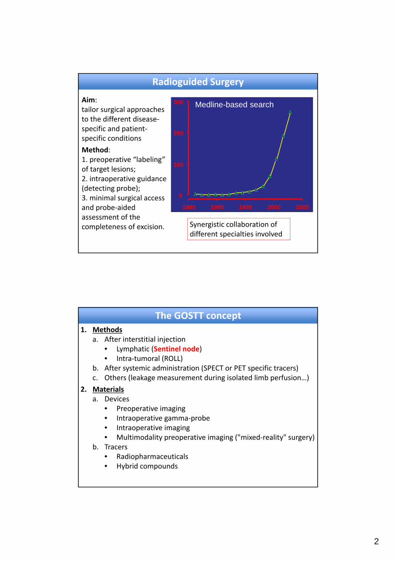
Radioguided Surgery
20052000199519901985
0
100
200
300 Medline-based searchAim: tailor surgical approaches to the different disease‐specific and patient‐specific conditions
Method: 1. preoperative “labeling” of target lesions;2. intraoperative guidance (detecting probe); 3. minimal surgical access and probe‐aided assessment of the completeness of excision. Synergistic collaboration of
different specialties involved
The GOSTT concept1. Methods
a. After interstitial injection • Lymphatic (Sentinel node)• Intra‐tumoral (ROLL)
b. After systemic administration (SPECT or PET specific tracers)c. Others (leakage measurement during isolated limb perfusion…)
2. Materialsa. Devices
• Preoperative imaging• Intraoperative gamma‐probe• Intraoperative imaging• Multimodality preoperative imaging ("mixed‐reality" surgery)
b. Tracers• Radiopharmaceuticals • Hybrid compounds

3
Artery
Vein
TumorSentinel Lymph Node
Secondary Lymph Nodes
Lymphatic vessel
Particles diffuse into interstitial fluid, drains into lymphatic vessels and regional nodes
First node involved in metastatic spread of tumour
The sentinel node concept
: Hot
• Common (guidelines)
– Breast
– Melanoma
– Gynecological malignancies
• Rare (research)
– Head and neck
– Urological malignancies
– Others
Sentinel Lymph Node Indications
4
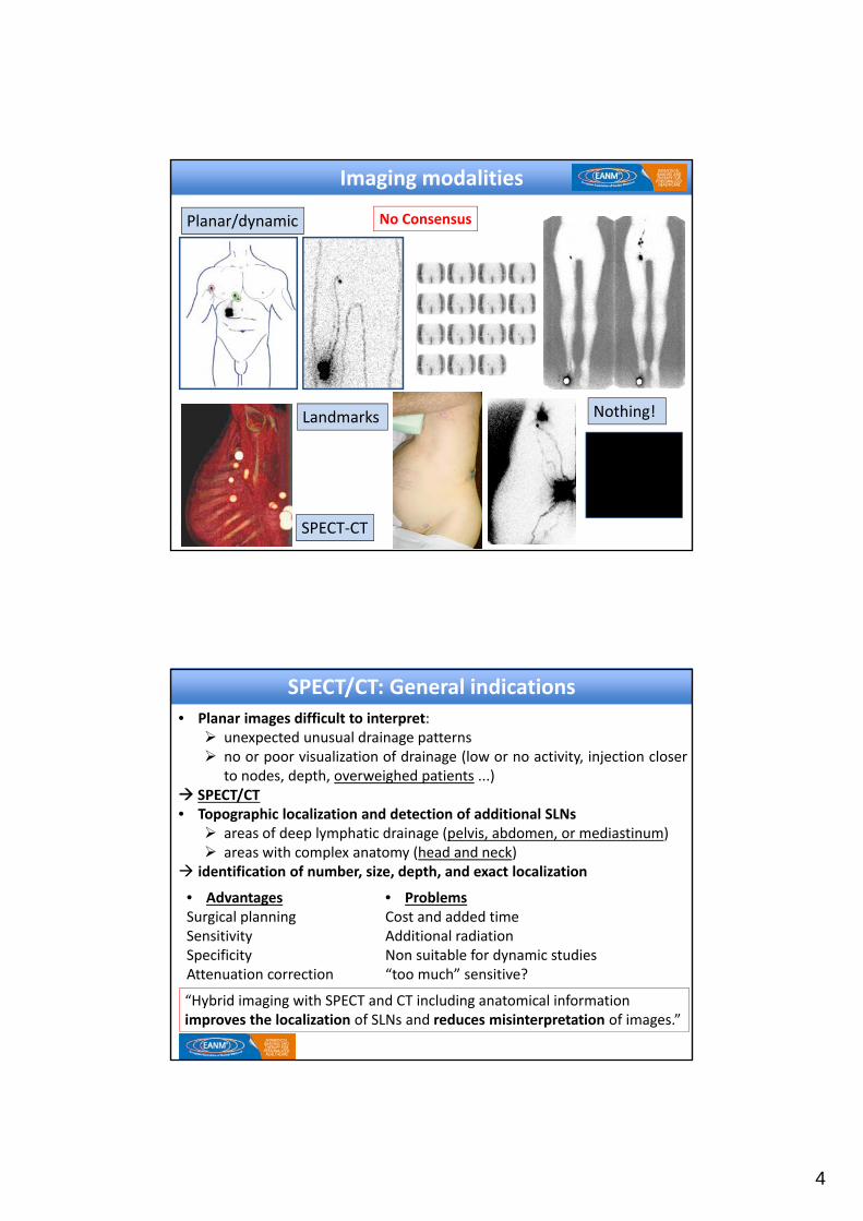
Imaging modalities
Planar/dynamic
SPECT‐CT
Landmarks
No Consensus
Nothing!
SPECT/CT: General indications
• Planar images difficult to interpret: unexpected unusual drainage patterns no or poor visualization of drainage (low or no activity, injection closer
to nodes, depth, overweighed patients ...) SPECT/CT• Topographic localization and detection of additional SLNs areas of deep lymphatic drainage (pelvis, abdomen, or mediastinum) areas with complex anatomy (head and neck)
identification of number, size, depth, and exact localization
• AdvantagesSurgical planningSensitivity SpecificityAttenuation correction
• ProblemsCost and added time Additional radiationNon suitable for dynamic studies“too much” sensitive?
“Hybrid imaging with SPECT and CT including anatomical information improves the localization of SLNs and reduces misinterpretation of images.”
5
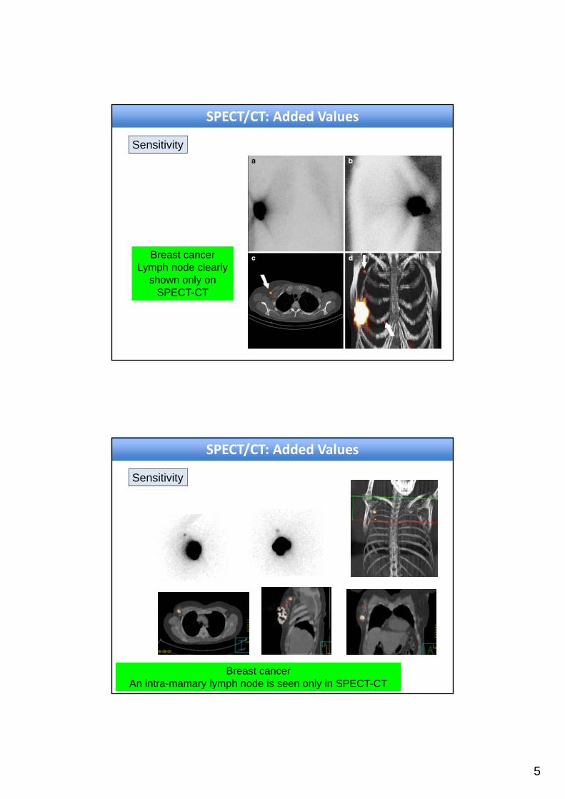
SPECT/CT: Added Values
Sensitivity
Breast cancerLymph node clearly
shown only on SPECT-CT
Breast cancerAn intra-mamary lymph node is seen only in SPECT-CT
SPECT/CT: Added Values
Sensitivity

6
Specific location of the SLN
Breast cancerLevel I and Level II axillar lymph
nodes clearly shown on SPECT-CT
SPECT/CT: Added Values
Breast cancerInternal mammary lymph nodes
Specific location of the SLN
SPECT/CT: Added Values
7
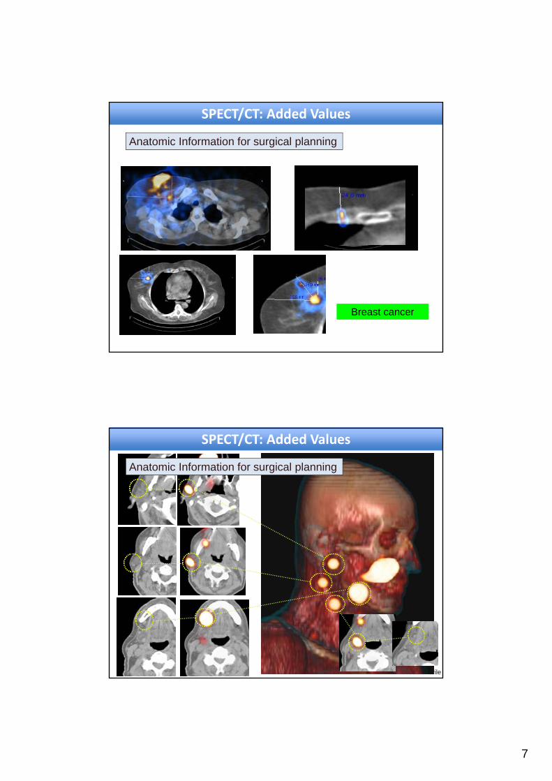
Breast cancer
Anatomic Information for surgical planning
SPECT/CT: Added Values
SPECT/CT: Added Values
F.Giammarile
Anatomic Information for surgical planning
8
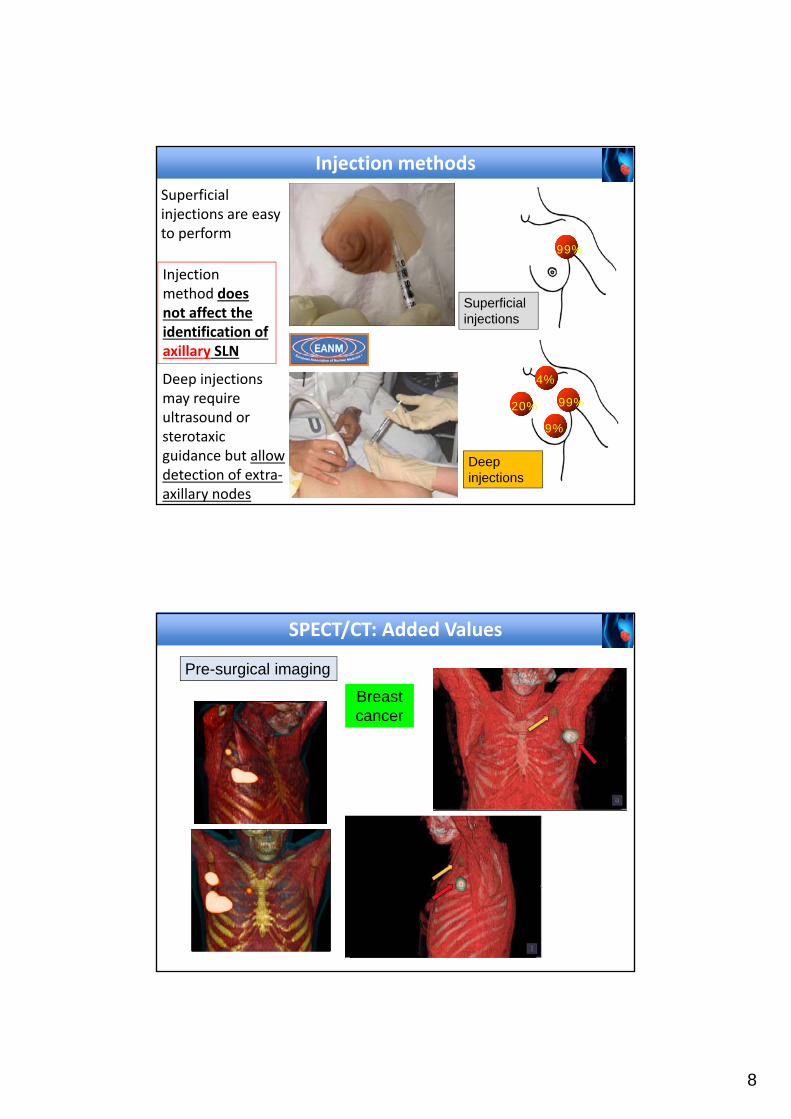
Injection methods
Injection method does not affect the identification of axillary SLN
4%
99%20%
9%
99%
Superficial injections
Deep injections
Superficial injections are easy to perform
Deep injections may require ultrasound or sterotaxic guidance but allow detection of extra‐axillary nodes
SPECT/CT: Added Values
Pre-surgical imaging
Breast cancer

9
SPECT/CT: Added Values
Pre-surgical imaging Breast cancer
Radioguided Occult Lesion Localisation (ROLL)Intratumoral administration guided by ultrasound/stereotaxisFollowed by immediate surgical resection, guided by probe
In non‐palpable suspected breast lesion replace hooked wired localization
(SLN in separate occasion)
10
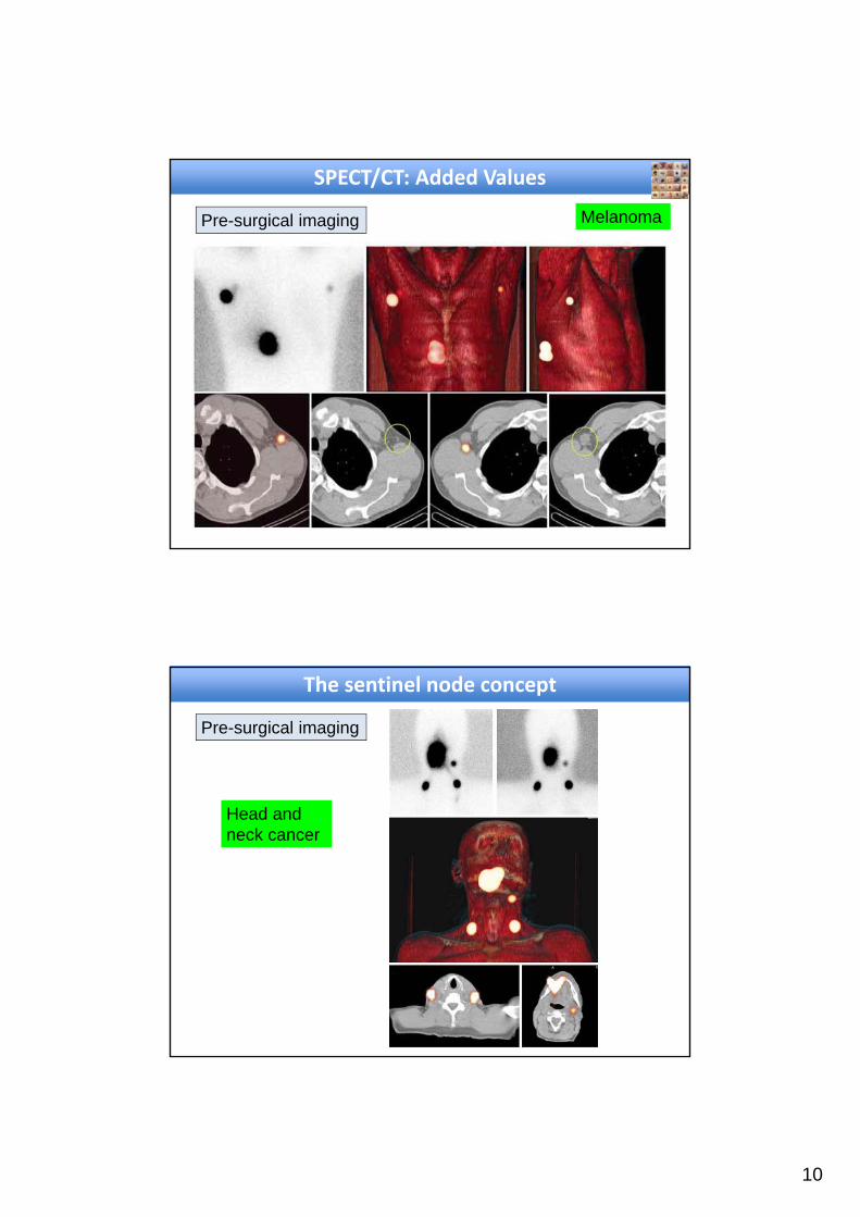
SPECT/CT: Added Values
Pre-surgical imaging Melanoma
The sentinel node concept
Pre-surgical imaging
Head and neck cancer

11
The sentinel node concept
Pre-surgical imaging
Cervical cancer
The sentinel node concept
Pre-surgical imaging
Vulvarcancer
12
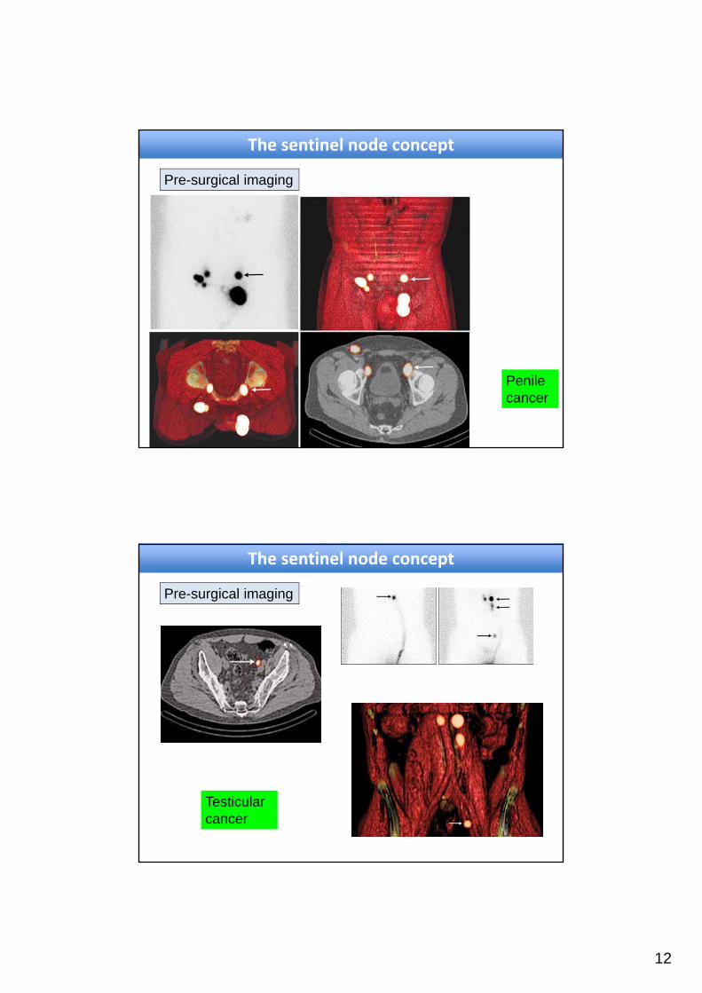
The sentinel node concept
Pre-surgical imaging
Penilecancer
The sentinel node concept
Pre-surgical imaging
Testicularcancer

13
Radiopharmaceuticals
Latest technological knowledge
Methods and instrumentation
Handle
Infrared localization
system
Optical camera
‐ 99mTc colloids (nano, sulfur) ‐ 99mTc phytates
‐ Better than colloids in breast cancer (533 patients, SurgToday, 2006)
– 198Au‐colloïd ‐ Uniformity of size (20 nm):
high lymph node uptake (15% vs 5%) (Nuc Med Biol, 2011)
‐ 99mTc‐Tilmanocept ‐ Receptor imaging
(Radio) pharmaceuticals for SLN
‐Diameter < 1nm
‐Coloration of urines (12‐24 hours) and teguments (24‐48 hours)
‐Long term tatooing effect
‐Anaphylactic shock 0.4‐1%
Eckert et al, 2013
Radiopharmaceuticals Blue Dyes

14
99mTc‐Tilmanocept
Receptor targeting agent
Lymphoseek ® • High affinity for CD 206 receptor
• Small size: 7nm, 19kDa
• Accumulation on macrophages and dendritic cells
• Internalized in lymphatic tissue
• High uptake and fast clearance from injection site
• Two phase III study in breast cancer (Baker el al. 2015 & Wallace et al. 2013):
• excellent concordance with blue and less surgically removed SLN. Sensitivity equal to colloids
“The advantages of this tracer include rapid clearance from the injection depot and low accumulation in second‐echelon nodes. This novel radiopharmaceutical might be of particular utility in patients with head and neck melanoma.”

15
Bimodal (hybrid) agents
Perspectives in Imaging Agents for Lymphatic Mapping
Assembled nanoparticulecontaining ICG
Fluorescent agent (ICG)
Simultaneous scintigraphic and optical imaging (near‐infrared fluorescence):
Tissue penetration signals Brouwer OR, Ann
Surg Oncol (2012)
Intrumentation

16
Endoscopic detection
Laparoscopic Gamma-Probe
Prostate carcinoma
Small field of view portable imaging devices
Portable gamma camera:Sentinella ®
Laser pointer centers targets on the screen
ANT
ANT axilar
OBLIQUE axilar
LATERAL Axilar

17
Small field of view portable imaging devices
Portable gamma camera: breast
57 year old patient with a breast cancer in left upper outer
quadrant
Images obtained with the portable gamma camera before and after SLN
excision:completeness of the SLN removal
Small field of view portable imaging devices
Portable gamma camera: melanoma
No SLN on pre‐operative imaging
Portable gamma camera: SLN is depicted
Increased resolution and sensitivity

18
Frontal melanoma with drainage to several nodes at different neck’s
level in right cervical basin
Images obtained with the portable gamma camera before and after
resection of 2 nodes:one more node is depicted
added value for close monitoring of the surgical procedure
Small field of view portable imaging devices
Portable gamma camera: melanoma
3D/2D tracking assisted probes
3‐D volume rendering SPECT
19
"Mixed‐Reality" Surgery(better 3D perception)
3D/2D tracking assisted probes
3‐D volume rendering SPECT
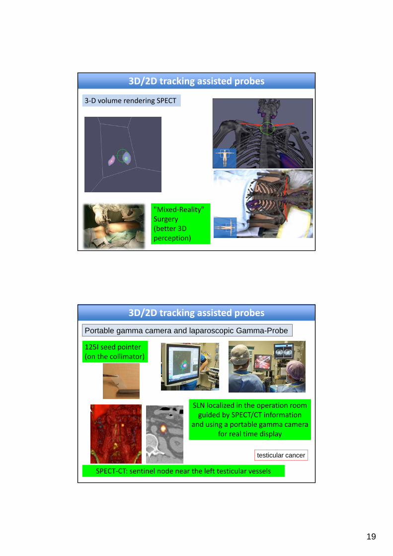
SPECT‐CT: sentinel node near the left testicular vessels
SLN localized in the operation room guided by SPECT/CT information
and using a portable gamma camera for real time display
testicular cancer
3D/2D tracking assisted probes
Portable gamma camera and laparoscopic Gamma-Probe
125I seed pointer (on the collimator)

20
Radioguided Surgery today
• Multimodality preoperative molecular imaging
Intraoperative imaging
Non‐imaging procedures
• are essential for optimizing surgical approaches in patient‐ and
disease‐specific conditions
• to reduce the invasiveness of procedures (positively impact health
economy)
• providing the standardization of the methods (guidelines)
Take‐home messages
Radioguided Surgery tomorrow
• Develop more specific tumor markers to delineate more precisely
the extent of damage (bimodal radioactive and fluorescent agents)
• Develop more sensitive detectors, miniaturized and ergonomic
• Integrate different surgical methods (imaging modalities, resection,
navigation)
Robot‐assisted endoscopic surgery
Virtual reality (mostly for training purposes), mixed reality and
augmented reality
Take‐home messages
21
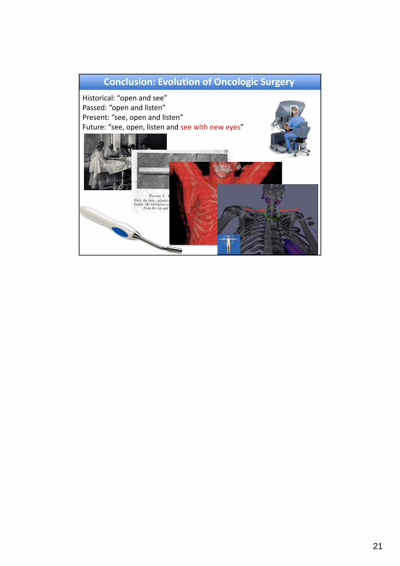
Historical: “open and see”Passed: “open and listen”Present: “see, open and listen”Future: “see, open, listen and see with new eyes”
Conclusion: Evolution of Oncologic Surgery