download - the american association of physicists in medicine
TRANSCRIPT

MARP, Inc.
Physicist’s Role in ACR MRAP Accreditation May 2010
Carl R. Keener, Ph.D., DABMP, [email protected]
MARP &Medical Radiation Physics, Inc.

MARP, Inc.
disclosure
ACR MRAP Physics subcommitteeACR MRAP phantom reviewerconsulting diagnostic physicist
GE, Siemens, Philips, Toshiba, Marconi, Hitachi, Fonar0.2T – 3TMRI surveys• 2006 –
17 surveys• 2007 –
33 surveys• 2008 –
48 surveys• 2009 –
43 surveys

MARP, Inc.
accreditation
United Healthcare will require accreditation for reimbursement by 4th quarter of 2009
MRAP (ACR)ICAMRL (IAC)outpatient imaging centersaccreditation started by Dec. 31, 2009
Medicare Improvements for Patients and Providers Act (MIPPA)
providers of advanced imaging services to be accredited by January 1, 2012
• Part BACRIACTJC

MARP, Inc.
ACR MRAP vs IAC ICAMRL
MARP MRI surveys43 accredited MRI clients
• 42 ACR (2 in process)• 1 IAC• 2 expired
54 MRI scanners
MARP CT surveys43 accredited CT clients
• 41 ACR (2 in process)• 2 IAC
146 CT scanners

MARP, Inc.
ACR accreditation process & phantom
ACR accreditation processonline application• information
• contact • site• personnel• MRI unit
• selection of modalities & exams• payment
submission• clinical Images• phantom Images
continuing requirements of accreditation
phantom & phantom tests

MARP, Inc.
online application
online applicationSection 1: • contact and general site information
Section 2:• MRI unit information• selection of modalities & exams
Section 3: • personnel information
• physicians• medical physicists / MR scientist• technologist
• payment
45-day submission window starts at payment

MARP, Inc.
online application
information• magnet • practice
credentials• physicians• technologists• physicists
modules• head• spine• MSK• body• MRA• cardiac
fees • Accreditation / Reinstate
• $2400 for 1 -
4 modules• $2600 for 5 modules• $2800 for 6 modules
• subsequent magnets / same site
• $2300 for 1 -
4 modules• $2500 for 5 modules• $2700 for 6 modules
• repeat• $800 for phantom or
clinical• $1600 for both
all magnets at site must be accredited45 day submission period begins

MARP, Inc.
qualifications & responsibilities
MRI supervising physician• responsible for MRI protocols• approves all aspects of the testing materials submission
before sending them to the ACR
lead MRI technologist• main contact person with ACR • should
be the primary person who completes accreditation forms and documents
qualified medical physicist / MR scientist• should
be responsible for supervising your facility’s weekly QC and the annual system performance evaluation
• MRI FAQs
v1.1.doc • closely involved with the phantom portion of your testing
materials submission• assist the supervising physician and lead technologist with
your routine clinical protocols
MARP, Inc.
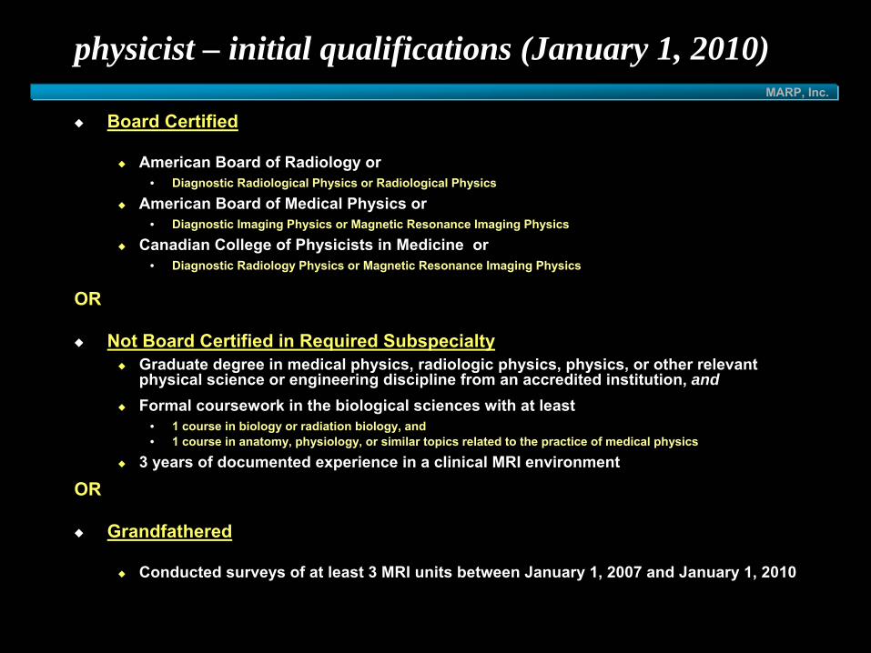
physicist – initial qualifications (January 1, 2010)
Board Certified
American Board of Radiology or• Diagnostic Radiological Physics or Radiological Physics
American Board of Medical Physics or• Diagnostic Imaging Physics or Magnetic Resonance Imaging Physics
Canadian College of Physicists in Medicine or• Diagnostic Radiology Physics or Magnetic Resonance Imaging Physics
OR
Not Board Certified in Required SubspecialtyGraduate degree in medical physics, radiologic physics, physics, or other relevant physical science or engineering discipline from an accredited institution, andFormal coursework in the biological sciences with at least
• 1 course in biology or radiation biology, and • 1 course in anatomy, physiology, or similar topics related to the practice of medical physics
3 years of documented experience in a clinical MRI environment
OR
Grandfathered
Conducted surveys of at least 3 MRI units between January 1, 2007 and January 1, 2010

MARP, Inc.
MR scientist
graduate degree in a physical science involving nuclear MR or MRI3 years documented experience in a clinical MRI environment
MARP, Inc.
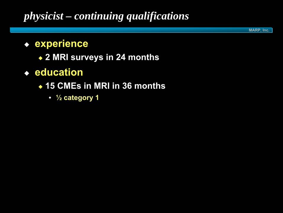
physicist – continuing qualifications
experience2 MRI surveys in 24 months
education15 CMEs in MRI in 36 months• ½
category 1

MARP, Inc.
ACR accreditation - initial questions????
when is the due date?has the initial fee been sent?
is there an ACR phantom?has it been orderedare there manufacturer phantoms?
is there a laser printer?is there a densitometer?
can CD-ROMs be burned?from scanner?from PACS?
how much will you do?evaluationphantomQCpaperwork /webwork for submissionclinical images
MARP, Inc.
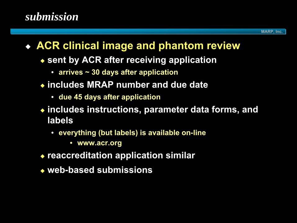
submission
ACR clinical image and phantom reviewsent by ACR after receiving application• arrives ~ 30 days after application
includes MRAP number and due date• due 45 days after application
includes instructions, parameter data forms, and labels• everything (but labels) is available on-line
• www.acr.org
reaccreditation application similarweb-based submissions

MARP, Inc.
clinical images
required from every magnet at practice location 4 sets of images
original 14 x 17 films • (hard copy submissions
only)• or refilmed from original
tapes or discs
electronic submissionCD-ROM with embedded viewermust include functions of:
• window/level• magnification• region of interest
• area• pixel mean• pixel standard deviation
• distance measurement• access to DICOM header
must be obtained within 30 days (before or after) of the acquisition of phantom images
MARP, Inc.
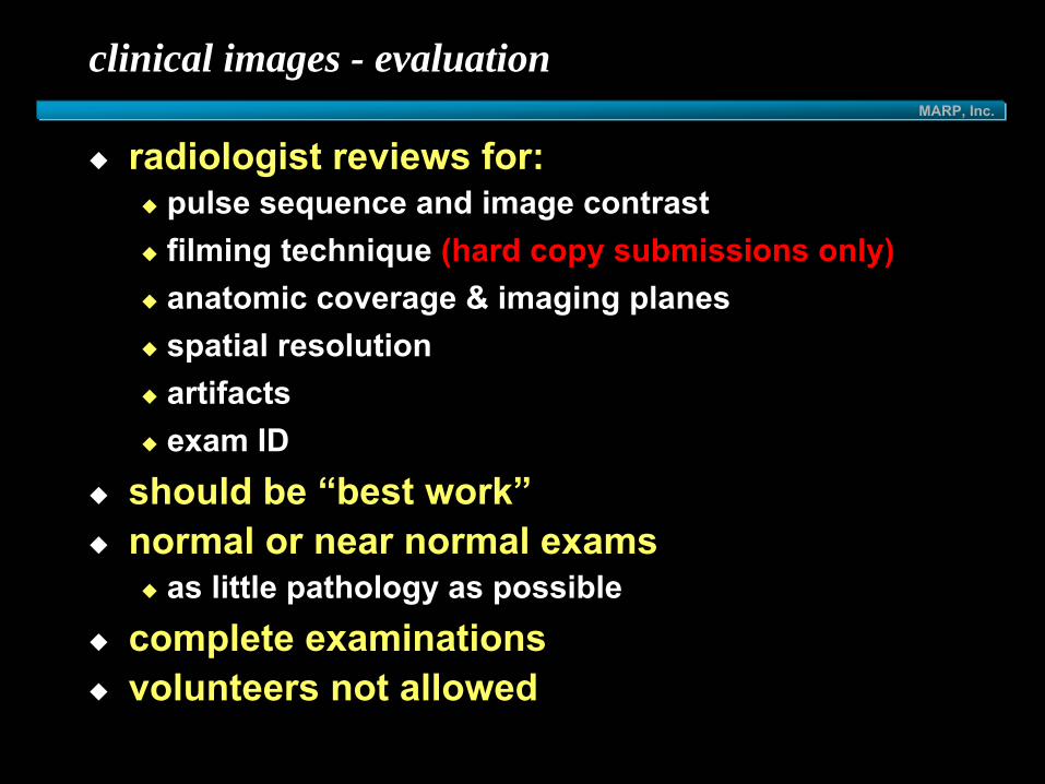
clinical images - evaluation
radiologist reviews for:pulse sequence and image contrastfilming technique (hard copy submissions only)anatomic coverage & imaging planesspatial resolutionartifactsexam ID
should be “best work”normal or near normal exams
as little pathology as possible complete examinationsvolunteers not allowed
MARP, Inc.
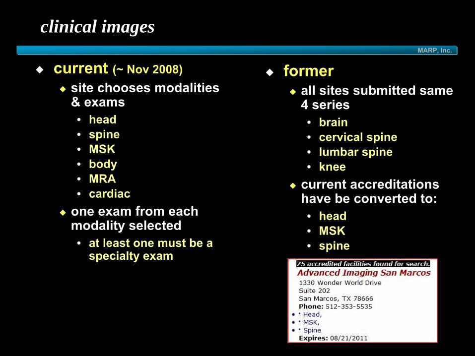
clinical images
current (~ Nov 2008)site chooses modalities & exams• head• spine• MSK• body• MRA• cardiac
one exam from each modality selected• at least one must be a
specialty exam
formerall sites submitted same 4 series• brain• cervical spine• lumbar spine• knee
current accreditations have be converted to:• head• MSK• spine

MARP, Inc.
clinical images
MSKKnee such as for internal derangementShoulder such as for internal derangementWrist such as for internal derangement*Elbow such as for internal
derangement*Forefoot for Morton’s neuroma*
bodyMale pelvis such as for prostate cancerRenalHepatobiliary to Include MRCP*Female pelvis such as for uterine or adnexal disease*
headBrain for transient ischemic attack (TIA)Internal auditory canal (IAC/temporal bone) for hearing lossBrain for suspected demyelinatingdisease*Pituitary with dynamic contrast enhancement*Orbits for vision loss*
spineLumbar SpineThoracic SpineCervical Spine*Cervical Spine with contrast for intramedullary disease*
* specialty exam
MARP, Inc.
clinical images
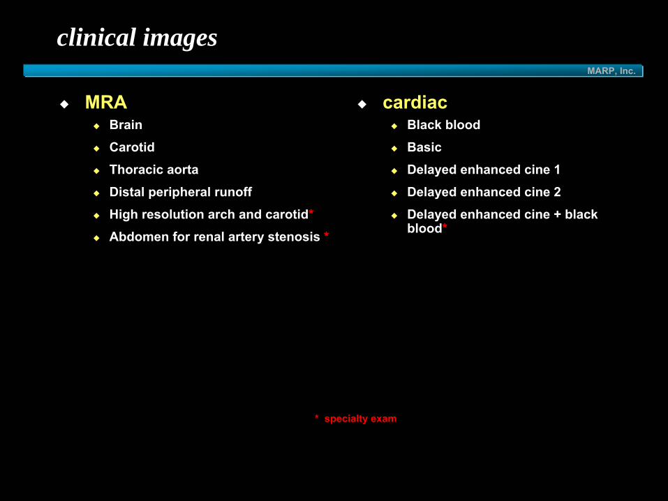
cardiacBlack bloodBasicDelayed enhanced cine 1Delayed enhanced cine 2Delayed enhanced cine + black blood*
* specialty exam
MRABrainCarotidThoracic aortaDistal peripheral runoffHigh resolution arch and carotid*Abdomen for renal artery stenosis *
MARP, Inc.
clinical images
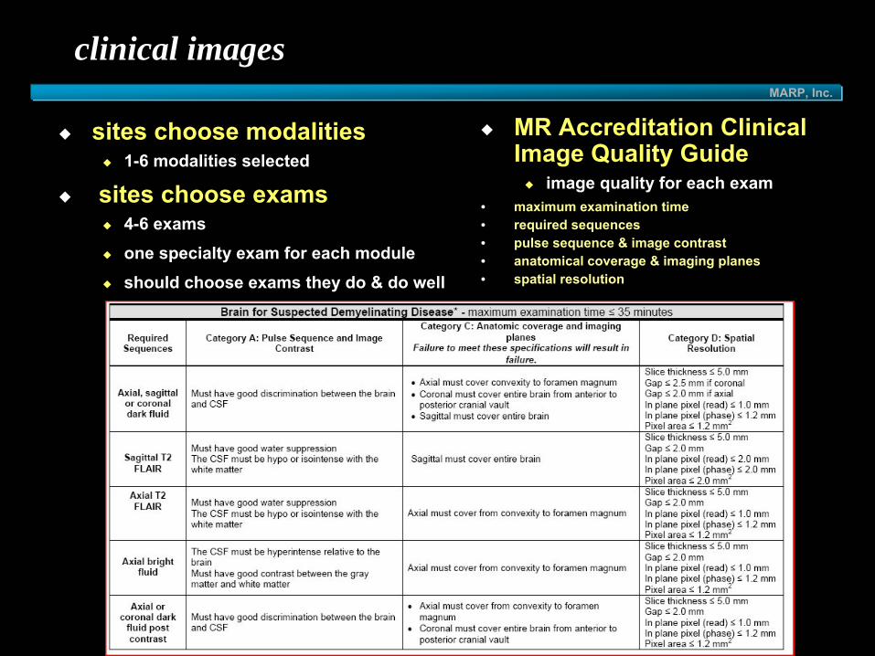
sites choose modalities1-6 modalities selected
sites choose exams4-6 examsone specialty exam for each moduleshould choose exams they do & do well
MR Accreditation Clinical Image Quality Guide
image quality for each exam• maximum examination time• required sequences• pulse sequence & image contrast• anatomical coverage & imaging planes• spatial resolution

MARP, Inc.
clinical images
all images (clinical & phantom) must be submitted by the due date on the labels sent by the ACReach of the 4 image types are labeled and placed in film jackets with completed parameter data form• pulse sequence• FOV• acquired matrix• slice thickness• slice gap
electronic submission• 2 CDROMS with embedded viewer
MARP, Inc.
quantitative phantom testing
MARP, Inc.
MRI accreditation phantom
specific ACR MRI accreditation phantom is usedspecific protocols for T1 and T2 are provided in the site instructions with the full application• phantom site scanning instructions at www.acr.org
each site is required to submit phantom images using the ACR protocol • and phantom images using its own routine T1 and T2
weighted scan protocol for head examinations
same phantom images for all modalities on full-sized magnet• smaller phantom used for extremity-only magnets

MARP, Inc.
phantom image submission - films & data required
films12-on-1 films of all 4 series
dataDicom PC CD-ROM• preferred method• each series in separate directory• make certain data are easily located• make certain images are accessible by Osiris software
• do not
use embedded viewer• do not
use compressed images
site archive format (tape or disk) is no longer acceptable

MARP, Inc.
phantom image data
provided by some manufacturers
• Fonar
available on newer workstations / PACS environments
• make certain images on CD can be located & opened by Osiris
provided by DesAcc, Inc. • HTML format on CD-ROM available
for• Elscint
(5¼")• GE (most 5¼" & most DAT)• Hitachi (5¼")• Philips (5¼" & 12")• Siemens (5¼" & 12")• Toshiba (5¼")
• $200-250 + $25 shipping • ~ 2-3 weeks turnaround• http://www.desacc.com• 312 930-5617
conversion of media to Dicom PC CD-ROM

MARP, Inc.
phantom image data – CD-ROM
available on newer workstations / PACS environmentsDICOM only CD
with DICOMDIRMRAP applications as of Nov 2007:
8798 units6052 facilities
label CD
MRAP accredited as of May 2010
5437 facilities431 facilities under review
ICAMRL accredited as of May 2010
268 facilities

MARP, Inc.
phantom data CD-ROM
uncompressed DICOM images256 x 256 x 2 = 131072 bytes

MARP, Inc.
phantom data CD-ROM
embedded readers often compress images

MARP, Inc.
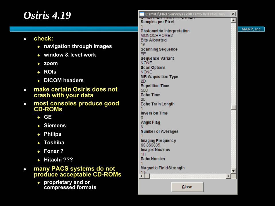
Osiris 4.19
free DICOM viewerused by ACR reviewersdownload at
http://www.sim.hcuge.ch/osiris/01_Osiris_Presentation_EN.htm make certain Osiris can open images

MARP, Inc.
make certain Osiris can open .dcm files
Osiris correctly opens• uncompressed
DICOM files
Osiris may not open• compressed files
• note: 61kB vs
131 kB• nonDICOM
files• proprietary files
MARP, Inc.
Osiris 4.19
check:navigation through imageswindow & level workzoomROIsDICOM headers
make certain Osiris does not crash with your datamost consoles produce good CD-ROMs
GESiemensPhilipsToshibaFonar ?Hitachi ???
many PACS systems do not produce acceptable CD-ROMs
proprietary and or compressed formats

MARP, Inc.
check images before submitting
compressed image wrong gap

MARP, Inc.
electronic submission

MARP, Inc.
continuing requirements of accreditation program
weekly QCannual testsACR has the right to perform random site and/or film inspections

MARP, Inc.
quality control
effective July 2005:for renewal, site must send:
• 3 months QC/printer data• annual survey report
• dated within 12 months• documentation of corrections
for failures
QC and annual survey are now required for initial applications too
effective August 2002:weekly tests (initially daily):
• central frequency• transmitter gain /attenuation• geometric accuracy• spatial resolution• low-contrast detectablilty• image artifact assessment
weekly:• laser film QC• visual checklist
annual:• physicist/MR scientist
performance evaluation & QC review

MARP, Inc.
NoseChin
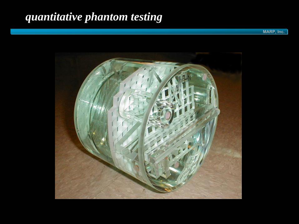
ACR MRI large phantom
J.M. Specialty Parts11689-Q Sorrento Valley RoadSan Diego, CA 92121(858) 794-7200
$1050previously $730
delivery6-8 weeks without MRAP number2-3 weeks with MRAP numberorder without MRAP, then call to add MRAP

MARP, Inc.
ACR MRI small phantom
J.M. Specialty Parts11689-Q Sorrento Valley RoadSan Diego, CA 92121(858) 794-7200
$780

MARP, Inc.
performance criteria
Phantom Test Guidance for the ACR MRI Accreditation Program
• describes tests• instructions• performance criteria• reasons for failure• available from www.acr.org• “large phantom”
= old phantom

MARP, Inc.
site scanning instructions
Site Scanning Instructions for use of the MRI Phantom for the ACR MRI Accreditation Program
• phantom positioning• pulse sequences• film and data instructions• sent to site with full
application• available from ACR

MARP, Inc.
phantom pulse sequences
2 ACR sequencesT1T2
identical for all sitesextremity-only sites use different ACR sequences for small phantom
2 site sequencesT1T2
site specificmay be used to increase SNR in order to pass low-contrast tests on low-field units

MARP, Inc.
ACR sequences
regular phantom1 sagittal localizer slice
• T1: SE• TR=200 ms, TE=20ms• 25 cm FOV, 256x256, 20mm slice• 1 NEX (NSA, NAQ, AVE)
2 sets of 11 axial slices• 25 cm FOV, 256x256• 1mm pixels• 5 mm slice, 5 mm gap• 1 NEX (NSA, NAQ, AVE)• T1:
• SE, TR=500 ms, TE=20 ms• T2:
• SE, TR=2000 ms, TE=20/80 ms • Use 2nd
echo onlyall sites must use the same ACR sequences
• slight modifications allowed if scanner cannot set them
• T1: SE, TR=515 ms, TE=20 ms (Toshiba Opart)
• T2: SE, TR=500 ms, TE=90/25 ms (Fonar)
• document other scan parameters on site data sheet
small phantom1 sagittal localizer slice
• T1: SE• TR=200 ms, TE=__ms• 12 cm FOV, 192x152, 20mm slice• 1 NEX (NSA, NAQ, AVE)
2 sets of 7 axial slices• 12 cm FOV, 192x152 • 0.625mm x 0.79mm pixels• 5 mm slice, 3 mm gap• 1 NEX (NSA, NAQ, AVE)• T1:
• SE, TR=500 ms, TE=20 ms• T2:
• SE, TR=2000 ms, TE=20 ms• only 1 echo

MARP, Inc.
required phantom tests
geometric accuracyhigh-contrast spatial resolutionslice thickness accuracyslice position accuracyimage intensity uniformitypercent signal ghostinglow-contrast object detectabilityimage artifacts

MARP, Inc.
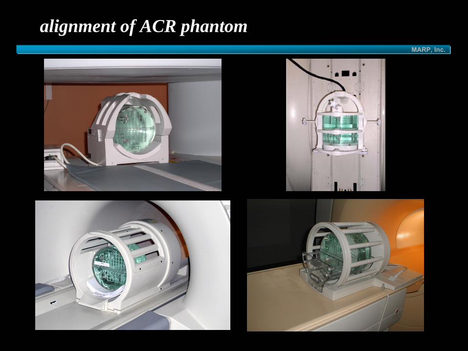
alignment of ACR phantom

MARP, Inc.
pre-scanning procedures
magnet should be checked by service engineer prior to acquisitionalignment is important
• center of head coil • computer paper
• straight• use bubble level
• centered SI, LR & AP• make localizer slice in all 3
planes• use grid to check
centering• record position for future
use
MARP, Inc.
alignment of ACR phantom

MARP, Inc.
sagittal localizer slice
Nose
Chin
A P
S
I
1
11
Tabl
e
Nose
Chin
Tabl
e

MARP, Inc.
sagittal localizer slice
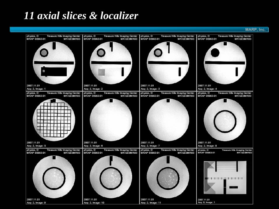
set up 11 axial slices for all 4 series• 5 mm thick with 5 mm gap
• small phantom : 7 axial slices, 5 mm slice, 3 mm gap
notes• if geometric accuracy is off, low contrast slices 8-11 may
not be accurate • phantom wedges and LCD insert 11 may not be perfectly
aligned• some units have protocols which number slices opposite of
the ACR recommendations• may not start renumbering at "1" for each series• make note of which image is 2nd
echo on T2
MARP, Inc.
11 axial slices & localizer

MARP, Inc.
7 axial slices & localizer on small phantom

MARP, Inc.
geometric accuracy
sagittal localizer & ACR axial T1 slices 1 & 5specific window & level:
window/level must be set separately for localizer & axial T1• both axial slices use same window/level
window as narrow as possibleset level where ½ of water is dark (mean)set window width = mean value & window level
= ½
mean
value
small phantom• sagittal localizer & ACR axial slices 1 & 3
MARP, Inc.
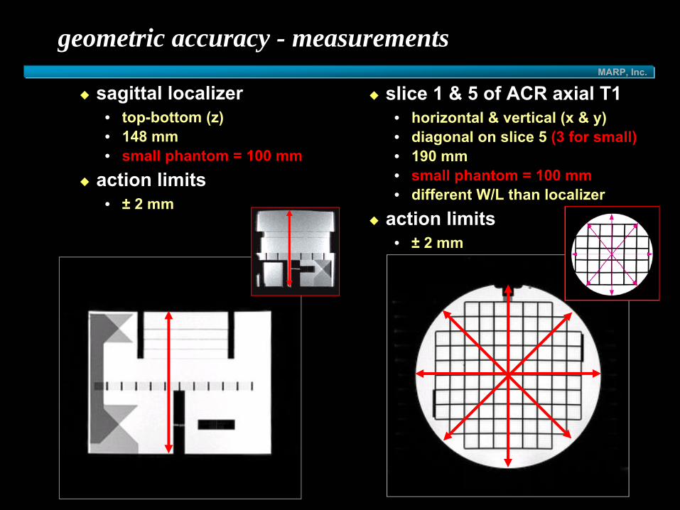
geometric accuracy - measurements
slice 1 & 5 of ACR axial T1• horizontal & vertical (x & y)• diagonal on slice 5 (3 for small)• 190 mm• small phantom = 100 mm• different W/L than localizer
action limits• ± 2 mm
sagittal localizer• top-bottom (z)• 148 mm• small phantom = 100 mm
action limits• ± 2 mm

MARP, Inc.
geometric accuracy - notes
image may be bowed • accurate at one location,
inaccurate at another • measure in the center
bubble may obscure top of slice
• measure at anglelocalizer & axial slices need different W/Loperator may know the actual values and aim for themopen short bore magnets may use geometric corrections
• Siemens –
Large FOV or 2D distortion correction

MARP, Inc.
1.1 mm 1.0 mm 0.9 mm
high-contrast spatial resolution
measurements• use slice 1 of ACR T1 & ACR T2• magnify slice 1 by 2 to 4• observe UL holes ; adjust window/level
• observe rows: if all 4 holes in a single
row are distinguishable, score image as resolved at this hole size
• view all three sets (1.1 mm, 1.0 mm, 0.9 mm)• score = smallest holes resolved
• repeat for LR array with columns
of holes
performance criteria: 1.0 mmsmall phantom
• 0.9 mm, 0.8 mm, 0.7 mm sets• pixel size = 0.625 x 0.79 mm• performance criteria: 0.8 mm
UL
LR

MARP, Inc.
slice thickness accuracy
slice 1 of ACR T1 & ACR T2crossed ramps (10:1 slope)measure mean
• magnify by 2 to 4• adjust window/level to see signal ramps • 2 ROIs• mean of middle of each signal ramp • take average
measure width• lower level to ½
average• set window at minimum• measure lengths of top and bottom ramps
calculate slice thicknessperformance criteria:• 5.0 ±
0.7 mm
( )( )bottomtop
bottomtop+×
×= 0.2 thicknessslice

MARP, Inc.
slice thickness - notes
• edges of ramps difficult to determine• Gibbs artifacts• noise (low field magnets)
• display may give max-min signal (or use relative scale)• use Osiris
• 1 mm measurement error = 1/10 mm error in slice thickness• same for small phantom
1.5 T 0.2 T

MARP, Inc.
slice thicknesses measurements
Hitachi Airis IIROI scale different than WL scale“Jump level to ROI mean”
• sets level to mean of ROI• displays mean using WL scale• manually reduce level to ½• manually reduce width to 1
Toshiba Opart & VantageROI scale different than WL scaleno simple workaroundburn CD
• use Osiris or Osirix

MARP, Inc.
11
A P
S
I
slice position accuracy
measurements• use slices 1 & 11 of ACR T1 & ACR T2
• only slice 1 of small phantom• magnify by 2 to 4 & adjust window/level• measure difference of left & right bars
• if left bar is longer assign a minus sign to the length
11
1
MARP, Inc.
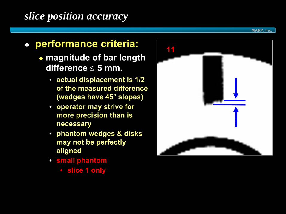
slice position accuracy
performance criteria: magnitude of bar length difference ≤ 5 mm.• actual displacement is 1/2
of the measured difference (wedges have 45°
slopes) • operator may strive for
more precision than is necessary
• phantom wedges & disks may not be perfectly aligned
• small phantom• slice 1 only
11
MARP, Inc.
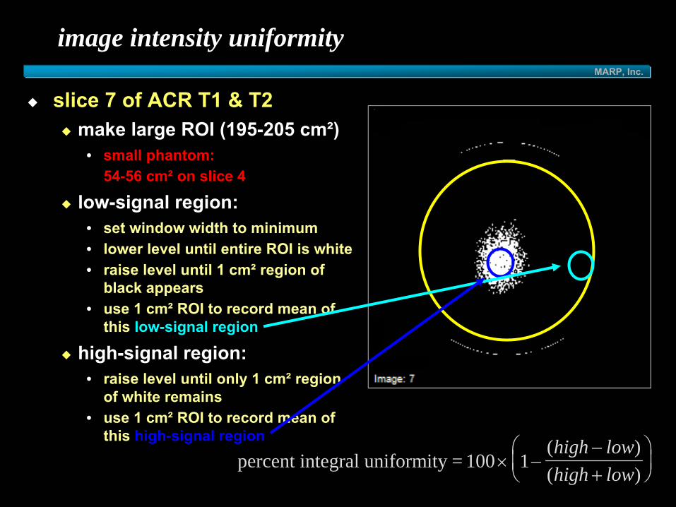
image intensity uniformity
slice 7 of ACR T1 & T2make large ROI (195-205 cm²)
• small phantom: 54-56 cm²
on slice 4
low-signal region: • set window width to minimum• lower level until entire ROI is white• raise level until 1 cm²
region of black appears
• use 1 cm²
ROI to record mean of this low-signal region
high-signal region: • raise level until only 1 cm²
region of white remains
• use 1 cm²
ROI to record mean of this high-signal region
percent integral uniformity = 100 × −−+
⎛⎝⎜
⎞⎠⎟1
( )( )high lowhigh low

MARP, Inc.
image intensity uniformity
performance criteria:PIU ≥
87.5%
• if there is not a well-defined high/low intensity level…
…..uniformity is very high!
same for small phantom
for 3.0T:PIU ≥
82% (July 2005)

MARP, Inc.
image intensity uniformity – 8-channel coils
smaller coilharder to setup
poorer uniformity
correctionsSurface Coil Intensity Correction (SCIC) – GE
no correction
SCIC
Prescan Normalization (Siemens)
“CLEAR”
(Philips)
“Quadrature” vs “SENSE”

MARP, Inc.
percent signal ghosting
use slice 7 of ACR T1make large ROI (195-205 cm²)*
• record mean• small phantom:
54-56 cm²
on slice 4
make 4 elliptical ROIs• 10 cm²
with 4:1 ratio• left, right, top, bottom• record mean of each
performance criteria: • ghosting ratio ≤
0.025 (2.5%)
ghosting ratio = large ROI
( ) ( )( ( )
top bottom left right+ − +×2
MARP, Inc.
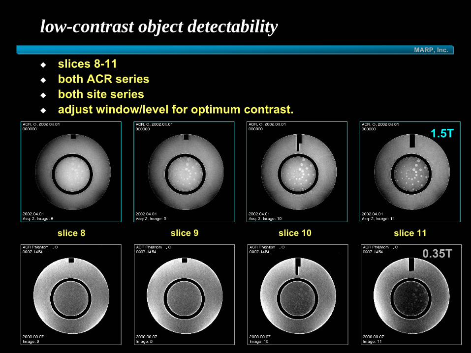
low-contrast object detectability
slices 8-11both ACR seriesboth site seriesadjust window/level for optimum contrast.
slice 8 slice 9 slice 10 slice 11
0.35T
1.5T

MARP, Inc.
low-contrast object detectability
4 slices with low-contrast holesslices 8-11
• decreasing contrast levels (11→ 8)
10 spokes per slice• 3 holes per spoke• decreasing size (clockwise)
count complete spokesall 3 disks must be discernible
• more apparent than background
end with last complete spokefor phantom review:
• all slices are counted• total score = discernible spokes
from all four slices

MARP, Inc.
low-contrast object detectability – small phantom
2 slices with low-contrast holesslices 6-7
• decreasing contrast levels (7→ 6)
10 spokes per slice• 3 holes per spoke• decreasing size (clockwise)

MARP, Inc.
low-contrast object detectability
performance criteriaeach ACR series should have a total score of at least 9 spokes• 4 slices for large phantom (9/40)• 2 slices for small phantom (9/20)
for 3.0T, the total score must be at least 37 spokes.must pass both ACR series or both site series

MARP, Inc.
low-contrast object detectability
causes of failure:incorrectly positioned slices
• contrast based on partial volume averaging
tilted phantomwarped slices / B0inhomogeneitycorrection softwareincorrect slice thicknessghostinginadequate SNR

MARP, Inc.
low-contrast object detectability
notes:several lower spokes may be visible but cannot be counted due to artifact obscuring one of the higher-level spokeswindow/level each slice separatelystart with highest contrast and move down.site may need to change their protocol • (especially for low field magnets)

MARP, Inc.
1.5 T 0.3 T
low-contrast: high vs. low field
• slice 11 -
ACR T1 series
MARP, Inc.
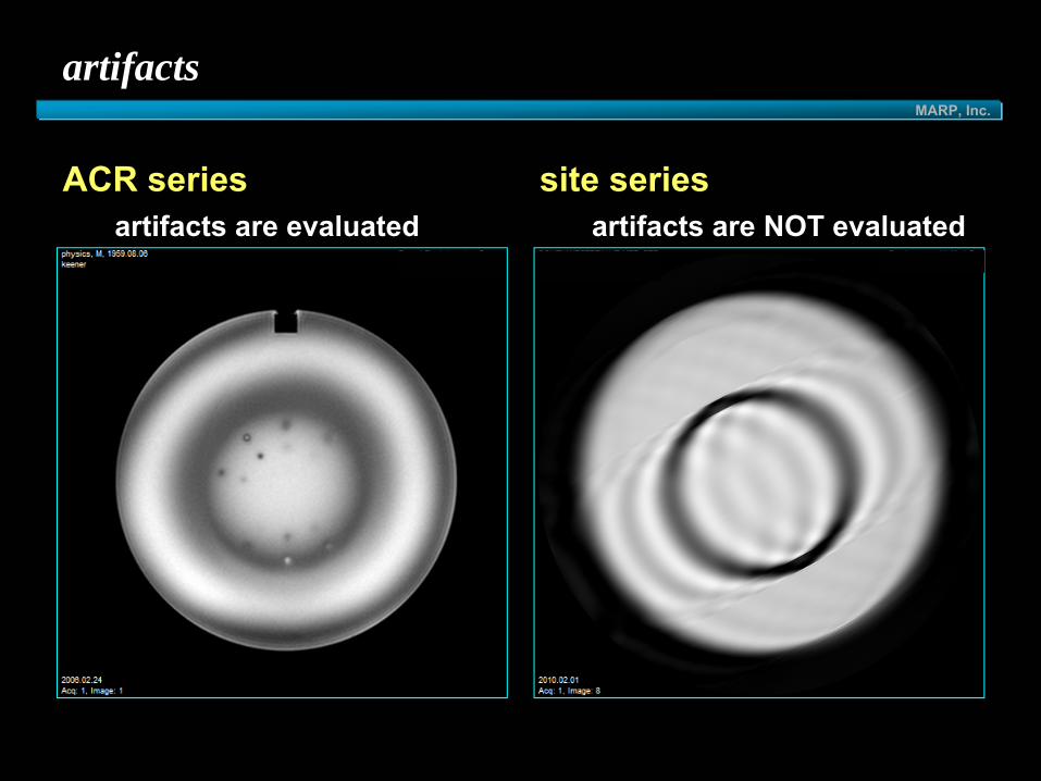
artifacts
ACR series artifacts are evaluated
site series artifacts are NOT evaluated

MARP, Inc.
artifacts
DC Offset (ACR series) acceptable
susceptibility (ACR series)questionable

MARP, Inc.
artifacts
wraparound (site series) acceptable
ghosting (ACR T2)questionable

MARP, Inc.
homogeneity
required for annual survey & initial evaluationconcerns
geometric distortion?problems with fat saturation?
methodsspectralphase-differencevisual distortion

MARP, Inc.
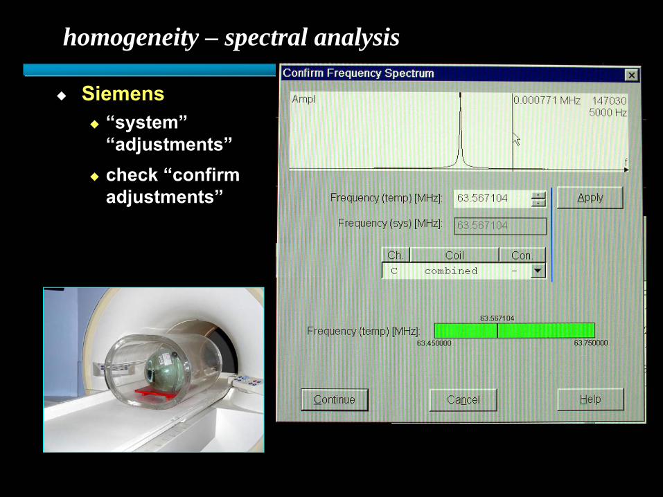
homogeneity – spectral analysis
measures frequency of signal-producing phantom in magnet
DSV• ↑
homogeneity at center & with ↓
FOV• Δ FOV…
Δ phantom sizespectrum displayed on screen (Hz)homogeneity measured in ppmcompare to specs or baseline values

MARP, Inc.
homogeneity – spectral analysis
manual prescanGE
EPI tuningElscint
FWHM (Hz)
volume ~ phantom
ppmHz
MHzFWHM
LarmorFrequency=
MARP, Inc.
homogeneity – spectral analysis
Siemens“system”“adjustments”check “confirm adjustments”

MARP, Inc.
phase-difference (Philips)
shim_checkoption under phantom studiesFFE, fixed technique & FOV
40 cm cylindrical phantom
holder to center phantom3 orientations
view real imagescount B→W transitions

MARP, Inc.
shim_check (Philips)
example:1.5T, FFE, TR=400, TE=16, 30º, 450 mm FOVB→W = 1 ppm
example:coin taped to phantom

MARP, Inc.
shim_check (Philips)
DHL
example:1.5T, FFE, TR=400, TE=16, 30º, 450 mm FOVB→W = 1 ppm

MARP, Inc.
homogeneity – geometric distortion
most low-field open magnets do not have software to check homogeneity
observe effects on geometric distortion
• lower bandwidthwarped images
7.4 kHz 3.6 kHz

MARP, Inc.
different bandwidths
measure distortion in frequency-encoding direction with different bandwidths (Clarke & Chen)
( ) ( )( ) ( )120
2121
2)(
BWBWFOVBxxBWBWppmMFH
−−××
=πγ
BW=33 Hz (8448 kHz)FE45 TR=256, TE=45, 7 mm, FA=70°, FOV=20cm
BW=244 Hz (62464 kHz)FE5.0 TR=256, TE=5, 7 mm, FA=20°, FOV=20cm
148.1 cm151.6 cm
SI 2.68 ppm
AP 0.84 ppm
LR 0.31 ppmSWOMRI

MARP, Inc.
coils
must be checked annually
SNRghostinguniformity

MARP, Inc.
volume coils
similar to head coiluniformity
• mean• high• low
SNR• mean• background noise
ghosting• mean• background signal• ghost signal (PE direction)
phased-array coils• may be treated as volume
if they have volume configuration

MARP, Inc.
surface coils
max SNR• max ROI• background noise
uniformity• subjective
ghosting• subjective
phased-array coils• may be treated multiple surface
coils if you can distinguish the location of the arrays

MARP, Inc.
phased-array coils
multiple types
~ volume coil
~ multiple surface coils(arrays distinguishable)
more complicated

MARP, Inc.
recommendations
be familiar with process• read QC manual, phantom guidance & site instructions• set up worksheet or computer program• let site know if they will pass phantom portion
scan early in the process• site may need to adjust protocols prior to acquisition of
clinical and phantom images• before application to let site know condition of magnet prior
to due date • magnet may be “in specs”
and still fail part of the test• magnet may need corrections prior to phantom submission

MARP, Inc.
recommendations
physician• two-month
window to get clinical images• may need to adjust protocols
service engineer• magnet should be in top shape prior to obtaining images
technologist• needs assistance in setting up QC• schedule ~ 5 hours

MARP, Inc.
conclusion
physicist• there is no reason a site should fail the phantom portion of
the MRI Accreditation program if they are being assisted by a physicist
• phantom materials submitted by physicist should be usable by ACR
• physicist should be able to perform all tests required by ACR

MARP, Inc.
application & submission resources
www.acr.orgapplication & submission documents • all changed ~ November 2008
phantom submission & site scanning documents• unchanged for large phantom• added small phantom ~ November 2008

MARP, Inc.
Physicist’s Role in ACR MRAP Accreditation May 2010
Carl R. Keener, Ph.D., DABMP, [email protected]
MARP &Medical Radiation Physics, Inc.