efeito do envelhecimento artificial acelerado,...
TRANSCRIPT

i
THAYLA HELLEN NUNES GOUVEIA
EFEITO DO ENVELHECIMENTO ARTIFICIAL
ACELERADO, CLAREAMENTO E MANCHAMENTO NAS
PROPRIEDADES FÍSICAS DE UM COMPÓSITO
NANOPARTICULADO
EFFECT OF ACCELERATED ARTIFICIAL AGING,
BLEACHING AND STAINING ON PHYSICAL PROPERTIES OF A
NANOFILLED COMPOSITE
Piracicaba
2015

ii

iii
UNIVERSIDADE ESTADUAL DE CAMPINAS
FACULDADE DE ODONTOLOGIA DE PIRACICABA
THAYLA HELLEN NUNES GOUVEIA
EFEITO DO ENVELHECIMENTO ARTIFICIAL ACELERADO,
CLAREAMENTO E MANCHAMENTO NAS PROPRIEDADES
FÍSICAS DE UM COMPÓSITO NANOPARTICULADO
EFFECT OF ACCELERATED ARTIFICIAL AGING, BLEACHING
AND STAINING ON PHYSICAL PROPERTIES OF A NANOFILLED
COMPOSITE
Orientadora: Profa. Dra. Debora Alves Nunes Leite Lima
Piracicaba
2015
Dissertação apresentada à Faculdade de Odontologia de Piracicaba da
Universidade Estadual de Campinas como parte dos requisitos exigidos
para obtenção do título de Mestra em Clínica Odontológica, na Área de
Dentística.
Dissertation presented to the Piracicaba Dental School of the University of
Campinas in partial fulfillment of the requirements for the degree of
Master in Dental Clinic in Operative Dentistry Area.
Este exemplar corresponde á versão final da
dissertação de mestrado da aluna Thayla Hellen Nunes
Gouveia, orientada pela Profa. Dra. Debora Alves
Nunes Leite Lima.
Assinatura do Orientador

iv

v

vi

vii
Resumo
O objetivo deste estudo in vitro foi avaliar as propriedades físicas de uma resina
composta nanoparticulada submetida ao envelhecimento artificial acelerado (AAA), acao
de agentes clareadores e manchamento extrínseco. O estudo foi dividido em 2
experimentos. O experimento 1 avaliou a influência do AAA e do peróxido de carbamida a
16% com diferentes espessantes na retenção do brilho (GU), cor ( ), microdureza (KHN)
e rugosidade média (Ra) de superfície do compósito. A análise qualitativa da superfície de
resina foi realizada através de microscopia de força atômica. Cem amostras cilíndricas
(7,0mm x 2,0mm) foram confeccionadas e distribuídas aleatoriamente em 10 grupos
(n=10). Somente metade dos grupos foram envelhecidos. Para o tratamento
clareador/espessante, os grupos com e sem AAA foram divididos em: peróxido de
carbamida 16% (PC) com carbopol, PC com natrosol, gel de carbopol, gel de natrosol e
sem tratamento (controle). Os dados obtidos foram analisados por meio de ANOVA dois
fatores e teste de Tukey. Para o as comparações múltiplas foram realizadas pelos teste
de Tukey e Dunnett. Os valores de rugosidade foram analisados pelos testes de Kruskal
Wallis, Dunn e Mann Whitney. O nível de significância considerado foi de 5%. O PC com
carbopol promoveu redução apenas na microdureza do compósito, diferindo
estatisticamente do controle. Para o PC com natrosol esta alteração não foi observada. O PC
com carbopol aumentou a rugosidade e diminuiu o brilho das resinas envelhecidas,
enquanto o natrosol só o briho foi reduzido, diferindo estatisticamente do grupo controle. O
Experimento 2 avaliou a influência dos tratamentos clareadores em diferentes
concentrações e manchamento com café na propriedades físicas anteriormente citadas da
resina nanoparticulada, submetidas ou não ao AAA. Duas amostras representativas de cada
grupo foram avaliadas em microscopia eletrônica de varredura. Cento e vinte amostras
cilíndricas foram confeccionadas, semelhante ao estudo anterior, distribuídas em 12 grupos
(n=10). Metade das amostras foram envelhecidas artificialmente. Para o tratamento
clareador/pigmentação, os grupos com e sem AAA, foram divididos em: peróxido de

viii
carbamida a 10% (PC 10%), peróxido de hidrogênio a 35% (PH 35%) e sem tratamento
clareador, com e sem pigmentação por café. Os dados foram coletados e submetidos a
ANOVA três fatores e teste de Tukey para comparações múltiplas. Para o as
comparacões múltiplas foram realizadas pelos testes de Tukey e Dunnett (α=0,05). Nas
resinas sem envelhecimento, os agentes clareadores reduziram a microdureza do compósito,
entretanto a rugosidade, brilho e cor não apresentaram alterações. Após o envelhecimento,
o PC 10% aumentou a rugosidade e diminuiu o brilho comparado ao PH 35%. A
pigmentação também reduziu a microdureza das resinas sem e com envelhecimento e
alterou a cor das resinas envelhecidas . Portanto, a ação dos géis clareadores
reduziu a microdureza do nanocompósito, sendo que a composição do gel clareador
influenciou nessa alteração. O AAA promoveu significativas alterações nas propriedades
físicas avaliadas, além de favorecer uma maior degradação da superfície da resina pelo
agente clareador e este efeito contribuiu para um maior manchamento extrínseco.
Palavras-chave: clareamento dental, resina composta, envelhecimento artificial
acelerado, pigmentação.

ix
Abstract
The aim of this in vitro study was to evaluate the physical properties of a
nanocomposite subjected to artificial accelerated aging (AAA), to the action of bleaching
agents and to extrinsic staining. This study was divided into two experiments. Experiment
1: evaluate the influence on a composite surface of AAA and 16% carbamide peroxide with
different thickeners, in gloss retention (GU), color (ΔE), microhardness (KHN) and
roughness (Ra). Qualitative analysis of the resin surface was performed by atomic force
microscopy. One hundred cylindrical samples (7.0mm x 2.0mm) were prepared and
randomly divided into 10 groups (n = 10). Only half of the groups were aged. For the
bleaching/thickening treatment, the groups with and without AAA were divided into
groups/ treatments: 16% carbamide peroxide (PC) with carbopol, PC natrosol, carbopol gel,
natrosol gel and untreated (control). Data were submitted to two-way ANOVA and Tukey’s
test for multiple comparisons. The multiple comparisons for were performed by using
Tukey’s and Dunnett’s tests, and roughness values was analysed by using Kruskal-Wallis,
Dunn’s, and Mann-Whitney’s tests (α =0.05). The PC with carbopol promoted a decreased
in the composite microhardness differing significantly from the control. As for CP +
natrosol this change in the microhardness was not observed. The treatment with CP +
carbopol increased the roughness and decreased the gloss of the aged resins, whereas
natrosol reduced only the gloss which differed statistically from the control. Experiment 2:
evaluate the influence of bleaching treatments with different concentrations and coffee
staining, in the physical properties of nanocomposite, submitted or not to AAA. Two
representative samples of each group were evaluated in scanning electron microscopy
(SEM). One hundred twenty cylindrical samples were prepared similar to the previous
study and divided into 12 groups (n = 10). Half of specimens were artificially aged. For the
bleaching/pigment treatment, groups with and without AAA, were divided into the
following groups: 10% carbamide peroxide (10% CP), 35% hydrogen peroxide (35% PH)
without bleaching treatment, with and without pigmentation of coffee. Data were submitted
to three-way ANOVA and Tukey’s test for multiple comparisons. The multiple

x
comparisons for were performed by using Tukey’s and Dunnett’s tests (α=0,05). The
resins without aging, the bleaching agents promoted a reduction in the composite
microhardness, however, in the roughness, gloss and color this changes were not observed.
After aging, the pigmentation also reduced the microhardness of the resins with and without
aging and altered the color of the aged resins (ΔE> 25). 10% PC increased roughness and
decreased gloss compared to 35% PH. Pigmentation also reduced the microhardness of the
resins with and without aging and change the color of the aging resins (ΔE> 25). The action
of bleaching agents reduced the microhardness of the nanocomposite, and this alteration
was influenced by the composition of the bleaching gel. The AAA promoted significant
changes in the evaluated physical properties and favored the further deterioration of the
resin surface by the bleaching, contributing to a greater extrinsic staining.
Key-words: dental bleaching, composite resin, accelerated aging, pigmentation.

xi
Sumário
Dedicatória xiii Agradecimento Especial xv Agradecimentos xvii
Introdução 1
Capítulo 1 6
Effect of At-Home Bleaching with Different Thickeners and Aging on
Physical Properties of a Nanocomposite
Capítulo 2 28
Evaluation of Physical Properties of a Nanocomposite after Aging, Bleaching, and
Staining
Conclusão 53 Referências 54 Apêndice 56 Anexo 72

xii

xiii
Dedicatória
À Deus, por todo seu amor manifestado em minha vida, fornecendo cada dia a capacidade
necessária para vencer os desafios encontrados.
Deus não escolhe os capacitados ou perfeitos, ele capacita, purifica, transforma, santifica,
converte, mais exige do escolhido um desejo sincero de entrega!
Pe. Luís Erlin.
Ao meu pai, Francisco, meu bem maior, por cada escuta, cada abraço e incentivo
constante. Agradeço por toda confiança em mim depositada e pelo seu amor. Te amo
painho!
À minha mãe, Margarida, minha fortaleza e porto seguro. Obrigada por existir e sempre
encorajar-me com suas palavras doces. Nada disso teria sentido sem você. Te amo mainha!
Pedacinhos de mim! Thaciano, Thais e Talita. Como sou feliz por ter vocês como meus
irmãos. Agardeço por todo amor a mim doado, pelo incentivo e por toda torcida.
Ao meu bem, Manoel Costa Neto, por entender que minha ausência foi necessária. Te
agradeço pelo seu amor sincero.
Para ter sucesso é necessário amar de verdade o que se faz.
Steve Jobs.

xiv

xv
Agradecimento Especial
À minha orientadora, Profa. Dra. Débora Alves Nunes Leite Lima, por acreditar em
mim! Agradeço pela oportunidade de ser sua aluna e permitir compartilhar todo seu
conhecimento comigo. Obrigada pelo convívio agradável, por suas palavras de incentivo. À
você professora todo meu respeito e carinho.
Você é minha ispiração!

xvi

xvii
Agradecimentos
A Faculdade de Odontologia de Piracicaba da Universidade de Campinas, na pessoa de seu
diretor, Prof. Dr. Prof. Dr. Guilherme Elias Pessanha Henriques e ao diretor associado
Prof. Dr. Francisco Haiter Neto.
A Coordenadora Geral da Pos-Graduacao da FOP/UNICAMP, Profa. Dra. Cínthia
Pereira Machado Tabchoury.
À Coordenadora do curso de Pós-Graduação em Clínica Odontólogica da FOP/UNICAMP,
Profa. Dra. Karina Gonzales Silvério Ruiz.
A secretaria da Coordenadoria de Pos-Graduacao da FOP/UNICAMP. Erica Alessandra
Pinho Sinhoreti e Ana Paula Carone, por toda atencao dispensada.
Ao Prof. Dr. Luis Alexandre Maffei Sartini Paulillo do Departamento de Odontologia
Restauradora, Area de Dentística por todo conhecimento compartilhado. Agradeço toda a
atenção e disponibilidade em ajudar sempre que o solicitei.
Ao Prof. Dr. Flávio Henrique Baggio Aguiar do Departamento de Odontologia
Restauradora, Area de Dentística Agradeço por toda sua contribuição no meu
desenvolvimento profissional. Obrigada por todo conhecimento doado.
Aos Professores do Departamento de Odontologia Restauradora, Area de Dentística, Prof.
Dr. Luis Roberto Marcondes Martins, Prof. Dr. Marcelo Giannini, Profa. Dra. Giselle
Maria Marchi Baron, pela honra de vivenciar seus ensinamentos, concedidos com todo
carinho durante a minha Pos-graduacao.
A Profa. Dra. Glaucia Maria Bovi Ambrosano, por sempre disponíbilizar um pouquinho
do seu tempo precioso, para me ouvir e ajudar nas analises estatísticas.

xviii
Aos professores que participaram do meu exame de qualificacao, Prof. Dr. Flávio
Henrique Baggio Aguiar (FOP/UNICAMP), Prof. Dr. Anderson Catelan
(PUC/CAMPINAS) e Profa. Dra. Maria Cecília Caldas Giorgi
(UEA), por todo tempo dedicado ao meu trabalho. A colaboracao dos senhores foi
fundamental para o aprimoramento deste estudo.
Aos meus professores da Graduação na Faculdade Católica Rainha do Sertão, Profa. Dra.
Maria Denise Rodrigues de Moraes e Prof. Dr. Sormani Queiroz, pelo primeiro
incentivo e por me apresentarem o caminho do pesquisa, e mesmo de longe pelo
acompanhamento da minha trajetória. Obrigada pela amizade e todo carinho que os
senhores tem por mim.
Aos meu amigos da Graduação na Faculdade Católica Rainha do Sertão, Everardo
Fonseca, Rafaela Costa e Elayne Marques Agradeço por toda torcida, todo incentivo. Amigos para vida toda!
Aos amigos da Turma de Mestrado e Doutorado do Curso de Pos-Graduacao em Clínica
Odontologica – Area de Concentracao Dentística – FOP/UNICAMP:
Suelem, obrigada pela sua amizade sincera e por toda ajuda durante a realização da minha
pesquisa, tenho por você um carinho enorme. Toda sua perseverança amiga é admirável e
me desperta para vencer as barreiras diárias. Nossa amizade será pra sempre!
Cristiane, Muito obrigada amiga por tudo. Agardeço pela oportunidade de te conhecer,
pelo companheirismo, pela segurança que você me passa a cerca do conhecimento
científico e da vida. Você muito especial.
Quero sua amizade sempre!
Waldemir, agradeço imensamente todo seu carinho comigo, por toda atenção e disposição
a me ajudar, te admiro muito. Sua inteligência e alegria de viver, estimula em mim o desejo
do saber. Obrigada pela nossa amizade!
Mary, só mary! Sua amizade e alegria me faz tão bem. Obrigada por todo convívio
agradável, pela amizade pura e verdadeira que temos.
Jéssica, te agradeço por carinho me doado, pela sua generosidade. Ë louvável todo seu
empenho e dedicação para com o próximo e todo o que faz vejo um toque de amor.
Obrigada pela sua amizade.

xix
Michele, agradeço por ser uma pessoa doce, sua paciência e mansidão me faz querer ser
uma pessoa melhor.
Marília, irmã de orientação. Obrigada por dividir comigo suas experiências, pelo carinho
que demostra ter por mim. Sua sede de crescimento me impulsiona a querer ser sempre
mais do que fui ontem.
Bruna, seu jeito simples revela o quanto sabia és. Obrigada por todo carinho e atenção que
tens por mim!
Diogo, admiro sua tranquilidade e sua determinação, sempre focado em cumprir suas
atribuições. Obrigada pelo carinho que sempre manifesta ter por mim e pelo convívio
agradável.
Isabel, agradeço de coração por toda ajuda doada e por todo carinho a mim manifestado.
A Juliana Públio, minha irmã mais velha na orientação. Sua disposição em sempre me
ajudar, em qualquer situação, me faz ter por você um carinho enorme. Quantos finais de
semana, quantas noites até tarde na faculdade, todas as palavras de incentivo. Sou muito
grata e verdadeiramente agradeço, humildemente por tudo.
A Núbia Pini, agradeço por te conhecido, pela sua generosidade, por toda compreensão,
pela amizade sincera. Tenho por você um carinho tão grande!
É muito bom ter você por perto, me sinto segura. Obrigada por me fazer enxergar que
posso muito mais do que imagino.
A Maria Jordão, Henrique, Lucinha, obrigada pela presença diária nessa jornada.
A secretaria da Area de Dentística Monica Barnabe, te agradeço por todo ajuda e
paciência, pelas conversas e por todo carinho que demostra ter por mim.
Ao biologo Adriano Martins, por todo apoio no uso do MEV e pela amizade construída.
Ao engenheiro Marcos, Area de Materiais Dentarios, Sempre disposto a me ajudar. Seu
caráter e inteligência me faz te admirar muito. Sou muito grata por tudo!

xx
Ao Laboratório LAMULT e aos funcionários, Rosane Palissari e Eduardo Favarão, por
todo auxilio no uso do AFM.
Ao órgão de fomento CAPES pela concessão da bolsa de estudo.
As amizades que Piracicaba me proporcionou:
Denise, my roommate e amiga. Te agradeço imensamente pelo convívio que tivemos e pela
amizade sincera que construímos.
Bruna, Flávia, Kamila, Cimara e Juliana Botelho, pela partilha do lar, pela oportunidade
de conviver e descobrir o quanto são pessoas maravilhosas.
Anna Carolina, Erika Condo, Mariana Anselmo pela linda amizade que temos, levarei
pra sempre no meu coração!!!
Ao oratório São Mario, meu lugar de encontro com Deus.
A todas as pessoas que contribuíram direta ou indiretamente para a realização deste
trabalho, meu muito obrigada!

1
Introdução
O uso de materiais restauradores estéticos, como as resinas compostas, tem
aumentado em consequência do desenvolvimento tecnológico na composição do material.
Excelentes propriedades mecânicas e estéticas, tem permitido a confecção de restaurações
diretas em dentes anteriores e posteriores, com características semelhantes ao dente natural
(Anfe et al., 2011; Karaarslan et al., 2013).
Entretanto, mesmo com muitos avancos, as resinas restauradoras ainda
apresentam limitacões, sobretudo em relacao a manutencao das características esteticas ao
longo do tempo. As propriedades esteticas como cor, brilho e lisura de superfície sao
essenciais na caracterizacao das restauracões. A cor dos compósitos pode alterar devido a
fatores de origem intrínseca e extrínseca, sendo a principal causa de substituicao das
restauracões (Pires-de-Souza et al., 2007; Vichi et al., 2009). Os fatores intrínsecos podem
desencadear reações físico-químicas no interior do material (Pires-de-Souza et al., 2007)
tornando-o mais susceptível a degradação, reduzindo a estabilidade da cor (Silva Leite et
al., 2014). Essas reações são dependentes da composição da matriz resinosa, da distribuição
e tamanho das partículas de carga, sistema foto-iniciador e o grau de ligações duplas de
carbono remanescentes (Aguiar et al., 2011). Os fatores extrínsecos podem interferir nas
características da resina atraves da absorcao e adsorcao de corantes, resultante de
contaminacao superficial externa proveniente da dieta alimentar e hábitos do paciente
(Sabatini et al., 2012).
A rugosidade da superfície dos materiais restauradores contribui para a
longevidade estética das restaurações, sendo dependente da composição do material,
técnica de polimento e da capacidade de resistir ao desgaste (Attin et al., 2004; Jung et al.,
2007). Em geral, as resinas compostas sofrem um desgaste ao longo do tempo, sendo este
irregular, em função da diferença de dureza entre a porção orgânica e inorgânica, o que
pode dificultar a obtenção de uma superfície lisa. Essas características estão diretamente
relacionadas com o brilho do material (Attin et al., 2004), uma vez que, quanto maior for a
rugosidade da superfície, maior é o grau de reflexão difusa da luz, o que resulta em menor

2
brilho (Antonson et al., 2011). Clinicamente, este fato promove alteração nas propriedades
ópticas, destacando o material restaurador em relação ao dente (Hafez et al., 2010).
As resinas compostas com nanopartículas, sao compositos que utilizam a
engenharia molecular ou nanotecnologia molecular para a produção de partículas de cargas
em tamanhos nanométricos (Silva Leite et al., 2014). Esse compósito tem sido amplamente
utilizado na odontologia, por apresentar excelentes propriedades mecânicas e estéticas. O
melhor desempenho mecânico se deve ao tamanho reduzido de suas partículas (20nm) que
propicia uma maior incorporacao de cargas pela presenca dos “nanoclusters”, que sao
aglomerados de nanopartículas. Alem disso, a formacao dos “clusters” permite que os
mesmos sejam desgastados e não necessariamente removidos durante o polimento,
permitindo excelente acabamento na superfície (Barakah et al., 2014), favorecendo baixa
rugosidade e consequentemente maior brilho (Lee et al., 2008; Furuse et al., 2008), o que
torna as restaurações esteticamente harmoniosas (Lee et al., 2008).
Embora muitos parametros relacionados com a estetica possam ser avaliados
imediatamente apos a confeccao das restauracões, a analise deve ser feita em longo prazo,
pois o envelhecimento pode causar significativas mudancas em suas propriedades (Paravina
et al., 2004). Dentre as principais alteracões clínicas encontradas nas resinas compostas
envelhecidas ha a mudanca de cor, que e afetada pela degradacao da superfície (Ferracane,
2006) e acúmulo de pigmentos (Rattacaso et al., 2011) interferindo no índice de refracao da
luz. Além disso, o envelhecimento tende a apresentar uma superfície rugosa, que favorece a
retencao de biofilme (Yap et al., 2002), podendo levar a recorrência de carie quando
associada a uma ma higienizacao, bem como a fratura da restauracao pela degradacao
interna do material decorrente da difusao de agua e produtos químicos (Carvalho et al.,
2003).
O envelhecimento artificial acelerado para teste em especimes nao metalicos
tem sido considerado um vantajoso método de envelhecimento dos materiais (Melo et al.,
2011). A metodologia permite ciclos repetitivos de exposição a fontes de luz com radiação
UV, associada a um processo de condensacao a temperatura constante, no qual a saliva e
simulada pela agua destilada com oxigênio gerado pelo proprio sistema (ASTM, 2006).
Este mecanismo permite analisar a degradacao do material em um curto período de tempo,

3
o que corresponderia a meses ou anos de uso na cavidade bucal, tornando um parâmetro
fundamental na busca por procedimentos que permita longevidade clínica.
Além do envelhecimento do material, outra preocupacao existente em relacao
aos compósitos dentais são as possíveis alterações decorrentes da ação do agente clareador
sobre as propriedades físicas, químicas, mecânicas e morfológicas das resinas compostas.
Devido ao aumento do número de pacientes que são submetidos aos tratamentos
clareadores e que apresentam restauracões esteticas, o uso do gel clareador pode vir a
comprometer as características dos compositos restauradores, sobretudo suas propriedades
opticas (Canay & Cehreli; 2003Attin et al., 2004).
Durner et al., 2014 ressaltou que a interação oxidativa dos agentes clareadores
com a matriz polimérica pode favorecer a lixiviação de componentes da estrutura
resultando na decomposição do material, o que influencia diretamente nas propriedades
físicas do compósito recém confeccionados. Além disso, outros estudos apontam que
compósitos envelhecidos sob várias condições físico-químicos, como por meio da luz
visível e irradiacao UV, mudancas de temperatura e umidade, tambem podem contribuir
para uma maior difusão do agente clareador pela formação de micro fraturas nas
restauracões, e assim intensificar a degradacao do composito (Ameye et al., 1981; Attin et
al., 2004) devido a sua alta capacidade oxidativa (Lima et al., 2008).
As principais alterações físicas, decorrentes da aplicação do gel clareador são o
aumento da rugosidade, redução da dureza, menor resistência da interface adesiva, bem
como alteração de cor e opacidade (Hafes et al., 2010; Gurbuz et al., 2012; Mendes et al.,
2012; Torres et al., 2012; Varanda et al., 2013). Entretanto, os estudos sao contraditorios
quanto a essas alterações, e os resultados obtidos tendem a variar de acordo com a
composição das resinas compostas, tipo de agente clareador utilizado, concentração do
peróxido, bem como a frequência e duração da aplicação dos clareadores (Attin et al.,
2004; Wattanapayungkul et al., 2004; Wang et al., 2011; Torres et al., 2012).
Alterações decorrentes do uso de agentes clareadores podem não estar
relacionadas apenas a aplicação de peróxido, mas também com a presença do agente
espessante. O polímero carboxipolimetileno (carbopol) e o espessante mais utilizado na
composição dos géis clareadores caseiros (Soares et al., 2006). Sua funcao e tornar a

4
composição líquida do produto clareador em gel. Assim, o espessante atua prevenindo que
o agente clareador escoe e seja ingerido pelo paciente, além de prolongar seu tempo de
liberação de íons oxigênio e manter um contato íntimo com a superfície dental (Oliveira et
al., 2007). Entretanto, esse espessante tem sido capaz de causar diminuição na microdureza
de materiais resinosos. Lima et al. (2008), verificaram que essa reducao na microdureza de
superfície ocorreu devido a susceptibilidade de monômeros, especificamente bisfenol A
glicol dimetacrilato (Bis- GMA) que forma a matriz resinosa dos compositos solúveis na
presenca do carbopol. Dessa forma, o presente estudo apresenta uma nova proposta em
substituir o carbopol pelo natrosol (hidroxietil celulose), que pode ser utilizado como
estabilizante, emulsificante e tambem como espessante. Possui características nao iônicas, e
seu uso pode ser recomendado com substancias acidas. Tendo em vista o exposto acima,
estudos que avaliem os efeitos dos geis clareadores sobre os materiais restauradores
tornam-se extremamente necessarios, principalmente sobre compositos envelhecidos.
Ainda e importante avaliar o grau de pigmentacao de compositos envelhecidos
quando submetidos ao tratamento clareador, visto que cada vez mais os pacientes têm
buscado por tratamento de manutenção da cor clareada dos dentes, e que os mesmos
apresentem restauracões esteticamente satisfatorias. Estudos têm abordado o manchamento
das resinas compostas em solucões pigmentantes proveniente da dieta alimentar,
imediatamente apos sua confeccao ou em um curto período de tempo (Guler et al., 2005;
Bayindir et al., 2012; Gonulol et al., 2012; Sabatine et al., 2012). Entretanto, o
envelhecimento do material e acao do agente clareador podem influenciar as propriedades
opticas e de superfície dos materiais restauradores que sao fatores importantes para um
resultado estético de qualidade. Desta forma, são necessários estudos que avaliem o
comportamento das restaurações ao longo do tempo, com o objetivo de favorecer maior
durabilidade das mesmas.
Com a finalidade de conhecer o comportamento do material restaurador
exposto a ação de agente químico e as adversidades da cavidade bucal, estimando melhor
longevidade clínica, o objetivo desse estudo foi avaliar a influência do envelhecimento
artificial acelerado e ação do gel clareador nas propriedades físicas de uma resina composta
nanoparticulada. No primeiro capítulo, foi analisado a influência de diferentes espessantes

5
presentes no gel clareador caseiro em um compósito envelhecido artificialmente sobre as
suas características de cor, brilho, rugosidade e microdureza superficial. No segundo
capítulo, foi analisado o efeito que a exposição ao envelhecimento artificial acelerado, a
ação que o clareamento (caseiro e consultório) e o agente pigmentante exercem sobre a cor,
brilho, rugosidade e microdureza superficial de uma resina composta nanoparticulada.

6
Capítulo 1
Effect of At-Home Bleaching with Different Thickeners and Aging on Physical
Properties of a Nanocomposite
Running Title: Effect of bleaching on physical properties of a nanocomposito
Clinical Relevance: The bleaching can change the properties of the composite resin, thus
compromising its clinical longevity. The harmful effects of bleaching also depends on the
thickener used, since the replacement of an anionic thickener with a non-ionic one can
cause less superficial degradation of the composite.
ABSTRACT
The aim of the present in vitro study was to evaluated the influence of 16%
carbamide peroxide (CP) containing different thickeners on the physical characteristics of a
nanocomposite resin submitted or not to accelerated artificial aging (AAA). One hundred
cylindrical samples (7.0 mm x 2.0 mm) were prepared and then randomly distributed into
two groups (n = 50) according to AAA. Also, each group was further divided into 5
subgroups (n = 10) depending on the bleaching/thickener treatment: CP + Carbopol, CP +
Natrosol, Carbopol, Natrosol, and no treatment (control). The physical properties tested
were the following: color (CIE Lab), gloss (GU), mean roughness (Ra), and Knoop
microhardness (KHN). Qualitative analysis of the resin surface was performed with atomic
force microscopy (AFM). Data were submitted to two-way ANOVA and Tukey’s test for
multiple comparisons. The multiple comparisons for were performed by using Tukey’s
and Dunnett’s tests, with roughness values being analysed by using Kruskal-Wallis,
Dunn’s, and Mann-Whitney’s tests (α = 0.05). The results demonstrate that among the
physical properties evaluated, CP + Carbopol promoted reduction in composite

7
microhardness only, thus differing statistically from the controls. As for CP + Natrosol,
such a change was not observed. The aging process reduced all the physical properties, thus
differing statistically from the without aging group. CP + Carbopol increased the roughness
and decreased the gloss of aged resins, whereas Natrosol reduced gloss only, which differed
statistically from the controls. AFM showed evidence of loss of organic matrix and
exposure to load particles in the aged samples. Therefore, the replacement of Carbopol with
Natrosol provided maintenance of the composite microhardness following bleaching. The
aging process reduced the physical properties evaluated, and some changes were enhanced
by the application of bleaching.
Key-words: bleaching agents, composite resin, accelerated aging, gloss,
microhardness, surface roughness, color stabilty
INTRODUCTION
The technological development in the formulation of composite resins has provided
better aesthetic and mechanical results. With the use of nanotechnology, there has been a
reduction in the size of load particles, which allows excellent polishing, greater resistance
to wear, and improvement of optical characteristics.1-3 This fact enables a more optimal
aesthetic treatment, with better mimetisation between restoration and tooth. However,
composite resins still have limitations, mainly regarding the maintenance of physical and
morphological properties over time.
With the aesthetic valorisation and the increasing search for dental bleaching, the
possible alterations resulting from the action of bleaching agents on composite resins have
been investigated. Increase in roughness, decrease in microhardness, reduction in adhesive
interface strength, and changes in color and opacity are among the main alterations.3-7
However, there is controversy in the literature regarding such alterations, and the results
found tend to change depending on composition of the composite resins, type of bleaching
agent used, and concentration of peroxide, including frequency and length of application of
these materials.6,8-10

8
In addition, the alterations resulting from the use of bleaching agents may not be
related only to the application of peroxide, but also to the presence of the thickening agent.
The thickener is responsible for making the bleaching agent’s liquid composition into gel,
thus extending the release time of oxygen ions and keeping the product in close contact
with dental surface.11 Among the thickeners, Carbopol is frequently used in the technique
of at-home bleaching12, although this agent has been associated with alterations in the
physical properties of composite resin, such as reduction in microhardness.13
In patients with direct aesthetic restorations who are submitted to bleaching
treatments, there is a direct contact between gel and restorative material. In this manner,
studies that evaluate the action of bleaching gels on composite resin are necessary as there
is no consensus in the literature on the effect of these products on restorative materials,
including its action on aged resins within the oral cavity over time.9
In view of this, the aim of the present study was to evaluate the influence of at-home
bleaching containing different thickeners on the physical properties, such as gloss, color,
roughness, and micro-hardness, of a nanocomposite resin submitted or not to accelerated
artificial aging.
MATERIALS & METHODS
One hundred cylindrical samples containing nanocomposite resin (Filtek Z350
XT®, 3M/ESPE, Sumaré, SP, Brazil), measuring 7.0 mm in diameter and 2.0 mm in
thickness, were prepared. The composite resin was inserted into the silicone matrix (Elite
HD + normal setting-© Zermack SpA- Badia Polesine (RO), Italy), which was previously
made, in one single increment by using a metal spatula (Goldstein XTS flex, Hu-friedy,
Chicago, USA). After insertion of the resin, the increment was covered with polyester strip
and glass slide under a 500g weight during 30 seconds. Samples were light-cured with a
light emitting diode (LED) (Valo - Ultradent Products Inc., South Jordan, UT, USA) at
irradiance 817 mW/cm2 for 20 seconds.
Irradiance was previously calculated by measuring the power of light source (mW)
with a powermeter (Ophir Laser Measurement, Jeruzalem, Israel), whose result was then
9
divided by the diameter of the LED light tip (cm2), which was measured with a digital
calliper (Digital caliper, model CD-15C, Mitutoyo, Japan).
After preparing the samples, they were stored for 24 hours at 37oC and 100%
relative humidity. After, the samples were submitted to the polishing process by using a
polishing machine (modelo APL-4; Arotec, Cotia, SP, Brazil). The top surface of each
sample was polished for 1 minute with #600, #1200 and #4000 grit silicon carbide
sandpaper discs (CARBIMET Paper Discs; Buehler, IL, EUA). Felt (TOP, RAM E SUPRA
- Arotec, Cotia, SP, Brazil) in association with diamond pastes (3, ½, and ¼ µm) were used
to finish the polishing process. A the interval between each polishing using sandpaper and
felt, the samples were cleaned for 5 minutes with deionised water in an ultrasonic bath
(ultra clearer USC-1450A/ Frequency 25kHz, UNIQUE, CE, Brazil) for removal of debris.
The samples were randomly distributed into two groups (n = 50) depending on the
aging. Also, each group was further divided into 5 subgroups (n = 10) according to the
bleaching treatment and thickening agent (Table 1).
The accelerated artificial aging (AAA) was performed in an EQ-UV natural
(Equilam, Diadema, SP, Brazil) weathering device operating at controlled ultraviolet light
radiation, which was emitted from a xenon light source at 0.71 W/cm2 and wavelength of
310 nm. The cycles consisted of 4 hour exposure to UV-B light and 4 hour condensation
for a 300 hour period at constant temperature of 50oC.14
For application of different bleaching treatments and thickening agents, both with
aging and without aging samples were subdivided into the following groups: 16%
carbamide peroxide (CP) + Carbopol, CP + Natrosol, Carbopol gel only, Natrosol gel only,
and no bleaching treatment. The bleaching treatment was performed during 14 days. The
bleaching was applied to the whole top surface of each sample. The samples were stored at
temperature of 37±2oC for 4 hours, thus simulating oral cavity. After each application, the
samples were thoroughly under running water for 1 minute, dried with absorbing paper, and
then stored again at 37±2oC and 100% relative humidity until the next application. The
samples receiving no treatment were also stored at temperature of 37±2oC and 100%
relative throughout the experiment. The pH values of the bleaching gels were measured
with a digital pH-meter for approximately 3g of each gel (Table 1).

10
Color Analysis
Color analysis was performed at the end of all treatments. The samples were placed
on Teflon device (sample holder) inside a light cabin (GTI mini matcher MM1e; GTI
Graphic Technology, Newburgh, NY, USA) to standardize the ambient light during the
measurement process. The samples were submitted to color reading by using a
spectrophotometer (Konica Minolta CM-700d spectrophotometer Konica Minolta
Investment, Shanghai, China), that was previously calibrated in accordance with to the
manufacturer’s instructions.
The values obtained were quantified by using the CIE Lab system as three
coordinates: L* (Luminosity, from 0 = black to 100 = white), a* (from axis -a = green to
axis +a = red), and b* (from axis –b = blue to axis +b = yellow). These coordinates define
the color of an object within a 3-D color space through the On Color QC Lite software
(Konica Minolta, Japan). Calculation of ΔE was obtained with the following formula: ΔE=
[(L1 – L0)2 + (a1 – a0)
2 + (b1 – b0)2]1/2.
Gloss (GU)
For analysis of the gloss, a glossmeter (ZGM 1120 Glossmeter – Zehntner Gmb
HTesting Instruments, Switzerland) was used with light beam projecting on the sample’s
surface at a 60° angle (ISO-Standards, ISSO 2813), which allows the mean gloss to be
evaluated. Four measurements were made, corresponding to each quadrant of the evaluated.
The mean reading was recorded as a unit of gloss.
Roughness (Ra)
The surface roughness was evaluated by using a rugosimeter (SV-3100S4 –
Mitutoyo, Toquio, Japao), which was properly calibrated for reading in accordance with the
ANSI requirements and equipped with a diamond tip (0.5 µm radius). The precision was of
0.01 µm, with cut-off value of 0.25 mm, reading length being 5 times the cut-off value
(1.25 mm), and average speed of 0.1 mm/s. The samples were parallel placed on the surface
of the equipment, marked with 3 equidistant points passing through the geometric center of
the sample to the border in order to guide the reading process. The readings were performed

11
as follows: the first one at 180o, the second one at 135o, and the third one at 90o. The mean
value of the three readings was considered as the final mean roughness value (Ra).
Knoop MicroHardness (KHN)
For analysis of the Knop microhardness, five indentations on the top surface of each
sample were performed as follows: the first indentation was centrally made and the other
four ones at a distance of 200 µm from the centre. A Knoop microhardness tester (HMV-
2000, Shimadzu Corporation, Toquio, Japao) with diamond indentator was used under a
50g load for 15 seconds. The mean value of the five indentations was calculated as being
the KHN value for each sample.
Atomic Force Microscopy (AFM)
For the quantitative analysis of the surface, two samples of each group were
randomly chosen after each treatment. Three-dimensional topographic images (15 µm x 15
µm) were obtained by using a profile meter mounted on the atomic force microscope (Easy
Scan 2, Nano surf, Boston, MA, EUA) operating in the tapping mode, with constant
variation between 31 and 71 N/m, wavelength of 225 µm, and resonance frequency of 160-
210 kHz. The images were processed by using the Gwyddion software (Gwyddion 2.29,
GNU General Public License).
Statistical Analysis
After exploratory and descriptive analysis, the data were submitted to statistical
tests. Gloss and microhardness were submitted to two-way analysis of variance (ANOVA)
and Tukey’s test. Color was assessed through the variable E on a factorial basis with
additional treatment, whereas multiple comparisons were performed with Tukey’s and
Dunnett’s tests. The data on surface roughness did not meet the assumptions for parametric
analysis and consequently they were assessed by using non-parametric Kruskal-Wallis’s,
Dunn’s, and Mann-Whitney’s tests. All the statistical tests were performed at significant
level of 5%.

12
Table 1. Composition, manufacturer, lot and ph value of the products used in the study
Materials Composition Manufacturer lot pH
Resin Filtek Supreme
XT- shade A2E
Resin matrix: Bis-GMA,UDMA, TEGDMA
and Bis-EMA
Filler type: Nanoclusters (0.6 and 1.4 um),
nanoparticle, zirconia, (5-20 nm) and silica (20
nm)
Filler content (%): 78.5% (weight), 59.5%
(volume)
3M/ESPE
Sumaré,SP,Brazil
1333000736 -
Bleaching 16%
Carbamide Peroxide +
Carbopol
Carbamide peroxide crystals 16%, deionized
water QS, mint flavorant 1%, carbopol QSP
10g.
Drogal manipulation 660289B Inicial: 6.52
After 4 h: 6.54
Bleaching 16%
Carbamide Peroxide +
Natrosol
Carbamide peroxide crystals 16%, deionized
water QS, mint flavorant 1%, Natrosol QSP
10g.
Drogal manipulation 660289C Inicial: 6.36
After 4 h: 6.54
Carbopol
Nostrabase ®
Gel anionic: aqua, Carbomero,
dissodium EDTA , Phenoxyethanol and
methylisothiazolinone, glycerin and
polyacrylate glycerin, aminomethylpropanol.
Drogal manipulation 660289A Inicial: 6.36
After 4 h: 6.34
Natrosol Nostrabase
®Gel no ionic: water, Carbomero,
dissodium EDTA , Phenoxyethanol and
methylisothiazolinone, glycerin and
polyacrylate glycerin, aminomethylpropanol.
Drogal manipulation 6602289 Inicial: 5.85
After 4 h: 5.78
Bis-GMA: bisphenol A diglycidyl methacrylate, UDMA: Urethane dimethacrylate, TEGDMA: Triethyleneglycol- dimethacrylate, Bis-EMA:
ethoxylated bis-phenol A methacrylate; EDTA: Ethylenediamine tetraacetic acid.

13
RESULTS
Color
The values listed in Table 2 refer to color change (ΔE). The highest mean values
were found in all with aging groups, statistically differing from the without aging groups (
≤ 0.05). When the bleaching treatments were compared in the with aging groups, a
statistically significant difference ( ≤ 0.05) was found as samples treated with CP had
lower ΔE values compared to controls, regardless of the thickener. However, there was no
significant difference in the groups treated with thickeners only (i.e. Carbopol and
Natrosol). In the without aging groups, there was no statistically significant difference
between them, although there was difference compared to groups treated with thickeners
only ( ≤ 0.05).
Table 2. Means (standard deviation) of ΔE as a function of the treatment and aging
Treatment Aging
Without With
16% Carbamide Peroxide + carbopol 1.66 (0.48) Ba * 13.96 (1.29) Aa
16% Carbamide Peroxide + natrosol 1.62 (0.53) Ba *13.82 (0.54) Aa
Carbopol 0.51 (0.29) Bc 15.08 (0.78) Aa
Natrosol 1.00 (0.27) Bb 15.27 (1.04) Aa
Without treatment - 15.27(0.65) Means followed by different letters (uppercase letters in the lines and lowercase letters in the columns)
indicate statistical differences ( ≤ 0.05). * It differs from the Without group with aging (
Surface Gloss
The results listed in Table 3 refer to the gloss on the surface. Lower mean values
were found in all with aging groups, statistically differing from the without aging groups (
≤ 0.05). The comparison of different treatments between with aging groups showed that CP
+ Carbopol had lower surface gloss ( ≤ 0.05). On the other hand, CP + Natrosol had
higher surface gloss compared to CP + Carbopol, but also had a lower gloss compared to
groups treated with thickeners only (Carbopol and Natrosol) and controls. No significant

14
differences were found between the latter groups ( 0.05). Without aging groups showed
no statistical difference between them ( 0.05).
Table 3. Means (standard deviation) of gloss as a function of the treatment and aging
Treatment Aging
Without With
16% Carbamide Peroxide + carbopol 87.68 (5.00) Aa 3.70 (2.86) Bc
16% Carbamide Peroxide + natrosol 89.94 (3.80) Aa 21.76 (5.29) Bb
Carbopol 89.90 (3.80) Aa 39.30 (2.60) Ba
Natrosol 89.90 (3.80) Aa 39.43 (5.02) Ba
Without treatment 89.96 (4.13) Aa 35.39 (3.81) Ba
Means followed by different letters (uppercase letters in the lines and lowercase letters in the columns)
indicate statistical differences ( ≤ 0.05).
Surface Roughness
The results listed in Table 4 shows that there was statistical difference between with
aging and without aging groups regarding surface roughness, the with aging group
presenting higher mean values ( ≤ 0.05). In the comparison between the treatments, the
with aging groups treated with CP + Carbopol had higher roughness values compared to
groups using Carbopol, Natrosol and controls. However, no statistical difference was found
between CP + Carbopol and CP + Natrosol, which had an intermediate value. The without
aging groups showed that treatments with Natrosol and CP + Natrosol had higher
roughness values compared to the control group. The treatments with Carbopol and CP +
Carbopol had intermediate values.
Table 4. Median (minimum and maximum) the roughness as a function of the treatment
and aging
Treatment Aging
Without With
16%Carbamide Peroxide + carbopol 0.18 (0.17;0.19) Bab 1.32 (0.20;1.74) Aa
16% Carbamide Peroxide + natrosol 0.19 (0.17;0.19) Ba 0.25 (0.20;0.32) Aab
Carbopol 0.18 (0.16;0.19) Bab 0.22 (0.15; 0.29) Ab
Natrosol 0.19 (0.17;0.20) Ba 0.21 (0.16;0.26) Ab
Without treatment 0.17 (0.15;0.18) Bb 0.22 (0.18;0.28) Ab Median followed by different letters (uppercase letters in the lines and lowercase letters in the columns)
indicate statistical differences ( ≤ 0.05).

15
Knoop Microhardness
Considering the values of microhardness listed in Table 5, can be observed that
statistically significant differences were found in with aging groups compared to without
aging groups, with the without aging groups having lower mean values ( ≤ 0.05). No
statistical differences were found between the different treatments in the with aging group
( 0.05). As for the without aging groups, the treatment with Carbopol had lower mean
values of surface microhardness, statistically differing from the control group ( 0.05).
However, the treatments with Natrosol and CP + Natrosol had values of microhardness
similar to those of the control group, with no statistical difference ( 0.05).
Table 5. Mean (standard deviation) of microhardness as a function of the treatment and
aging
Treatment Aging
Without With
16% Carbamide Peroxide + carbopol 96.50 (6.17) Abc 58.83 (3.87) Ba
16% Carbamide Peroxide + natrosol 101.88 (4.50) Aab 57.15 (6.17) Ba
Carbopol 92.52 (5.70) Ac 56.07 (5.78) Ba
Natrosol 101.56 (4.68) Aab 58.33 (6.66) Ba
Without treatment 106.17 (4.43) Aa 62.41 (5.65) Ba Mean followed by different letters (uppercase letters in the lines and lowercase letters in the columns) indicate
statistical differences ( ≤ 0.05).
Atomic Force Microscopy
Three-dimensional images of the surface of the Filtek Z350 XT nanocomposite
resin showed morphological alterations after 300 hours of accelerated artificial aging
(Figure 2), regardless of the treatment performed. This result was evidenced by the loss of
matrix and exposure to load particles in comparison to without aging groups (Figure 1).
In the without aging groups, small alterations in roughness were detected by AFM
(Figure 1). However, the 3D images no showed any evidence of loss of load particles nor
disorganised organic matrix following bleaching treatment with thickeners (Figure 1 –
a/b/c/d) compared to the control group (Figure 1e).

16
The with aging group treated with CP + Carbopol (Figure 2a) was found to have a
more pronounced loss of resin matrix, including loss of inorganic particles, thus showing a
rougher surface compared to the group treated with CP + Natrosol, whose roughness was
lower (Figure 2b). Evaluation of 3D images of the with aging groups treated with
thickeners only (Figures 2c and 2d) also had loss of organic matrix, a result similar to that
of the with aging group no treatment (Figure 2e), whose roughness was similar.

17
Figure 1. Three-dimensional topographic images of composite resin surfaces without aging and the treatments produced by AFM: a) 16% PC +
carbopol b) 16% PC 16% + natrosol c) carbopol, d) natrosol, e) without treatment – control (15 μm x 15 μm).
(e)
(a) (b)
(c)
(d)

18
(a)
(c)
(b)
(e)
(d)
Figure 2. Three-dimensional topographic images of composite resin surfaces with aging and the treatments produced by AFM: a) 16% CP +
carbopol, b) 16% CP + natrosol, c) carbopol, d) natrosol, e) without treatment – control (15 μm x 15 μm).

19
DISCUSSION
The effect of at-home bleaching treatment on the physical properties of a
nanocomposite was evaluated in the present study. The results showed that 16% carbamide
peroxide (CP) + Carbopol reduced the microhardness of the nanocomposite. This result is
in accordance with Lima and others13 who associate the presence of thickener (present in
the carbamide peroxide-based gels) with the decrease in the microhardness of resin-based
materials, due the monomer bisphenol A-glycidyl methacrylate (Bis-GMA) is susceptible
to the solubility in the presence of Carbopol. In this study, only application of Carbopol to
the surface of the composite resin was also capable to reduce the microhardness values
compared to the control group.
Carbopol (carboxypolymethylene) is a synthetic hydrosoluble polymer used as gel-
forming agent in aqueous systems to stabilise emulsions and to make solutions viscous.12
The changes in the resin microhardness by Carbopol have been attributed to its low pH 11 as
this thickener is derived from a carboxylic acid, whose acidic pH can contribute to the
composite degradation. Carbopol must be buffered to neutral pH to become inert before
being inserted into the bleaching gel composition.12 Therefore, the pH of the solutions was
measured (Table 1), with Carbopol being adequately neutralised and its acidic potential
disregarded in the present study. Nevertheless, Carbopol has high-ionic characteristics and
can react with polymeric matrix components like the monomers, particularly the Bis-GMA.
According to Wu & Mckinney15, this monomer is highly susceptible to chemical softening
because of the high levels of solubility, which can justify the result found.
Natrosol (hydroxyethylcellulose) is a cellulose-based polymer largely used in
various materials, such as: latex emulsions, water-soluble resins, surfactants and detergents,
anti-foaming agents, plastifying agents, and organic solvents as they have excellent
compatibility with different types of composites. As a result, its use in bleaching was
proposed because this polymer has non-ionic characteristics and its use can be
recommended in acidic substances as it has high pH stability (2.0 to 12.0). Thus, the results
of the present study demonstrated that Natrosol used alone as well as in association with CP
did not change the microhardness of the composite resin, since no statistically differences
were found compared to controls.

20
Microhardness was found to be decreased in all groups when dental composites
were artificially aging. Artificial aging was used to allow material degradation to be
analysed in short time, that is, corresponding to months or years within the oral cavity. The
aging effect is observed through the action of both UV radiation and water on the polymer
plastification. According to Ferracane and others16 plastification is the result of the
presence of water inside the matrix or at the matrix/load particle interface, causing
relaxation or softening of the bonds, which reduces the microhardness of the composite
resin. However, no statistical differences were found between the different bleaching
treatments and controls regarding the microhardness of the composite after artificial aging.
Thus, it was found that application of bleaching gels was not able to enhance the decrease
of microhardness caused by artificial aging.
With regard to the surface roughness of the composite, statistical differences were
found in the without aging groups as the treatments with CP + Natrosol and Natrosol only
showed higher mean values compared to controls, but with no statistical differences
compared to treatments with CP + Carbopol and Carbopol only. However, despite the
differences found in the groups, the roughness values were lower than 0.2 µm (Table 1 and
Figure 2), which characterises a low-roughness surface. In addition, this value is considered
the threshold for bacterial adhesion.17
This change in roughness by the treatment with bleaching gel + Natrosol did not
affect the surface gloss of the composite resin, a result also observed in all other treatments
without aging resin. This may be attributed to the homogeneity between matrix and load
particle at the surface of the nanocomposite. This resin has an inorganic portion consisting
of weakly bonded aggregates of silicone nanoparticles (SIO2) and zirconium-silica
(SIO2/ZrO), thus forming agglomerates or nanoclusters. This fact allows a greater amount
of load particles to be inserted, which are then distributed throughout the whole matrix.
This, in turn, decreases the formation of areas made up of organic matrix only, making the
resin more unstable to chemical agents.18 Therefore, composites with uniform surface in
relation to the quantity of matrix and charges can suffer minor changes resulting from the
action of bleaching agents. In addition, the formation of clusters allows them to be eroded

21
and not necessarily removed during the polishing process, which enables excellent surface
finishing19 and gloss.
Groups submitted to AAA had higher roughness values and lower gloss values. This
happened because the aging process by using light and condensation has possibly degraded
the organic matrix of the resin, resulting in leaching of inorganic components (Figure 1),
which may have contributed to an increase in the porosity.20 Therefore, the surface gloss is
directly influenced by the roughness. Studies show that the light beam inciding on a smooth
surface is directly reflected at a defined angle, whereas on a rough surface the light beam is
reflected at several angles, that is, on a diffuse basis.21,22 Therefore, part of the light
inciding on the rough surface is dissipated, resulting in a lower gloss value.
When different bleaching treatments were evaluated in the with aging groups, the
use of CP + Carbopol had higher roughness values and lower gloss values compared to
control group. Rattacaso and others20 demonstrated that changes in the resin’s surface
resulting from bleaching agents occur due to the action of free radicals, high oxidative
capacity, and presence of water. The latter is the by-product from the dissociation of
hydrogen peroxide, which can accelerate hydrophilic degradation of the resin and
disorganise the matrix, thus increasing roughness and altering the surface gloss. On the
other hand, the group treated with CP + Natrosol had no change in gloss, with statistically
significant difference compared to control group. However, when compared to the group
treated with CP + Carbopol, higher gloss values were found. This result suggests that the
use of CP + Natrosol is more likely to be effective in the maintenance of the surface gloss.
The color of the resin was assessed according to the CIE system (Commission
Internationale de L'Eclairage) (L*a*b*), which quantifies the general color variation ( ).
According to Janda and others23 when = 0-2, the color change is clinically
imperceptible; = 2-3, color change is little perceptible; > 3-8, color change is
moderately perceptible; and > 8, color change is intensively perceptible. The results of
the present study show that variation in color was very small and clinically imperceptible in
the without aging group, regardless of the treatment used ( < 2). Therefore, different
bleaching treatments were not able to alter the color of without aging samples.

22
Groups submitted to accelerated artificial aging showed significant changes in
color, regardless of the treatment. Variation in color was found to be intensively perceptible
( 13), which occurred due to physical-chemical reactions unleashed by UV light
radiation.24 This may be attributed to several factors such as: presence of residual
camphorquinone25, a yellow-colored co-initiator degrading over time and which promotes
changes in the color of the composite,26 even when used in small amounts from 0.03 to
0.1%; oxidation of tertiary amines (co-initiators), which form by-products derived from
light activation (these activated molecules can react with oxygen, aromatic groups or small
organic molecules incorporated during manipulation of the material, resulting in the
formation of a system called chromosphere, which increases the absorption of visible light
and yellows the material under influence of light and heat)27,28 all these conditions were
present in the AAA process used in the present study. Also, the darkening of the composite
resin following AAA may still be attributed to the hydrophilic characteristics of the
monomers present in the resin. Bis-GMA and TEGDMA have water absorption rates of 3-
6% and 0-1%, respectively.7 In fact, the presence of water leads to changes in the color of
the resin, a consequence of the alteration in the light refraction index.29
When with aging groups were treated with CP, regardless of the thickener, lower
values of were observed, thus demonstrating that surface degradation promoted a
subtle change in the color of the composite. Therefore, we understand that the replacement
of the thickening agent did not interfere with the color change in these groups. In general,
when exposed to bleaching treatments, the color change mechanism in the composite resin
includes oxidation of surface pigments, oxidation of amines (which are responsible for
color stability over time), or breakdown of the resin matrix (through leaching of
monomers).7,30 These factors seem to explain this slight variation in color reported in the
present study. Dental composite can absorb water and chemical products through both
polymeric matrix and matrix-charge interface, resulting in the softening of the material.
This can favour the leaching of components in its structure, which enhances its degradation
3,8,31,32 and makes the color of the composite lighter. In addition, studies have reported that
changes in temperature and humidity can promote greater degradation in composites aged
under various physical-chemical conditions (e.g. visible light and UV irradiation), which

23
favours deeper penetration of the bleaching agent through micro-fractures in the
restorations.8,31 Therefore, a greater loss of organic and inorganic material from the surface
may have resulted in color change, making the samples lighter in these groups.
The samples were qualitatively analysed for morphological changes in their surfaces
by using atomic force microscopy (AFM). This methodology allows the materials to be
structurally characterised through visualisation of high-resolution 3D topographic images.2
For the without aging samples, regardless of the bleaching treatment, the AFM cantilever
sensor showed no significant irregularity, such as loss of inorganic particles, thus
evidencing disorganised organic matrix on the surface of the samples (Figure 1a-e). A low-
roughness surface was observed in the present study, which was similar to that in all
groups, even after bleaching treatments (Figure 1 a/b/c/d/e). This result allows us to state
that bleaching treatments had minimal effects on the samples. However, all the with aging
groups showed pronounced loss of organic matrix with exposure of load particles (Figure
2a-e). The variation demonstrated in the roughness tests and AFM revealed a surface
roughness of 0.2 µm, even areas above 1 µm following AAA. Bollen and others33 reported
that values above 0.2 µm contribute to plaque accumulation, which in turn increases the
risk of caries lesion and periodontal diseases. Therefore, from the clinical view, aged
composites can compromise the material’s longevity and the patient’s oral health.
Based on the results of the present study, one can find that aged composite resin has
produced relevant changes in the physical properties evaluated. Also, some effects were
enhanced after application of bleaching agents, thus compromising aesthetic aspects
regarding these materials. As for without aging composites, changes were found to be
minimal and dependent on the type of bleaching agent, which might have suggested a
judicious selection of the bleaching product, since the microhardness values were reduced
in some compositions.
CONCLUSION
Changes in the physical properties of the nanocomposite by whitening agents vary
depending on the thickener used. The replacement of Carbopol with Natrosol has provided
maintenance of the micro-hardness of the composite following whitening, whereas the

24
aging process reduced all the physical properties evaluated, and some changes were
enhanced by the application of bleaching.
ACKNOWLEDGMENTS
The authors would like to thank the Drogal Manipulations for the products
donated for this study, the laboratory (LAMULT, IFGW – Unicamp) for the use of the
atomic force microscope, and also the Coordination for the Improvement of Higher
Education Personnel (CAPES) for the scholarship granted.
REFERENCES
1. Ferracane, JL (2011) Resin composite - state of the art Dental Materials 27(1) 29-38.
2. Varanda E, Prado MD, Simao R & Dias KRHC (2013) Effect of In-office Bleaching
Agents on the Surface Roughness and Morphology of Different Dental Composites: An
AFM Study Microscopy Research and Technique 76(5) 481–485.
3. Andrade ICGB, Basting TR, Rodrigues JA, Amaral FLB, Turssi CP & Franca FMG
(2014) Microhardness and color monitoring of nanofilled resin composite after bleaching
and staining European Journal of Dentistry 8(2) 160-165.
4. Gurbuz A, Ozkan P, Yilmaz K, Yilmaz B & Durkan R (2013) Effect of at-home
whitening strips on the surface roughness and color of a composite and an ormocer
restorative material Journal of Prosthodontics 22(1) 69-73.
5. Mendes APKF, Barceleiro MO, Reis RSA, Bonato LL & Dias KRHC (2012) Changes in
Surface Roughness and Color Stability of Two Composites Caused by Different Bleaching
Agents Brazilian Dental Journal 23(6) 659-666.
6. Torres CRG, Ribeiro CF, Bresciani E & Borges AB (2012) Influence of Hydrogen
Peroxide Bleaching Gels on Color, Opacity, and Fluorescence of Composite Resins
Operative Dentistry 37(5) 526-531.
7. Hafez R, Ahmed D, Yousry M, El-Badrawy W & El-Mowafy O (2010) Effect of In-
Office Bleaching on Color and Surface Roughness of Composite Restoratives European
Journal of Dentistry 4(2) 118-127.

25
8. Attin, T, Hannig C, Wiegand A & Attin R (2004) Effect of bleaching on restorative
materials and restorations-a systematic review Dental Materials 20(9) 852-861.
9. Wattanapayungkul P, Yap AU, Chooi KW, Lee MF, Selamat RS & Zhou RD (2004) The
effect of home bleaching agents on the surface roughness of tooth-colored restoratives with
time Operative Dentistry 29(4) 398–403.
10. Wang L, Francisconi LF, Atta MT, Dos Santos JR, Del Padre NC, Gonini AJ &
Fernandes KB (2011) Effect of bleaching gels on surface roughness of nanofilled
composite resins European Journal Dentistry 5(2) 173-179.
11. Oliveira GPF, Toyoshim AER, Soldani P, Bueno ALN, Cassoni A, Amaral C &
Rodrigues JA (2007) Efeito do carbopol como agente espessante sobre a rugosidade
superficial do esmalte na tecnica de clareamento dental caseiro Revista de Odontologia da
UNESP 36(2)113-119.
12. Soares JC, Silva NR, Quagliatto PS & Campos RE (2006) Avaliacao clínica de
clareamento caseiro com gel de peroxido de carbamida industrializado e manipulado em
farmácia Revista de Odontologia da UNESP 35(1) 69-74.
13. Lima DNAL, Sversut de Alexandre R, Martins ACM, Aguiar FHB, Ambrosano GMB
& Lovadino JR (2008) Effect of curing lights and bleaching Agents on physical properties
of a hybrid composite Resin Journal Esthetic and Restorative Dentistry 20(4) 266-275.
14. ASTM Standards G154-00a (2006) Standard practice for operating fluorescent light
apparatus for UV exposure of non-metallic materials. West Conshohocken (PA): Annual
Book of ASTM Standards 14(4) 646–654.
15. Wu W & Mckinney JE (1982) Influence of chemicals on wear of dental composites
Journal of Dental Research 61(10) 1180-1183.
16. Ferracane, JL Hygroscopic and hydrolytic effects in dental polymer networks (2006)
Dental Materials 22(3) 211-222.
17. Yap AU, Lye KW & Sau CW (1997) Surface characteristics of tooth-colored
restoratives polished utilizing different polishing systems Operative Dentistry 22(6) 260-
265.
18. Polydorou O, Hellwig E, Auschill TM (2006) The Effect of Different Bleaching Agents
on the Surface Texture of Restorative Materials Operative Dentistry 31(4) 473-480.

26
19. Barakah HM & Taher NM (2014) Effect of polishing systems on stain susceptibility
and surface roughness of nanocomposite resin material Journal of Prosthetic Dentistry
112(3) 627-631.
20. Rattacaso RMB, Garcia LFR, Aguilar FG S & Pires-de-Souza CFCP (2011) Bleaching
Agent Action on Color Stability, Surface Roughness and Microhardness of Composites
Submitted to Accelerated Artificial Aging European Journal of Dentistry 5(2) 143-149.
21. Ardu S, Braut V, Uhac I, Benabachir N, Feilzer AJ & Krejci I (2009) Influence of
mechanical and chemical degradation on surface gloss of resin composite materials
American journal of dentistry 22(5) 264-268.
22. Egilmez F, Ergun G, Cekic-Nagas I, Vallittu PK & Lassila LV (2012) Estimation of the
surface gloss of dental nanocomposites as a function of color measuring geometry
American journal of dentistry 25(4) 220-226.
23. Janda R, Poulet JF, Latta M, Steffin G & Ruttermann S (2005) Color stability of resin-
based filling materials after aging when cured with plasma or halogen light European
Journal of Oral Sciences 113(3) 251–257.
24. Wozniak WT, Moser JB, Willis E & Stanford JW (1985) Ultraviolet light stability of
composite resins Journal of Prosthetic Dentistry 53(2) 204-209.
25. Sarafianou A, Iosifidou S, Papadopoulus T & Eliades G (2007) Color stability and
degree of cure of direct composite restoratives after accelerated aging Operative Dentistry
32(4) 406-411.
26. Dlugokinski MD, Cauguman FW & Rueggeberg FA (1998) Assessing the effect of
extraneous light on photoactivated resin composites Journal of the American Dental
Association 129(8) 1103-1109.
27. Brahim D, Garcia LFR; Cruvinel DR; Sousa ABS & Pires-de-Souza FCP (2012) Color
stability of modern composites subjected to different periods of accelerated artificial aging
Brazilian Dental Journal 23(5) 575-580.
28. Oliveira, DCRS, Souza-Júnior EJ, Prieto LT, Coppini RRM, Maia, RR & Paulillo,
LAMS (2014) Color Stability and Polymerization Behavior of Direct Esthetic Restorations
Journal of Esthetic and Restorative Dentistry 26(4) 288–295.
29. Vichi A, Ferrari M & Davidson CL (2009) Color and opacity variations in three

27
diferente resin-based composite products after water aging Operative Dentistry 20(6) 530-
534.
30. Monaghan P, Trowbridge T & Lautenschlager E (1992) Composite resin color change
after vital tooth bleaching Journal of Prosthetic Dentistry 67(6) 778-781.
31. Ameye C, Lambrechts P & Vanherle G (1981) Conventional and microfilled composite
resins. Part 1: Color stability and marginal adaption Journal of Prosthetic Dentistry 46(6)
623-630.
32. Okte Z, Villalta P, García-Godoy F, Lu H & Powers JM (2006) Surface Hardness of
Resin Composites After Staining and Bleaching Operative Dentistry 31(5) 623-628.
33. Bollen CM, Lambrechts P & Quirynen M (1997) Comparison of surface roughness of
oral hard materials to the threshold surface roughness for bacterial plaque retention: A
review of the literature Dental Materials 13(4) 258–269.

28
Capítulo 2
Evaluation of Physical Properties of a Nanocomposite after Aging, Bleaching, and
Staining
Clinical Significance: The maintenance treatment of dental bleaching exposes composite
resins to the action of bleaching agent and they do not need to be necessarily replaced
thereafter. However, the bleaching can compromise the physical properties of aged
composites, thus favoring extrinsic staining.
ABSTRACT
Purpose: The objective of the in vitro study was to assess the effect accelerated
artificial aging, bleaching agents and coffee staining on the color, gloss, roughness and
microhardness of a nanocomposite. Materials and Methods: One hundred twenty samples
(7 mm x 2 mm) were prepared and randomly divided into 2 groups (n = 60) depending on
the aging. Each group was further subdivided into 6 subgroups (n = 10) according to the
bleaching and staining as follows: 10% carbamide peroxide (10% CP), 10% CP + staining,
35% hydrogen peroxide (35% HP), 35% HP + staining, without bleaching treatment (WB)
and WB + staining. Scanning electron microscopy was performed for qualitative analysis of
the resin surface. Data were submitted to three-way analysis of variance (ANOVA) and
Tukey’s test for multiple comparisons. As for , multiple comparisons were performed by
using Tukey’s and Dunnett’s tests ( = 0.05). Results: The bleaching reduced significantly
the microhardness of the nanocomposite in without aging groups. All physical properties
were found to be negatively changed after the aging process, with bleaching treatment with
10% CP increasing significantly the roughness and loss of gloss compared to the 35% HP.
Staining reduced microhardness of both with and without aging nanocomposite as well as
the color of the former ( E > 25), a significantly different result compared to controls.
Conclusions: The bleaching reduced the microhardness of the nanocomposite, whereas the

29
aging process changed all the properties studied. The bleaching favored extrinsic staining
of the with aging nanocomposite.
Key-words: Dental bleaching, composite resin, accelerated artificial aging, pigmentation.
INTRODUCTION
The use of esthetical restorative materials such as composite resins has increased as
a result from technological development in their composition. In addition, their excellent
esthetical properties have allowed restorations to be made with characteristics increasingly
similar to those of the natural tooth.1,2 The esthetical appearance of restorative materials is
directly related to their optical properties,3 although the longevity of these properties is still
a limiting factor.
The staining of restorative materials is one of the main alterations compromising
their esthetical quality,4,5 thus being one of the most relevant causes of replacement of
restorations.6 It is known that the color of composite resins can be altered due to intrinsic
factors resulting from physical-chemical reactions unleashed within the material, which is
dependent on matrix composition, type and size of the load particles, photo-initiator, and
degree of conversion.7 In addition, extrinsic factors also interfere with the color through
absorption and adsorption of dyes as a result of external surface contamination from food.4,5
Among the surface characteristics of the composite resin, one can highlight gloss
and roughness, which are properties directly related to the composition of resin-based
materials, mainly regarding the type of load particle and amount of organic matrix.8 Within
this context, nanoparticulate resins can bring advantages because they possess low
roughness and consequently more gloss.9,10 In addition, small load particles allow a large
amount of them to be inserted into the composite, thus providing better mechanical
properties,11 which gives to this material better esthetical result and more longevity of the
restorative treatment.9
Composite resins can also have changes in their properties as a result of the action
of chemical agents, such as the bleaching gels.12 The literature is divergent regarding this
finding as some studies show that these agents can interfere with the physical-chemical
properties of composite resins on the one hand, which can be translated into alterations in

30
the surface morphology due to increase in roughness and changes in color and
microhardness,12,13 whereas others disagree with such alterations on the other hand.14-16
However, it is questionable whether these changes can be more evident in aged composites,
since penetration of bleaching gel can be more favored by alterations in the composites
over time.
As it is common that patients with esthetical restorations seek bleaching treatment
for maintenance of their bleached teeth, it becomes necessary that further studies on the
inevitable contact between bleaching agent and restorative material are conducted as
possible changes in the surface of the composite can allow extrinsic pigments to be
accumulated, thus compromising the longevity of the restoration.
From this assumption, the objective of this in vitro study was to assess the effect of
accelerated artificial aging, bleaching treatment, and staining agent on color, gloss,
roughness and microhardness of the surface of a nanoparticulate composite resin.
MATERIALS & METHODS
One hundred twenty cylindrical samples containing nanocomposite resin (Filtek
Z350 XT®, 3M/ESPE, Sumaré, SP, Brazil), measuring 7.0 mm in diameter and 2.0 mm in
thickness, were prepared. The composite resin was inserted into the silicone matrix (Elite
HD + normal setting-© Zermack SpA- Badia Polesine (RO), Italy), which was previously
made, in one single increment by using a metal spatula (Goldstein XTS flex, Hu-friedy,
Chicago, USA). After insertion of the resin, the increment was covered with polyester strip
and glass slide under a 500g weight during 30 seconds. Samples were light-cured with a
light emitting diode (LED) (Valo - Ultradent Products Inc., South Jordan, UT, USA) at
irradiance 817 mW/cm2 for 20 seconds.
Irradiance was previously calculated by measuring the power of light source (mW)
with a powermeter (Ophir Laser Measurement, Jeruzalem, Israel), whose result was then
divided by the diameter of the LED light tip (cm2), which was measured with a digital
calliper (Digital caliper, model CD-15C, Mitutoyo, Japan).
After preparing the samples, they were stored for 24 hours at 37oC and 100%
relative humidity. After, the samples were submitted to the polishing process by using a

31
polishing machine (modelo APL-4; Arotec, Cotia, SP, Brazil). The top surface of each
sample was polished for 1 minute with #600, #1200 and #4000 grit silicon carbide
sandpaper discs (CARBIMET Paper Discs; Buehler, IL, EUA). Felt (TOP, RAM E SUPRA
- Arotec, Cotia, SP, Brazil) in association with diamond pastes (3, ½, and ¼ µm) were used
to finish the polishing process. A the interval between each polishing using sandpaper and
felt, the samples were cleaned for 5 minutes with deionised water in an ultrasonic bath
(ultra clearer USC-1450A/ Frequency 25kHz, UNIQUE, CE, Brazil) for removal of debris.
The samples were randomly divided into two groups (n = 60) according to the
aging. Also, each group was subdivided into six subgroups (n = 10) according to bleaching
treatment and staining agent (Figure 1 and Table 1).
The accelerated artificial aging (AAA) was performed in an EQ-UV natural
weathering device (Equilam, Diadema – SP, Brazil) operating at total energy of 389.2
kJ/m2 for exposure of the samples to controlled ultraviolet light radiation, which was
Figure 1 - Experimental design and presentation of the study groups and performed analyses.

32
emitted from a xenon light source at irradiance of 0.71 W/cm2 and wavelength of 310 nm.
The cycles consisted of 4 hour exposure to UV-B light and 4 hour condensation for a 300
hour period at constant temperature of 50oC.17
The bleaching treatments were performed according to the manufacturer’s
instructions and the following protocols:
10% carbamide peroxide (Whiteness Perfect 10 % - FGM): the gel was applied
onto the top surface of the resins for 21 days consecutively, remaining in contact
for 4 hours in oven at 37oC±2.
35% hydrogen peroxide (Whiteness HP MAXX - FGM): the gel was applied
onto the top surface of the resins in three applications of 15 minutes each (total
of 45 minutes) in three bleaching sessions at intervals of 7 days.
After each application, the gel was removed from the surface of the samples by
using plastic cotton swabs (Cotonetes- Johnson & Johnson, Brazil). Next, the samples were
abundantly washed with running water for 1 minute, dried with absorbing paper, and stored
at 100% relative humidity and temperature of 37oC±2.
To evaluate the influence of pH on the bleaching gels on the surface of the samples,
they were measured in duplicate by using a digital pH-meter (Procyon, São Paulo, Brazil)
with approximately 3g of each gel (Table 1).
The samples submitted to extrinsic staining had their surfaces (bottom and lateral)
covered with sticky wax (ASFER Industria Química Ltda. Brazil) so that only the top
surface was exposed to the staining solution. The samples were stained with coffee solution
on a daily basis by dissolving 3.6 g of coffee (Cafe Nescafe Classic, Sao Paulo, Brazil) into
300 ml of boiled distilled water. After 10 minutes of manipulation, the samples were stored
in this solution for 7 days and kept in oven at 37oC±2, simulating the temperature of the
oral cavity. The solution was replaced every day. At each replacement, the samples were
abundantly washed with running water for 5 minutes and then dried with absorbing paper
before color analysis. Coffee pH was also measured as similarly as the bleaching gel, with
approximately 30 ml of solution (Table 1).

33
The samples receiving no treatment and those submitted to bleaching and/or aging
process were stored at 100% relative humidity and temperature of 37oC±2 throughout the
whole experiment. After finishing all the procedures, analyses were conducted as follows:
Color Analysis
Color analysis was performed at the end of all treatments. The samples were placed
on Teflon device (sample holder) inside a light cabin (GTI mini matcher MM1e; GTI
Graphic Technology, Newburgh, NY, USA) to standardize the ambient light during the
measurement process. The samples were submitted to color reading by using a
spectrophotometer (Konica Minolta CM-700d spectrophotometer Konica Minolta
Investment, Shanghai, China), that was previously calibrated in accordance with to the
manufacturer’s instructions.
The values obtained were quantified by using the CIE Lab system as three
coordinates: L* (Luminosity, from 0 = black to 100 = white), a* (from axis -a = green to
axis +a = red), and b* (from axis –b = blue to axis +b = yellow). These coordinates define
the color of an object within a 3-D color space through the On Color QC Lite software
(Konica Minolta, Japan). Calculation of E was obtained with the following formula: ΔE=
[(L1 – L0)2 + (a1 – a0)
2 + (b1 – b0)2]1/2.
Gloss (GU)
For analysis of the gloss, a glossmeter (ZGM 1120 Glossmeter – Zehntner Gmb
HTesting Instruments, Switzerland) was used with light beam projecting on the sample’s
surface at a 60° angle (ISO-Standards, ISSO 2813), which allows the mean gloss to be
evaluated. Four measurements were made, corresponding to each quadrant of the evaluated.
The mean reading was recorded as a unit of gloss.

34
Roughness (Ra)
The surface roughness was evaluated by using a rugosimeter (SV-3100S4 –
Mitutoyo, Toquio, Japao), which was properly calibrated for reading in accordance with the
ANSI requirements and equipped with a diamond tip (0.5 µm radius). The precision was of
0.01 µm, with cut-off value of 0.25 mm, reading length being 5 times the cut-off value
(1.25 mm), and average speed of 0.1 mm/s. The samples were parallel placed on the surface
of the equipment, marked with 3 equidistant points passing through the geometric center of
the sample to the border in order to guide the reading process. The readings were performed
as follows: the first one at 180o, the second one at 135o, and the third one at 90o. The mean
value of the three readings was considered as the final mean roughness value (Ra).
Knoop Microhardness (KHN)
For analysis of the Knop microhardness, five indentations on the top surface of each
sample were performed as follows: the first indentation was centrally made and the other
four ones at a distance of 200 µm from the center. A Knoop microhardness tester (HMV-
2000, Shimadzu Corporation, Toquio, Japao) with diamond indentator was used under a
50g load for 15 seconds. The mean value of the five indentations was calculated as being
the KHN value for each sample.
Scanning Electron Microscopy (SEM)
In order to observe the matrix topography as well as the size and geometry of the
load particles, two samples from each group were randomly selected at the end of the study.
The samples were coated with gold alloy and photomicrographs of representative areas
were taken at magnification of 1000x by means of a scanning electron microscope (JEOL-
JSM 5600 LV - Tokyo, Japan).
Statistical Analysis
After exploratory data analysis, the variable E was submitted to analysis of
variance (ANOVA) on a factorial basis, with multiple comparisons being performed with
Tukey’s test and additional treatment with Dunnett’s tests. Three-way ANOVA and

35
Tukey’s test were used for the other variables (i.e., color, gloss, roughness, and
microhardness). Significance level was set at 5% in all analyses.

36
Table 1. Composition, manufacturer, lot and ph value of the products used in the study Materials Composition Manufacturer Lot pH
Resin Filtek Supreme
XT- shade A2E
Resin matrix: Bis-GMA,UDMA, TEGDMA
and Bis-EMA.
Filler type: Nanoclusters (0.6 and 1.4 um),
nanoparticle, zirconia, (5-20 nm) and silica (20 nm)
Filler contente (%): 78.5% (weight), 59.5%
(volume)
3M/ESPE Sumaré,SP,Brazil 133300073
6
-
Whiteness Perfect
10%
10% Carbamide peroxide, carbopol neutralized,
potassium nitrate, sodium fluoride, humectant
glycol, deionized water.
FGM, Joinville, SC, Brazil 150413 6.02
Whiteness HP Maxx
35%
35% Hydrogen peroxide, thickener, dye mixture,
glycol, inorganic filler and deionized water.
FGM, Joinville, SC, Brazil 260214 5.61
Nescafé Classic Ground beans 50g Nestlé São Paulo, SP, Brazil 42443055 4.97
Bis-GMA: bisphenol A diglycidyl methacrylate, UDMA: Urethane dimethacrylate, TEGDMA: Triethyleneglycol- dimethacrylate, Bis-EMA:
ethoxylated bisphenol A methacrylate.

37
RESULTS
Color
The values listed in Table 2 refer to color change (ΔE). All with aging groups had
higher mean values of ΔE, regardless of bleaching treatment and staining, differing
statistically from the without aging groups ( ≤ 0.05). When the different bleaching
treatments were compared regarding with aging and stained samples, groups treated with
10% CP and 35% HP had higher mean values, thus differing statistically from the without
bleached group but not between each other ( ≤ 0.05). In the aged samples with no
staining, groups treated with 10% CP and 35% HP had lower mean values, differing
statistically from the without bleaching group but not between each other ( ≤ 0.05). When
staining was evaluated in the whit aging samples, all stained groups were found to have
higher mean values of ΔE compared to without staining groups under the same conditions
of aging and treatment ( ≤ 0.05). In the without aging samples with staining, groups
treated with 10% CP and 35% HP differed statistically between each other compared to
control ( ≤ 0.05), although this result was observed neither in the without aging group nor
in the non-stained group as both did not differ from each other ( 0.05). When staining
was evaluated in the without aging groups, the stained samples had higher mean values of
ΔE compared to the non-stained groups under the same conditions of aging and treatment
( ≤ 0.05).
Table 2. Mean (standard deviation) of ∆E as a function of the treatments, pigmentation e
aging. Pigmentation Treatament Aging
With Without
* With 10% Carbamide peroxide #27.15 (1.61) Aa #2.81 (0.47) Ba
35% Hydrogen peroxide #27.70 (1.76) Aa #1.85 (0.26) Bb
Without bleaching 25.15 (0.76) 2.30 (0.39)
Without 10% Carbamide peroxide #12.63 (0.54) Aa 0.95 (0.49) Ba
35% Hydrogen peroxide #11.29 (0.89) Aa 1.05 (0.48) Ba
Without bleaching 14.89 (0.75) - Means followed by different letters (uppercase letters in the lines and lowercase letters in the columns)
indicate statistical differences ( ≤ 0.05). *Differs without pigmentation group at the same level of aging and
treatment (ρ≤0,05). # Differs without bleaching group at the same level of pigmentation and aging (ρ ≤0,05).

38
Surface Gloss
In the results listed in Figure 2, one can observe significant differences in the
comparison between all treatments with and without aging ( ≤ 0.05). With aging groups
showed the lowest values of gloss, regardless of bleaching treatment and staining,
compared to the without aging groups. In the comparison of treatments for with aging
groups only, stained and non-stained, the groups treated with 10% CP had lower values of
gloss, differing statistically from the groups treated with 35% HP and those with no
bleaching ( ≤ 0.05), with the latter presenting no differences between each other (
0.05). However, when staining was evaluated in with aging groups, all stained samples
differed from the non-stained ones under the same conditions of aging and treatment,
presenting higher values of gloss ( ≤ 0.05). By comparing the different bleaching
treatments in the without aging groups, stained and non-stained, no statistically significant
difference was found ( 0.05).
Figure 2 - Box-plot chart of the brightness values as a function of treatments, pigmentation
and aging. Different letters differ (compare case aging and lower case letters compare the
bleaching treatments) (ρ ≤0,05). * Differs without pigmentation group at the same level of
aging and treatment (ρ≤0,05). S= stained, NS= non-stained

39
Surface Roughness
The results listed in Figure 3 indicate that there were statistical differences between
the different treatments with and without aging ( 0.05). With aging groups had higher
values of roughness, regardless of bleaching treatment and staining, compared to the
without aging groups. In the comparison between treatments for with aging samples stained
and non-stained, groups treated with 10% CP had higher values of roughness compared to
those treated with 35% HP, with both being statistically different between each other and
from non-bleached groups. However, when pigmentation was evaluated in these groups, all
stained samples differed from non-stained ones under the same conditions of aging and
treatment ( ≤ 0.05). As for the without aging groups, no statistically significant differences
were found between the different bleaching treatments stained and non-stained ( 0.05).
Figure 3 - Bar graph of roughness values as a function of treatments, aging and pigmentation.
Different letters differ (compare case aging and lower case letters compare treatments within
each level of pigmentation) (ρ ≤0,05). * Differs without pigmentation group at the same level of
aging and treatment (ρ ≤0,05). S= stained, NS= non-stained

40
Knoop Microhardness
Considering the values of microhardness listed in Figure 4, significant differences
were found between all treatments for both with aging and without aging samples ( ≤
0.05). With aging groups had lower values of microhardness compared to the without aging
groups, regardless of bleaching treatment and staining.
When different treatments were assessed in the with aging groups, those treated
with 10% CP and 35% HP had lower values of microhardness compared to the without
bleached group, regardless of the staining. This finding, however, was not observed in the
without aging groups.
As for the staining factor, the evaluation of with aging groups showed that all
stained groups differed from the non-stained groups as the former had lower values of
microhardness, regardless of the bleaching treatment, under the same conditions of aging
and treatment. In the without aging groups, all the stained groups also differed from the
non-stained ones as the former had lower values of microhardness, regardless of the
bleaching treatment used.
Figure 4 - Bar graph of microhardness values as a function of treatments,
pigmentation and aging. Different letters differ (compare case aging and lower case
letters compare treatments within each level of pigmentation) (ρ ≤0,05). * Differs
without pigmentation groups at all levels and aging treatment (ρ ≤0,05).

41
Scanning Electron Microscopy
Photomicrographs of the surfaces of the composite resins with 300 hours under
AAA showed morphological alterations compared to the without aging groups (Figures 5
and 6).
In the without aging and non-stained groups, only those treated with 10% CP had a
slight loss of organic matrix (Figure 5-d), differing from the groups treated with 35% HP
and without treatment (Figure 5-e/f). Without aging and stained groups had no significant
difference between each other (Figure 5-a/b/c).
With aging groups without staining treated with 35% HP and 10% CP had a
clear loss of organic matrix as load particles were exposed and there was presence of cracks
(Figure 6-e/d) compared to the aged groups without bleaching treatment (Figure 6-f). When
these groups (Figure 6-e/d) are compared to each other, one can observe that the treatment
with 10% CP resulted in a more degraded surface of the organic matrix and enhanced
exposure of load particles, which was characterized by the presence of pores and numerous
cracks (Figure 6-d).
The surface of with aging and stained samples treated with 35% HP and 10% CP
also exhibited presence of cracks (Figure 6-b/a) compared to the aged group without
staining (Figure 6-c). The comparison between stained groups treated with 35% HP and
10% CP showed again that the latter treatment resulted in a greater amount of cracks, which
let the surface totally with a cracked appearance (Figure 6-a).

42
a
b
c
d
e
f
Figure 5 - SEM observation of composite resin surfaces without aging and the treatments: a) 10% CP
stained, b) 35% HP stained, c) Without bleaching stained, d) 10% CP non-stained, e) 35% HP non-
stained, f) Without bleaching non-stained, at 1000× magnification.

43
a
c
b
d
e
f
Figure 6 - SEM observation of composite resin surfaces with aging and the treatments: a) 10% CP
stained, b) 35% HP stained, c) Without bleaching stained, d) 10% CP non-stained, e) 35% HP non-
stained, f) Without bleaching non-stained, at 1000× magnification.

44
DISCUSSION
The action of bleaching at different concentrations, aging and staining on the
physical properties of a nanocomposite were evaluated in the present study. The results
showed that 10% CP and 35% HP decreased the microhardness of the composite resin
compared to controls (without bleaching samples). This probably occurred because
hydrogen peroxide is capable of generating free radicals, which can lead to degradation of
the polymer chain of the resin and rupture of the load particles/matrix bond. This alteration
can favor the loss of particles and water absorption, thus reducing both surface integrity and
microhardness of the composite resin.18,19
After the accelerated artificial aging (AAA) by means of exposure to UV light and
condensation, the samples were found to have lower values of microhardness in all groups
compared to the without aging resins. In the present study, AAA was proposed to check
changes occurring over time in the restorative material and the influence of bleaching gels
in these composites. The results show evidence that aging caused plastification of the
polymer. The process of plastification occurs when water penetrates into the matrix
structure or load particules/matrix interface, which promotes relaxation or softening of the
bonds and consequent decrease of microhardness.20
It was observed that in the bleaching treatment, regardless of the concentration
(10% CP and 35% HP), with aging and bleaching resins had even lower values of
microhardness compared to without bleaching ones. This finding shows that without resins,
under various physical-chemical conditions (e.g. visible light and UV radiation, changes in
temperature, and humidity), can promote greater degradation and favor a deeper penetration
of the bleaching agent through the microfractures in the restorations12,21 and therefore
reduce the microhardness of the material.
In the present study, the influence of staining solutions on composites was also
evaluated after bleaching and/or AAA. Coffee was chosen as staining solution because it
was demonstrated that this substance has a high capacity of staining the composite resin.22
The results showed that coffee staining reduced the microhardness in all groups with aging
and without aging samples, regardless of the bleaching treatment. This alteration can be
attributed to the composition of resin matrix and its susceptibility to the softening caused

45
by low-pH beverages. Acidic beverages and foods promote erosion and dissolution of the
surface components of the resin, in addition to facilitating leaching of the components of its
structure.23 As coffee had a pH of 4.97 in the present study and resin matrix has
hydrophobic characteristics, the reduction of the surface microhardness could be explained.
With regard to the surface roughness and gloss, no statistically significant difference
was found in the different bleaching treatments. This result is attributed to the
characteristics of the polymer formed. Nanocomposite resins have agglomerates or
“nanoclusters”, which allows a greater amount of load particles to be inserted into the
polymer on a uniformly distributed basis. This minimises the formation of areas with
greater amount of organic matrix. The influence of bleaching on composite resins is
decreased by the fact that resin matrix is more susceptible to both water absorption and
chemical products.24 Therefore, the action of hydrogen peroxide was not able to affect
significantly the surface of these composite resins. Moreover, the formation of clusters
allows resins to be eroded and not necessarily removed during the polishing process, thus
favoring a better polishing25 and excellent gloss.
After AAA, it was possible to observe a decrease in gloss values and increase in
roughness values compared to the without aging samples. Paravina and colleagues26
reported that chemical degradation and increase in surface roughness are changes seen
following AAA, making the surface texture of the composite irregular, which directly
affects the gloss. Therefore, the greater the surface roughness, the higher the degree of
diffuse reflection, thus affecting negatively the gloss.27 The type of bleaching treatment
influenced negatively both roughness and gloss of the with aging resins. The group treated
with 10% CP had a higher value of roughness and a more significant reduction in gloss
compared to the groups treated with 35% HP. This result can be attributed to the
composition of the bleaching agent used, since 10% CP has carbopol in its formulation,
which is a thickener characterized by high ionic potential. This thickener can react with the
components of the polymer matrix, such as the monomer bisphenol A-glycidyl
methacrylate (Bis-GMA), and intensify the surface degradation. According to Wu &
Mckinney28, this monomer is highly susceptible to chemical softening because of its high
levels of solubility, which might explain the result found here.

46
Application frequency, contact time between at-home bleaching agent and
composite resin, and type of polymer matrix formed can have influenced the changes
promoted by the bleaching agent. Malkondu and colleagues29 stated that degradation of the
composite by the bleaching agent depends on the penetration depth of the hydrogen
peroxide into the restorative material. However, composites with reticulated chains formed
from high-molecular-weight monomers (UDMA and Bis-EMA) make penetration of the
bleaching agent difficult, thus being necessary a longer time for its diffusion into the
material. This may have contributed to the reduction in gloss and increase in roughness
found in with aging resins treated with 10% CP, since at-home treatment allows longer
exposure of gel to the resin as well as a daily application regimen.
Evaluation of the staining in with aging samples has shown lower values of
roughness, regardless of the bleaching treatment used. This result probably occurred
because coffee particles filled the irregularities formed by AAA, making the surface a little
smoother, thus improving light reflection and also resulting in higher values of gloss. This
was corroborated by the results found by Reis and colleagues30 and Azer and colleagues31,
who reported that in rough surfaces resulting from degradation there is an increase in the
surface area for adsorption of pigments, thus leading to more staining. On the other hand,
no significant changes were found in the without aging samples regarding roughness and
gloss. This happens due to the fact that the pigment cannot adhere to a smooth surface to
allow its absorption into the resin matrix, thus causing the staining.
For evaluation of the color change ( ), it is accepted that color variation in
composite resins is imperceptible to human eyes for < 1 clinically acceptable for 1 <
< 3.3, and clinically unacceptable for 3.3. In the latter case, replacement of the
material is recommended.32 In the present study, the bleaching gels tested (10% CP and
35% HP) were not able to modify the color of the without aging resins ( < 2).
After being submitted to AAA, the samples were found to have significant changes
in color ( = 14), being clinically unacceptable. The presence of light can unleash
physical-chemical reactions in the composite resin, such as oxidation of monomers, which
did not react, and amines. According to Janda and colleagues33 in the photo-initiator
systems, there are tertiary aromatic (or aliphatic) amines which act like accelerators. All

47
amines are known for forming by-products during the polymerization reaction, which tend
to produce a yellow-brown-reddish color under the influence of light or heat. The
monomers TEGDMA and Bis-GMA, which are present in the composite resin studied, are
more likely to absorb water.20,34 Consequently, the presence of water leads to alterations in
dispersion and absorption of light, which in turn interferes with light reflection and resin
translucency.35 Taken all together, these factors can explain the color variation in the
composite resin used in the present study.
When with aging samples were bleached, it was found that there was a reduction in
the values of . This finding has shown that surface degradation promoted a color change
in the composite resin ( > 12). The mechanism involved in the color change following
exposure to bleaching agents is related to oxidation of pigments existing in the surface,
oxidation of amines accounting for color stability over time, or breakdown of the resin
matrix through leaching of monomers.36,37 All these can explain the color change.
Without aging resins and staining showed < 3, regardless of the bleaching agent
used. This change in color is clinically acceptable. The nanoparticulate resin is composed
by very small load particles, which allows a greater amount of load particles to be inserted
and better distribution within the resin matrix. This fact allows no greater staining as the
proportion of areas filled with only resin matrix is decreased,26 preventing a greater
absorption of pigments, as described earlier. Also, the surface smoothness of the composite
resin decreases the retention of pigments. On the other hand, aged samples with staining
showed the greatest variations in color, with > 25. This variation is highly perceptible
and clinically inacceptable. The degradation promoted by AAA and bleaching agents
favored the increase in roughness due to the loss of organic matrix, mainly load particles,
thus exposing larger areas of organic matrix and making the resin more susceptible to
absorption of water and pigments.38
Changes in the surface of the composite resin were observed by using scanning
electron microscopy. Without aging samples with staining treated with different bleaching
exhibited smooth, uniform surface and absence of pore, fissures or cracks (Figure 5). Janus
and colleagues39 report that nanocomposite resins have a surface completely filled with
rounded load particles, grouped and organized in a polymer matrix. Therefore, these

48
characteristics decrease the susceptibility to the effects of both bleaching and staining
agents (Figure 5-b/c/e/f). Only the group treated with 10% CP (Figure 5-d) had a slight loss
of organic matrix as it was possible to observe a greater exposure of load particles.
Matrix degradation is characterized by the exposure of load particles as a result of
the rupture of chemical bonds within the polymer. Therefore, these changes promote
alterations in the composite resin by the increase in the number of pores, loss of particles or
removal of clusters. In the with aging groups bleached with 10% CP and 35% HP, it was
possible to observe such characteristics (Figure 6-d/e). After the staining, the surface of the
resin was found to be smoother despite the countless cracks. This probably occurred
because the surface may have been filled with pigments from coffee (Figure 6-a/b).
The presence of cracks following AAA is reported in the literature40 (Figure 6-c/f),
and an increase in this occurrence was found following bleaching treatments (Figure 6-d/e).
Wattanapayungkul and colleagues18 observed that the surface of a methacrylate-based
composite resin exhibited numerous cracks and pits after a 2-week treatment with 10% CP,
with significant increase in roughness. The authors attribute this finding to water
penetration and hydroscopic expansion of the composite resin. Moreover, they report that
the high oxidant capacity of the hydrogen peroxide due to formation of free radicals affect
the interface matrix-particles as well as the polymer chains of the matrix, resulting in a
greater propagation of cracks. Similarly, in the present study, the groups treated with 10%
CP showed greater propagation and expansion of these cracks after staining, thus letting the
surface with a cracked appearance (Figure 6-a).
The long-term maintenance of the esthetical characteristics is desirable in direct
restorative procedures. In this manner, the aging of the material in association with the
action of bleaching and staining agents can compromise the treatment for esthetical
rehabilitation with nanocomposite.
CONCLUSION
The action of bleaching has reduced the microhardness of a nanoparticulate
composite resin. The treatment with 10% CP enhanced the loss of gloss and increased the

49
roughness of with aging resins. In addition, AAA favored the surface degradation of the
material by the bleaching agent, contributing to more extrinsic staining.
ACKNOWLEDGMENTS
The authors would like to thank the Coordination for the Improvement of
Higher Education Personnel (CAPES) for the scholarship granted.
REFERENCES
1. Karaman S, Tuncer D, Firat E, Ozdemir OS. Influence of different staining beverages on
color stability, surface roughness and microhardness of silorane and methacrylate-based
composite resins. J Contemp Dent Pract 2014;15(3):319-25.
2. Anfe TEA, Agra CM, Vieira GF. Evaluation of the Possibility of Removing Staining by
Repolishing Composite Resins Submitted to Artificial Agingjerd. J Esthet Restor Dent
2011;23(4):260–67.
3. Hosoya Y, Shiraishi T, Odatsu T, et al. Effects of polishing on surface roughness, gloss,
and color of resin composites. Int J Oral Sci 2011; 53(3):283-91.
4. Janda R, Poulet JF, Latta M, et al. Color stability of resin-based filling materials after
aging when cured with plasma or halogen light. Eur J Oral Sci 2005;113(3):251–7.
5. Sabatini C, Campillo M, Aref J. Color Stability of Ten Resin-Based Restorative
Materials. J Esthet Restor Dent 2012; 24(3):185–99.
6. Pires-de-Souza F, Garcia L, Hamida H, Assirati L. Color stability of composites
subjected to accelerated aging after curing using either a halogen or a light emitting diode
source. Braz Dent J 2007;18(2):119–21.
7. Asmussen E, Hansen EK. Surface discoloration of restorative resins in relation to surface
softening and oral hygiene. Scand J Dent Res. 1986; 94: 174-7.
8. Jasse FF, Campos AD, Lefever D, et al. Influence of filler charge on gloss of composite
materials before and after in vitro toothbrushing. J Dent 2013;41(5): 41-4.

50
9. Lee YK. Influence of filler on the difference between the transitted and reflected colors
of experimental resin composite. Dent Mater 2008;24(9):1243-7.
10. Furuse AY, Gordon K, Rodrigues FP, et al. Color-stability and gloss- retention of
silorane and dimetacrylate composite with accelerated aging. J Dent 2008;36(11): 945-52.
11. Salgado VE, Cavalcante LM, Silikas N, Schneider LF J. The influence of nanoscale
inorganic content over optical and surface properties of model composites. J Dent 2013;
41(5): 45–53.
12. Attin T, Hannig C, Wiegand A, Attin R. Effect of bleaching on restorative materials and
restorations—a systematic review. Dent Mater 2004; 20(9): 852-86.
13. Moraes RR, Marimon JLM, Schneider LFJ, et al. Carbamide peroxide bleaching agents:
Effects on surface roughness of enamel, composite and porcelain. Clin Oral Investig
2006;10(1): 23-8.
14. Langsten RE, Dunn WJ, Hartup GR, Murchison DF. Higher- concentration carbamide
peroxide effects on surface roughness of composites. J Esthet Restor Dent 2002;14(2): 92-
6.
15. Turker SB, Biskin T. Effect of three bleaching agents on the surface properties of three
different esthetic restorative materials. J Prosthet Dent 2003;89(5):466-73.
16. Yalcin F, Gürgan S. Effect of two different bleaching regiments on the gloss of tooth
colored restorative materials. Dent Mater 2005; 21(5): 464–68.
17. ASTM Standrds G154-00a. Standard practice for operating fluorescent light apparatus
for UV exposure of nonmetallic materials. Annual Book of ASTM. Penn- Sylvania: United
States 2006: 646-654.
18. Wattanapayungkul P, Yap AU, Chooi KW, et al. The effect of home bleaching agents
on the surface roughness of tooth-colored restoratives with time. Oper Dent 2004;
29(4):398–403.
19. Durner J, Obermaier J, Ilie N. Investigation of different bleaching conditions on the
amount of elutable substances from nano-hybrid composites. Dent Mater 2014;30(2):192-9.
20. Ferracane JL. Hygroscopic and hydrolytic effects in dental polymer networks. Dent
Mater 2006;22(3):211-22.
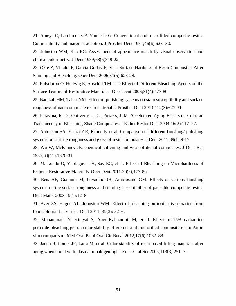
51
21. Ameye C, Lambrechts P, Vanherle G. Conventional and microfilled composite resins.
Color stability and marginal adaption. J Prosthet Dent 1981;46(6):623- 30.
22. Johnston WM, Kao EC. Assessment of appearance match by visual observation and
clinical colorimetry. J Dent 1989;68(6)819-22.
23. Okte Z, Villalta P, García-Godoy F, et al. Surface Hardness of Resin Composites After
Staining and Bleaching. Oper Dent 2006;31(5):623-28.
24. Polydorou O, Hellwig E, Auschill TM. The Effect of Different Bleaching Agents on the
Surface Texture of Restorative Materials. Oper Dent 2006;31(4):473-80.
25. Barakah HM, Taher NM. Effect of polishing systems on stain susceptibility and surface
roughness of nanocomposite resin material. J Prosthet Dent 2014;112(3):627-31.
26. Paravina, R. D., Ontiveros, J. C., Powers, J. M. Accelerated Aging Effects on Color an
Translucency of Bleaching-Shade Composites. J Esthet Restor Dent 2004;16(2):117–27.
27. Antonson SA, Yacizi AR, Kilinc E, et al. Comparison of different finishing/ polishing
systems on surface roughness and gloss of resin composites. J Dent 2011;39(1):9-17.
28. Wu W, McKinney JE. chemical softening and wear of dental composites. J Dent Res
1985;64(11):1326-31.
29. Malkondu O, Yurdaguven H, Say EC, et al. Effect of Bleaching on Microhardness of
Esthetic Restorative Materials. Oper Dent 2011:36(2);177-86.
30. Reis AF, Giannini M, Lovadino JR, Ambrosano GM. Effects of various finishing
systems on the surface roughness and staining susceptibility of packable composite resins.
Dent Mater 2003;19(1):12–8.
31. Azer SS, Hague AL, Johnston WM. Effect of bleaching on tooth discoloration from
food colourant in vitro. J Dent 2011; 39(3): 52–6.
32. Mohammadi N, Kimyai S, Abed-Kahnamoii M, et al. Effect of 15% carbamide
peroxide bleaching gel on color stability of giomer and microfilled composite resin: An in
vitro comparison. Med Oral Patol Oral Cir Bucal 2012;17(6):1082–88.
33. Janda R, Poulet JF, Latta M, et al. Color stability of resin-based filling materials after
aging when cured with plasma or halogen light. Eur J Oral Sci 2005;113(3):251–7.

52
34. Roselino LMR, Cruvinel DRC, Chinelatti MA, Pires-de-Souza FCPP. Effect of
brushing and accelerated ageing on color stability and surface roughness of composites. J
Dent 2013;41(5):54–61.
35. Vichi A, Ferrari M, Davidson CL. Color and opacity variations in three diferente resin-
based composite products after water aging. Dent Mater 2009;20(6):530-534.
36. Monaghan P, Trowbridge T, Lautenschlager E. Composite resin color change after vital
tooth bleaching. J Prosthet Dent 1992;67(6):778-781.
37. Hafez R, Ahmed D, Yousry M, et al. Effect of In-Office Bleaching on Color and
Surface Roughness of Composite Restoratives. Eur J Dent 2010; 4(2):118-27.
38. Fontes ST, Fernandez MR, Moura CM, Meireles SS. Color stability. Color stability of a
nanofill: effecte different immersion media. Oral Sci. 2009;17(5):388-91.
39. Janus G, Fauxpointa Y, Arntzc H, et al. Surface roughness and morphology of three
nanocomposites after two different polishing treatments by a multitechnique. Dent Mater
2010;26(5):416–25.
40. Lu H, Powers JM. Color stability of resin cements after accelerated aging. Am J Dent.
2004;17(5):354-8.

53
Conclusão
De acordo com os resultados obtidos nesse estudo, pode-se concluir que:
- As alterações das propriedades físicas do nanocompósito pelos clareadores
variaram conforme a composição dos mesmos. A substituição do espessante carbopol pelo
natrosol no clareamento com peróxido de carbamida a 16 % propiciou a manutenção da
microdureza;
- O envelhecimento artificial acelerado promoveu significativas alterações nas
propriedades físicas do nanocompósito, como o aumento da rugosidade, diminuição do
brilho e microdureza. Ainda, a cor apresentou mudanças perceptíveis e inaceitáveis
clinicamente;
- O peróxido de carbamida a 10 % intensificou a perda de brilho e aumentou a
rugosidade da resina após envelhecimento artificial acelerado;
- A alteração de superfície do nanocompósito desencadeada pelo processo de
envelhecimento e desafio químico decorrente do clareamento contribuiu para uma maior
pigmentação extrínseca.

54
Referências*
Aguiar FH, Georgetto MH, Soares GP, Catelan A, Dos Santos PH, Ambrosano GM,
Figueroba SR, Lovadino JR. Effect of different light-curing modes on degree of
conversion, staining susceptibility and stain's retention using different beverages in a
nanofilled composite resin. J Esthet Restor Dent. 2011; 23(2): 106-14.
Bayindir F, Kurklu D, Yanikoglu N.D. The effect of staining solutions on the color stability
of provisional prosthodontic materials. J Dent. 2012; 40(2): 41-6.
Canay S, Cehreli MS. The effect of current bleaching agents on the color of light-
polymerized composites in vitro. J Prosthet Dent. 2003; 89(5): 474-78.
Carvalho PRB, Meneses Filho FP, Silva CHV. Etiology and prevention of color change in
composite esthetic restorations. Inter J Dent. 2003; 2(1): 236-40.
Durner J, Obermaier J, Ilie N. Investigation of different bleaching conditions on the amount
of elutable substances from nano-hybrid composites. Dent Mater. 2014; 30(2): 192-9.
Gonulol N, Yilmaz F. The effects of finishing and polishing techniques: on surface
roughness and color stability of nanocomposites. J Dent. 2012; 40(2): 64-70.
Guler AU, Yilmaz F, Kulunk T, Guler E, Kurt S. Effects of different drinks on stainability
of resin composite provisional restorative materials. J Prosthet Dent. 2005; 94(2): 118-24.
* De acordo com a normativa da FOP/Unicamp baseada na norma International Committee of Medical
Journal Editors – Grupo de Vancouver. Abreviatura dos periódicos em conformidade com o Medline.

55
Jung M, Eichelberger K, Klimek J. Surface geometry of four nanofiller and one hybrid
composite after one-step and multiple-step polishing. Oper Dent. 2007; 32(4): 347-55.
Melo MA, Moysés MR, Santos SG, Alcântara CE, Ribeiro JC. Effects of different surface
treatments and accelerated artificial aging on the bond strength of composite resin repairs.
Braz Oral Res. 2011; 25(6): 485-91.
Silva Leite MLA, Silva FDMC, Meireles SS, Duarte RM, Andrade AK. The effect of
drinks on color stability and surface roughness of nanocomposites. Eur J Dent. 2014; 8(3):
330-6.
Yap AU, Mor BY. Surface finish of a new hybrid aesthetic restorative material. Operative
Dentistry. 2002; 27(2); 161-66.

56
Apêndice
DETALHAMENTO DAS METODOLOGIAS
1. Delineamento Experimental
Artigo 1
Unidades experimentais: 100 amostras cilíndricas de resinas compostas nanoparticuladas
(Filtek Z350 XT®, 3M/ESPE), Figura1.
Fatores em estudo:
- Envelhecimento (2 níveis):
Com envelhecimento
Sem envelhecimento
- Tratamento (5 níveis ):
Clareamento caseiro (Peróxido de carbamida 16% + carbopol)
Clareamento caseiro (Peróxido de carbamida 16% + natrosol)
Carbopol
Natrosol
Sem clareador
Variáveis de resposta:
Brilho
Cor (ΔE)
Rugosidade superficial (Ra)
Microdureza (KHN)

57
Divisão dos Grupos:
As amostras foram divididas em 10 grupos (n=10), de acordo com o
envelhecimento e tratamento clareador (Figura1).
Figura 1: Grupos de estudo.
Artigo 2
Unidades experimentais: 120 amostras cilíndricas de resinas compostas (Filtek Z350 XT®
3M/ESPE), Figura 2.
Fatores em estudo:
- Envelhecimento (2 níveis):
Com envelhecimento
Sem envelhecimento
- Clareamento (3 níveis)
Peróxido de carbamida 10% (Whiteness Perfect 10% - FGM )
Peróxido de hidrogênio 35% (Whiteness HP MAXX-FGM)

58
Sem clareamento
- Pigmentação (2 níveis)
Com pigmentação
Sem pigmentação
Variáveis de resposta:
Cor
Brilho
Rugosidade
Microdureza
Divisão dos Grupos:
As amostras foram divididas em 12 grupos (n=10)(Figura 2).
Figura 2: Grupos de estudo.
2. Preparo das amostras (Artigo 1 e 2)
Amostras cilíndricas (7,0 mm de diâmetro e 2,0mm de espessura) Foram
confeccionadas com a resina composta nanoparticulada (Filtek Z350 XT®
, 3M/ESPE) na
cor A2 (Figura 1). Com o auxílio de uma espátula de inserção (Goldstein XTS flex, Hu-

59
friedy, EUA) a resina composta foi inserida em incremento único de 2,0mm em uma matriz
de silicone por adição (Elite HD + normal setting-© Zermack SpA- Badia Polesine (RO),
Italy) obtida através da moldagem de um modelo metálico. Para evitar qualquer
deformação, e permitir a padronização das amostras durante a confecção, a matriz de
silicone foi revestida por um tupo de PVC (Figura 2-a). Em seguida, a resina foi coberta
com uma tira de poliéster e lâmina de vidro sob um peso de 500g durante 30 segundos, para
assegurar a compactação e eliminação de bolhas de ar na resina não polimerizada. A
fotoativação das amostras foi realizada por 20 segundos por LED (Valo – Ultradent
Products Inc., South Jordan, UT, USA) com irradiância de 817 mW/cm2 em que a ponta do
fotoativador ficou o mais próximo possível da amostra sem no entanto toca-la (Figura 2).
(c) (b)
Figura 1: Resina composta de nanopartículas Filtek Z350 XT.
Figura 2: Confecção da matriz de silicone e preparo da amostra: a) Modelo metálico e
moldagem de silicone, b) Inserção da resina composta com auxílio de espátula de
inserção, c) Completa inserção da resina composta, d) Adaptação da tira de poliéster e
lâmina de vidro, e) Compactação da resina composta sob um peso de 500g, f)
Fotoativação da resina.

60
Logo após a confecção das amostras, estas foram armazenadas por 24 horas em
estufa a 37oC a 100% de umidade relativa para completa polimerização. Em seguida, foram
submetidas ao processo de polimento na politriz giratória (modelo APL-4; Arotec, SP,
Brasil). A face superior de cada espécime foi polida por 1 minuto cada em disco de lixa de
carbeto de silício, utilizadas nas granulações #600, #1200 e #4000 (CARBIMET Paper
Discs; Buehler, IL, EUA).
Para a finalização do polimento, utilizou-se discos de feltro e pasta diamantada
(Arotec, Cotia, SP, Brasil) nas seguintes gradacões: feltro marrom com pasta de granulacao
3μm, feltro verde com pasta de granulacao 1⁄2μm, feltro azul com pasta de granulacao
1⁄4μm e feltro azul sem pasta (Figura 3).
Entre cada lixa e cada feltro, as amostras foram submetidas à limpeza em cuba
ultrassônica (ultra clearer USC-1450A/ Frequência 25kHz, UNIQUE-Brasil) com água
destilada por 5 minutos, e ao final do processo de polimento por 10 minutos, com troca de
água a cada 5 minutos para eliminação de resíduos, ficando a amostra com um aspecto liso
e brilhante.
(a)
Figura 3: Polimento das amostras: a) Politriz giratória, b) Lixas de carbeto de silício, c)
Feltros e pastas diamantadas, d) Amostras finalizadas.

61
3. Protocolo do Envelhecimento Artificial Acelerado
Para a realização do envelhecimento, as amostras foram fixadas com cola de
silicone (POLYSTIC®
, Pulvitec, Brasil) em placas metálicas específicas do equipamento.
Em seguida, foram levadas à uma máquina de intemperismo natural para o envelhecimento
artificial acelerado, específico para objetos não metálicos (EQ-UV Equilam, Diadema - SP,
Brasil). Esse equipamento é composto por uma rede de quatro fontes de luz, as quais ficam
à uma distância de 50 mm em relação as amostras, concentrada na região do ultravioleta B
que simula a luz do dia. A humidade é promovida pelo processo de condensação ativados
em ciclos separados automaticamente e repetidos sucessivamente.
Neste estudo padronizou-se o programa II, que utiliza uma energia total de
389,2 kJ/m2 para a exposição das amostras com irradiacao controlada de luz ultravioleta
emitida por uma fonte de luz xenônio com irradiancia de 0,71 W/m2 no comprimento de
onda de 310 nm. Os ciclos consistiram em 4 horas de exposicao a luz UV-B e 4 horas de
condensação por um tempo total de 300h em temperatura constante de 50 oC (ASTM,
2006) (Figura 4).
Figura 4: Envelhecimento das amostras: a) Amostras fixadas em placas específicas do equipamento, b)
Máquina de envelhecimento artificial acelerado, c) Amostras sem e com envelhecimento.

62
4. Protocolo do Tratamento Clareador
4.1 Tratamento clareador (Artigo 1)
No primeiro estudo utilizou-se gel de Peróxido de carbamida a 16% com
carbopol, gel de Peróxido de carbamida a 16% com o natrosol, gel de carbopol e gel de
natrosol. Diariamente, por um período de 14 dias, as amostras receberam uma sessão de
tratamento clareador. O gel foi aplicado na superfície de topo das amostras, permanecendo
em contato com a mesma por 4 horas. As amostras foram fixadas em placas de vidro com o
auxílio de uma fita dupla face e inseridos em um aparato fechado contendo água
deionizada, sem no entanto toca-las, em estufa a 37oC ± 2, simulando a umidade da
cavidade bucal (Figura 5).
Após o tempo de 4 horas em contato com a superfície das amostras, o gel foi
removido da superfície da amostra com hastes flexíveis de plástico com algodão em suas
pontas (Cotonetes- Johnson & Johnson, Brasil), em seguida, lavadas em água corrente por
1 minuto, secadas com papel absorvente (Kleenex – Kimberly-Clark, Brasil) e armazenadas
em 100% de umidade relativa e levada a estufa a uma temperatura de 37oC ± 2 (Figura 6).
Figura 5: Uso dos géis: a) Géis clareadores (peróxido de carbamida 16% com carbopol,
peróxido de carbamida 16% com natrosol, gel de carbopol, gel de natrosol) b) Forma de
armazenamento durante do tratamento dos géis sobre as amostras.

63
As amostras que nao receberam tratamento foramarmazenadas em 100% de umidade
relativa em estufa a 37oC ± 2, durante todo o estudo.
4.2 Tratamento clareador (Artigo 2)
Após serem fixadas na placa de vidro com o auxílio de fita dupla face e
inseridos em um aparato contendo água deionizada, os tratamentos clareadores no segundo
estudo foram realizados de acordo com as instruções do fabricante.
Peróxido de carbamida 10% (Whiteness Perfect 10 % - FGM): O gel foi
aplicado sobre a superfície de topo das resinas por 21 dias consecutivos, permanecendo em
contato por 4 horas em estufa a 37 oC ± 2. O aparato com água foi fechado com o objetivo
de simular a humidade da cavidade bucal (Figura 7).
Figura 6: Finalização do tratamento: a) Remoção do gel, b) Lavagem, c)
Secagem da amostra b) Armazenamento.
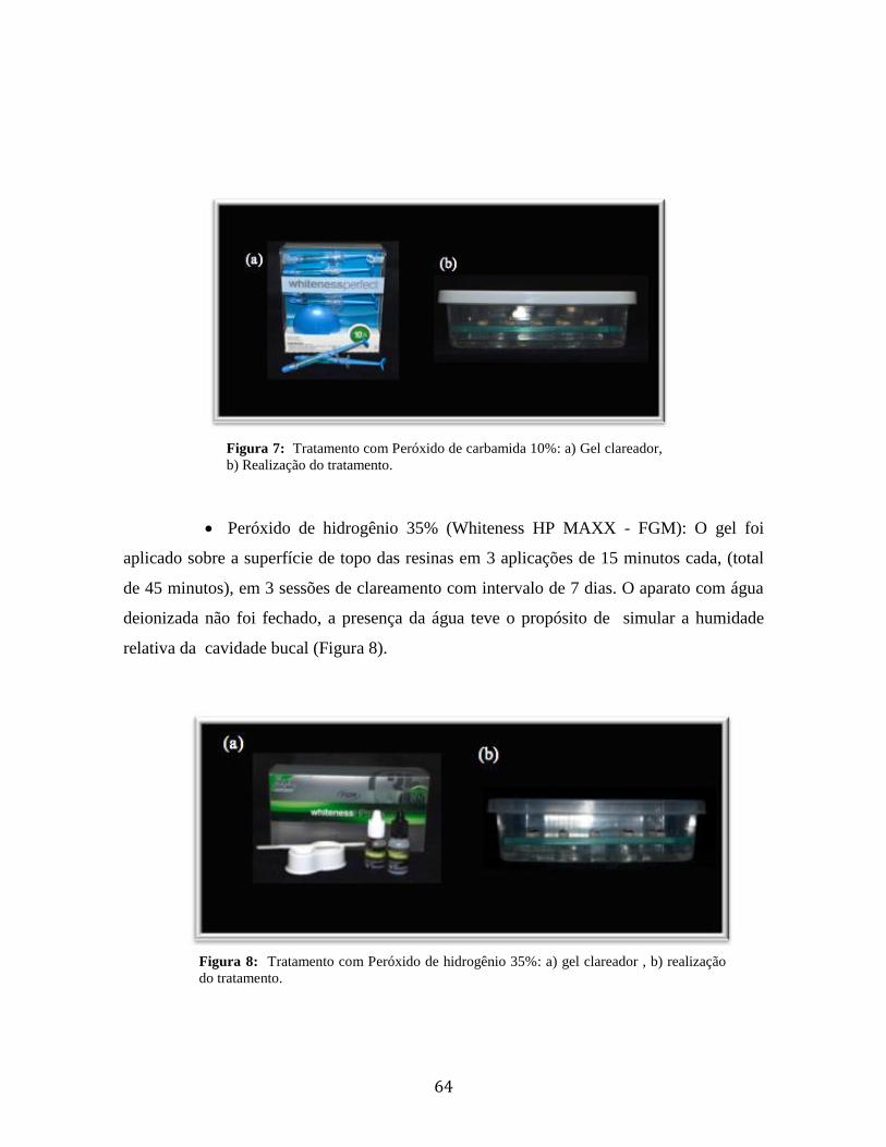
64
Peróxido de hidrogênio 35% (Whiteness HP MAXX - FGM): O gel foi
aplicado sobre a superfície de topo das resinas em 3 aplicações de 15 minutos cada, (total
de 45 minutos), em 3 sessões de clareamento com intervalo de 7 dias. O aparato com água
deionizada não foi fechado, a presença da água teve o propósito de simular a humidade
relativa da cavidade bucal (Figura 8).
Figura 7: Tratamento com Peróxido de carbamida 10%: a) Gel clareador,
b) Realização do tratamento.
Figura 8: Tratamento com Peróxido de hidrogênio 35%: a) gel clareador , b) realização
do tratamento.

65
Após cada aplicação, o gel foi removido da superfície da amostra com hastes
flexíveis de plástico com algodão em suas pontas (Cotonetes- Johnson & Johnson, Brasil),
ao final foram lavadas abundantemente em água corrente por 1 minuto, secadas com papel
absorvente e armazenadas em 100% de umidade relativa a uma temperatura de 37 ºC ± 2
(Figura 9).
5. Protocolo de manchamento (Artigo 2)
As amostras a serem submetidas a pigmentação extrínseca tiveram suas laterais
e superfície base recobertas com cera pegajosa (Cera pegajosa em bastão ASFER Indst.
Química Ltda.), para que apenas a superfície topo ficasse exposta à pigmentação. A solução
pigmentante foi obtida através da dissolução 3,6 g de cafe (Cafe Nescafe Classic, Sao
Paulo, Brasil) em 300 ml de água destilada fervida. Após 10 minutos de manipulação da
água e do pó, a solução foi filtrada com papel filtro. As amostras foram armazenadas nessa
solução por 7 dias em estufa 37oC ± 2, simulando a temperatura da cavidade bucal.
Diariamente a solução foi trocada, e a cada troca as amostras eram lavadas em água
corrente por 5 minutos, para remoção de borra. Ao final do período de manchamento e
Figura 9: Finalização do tratamento: a) Remoção do gel a base de peróxido de
hidrogênio, b) Remoção do gel a base de peróxido de carbamida c) Lavagem, d) Secagem
da amostra, e) Armazenamento.

66
armazenamento, as amostras foram novamente lavadas por 5 minutos e secadas com papel
absorvente previamente a análise da cor (Figura 10).
6. Análise de cor (Artigo 1 e 2)
A análise de cor foi realizada após os tratamentos de envelhecimento,
clareamento e pigmentação, de acordo com cada grupo nos respectivos estudos. As
amostras foram posicionadas em um dispositivo de teflon (porta amostra) dentro de uma
Câmara de luz (GTI Mini Matcher MM1e, GTI Graphic Technology Inc., Newburgh, NY,
USA) para padronização do ambiente de cada leitura, sendo utilizado a opção luz do dia.
Foi utilizado o espectrofotômetro Konica Minolta CM-700d previamente
calibrado de acordo com as instruções do fabricante. As amostras foram lidas e os valores
obtidos foram analisados no programa On Color QC Lite (Konoca Minolta, Japão) em
computador (Figura 11).
Figura 10: Manchamento das amostras: a) Amostras recobertas com cera,
b) Café, c) Imersão na solução, d) Amostras (sem envelhecimento, sem e
com pigmentação, envelhecidas sem e com pigmentação).

67
7. Brilho de superfície (Artigo 1 e 2)
Os valores de brilho das superfícies das amostras foram mensurados com
medidor de brilho (ZGM 1120 GLOSSMETER – Zehntner Testing Instruments, Sissach,
Suíça) ( Figura 12 a). O princípio de mensuracao do aparelho e baseado em um feixe de luz
que incide na superfície da amostra em ângulos de 200, 600 e 850. Nesse estudo padronizou-
se a angulação 60o, que permite avaliar o brilho médio da superfície (ISO-Standards, ISSO
2813). Para a calibracao do aparelho, foi utilizada uma amostra padronizada de vidro negro
polido (calibrador) ( Figura 12 b/c), fornecida pelo fabricante. Para cada amostra, quatro
mensuracões foram realizadas, uma em cada quadrante. A media das leituras foi registrada
como valor da unidade de brilho para cada amostra (Gloss Unit - GU). Para evitar a
influência da iluminacao externa, utilizou-se um dispositivo metálico com dimensões
Figura 11: Posicionamento das amostras durante a leitura da cor: a)
Espectrofotômetro, b) Porta amostra, c) Câmera de luz, d) Leitura de
cor.

68
semelhante as amostras do estudo, que ao ser pressionada pela base do medidor de brilho,
impedia a entrada da luz externa (Figura 12 d/e).
8. Rugosidade Superficial (Artigo 1 e 2)
A rugosidade superficial foi avaliada por meio de um rugosímetro (SV-3100S4 – Mitutoyo,
Toquio, Japao) (Figura 13-a). As amostras foram plan
ificadas em uma base de acrílico com auxílio de cera utilidade em um
planificador (Figura 13-b) e sequencialmente posicionadas paralelas a superfície do
equipamento. Marcações em três pontos equidistantes guiaram a leitura da ponta de
diamante do rugosímetro , para que o mesmo passasse pelo centro geométrico da amostra
até a borda. A primeira leitura foi realizada em 180o, em seguida as amostras foram
Figura 12: medição do brilho de superfície: a) Medidor de brilho, b) calibrador, c)
medidor de brilho acoplado ao calibrador, d) suporte metálico para guiar a leitura, e)
Posição da amostra para posterior encaixe do medidor.

69
posicionadas em 135o e a terceira leitura em 90o. A média das três leituras considerou-se a
rugosidade média (Ra) de cada superfície (Figura 13 c).
9. Microdureza Knoop (Artigo 1 e 2 )
A análise da microdureza foi realizada sobre a superfície de topo de cada
amostra utilizando o microdurômetro (HMV-2000, Shimadzu Corporation, Toquio, Japao)
(Figura 14 a) com um indentador de diamante Knoop, sob uma carga de 50g por 15
segundos. Cinco medições Knoop foram feitas sobre a superfície superior de cada amostra:
uma no centro e outras 4 a uma distância de 200 m a partir do centro. A média dos 5
valores das indentações foi calculado como o valor de KHN para cada espécime (Figura 14
b/c).
Figura 13: Medição da rugosidade: a) Rugosímetro, b) Detalhe da ponta do
rugosímetro na amostra, c) Posição e leitura da rugosidade.

70
10. Microscopia de Força Atômica (Artigo 1)
Para análise qualitativa da superfície, duas amostras de cada grupo foram
escolhidas aleatoriamente após cada tratamento. Para tanto, as amostras foram lavadas em
banho ultrassônico e secadas em papel absorvente. Em seguida, as amostras foram fixadas
com auxílio de fita dupla face no suporte metálico e acoplada no microscópio de força
atômica (Easy Scan 2, Nano surf, Boston, MA, EUA) (Figura 15). Assim, imagens
topográficas em 3D de tamanho 15 m x 15 m foram obtidas para cada grupo.
Figura 14: Medição da microdureza: a) Microdurômetro b) Posição e
leitura da dureza, c) Esquema grafico das indentacões.

71
11. Microscopia Eletrônica de Varredura (Artigo 2)
Com a finalidade de observar qualitativamente a superfície topográfica da
matriz resinosa, bem como o tamanho e geometria da partículas de carga. Foi utilizado o
microscópio eletrônico de varredura MEV (JEOL- JSM 5600 LV- Tokio, Japao). Duas
amostras de cada grupo foram selecionadas ao final de todo o experimento. As amostras
foram lavadas em cuba ultrassônica e secadas em papel absorvente, em seguida,
armazenadas em sílica gel por 48hs. Em sequência foram fixadas em discos metálicos para
cobertura com ouro. Desta forma, foram obtidas fotomicrografias de áreas representativas
de cada grupo no estudo com uma ampliação 1000X. (Figura 16).
(a)
Figura 15: AFM: a) Microscópio de força atômica b) Posição da amostras no
microscópio c) Amostras fixadas no suporte metálico do microscópio.
Figura 16: MEV: a) Metalizador, b) Amostras com metalização, b) Microscópio eletrônico
de varredura.

72
Anexo COMPROVAÇÃO DE SUBMISSÃO
Artigo 1

73
Artigo 2