http:// tour of the cell lecture 4
TRANSCRIPT

http://www.steve.gb.com
Tour of the CellLecture 4
2
Much of the text material in the lecture notes is from our textbook, “Essential Biology with Physiology” by Neil A. Campbell, Jane B.
Reece, and Eric J. Simon (2004 and 2008). I don’t claim authorship. Other sources were sometimes used, and are noted.

3
Outline
• Drugs that target cells• Microscopic world of cells• Microscopy• Cell types • Cell components and functions• Origin of membranes• Words and terms to know• Possible test items

4
American Civil War
http://www.a2zcds.comhttp://nmhm.washingtondc.museum
During the American Civil War, many soldiers died from infections in the treatment of their wounds (possibly as many as actually died on the
battlefield).

5
Penicillin
• Antibiotics derived from microorganisms disable or kill bacteria.• In the 1920s, Alexander Fleming discovered penicillin when he observed
that mold prevented the growth of bacteria that he was trying to cultivate in bread.
• Fleming recognized the value of an agent that inhibits bacterial growth, and the age of antibiotics was born.
• The death rates from diseases such as bacterial pneumonia and surgical infection dropped substantially once antibiotics were introduced and then widely used. �
• The discovery of penicillin is a famous case of ‘serendipity.’

6
Bread Mold
http://www.sciencemore.com

7
Antibiotic Actions
• Antibiotics destroy bacteria or inhibit their growth.• Penicillin works by disrupting the synthesis of the cell walls in bacteria.• Erythromycin binds to a structure that synthesizes proteins found only in
bacterial cells.• Ciprofloxacin, used in treating anthrax infections, targets an enzyme that
helps maintain the genetic structure of bacteria.
http
://w
eb.e
du

8
Cell Theory
• Cells were first described in 1665 by the British scientist, Robert Hooke, while examining a thin slice of cork through a microscope.
• Over the next two centuries, cells were found in all organisms examined under a microscope.
• By the mid-1800s the accumulation of evidence (through the process of inductive reasoning) led to the cell theory: all organisms are composed of cells.
Microscopic view of commercial cork obtained from Cork Oak
http
://in
stru
ct1.
cit.c
orne
ll.ed
u
• The theory was later expanded to include observations that new cells arise from previously existing cells.

9
Cellular Structure of Cork
http://farm1.static.flickr.com
Robert Hooke’s drawing

10
Grove of Cork Oak
http://cache.eb.com
http://www.isa.utl.pt
11
Microscopic World of Cells
• Each cell in a living organism is very complex.• Cells must be very small for materials to move in and out of the cell to
meet its needs.• A modern jet aircraft, if it was reduced to the size of a cell, would seem
simple in comparison.• Organisms are single-cellular, such as bacteria and protista, and multi-
cellular as animals, plants, and most fungi.• The human body has many trillions of cells that work together to perform
specific functions.

12
A Few Types of Neurons
http
:ww
w.s
emap
horin
.com

13
Light Microscope
A portable microscope similar to the one Darwin
used on the H.M.S Beagle
http://www.hps.cam.ac.uk
http://www.meijitechno.com
Modern lab and classroom version

14
LM Operation
• Light microscopes (LMs) were first developed during the Renaissance period.
• Visible light passes through the specimen—the lens enlarges the image and projects it onto the human eye or camera.
• Modern light microscopes have compound lenses to reduce chromatic (color) aberration and spherical aberration for improving the quality of the viewed image.
15
Magnification and Resolving Power
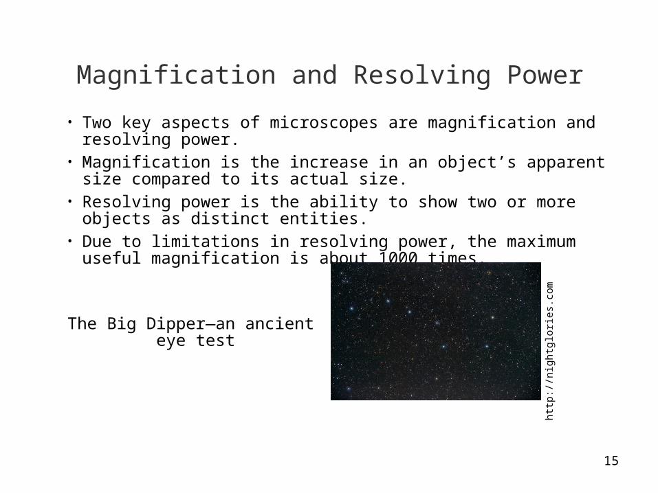
• Two key aspects of microscopes are magnification and resolving power.• Magnification is the increase in an object’s apparent size compared to
its actual size.• Resolving power is the ability to show two or more objects as distinct
entities.• Due to limitations in resolving power, the maximum useful magnification
is about 1000 times.
http
://n
ight
glor
ies.
com
The Big Dipper—an ancient eye test

16
LM Micrograph
http://www.steve.gb.com
Cross-section of bamboo showing its internal vasculature.

17
LM Micrograph
Cell structure in an elodea leaf
http://marby.online.com

18
LM Micrograph
http://www7.ocn.ne.jp
Cross section through a buttercup stem

19
Red blood cells and a stained white blood cell
http://www.uwash.edu
LM Micrograph

20
LM Micrograph
Coronal cross-section of a rat brain
http://www.emsdiasum.com

21
Electron Microscope
http://www.usaft.af.mil
An electron microscopy lab

22
EM Operation
• The study of the structure of cells continued to advance once electron microscopes (EMs) were developed in the 1950s.
• Electron microscopes use beams of electrons rather than light to explore the very small world.
• The resolving power is much higher than for light microscopes, allowing for much higher useful magnifications.
• Electron micrographs can be produced at magnifications of 100,000 or higher.

23
Types of Electron Microscopes
• Scanning electron microscopes are used for studying the surfaces of cells.
• Transmission electron microscopes are used for exploring the internal structure of cells.
• Light microscopes can be used with live or prepared (dead) specimens, while electron microscopes can only be used with prepared specimens.

24
EM Micrograph
http://www.allergy-details.com
A ‘potpourri’ of pollens

25
EM Micrograph
http://www.microscopy-uk.org
Blood—neutrophils and lymphocytes
26
EM Micrograph
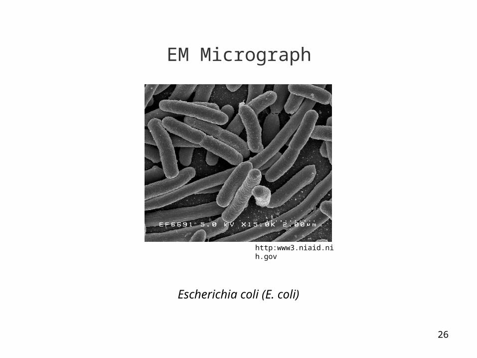
http:www3.niaid.nih.gov
Escherichia coli (E. coli)
27
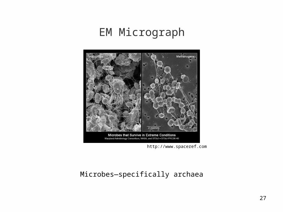
EM Micrograph
http://www.spaceref.com
Microbes—specifically archaea
28
We now examine cells and their organelles

29
Cell Types
• Prokaryotic cells—evolutionary much older cells, and much simpler in structure than eukaryotic cells. Bacteria, for example, are prokaryotes.
• Eukaryotic cells—much more complex internal structure. All animal and plant cells are eukaryotes.
• Prokaryotic cells are much smaller than eukaryotic cells and do not have a true nucleus—they have a nucleoid region containing genetic material.

30
Prokaryotic Cell
The bacteria E. coli dividing
http://www.bio.mtu.edu
A highly-stylized representation
http://www.cod.edu

31
Eukaryotic Cell
http://www.cod.edu
A highly-stylized representation
Electron micrograph—the nucleus and endoplasmic reticulum are prominent
http://www.steve.gb.com
32
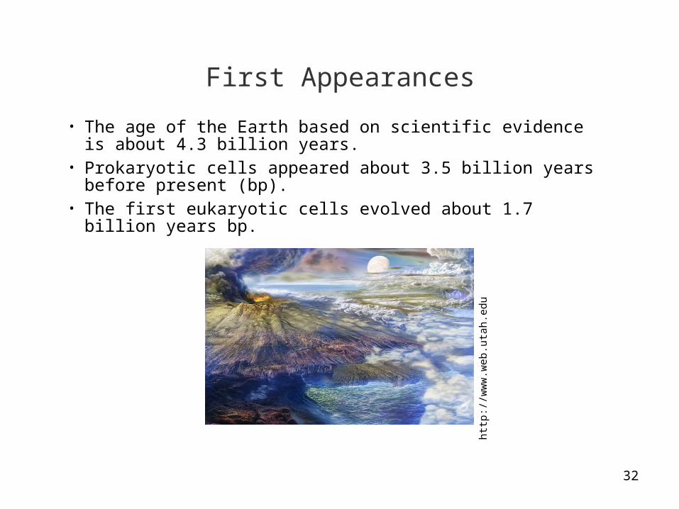
First Appearances
• The age of the Earth based on scientific evidence is about 4.3 billion years.
• Prokaryotic cells appeared about 3.5 billion years before present (bp).• The first eukaryotic cells evolved about 1.7 billion years bp.
http
://w
ww
.web
.uta
h.ed
u
33
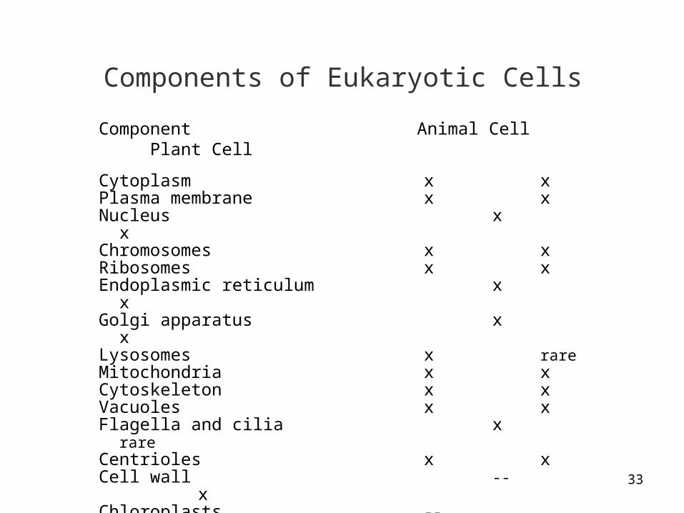
Components of Eukaryotic Cells
Component Animal Cell Plant Cell
Cytoplasm x xPlasma membrane x xNucleus x xChromosomes x xRibosomes x xEndoplasmic reticulum x xGolgi apparatus x xLysosomes x rareMitochondria x xCytoskeleton x xVacuoles x xFlagella and cilia x rareCentrioles x xCell wall -- xChloroplasts -- xCentral vacuole -- x

34
Cytoplasm
Electron micrographhttp://www.danforthcenter.org
35
Cytoplasm
• The cytoplasm is the region of the cell between the nucleus and plasma membrane.
• It contains various organelles suspended in a fluid known as the cytosol.• Each organelle is adapted to perform specific functions, as we will discuss. • Most organelles in eukaryotic cells are enclosed by their own membranes.
36
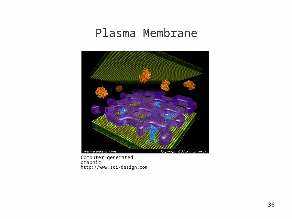
Plasma Membrane
Computer-generated graphichttp://www.sci-design.com
37
Plasma Membrane
• Cells have a plasma membrane separating the interior and exterior of the cell—they are also known as the intracellular and extracellular spaces.
• The membrane consists primarily of phospholipids and proteins in a ‘fluid mosaic.’
• The plasma membrane regulates the traffic of molecules moving into and out of the cell.
• It is selectively permeable—that is, the membrane allows some molecules to pass through while preventing the passage of others.
• Transport proteins embedded in the plasma membrane allow the passage of other molecules such as glucose molecules.
38
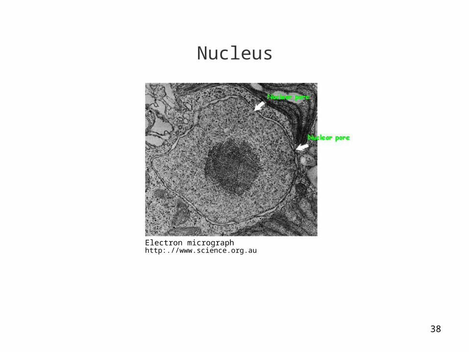
Nucleus
Electron micrographhttp:.//www.science.org.au
39
Nucleus
• The nucleus has a double membrane known as the nuclear envelope.• The double membrane is similar in structure to the plasma membrane.• Pores in the membrane allow the passage of material between nucleus
and cytoplasm.• DNA molecules and associated proteins form long fibers in the nucleus
called chromatin.• The nucleus also contains a ball-like mass (the nucleolus) that produces
the component parts of ribosomes.

40
Chromatin and DNA
Computer-generated graphicsBoth images from http://www.cgl.ucsf.edu
Unpacked
Packed

41
Ribosomes
Computer-generated graphichttp://rna.ucsc.edu

42
Ribosomes
• Ribosomes are located in the cytoplasm, near the cell nucleus, where they synthesize proteins.
• Some ribosomes make proteins that will be dissolved in the cytoplasm, while others make proteins for the plasma membrane or secretion by the cell.
• DNA transfers genetic information via messenger RNA to the ribosomes to synthesize proteins.
• We will discuss DNA and RNA in future lectures on the genetic basis of life.
43
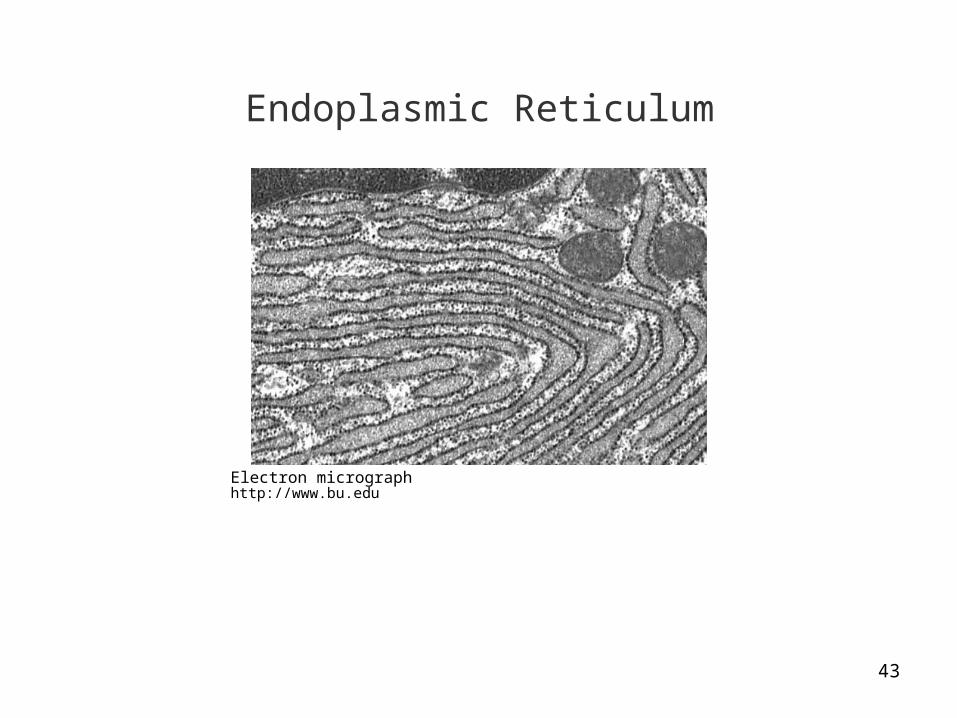
Endoplasmic Reticulum
Electron micrographhttp://www.bu.edu

44
Endoplasmic Reticulum
• The endoplasmic reticulum (ER) produces many types of molecules of life.• It is a complex system of tubes and sacs running through the cytoplasm.• Rough ER has the visual appearance of roughness due to ribosomes that
stud its exterior surface. • The products of ribosomes are modified in the rough ER and sent on their
way in transport vesicles.• Cells that secrete substantial amounts of protein, such as salivary glands,
are rich in rough ER.
45
Smooth ER
• Smooth ER synthesizes lipids, among other biological molecules.• It lacks the embedded ribosomes found in the membrane of rough ER.• Cells in the ovaries and testes are rich in smooth ER, and produce the
steroids, estrogen and testosterone.

46
Detoxification
• Smooth ER in the cells of the liver produce enzymes for the detoxification of drugs and poisons in the blood.
• Smooth ER increases in liver cells when exposed to the chemicals in some drugs.
• As a result, the body increases its tolerance to a chemical, requiring higher dosages to achieve the same effect.
• An increase in tolerance to some drugs is a hallmark of addiction—it has a firm biological basis.

47
Golgi Apparatus
Electron micrographhttp://www.bu.edu

48
Golgi Apparatus
• The Golgi apparatus is named for its discover, the Italian scientist, Camillo Golgi.
• It works with the ER to refine, store, and distribute molecules synthesized in the cell.
• Products manufactured in the ER reach the Golgi apparatus via transport vesicles.
• Enzymes in the Golgi apparatus modify many of the products from the ER.• The Golgi apparatus tags proteins with ‘addresses’ for their destinations
within the cell.• Containers known as vesicles budding from the Golgi apparatus distribute
molecules to other organelles.
49
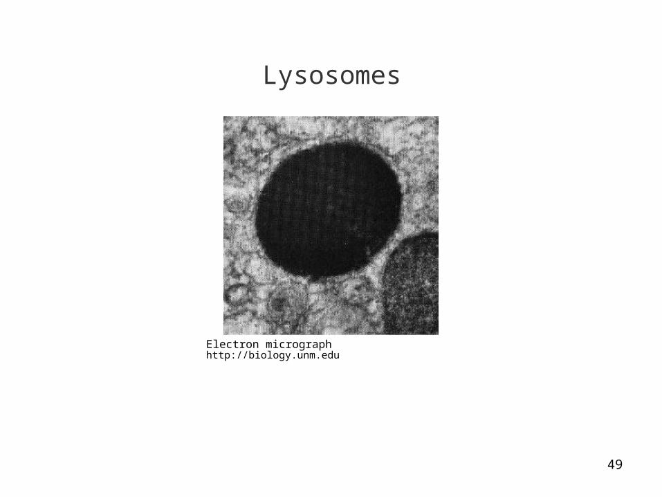
Lysosomes
Electron micrographhttp://biology.unm.edu
50
Lysosomes
• A lysosome is a membrane-enclosed sac of enzymes needed for cellular digestion.
• Lysosomes provide a compartment for chemical digestion to prevent self-destruction of the cell.
• Enzymes breakdown macromolecules including proteins, glycogen, fats, and nucleic acids.
• The molecules produced from the cellular digestive process nourish the cell.
51
Other Functions
• Other lysosomes serve as recycling centers by engulfing and digesting damaged organelles and making the molecules available for forming new organelles.
• Lysosomes in white blood cells ingest bacteria—the enzymes destroy the bacterial cell walls.
• Another type destroys the webbing joining the fingers in human embryos.

52
Mitochondria
Electron micrographhttp://is2.okcupid.com
53
Mitochondria
• Mitochondria are responsible for cellular respiration in harvesting energy to perform cellular work.
• Sugars and other food molecules are converted to a form of energy known as ATP.
• The inner membrane of mitochondria contains many folds to increase the surface area and maximize ATP output.
• We will discuss harvesting of chemical energy when we discuss the Krebs cycle.
Mitochondria—plural; mitochondrion—singular
54
Cytoskeleton
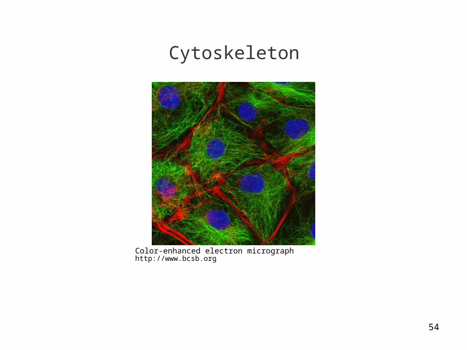
Color-enhanced electron micrographhttp://www.bcsb.org

55
Cytoskeleton
• Microtubules in the cytoplasm form a network of fibers known as the cell’s cytoskeleton
• They provides structural support and means for specialized movements.• Microtubules are constructed of proteins to provide structural support,
which is important in animal cells that don’t have semi-rigid cell walls.• The microtubules hold the organelles in place in the cytoplasm and guide
the movement of vesicles.

56
Additional Functions
• Microtubules also guide the movement of chromosomes when cells divide.• Unlike a bony skeleton, the cytoskeleton can be dismantled in one part of
the cell to reform in a new location.• This process occurs through the removal and replacement of its units of
proteins.• It contributes to the crawling motion of amoeba and the movement of white
blood cells.

57
Vacuoles
Electron micrographhttp://www.pharmacology.com
Synaptic vesicles (a type of vacuole) in a neuron
58
Vacuoles
• Vacuoles are membrane-enclosed sacs that bud from the Golgi apparatus endoplasmic reticulum, and plasma membrane.
• They differ in size depending on their functions.• The synaptic vesicles in the end buttons of neurons contain transmitter
substances released in response to nerve impulses that signal to other neurons.
• Other vacuoles form in the plasma membrane to engulf food so it can be transported to the lysosomes.
• Plant cells often have a large central vacuole as we will discuss later in the lecture.

59
Flagella
Human sperm
Electron micrographhttp://www2.sunysuffolck.edu

60
Flagella
• The microtubules found in some eukaryotic cells have appendages that can move.
• Flagella propel cells through an undulating, whip-like motion. • Flagella generally occur singly—for example, in sperm which must travel
the female reproductive tract to fertilize the ovum (or egg).• Problems with flagella can result in male infertility, as we will discuss in the
lecture on sexual reproduction.
Flagella—plural; flagellum—singular

61
Cilia
Human oviduct
Electron micrographhttp://www.talbotcentral.ucr.edu
62
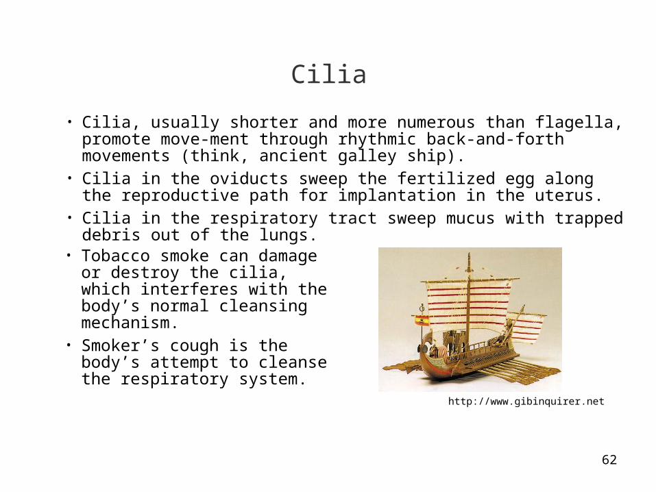
Cilia
• Cilia, usually shorter and more numerous than flagella, promote move-ment through rhythmic back-and-forth movements (think, ancient galley ship).
• Cilia in the oviducts sweep the fertilized egg along the reproductive path for implantation in the uterus.
• Cilia in the respiratory tract sweep mucus with trapped debris out of the lungs.
• Tobacco smoke can damage or destroy the cilia, which interferes with the body’s normal cleansing mechanism.
• Smoker’s cough is the body’s attempt to cleanse the respiratory system.
http://www.gibinquirer.net

63
Extracellular Matrix
• Most animal cells secrete a thick, sticky coat known as the extracellular matrix.
• The coat helps hold cells together in tissues, and provides protective and supportive functions.

64
Cell Junctions
• Adjacent cells in many animal tissues are connected by cell junctions.• Tight junctions bind cells together to form a leak-proof sheet of tissue (such
as in the intestines).• Anchoring junctions bind cells together, but allow some molecules to pass
among the extracellular spaces.• Communicating junctions are channels that enable H2O and other small
molecules to flow among neighboring cells.
65
Centrioles
Computer-generated graphichttp://www.sparkleberrysprings.com

66
Centrioles
• Centrioles are canned-shaped structures made of microtubules that support cell division.
• We will discuss their function when we cover mitosis and meiosis in later lectures.
67
Plant-Specific Cell Components
Silver Maple Tree, Queens, New Yorkhttp://graphics8.nytimes.com

68
Cell Wall
Electron micrographhttp://www.aber.ac.uk
69
Cell Wall
• A plant cell is encased by a semi-rigid wall on the exterior of the plasma membrane.
• The wall protects the cell, maintains its shape, and keeps it from absorbing too much water.
• Cell walls are collectively strong enough to hold up even the tallest of trees against the force of gravity.
• They are formed from cellulose embedded in a matrix of lignin and other molecules.
• This arrangement is similar to the construction of steel-reinforced concrete and fiberglass.
• Plant cells have junctions and channels that enable water and other small molecules to pass among cells.
70
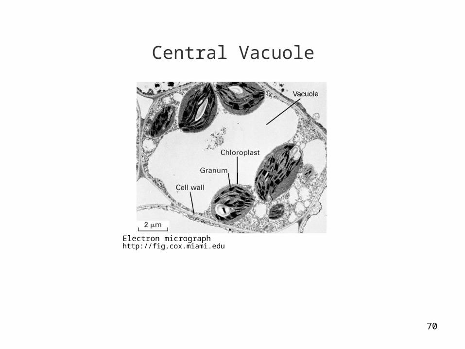
Central Vacuole
Electron micrographhttp://fig.cox.miami.edu

71
Central Vacuole
• Plant cells may have a large central vacuole to store nourishment, water, and even poisons that protect against plant-eating animals.
• Some central vacuoles contain pigments to provide color that can attract pollinating insects.

72
Chloroplasts
Electron micrographhttp:/www.bio.ic.ac.uk

73
Photosynthesis
• Most of the living world requires energy provided by photosynthesis.• Photosynthesis involves the conversion of sunlight to sugars and other
energy-rich molecules.• Oxygen (O2) is produced as a by-product and released into the atmos-
phere.

74
Chloroplasts
• Chloroplasts are the organelles in plants and protists that perform photo-synthesis.
• The flat, disk-shaped objects in the electron micrograph are grana, where photosynthesis takes place.
• We will discuss photosynthesis when we cover the Calvin cycle in a later lecture.

75
Venus Fly Trap
http://www.mooseyscountrygarden.com
One of the few types of plants to contain lysosomes and digestive enzymes.

76
Origin of Cell Membranes
http
://n
atur
alsc
ient
ist.
blog
spot
.com
Oil-in-vinegar analogy
77
Membrane Formation
• The plasma membrane is the cell boundary—without a membrane a cell would not be a separate unit.
• The formation of membranes was probably one of the earliest events in the evolution of life.
• Phospholipids, found in membranes, were likely among the first organic molecules to be formed through chemical reactions on Earth before life originated.
78
Phospholipids
• Phospholipids spontaneously assemble into membrane-like structures when mixed with water.
• Assembly requires no genetic instructions—the organization depends only on their hydrophobic and hydrophilic characteristics of phospholipids.
• The membranes provided a package for new organelles as they developed early on.

79
Words and Terms to Know
• Antibiotic• Cell junction• Cell theory• Cell wall• Central vacuole• Chloroplast• Chromatin• Cilia• Cytoplasm• Cytoskeleton• Cytosol• Electron microscope• Endoplasmic reticulum
•Flagellum •Golgi apparatus•Light microscope•Lysosome•Mitochondrion•Nucleoid region•Nucleus•Organelle•Phospholipid•Plasma membrane•Ribosome•Vacuole

80
Possible Test Items
1. Describe the biological mechanisms and effects of antibiotics such as penicillin and erythromycin.
2. What is the cell theory? What implications does it have for modern biology?
3. Describe the light microscope and electron microscope. What are three major differences?
4. List and describe four similarities or differences between prokaryotic and eukaryotic cells.
5. Describe the locations and functions of five organelles found in most eukaryotic cells.
6. Describe the locations and function of three features of plant cells not found in animal cells.