neuroimaging research on ageing, maria del c. valdés
TRANSCRIPT

Neuroimaging research
on ageing,
neurodegenerative and
cerebrovascular diseases
Assess brain biomarkers reflected in
micro/macrostructural changes and study
their impact on cognition and health
Maria del C. Valdés
Hernández
Row Fogo Lecturer in Image
Analysis
Centre for Clinical Brain
Sciences
University of Edinburgh

The Dementia Research Institute at the University of Edinburgh

Clinical practice vs. clinical research
Clinical practice Clinical research
Validation in clinical trials, phases 1 to 4
http://uk.gsk.com/en-gb/research/trials-in-people/clinical-trial-phases/
Validation in synthetic images, phantoms, images from clinical and/or
research protocols and against published results
Users are patients/caregivers/practitioners,
therefore tailored for individual use by specific patient populations
Users are researchers,therefore tailored for broad use and
management of big data
User-friendly interface with possibilities of user interaction
Fully automatic methods preferable with minimal user input
Real-time output required Real-time output not required. Batch background processing preferred
Cost-effective Cost-effectiveness not relevant

Clinical practice - SymptomApp
Corbett and Valdés Hernández @ Future Health Product Forge
https://www.youtube.
com/watch?v=qc8a4
pp5lAc

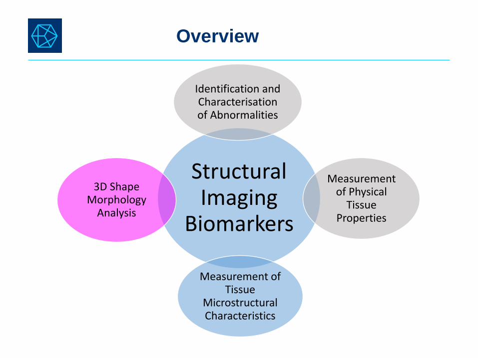
Overview
Structural Imaging
Biomarkers
Identification and Characterisation of Abnormalities
Measurement of Physical
Tissue Properties
Measurement of Tissue
Microstructural Characteristics
3D Shape Morphology
Analysis
Clinical Research

Structural Imaging
Biomarkers
Identification and Characterisation of Abnormalities
Measurement of Physical
Tissue Properties
Measurement of Tissue
Microstructural Characteristics
3D Shape Morphology
Analysis
Overview

How common are they?
Incidental findings Systematic Review BMJ 2009 339:b3016
(16 studies, involving 19,559 people)
Brain Abnormalities

Are all abnormalities incidental findings?
Brain Abnormalities
Sandeman EM, Valdes Hernandez MC, Morris Z, et al. (2013) Incidental Findings on Brain MR Imaging in Older
Community-Dwelling Subjects Are Common but Serious Medical Consequences Are Rare: A Cohort Study. PLOS
ONE 8(8): e71467. https://doi.org/10.1371/journal.pone.0071467
https://journals.plos.org/plosone/article?id=10.1371/journal.pone.0071467
Number (%) of subjects with incidental findings (IF), from a birth cohort of 700 (~72years old) individuals (determined against normal ageing templates)

Are all incidental findings clinically relevant?
Brain Abnormalities
Booth TC, Boyd-Ellison JM (2015) The Current Impact of Incidental Findings Found during
Neuroimaging on Neurologists’ Workloads. PLOS ONE 10(2): e0118155.
https://doi.org/10.1371/journal.pone.0118155
https://journals.plos.org/plosone/article?id=10.1371/journal.pone.0118155

T2 hyper-
inten-
sities
Strokes
and
lacunes
Mineral
deposits
and
haemor-
rhages
Peri-
vascular
spaces
Vascular disease
Brain Vascular Abnormalities

T2 hyper-
inten-
sities
Strokes
and
lacunes
Mineral
deposits
and
haemor-
rhages
Peri-
vascular
spaces
Vascular disease
Brain Vascular Abnormalities

T2 hyperintensities and the white matter

Prefrontal
brain slice
of an 88-yrs
old female
with
Alzheimer’s
disease
FLAIR
MRI
Histochemically
stained
hemisphere
Bodian Silver stained sections
Figure adapted from Gouw AA et al. Brain (2008), 131:3286-3298
T2 hyperintensities and the white matter

T2 hyperintensities and the white matter
Meijboom et al. 2018 (under review)

T2 hyperintensities and the white matter
https://sourceforge.net/projects/bric1936/files/

https://github.com/febrianrachmadi/lots-iam-gpu
T2 hyperintensities and the white matter

https://github.com/febrianrachmadi/lots-iam-gpu
T2 hyperintensities and the white matter
FLAIR MRI 2D Patch-CNN (DeepMedic) Patch-uNet
LOTS-IAM LST-LGA MVQ-100

https://github.com/febrianrachmadi/lots-iam-gpu
T2 hyperintensities and the white matter
FLAIR MRI with WMH Label 2D Patch-CNN (DeepMedic) Patch-uNet
LOTS-IAM LST-LGA MVQ-100

https://link.springer.com/chapter/10.1007/
978-3-030-00931-1_58
T2 hyperintensities and the white matter
Automatic Irregular Texture Detection in Brain MRI Without Human Supervision
Rachmadi, Valdés Hernández and KomuraLNCS, volume 11072 MICCAI 2018 pp 506-513
Extended version in:https://www.biorxiv.org/content/early/2018/07/23/334292

T2 hyper-
inten-
sities
Strokes
and
lacunes
Mineral
deposits
and
haemor-
rhages
Peri-
vascular
spaces
Vascular disease
Brain Vascular Abnormalities

The multispectral approach – cortical stroke lesions
Study on 57 patients with mild to moderate cortical strokes
(Poster at the Annual Meeting of the European Stroke Organisation, 2017,
Christopher and Valdés Hernández)

Volumetric differences between FLAIR
assessments guided by Diffusion-
weighted imaging (DWI) vs. blind to DWI
Volumetric differences between
assessments using FLAIR vs. T1-
weighted
The multispectral approach – cortical stroke lesions
Measurements were not associated with the clinical parameters evaluated:
age, basal ganglia perivascular spaces burden, blood pressure, pulse
frequency, small vessel disease load, lesion or atrophy scores, and all
associations yielded similar B and p values.

The multispectral approach – cortical stroke lesions

T2 hyper-
inten-
sities
Strokes
and
lacunes
Mineral
deposits
and
haemor-
rhages
Peri-
vascular
spaces
Vascular disease
Brain Vascular Abnormalities
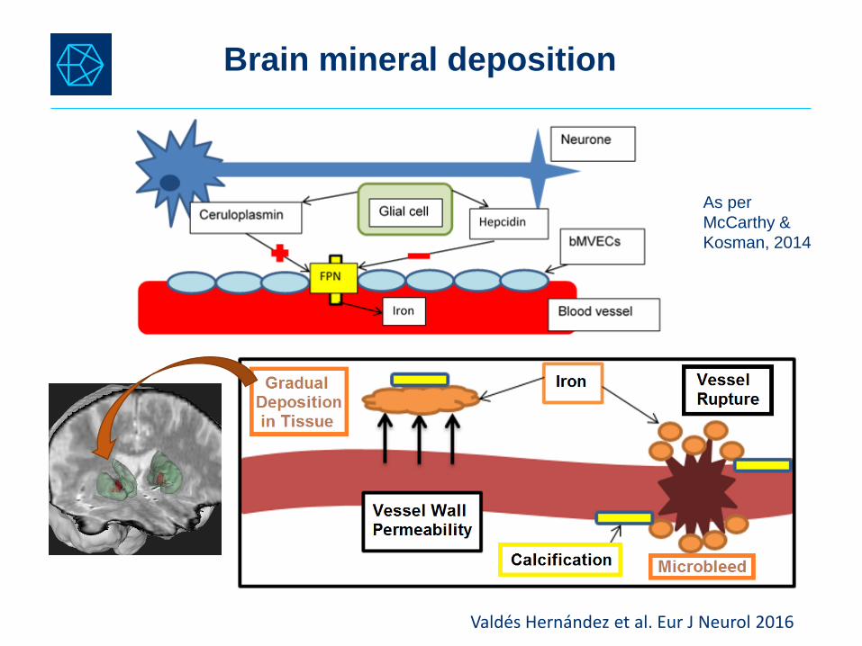
Brain mineral deposition
As per
McCarthy &
Kosman, 2014
Valdés Hernández et al. Eur J Neurol 2016

T1W
T2*W
Ca Fe
Glatz A. et al NeuroImage 2014
Brain mineral deposition in routine clinical MRI scans
Valdés Hernández et al. JMRI 2014

Ca+ ions trapped in Alginate gel have an effect on the MRI signal: Formation of
hypointensities in GRE and hyperintensities in T1W; If Ca+ density increases (e.g.
CaCO3) then the water density goes down and hypointensities are visible in T1W.
Outer ring: CaCl2 solutions and Alginate Gel (and one MnCl2solution without Alginate)
Glatz A. et al NeuroImage 2014
Brain mineral deposition – Validation in phantoms

Trapped Cu+ ions cause pronounced hypointensities in GRE, FLAIR and
hyperintensities in T1W (Cu5, mostly contains Cu+ ions dissolved in water).
Inner ring: CuSO4 solutions and Alginate Gel
Glatz A. et al NeuroImage 2014
Brain mineral deposition – Validation in phantoms

T2 hyper-
inten-
sities
Strokes
and
lacunes
Mineral
deposits
and
haemor-
rhages
Peri-
vascular
spaces
Vascular disease
Brain Vascular Abnormalities

http://datashare.is.ed.ac.uk/handle/10283/2216
Gonzalez-Castro and Valdés Hernández Clin Sci 2017
Enlarged perivascular spaces – Automatic scoring

Predictors for Model 1: age, total atrophy, hypertension, Fazekas score, whether the
patient had a previous lacunar infarct or not, index stroke subtype and SVD score.
Predictors for Model 2: the same as Model 1 with the exception of SVD score.
Predictors for Model 3: the same as Model 1 with the exception of Fazekas score
and whether the patient had a previous lacunar infarct or not, as these two
parameters are contemplated within the SVD score.
No SVD score
Enlarged perivascular spaces – Automatic scoring ?
Gonzalez-Castro and Valdés Hernández Clin Sci 2017

Enlarged perivascular spaces – Automatic quantification
Ballerini et al. 2018

Are these biomarkers related?
Valdés Hernández et al. Eur J Neurol 2016
Valdés Hernández et al. 2018 (under review)

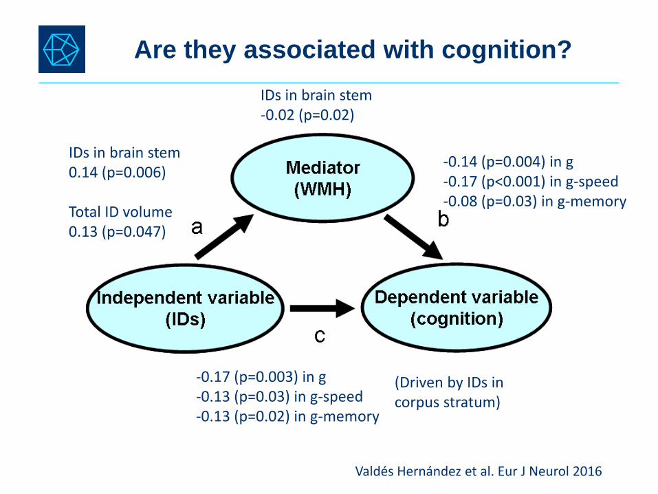
Are they associated with cognition?
Microvesseldysfunction
Cognition in late adulthood
Childhood intelligence
Deary et al. 2009
Penke et al. 2012
Perivascular spaces
Brain mineral
deposition
Valdés Hernández et al. 2018 (under review)

Bivariate correlations
G-factor G-speed G-memory
% ID volin ICV
% CSO-PVS vol in ICV
CSO-PVS count
% WMH vol in ICV
Age
G-factor 1
G-speed 0.74** 1
G-memory 0.68** 0.46** 1
% ID vol
in ICV
-0.090* -0.077 -0.087 1
%CSO-PVS
vol in ICV
-0.095* -0.11* -0.10* 0.13** 1
CSO-PVS
count
-0.011 -0.033 -0.060 0.14** 0.83** 1
% WMH vol
in ICV
-0.18** -0.25** -0.14** 0.039 0.48** 0.19** 1
Age -0.14** -0.15** -0.040 -0.027 0.084 0.063 0.20** 1
Are they associated with cognition?
Valdés Hernández et al. Eur J Neurol 2016
IDs in brain stem 0.14 (p=0.006)
Total ID volume0.13 (p=0.047)
-0.14 (p=0.004) in g-0.17 (p<0.001) in g-speed-0.08 (p=0.03) in g-memory
IDs in brain stem -0.02 (p=0.02)
-0.17 (p=0.003) in g-0.13 (p=0.03) in g-speed-0.13 (p=0.02) in g-memory
(Driven by IDs in corpus stratum)

Structural Imaging
Biomarkers
Identification and Characterisation of Abnormalities
Measurement of Physical
Tissue Properties
Measurement of Tissue
Microstructural Characteristics
3D Shape Morphology
Analysis
Overview

Thrippleton, Valdés Hernández and Wardlaw (under review)
Physical tissue properties

1) Second order statistical textural features (e.g. extracted from a Grey Level
Co-occurrence Matrix)
Physical tissue properties – Texture analysis
Valdés Hernández and Gonzalez-Castro et al.
Front Neurol 2017

m
m
m
𝐫𝐞𝐥𝐚𝐭𝐢𝐯𝐞 𝐦𝐞𝐚𝐧 𝐢𝐧𝐭𝐞𝐧𝐬𝐢𝐭𝐲 =𝐦𝐞𝐚𝐧 𝐚𝐛𝐬𝐨𝐥𝐮𝐭𝐞 𝐢𝐧𝐭𝐞𝐧𝐬𝐢𝐭𝐲
𝐦𝐚𝐱𝐢𝐦𝐮𝐦 𝐢𝐧𝐭𝐞𝐧𝐬𝐢𝐭𝐲 𝐢𝐧 𝐭𝐢𝐬𝐬𝐮𝐞 𝐭𝐲𝐩𝐞∙ 𝟏𝟎𝟎%
0
0 1 3
1 1 2 3 3
1 2 2
3
𝐫𝐞𝐥𝐚𝐭𝐢𝐯𝐞 𝐒𝐃 𝐢𝐧𝐭𝐞𝐧𝐬𝐢𝐭𝐲 =𝐒𝐃 𝐨𝐟 𝐑𝐎𝐈 𝐢𝐧𝐭𝐞𝐧𝐬𝐢𝐭𝐢𝐞𝐬
𝐦𝐚𝐱𝐢𝐦𝐮𝐦 𝐢𝐧𝐭𝐞𝐧𝐬𝐢𝐭𝐲 𝐢𝐧 𝐭𝐢𝐬𝐬𝐮𝐞 𝐭𝐲𝐩𝐞∙ 𝟏𝟎𝟎%
𝐫𝐞𝐥𝐚𝐭𝐢𝐯𝐞 𝐦𝐚𝐱𝐢𝐦𝐮𝐦 𝐢𝐧𝐭𝐞𝐧𝐬𝐢𝐭𝐲 =𝐦𝐚𝐱𝐢𝐦𝐮𝐦 𝐑𝐎𝐈 𝐢𝐧𝐭𝐞𝐧𝐬𝐢𝐭𝐲
𝐦𝐚𝐱𝐢𝐦𝐮𝐦 𝐢𝐧𝐭𝐞𝐧𝐬𝐢𝐭𝐲 𝐢𝐧 𝐭𝐢𝐬𝐬𝐮𝐞 𝐭𝐲𝐩𝐞∙ 𝟏𝟎𝟎%
2) First order statistical textural features
Physical tissue properties – Texture analysis
Viksne and Valdés Hernández et al. 2015 MIUA, Lincoln, UK
http://miua.blogs.lincoln.ac.uk/files/2015/07/MIUA2015_Proceedings_Final.pdf (p 66)

In ROIs
on FLAIR and T1-weighted
In tissues and CSF
on pre-/post-Gd FLAIR
Viksne and Valdés Hernández et al. 2015 MIUA,
Lincoln, UK
http://miua.blogs.lincoln.ac.uk/files/2015/07/MIU
A2015_Proceedings_Final.pdf (p 66)
Valdés Hernández and Gonzalez-Castro et al.
Front Neurol 2017
Physical tissue properties – Texture analysis

Perivascular spaces in
basal ganglia
Total scores of white
matter hyperintensity
burden
Small Vessel Disease
scores
GLCM Contrast (variability) GLCM Homogeneity
Disease progression
Physical tissue properties – Texture analysis
In relation to:
Structural Imaging
Biomarkers
Identification and Characterisation of Abnormalities
Measurement of Physical
Tissue Properties
Measurement of Tissue
Microstructural Characteristics
3D Shape Morphology
Analysis
Overview

Pre-requisites:
1) Smooth surface representing individual shape characteristics
2) Inter-subject point-to-point shape correspondence
3) Robust restoration of individual shape details across large variations
of shape and size
3D Shape Morphology Analysis
Image courtesy of J. Kim, KAIST, South Korea

Progressive deformation – Kim and Park
http://www.nitrc.org/projects/
dtmframework/
3D Shape Morphology Analysis
Kim and Valdés Hernández et al. IEEE TMI 2015

General memory < SD General memory > SD
Left
Hippocampus
Right
Hippocampus
3rd.
ventricle
Example - Regional deformation pattern for general memory
3D Shape Morphology Analysis
Valdés Hernández, Cox et al. NBA 2016

Structural Imaging
Biomarkers
Identification and Characterisation of Abnormalities
Measurement of Physical
Tissue Properties
Measurement of Tissue
Microstructural Characteristics
3D Shape Morphology
Analysis
Overview

(a) Fractional Anisotropy
(b) T1 pulse
(c) Magnetisation
Transfer Ratio
(d) Mean Diffusivity
(e) Radial diffusivity
(f) Axial diffusivity
Tissue Microstructure
Image courtesy of M.E. Bastin, University of Edinburgh, UK

Muñoz Maniega, Valdés Hernández et al. ISMRM 2010
Tissue Microstructure

Some of the severe lesions at baseline correspond to tissue
loss or remain severe (i.e. very hyperintense) after a year.
WMH at baseline only WMH unchanged WMH at follow-up only
Tissue Microstructure –Longitudinal changes
Valdés Hernández et al. AHA 2015

Tissue Microstructure –Longitudinal changes
Valdés Hernández et al. AHA 2015

Row Fogo Charitable Trust
Development of the software/methods on this presentation
received funds from:
Funding