potential transmission of avian influenza a (h5n1) virus ... · 3 who/sde/wsh/06.1 review of latest...
TRANSCRIPT
Review of latest available evidence on potential transmission of avian influenza (H5N1) through water and sewage and ways to reduce the risks to human
health
Last updated 10/10/2007
Water, Sanitation and Health Public Health and Environment
Geneva 2006
1
2
Review of latest available evidence on potential transmission of avian influenza (H5N1) through water and sewage and ways to reduce the risks to human health WHO/SDE/WSH/06.1
© World Health Organization 2006
All rights reserved. Publications of the World Health Organization can be obtained from WHO Press, World Health Organization, 20 Avenue Appia, 1211 Geneva 27, Switzerland (tel: +41 22 791 3264; fax: +41 22 791 4857; email: [email protected]). Requests for permission to reproduce or translate WHO publications – whether for sale or for noncommercial distribution – should be addressed to WHO Press, at the above address (fax: +41 22 791 4806; email: [email protected]).
The designations employed and the presentation of the material in this publication do not imply the expression of any opinion whatsoever on the part of the World Health Organization concerning the legal status of any country, territory, city or area or of its authorities, or concerning the delimitation of its frontiers or boundaries. Dotted lines on maps represent approximate border lines for which there may not yet be full agreement. The mention of specific companies or of certain manufacturers’ products does not imply that they are endorsed or recommended by the World Health Organization in preference to others of a similar nature that are not mentioned. Errors and omissions excepted, the names of proprietary products are distinguished by initial capital letters. All reasonable precautions have been taken by the World Health Organization to verify the information contained in this publication. However, the published material is being distributed without warranty of any kind, either expressed or implied. The responsibility for the interpretation and use of the material lies with the reader. In no event shall the World Health Organization be liable for damages arising from its use. Printed by the WHO Document Production Services, Geneva, Switzerland
3
WHO/SDE/WSH/06.1
Review of latest available evidence on potential transmission of avian influenza (H5N1) through water and sewage and ways to reduce the risks to human
health
Last updated 10/10/2007
This document is periodically revised as more information becomes available. Questions and answers are being separately developed and will also be periodically updated. To this end, we welcome comments, which can be submitted via email to [email protected] The most up-to-date version of this document can be accessed at: http://www.who.int/water_sanitation_health/emerging/ai review.pdf.
4
Purpose and Scope The purpose of this document is to summarize the latest available studies and findings on avian influenza pertaining to water resources, water supplies, sanitation (human excreta, sewerage systems and health care waste) and hygiene. Based on this information, we discuss the evidence of avian influenza-related risks to human health associated with these areas of interest. We also lay the groundwork for risk management strategies that will evolve as we improve our understanding of the virus. The document is intended to serve as the scientific basis to inform technical briefing notes, including questions and answers, directed at public health authorities and others involved in pandemic influenza planning. Non-technical question and answer reviews also are planned to provide information and recommendations that are derived from this document and that are of interest to the general public. Although the “pandemic H5N1” virus does not yet exist, it may behave like avian influenza H5N1 virus in many respects. In this paper, information on avian H5N1 virus is provided where available, but data on other avian influenza viruses are also provided to help fill knowledge gaps until we learn more. As new or revised information is received both this document and the questions and answer reviews will be periodically updated. For information about the current situation or for answers to frequently asked questions about avian influenza and the possible progression to pandemic influenza, please visit WHO's website at http://www.who.int/csr/disease/avian_influenza/en/index.html. Background There are three types of influenza viruses: A, B and C. Influenza A viruses can infect humans, birds, pigs, horses, and other animals, but wild birds are the natural hosts for these viruses. Influenza A viruses can cause pandemics. Influenza B viruses are usually found only in humans and generally are associated with less severe epidemics than influenza A viruses. Influenza C viruses cause mild illness in humans and are not a significant concern for human health. Only influenza A viruses are discussed in this document. Influenza A viruses have different subtypes, defined by the H (haemagglutinin) and N (neuraminidase) proteins on the surface of the virus. The H subtypes are epidemiologically most important, as they govern the ability of the virus to bind to and enter cells, where multiplication of the virus then occurs. The N subtypes govern the release of newly formed virus from the cells. Some subtypes have low pathogenicity (the capacity to cause disease) and others that have high pathogenicity. Human influenza, the seasonal affliction that causes symptoms such as fever, cough, sore throat and headaches, is caused by human influenza A viruses. Three human influenza A subtypes: H1N1, H1N2 and H3N2 have caused major outbreaks in humans. Avian influenza or bird flu is less well known, although it has captured media attention in recent years. Avian influenza is an infection caused by avian influenza A viruses, with
5
transmission normally occurring between birds. Less commonly, avian influenza A has infected pigs, and on rare occasions, humans. The subtype of avian influenza A virus known as H5N1 is very contagious among birds and can cause significant mortality in some avian species. In the rare instances that the virus is transmitted from birds to humans, H5N1 can cause pneumonia, multi-organ failure and often death. As of 12 May 2006, there have been 208 cases of transmission to humans, with 115 fatalities reported to WHO (these statistics are frequently updated by WHO and can be found at http://www.who.int/csr/disease/avian_influenza/country/cases_table_2006_03_24/en/index.html). Wild waterfowl are considered the natural reservoir of all avian influenza A viruses. Most infected birds exhibit no symptoms, even when they are excreting large quantities of infectious virus. These asymptomatic birds act as “silent” reservoirs of the virus, perpetuating its transmission to other birds. Domestic waterfowl (e.g., ducks) may also act as a two-way intermediary in the transmission pathway of avian influenza between wild waterfowl and domestic terrestrial poultry (e.g., chickens). Although usually transmitted from wild birds as a virus of low pathogenicity, it may mutate during replication in domestic poultry and highly pathogenic avian influenza (HPAI) strains may arise. The overriding concern with respect to the HPAI H5N1 virus is that it may change into a form that is highly infectious for humans and that spreads easily from person to person. This could mark the start of a global outbreak or pandemic. No one will have immunity to the virus, as no one will have been exposed to it or developed antibodies. No vaccine with guaranteed efficacy can be prepared in advance of such an outbreak, because the causative virus does not yet exist. Potential vaccines are being prepared and stockpiled in advance, in the hopes that they may match a pandemic strain. A pandemic strain also may have characteristics of pathogenicity that will not be immediately known. Such changes may have implications for the efficacy of control measures established during pre-pandemic planning. However, we do know that some actions, such as strengthening personal hygiene practices, will reduce human-to-human transmission and help stop or slow the spread of pandemic virus. Routes of entry into water Birds infected with avian influenza virus shed large quantities of virus in their faeces as well as in their saliva and nasal secretions. Shedding occurs in the first two weeks of infection (1). It has been shown that one infected duck excretes up to 1010 EID50 (the median egg infective dose) in 24 hours (2). As ducks are known to excrete 7.5–10 kg of faeces per year and geese excrete 12.5–15 kg (3), infected waterfowl may be able to excrete up to 3 × 10⁹ EID50 per gram faeces. It is likely that infected droppings or other secretions from both symptomatic and asymptomatic migratory waterfowl will enter water environments where the birds gather. Avian influenza virus has been isolated from unconcentrated water from six lakes in
6
Canada where ducks gathered and deposited large amounts of faeces (4); from lakes in the United States (5,6); and from concentrated pond water from Hong Kong (7). There are no quantitative data available on levels of H5N1 virus in lake water where waterfowl gather, although its detection in unconcentrated water and in small sample volumes suggests that levels are relatively high. Besides direct deposition of faeces into lake water by migratory waterfowl, it has been suggested that faecal waste from duck and chicken farms may spread to bodies of water via wind, surface runoff or possibly enter groundwater through disposal and composting of waste on poultry farms. Routes of entry into sewage In addition to birds, avian influenza virus may be shed in the faeces of mammals, including infected humans, domestic animals and livestock. The H5N1 virus could potentially enter into sewage in urine or faeces excreted by infected humans or in animal waste that is combined with human sewage. Information on the excretion of H5N1 viruses in urine or faeces by mammalian species, including humans, is exceedingly limited and unlikely to be representative of a potential future human pandemic strain. The isolation of the H5N1 virus from the faeces of a child presenting with diarrhoea followed by seizures, coma and death (8) suggests that the virus may be excreted by infected humans and can enter sewage in this manner. No virus was detected in the urine of this patient (8). Persistence in water Avian influenza viruses can persist for extended periods of time in water, although quantitative information on the subtype H5N1 is lacking. Data and findings from studies on other avian influenza subtypes are presented below and summarized in Annex 1. One study (2) showed the avian influenza subtype H3N6 resuspended in Mississippi River (USA) water was detected for up to 32 days at 4°C and was undetectable after 4 days at 22°C. The data showed a decrease of about 4 logarithmic units (LU) in 32 days at 4°C (T90, the time taken to eliminate 90% of the virus in the sample, was estimated to be 8 days) and of more than 8 LU at 22°C (estimated T90 = 0.5 days). In a second study (9), which used five low-pathogenicity avian influenza viruses (H3N8, H4N6, H6N2, H12N5, and H10N7), infectivity of virus in distilled water (initial concentration 106 TCID50, or median tissue culture infective dose, per ml) was retained for up to 207 days at 17°C and 102 days at 28°C. The T90 ranged between 21 and 32 days at 17°C and between 5 and 17 days at 28°C, depending on the strain. In a study that showed a high level of positive water samples (23%) for a strain of influenza A virus in a lake where ducks were nesting, the proportion of positive samples remained high (14%) in the autumn after the ducks had left for migration, indicating that the virus is able to persist in water (6). Other studies of the persistence of avian influenza viruses in water have shown that these viruses persist for different periods of time depending on temperature, pH and salinity.
7
For example, one study showed that viruses survived longest (T90 = 17 days) at 17°C, with low levels of salinity and a pH of 8.2 (17). The shortest viability was observed when virus was exposed to 28°C with 20 ppt salinity and a pH of 8.2 (T90 = 1.5 days) (10). Persistence in sewage, excreta and animal wastes Data and findings from studies on persistence of various avian influenza subtypes in untreated sewage, waste and animal faeces are summarized in Annex 1. No specific information is available on factors affecting the persistence of H5N1 virus in sewage or on the effect of waste treatment processes on H5N1 concentrations. Virus concentrations are reduced at different rates and to various extents in both human and animal waste treatment processes, depending on conditions, but they are not completely eliminated. Furthermore, virus concentrations may be enriched in certain treated or separated waste fractions (such as waste solids) by sedimentation and solid–liquid separation processes (11). Studies on the survival of viruses in human faecal wastes and agricultural animal wastes have indicated that persistence is dependent on several factors, including the virus type, waste type, temperature and other environmental conditions and processes (11). The period of avian influenza infectivity in bird faeces and secretions depends on pH and temperature conditions, but generally four weeks after infection, avian influenza virus can no longer be detected (1). Ducks infected with the H5N1 virus have been found to shed the virus at high titres from the trachea as well as from the cloacae, with peak levels of virus shedding after three days (12,13). H5N1 virus was isolated from duck faeces on day 3 post-infection (ducks had been infected with 106 EID50 of virus in 1 ml volume) at 2.25–3.75 log10 EID50 per gram fresh faeces, but became undetectable after the faeces were dried overnight at room temperature (20°C). Virus titres declined in wet faeces kept at 25°C but remained detectable for 7 days. When the wet faeces were stored at 4°C, the virus remained viable when testing ceased at day 20. At 37°C, the viruses remained detectable in wet faeces until day 4 (two viruses) and day 6 (two viruses) (14). These results suggest that freshly deposited faeces are highly infective (at any temperature), that H5N1 in faecal deposits on land may be more rapidly inactivated than H5N1 in water and that the virus’s survival decreases at higher temperatures. H5N1 typically persists in colder temperatures and produces outbreaks during the colder months of the year (15). However, recent (unpublished) studies mentioned above have shown that current H5N1 strains survive longer in faeces at warmer temperatures than previously circulating viruses (14), which may explain how the virus has resurfaced in summer months in Asia.
8
Does the release of antiviral compound into sewers lead to influenza virus resistance in nature? Potential concern has been raised regarding the possibility that an influenza-specific antiviral used for treatment and prophylaxis (oseltamivir) is not removed or degraded during normal sewage treatment. This may lead to concentrations in natural waters under circumstances such as an influenza pandemic, during which the drug will be administered to large numbers of population, reaching levels to which influenza viruses in nature may develop resistance. This would then increase the risk that influenza viruses being introduced to human beings may become resistant. To date, there is no credible evidence for such concern because levels of this antiviral compound in wastewater will be low and probably lower than levels needed for biological activity of oseltamivir in waste water (order of uM in vitro). Therefore, it should not be concluded that biological events will occur in a virus (an obligate, intracellular biological agent) or its host (cells) that will lead to the emergence of viral resistance to the therapeutic drug as a result of its presence in wastewater. The activated sludge process of biological wastewater treatment has a mean residence time of 4-8 (conventional) to 24 (extended aeration) hours. The ability of an anti-influenza virus chemical as oseltamivir to survive to such a biologically active exposure and biodegradation and biotransformation process is uncertain and has not been adequately studied. Exposure of influenza viruses in (waste) water to an antiviral compound may lead to chemical reactions between the virus and the antiviral chemical, such that they would react and somehow result in a complex that would lead to biological selection of resistant mutants. However, in order for biological selection of resistant virus mutants to occur, there must be a living virus host (i.e. a mammal or bird) involved in the process. The influenza virus and the antiviral compound must be present together in a living host, perhaps as a complex, for virus-host interactions to occur that would lead to virus mutations resulting in virus resistance to the compound. These genetic selection reactions or mutations will not occur de novo in the extracellular environment, such as in sewage or water. To assume that such a biological selection event can simply occur because the viruses and the chemicals are both present in the water or wastewater environment is both hypothetical and inconsistent with the known role of the biological host in the needed biological processes that result in selection of resistant mutant viruses. Persistence in other media relevant to hygiene interventions Knowing the persistence of virus in or on media such as dust, fomites, air and aerosols helps inform effective application of hygiene practices intended to minimize transmission of pandemic influenza virus, should it arise. Data and findings from studies on persistence of various influenza viruses for these media are summarized in Annex 1.
9
Site(s) of infection Infection refers to the entry and multiplication of a pathogenic agent in the body of humans or animals. Infection does not necessarily result in disease, as is the case with asymptomatic wildfowl carrying low pathogenic avian influenza viruses. Additionally, the site of replication is not always the same as the point of entry of a pathogen into the body, which could have implications for the types of environmental management interventions that would be implemented in the event of pandemic influenza. The site of virus replication in mammals may vary and it is important to note that both viral and host factors may influence tissue tropism (16) (i.e., the types of cells and tissues infected by a virus). In view of the greater number of virus isolations from the cloaca than from the trachea of domestic ducks, avian influenza viruses replicate primarily in the gastrointestinal tract of infected waterfowl (2,17). However, in recent studies of H5N1 (12), juvenile mallards were inoculated with 23 different H5N1 avian influenza viruses isolated in Asia between 2003 and 2004. Viruses replicated to higher levels in the trachea than in the cloaca of both inoculated and contact birds. Based on these findings, the authors suggest that the digestive tract may not be the main site of H5N1 influenza virus replication in ducks. In humans, influenza A virus replicates primarily in the respiratory tract. However, the clinical presentation of infection in humans by H5N1 avian influenza virus provides additional suggestive evidence concerning site of infection. The frequent occurrence of diarrhoea in infected humans and the detection of viral RNA in most faecal samples tested (and infectious virus in one faecal sample) suggest that H5N1 virus may replicate in the human gastrointestinal tract (8,18,19). Diarrhoea in humans arising from H5N1 avian influenza appears to be more common than in influenza due to human influenza viruses and may precede respiratory manifestations by up to one week (19). In fact, in the case of two children who died from H5N1 infection (8), both presented with diarrhoea without apparent respiratory symptoms. Although symptoms of avian influenza in humans from viruses other than H5N1 include eye infections, it should be noted that patients with H5N1 avian influenza have rarely had conjunctivitis (19). Thus, the conjunctival mucosa may be a point of entry for H5N1 infection, but is not likely a site of viral replication. Routes of transmission In managing environmental risks we need to understand the routes of virus transmission; that is, how it moves through the environment and eventually reaches a site of infection. For human influenza viruses, transmission occurs primarily by inhalation of infectious droplets or airborne droplet nuclei (aerosols), and direct, or possibly indirect (fomite), contact followed by transfer to the upper respiratory tract via the nose, mouth or
10
conjunctival mucosa (eyes) (19). Bird-to-human transmission of avian influenza virus is likely to encompass these routes, as well as others, including ingestion, for example ingestion of contaminated water, although there is, as yet, no evidence of this reported (18). Evidence is lacking on the mechanisms by which the avian influenza H5N1 crosses the species barrier from birds to infect humans. Most cases of H5N1 infection in humans are the result of direct contact with poultry or with objects or surfaces contaminated with faeces from infected poultry (with a few cases of suspected human-to-human contact). These observations suggest either a respiratory (e.g. inhalation of infectious aerosols or droplets) or faecal–oral route of transmission from birds to humans. However, based on the findings of the recent studies with H5N1 virus described earlier (12), in which viruses replicated to higher levels in the trachea than in the cloaca of both inoculated and contact birds, the faecal–oral route may not be the main transmission path for some influenza A subtypes including H5N1. A potential faecal-oral route raises the question of whether human faeces could be a source of transmission. The observation of virus in the faeces of infected humans does not necessarily mean that there is a faecal–oral route of infection for humans or even a faecal–respiratory route, but it may have implications for loads in sewer systems and aerosol generation where outbreaks in humans occur. Given the survival of H5N1 in the environment, several modes of environmental transmission are theoretically possible (19). Examples include oral ingestion or aspiration of contaminated water during swimming, direct intranasal or conjunctival inoculation during exposure to contaminated water, and contamination of hands from infected fomites with subsequent self-inoculation. Inhalation or contact with self-inoculation of untreated poultry faeces used as fertilizer presents other possible routes. There is very little information on the role of water in the transmission of influenza viruses among waterfowl or to other animals, including humans. One study (7) suggests that the faecal–water–oral route is probably significant in transmission of the virus between birds, in view of the greater number of virus isolations from the cloaca than from the trachea of domestic ducks. The authors suggest that a cycle of waterborne transmission and maintenance of influenza viruses exists within the duck communities of southern China and that it is conceivable that virus transmission could occur in this manner to other susceptible animals, including humans. This conclusion was supported by another study in which influenza A virus was isolated throughout the year from faeces and/or pond water where dabbling ducks nested in Alaska, USA; recurrent faecal contamination of pond water led to transmission of the virus to uninfected ducks (6). The fact that waterfowl excrete influenza viruses into water does not confirm waterborne transmission between birds; nor does it offer an indication of the extent of the risk of infection to humans exposed to the water. Some other viruses are likewise excreted into water environments but are not known to be transmitted to a meaningful extent via that route (Willie Grabow; personal communication, 13 Nov 2005). Fomites are any inanimate object (e.g., linens, money, clothing, dishes, doorknobs, toys etc.) that can transmit infectious agents from one person to another. Transmission of
11
influenza viruses by fomites has not been extensively documented but is believed to occur. One study (20) observed that the transmission of influenza virus from donors shedding large viral loads could occur for 2-8 hr via stainless steel surfaces and for a few minutes via paper tissues. Influenza A virus placed on hard, nonporous surfaces (steel and plastic) could be cultured from the surfaces at diminishing titres for <24 to 48 hours and from cloth, paper, and tissues for <8 to 12 hours at conditions of 35% to 40% humidity and a temperature of 28°C. Higher humidity shortened virus survival. Infectious virus on nonporous surfaces could be transferred to hands 24 hours after the surface was contaminated, while tissues could transfer virus to hands for 15 minutes after the tissue was contaminated. On hands, virus concentration fell by 100- to 1,000-fold within 5 min after transfer. The authors concluded that transmitting infection from the surfaces tested would require a high titre of virus (around 100 TCID50/mL) on the surface; such titres can be found in nasal secretions at an early stage of illness. Thus, under conditions of heavy environmental contamination, the transmission of influenza virus via fomites may be possible. Minimizing virus transmission: focus on hygiene To date, human infections with avian influenza viruses in circulation since 1997 have not resulted in sustained human-to-human transmission. If the current avian influenza H5N1 virus changes to produce a strain that is more transmissible among humans, it could signal the start of a pandemic. Strengthening personal hygiene practices to reduce human-to-human transmission will help stop or slow the spread of pandemic virus. As important is the need to keep hospitals safe and protect patients and healthcare workers from infection. Given the uncertainty about the exact modes by which avian influenza, including the current H5N1 strains, may be transmitted between humans, enhanced infection control precautions for patients with suspected or confirmed avian influenza infection appear warranted. There is the need to minimize infection opportunities because every infection presents a chance of genetic mutation that might give a rise to pandemic virus. Hygiene in domestic and community settings Personal hygiene includes individual practices that serve to promote or preserve health such as habits of cleanliness. In the case of highly contagious diseases such as influenza, special attention should be paid to personal behaviour in community settings as well as the household. Public education, including public health messages, is an important part of national and local planning for pandemic influenza. WHO has issued guidance on personal hygiene in health care settings (see next section), but such guidance is based on general transmission patterns of seasonal human influenza. It is not known how effective this guidance would be in slowing the pandemic spread of a new virus strain. Thus, any recommendation that WHO provides before a pandemic, and even once a pandemic starts, may be modified once more information about the pandemic strain (such as its infectivity and pathogenicity) is obtained.
12
However, there are basic good health habits that will help reduce the spread of influenza virus in the home, school or other community setting. These include hand washing, cough etiquette and surface cleaning as described in Annex 2. Additionally, there are several studies and reports that identify hygienic practices that may prevent infection and reduce the transmission of infectious pathogens (21,22,23,24). Hygiene in healthcare settings WHO has produced comprehensive guidance pertaining to infection control in healthcare settings, with special emphasis on avian influenza issues (available at: http://www.who.int/csr/disease/avian_influenza/guidelinestopics/en/index3.html). Strong hygiene practices are always a critical component of infection control. The key recommendations for hygiene practice during the care of patients with suspected or confirmed avian influenza infection are provided in Annex 3. As indicated above, the guidance should be used for planning purposes, but may be modified as the epidemiology of avian influenza or pandemic influenza evolves. Assessment and management of environmental risks If the site(s) of infection and the routes of transmission are known, it is possible to identify exposure scenarios that might pose unacceptable risks to human health1. Combined with estimates of exposure concentrations and knowledge of the dose–response relationship (obtained from infectivity studies or by other means), this information may be used to develop quantitative risk assessments for these exposure pathways. From the limited information available for the current strain of H5N1 virus, it appears that the current risk of human infection is small or nonexistent for environmental exposure pathways associated with virus transmission through water, sewage or fomites. However, should evidence emerge that a highly pathogenic form of the virus is adapting better to humans, the risks presented by environmental media might increase. As a framework for guidance on the management of environmental risks that would be needed in the event of pandemic influenza, we identify a general set of potential exposure scenarios below. Possible prevention and control measures are discussed with the understanding that measures will need to be tailored to the specific settings and conditions faced by potentially exposed individuals. These scenarios retain the assumption that humans can be infected by respiration, contact (with transfer to eyes, nose or mouth) or ingestion of the virus. Scenario 1: Consumption of virus-contaminated drinking water.
1 Note on infection versus disease: For infectious diseases, an individual may become infected without disease symptoms for short or long periods of time. During this time, such individuals still may be infectious. When infection without disease is sustained, this is referred to as "carrier state." As yet, no information about the carrier state is available for the H5N1 virus. Thus, the current focus of control is on preventing exposure leading to infection, thereby controlling the potential for disease.
13
Sources of drinking water that may be susceptible to avian influenza virus contamination include surface water bodies (e.g., reservoirs, ponds, lakes and rivers), groundwater aquifers and rainwater collection systems. Of these sources, open water bodies where infected waterfowl gather are the most likely potential route of entry of virus into the drinking water supply. The risk to humans will depend on a variety of factors, including the use to which the water is put, the health status of the humans (e.g. immunocompromised versus healthy), and whether or not the water is treated. In open water bodies, many variables will influence the probability of virus contamination and the persistence/survival of these viruses, including whether the stored water is on a natural flyway for waterfowl, the number of birds stopping over, the availability of food for waterfowl in that water, the time of year, temperature, pH and the length of time between deposition of faecal material and the draw off of water. The probability of having detectable levels of influenza viruses in such bodies of water is likely to be higher in the seasons when migratory birds are passing through the area. There is no available epidemiological evidence associating wildfowl excreta or death in storage reservoirs of either treated or untreated drinking water with avian influenza disease in humans. Moreover, qualitative and quantitative risk assessments for avian influenza infection of humans through consumption of contaminated drinking water in France and the Netherlands, respectively, estimated a daily risk of infection to be below health-based standards (25,26). Nevertheless, it is theoretically possible that transmission of avian influenza could occur in specific local circumstances were a series of factors to coincide. These could include wildfowl and domestic poultry contamination of storages, distribution of water without treatment (including without disinfection); and household use that gave rise to an infectious exposure, possibly through the oral ingestion of contaminated water, or conjunctival inoculation through face washing with contaminated water or contaminated hands. Prevention and control measures can be suggested to minimize, if not eliminate, the risk from the consumption of contaminated water. Water supplies receiving treatment as recommended in the WHO Guidelines for Drinking Water Quality2 are unlikely to pose a significant risk of infection even if infected waterfowl are present in source waters. Due to their structure, influenza viruses are relatively susceptible to disinfectants, including oxidizing agents such as chlorine and ozone. They are also readily inactivated by heating, so boiling would also be effective3 Authorities charged with managing any potential risk may consider ensuring chlorination or alternative disinfectant residuals be maintained throughout distribution. For effective disinfection of adequately pre-treated water, there should be a residual concentration of
2 Including, for example, a residual concentration of free chlorine of ≥0.5 mg/litre after at least 30 min contact time at pH <8.0. WHO (2004) Guidelines for Drinking Water Quality, Third Edition, p 194; Geneva, World Health Organization Available at http://www.who.int/water_sanitation_health/dwq/gdwq3/en/index.html 3 WHO Guidelines for Drinking Water Quality recommend bringing water to a rolling boil to assure adequate water disinfection.
14
free chlorine of at least 0.5 mg/litre after at least 30 min contact time at pH <8.0. Additionally, the residual concentration leaving the treatment plant should be capable of maintaining a free chlorine concentration of 0.2 mg/litre throughout the distribution system. Where household water safety is suspect, authorities should consider advising families to treat their drinking water with available and acceptable household-level interventions, including home chlorination (addition of bleach) and boiling, that are effective at inactivating viruses. Guidance for household-level interventions is provided in Annex 4 Scenario 2: Bathing/Recreational contact with virus-contaminated water bodies. Potential modes of transmission to humans of avian influenza virus during recreational use or bathing in contaminated water include oral ingestion, aspiration of contaminated water and direct intranasal or conjunctival inoculation. The potential for exposure is greatest in open bodies of water receiving significant waterfowl populations depending, in part, on the number of birds and the time of year. While the majority of human cases of H5N1 have been attributed to close contact with infected poultry or poultry faeces, the possibility of developing the disease following swimming or bathing in water with high loads of avian influenza virus has been suggested in two cases in Vietnam (27). Disease reportedly developed in a 35-year-old woman and an unrelated 9-year-old girl following swimming in water. The water in which the 35-year-old women bathed had reportedly been used for the disposal of dead poultry. However, it is not possible to conclusively attribute the infection to aspiration or ingestion of this potentially contaminated water, or to another type of exposure. Another potential case occurred in Cambodia where an 8-year-old female may have been exposed to the virus through recreational contact with contaminated water, where asymptomatic ducks may have shed virus into the pond. However, again, there is no evidence to confirm this hypothesis (Dr. Megge Miller, WHO Cambodia; personal communication, 24 May 2006). Data on both exposure and effects assessment for avian influenza viruses in recreational exposures are lacking. Risk assessments for recreational exposure to other viruses may be useful as a model. For example, an assessment of adenoviruses in recreational water (28) modeled the risk of infection due to consumption during swimming. Water bodies used for recreational purposes are almost never treated, and treatment may not be feasible. Authorities concerned can manage any potential risk from highly contaminated waters by advising the public of this risk, and, where a high degree or risk has been identified, by restricting human access to potentially hazardous sites. Scenario 3: Exposure to poorly managed excreta and wastewater that harbour the virus. There is some evidence to show that H5N1 is excreted in human faeces (8), and all influenza A viruses can be transmitted in the faeces of avian species. Human and animal excreta are most often managed separately. However, there are settings and scenarios
15
where animal waste may be combined with human waste. If the sewage from poultry houses is mixed with human sewage, for example, there could be a risk of transmitting influenza through interconnected sewage pipes and non-sealed venting. In such situations, prevention and control measures should focus on reducing airborne droplet and aerosol transmission. Sewers should never be vented through human living quarters. Other means of excreta disposal where aerosol formation is unlikely, such as latrines, probably represent an extremely low risk of virus transmission. Scenario 4: Occupational exposure at sewage treatment facilities. Under current conditions, where the virus has not adapted to humans, there is no significant risk to sewage workers, provided that poultry house discharge is not included with human sewage. If human-to-human transmission of HPAI virus increases significantly, human excreta in outbreak areas could contain avian influenza viruses shed by humans and the exposure risks to sewage workers would need to be reconsidered. Based on the epidemiological evidence for this human adapted form of the virus, contaminated sewage might need treatment to reduce virus numbers, and measures might be needed to reduce aerosol generation and exposure of individuals to aerosols (e.g., use of protective clothing, such as masks). As mentioned above, although there is no specific information available on the response of H5N1 virus to waste treatment processes, virus concentrations are generally reduced at various rates and to various extents in both human and animal waste treatment processes, but they are not completely eliminated (11). If the human sewage is mixed with contaminated animal waste, similar precautions would need to be taken. Future research directions The virus that may one day bring about a new influenza pandemic does not yet exist. In the absence of virus-specific information, studies of existing potential precursors, such as the avian influenza virus H5N1, and related subtypes can shed light on potential risk factors and thus aid decision makers in planning and preparedness activities. With respect to water, sanitation and hygiene, little is known about the specific risk factors posed by H5N1 avian influenza for the human population. There is a paucity of information on the presence and stability of the H5N1 virus in water and sewage. Further, the site(s) of infection and the route(s) of transmission of H5N1 avian influenza virus that presently causes disease in some birds and, in rare instances, humans are likely to change in future pandemic strains. When H5N1 cases in humans arise, the focus is often on collection of specimen samples for case management and epidemiological data to trace the origin of disease. Opportunities for environmental sample collection that could help fill the knowledge gaps described in this report are often overlooked. Additional data on the survival of the H5N1 virus in water and sewage under field conditions would improve exposure assessment. Quantitative estimates of the range of influenza virus concentrations in bird faeces and in waters in which birds have congregated would provide better information on the potential viral loads that could be shed into the environment. From this, virus
16
concentration could be estimated based on dilution factors of faeces in water resources. Thus, there is a need for environmental sampling protocols that can provide investigators with well documented, representative samples while recognizing the time-constrained, resource-limited conditions faced by the field team. More information is needed about the inactivation of avian influenza H5N1 viruses in water and in avian waste. Specifically, information on the means and methods for effective disinfection at all levels of water and wastewater treatment (household, healthcare settings, community systems). In the event a new strain of virus with efficient human-to-human transmission emerges, the window of opportunity to intervene could be very small. Rapid response and effective infection control in healthcare and community settings will require a number of public health intervention strategies. While standard precautions and good hygiene practice have been developed for seasonal human influenza, these need to be underpinned by virus-specific research on the efficacy and effectiveness of barrier and behavioural approaches to infection control. Acknowledgements The preparation of an initial draft by Robert Webster and Diane Hulse-Poste, both of St. Jude Children's Research Hospital, Memphis, Tennessee is gratefully acknowledged. Thanks are also due to the following individuals who contributed to the writing or provided constructive comments: Albert Bosch, University of Barcelona, Barcelona, Spain; Ana Maria de Roda Husman, National Institute of Public Health and the Environment (RIVM), Bilthoven, The Netherlands; Christophe Gantzer, Laboratoire de Chimie Physique et Microbiologie pour l'Environnement (LCPME), Nancy, France; Chuck Gerba, University of Arizona, Tucson, Arizona, USA; Willie Grabow, retired (formerly of University of Pretoria, Pretoria, South Africa); Huang Baoxu, China Epizootiology Centre Qingdao, China; Gertjan Medema, Kiwa Water Research, Nieuwegein, The Netherlands; Pierre Payment, University of Quebec, Montreal, Quebec, Canada; Mark Sobsey, University of North Carolina, Chapel Hill, North Carolina, USA; Marla Sheffer of Ottawa, Ontario, Canada; Melita Stevens, Melbourne Water, Melbourne, Victoria, Australia; Gary Toranzos, University of Puerto Rico, Rio Piedras, Puerto Rico; Carl-Henrik von Bonsdorff, Helsinki University, Helsinki, Finland; and Peter Wyn-Jones, University of Sunderland, Sunderland, United Kingdom. This input from the following WHO staff working on influenza-related issues and water and sanitation, is also very much appreciated: Houssain Abouzaid, Roger Aertgeerts, Lisa August, Jamie Bartram, Yves Chartier, Alice Crosier, Keiji Fukuda, Bruce Gordon, Peter Horby, Megge Miller, Michael Perdue, Cristiana Salvi, Suzanne Scheele, Carmem Pessoa Da Silva, Hiroko Takasawa, Terrence Thompson, Wenjie Wang. References and Further Reading
17
1. Cardona CJ (undated) Avian influenza. University of California Davis Veterinary Medicine Extension, Davis, California. Available online at http://www.vetmed.ucdavis.edu/vetext/INF-PO_AvianInfluenzaFS.html (accessed 20 March 2006).
2. Webster RG, Yakhno M, Hinshaw VS, Bean WJ and Murti KG (1978) Intestinal influenza: replication and characterization of influenza viruses in ducks. Virology, 84(2):268–278.
3. Regional Lead Centre in China Asian-Pacific Regional Research and Training Centre for Integrated Fish Farming Wuxi, China (1985) Chapter III. Pond fertilization and fish feed in: Training manual: Integrated fish farming in China. Network of Aquaculture Centres in Asia (NACA), Bangkok, Thailand, September 1985, (NACA/TR/85/11). Available online at http://www.fao.org/docrep/field/003/AC233E/AC233E03.htm.
4. Hinshaw VS, Webster RG and Turner B (1979) Water-borne transmission of influenza A viruses? Intervirology, 11:66–68.
5. Halvorson D et al. (1983) Epizootiology of avian influenza — simultaneous monitoring of sentinel ducks and turkeys in Minnesota. Avian Diseases, 27:77–85.
6. Ito T, Okazaki K, Kawaoka Y, Takada A, Webster RG and Kida H (1995) Perpetuation of influenza A viruses in Alaskan waterfowl reservoirs. Archives of Virology, 140:1163–1172.
7. Markwell DD and Shortridge KF (1982) Possible waterborne transmission and maintenance of influenza viruses in domestic ducks. Applied and Environmental Microbiology, 43(1):110–116. Available online at http://pubmedcentral.gov/pagerender.fcgi?artid=241789&pageindex=1#page
8. De Jong MD, Van Cam B, Qui PT, Hien VM, Thanh TT, Hue NB, Beld M, Phuong LT, Khanh TH, Ha DQ and Ferrar J (2005) Fatal avian influenza A (H5N1) in a child presenting with diarrhea followed by coma. The New England Journal of Medicine, 352:686–691.
9. Stallknecht DE et al. (1990) Persistence of avian influenza viruses in water. Avian Diseases, 34:406–411.
10. Stallknecht DE et al. (1990) Effects of pH, temperature, and salinity on persistence of avian influenza viruses in water. Avian Diseases, 34:412–418.
11. Sobsey MD and Meschke JS (2003) Virus survival in the environment with special attention to survival in sewage droplets and other environmental media of fecal or respiratory origin. Draft (dated 21 August 2003) prepared for a World Health Organization meeting (International SARS Symposium) held on 23–25 September 2003 in Rome, Italy.
12. Sturm-Ramirez KM, Hulse-Post DJ, Govorkova EA, Humberd J, Seiler P, Puthavathana P, Buranathai C, Nguyen TD, Chaisingh A, Long HT, Naipospos TSP, Chen H, Ellis TM, Guan Y, Peiris JSM and Webster RG (2005) Are ducks contributing to the endemicity of highly pathogenic H5N1 influenza virus in Asia? Journal of Virology, 79(17):11269–11279.
13. Hulse-Post DJ, Sturm-Ramirez KM, Humberd J, Seiler P, Govorkova EA, Krauss S, Scholtissek C, Puthavathana P, Buranathai C, Nguyen TD, Long HT, Naipospos TSP, Chen H, Ellis TM, Guan Y, Peiris JSM and Webster RG (2005) Role of domestic ducks in the propagation and biological evolution of highly pathogenic
18
H5N1 influenza viruses in Asia. Proceedings of the National Academy of Sciences of the United States of America, 102(30):10682–10687.
14. Hulse-Post DJ and Webster RG (2005) Unpublished data. Memphis, Tennessee, St. Jude Children’s Research Hospital, Department of Infectious Diseases, Division of Virology.
15. Li KS, Guan Y, Wang J, Smith GJD, Xu KM, Duan L, Rahardjo AP, Puthavathana P, Buranathai C, Nguyen TD, Estoepangestie ATS, Chaisingh A, Auewarakul P, Long HT, Hanh NTH, Webby RJ, Poon LLM, Chen H, Shortridge KF, Yuen KY, Webster RG and Peiris JSM (2004) Genesis of a highly pathogenic and potentially pandemic H5N1 influenza virus in eastern Asia. Nature, 430:209–213.
16. Kawaoka Y, Bordwell E and Webster RG (1987) Intestinal replication of influenza A viruses in two mammalian species. Archives of Virology, 93:303–308.
17. Slemons RD and Easterday BC (1978) Virus replication in the digestive tract of ducks exposed by aerosol to type-A influenza. Avian Diseases, 22:367–377.
18. Hayden F and Croisier A (2005) Transmission of avian influenza viruses to and between humans. The Journal of Infectious Diseases, 192(8):1311–1314.
19. The Writing Committee of the World Health Organization (WHO) Consultation on Human Influenza A/H5 (2005) Avian influenza A (H5N1) infection in humans. The New England Journal of Medicine, 353(13):1374–1385.
20. Bean B, Moore BM, Sterner B, Peterson LR, Gerding DN, Balfour HH Jr. (1982) Survival of influenza viruses on environmental surfaces. The Journal of Infectious Diseases; 146(1):47–51.
21. International Scientific Forum on Home Hygiene (2002) Guidelines for prevention of infection and cross-infection in the domestic environment.
22. International Scientific Forum on Home Hygiene (2002) Hygiene procedures in the home and their effectiveness: a review of the scientific evidence base.
23. International Scientific Forum on Home Hygiene (in press) Home Hygiene in Developing Countries - Prevention of infection in the home and the peri-domestic setting: A training resource on hygiene for teachers, community nurses, community workers and other health professionals in developing countries.
24. Barker J, Stevens D, Bloomfield SF (2001) Spread and prevention of some common viral infections in community facilities and domestic homes. Journal of Applied Microbiology, 91:7-21.
25. L’Agence française de sécurité sanitaire des aliments (AFSSA) (2006) Avis de l'Afssa en date du 15 Mars 2006 relatif à l'évaluation qualitative du risque sanitaire pour l'homme lié à la présence dans l'eau destinée à la consommation humaine et dans divers effluents aqueux de virus influenza hautement pathogène, dans le cas d'une épizootie ou dans le cas d'une épidémie humaine [AFFSA advice, dated 15 March 2006, in relation to the qualitative sanitary risk evaluation associated with highly pathogenic influenza viruses present in water intended for human consumption and in various effluents in the case of an epizooty or human epidemic]. Available online at http://www.afssa.fr/Object.asp?IdObj=34669&Pge=0&CCH=060609092515:26:4&cwSID=41BCD42F351B497A866D8D128481C0C9&AID=0.
19
26. Schijven JF, Teunis PFM and de Roda Husman AM (2005) Quantitative risk assessment of avian influenza virus infection via water. Report in prepared for the Environmental Inspectorate for the Netherlands (RIVM report 703719012).
27. Cooperative Research Centre (CRC) (2005) Avian influenza: Is there a risk to water supplies? Health Stream, Issue 40, December 2005. Available online at http://www.waterquality.crc.org.au/hsarch/hs40m.htm (accessed 20 March 2006).
28. Van Heerden J, Ehlers MM, Vivier JC and Grabow WOK (2005) Risk assessment of adenoviruses detected in treated drinking water and recreational water. Journal of Applied Microbiology, 99:926-933.
Further Reading:
CDC. Key facts about avian influenza (Bird Flu) and avian influenza A (H5N1) virus. Atlanta, Georgia, United States Department of Health and Human Services, Centers for Disease Control and Prevention. Available online at http://www.cdc.gov/flu/avian/gen-info/facts.htm (updated 7 February 2006; accessed 20 March 2006).
WHO (2005) Avian influenza. Geneva, World Health Organization. Online at http://www.who.int/csr/disease/avian_influenza/en/ (updated 9 February 2006; accessed 13 February 2006).
WHO. Guidelines for drinking-water quality (available at http://www.who.int/water_sanitation_health/dwq/gdwq3/en/index.html).
WHO. Guidelines for safe recreational waters Volume 1 - Coastal and fresh waters (available at http://www.who.int/water_sanitation_health/bathing/srwe1/en/).
WHO. Non-pharmaceutical interventions: their role in reducing transmission and spread. Available from http://www.who.int/csr/disease/avian_influenza/pharmaintervention2005_11_3/en/index.html
World Health Organization Writing Group (2006) Nonpharmaceutical public health interventions for pandemic influenza, national and community measures. Emerging Infectious Diseases; 12:88–94.
20
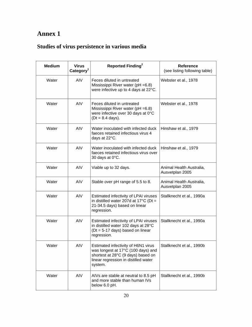
Annex 1 Studies of virus persistence in various media
Medium Virus Category1
Reported Finding2 Reference (see listing following table)
Water AIV Feces diluted in untreated Mississippi River water (pH =6.8) were infective up to 4 days at 22°C.
Webster et al., 1978
Water AIV Feces diluted in untreated Mississippi River water (pH =6.8) were infective over 30 days at 0°C (Dt = 8.4 days).
Webster et al., 1978
Water AIV Water inoculated with infected duck faeces retained infectious virus 4 days at 22°C.
Hinshaw et al., 1979
Water AIV Water inoculated with infected duck faeces retained infectious virus over 30 days at 0°C.
Hinshaw et al., 1979
Water AIV Viable up to 32 days. Animal Health Australia, Ausvetplan 2005
Water AIV Stable over pH range of 5.5 to 8. Animal Health Australia, Ausvetplan 2005
Water AIV Estimated infectivity of LPAI viruses in distilled water 207d at 17°C (Dt = 21-34.5 days) based on linear regression.
Stallknecht et al., 1990a
Water AIV Estimated infectivity of LPAI viruses in distilled water 102 days at 28°C (Dt = 5-17 days) based on linear regression.
Stallknecht et al., 1990a
Water AIV Estimated infectivity of H6N1 virus was longest at 17°C (100 days) and shortest at 28°C (9 days) based on linear regression in distilled water system.
Stallknecht et al., 1990b
Water AIV AIVs are stable at neutral to 8.5 pH and more stable than human IVs below 6.0 pH.
Stallknecht et al., 1990b
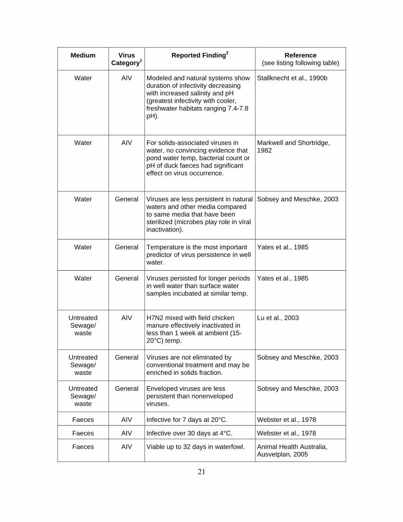
21
Medium Virus Category1
Reported Finding2 Reference (see listing following table)
Water AIV Modeled and natural systems show duration of infectivity decreasing with increased salinity and pH (greatest infectivity with cooler, freshwater habitats ranging 7.4-7.8 pH).
Stallknecht et al., 1990b
Water AIV For solids-associated viruses in water, no convincing evidence that pond water temp, bacterial count or pH of duck faeces had significant effect on virus occurrence.
Markwell and Shortridge, 1982
Water General Viruses are less persistent in natural waters and other media compared to same media that have been sterilized (microbes play role in viral inactivation).
Sobsey and Meschke, 2003
Water General Temperature is the most important predictor of virus persistence in well water.
Yates et al., 1985
Water General Viruses persisted for longer periods in well water than surface water samples incubated at similar temp.
Yates et al., 1985
Untreated Sewage/
waste
AIV H7N2 mixed with field chicken manure effectively inactivated in less than 1 week at ambient (15-20°C) temp.
Lu et al., 2003
Untreated Sewage/
waste
General Viruses are not eliminated by conventional treatment and may be enriched in solids fraction.
Sobsey and Meschke, 2003
Untreated Sewage/
waste
General Enveloped viruses are less persistent than nonenveloped viruses.
Sobsey and Meschke, 2003
Faeces AIV Infective for 7 days at 20°C. Webster et al., 1978
Faeces AIV Infective over 30 days at 4°C. Webster et al., 1978
Faeces AIV Viable up to 32 days in waterfowl. Animal Health Australia, Ausvetplan, 2005
22
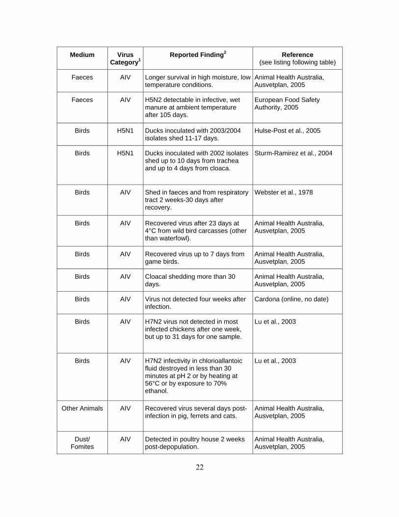
Medium Virus Category1
Reported Finding2 Reference (see listing following table)
Faeces AIV Longer survival in high moisture, low temperature conditions.
Animal Health Australia, Ausvetplan, 2005
Faeces AIV H5N2 detectable in infective, wet manure at ambient temperature after 105 days.
European Food Safety Authority, 2005
Birds H5N1 Ducks inoculated with 2003/2004 isolates shed 11-17 days.
Hulse-Post et al., 2005
Birds H5N1 Ducks inoculated with 2002 isolates shed up to 10 days from trachea and up to 4 days from cloaca.
Sturm-Ramirez et al., 2004
Birds AIV Shed in faeces and from respiratory tract 2 weeks-30 days after recovery.
Webster et al., 1978
Birds AIV Recovered virus after 23 days at 4°C from wild bird carcasses (other than waterfowl).
Animal Health Australia, Ausvetplan, 2005
Birds AIV Recovered virus up to 7 days from game birds.
Animal Health Australia, Ausvetplan, 2005
Birds AIV Cloacal shedding more than 30 days.
Animal Health Australia, Ausvetplan, 2005
Birds AIV Virus not detected four weeks after infection.
Cardona (online, no date)
Birds AIV H7N2 virus not detected in most infected chickens after one week, but up to 31 days for one sample.
Lu et al., 2003
Birds AIV H7N2 infectivity in chlorioallantoic fluid destroyed in less than 30 minutes at pH 2 or by heating at 56°C or by exposure to 70% ethanol.
Lu et al., 2003
Other Animals AIV Recovered virus several days post-infection in pig, ferrets and cats.
Animal Health Australia, Ausvetplan, 2005
Dust/ Fomites
AIV Detected in poultry house 2 weeks post-depopulation.
Animal Health Australia, Ausvetplan, 2005
23
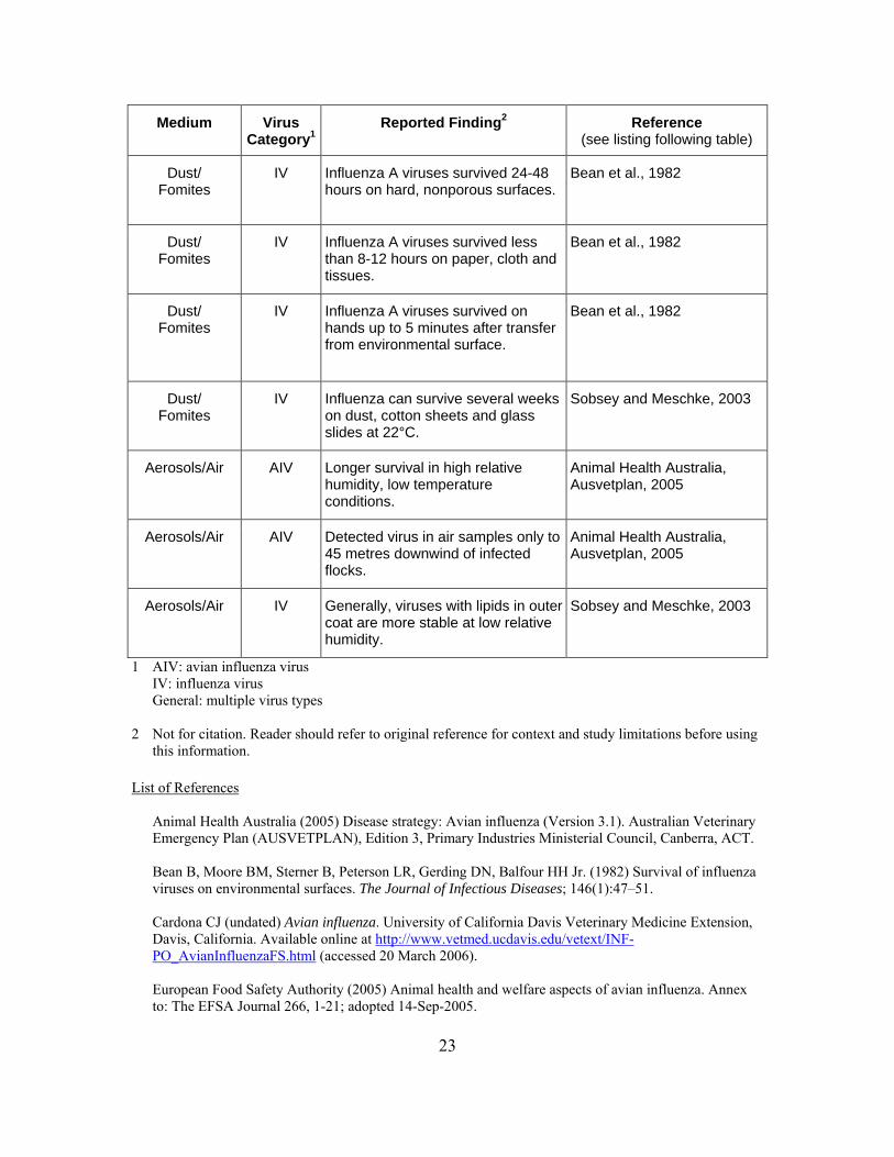
Medium Virus Category1
Reported Finding2 Reference (see listing following table)
Dust/ Fomites
IV Influenza A viruses survived 24-48 hours on hard, nonporous surfaces.
Bean et al., 1982
Dust/ Fomites
IV Influenza A viruses survived less than 8-12 hours on paper, cloth and tissues.
Bean et al., 1982
Dust/ Fomites
IV Influenza A viruses survived on hands up to 5 minutes after transfer from environmental surface.
Bean et al., 1982
Dust/ Fomites
IV Influenza can survive several weeks on dust, cotton sheets and glass slides at 22°C.
Sobsey and Meschke, 2003
Aerosols/Air AIV Longer survival in high relative humidity, low temperature conditions.
Animal Health Australia, Ausvetplan, 2005
Aerosols/Air AIV Detected virus in air samples only to 45 metres downwind of infected flocks.
Animal Health Australia, Ausvetplan, 2005
Aerosols/Air IV Generally, viruses with lipids in outer coat are more stable at low relative humidity.
Sobsey and Meschke, 2003
1 AIV: avian influenza virus IV: influenza virus General: multiple virus types 2 Not for citation. Reader should refer to original reference for context and study limitations before using
this information. List of References
Animal Health Australia (2005) Disease strategy: Avian influenza (Version 3.1). Australian Veterinary Emergency Plan (AUSVETPLAN), Edition 3, Primary Industries Ministerial Council, Canberra, ACT. Bean B, Moore BM, Sterner B, Peterson LR, Gerding DN, Balfour HH Jr. (1982) Survival of influenza viruses on environmental surfaces. The Journal of Infectious Diseases; 146(1):47–51. Cardona CJ (undated) Avian influenza. University of California Davis Veterinary Medicine Extension, Davis, California. Available online at http://www.vetmed.ucdavis.edu/vetext/INF-PO_AvianInfluenzaFS.html (accessed 20 March 2006). European Food Safety Authority (2005) Animal health and welfare aspects of avian influenza. Annex to: The EFSA Journal 266, 1-21; adopted 14-Sep-2005.
24
Hinshaw VS, Webster RG and Turner B (1979) Water-borne transmission of influenza A viruses? Intervirology, 11:66–68. Lu H, Castro AE, Pennick K, Liu J, Yang Q, Dunn P, Weinstock D, and Henzler D (2003) Survival of avian influenza virus H7N2 in SPF chickens and their environments. Avian Diseases, 47(s3):1015-1021. Markwell DD and Shortridge KF (1982) Possible waterborne transmission and maintenance of influenza viruses in domestic ducks. Applied and Environmental Microbiology, 43(1):110-116. Sobsey MD and Meschke JS (2003) Virus survival in the environment with special attention to survival in sewage droplets and other environmental media of fecal or respiratory origin. Draft (dated 21 August 2003) prepared for a World Health Organization meeting (International SARS Symposium) held on 23-25 September 2003 in Rome, Italy. Stallknecht DE, Shane SM, Kearney MT, and Zwank PJ (1990) Persistence of avian influenza viruses in water. Avian Diseases, 34:406-411. Stallknecht DE, Kearney MT, Shane SM, and Zwank PJ (1990) Effects of pH, temperature, and salinity on persistence of avian influenza viruses in water. Avian Diseases, 34:412-418. Sturm-Ramirez KM, Hulse-Post DJ, Govorkova EA, Humberd J, Seiler P, Puthavathana P, Buranathai C, Nguyen TD, Chaisingh A, Long HT, Naipospos TSP, Chen H, Ellis TM, Guan Y, Peiris JSM and Webster RG (2005) Are ducks contributing to the endemicity of highly pathogenic H5N1 influenza virus in Asia? Journal of Virology, 79(17):11269-11279. Webster RG, Yakhno M, Hinshaw VS, Bean WJ and Murti KG (1978) Intestinal influenza: replication and characterization of influenza viruses in ducks. Virology, 84(2):268-278. Yates MV, Gerba CP and Kelley LM (1985) Virus persistence in groundwater. Applied and Environmental Microbiology, 49(4):778-781.
25
Annex 2 Hygiene in Domestic and Community Settings Precautions for the home Basic good health habits that will help reduce the spread of influenza virus in the home include:
• Cover your mouth and nose with a tissue when coughing or sneezing.
• Wash your hands often, especially:
− Before, during, and after you prepare food − Before you eat − After you use the bathroom − After handling animals or animal waste − When your hands are dirty, and − More frequently when someone in your home is sick.
• Avoid touching your eyes, nose or mouth. Infections are often spread when a person touches something that is contaminated with germs and then touches his or her eyes, nose, or mouth.
Cleaning and disinfection of household surfaces likely to be contaminated by infectious secretions appears worthwhile, but no evidence supports the efficacy of widespread disinfection of air. Precautions for schools As part of pandemic influenza planning, special attention should be given to teaching staff, children, and their parents on how to limit the spread of infection. Programs should already be teaching these things (for example, use good hand washing; cover the mouth when coughing or sneezing; clean toys frequently) to build habits that protect children from influenza disease in general. Handwashing and respiratory hygiene/cough behaviours should be routine for all and strongly encouraged in public health messages. Such practices should be facilitated by making hand-hygiene facilities available in schools. Keep a good supply of things you will need to help control the spread of infection (soap, paper towels, and tissues). Store the supplies in easy-to-find places.
26
Further Reading Bean B, Moore BM, Sterner B, Peterson LR, Gerding DN, Balfour HH Jr. (1982) Survival of influenza viruses on environmental surfaces. Journal of Infectious Disease; 146:47–51.
Centers for Disease Control and Prevention; Schools, Childcare Providers, and Parents. Available at http://www.cdc.gov/flu/school/.
World Health Organization Writing Group (2005) Non-pharmaceutical interventions: their role in reducing transmission and spread. Available from http://www.who.int/csr/disease/avian_influenza/pharmaintervention2005_11_3/en/index.html
World Health Organization Writing Group (2006) Nonpharmaceutical public health interventions for pandemic influenza, national and community measures. Emerging Infectious Diseases;12(1):88–94.
27
Annex 3
Water, Sanitation, Hygiene and Waste management in Health Care Settings relevant to Influenza A (H5N1) infection
Drinking water Avian influenza viruses can persist for extended periods of time in water depending on temperature, pH and salinity but information on environmental persistence of H5N1 in water is lacking 1. As influenza A (H5N1) infection is frequently accompanied by gastrointestinal symptoms, it could indicate that H5N1 may enter the host through a faecal-oral route. Based on the little evidence available there is a small but potential risk. WHO recommends 2 the following guidelines, applicable to health care settings, which may be adapted to national standards.
1. Microbiological quality E. coli or thermotolerant coliform bacteria must not be detectable in any 100-ml sample
2. Disinfection Free residual chlorine content at discharge points:
- 0.2-0.5mg/l (pH < 8) minimum 30 minutes contact time
- 0.4-1.0mg/l (pH > 8) minimum 60 minutes contact time
3. Chemical and radiological quality Water meets WHO Guidelines for Drinking-Water Quality or national standards concerning chemical and radiological parameters
4. Turbidity Maximum turbidity is 5 NTU
5. Acceptability There are no tastes, odours or colours that would discourage consumption of the water
See also Guidelines for drinking-water quality, WHO 3
Hand hygiene Hand hygiene remains a prerequisite requirement to prevent the transmission of diseases. In environments where H5N1 may be present, hand hygiene, which includes hand washing and the use of alcohol-based hand rubs, is critical to prevent possible inoculation of the nose, mouth, and conjunctivae by contaminated hands. Hand hygiene is also necessary to limit the potential for transmission of other nosocomial infections.
28
When visibly dirty or contaminated with proteinaceous material, or visibly soiled with blood or other body fluids, wash hands with either a plain or antimicrobial soap and water and rub hands together vigorously for at least 30 seconds, covering all surfaces of the hands and fingers. Rinse hands with water and dry thoroughly with a disposable towel. Use towel to turn off the faucet.4 If hands are not visibly dirty, use an alcohol-based preparation. Many studies have demonstrated that influenza, an enveloped virus, is susceptible to alcohols when tested in vitro5 and in vivo testing with a 95% ethyl alcohol hand disinfectant reduced influenza virus on hands by a log10 reduction > 2.5.6 Ethyl alcohol has greater activity against viruses than isopropyl alcohol 7, therefore, ethyl alcohol-based hand disinfection products may be preferred over isopropyl alcohol products in settings where transmission of influenza H5N1 is of concern.
When decontaminating hands with an alcohol-based hand rub, apply product to palm of one hand and rub hands together, covering all surfaces of hands and fingers, until hands are dry. Follow the manufacturer's recommendations regarding the volume of product to use.4 Soiled surfaces • Cleaning MUST precede disinfection. Items and surfaces cannot be disinfected if they
are not first cleaned of any kind of organic matter (patient's excretions, secretions, dirt, soil, etc). Disinfection is a process of killing microorganisms without complete sterilization. Avian influenza virus is inactivated by a range of disinfectants, including:
− phenolic disinfectants − quaternary ammonia compounds − peroxygen compounds − sodium hypochlorite (household bleach) or calcium hypochlorite, see below − alcohol (see below) − germicides with a tuberculocidal claim on the label − other registered/licensed disinfectants
• Use manufacturer’s recommendations for use/dilution, contact time, and handling.
• Because mycobacteria have the highest intrinsic level of resistance among the vegetative bacteria, viruses, and fungi, any germicide with a tuberculocidal claim on the label (i.e., an intermediate-level disinfectant) is considered capable of inactivating influenza.8
• Patient rooms/areas should be cleaned at least daily and terminally cleaned at discharge. In addition to daily cleaning of floors and other horizontal surfaces, clean frequently touched surfaces (e.g., medical equipment, bed rails, bedside and over-bed tables, TV controls, call buttons, telephones, bathroom surfaces including safety/pull-up bars, doorknobs, commodes, ventilator surfaces and plastic curtains) twice daily, if possible.
29
• Keep areas around the patient free of unnecessary supplies and equipment to facilitate daily cleaning.
• Bed curtains should be laundered if visibly soiled and/or at hospital discharge.
• Paper sheeting that is changed between patients is appropriate for patient examination tables in outpatient areas.
• Do not spray (i.e., fog) occupied or unoccupied rooms with disinfectant. This is a potentially dangerous practice that has no proven disease control benefit.
Alcohol
Alcohol is effective against influenza virus, however 70% ethyl alcohol is a powerful broad-spectrum germicide and is considered generally superior to isopropyl alcohol. Alcohol is often used to disinfect small surfaces (e.g., rubber stoppers of multiple-dose medication vials, and thermometers) and occasionally external surfaces of equipment (e.g., stethoscopes and ventilators). Because alcohol is flammable, its use as surface disinfectant should be limited to small surface areas and be used in well-ventilated spaces only. Alcohol may also cause discoloration, swelling, hardening, and cracking of rubber and certain plastics after prolonged and repeated use.
Sodium hypochlorite (bleach)
Bleach is a strong and effective disinfectant, but it is readily inactivated in the presence of organic material. Its active ingredient, sodium hypochlorite, is effective in killing bacteria, fungus and viruses, including influenza virus. Diluted household bleach works at variable exposition times (from 10 to 60 min), is widely available at a low cost, and can be recommended for the disinfection of health care facilities. However, bleach irritates mucous membranes, the skin and the airway, decomposes under heat or light, and reacts readily with other chemicals. Therefore, caution is advised when bleach is used. Improper use of bleach may reduce its effectiveness for disinfection and also lead to accidents, which can be harmful to health.
Procedures for preparing/using diluted bleach • Use mask, rubber gloves, plastic apron and goggles (recommended).
• Mix and use bleach solutions in well-ventilated areas.
• Mix bleach with cold water because hot water decomposes the active ingredient of bleach and renders it ineffective.
30
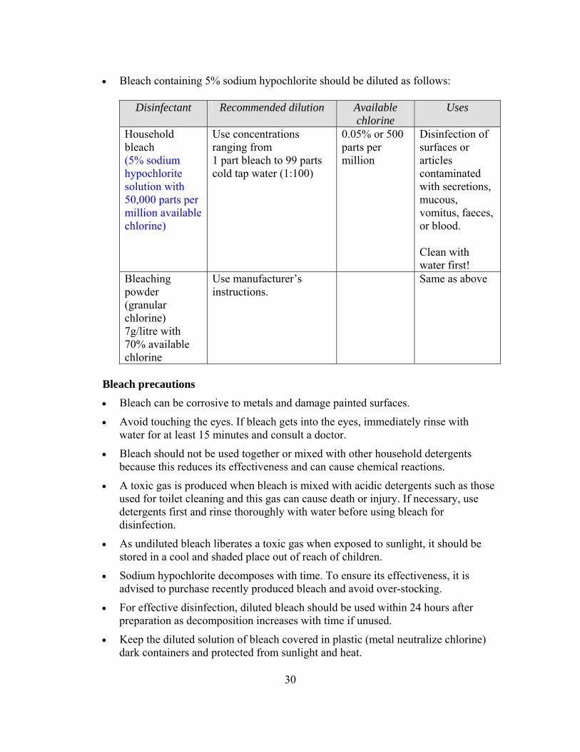
• Bleach containing 5% sodium hypochlorite should be diluted as follows:
Disinfectant Recommended dilution Available chlorine
Uses
Household bleach (5% sodium hypochlorite solution with 50,000 parts per million available chlorine)
Use concentrations ranging from 1 part bleach to 99 parts cold tap water (1:100)
0.05% or 500 parts per million
Disinfection of surfaces or articles contaminated with secretions, mucous, vomitus, faeces, or blood. Clean with water first!
Bleaching powder (granular chlorine) 7g/litre with 70% available chlorine
Use manufacturer’s instructions.
Same as above
Bleach precautions • Bleach can be corrosive to metals and damage painted surfaces.
• Avoid touching the eyes. If bleach gets into the eyes, immediately rinse with water for at least 15 minutes and consult a doctor.
• Bleach should not be used together or mixed with other household detergents because this reduces its effectiveness and can cause chemical reactions.
• A toxic gas is produced when bleach is mixed with acidic detergents such as those used for toilet cleaning and this gas can cause death or injury. If necessary, use detergents first and rinse thoroughly with water before using bleach for disinfection.
• As undiluted bleach liberates a toxic gas when exposed to sunlight, it should be stored in a cool and shaded place out of reach of children.
• Sodium hypochlorite decomposes with time. To ensure its effectiveness, it is advised to purchase recently produced bleach and avoid over-stocking.
• For effective disinfection, diluted bleach should be used within 24 hours after preparation as decomposition increases with time if unused.
• Keep the diluted solution of bleach covered in plastic (metal neutralize chlorine) dark containers and protected from sunlight and heat.
31
Transmission through aerosol or dust A potential risk exists through possible direct inoculation (e.g., via inhalation or direct impact) into the respiratory (nose/mouth) or conjunctival (eyes) mucosa. To mitigate this route of transmission, cleaning methods that do not produce aerosols or resuspended particulates should be used. Avoid dusting methods that disperse dust (e.g., feather dusting) and wet-dust horizontal surfaces by moistening a cloth with a water.
The optimal combination of PPE for preventing H5N1 transmission during aerosol-generating procedures has not been determined, but PPE should cover the torso, arms, and hands as well as the eyes, nose, and mouth.
• Respiratory protection for aerosol-generating procedures During aerosol-generating procedures, there must be minimal respirator face-seal leakage to fully protect health care workers from exposure to small-particle aerosols. The following respiratory protection options should be considered:
− Disposable particulate respirators (particulate masks) (e.g., N-95 or equivalent) are the minimum level of respiratory protection required for health care workers performing aerosol-generating procedures.
− To ensure adequate protection, health care workers should be fit tested to the disposable respirator (particulate mask) model that they will wear and know how to perform a user seal check. A user seal check should be performed each time a respirator is put on, before entering the patient room.
− Some factors to consider when choosing respirators in this setting include availability, impact on mobility, impact on patient care, potential for exposure to higher levels of aerosolized respiratory secretions, and potential for reusable respirators (particulate masks) to serve as fomites for transmission.
• Engineering controls for aerosol-generating procedures
Perform the procedure in a negative pressure room (if available); if a negative pressure room/area cannot be created 8, 9:
− Perform the procedure in a private room, away from other patients.
− If possible, increase air exchanges, create a negative pressure relative to the hallway, and avoid recirculation of the room air.
− If recirculation of air from such rooms is unavoidable, pass the air through a HEPA filter before recirculation.
− Cleaning devices, such as portable HEPA filtration units, may be used to further reduce the concentration of contaminants in the air.
Keep doors closed except when entering or leaving the room, and minimize entry and exit during the procedure.
32
Linen and laundry of an H5N1 infected patients Standard precautions are recommended for linen and laundry that might be contaminated with respiratory secretions from suspected or confirmed H5N1 infected patients:8
Place soiled linen directly into a laundry bag in the patient’s room. Contain linen in a manner that prevents the linen bag from opening or bursting during transport and while in the soiled linen holding area.
• Wear gloves and gown and mask when directly handling soiled linen and laundry (e.g., bedding, towels, personal clothing) as per standard precautions.
• Do not shake or otherwise handle soiled linen and laundry in a manner that might create an opportunity for disease transmission or contamination of the environment.
• Wear gloves for transporting bagged linen and laundry.
• Perform hand hygiene after removing gloves that have been in contact with soiled linen and laundry.
• Wash with soap, rinse then soak in 0.5% chlorine and dry linen according to routine standards and procedures.
Waste management Standard precautions when working with clinical and nonclinical solid waste∗ that may be contaminated with avian influenza H5N1 virus are required:
• All waste generated in the isolation room/area should be disposed of in suitable containers or bags.
• Used patient care supplies that are not likely to be contaminated (e.g., paper wrappers) may be discarded as nonclinical waste.
• All waste from an isolation room/area that may be contaminated with influenza A (H5N1) should be treated as clinical (infectious) waste and “red or yellow-bagged” or labelled as “biohazard” and should be treated and disposed of as per facility policy and in accordance with national regulations pertaining to such waste.
• One waste disposal bag is usually adequate, providing waste can be placed in the bag without contaminating the outside of the bag. If that is not possible, two bags are needed (double bagging).
∗ Clinical waste includes waste directly associated with blood, body fluids secretions and excretions; laboratory waste that is directly associated with specimen processing, human tissues, including material or solutions containing free-flowing blood, and animal tissue or carcasses used for research; and also includes discarded sharps. Nonclinical waste is routine waste generated during the care and treatment of suspected or confirmed HPAI patients.
33
• Currently, there is no evidence to suggest that the presence of H5N1 virus in liquid waste such as urine or faeces requires a change in the protocols used to manage this type of infectious waste.
• Outside of the isolation room/area, gloves should be worn when handling waste and hand hygiene should be performed after glove removal.
Dishes and eating utensils Use standard precautions for handling dishes and eating utensils used by patients with suspected or confirmed influenza A (H5N1) infection:
• Wear gloves when handling patient trays, dishes, and utensils.
• Clean reusable items first by washing with soap and water either manually or in a dishwasher at the recommended water temperature. Then disinfect items by either boiling in water or soaking in chlorine.
• Disposable items or left over from food should be discarded with other infectious waste.
References:
1. Department of Population Health College of Veterinary Medicine, University of Georgia (2005) Highly Pathogenic Avian Influenza Virus H5N1 and Wild Birds.
2. WHO. Guidelines to be used for producing minimum standards for water, sanitation, hygiene and waste management in health-care settings, with specific reference to developing countries, draft under progress.
3. WHO (2004) Guidelines for Drinking-water Quality, third edition. 4. Buxton Bridges CB, Kuehnert MJ, Hall CB (2003) Transmission of influenza:
implications for control in health care settings. Clinical Infectious Diseases; 37(8):1094-1101.
5. WHO (2005) Consensus document on the epidemiology of severe acute respiratory syndrome (SARS) WHO/CDS/CSR/GAR/2003. Available at: http://www.who.int/csr/sars/en/WHOconsensus.pdf. Accessed 14 November.
6. CDC (2002) Guideline for Hand Hygiene in Health-Care Settings. Morbidity and Mortality Weekly Report; 51(RR16):1-44.
7. Ali J, Dolan M, Fendler E, Larson E (2000) Alcohols. In: Block S, ed. Disinfection, sterilization and preservation. 5 ed. Philadelphia: Lippincott, Williams, and Wilkins; 229-253.
8. Schurmann W, Eggers H (1983) Antiviral activity of an alcoholic hand disinfectant: comparison of the in vitro suspension test with in vivo experiments on hands, and on individual fingertips. Antiviral Research; 3:25-41.
9. Rosenbaum R, Benyo J, O'Connor R, et al (2004) Use of a portable forced air system to convert existing hospital space into a mass casualty isolation area. Annals of Emergency Medicine; 44(6):628-634.
34
Additional Reading:
Bean B, Moore B, Sterner B, Peterson L, Gerding D, Balfour HJ (1982) Survival of influenza viruses on environmental surfaces. Journal of Infectious Diseases; 46(1):47-51.
Buxton Bridges C, Katz JM, Seto WH, et al. (2000) Risk of influenza A (H5N1) infection among health care workers exposed to patients with influenza A (H5N1), Hong Kong. Journal Infectious Diseases; 181(1):344-348.
CDC (2003) Guidelines for Environmental Infection Control in Health-Care Facilities. Morbidity and Mortality Weekly Report; 52(RR10):1-42.
Fowler R, Guest C, Lapinsky SE, et al. (2004) Transmission of Severe Acute Respiratory Syndrome during Intubation and Mechanical Ventilation. American Journal of Respiratory and Critical Care Medicine; 169:1198-1202.
Goldmann D (2000) Transmission of viral respiratory infections in the home. Pediatric Infections Disease Journal; 19(10 Suppl):S97-102.
Healing TD, Hoffman PN, Young SEJ (1995) The infection hazards of human cadavers. Communicable Disease Report Weekly; 5(5):R61-68.
Katz JM, Lim W, Bridges CB, et al. (1999) Antibody response in individuals infected with avian influenza A (H5N1) viruses and detection of anti-H5 antibody among household and social contacts. Journal Infectious Diseases; 180(6):1763-1770.
Klein M, Deforest A (1991) Principles of Viral Inactivation. Disinfectants, Sterilization, and Preservation. 3 ed. Philadelphia: Lea and Febiger; 222-434.
Lidwell OM (1974) Aerial dispersal of micro-organisms from the human respiratory tract. Society for Applied Bacteriology Symposium Series; 3(135-154).
Loosli D, Lemon H, Robertson O, Appel E (1943) Experimental airborne influenza infection. I. Influence of humidity on survival of virus in air. Proceedings of the Society for Experimental Biology and Medicine; 53:205-206.
Sehulster L, Chinn R (2003) Guidelines for environmental infection control in health-care facilities. Recommendations of CDC and the Healthcare Infection Control Practices Advisory Committee (HICPAC). Morbidity and Mortality Weekly Report; 52(RR-10):1-42.
WHO (2006) Infection control recommendations for avian influenza in health-care facilities, Aide-Memoire, Available at: http://www.who.int/csr/disease/avian_influenza/guidelinestopics/en/index3.html
WHO (2006) Avian Influenza A (H5N1) in Humans - Interim Infection Control Guideline for Health Care Facilities, revised February 2006.
WHO (2005) Guidelines on Hand Hygiene in Health Care (Advanced Draft), May 2005.
35
WHO (2005) Inter-country-consultation: influenza A/H5N1 in humans in Asia: Manila, Philippines, 6-7 May. 2005.
Zhong N, Zeng G (2003) Our strategies for fighting severe acute respiratory syndrome (SARS). American Journal of Respiratory and Critical Care Medicine; 168:7-9.
36
Annex 4 Household drinking water disinfection using chlorine Using chlorine Chlorine is a chemical of choice used for the disinfection and decontamination of water and
particularly drinking water because of its strong oxidizing properties against almost all pathogenic viruses and bacteria which can be transmitted in water.
Chlorine is found in powder, tablets, gas or already made solution in plastic bottles.
Common household bleach has between 4-5% of active chlorine and is in liquid form. Chlorine should be stored in airtight glass or plastic containers and protected from excessive
light and heat to maintain its active power level. It must be stored out of reach of children. The amount of chlorine required should be determined on a case-by-case basis as it depends
on the quantity and on the quality of water (mainly turbidity and pH). For example, 4 to 5 drops of a chlorine are typically needed to treat 1 litre of clear water.
Local recommendations on treatment with chlorine should include the time needed for
chlorine to have an effect (e.g., WHO Guidelines for Drinking Water Quality recommend a minimum of 30 minutes contact time to ensure inactivation of pathogenic micro-organisms).
The residual free chlorine is the concentration of chlorine left in water after initial treatment.
It has the power to react on subsequent contamination of the water. Residual disinfectant makes an important contribution to ensuring microbial safety of drinking-water (e.g., WHO Guidelines for Drinking Water Quality recommend a minimum residual concentration of free chlorine of ≥0.5 mg/litre after at least 30 minutes contact time).
Turbid water has suspended particles that can protect micro-organisms from chlorine. A
pretreatment such as sedimentation (let particles settle down) will be required first. Keeping treated drinking water safe Once your chlorine dosage is set, test the free residual chlorine from time to time as water
quality may change (e.g. once a month). Storing drinking water Use a dedicated container for storing safe drinking water. Protect safe water from contact with animals. If affordable, use a covered storage container, equipped with a tap and a lid. Keep a dedicated cup attached to the storage container to transfer water. This prevents cross
contamination of the water by the use of dirty or used cups. Keep storage containers in the shade as light and heat will decrease residual free chlorine
levels.
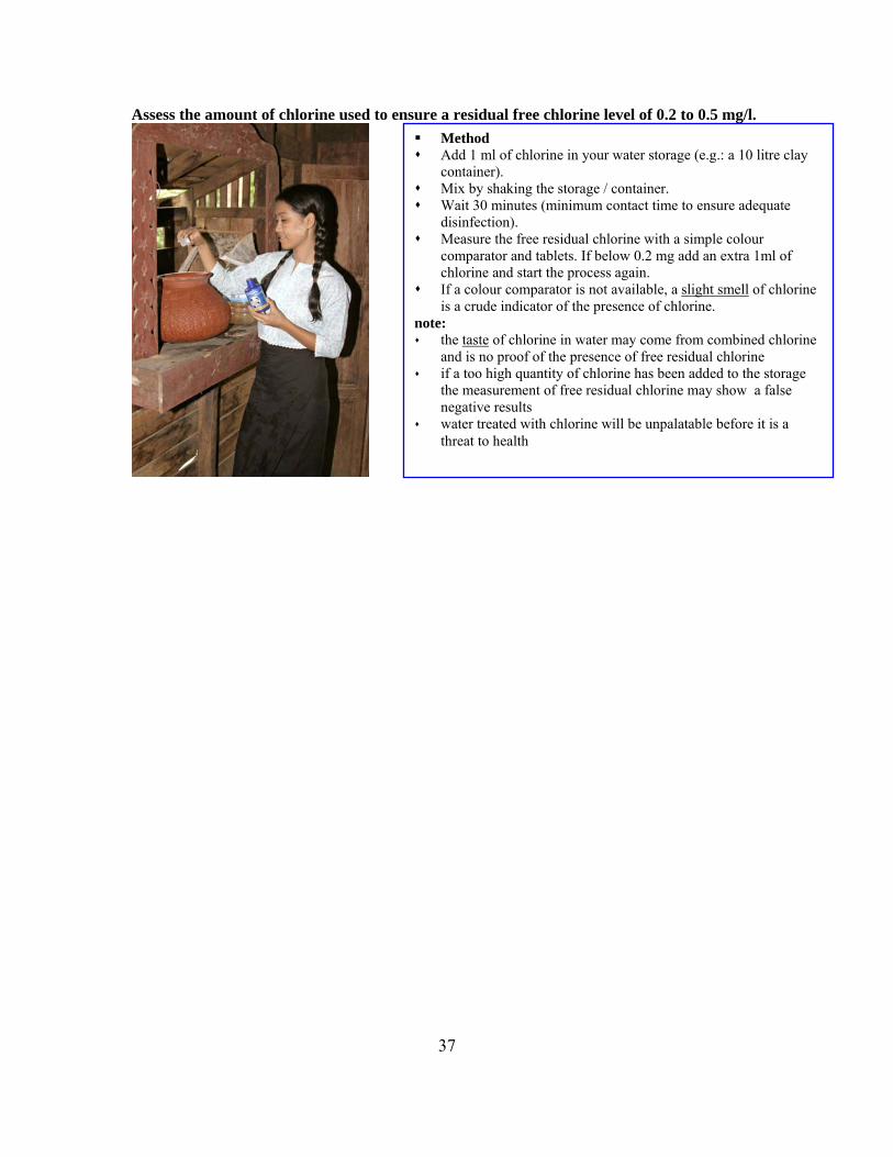
Assess the amount of chlorine used to ensure a residual free chlorine level of 0.2 to 0.5 mg/l. Method Add 1 ml of chlorine in your water storage (e.g.: a 10 litre clay
container). Mix by shaking the storage / container. Wait 30 minutes (minimum contact time to ensure adequate
disinfection). Measure the free residual chlorine with a simple colour
comparator and tablets. If below 0.2 mg add an extra 1ml of chlorine and start the process again.
If a colour comparator is not available, a slight smell of chlorine is a crude indicator of the presence of chlorine.
note: the taste of chlorine in water may come from combined chlorine
and is no proof of the presence of free residual chlorine if a too high quantity of chlorine has been added to the storage
the measurement of free residual chlorine may show a false negative results
water treated with chlorine will be unpalatable before it is a threat to health
37