treatment of hyper-granulated limb wounds in horsesvetmedmosul.org/ijvs/media/11-2-7e.pdf ·...
TRANSCRIPT

Iraqi Journal of Veterinary Sciences, Vol. 25, No. 2, 2011 (71-80)
71
Treatment of hyper-granulated limb wounds in horses
O. A. Bader1 and M. J. Eesa
2
1 Department of Surgery and Theriogeniology, College of Veterinary Medicine, University of Mosul, Mosul,
2 Department of Surgery and Obstetrics, College of Veterinary Medicine, University of Baghdad, Baghdad, Iraq
(Received February 26, 2008; Accepted May 19, 2010)
Abstract
This study was performed to investigate the different methods of treating hyper granulation tissue on experimentally
induced wounds in equine limbs. Wounds were induced by removal of a skin patch and subcutaneous tissue for about 5-7 cm
width and 6-8 cm in length from the dorsal and lateral aspect of the fore and hind limbs below the carpal and tarsal joints. The
wounds were left open without treatment and the animals were trained 2-2.5 hours every day for about 3-5 weeks until hyper
granulation tissue was developed. The schedule for the treatment of hyper granulation was divided into five groups each
contained eight wounds of hyper granulation tissue; each main group was divided into two subgroups. The subgroups of first,
second, third, fourth and fifth groups were treated by the following schedules: bandage alone; copper sulphate ointment 10%;
silver nitrate ointment 2%; red mercury ointment 11%; and laser therapy (at a total dose of 9.72 Joule / cm2) respectively.
While the second subgroups were treated by surgical resection of the hyper granulation tissue, followed by the same treatments
applied on the first subgroup. The bandage for all experimental groups was changed every 48 hours until healing was occurred.
The clinical and histological observation of the first group revealed that the healing take long period comparing with other
groups. The mean of wound healing were 65 days in non surgical removal of hyper granulation tissue subgroup, while 57 days
in surgical removed of hyper granulation tissue subgroup. The results of the second, third, fourth groups revealed that the
caustic material especially red mercury has a role in healing processes through depressing the hyper granulation tissue. The
mean of wound healing of the second group was 42.25 days in non surgical removal of hyper granulation tissue subgroup
while 37.25 days in surgically removed hyper granulation tissue subgroup. In the third group the mean of wound healing was
45.75 days in non surgical removal of hyper granulation tissue subgroup while 44.75 days in surgically removed hyper
granulation tissue subgroup. While in the fourth group the mean of wound healing was 39 days in non surgical removal of
hyper granulation tissue subgroup while 36 days in surgically removed hyper granulation tissue subgroup. In the fifth group
the clinical and histological observation revealed that the using of laser lead to reduce the period for wound healing
significantly comparing with other groups. The mean of wound healing was 25 days in non surgical removal of hyper
granulation tissue subgroup while 20 days in surgically removed hyper granulation tissue subgroup, so that the laser was the
best in this study and the using of surgical removal is better than of non surgical removal.
Keywords: Hyper granulation; Limb wounds; Equine. Available online at http://www.vetmedmosul.org/ijvs
الخيولفي قوائم الفرط الحبيبي لجروج ال �جع
٢و محمد جواد عيسى ١عمر عادل بدر
،، كلية الطب البيطري، جامعة الموصل، الموصلالجراحة وعلم تناسل الحيوانفرع ١ العراق بغداد،، كلية الطب البيطري، جامعة بغداد ،الجراحة والتوليدفرع ٢
الخ�صة
. ي الجروح المحدثة تجريبيا في قوائم الخيلأجريت ھذه الدراسة لبيان تأثير طرائق ع&جية مختلفة لع&ج حالة فرط النسيج الحبيبي ف
سم عرضا وتم إحداث الجروح ٧- ٥سم طو: و ٨- ٦تم إحداث الجروح بإزالة جزء من الجلد والنسيج تحت الجلد بمساحة تراوحت بين

Iraqi Journal of Veterinary Sciences, Vol. 25, No. 2, 2011 (71-80)
72
ة ومن دون ع&ج وتم في الجھة الظھرية والوحشية لك& القائمتين ا@مامية والخلفية أسفل مفصل الرسغ بعد ذلك تركت الجروح مفتوحتم .أسابيع لحين ظھور فرط النسيج الحبيبي بشكل واضح ٥-٣ساعتين ونصف ولمدة –ترويض حيوانات التجربة يوميا لمدة ساعتين
. جروح وكل مجموعة رئيسية قسمت إلى مجموعتين ثانويتين ٨تقسيم حيوانات التجربة إلى خمس مجاميع رئيسية ضمت كل مجموعة لوحدھا ، مرھم يع الثانوية ا@ولى للمجاميع الرئيسية ا@ولى والثانية والثالثة والرابعة والخامسة باستخدام العصابة عولجت المجام
على ) ٢سم/ جول ٩.٧٢بجرعة كلية (والليزر % ١١ ، مرھم الزئبق ا@حمر% ٢، مرھم نترات الفضة % ١٠كبريتات النحاس ية فقد عولجت باستخدام اPزالة الجراحية للنسيج الحبيبي الزائد ومن ثم تم وضع نفس المواد السابقة أما في المجاميع الثانوية الثان. التوالي
أوضحت النتائج السريرية والنسجية أن المجموعة ا@ولى استغرقت وقتا طوي& مقارنة مع .التي تم وضعھا في المجاميع الثانوية ا@ولىيوما أما في ٦٥م الجروح في المجموعة الثانوية التي لم يتم إجراء اPزالة الجراحية فيھا باقي المجاميع ا@خرى حيث بلغ معدل التئا
بينت المجاميع الثانية والثالثة والرابعة التي استخدمت فيھا المواد .يوما ٥٧المجموعة الثانوية التي تم إجراء اPزالة الجراحية فيھا عبت دورا مھما في التئام الجروح من خ&ل تثبيط نمو النسيج الحبيبي المفرط حيث كان الكاوية وخاصة الزئبق ا@حمر أن ھذه المواد ل
يوما أما في المجموعة الثانوية ا@خرى كان ٤٢.٢٥معدل التئام الجروح في المجموعة الثانوية التي لم يتم إجراء اPزالة الجراحية فيھا وعة الثالثة كان معدل التئام الجروح في المجموعة الثانوية التي لم يتم إجراء أما في المجم. يوما للمجموعة الرئيسية الثانية ٣٧.٢٥
بينما في المجموعة الرابعة كان معدل التئام . يوما ٤٤.٧٥يوما أما في المجموعة الثانوية ا@خرى كان ٤٥.٧٥اPزالة الجراحية فيھا أشارت .يوما ٣٦يوما أما في المجموعة الثانوية ا@خرى كان ٣٩حية فيھا الجروح في المجموعة الثانوية التي لم يتم إجراء اPزالة الجرا
النتائج السريرية والنسجية للمجموعة الخامسة أن استخدام الليزر أدى إلى تقليل فترة التئام الجروح وبشكل ملحوظ مقارنة مع المجاميع يوما أما في المجموعة الثانوية ٢٥يتم إجراء اPزالة الجراحية فيھا ا@خرى حيث بلغ معدل التئام الجروح في المجموعة الثانوية التي لم
لذلك عد الليزر ا@فضل في ھذه التجربة أيضا فان إجراء اPزالة الجراحية لفرط النسيج الحبيبي يعد أفضل من . يوما ٢٠ا@خرى كان .عدم إجرائھا
Introduction
Equine limbs suffer traumatic wounds more than other
animals and these wounds mostly heal by second intention
healing which may be associated with some complications
such as wound infections, tumor or hyper granulation tissue
(1-3). The natural response of the body to form granulation
tissue was resists infection and helps to full cavity of
wounds, beside that creates a good surface for migration of
epithelial cells to complete healing (4,5). Many factors may
help the formation of hyper granulation tissue in equine
limbs such as body size, age of animal, wound location,
early wound care such as topical medical application,
bandage and casting help to minimize hyper granulation
tissue formation (1,6-10). Treatments of hyper granulation
tissue depend on the amount of excessive tissue formation
and duration of time, many methods were used in this field
such as caustic material (11-13), cryogenic surgery (14,15),
surgical resection which is regarded as a good method
because it dose not affect on epithelization when compared
with chemical material (3,7,11), topical steroids (16), skin
grafting was used after resection of hyper granulation tissue
from a large area (7,17) and low-power laser therapy in
high doses (18). The aim of this study was to find a good
method for treating hyper granulation tissue through
comparing several methods of treatment.
Materials and methods Twenty horses of local breed 3-8 years in age, 250-400
Kg in weight and from both sexes were used in this study.
The animals were healthy and did not suffer from any
affection.
Inducing hyper granulation tissue Animals were anaesthetized by using acepromazine
(0.02 mg/kg B.W) intravenously as a premedication. Ten
minutes later using thiopental sodium (1g/90 kg B.W)
intravenously and repeated at a half doses if needed.
Clipping and shaving the area of lateral and dorsal aspect of
metacarpal and metatarsal bones. Wound was induced by
removal of a skin patch with subcutaneous tissue for about
5-7 cm width and 6-8 cm length from the dorsal and lateral
aspect of fore and hind limbs of metacarpal and metatarsal
bones (Figure 1 a). The wounds were left open and the
animals were trained daily for 2-2.5 hours/ day for about 3-
5 weeks, until hyper granulation tissue was developed
(Figure 1B).
Treatment The experimental animals were divided into five groups,
each contained four animals and each group was divided
into two equal subgroups, each subgroups consisted of four
wounds; First group (Control): The first four wounds were
treated by application of pressure bandage over the hyper
granulation tissue while the other four wounds were treated
by surgical resection of hyper granulation tissue to the level
of skin, then application of pressure bandage. Second
group: The first four wounds were treated by using copper
sulphate ointment10% locally with pressure bandage, while
the other four wounds were treated by surgical removal of
hyper granulation tissue and using copper sulphate ointment

Iraqi Journal of Veterinary Sciences, Vol. 25, No. 2, 2011 (71-80)
73
10% with pressure bandage. Third group: The first four
wounds were treated by application of silver nitrate
ointment 2% locally with pressure bandage and the other
four wounds were treated by surgical resection of hyper
granulation tissue, then using silver nitrate ointment 2%
with bandage. Fourth group: The first four wounds were
treated by application of red mercury ointment 11% locally
with pressure bandage, other four wounds were treated by
surgical resection of hyper granulation tissue and using red
mercury ointment 11% with bandage. Fifth group: The first
four wounds were treated by laser radiation using Helieum-
neiun (He-Ne), Infrared (IR) with bandage, (He- Ne 6x10 -3
and IR 3x10 -3
). Twelve sessions, one day rest between 3
sessions, the duration of radiation was 18 minute for each
day. The amount of energy of He- Ne (0.54 J/cm2) and
(0.27 J/cm2)
for IR in each session. The total amount of
energy of He- Ne, IR for twelve sessions was 6.48 J/cm2,
and 3.24 J /cm2, respectively, so that the total amount was
9.72 J/cm2, other four wounds were treated by surgical
resection of hyper granulation tissue, then using laser
radiation as same in above with pressure bandage.
In all first subgroups, bandage was changed every 48
hours until hyper granulation tissue became at the level of
skin and epithelial cells migrate to the area, while in the
second subgroups on which hyper granulation resection
were performed (Figure 1C), the bandage was remained
until wound became stable and epitheliazation started to
invade the area. Clinical follow up of the animals during
experimental period was practiced i.e. animal's appetite,
wounds condition and degree of healing, lameness, and
measurement of the thickness of hyper granulation tissue
before and after treatment by using a caliper.
Biopsies were taken from the hyper granulation tissue
before and after treatments at the period of clinical healing
from all groups to identify the nature of healing and to
compare the degree of healing between these five groups.
Hematoxylin and Eosin, MasonTtrichrom and Blue Alishin
staining (19 - 21) were used to investigate the nature of
tissue.
Statistical analyse were done by using analysis of
variance. The difference was determined by LSD at the
level of (P<0.05), and using statistical program SPSS
(SPSS Inc).
Results
The clinical findings revealed that the average thickness
of hyper granulation tissue were 2 - 2.25 cm after 3-5
weeks from wounds inducing (Figure 1B).
First group: in non surgical resection subgroup, pus
formation was observed during dressing but there was no
sensation, lameness or bleeding. While in surgical excision
of hyper granulation tissue subgroup, the signs were the
same as above but less pus formation was observed.
Second group: the clinical signs were similar to that
seen in the first group, but less in degree.
Third group: in non surgical resection subgroup,
excessive amount of pus and bleeding observed during
bandage change but the discharge decreased gradually,
tissue sloughing especially at the central lesion, pain and
lameness, were observed. The signs of surgical resection
subgroup were similar to the non surgical resection once.
Fourth group: both the surgical and non surgical
resection subgroups, show similar clinical signs in the form
of little amount of pus exhibited during bandage change, no
bleeding, pain or lameness during treatment were observed.
Fifth group: in non surgical resection of hyper
granulation tissue subgroup the signs revealed that the
superficial layers of the hyper granulation tissue dissolved
after 10 minutes of laser radiate, and at the end of each
radiation session, the tissues appeared as it was cooked.
There was no pain or lameness, with little pus but without
bleeding at the time of bandage changing.
Clinically the partial or complete wound healing after
treatment as in 1st, 2
nd, 3
rd, 4
th and 5
th groups were appeared
in (Figures 1D), (Figures 2A, 2B, 2C and 2D) and (Figures
3A, 3B, 3C and 3D).
In all experimental animals wound healing of hind limbs
was faster than of fore limbs, and of lateral aspect was
faster than wounds located on dorsal aspect of both fore and
hind limbs.
Different degrees of tissue depression were recorded by
using caliper in non surgical resection of hyper granulation
tissue of experimental subgroups, after using pressure
bandage in first group and different caustic material in 2nd
,
3rd
and fourth group or laser therapy as in fifth group. This
variation of depression was summarized in Table 1. The
mean degrees of wound healing for the experimental groups
were recorded and summarized in Table 2.
The histopatholgical findings of the hyper granulation
tissue before treatment revealed severe proliferation of
fibroblast with minimal amount of collagen fibers. There
was severe infiltration of mononuclear and multinuclear
inflammatory cells around newly blood vessels (Figure 4
A).While the microscopic findings for the biopsies taken at
the period of clinical healing revealed that the formation of
epithelial cells from the edge of skin and invade the area to
cover it (Table 3).

Iraqi Journal of Veterinary Sciences, Vol. 25, No. 2, 2011 (71-80)
74
Figure 1: (A) Surgical resection of skin flap from lateral surface of fore limb. (B) Hyper granulation tissue formation after 35
days of inducing wound. (C) Complete resection of hyper granulation tissue after 5 weeks of inducing wound. (D) Partial
wound healing after 40 days of treatment in non surgical resection of hyper granulation tissue in control group.
Table 1: Mean and standard error of depression of non surgical resection of hyper granulation tissue. The different letters
means significances difference at the level of P<0.05.
Days Control Copper sulphate Silver nitrate Red mercury Laser
2 19.4±0.2646 a 17.2±0.4262 b 17.1±0.2780 b 17.19±0.4546 b 15.3±0.263 c
6 17.1±0.6884 a 13.5±0.4491 b 14.55±0.2483 b 14.0±0.5105 b 1.7±0.2944 c
10 15.1±0.5598 a 8.1±0.4328 b 9.2±0.2750 c 6.1±0.3198 d -----
14 12.9±0.3851 a 3.4±0.3425 b 5.7±0.1871 c 1.5±0.2217 d -----
18 10.6±0.3816 a ----- 2.4±0.1291 c ----- -----
22 8.3±0.5023 a ----- 0.4±0.1291 b ----- -----
26 5.3±0.4516 a ----- ----- ----- -----
30 2.6±0.2415 a ----- ----- ----- -----
34 0.8±0.2926 a ----- ----- ----- -----
Mean 10.9±0.7402 a 5.4±0.8269 b 6.2±0.2850 b 5.0±0.8503 b 2.5±0.7025 c

Iraqi Journal of Veterinary Sciences, Vol. 25, No. 2, 2011 (71-80)
75
Figure 2: (A) Partial wound healing after 35 days of treatment in surgical resection of hyper granulation tissue in control
group. (B) Partial wound healing after 35 days of treatment in non surgical resection of hyper granulation tissue in second
group. (C) Complete wound healing after 35 days of treatment in surgical resection of hyper granulation tissue in second
group. (D) Partial wound healing after 40 days of treatment in surgical resection of hyper granulation tissue in third group.
Table 2: Show the mean of wound healing in experimental groups. The different letters means significances difference at the
level of P<0.05.
Groups Surgical resection Degree of healing /days
Control With out surgical resection 65±0.9129 a
With surgical resection 57.25±1.25 b
Copper sulphate With out surgical resection 42.25±2.2867 ce
With surgical resection 37.25±2.0156 cf
Silver nitrate With out surgical resection 45.75±2.1747 b
With surgical resection 44.75±1.75 e
Red mercury With out surgical resection 39±1.9579 cg
With surgical resection 36±2.2730 dfg
Laser With out surgical resection 25±1.0801 h
With surgical resection 20±0.8165 h

Iraqi Journal of Veterinary Sciences, Vol. 25, No. 2, 2011 (71-80)
76
Figure 3: (A) Partial wound healing after 20 days of treatment in non surgical resection of hyper granulation tissue in fourth
group. (B) Partial wound healing after 18 days of treatment in surgical resection of hyper granulation in fourth group. (C)
Partial wound healing after 15 days of laser treatment in non surgical of hyper granulation tissue in fifth group. (D) Partial
wound healing after 11 days of laser therapy in surgical resection of hyper granulation tissue in fifth group.
Discussion
Healing of equine limb's wounds are usually associated
with complications such as hyper granulation tissue,
infection by bacteria, virus or fungus and may affect by
tumor (22,23). The proliferation stage of wound healing in
equine is important due to the formation of granulation
tissue to repair the wound not stay at the level of skin edge,
but continue to grow over the level of skin especially at the
lower limbs (24).
In this study open wound induced in lower limbs after
3-5 weeks of training produced hyper granulation tissue. Its
formation might be due to high skin tension and excessive
movement of the area, in addition the decrease of blood
supply at this region. This opinion is in agreement with
authors (1), while (23) said that the formation of hyper
granulation tissue in equine limbs caused by epidermal
growth factors and fibroblast growth factor.
The results indicated that wound healing of distal fore
limbs were slower than distal hind limbs at the same
location, this may be due to skin tension or anatomical
variation and this coincide with (6). In addition to that my
opinion of this result may be due to over 60-70 % of body
weight loaded by fore limbs so that it gets more tension and
movement than hind limbs.
The wound healing of lateral aspect of fore and hind
limbs was faster than dorsal aspect of fore and hind limbs.
This phenomenon may be due to the tension which is more
in dorsal surface than in lateral aspect beside that the blood
supply was less in dorsal aspect than in lateral aspect, this
fact coincides with (6).

Iraqi Journal of Veterinary Sciences, Vol. 25, No. 2, 2011 (71-80)
77
Figure 4: (A) Granulation tissue, which characterize by proliferation of fibroblast (a) and infiltration of inflammatory cells (b)
and collagen fibers(c). H&E, 100 X. (B) Dense collagen fibers formation (a) with newly blood vessels (b). Non surgical
resection of hyper granulation tissue of control group. Mason Trichrom, 100 X. (C) Congestion of blood vessels (a),
proliferation of collagen fibers (b), epithelization appeared (c). Non surgical resection of hyper granulation tissue of second
group. H&E, 75 X. (D) Congestion of blood vessels (a), proliferation of collagen fibers (b). Surgical resection of hyper
granulation tissue of second group. Mason Trichrom, 100 X.
Surgical removal of hyper granulation tissue promote
healing, this may be due to the immediate resection of
hyper granulation tissue made the area at the level of skin
so it gives more chance for proliferation of epithelial cells
to migrate into the excision site. This appears in results of
surgical resection of hyper granulation tissue in first group
as compare with non surgical resection of hyper granulation
tissue of the same group (Table 2), this agreement with
other workers (11).
The role of copper sulphate ointment on depression of
hyper granulation tissue appeared from results in (Table 2).
The mean degree of wound healing were 42.25 ± 2.2867
and 37.25 ± 2.0156 days of non surgical and surgical
resection of hyper granulation tissue respectively. This
means that copper sulphate can be able to depress hyper
granulation tissue and lead to enhance healing. The
histopathological findings was documented these results
and there was significant difference between this group and
first group (Table 3), this fact coincides with (13).
The mean degree of wound healing was 45.75 ± 2.1747
days with non surgical and 44.75 ± 1.75 days for surgical
resection of hyper granulation tissue in third group (Table
2). This indicated that silver nitrate ointment has the ability
to depress hyper granulation tissue and enhance healing
Iraqi Journal of Veterinary Sciences, Vol. 25, No. 2, 2011 (71-80)
78
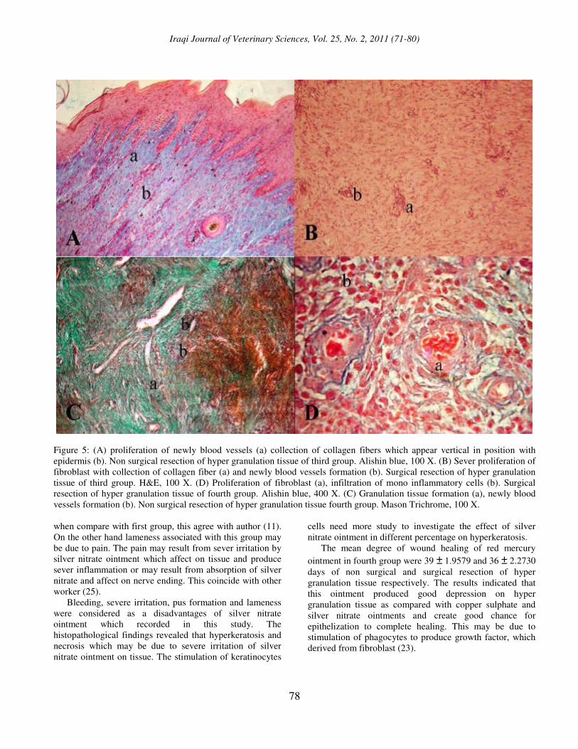
Figure 5: (A) proliferation of newly blood vessels (a) collection of collagen fibers which appear vertical in position with
epidermis (b). Non surgical resection of hyper granulation tissue of third group. Alishin blue, 100 X. (B) Sever proliferation of
fibroblast with collection of collagen fiber (a) and newly blood vessels formation (b). Surgical resection of hyper granulation
tissue of third group. H&E, 100 X. (D) Proliferation of fibroblast (a), infiltration of mono inflammatory cells (b). Surgical
resection of hyper granulation tissue of fourth group. Alishin blue, 400 X. (C) Granulation tissue formation (a), newly blood
vessels formation (b). Non surgical resection of hyper granulation tissue fourth group. Mason Trichrome, 100 X.
when compare with first group, this agree with author (11).
On the other hand lameness associated with this group may
be due to pain. The pain may result from sever irritation by
silver nitrate ointment which affect on tissue and produce
sever inflammation or may result from absorption of silver
nitrate and affect on nerve ending. This coincide with other
worker (25).
Bleeding, severe irritation, pus formation and lameness
were considered as a disadvantages of silver nitrate
ointment which recorded in this study. The
histopathological findings revealed that hyperkeratosis and
necrosis which may be due to severe irritation of silver
nitrate ointment on tissue. The stimulation of keratinocytes
cells need more study to investigate the effect of silver
nitrate ointment in different percentage on hyperkeratosis.
The mean degree of wound healing of red mercury
ointment in fourth group were 39 ± 1.9579 and 36 ± 2.2730
days of non surgical and surgical resection of hyper
granulation tissue respectively. The results indicated that
this ointment produced good depression on hyper
granulation tissue as compared with copper sulphate and
silver nitrate ointments and create good chance for
epithelization to complete healing. This may be due to
stimulation of phagocytes to produce growth factor, which
derived from fibroblast (23).

Iraqi Journal of Veterinary Sciences, Vol. 25, No. 2, 2011 (71-80)
79
Figure 6: (A) Collection of collagen fiber (a), newly blood
vessels formation (b). Non surgical resection of hyper
granulation tissue of laser therapy. Mason Trichrom, 200 X.
(B) Infltration of mononuclear inflammatory cells (a)
through collagen fibers. Surgical resection of hyper
granulation tissue of laser therapy. Mason Trichrom, 400 X.
The results of laser therapy were exhibited that there
was significant difference in between this group and other
groups of this study. The mean degree of wound healing
were 25± 1.6801 and 20± 0.8165 days in non surgical and
surgical resection respectively. The ability of laser to
depress hyper granulation tissue may be due to depression
of growth factor which associated with granulation tissue
formation, beside that the laser may affect on proliferation
of macrophage which contribute to stimulate fibroblast to
produce collagen fiber, this agreed with (26,27).
Laser therapy caused increasing temperature which
change the nature of protein or lead to vaporization of
liquid in the tissue. Laser leads to increase temperature of
water in the intracellular cells which causes burst of cells
(28).
There was no pain in laser therapy, this may be due to
the increasing endorphin, metenkephalin and beta-
endorphin which decrease pain, this agree with (26,27).
Table 3: Show histopatholgical study of all groups and
subgroups.
Groups
Non surgical resection
of hyper granulation
tissue
Surgical resection
of hyper
granulation tissue
Control
Excessive collagen
fibers formation
mixed with sever
mononuclear
inflammatory cells.
Minimal proliferation
of newly blood
vessels (Figure 4A).
Fibroplasias with
dense collagen
fibers. Mild
infiltration of
mononuclear
inflammatory cells.
Sever proliferation
of newly blood
vessels like cluster
(Figure 4B).
Copper
sulphate
Fibroplasias with
infiltration of
inflammatory cells.
Newly blood vessels
formation and
epithelization was
appeared (Figure 4C).
Sever fibroplasias
and newly blood
vessels formation
with infiltration of
mononuclear
inflammatory cells
(Figure 4D).
Silver
nitrate
Sever fibroblastic,
nidus of inflammation
with infiltration of
inflammatory cells.
Sever collection of
collagen fibers and
blood vessels to form
a rod like taken the
vertical position with
epidermal layer
(Figure 5A).
Fibroplasias and
there was nidus of
newly blood
vessels which
surrounded by
inflammatory cells.
Precipitate of
hemosidrin.
Necrosis of
epidermal layer
(Figure 5B).
Red
mercury
Proliferation of
fibroblast and
collection of collagen
fiber. Sever
infiltration of
mononuclear and
multinuclear
inflammatory cells
(Figure 5C).
Sever proliferation of
fibroblast, decrease
the amount of
collagen fibers,
infiltration of
mononuclear cells
around newly blood
vessels (Figure 5D).
Laser
Mature granulation
tissue which mixed
with mononuclear
inflammatory cells.
Formation of newly
blood vessels (Figure
6A).
Collagen fibers
which infiltrated
with mononuclear
inflammatory cells.
Proliferation of
newly blood vessels
(Figure 6B).

Iraqi Journal of Veterinary Sciences, Vol. 25, No. 2, 2011 (71-80)
80
Conclusions
This study indicated that wounds healing of surgical
resection of hyper granulation tissue was faster than non
surgical resection, and the wound healing of distal fore
limbs were slower than distal hind limbs and the dorsal
aspect of distal limbs were slower than lateral aspect of
limbs.
Different caustic materials were used in this study
which lead to depress hyper granulation tissue at varying
level and promote healing at different degrees. The
disadvantage of silver nitrate ointment causes necrosis,
slough, pain and lameness, while red mercury ointment
gave best results when compared with other caustic
materials. In addition to that the literature exhibit that red
mercury ointment not used by other authors in treatment of
hyper granulation tissue in equine.
The laser therapy gets the best results when compare with
other methods used in this study. The literature appeared
that the laser therapy not widely studied in this field, in
addition to that the histopathological study not used by
other worker to document the results specially in laser
therapy of hyper granulation tissue.
References
1. Jacobs KA, Leach DH, Fretz PB, Townend HGG. Comparative
aspects of the healing of excisional wounds on the leg and body of
horses. Vet Surg.1984;13:83-90.
2. Monahan CM, Champman MR, Taylor HW, French DD, Klei TR,
Knottenbelt DC. Equine wound management: Are there significant
differences in healing at different sites on the body. Vet
Dermatol.1997;8(4):273-290.
3. Lindsay WA. Wound treatment in horses: what to know about second
intention healing. Equine pract.1988;10:396-403.
4. Lee AG, Swaim SF. Granulation tissue: how to take advantage of it in
management of open wounds. Como contin Educ pract
Vet.1988;10:163.
5. Stashak TS.Equine wound management.1st ed, Philadelphia, lea and
Febiger.1991; pp:40-44.
6. Bertone AL, Sullins KE, Stashak TS, Norrdin RW. Effect of wound
location and the use of topical collagen gel on exuberant granulation
tissue formation and wound healing in the horse and pony.AMJ Vet
Res. 1985;45(7):1438-1444.
7. Bertone A.L. Management of exuberent granulation tissue. Vet Clinc
North Amer.. Equine Pract 1989; 5 (3) :551-562.
8. Wilmink JM, Stolk PWT, van Weeren PR, Barneveld A. Differences
in second intention wound healing between horses and ponies:
Macroscopic aspects. Equine Vet J.1999;31:53-60.
9. Wilmink JM, Nederbraagt H, van Weeren PR, Stolk PWT, Barneveld
A. Differences in wound contraction between horses and ponies :the in
vitro contraction capacity of fibroblast. Equine Vet J.2001;33:499-
505.
10. Harris B, Eaglstein WH, Falanga V. Basal cell carcinoma arising in
venous ulcers and mimicking granulation tissue. J Dermatol Surg
Oncol. 1993;19(2):150-152.
11. Ovington LG. What to do with too much of a good thing. Cover
wound care.1999;4(4):680-681.
12. Cultar RS.Copper sulphate treatment of proud flesh. Vet
Rec.1973;92(26):711-712.
13. Frank ER. Wounds and infection. In Veterinary surgery 7th ed. Jain
SK.CBS Publishers and distributors. India.1981;pp:47-48.
14. Fretz PB, Martin GS, Jacobs KA. Treatment of exuberant granulation
tissue in the horse: Evaluation of four methods. Vet
Surg.1983;12:137-140.
15. Seim HB. Mechanisms of cold induced cellular death. Vet clinc North
Amer Small Anim Pract.1980;10:755.
16. Barber SM, Caron J. The effect of bandaging on second intention
wound healing in the horse. Vet Surg.1987;16:82.
17. Stashak TS. Free skin grafting in the horses. Vet Clinc North Amer
Large Anim Pract.1984;12:315.
18. Worth M. Low level laser therapy provides new treatment
possibilities. Wound Equine Vet 1998;3(3):7-18.
19. Luna LG. Manual of histologic staining methods of the armed forces
institute of pathology,3rd ed. MeGraw Hill Book Co., Inc,New York,
USA.1968;pp:94-95.
20. Drury RAB, Wallington EA. Carleton's histological techniques 5th ed.
Oxoford university press,London.1980;pp:57-68,140-142.
21. Pearse AGE. Histochemistry theoretical and applied 4th ed. Analytical
technology, Churchill Livingston, Edinburgh. 1985;pp:1,849.
22. Miller RM.Sound and healthy :proud flesh. Western Horseman.
1993;58(4):33-34.
23. Cochrance CA, Freeman KL, Knottenbelt DC. Effect of growth
factors on the characteristics of cells associated with equine wound
healing and sarcoid formation. Wound Rep Reg.1996;4(1):65-85.
24. Bertone AL. Principles of wound healing. Vet Clin North Am :Equine
Pract. 1989 b;5(3):449-463.
25. Sibbald RG, Williamson D, Orsted HL, Campbell K, Keast D, Krasner
D, Sibbald D. Preparing the wound bed-debridement, bacterial balance
and moisture balance. Ostomy wound management. 2000;46(11):14-
35.
26. Ebert DW, Bertone AL, Robert C. Effect of irradiation with a low-
intensity diod laser on the metabolism of equine articular cartilage in
vitro. Am J Vet Res.1998;59(12):1613-1618.
27. Ramey DW, Basford JR. Laser therapy in horse. Compendium
2000;22(3):263-271.
28. Klause SE, Roberts SM. Lasers and Veterinary surgery. Compendium
small Anim.1990;12(11):1565-1576.