viability and growth of mouse embryos after in vitro...
TRANSCRIPT

/. Embryol. exp. Morph. Vol. 23, 3, pp. 693-704, 1970 693 Printed in Great Britain
Viability and growth of mouse embryos after in vitro culture and fusion
By PATRICIA BOWMAN 1 AND A N N E M c L A R E N 2
From the Institute of Animal Genetics, University of Edinburgh
S U M M A R Y
About 80 % of 8-cell mouse eggs developed to the blastocyst stage in culture, whether the zona pellucida was left intact, or removed with pronase (pre-incubated and dialysed) and the eggs then cultured singly or as fused pairs. When pronase was used without prior incubation and dialysis, the success rate was reduced to 50 %.
After transfer to uterine foster-mothers, 20-30 % of apparently normal blastocysts cultured with or without the zona, singly or fused, developed into live foetuses, compared with over 50 % of control blastocysts taken directly from the uterus. Some of the excess mortality of cultured embryos took place before implantation and some soon after.
The foetuses derived from cultured blastocysts averaged 01 g lighter than those derived from control uterine blastocysts similarly transferred. No differences in the weights of the placentae were observed. Foetal and placental weights were unaffected by whether the eggs had been cultured singly or fused, implying that growth regulation of fused embryos is complete by the 17th day of gestation.
The longer the eggs were maintained in culture, the lower was their viability after transfer, and the lighter were the foetuses derived from them.
INTRODUCTION
Culture of mouse embryos from the 2- or 8-cell stage through to the blastocyst stage is today a routine procedure in many laboratories, using a variety of media and culture techniques (see Mintz, 1967a). The capacity of cultured blastocysts for further growth and development after transfer to uterine foster-mothers has, however, been critically assessed on relatively few occasions. The successful development and birth of mice grown from 8-cell stage to blastocyst on chemically defined media was first reported by McLaren & Biggers (1958) : the yield of Jive young was about 20 % from either cultured or control blastocysts. Using a different medium, Biggers, Moore & Whittingham (1965) found that only 7-5 % of embryos cultured from 2-cell stage to blastocyst developed into live foetuses, but gave no control data for their success rate with uterine blastocysts. In a similar but more extensive experiment, Gates (1965) obtained about 50 % of live foetuses from either cultured or control blastocysts when the faster cleaving embryos were selected for transfer, but less than 20 % when the more slowly cleaving embryos were used.
1 Author's address: Department of Genetics, University of Edinburgh, Scotland. 2 Author's address: Agricultural Research Council Unit of Animal Genetics, West Mains
Road, Edinburgh EH9 3JN, Scotland.
694 P . B O W M A N A N D A. M c L A R E N
As far as development in ectopic sites is concerned, the appearance of blastocysts proves a poor criterion on which to assess their developmental potential (Billington, Graham & McLaren, 1968). Different culture media, which supported development from 2- or 8-cell stage to blastocyst equally effectively, differed markedly in the extent to which the resulting blastocysts, after transfer to kidney or testis, gave rise to embryonic tissues rather than merely to trophoblast.
Many experimental manipulations can be carried out on pre-implantation mouse embryos during the period of culture. In particular, the zona pel-lucida can be removed with pronase (Mintz, 1962) and the embryos cultured naked; also two or more embryos can be fused together at the 8-cell stage (Tarkowski, 1961; Mintz, 1964; for review of literature, see McLaren, 1969«). Little is known of the effect of these treatments on viability and growth of embryos either in vitro or after transfer: Mintz (1961b) mentions that 45 % of her fused blastocysts survived at least till birth, while Tarkowski (1961) and Myst-kowska & Tarkowski (1968) report respectively 21 % and 7 % successful development of embryos from fused blastocysts, but neither group give any control data on untreated blastocysts.
The aim of the present work was to compare the viability and post-implan-tational growth rate of control, uterine blastocysts transferred to foster-mothers, with that of blastocysts cultured with the zona intact, without the zona singly, or without the zona and fused. The effect of zona removal and fusion on development in culture was also assessed.
MATERIALS AND METHODS
All mice belonged to the randomly bred Q strain. Eggs were obtained from the oviducts at the 8-cell stage, on the morning of the 3rd day of pregnancy (vaginal plug = 1st day), and unless otherwise stated were maintained in culture for 50-54 h. Control blastocysts were obtained from the uteri of females on the afternoon of the 4th day of pregnancy. Recipient females, mated to vasectomized males, were used on the afternoon of the 3rd day of pseudopregnancy, and were killed on the 17th day (16^ days p.c.).
The variance of foetal and placental weight between females is very high at this stage of gestation, but is relatively low between the two uterine horns of the same female (McLaren, 1965/?). In order to eliminate the maternal contribution to foetal growth as a source of variation, control blastocysts were transferred to one horn and cultured to the other horn of the same female, alternating control and cultured between right and left horns, and all weight comparisons were made between horns within females.
Eight-cell eggs and blastocysts were recovered in phosphate-buffered saline. Culture was in microdrops under liquid paraffin (Brinster, 1963), using the medium of Brinster (1965) except that the concentration of bovine serum
Uterine development of cultured embryos 695
albumin was increased to 3 mg/ml. The incubator was maintained at about 36 °C, and gassed with 10 % C02 in air.
For naked and fused eggs, the zona pellucida was removed by placing the eggs in pronase (05 % in phosphate-buffered saline with 10 mg/ml of polyvinylpyrrolidone) for about 10 min at room temperature. When the zonae were seen to be swelling and thinning, the eggs were removed and rinsed in three changes of culture medium before being put into the culture dishes. Naked and fused eggs were always cultured singly in microdrops, to avoid spontaneous fusion. In our later experiments, the pronase solution was incubated for 2 h at 37 °C and then dialysed against phosphate-buffered saline overnight at 4 °C (Mintz, 1961 a). For fusion, two naked 8-cell eggs were held together for a few seconds with fine forceps at room temperature in drops of culture medium.
The transfer technique was essentially that of McLaren & Michie (1956), modified as described by McLaren (19696). Before transfer, cultured blastocysts were well rinsed in the transfer medium (10 % calf serum in phosphate-buffered saline) so as to prevent any oil being carried over from the culture dishes. The maximum number of eggs transferred to a horn was 8; the average over the whole experiment was 4-5 eggs per horn.
For each recipient, cultured blastocysts were transferred to one uterine horn and blastocysts direct from a donor uterus to the other. The gain in precision when comparisons can be made within the same female far outweighs the slight blurring effect of transmigration of blastocysts between horns. Even with egg transfer, this is unlikely to affect more than a small percentage of blastocysts (McLaren & Michie, 1954).
At autopsy, the implants in each uterine horn were counted, and classified into live foetuses, middle and late deaths and early deaths ('moles'). Each live foetus and its placenta was separately weighed on a Cahn Gram electrobalance to the nearest 1 mg and 0-1 mg respectively; some placentae were then left for 96 h in an oven at 150 °C and dry weights taken to the nearest 001 mg.
To assess the significance of differences in blastocyst viability between groups, for each recipient the percentage of transferred blastocysts developing into foetuses was subjected to angular transformation, and an analysis of variance carried out. To analyse the effect of culture on the growth of foetuses and placentae, the mean foetal and placental weight was first calculated for each uterine horn separately, and the difference between the two horns found (control minus cultured). For this part of the analysis only females bearing at least one live foetus in each horn could be used. A weighted analysis of variance was performed on the horn differences, using a weighting factor L x R\(L + R), whereLandjRare the number of live foetuses in the left and right horn respectively. This procedure takes account of the fact that estimates of difference are more reliable when they are based on larger litters and litters more evenly distributed between horns.
696 P. BOWMAN AND A. McLAREN
RESULTS
Viability of embryos in vitro
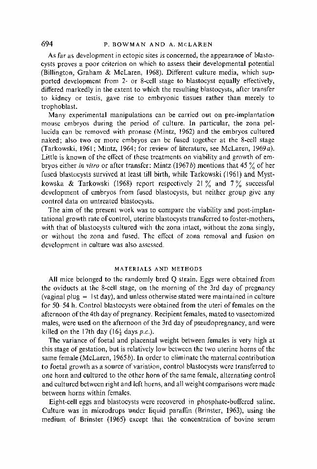
Of the 584 8-cell eggs cultured in intact zonae during the experimental period (8 months), 408 developed into blastocysts, giving an overall success rate of 69-8 %. For those occasions on which naked or fused eggs were also cultured, the control success rate was rather higher, over 80 % (Table 1). When untreated pronase was used, the percentage of blastocysts developing from naked eggs, single or fused, was only about 50 %, significantly below the control level; but prior incubation and dialysis (see Material and Methods section) abolished the toxic effect of the pronase and all three groups then yielded a very similar percentage of blastocysts.
Table 1. The effect of removing the zona, with or without subsequent fusion, on the development of 8-cell eggs to blastocysts in vitro
Conditions of culture
No. of eggs
cultured
Blastocysts A.
Significance of difference
from control* Treatment Conditions of culture
No. of eggs
cultured 1
No. / o
Significance of difference
from control*
Pronase untreated
Pronase pre-incubated and dialysed
In zona Naked Fused
In zona Naked Fused
134 148 86
144 115 J 23
116 75 43
120 98
100
86-6 50-7 500
83-3 85-2 81-3
P < 0001 P < 0001
P > 0-2 P > 0-2
* To take account of heterogeneity between culture days, the significance levels were assessed by the log odds method of McLaren (1952), using only data from those days when naked (or fused) and control (in zona) eggs had been cultured, and weighting each estimate by the reciprocal of its variance to give a combined estimate of the proportional change in the odds.
Viability of embryos after transfer
Eight per cent of our recipient females had no implants in either horn. We observe a similar failure rate among females mated to fertile males; the nonpregnant females have therefore been excluded from the analysis.
Most of the remaining variation in success rate of egg transfer is due to the operation itself, rather than to the reproductive efficiency of the recipients. There is therefore little to be gained by comparing the results of transfers of experimental and control eggs to the two uterine horns of individual females, unlike the situation in the analysis of foetal and placental weight. The uterine horn has therefore been taken as the unit of analysis, and a weighted regression analysis carried out. The three groups of control, uterine blastocyst transfers, corresponding to transfer of'in zona,' 'naked' or 'fused' blastocysts into the opposite
Uterine development of cultured embryos 697
horn, gave very similar results and have been combined. There was also no significant difference in the viability of blastocysts derived from pronase-treated eggs, according to whether or not the pronase had been pre-incubated and dialysed : that is, the toxic effect of the pronase appears to be an ' all-or-none ' effect, either killing the eggs before the blastocyst stage or leaving them unharmed. These groups have therefore also been combined.
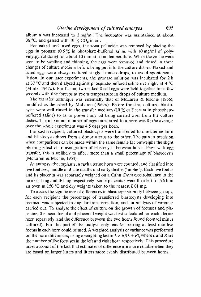
Table 2. The effect of culture, with or without the zona, singly or fused, on the viability of mouse embryos after transfer to uterine foster-mothers
No. of
Implants A
No. of ( Middle No. of eggs and Significance of
re trans late / o difference Blastocysts cipients ferred Live deaths Moles live from uterine*
Uterine 72 328 184 7 43 56-1 — Cultured 35 180 53 2 43 29-4 P < 0001
in zona Cultured 14 57 12 0 10 21 1 P < 0001 naked
Cultured 23 93 29 f 3 18 31-2 P < 0001 fused
* To take account of heterogeneity between transfer operations, the percentage of transferred eggs developing into live foetuses for each operation was subjected to angular transformation and an analysis of variance carried out, weighting by the number of eggs transferred.
t Of the 20 fused embryos that were sexed, 14 were male and only 6 female. The predominance of phenotypic males is characteristic of chimaeras (McLaren & Bowman, 1969).
The results are set out in Table 2. The percentage of live foetuses developing from cultured blastocysts did not differ significantly among eggs cultured with or without the zona, singly or fused; on the other hand the decrease in viability of cultured blastocysts compared with control, uterine blastocysts was highly significant (P < 0-001 for each of the three cultured groups).
The proportion of total implantation sites which consisted of 'moles' (i.e. decidua unaccompanied by any macroscopically visible embryonic remnant) was significantly higher for all three groups of transferred blastocysts than for uterine blastocysts (Table 2). The increased number of moles was not, however, sufficient entirely to account for the deficit of live embryos developing from cultured blastocysts. This suggests that some of the cultured blastocysts failed to elicit a decidual reaction after transfer, while others induced a decidual reaction in the recipient uterus, but died soon after implantation. In addition, some of the moles must represent deciduomata induced by the transfer operation itself, since on one or two occasions the number of moles observed in the uterine horn at autopsy was greater than the number of blastocysts transferred. For this reason success rates have been calculated in terms of live foetuses rather than total implantation sites.
45 E M B 23

698 P. BOWMAN AND A. McLAREN
Foetal and placental weight
Because of the large variation between females in foetal and placental weight, only those pregnancies were considered in which there was at least one live foetus in each horn. This meant that the growth of cultured and control embryos could be compared within the same female. Mean foetal and placental weights for each uterine horn were first calculated. Foetuses developed from uterine blastocysts were heavier than those from cultured blastocysts in all three groups
Fig. 1. Mean weights of foetuses and placentae (wet and dry). Each group derived from cultured blastocysts (cultured in zona, naked or fused) is compared with its own control group, derived from uterine blastocysts transferred to the opposite horn of the same female. Numbers of recipient females and eggs transferred in each group are given in Table 3. £?, Control; CD, Cultured.
(Fig. 1). In absolute foetal weight, the 'in zona' and 'fused' groups resembled one another very closely, both for control and for cultured values, while the seven females in the 'naked' group had heavier foetuses. Placental weight, both wet and dry, was similar in all three groups and did not appear to be affected by the previous history of the transferred blastocysts.
In order to assess the significance of the difference in weight of foetuses derived from control and cultured blastocysts, the control-minus-cultured difference between the two uterine horn means was calculated for each female. A weighted analysis of variance was carried out on these differences, as described
Uterine development of cultured embryos 699
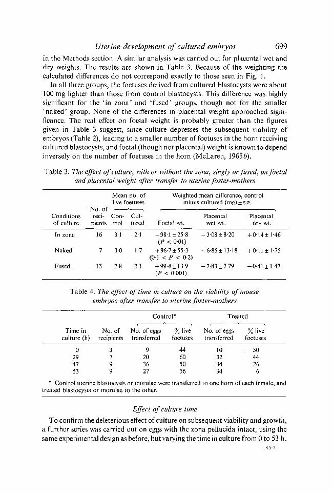
in the Methods section. A similar analysis was carried out for placental wet and dry weights. The results are shown in Table 3. Because of the weighting the calculated differences do not correspond exactly to those seen in Fig. 1.
In all three groups, the foetuses derived from cultured blastocysts were about 100 mg lighter than those from control blastocysts. This difference was highly significant for the 'in zona' and 'fused' groups, though not for the smaller 'naked' group. None of the differences in placental weight approached significance. The real effect on foetal weight is probably greater than the figures given in Table 3 suggest, since culture depresses the subsequent viability of embryos (Table 2), leading to a smaller number of foetuses in the horn receiving cultured blastocysts, and foetal (though not placental) weight is known to depend inversely on the number of foetuses in the horn (McLaren, 19656).
Table 3. The effect of culture, with or without the zona, singly or fused, on foetal and placental weight after transfer to uterine foster-mothers
No. of reci
pients
Mean no. of live foetuses / * *
Con- Cul-trol tured
Weighted minus
mean difference, control cultured (mg)±s.E.
Conditions of culture
No. of reci
pients
Mean no. of live foetuses / * *
Con- Cul-trol tured Foetal wt.
Placental wet wt.
Placental dry wt.
In zona
Naked
Fused
16
7
13
3-1
30
2-8
2-1
1-7
2-1
+ 98-1 ±25-8 (P < 001)
+ 96-7±55-3 (O-J < P < 0-2)
+ 99-4 ± 13-9 (P < 0001)
- 3 0 8 ±8-20
-6-85 ±13-18
-7-83 ±7-79
+ 0-14±l-46
+ 0-11 ±1-75
-0-41 ±1-47
Table 4. The effect of time in culture on the viability of mouse embryos after transfer to uterine foster-mothers
Control* Treated
Time in No. of No. of eggs % live culture (h) recipients transferred foetuses
No. of eggs transferred
% I've foetuses
0 29 47 53
9 20 36 27
44 60 50 56
10 32 34 34
50 44 26 6
* Control uterine blastocysts or morulae were transferred to one horn of each female, and treated blastocysts or morulae to the other.
Effect of culture time
To confirm the deleterious effect of culture on subsequent viability and growth, a further series was carried out on eggs with the zona pellucida intact, using the same experimental design as before, but varying the time in culture from 0 to 53 h.
45-2
700 P. BOWMAN AND A. McLAREN
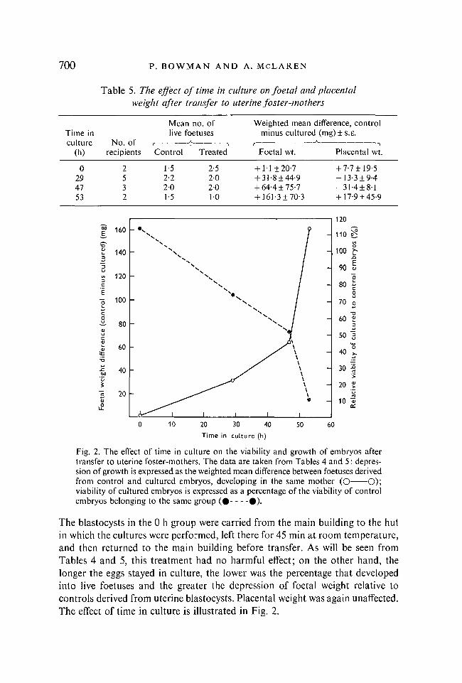
Table 5. The effect of time in culture on foetal and placental weight after transfer to uterine foster-mothers
Mean no. of Weighted mean difference, control Time in live foetuses minus cultured (mg) ± S.E. culture No. of culture No. of f
(h) recipients Control Treated Foetal wt. Placental wt.
0 2 1-5 2-5 + 1-1 ±20-7 +7-7±J9-5 29 5 2-2 2 0 + 31-8 ±44-9 -13-3 ±9-4 47 3 2 0 2 0 + 64-4 ±75-7 - 3 1 -4 ±8-1 53 2 1-5 10 + 161-3 ±70-3 +17-9 ±45-9
120 'M E, 160 - no e
V T3 o 0) 1_ 140 - s 100 >r 3 - O
V,
90 « "5 U
N - 90 « in 120 - V "Ö 3 C N. 80 « E c
0 "5 100 70 g
v w v. C N -a
(CO
80 _ N 60 e 3
<u N . w
u c
50 3 Ol
60 _ > " \ \ „ o i t \ 40 x
\ *-» X I \ 'JZ
ight
40 yS \ \ 30 |
> <u \ S \ \ — 20 J 2 20 \ 'Z3
% e — 10 "5 £ CÙ
0 10 20 30 40 50 60
Time in culture (h)
Fig. 2. The effect of time in culture on the viability and growth of embryos after transfer to uterine foster-mothers. The data are taken from Tables 4 and 5: depression of growth is expressed as the weighted mean difference between foetuses derived from control and cultured embryos, developing in the same mother (O O); viability of cultured embryos is expressed as a percentage of the viability of control embryos belonging to the same group ( • #) .
The blastocysts in the 0 h group were carried from the main building to the hut in which the cultures were performed, left there for 45 min at room temperature, and then returned to the main building before transfer. As will be seen from Tables 4 and 5, this treatment had no harmful effect; on the other hand, the longer the eggs stayed in culture, the lower was the percentage that developed into live foetuses and the greater the depression of foetal weight relative to controls derived from uterine blastocysts. Placental weight was again unaffected. The effect of time in culture is illustrated in Fig. 2.
Uterine development of cultured embryos 701
DISCUSSION
Although all the blastocysts that we transferred to uterine foster-mothers looked normal, the viability of those which had been cultured for two days in vitro was not much more than half that of blastocysts taken directly from the uterus. This finding is consistent with the observation of Billington etat. (1968), that some culture media which would allow the development of a high proportion of eggs into blastocysts of grossly normal appearance were nonetheless inadequate to permit subsequent embryonic development after transfer to an ectopic site. The culture medium used in the present experiment, on the other hand, was one of those that allowed the development of blastocysts which were not only normal in appearance, but also were indistinguishable from uterine blastocysts in their developmental potential on transfer to the kidney (Billington et al. 1968). Nonetheless, on transfer to the uterus, known to be a more exacting environment (McLaren, 1965Ö), their viability proved to be greatly reduced.
It seems, therefore, that the relative merits of different culture media for mammalian embryos cannot be fully assessed without recourse to the uterus.
Our experiment on varying the time in culture (see Fig. 2) suggests either that the effect of the suboptimal culture medium is cumulative, or that it exerts a more drastic effect upon older embryos, i.e. that blastocysts are more susceptible than are 8-cell stages or morulae.
McLaren & Biggers (1958) found no significant effect of previous culture on the viability of transferred blastocysts: but the numbers were small, especially in the control series, and the overall success rate following transfer was low. They found, as we did, that the embryos derived from cultured blastocysts weighed less on average than those from control blastocysts: on the small numbers available, the difference proved not to be significant but, in the light of the present findings, it was probably a real effect of culture.
Gates (1965) reported that 'fast-cleaving' eggs developed equally well whether cultured or not, and 'slow-cleaving' eggs developed equally poorly; but without further information on the proportion of fast- and slow-cleaving eggs in the cultured and control groups respectively a direct comparison of his data with our own is not possible.
In the present experiment removal of the zona with pronase, subsequent culture of the eggs in the zona-free condition, and the further manipulations required to produce fusion, had no additional harmful effects upon the eggs over and above those associated with culture. The lack of toxicity of the pronase makes an interesting contrast to the finding of McLaren (1969 b), that removal of the zona at the blastocyst stage with a crude pronase solution markedly reduced the viability of the embryos on transfer. The difference is probably due to the treatment of the pronase rather than the stage of the eggs, since Bronson & McLaren (1970) found that removal of the blastocyst zona with pronase before
702 P. BOWMAN AND A. McLAREN
transfer to the oviduct was lethal if crude pronase was used, but harmless if the enzyme had been pre-incubated and dialysed.
The mouse uterus, with its two more or less discrete horns, is admirably suited to provide within-female comparisons of the weight of foetuses derived from treated and control eggs. As an example of the efficiency of such an experimental design, in the present work we would have needed to carry out ten times as many transfer operations to achieve the same level of statistical significance if cultured and control blastocysts had been transferred to different females.
Why should culture during the pre-implantation stage consistently depress foetal but not placental weight? One possible interpretation is that the embryo-forming inner cell mass is more sensitive to suboptimal conditions of culture than are the trophoblast cells which give rise to the placenta. In support of this is the observation (Billington et al. 1968) that mouse eggs cultured to blastocyst stage in certain media and then transferred to the testis give rise to vigorous trophoblastic outgrowths but no embryonic tissue, while other media permit subsequent development of both embryo and trophoblast. Alternatively, it may be that the effect of culture is not to depress growth directly, but merely to delay development. At the stage of pregnancy at which our autopsies were performed, placental weight has reached a plateau but foetal weight is increasing rapidly : a delay of a few hours would therefore be expected to reduce foetal weight while leaving placental weight unaffected.
Our most striking finding is of the very exact growth regulation that fused embryos and their placentae have achieved by 16 - days p.c. At the time of transfer, fused blastocysts contain twice as many cells as do single cultured blastocysts (Patricia Bowman, unpublished observations); yet the degree to which the mean weight of the foetuses derived from cultured blastocysts fell short of that of the controls was virtually the same (see Table 3), whether the blastocysts were fused or single. As far as we know, this is the first quantitative demonstration of growth regulation after fusion, though Tarkowski (1961) reported that fused embryos examined on the 10th—14th day of gestation were 'within the range of normal size variation', and Mintz (1968) states : ' Within a few days after implantation, size regulation occurs, yielding embryos which are once again of normal size.'
When the converse experiment was performed, and one blastomere destroyed at the 2-cell stage, embryonic size continued to be approximately halved until about the 10th day of gestation but, by full term, regulation was complete and birth weight was normal (Tarkowski, 1959). Placental size was not examined.
Although the foetuses developed from fused blastocysts showed no pheno-typic evidence of chimaerism, since all eggs were derived from the same strain, the following lines of evidence suggest that, in most if not all cases, fusion was complete: (1) The eggs were examined carefully during the period of culture, and any in which fusion appeared to be incomplete were discarded. (2) The resulting foetuses showed a predominantly male sex ratio (see footnote to Table 2),

Uterine development of cultured embryos 703 as is characteristic of chimaeras. (3) A parallel series of fusions between eggs of contrasted genotypes yielded young, most of which showed overt chimaerism (McLaren & Bowman, 1969).
RÉSUMÉ Viabilité et croissance d'embryons de souris ayant fusionné en culture in vitro
Environ 80 % des œufs de souris de 8 cellules se sont développés en culture jusqu'au stade blastocyste, que la zone pellucide ait été laissée intacte ou enlevée avec de la pronase (préincubée et dialysée), et que les œufs aient été cultivés alors isolés ou fusionnés par paires. Quand on utilise la pronase avant l'incubation et la dialyse, les proportions de succès sont réduites à 50 %.
Après transfert dans des utérus de mères 'adoptives', 20 à 30% de blastocystes apparemment normaux cultivés avec ou sans la zone, séparément ou par paires, évoluent en fœtus vivants, en comparaison de plus de 50 % de blastocystes témoins prélevés directement dans les utérus. La plus grande mortalité des embryons cultivés se produit en partie avant, en partie aussitôt après l'implantation.
Les fœtus provenant de blastocystes cultivés sont en moyenne plus légers de 0,1 g que ceux prélevés de blastocystes témoins transplantés de la même façon. On n'a observé aucune différence dans le poids des placentas. Les poids des fœtus et des placentas ne montrent pas de différence, que les œufs aient été cultivés séparément ou par couples, ce qui implique que la régulation de croissance des œufs fusionnés est complète au 17ème jour de la gestation.
Plus les œufs ont été maintenus en culture, moins est grande leur viabilité après transplantation, et plus les foetus qui en dérivent sont légers.
We are grateful to the Ford Foundation for financial support.
REFERENCES
BIGGERS, J. D., MOORE, B. D. &WHITTINGHAM,D. G. (1965). Development of mouse embryos in vivo after cultivation from 2-cell ova to blastocysts in vitro. Nature, Lond. 206, 734-5.
BILLINGTON, W. D., GRAHAM, C. F. & MCLAREN, A. (1968). Extra-uterine development of mouse blastocysts cultured in vitro from early cleavage stages. / . Embryo!, exp. Morph. 20, 391-400.
BRINSTER, R. L. (1963). A method for in vitro cultivation of mouse ova from 2-cell to blastocyst. Expl Cell Res. 32, 205-8.
BRINSTER, R. L. (1965). Studies on the development of mouse embryos in vitro. IV. Interaction of energy sources. / . Reprod. Fert. 10, 227-40.
BRONSON, R. & MCLAREN, A. (1970). Transfer to the mouse oviduct of eggs with and without the zona pellucida. J. Reprod. Fert. 22. (in the press.)
GATES, A. H. (1965). Rate of ovular development as a factor in embryonic survival. In Reimplantation Stages of Pregnancy. Ed. G. E. W. Wolstenholme and M. O'Connor. London: Churchill.
MCLAREN, A. (1952). Section 4.12 in Factors affecting Susceptibility to Neurotropic Viruses in Rodents. D.Phil. Thesis, Oxford University.
MCLAREN, A. (1965 A). Maternal factors in nidation. In Symp. on The Early Conceptus, Normal and Abnormal. Ed. W. W. Park. Edinburgh and London: Livingstone.
MCLAREN, A. (19656). Genetic and environmental effects on foetal and placental growth in mice. / . Reprod. Fert. 9, 79-98.
MCLAREN, A. (1969ö). Recent studies on developmental regulation in Vertebrates. In Handbook of Molecular Cytology. Ed. A. Lima-de-Faria, pp. 639-55. Amsterdam: North Holland Publishing Co.
MCLAREN, A. (1969/)). Transfer of zona-free mouse eggs to uterine foster-mothers. J. Reprod. Fert. 19, 341-6.
MCLAREN, A. & BIGGERS, J. D. (1958). Successful development and birth of mice cultivated in vitro as early embryos. Nature, Lond. 182, 877-8.
704 P. B O W M A N A N D A. M c L A R E N
MCLAREN, A. & BOWMAN, P. (1969). Mouse chimaeras derived from fusion of embryos differing by nine genetic factors. Nature, Lond. 21A, 238-40.
MCLAREN, A. & MICHIE, D. (1954). Transmigration of unborn mice. Nature, Lond. 174, 844. MCLAREN, A. & MICHIE, D. (1956). Studies on the transfer of fertilized mouse eggs to
uterine foster-mothers. I. Factors affecting the implantation and survival of native and transferred eggs. / . exp. Biol. 33, 394-416.
MINTZ, B. (1962). Experimental study of the developing mammalian egg: removal of the zona pellucida. Science, N. Y. 138, 594-5.
MINTZ, B. (1964). Formation of genetically mosaic mouse embryos, and early development of 'lethal (t12/t12)-normaV mosaics. / . exp. Zool. 157, 273-92.
MINTZ, B. (1967a). Mammalian embryo culture. In Methods in Developmental Biology, Ed. F. H. Wilt & N. K. Wessells. New York: T. Y. Crowell.
MINTZ, B. (19676). Gene control of mammalian pigmentary differentiation. I. Clonal origin of melanocytes. Proc. natn. Acad. Sei. U.S.A. 58, 344-51.
MINTZ, B. (1968). Hermaphroditism, sex chromosomal mosaicism and germ cell selection in allophenic mice. VIII Bienn. Symp. Anim. Reprod., J. Anim. Sei. 27, Suppl. 1, 51-60.
MYSTKOWSKA, E. T. & TARKOWSKI, A. K. (1968). Observations on CBA-p/CBA-T6T 6 mouse chimeras. / . Embryo!, exp. Morph. 20, 33-52.
TARKOWSKI, A. K. (1959). Experiments on the transplantation of ova in mice. Acta theriol. 2, 251-66.
TARKOWSKI, A. K. (1961). Mouse chimaeras developed from fused eggs. Nature, Lond. 190, 857-60.
(Manuscript received 9 July 1969)