volume 2 number 11 november 2014 pages 1537–1716
TRANSCRIPT

Biomaterials Science
www.rsc.org/biomaterialsscience
ISSN 2047-4830
PAPERLisa A. Sawicki and April M. Kloxin Design of thiol–ene photoclick hydrogels using facile techniques for cell culture applications
Themed issue: Stem cell–materials interactions
Volume 2 Number 11 November 2014 Pages 1537–1716
BiomaterialsScience
PAPER
Cite this: Biomater. Sci., 2014, 2, 1612
Received 27th May 2014,Accepted 14th August 2014
DOI: 10.1039/c4bm00187g
www.rsc.org/biomaterialsscience
Design of thiol–ene photoclick hydrogels usingfacile techniques for cell culture applications†
Lisa A. Sawickia and April M. Kloxin*a,b
Thiol–ene ‘click’ chemistries have been widely used in biomaterials applications, including drug delivery,
tissue engineering, and controlled cell culture, owing to their rapid, cytocompatible, and often orthogonal
reactivity. In particular, hydrogel-based biomaterials formed by photoinitiated thiol–ene reactions afford
spatiotemporal control over the biochemical and biomechanical properties of the network for creating
synthetic materials that mimic the extracellular matrix or enable controlled drug release. However, the use
of charged peptides functionalized with cysteines, which can form disulfides prior to reaction, and vinyl
monomers that require multistep syntheses and contain ester bonds, may lead to undesired inhomogen-
eity or degradation under cell culture conditions. Here, we designed a thiol–ene hydrogel formed by the
reaction of allyloxycarbonyl-functionalized peptides and thiol-functionalized poly(ethylene glycol).
Hydrogels were polymerized by free radical initiation under cytocompatible doses of long wavelength
ultraviolet light in the presence of water-soluble photoinitiators (lithium acylphosphinate, LAP, and 2-
hydroxy-1-[4-(2-hydroxyethoxy)phenyl]-2-methyl-1-propanone, Irgacure 2959). Mechanical properties
of these hydrogels were controlled by varying the monomer concentration to mimic a range of soft tissue
environments, and hydrogel stability in cell culture medium was observed over weeks. Patterns of bio-
chemical cues were created within the hydrogels post-formation and confirmed through the incorpor-
ation of fluorescently-labeled peptides and Ellman’s assay to detect free thiols. Human mesenchymal
stem cells remained viable after encapsulation and subsequent photopatterning, demonstrating the utility
of the monomers and hydrogels for three-dimensional cell culture. This facile approach enables the for-
mation and characterization of hydrogels with well-defined, spatially-specific properties and expands the
suite of monomers available for three-dimensional cell culture and other biological applications.
Introduction
Click chemistries for the formation and modification of bio-materials have garnered significant and growing interest fornumerous applications, including drug delivery, tissue engin-eering, and controlled cell culture.1,2 A number of functionalgroups undergo efficient and highly selective click reactionsunder a variety of cytocompatible conditions, making themwell suited for the manipulation of biomaterial properties inthe presence of cells.3,4 These reactions include radicallyinitiated thiol–ene and thiol–yne,5,6 thiol-Michael addition,7,8
spontaneous reaction of azides with strained alkynes,9,10 andspontaneous reaction of tetrazine with norbornene and trans-
cyclooctene,11,12 which have been used to examine the effectsof matrix properties on cell behavior,6,7,9,11 to label cells andbiomolecules,10,12 and to form carriers for drug delivery.13
Amongst these, thiol–ene click chemistries have been exam-ined broadly for the formation and modification of hydrogel-based biomaterials owing to their ease of use and the avail-ability of thiols in many biomolecules.14
Hydrogels formed by thiol–ene click reactions have beenconstructed with a range of cytocompatible polymers andcopolymers, such as poly(ethylene glycol) (PEG),15 hyaluronicacid,16 and poly(ethylene glycol)–poly(lactic acid),17 and modi-fied with peptides and proteins, such as GPQG↓IWGQ,18
IPVS↓LRSG,18 and RGDS,19 to impart specific biologicalactivity.16,20 Various vinyl functional groups have been investi-gated for this purpose, including norbornene,19 vinyl sulfone,8
and allyl ether.21 For example, the Michael-type addition ofthiols on peptides with vinyl groups (‘ene’s) on vinyl sulfone-modified PEG has been widely employed to design hydrogelswith controlled, cell-responsive properties for use in drugdelivery or tissue engineering.8,22 These reactions proceed via
†Electronic supplementary information (ESI) available. See DOI: 10.1039/c4bm00187g
aDepartment of Chemical and Biomolecular Engineering, University of Delaware,
Newark, DE 19716, USA. E-mail: [email protected] of Materials Science and Engineering, University of Delaware, Newark,
DE 19716, USA
1612 | Biomater. Sci., 2014, 2, 1612–1626 This journal is © The Royal Society of Chemistry 2014
Ope
n A
cces
s A
rtic
le. P
ublis
hed
on 0
1 Se
ptem
ber
2014
. Dow
nloa
ded
on 1
2/1/
2021
7:4
6:41
AM
. T
his
artic
le is
lice
nsed
und
er a
Cre
ativ
e C
omm
ons
Attr
ibut
ion
3.0
Unp
orte
d L
icen
ce.
View Article OnlineView Journal | View Issue
a step growth mechanism,5,14 resulting in a homogeneousnetwork structure with robust mechanical properties for appli-cations in cell culture and delivery.23
Photoinitiated thiol–ene systems are particularly attractivefor hydrogel formation and modification because they allowuser-directed control over the presentation of biophysical orbiochemical cues in space and in time to promote specific cel-lular functions and toward mimicking the dynamic structureor composition of the native extracellular matrix (ECM)in vitro.24,25 Peptides modified with cysteines and polymersmodified with acrylates (mixed step and chain growth mechan-ism) or norbornenes (step growth mechanism) have beenextensively used owing to their rapid reaction under cyto-compatible photopolymerization conditions.19,26,27 For example,Fairbanks et al. first demonstrated that norbornene-modifiedPEG reacts within minutes with cysteine-modified, enzymati-cally degradable crosslinking peptides in the presence of aradical initiator to form hydrogels by step growth free radicalpolymerization.19 This strategy (vinyl-modified PEG) has beenused to encapsulate a number of cell types including, but notlimited, to osteoblasts, chondrocytes, mesenchymal stem cells(MSCs), and smooth muscle cells.28 These chemistries alsohave been used to create new biomaterial systems, such as ahydrogel formed by the reaction of norbornene-modified hya-luronic acid with a dithiol crosslinker and modified with pat-terns of biochemical cues at select time points.16
Despite their great utility, there are a few potential concernswhen using these existing thiol–ene photoclick systems.Recently, Shih and Lin observed that ester bonds present inpolymers modified with various vinyl groups (e.g., acrylic acidor norbornene carboxylic acid) degrade over relatively shorttimes in water or cell culture conditions (i.e., days to weeks),where the hydrolysis rate is affected by the incorporation ofdifferent charged peptide sequences.29 Preprogrammed degra-dation afforded by hydrolysis allows cell spreading within thematrix; however, it is often desirable for the rate of degradationto respond dynamically to cell secreted enzymes or an exter-nally-applied stimulus (e.g., light). Toward designing alternatesystems with controlled degradation (e.g., cell-secretedenzymes or light), polymer precursors modified with aminefunctional groups instead of hydroxyls have been utilized,introducing more water-stable amide bonds upon reactionwith carboxylic acid-containing functional groups.30,31 Despitethis increased stability, there typically is increased cost or syn-thetic processing associated with using these materials.Additionally, the formation of disulfide bonds betweencysteine-modified charged peptides32 before reaction maydeplete the concentration of thiols present in the reactionsolution, resulting in an off-stoichiometry mixture, defects inthe network structure, and slower polymerization times.33,34
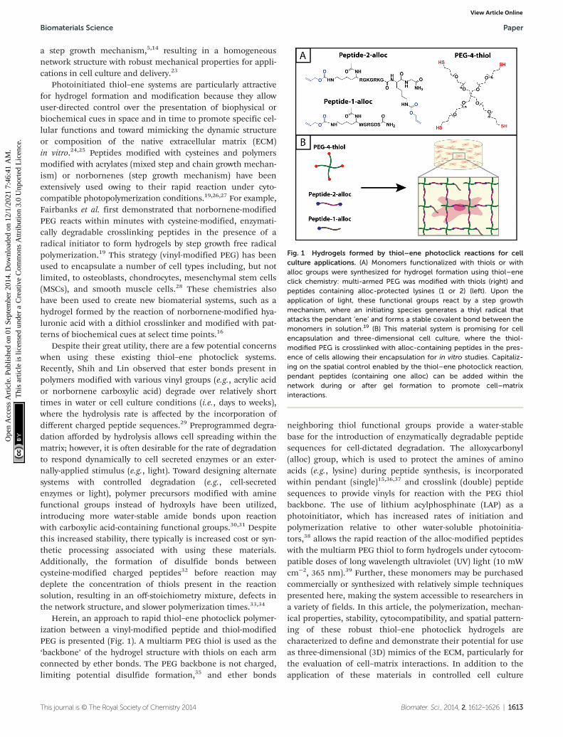
Herein, an approach to rapid thiol–ene photoclick polymer-ization between a vinyl-modified peptide and thiol-modifiedPEG is presented (Fig. 1). A multiarm PEG thiol is used as the‘backbone’ of the hydrogel structure with thiols on each armconnected by ether bonds. The PEG backbone is not charged,limiting potential disulfide formation,35 and ether bonds
neighboring thiol functional groups provide a water-stablebase for the introduction of enzymatically degradable peptidesequences for cell-dictated degradation. The alloxycarbonyl(alloc) group, which is used to protect the amines of aminoacids (e.g., lysine) during peptide synthesis, is incorporatedwithin pendant (single)15,36,37 and crosslink (double) peptidesequences to provide vinyls for reaction with the PEG thiolbackbone. The use of lithium acylphosphinate (LAP) as aphotoinitiator, which has increased rates of initiation andpolymerization relative to other water-soluble photoinitia-tors,38 allows the rapid reaction of the alloc-modified peptideswith the multiarm PEG thiol to form hydrogels under cytocom-patible doses of long wavelength ultraviolet (UV) light (10 mWcm−2, 365 nm).39 Further, these monomers may be purchasedcommercially or synthesized with relatively simple techniquespresented here, making the system accessible to researchers ina variety of fields. In this article, the polymerization, mechan-ical properties, stability, cytocompatibility, and spatial pattern-ing of these robust thiol–ene photoclick hydrogels arecharacterized to define and demonstrate their potential for useas three-dimensional (3D) mimics of the ECM, particularly forthe evaluation of cell–matrix interactions. In addition to theapplication of these materials in controlled cell culture
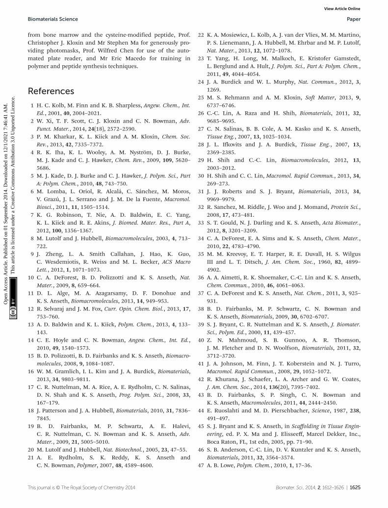
Fig. 1 Hydrogels formed by thiol–ene photoclick reactions for cellculture applications. (A) Monomers functionalized with thiols or withalloc groups were synthesized for hydrogel formation using thiol–eneclick chemistry: multi-armed PEG was modified with thiols (right) andpeptides containing alloc-protected lysines (1 or 2) (left). Upon theapplication of light, these functional groups react by a step growthmechanism, where an initiating species generates a thiyl radical thatattacks the pendant ‘ene’ and forms a stable covalent bond between themonomers in solution.19 (B) This material system is promising for cellencapsulation and three-dimensional cell culture, where the thiol-modified PEG is crosslinked with alloc-containing peptides in the pres-ence of cells allowing their encapsulation for in vitro studies. Capitaliz-ing on the spatial control enabled by the thiol–ene photoclick reaction,pendant peptides (containing one alloc) can be added within thenetwork during or after gel formation to promote cell–matrixinteractions.
Biomaterials Science Paper
This journal is © The Royal Society of Chemistry 2014 Biomater. Sci., 2014, 2, 1612–1626 | 1613
Ope
n A
cces
s A
rtic
le. P
ublis
hed
on 0
1 Se
ptem
ber
2014
. Dow
nloa
ded
on 1
2/1/
2021
7:4
6:41
AM
. T
his
artic
le is
lice
nsed
und
er a
Cre
ativ
e C
omm
ons
Attr
ibut
ion
3.0
Unp
orte
d L
icen
ce.
View Article Online
models, we believe this approach may be useful for the in situmodification of assembling peptides (e.g., adding functional-ities to supramolecular structures to allow electrical conduc-tion, enhance imaging, or promote specific biologicalinteractions)40,41 and even in membrane applications (e.g.,forming stable, charged PEG-based membranes forbatteries).42
Materials and methodsSynthesis of PEG-thiol macromer
Poly(ethylene glycol)-tetrathiol (PEG4SH) is commercially avail-able (JenKem Technology USA, Creative PEGWorks) or canbe synthesized as was done here using a modified version ofpublished protocols.43 Briefly, four-arm PEG (Mn ∼ 20 000 gmol−1, 10 g) (JenKem USA) was dissolved in anhydrous tetra-hydrofuran (THF, 70 mL) (Fisher Scientific) and purged withargon, and argon-purged sodium hydroxide (NaH, 4× molarexcess with respect to –OH groups) (Sigma Aldrich) suspendedin THF was transferred via cannula under argon to the dis-solved PEG. Allyl bromide (3× molar excess with respect to–OH groups) (Acros Organics) dissolved in 30 mL of THF sub-sequently was added. The PEG-allyl solution was refluxed over-night at 40 °C under argon and precipitated in ice cold ethylether to generate allyl ether-modified PEG (PEG4AE). ThePEG4AE was dissolved in dichloromethane (40 mL) (FisherScientific) with a photoinitiator (2,2-dimethoxy-1,2-diphenyl-ethan-1-one, I651, 0.5% w/w) (Acros Organics) and trace tri-fluoroacetic acid (TFA, ∼100 μL) (Acros Organics) and purgedwith argon. Thioacetic acid (2× molar excess with respect toallyl) (Acros Organics) was added, and the solution was purgedwith argon and subsequently exposed to UV light (365 nm at10–15 mW cm−2 for 45 minutes) to yield PEG-thioacetate(PEG4TA) after precipitation in ice cold diethyl ether. Last,PEG4TA was dissolved in 60–70 mL of water and purged withargon. An equal volume of 1 M sodium hydroxide (FisherScientific) purged with argon was added to the PEG4TA (0.5 Mfinal concentration) to generate the thiol end groups on thefinal PEG4SH product. The reaction immediately was neutral-ized with hydrochloric acid (final pH 1–2) (Fisher Scientific)and PEG4SH extracted with chloroform and trace TFA (toprevent disulfide formation) and precipitated in ice colddiethyl ether. To wash and collect all intermediates and thefinal product after precipitation, samples were centrifuged at0 °C for 20 minutes at 4400 rpm for a total of 3 washes anddessicated under vacuum at room temperature overnight. Allintermediates and the final product were characterized withproton nuclear magnetic resonance (1H NMR) in DMSO:PEG4AE 5.1–5.2 (m, 1H) 5.2–5.3 (m, 1H) 5.8–5.9 (m, 1H);PEG4TA 2.3 (s, 3H); PEG4SH 2.3 (m, 1H) for a single arm ofthe tetrafunctional monomer (ESI Fig. S1†).
Synthesis of alloc-functionalized peptides
The pendant cell adhesion sequence K(alloc)GWGRGDS(RGDS), a ubiquitous sequence found in many ECM proteins
including fibronectin and vitronectin,44 was synthesized topromote cell adhesion (amino acid(s) with reactive functionalgroups in bold). Non-degradable, water-soluble crosslinkingsequences were synthesized: K(alloc)RGKGRKGK(alloc)G37
(RGKGRK2alloc) (primary sequence used in hydrogel develop-ment) and K(alloc)GKGWGKGK(alloc)G (GKGWGKG2alloc)and CGKGWGKGCG (GKGWGKG2SH) (sequences withreduced charge and including tryptophan for easily assessingtheir concentration). Additionally, an enzymatically degrad-able, water-soluble crosslinking sequence KK(alloc)-GGPQG↓IWGQGK(alloc)K (GPQGIWGQ2alloc) (broadly degrad-able by matrix metalloproteinases (MMP)-1, 2, 3, 8 and 9)18
was synthesized to promote cell viability and allow spreadingin longer cell culture and photopatterning experiments. Eachwas synthesized by standard solid phase peptide synthesis(SPPS) techniques using Fmoc chemistry on MBHA rink amideresin (0.59 mmol g−1; 0.25 mmol scale) (Novabiochem) with apeptide synthesizer (Protein Technologies PS3). Fmoc-pro-tected amino acids, including the commercially-availablealloc-protected lysine, and o-(benzotriazol-1-yl)-N,N,N′,N′-tetra-methyluronium hexafluorophosphate (HBTU) (4× excess)(Chem-Impex International) were loaded into cartridges andcoupled on resin. Fmoc deprotection was carried out using20% piperidine (Sigma Aldrich) in N,N-dimethylformamide(DMF) (Fisher Scientific) prior to each amino acid coupling in0.4 M methylmorpholine in DMF. Peptide products werecleaved in 95% v/v trifluoroacetic acid (TFA), 2.5% v/v triisopro-pylsilane (TIPS) (Acros Organics), and 2.5% v/v water with 5%w/v dithiothreitol (DTT) (Research Products InternationalCorporation) to prevent disulfide formation and 2.5% w/v phenol(Sigma Aldrich) to protect tryptophan (W). After cleavage fromthe resin, peptides were precipitated in ice cold diethyl ether,centrifuged at 3000 rpm and 4 °C for 5 minutes for a total ofthree washes and dessicated under vacuum overnight at roomtemperature. Dry raw peptide product was purified by high-performance liquid chromatography (HPLC) and analyzed bymatrix-assisted laser desorption/ionization (MALDI, crystal-lized with α-cyano-4-hydroxycinnamic acid, Acros Organics) orelectrospray ionization (ESI) mass spectrometry to confirm syn-thesis of each desired peptide (ESI Fig. S2†).
A fluorescently-labeled pendant peptide, Alexa Fluor 488-AhxWGRGDSK(alloc)G (AF488RGDS), also was designed forphotopatterning experiments using published protocols.37
After Fmoc deprotection of Ahx on the N′-terminus of thepeptide, 1 mg Alexa Fluor® 488 Carboxylic Acid, 2,3,5,6-Tetra-fluorophenyl Ester, 5-isomer (Invitrogen) was stirred with0.25 mmol peptide on resin in 4 mL DMF and 50 μL N,N′-diiso-propylethylamine (DIPEA) (Chem-Impex International) over-night. The peptide was cleaved from resin, precipitated, andanalyzed by HPLC and ESI mass spectrometry (ESI Fig. S2†).
Synthesis of LAP initiator
The LAP initiator was synthesized using previously-describedmethods.38 Briefly, 2,4,8-trimethylbenzoyl chloride (1.6 g,0.009 mol) (Sigma Aldrich) was added to dimethyl phenylphos-phonite (1.5 g, 0.009 mol) (Acros Organics) and reacted over-
Paper Biomaterials Science
1614 | Biomater. Sci., 2014, 2, 1612–1626 This journal is © The Royal Society of Chemistry 2014
Ope
n A
cces
s A
rtic
le. P
ublis
hed
on 0
1 Se
ptem
ber
2014
. Dow
nloa
ded
on 1
2/1/
2021
7:4
6:41
AM
. T
his
artic
le is
lice
nsed
und
er a
Cre
ativ
e C
omm
ons
Attr
ibut
ion
3.0
Unp
orte
d L
icen
ce.
View Article Online
night at room temperature under argon. Lithium bromide (4×molar excess) (Sigma Aldrich) in 2-butanone (Sigma Aldrich)was added to the reaction solution and heated to 50 °C for10 minutes. The white precipitate was filtered and rinsed3 times with 2-butanone, and the final powder product driedand analyzed by 1H NMR, matching literature (ESI Fig. S3†).38
Hydrogel formation
All monomers and initiators were prepared in Dulbecco’sphosphate buffered saline (PBS) (Life Technologies) immedi-ately before polymerization. For the various experimentsdescribed below, solutions of PEG4SH, RGKGRK2alloc (unlessnoted otherwise), and RGDS (7.5, 10, 12.5 wt% with respect toPEG, 2 mM RGDS) were prepared at stoichiometric ratios ofthiol functional groups to alloc functional groups (1 : 1 SH :alloc) and containing a photoinitiator, either LAP (1.1 and2.2 mM) or Irgacure 2959 (I2959) (2.2 mM). Hydrogels wereformed upon irradiation of the monomer-initiator solutionwith cytocompatible doses of long wavelength UV light(365 nm at 10 mW cm−2, International Light IL1400A Radio-meter/Photometer) in the specific geometries described below.
Rheometry
Hydrogels were formed in situ on a photorheometer (TA AR-G2with UV light attachment, Exfo Omnicure Series 2000 lightsource, 365 nm filter, SilverLine UV Radiometer M007-153) toestimate the polymerization times for different initiator typesand monomer concentrations. I2959 (2.2 mM) or LAP (1.1 or2.2 mM) photoinitiators were added to 10 wt% PEG monomersolutions containing stoichiometrically balanced amounts(1 : 1 SH : alloc) of RGKGRK2alloc to compare the effects ofinitiator type on polymerization time (n = 3). PEG monomersolutions (7.5, 10, and 12.5 wt%) containing stoichiometricallybalanced amounts of RGKGRK2alloc and RGDS (2 mM) weremixed with 2.2 mM LAP to compare the effects of monomerconcentration on polymerization time (n = 6). Finally, PEG4SHor PEG4AE monomer solutions (10 wt%) containing stoichio-metrically balanced amounts of alloc (RGKGRK2alloc,GKGWGKG2alloc, GPQGIWGQ2alloc) or thiol-modified cross-linkers (PEG2SH, GKGWGKG2SH) were mixed with 2.2 mMLAP to compare the effects of crosslinker and functional groupchemistry on polymerization time (n = 3). These solutions wereplaced between parallel plates (8 mm diameter, 200 μm gap)and UV light (365 nm at 10 mW cm−2) applied 1 minute afterstarting rheometric measurements. Storage (G′) and lossmoduli (G″) were recorded over time at 2% applied strain and6 rad s−1 frequency. From the data, an approximate time forcomplete gelation was defined to be when the percent changein modulus between consecutive data points was less than0.1%.
For swollen modulus experiments, 7.5, 10, and 12.5 wt%hydrogels were polymerized within a 1 mm thick mold(2 microscope slides treated with Rain-X separated by a 1 mmrubber gasket). After polymerization, discs (8 mm diameter)were punched from the gel slab and swollen overnight in PBS.Strain sweeps (1 rad s−1 frequency, 1–100% strain) and
frequency sweeps (1–100 rad s−1 frequency, 5% strain) wereconducted on swollen gels to determine the linear viscoelasticregime for the material. The swollen gels were then placedbetween parallel plates on the rheometer and G′ and G″ weremeasured at 5% strain and 5 rad s−1 frequency (within thelinear viscoelastic regime) (n = 6).
Hydrogel swelling
Experiments to determine volumetric swelling ratios (Q) wereperformed on 7.5, 10, and 12.5 wt% hydrogels. Discs (8 mmdiameter) were punched from gels polymerized between glassslides separated by a 1 mm thick gasket, ensuring sufficientmass for measuring dry weight, and swollen overnight in PBS.After recording swollen mass (Ms), the gels were lyophilizedand the dry masses were measured (Md) (n = 6). Volumetricswelling ratio was calculated by the relationships:
q ¼ Ms
Md; Q ¼ 1þ ρpolymer
ρsolventðq� 1Þ
where q is the mass swelling ratio, ρpolymer = 1.07 g mL−1 45 forPEG, and ρsolvent = 1.00 g mL−1 for PBS.
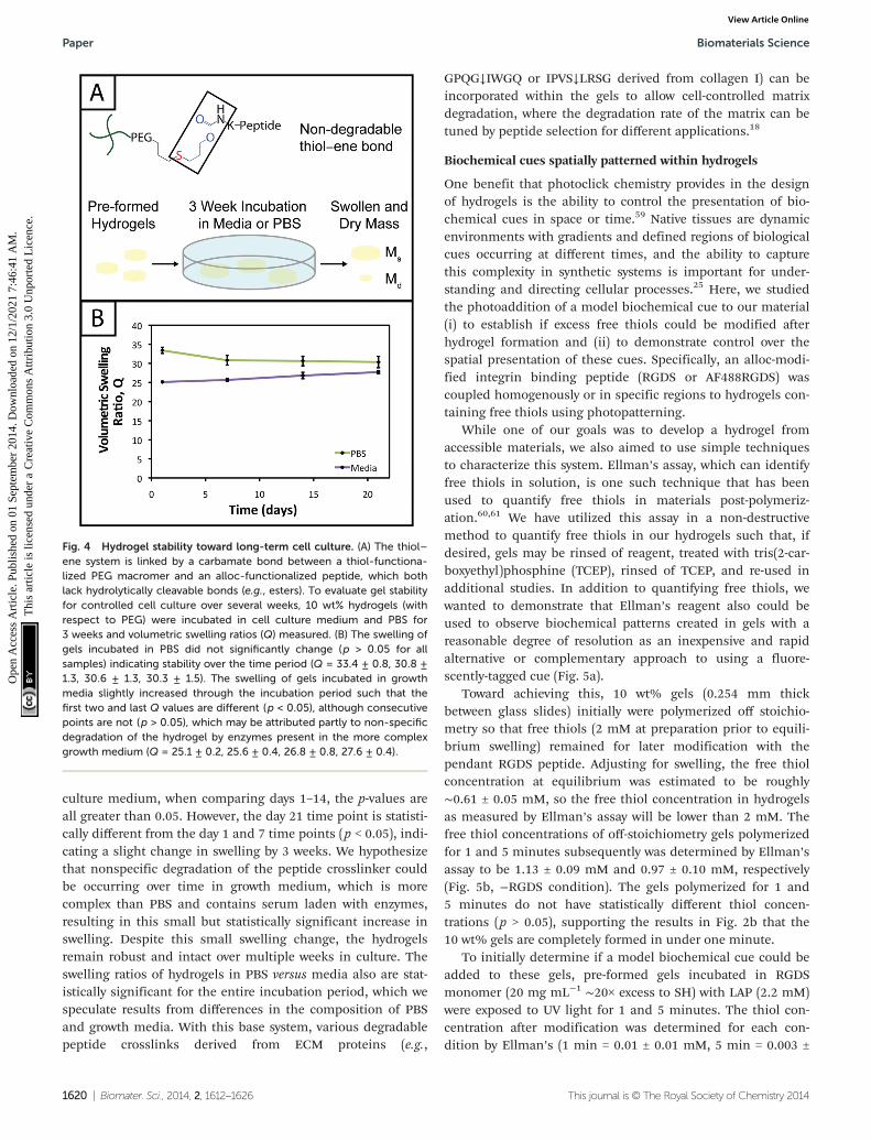
Experiments to determine gel stability after polymerizationwere performed on gels incubated in PBS and cell culturemedium at 37 °C over a 3 week time course. Gels (10 wt%)were polymerized for 5 minutes in 5 mm diameter molds(1 mL syringes with tips cut off ) under sterile conditions andplaced in sterile PBS and cell culture medium. Ms and Md wererecorded for the gels after 1, 7, 14, and 21 days (n = 6). Valuesfor the volumetric swelling ratio (Q) were calculated asdescribed above.
Detection of unreacted thiols
To initially quantify the photoaddition of biochemical cues,hydrogels (10 wt% with respect to PEG) were polymerized (1 or5 minutes) between glass slides separated by a 0.254 mm thickgasket (McMaster-Carr) and off-stoichiometry such thatapproximately 2 mM free thiol remained in the unswollen gelafter polymerization. Discs (5 mm diameter) were punchedfrom these gels for further treatment and analysis. Half of thegel discs were swollen in PBS containing LAP initiator(2.2 mM) and excess pendant peptide (20 mg mL−1, K(alloc)-GWGRGDS) and incubated at room temperature for 1 hour.After 1 hour, these gels were exposed to UV light for 1 or5 minutes to initiate the photoaddition of the RGDS. Theother half of the gels remained in PBS as a control. Free thiolconcentrations in the gels were quantitatively detected byEllman’s assay as described below.
Briefly, the swollen volume of the gels was predicted usingthe measured Q value (estimated at 19.3 μL). Ellman’s reactionbuffer (20.7 μL) containing 0.1 M sodium phosphate (SigmaAldrich) and 1 mM ethylenediaminetetraacetic acid (SigmaAldrich) at pH 7.5–8 was added to the gels for a total volume of40 μL. Ellman’s reagent (7.2 μL, 4 mg in 1 mL reaction buffer)(Fisher Scientific) was diluted in 360 μL of reaction buffer andadded to each well containing a gel. Gels were incubated inthe reagent for 1 hour and 30 minutes, the estimated time for
Biomaterials Science Paper
This journal is © The Royal Society of Chemistry 2014 Biomater. Sci., 2014, 2, 1612–1626 | 1615
Ope
n A
cces
s A
rtic
le. P
ublis
hed
on 0
1 Se
ptem
ber
2014
. Dow
nloa
ded
on 1
2/1/
2021
7:4
6:41
AM
. T
his
artic
le is
lice
nsed
und
er a
Cre
ativ
e C
omm
ons
Attr
ibut
ion
3.0
Unp
orte
d L
icen
ce.
View Article Online
the diffusion of the yellow NTB2− dianion out of the gel so thatthe supernatant and gel colors match (by visual inspection).Finally, a calibration curve of L-cysteine hydrochloride mono-hydrate (Sigma Aldrich) (0–2 mM) was made to calculate theconcentration of thiols detected in each gel. Absorbance ofeach condition was measured at 405 nM (Biotek Synergy H4automated plate reader).
To determine the free thiol concentration in conditions forphotopatterning in the presence of encapsulated cells, 10 wt%gels were polymerized in syringe tips (20 μL) such that approxi-mately 2 mM free thiol remained in the unswollen gel afterpolymerization. Gels polymerized for 1 and 5 minutes wereplaced immediately in PBS as a control (n = 3). Additional gelspolymerized for 1 minute immediately were placed in solu-tions of PBS containing 3 mg mL−1 RGDS and 2.2 mM LAPand incubated at 37 °C for 30 minutes (n = 3) or 1 hour30 minutes (n = 3). After incubation, these gels were exposedto a second dose of UV light for 1 minute to attach the bio-chemical cue (RGDS) to remaining free thiols. Free thiol con-centrations in the gels were quantitatively detected by Ellman’sassay as described above, accounting for larger gel size(swollen volume = 84.8 μL; add 15.2 μL of PBS to gel in wellplate for 100 μL total volume; add 18 μL Ellman’s reagent in900 μL Ellman’s buffer to each well).
Spatially-specific photopatterning of biochemical cues
Hydrogels (10 wt%) were polymerized between glass slidesspaced by a 0.254 mm gasket and off-stoichiometry to have afinal free thiol concentration of 2 mM within the as preparedgel (prior to equilibrium swelling). The hydrogel was left onone of the glass slides for subsequent treatments and rinsedwith PBS for 1 hour. Rinsed gels were placed in solution con-taining pendant peptides (AF488RGDS or RGDS) mixed with2.2 mM LAP initiator for 1 hour and 30 minutes to allowdiffusion of the peptides and initiator into the gel networkprior to subsequent patterning. Photomasks with lines ofincreasing thickness (0.2–1 mm width) or square patterns(0.4 mm edge) purchased from Advanced Reproductions Cor-poration were placed ink-side down on top of the samples andexposed to collimated UV light (Inpro Technologies collimat-ing adaptor, Exfo Omnicure Series 2000 light source) for1 minute (365 nm at 10 mW cm−2). Gels were rinsed 3× for40 minutes each with PBS to remove excess pendant peptideafter photoaddition. Samples containing the patternedAF488RGDS were imaged with a confocal microscope (Zeiss510 NLO). Ellman’s reagent was applied to the gels containingRGDS and imaged immediately on a stereomicroscope (ZeissStemi 2000-C).
Culture and encapsulation of human mesenchymal stem cells
Human mesenchymal stem cells (hMSCs) isolated fromhuman bone marrow (Lonza)46 were cultured on tissue-culturetreated polystyrene in cell culture medium46 and harvested at∼70–80% confluency (Passage 2, 3) for experiments. For evalu-ating the effects of light, cells were trypsinized from cultureplates, counted (hemacytometer), centrifuged (5 minutes,
1000 rpm), and plated at a density of 20 000 cells cm−2 in96-well plates. For cell encapsulation and photopatterningexperiments, cells were trypsinized from culture plates,counted (hemocytometer), centrifuged (5 minutes, 1000 rpm),and resuspended at desired densities in monomer solution(10 wt%) with and without RGDS. The mixtures of cells inmonomer solution were polymerized in syringe molds at cyto-compatible wavelengths and doses of UV light (365 nm at10 mW cm−2), encapsulating cells within the hydrogel matrix.
Metabolic activity of hMSCs in photopatterned and non-patterned hydrogels
Cells were suspended in monomer solution (10 wt%, 3000cells μL−1) containing 2 mM RGDS and polymerized in syringetip molds (20 μL) for 1 and 5 minutes (n = 6, non-patterned).Immediately after polymerization, gels were placed in cellculture medium to rinse out unreacted monomer and photo-initiator (30 minutes). After rinsing, the medium was replacedwith fresh medium and gels were incubated at 37 °C for sub-sequent analysis. For photopatterned gels, cells were sus-pended in monomer solution (10 wt%, 3000 cells μL−1)without RGDS and polymerized for 1 minute such that 2 mMfree thiols remained in the unswollen gel for subsequentmodification. After polymerization, the gels were incubated inPBS containing 3 mg mL−1 RGDS and 2.2 mM LAP for30 minutes or 1 hour 30 minutes at 37 °C before exposure to asecond dose of UV light (1 minute) to covalently link RGDSwithin the network (n = 6). Patterned gels were immediatelyplaced in cell culture medium (30 minutes) to rinse out excessmonomer and photoinitiator. At 1 and 3 days post-encapsula-tion (D1 and D3), metabolic activity was assessed by CellTiter96 (Promega) (n = 3 each condition, each time point).
To assess the effect of light alone on cell function, platedcells (20 000 cells cm−2) were exposed to UV light (1 min of365 nm at 10 mW cm−2). Metabolic activity was assessed byCellTiter 96 at D1 and D3 compared to control (no light) (n = 3each condition, each time point).
Viability of hMSCs in photopatterned and non-patternedhydrogels
To initially study the viability of cells encapsulated in hydro-gels, 3000 cells μL−1 were encapsulated in non-degradable gels(10 wt%, 2 mM RGDS before swelling) polymerized for 1 and5 minutes. Additional studies were performed to determinethe effect of cell density on viability post-encapsulation, withcells encapsulated in non-degradable gels (10 wt%, 2 mMRGDS before swelling) at 3000 and 30 000 cells μL−1. Viabilitywas quantified at 3 days post-encapsulation with a LIVE/DEAD® Viability/Cytotoxicity Kit for mammalian cells (Invitro-gen), and gels were imaged with a confocal microscope (Zeiss510 NLO).
To study the viability of cells in photopatterned hydrogelsover longer times in culture, cells were encapsulated in gels(10 wt%, 20 μL, 3000 cells μL−1) crosslinked with the degrad-able (GPQGIWGQ2alloc) peptide sequence such that 2 mMfree thiol remained in unswollen gels post-polymerization
Paper Biomaterials Science
1616 | Biomater. Sci., 2014, 2, 1612–1626 This journal is © The Royal Society of Chemistry 2014
Ope
n A
cces
s A
rtic
le. P
ublis
hed
on 0
1 Se
ptem
ber
2014
. Dow
nloa
ded
on 1
2/1/
2021
7:4
6:41
AM
. T
his
artic
le is
lice
nsed
und
er a
Cre
ativ
e C
omm
ons
Attr
ibut
ion
3.0
Unp
orte
d L
icen
ce.
View Article Online
(1 minute). Gels were placed in PBS containing 3 mg mL−1
RGDS and 2.2 mM LAP for 1 hour and exposed to a seconddose of UV light (1 minute) to allow attachment of RGDS tothe network. Viability was assessed 6 days after encapsulationwith the LIVE/DEAD® Viability/Cytotoxicity Kit for mammaliancells, providing time for hMSCs to partially degrade and attachto the hydrogel matrix.
Results and discussion
Click chemistries for hydrogel formation are of interest inmany biomaterials applications. Their efficient reactionsunder mild conditions enable hydrogel formation and modifi-cation in the presence of proteins and cells,3,28 which isespecially useful for designing materials that mimic nativetissue environments in vitro for cell culture. Light-mediatedthiol–ene click reactions in particular are of great utility forcontrol over the presentation of biomechanical and biochemi-cal cues in space and time within these systems. Here, wedescribe a new approach to utilizing thiol–ene chemistry forhydrogel formation and spatially-specific patterning in cellculture applications with alloc-functionalized peptides andthiol-terminated PEG. This strategy enables rapid and consist-ent polymerization of hydrogels controlled by the applicationof light, the formation of a stable bioinert base matrix, and thespatial presentation of biochemical cues within the hydrogelnetwork.
Initiator selection allows rapid polymerization undercytocompatible conditions
Thiol–ene reactions for biomaterial applications can occurspontaneously in aqueous solutions in the presence of a basecatalyst or upon the introduction of free radicals, dependingon vinyl group selection.14,47 For example, base-catalyzedpolymerization of hydrogels in the presence of cells byMichael-type addition reactions between thiols and vinyl sul-fones or maleimides has been used to understand cell behav-ior, invasion, and differentiation in synthetic mimics of theECM.7,22 Additionally, for control over when and where thereaction takes place, polymerizations of hydrogels by a photo-initiated, free radical step growth reaction between thiols andvinyls (e.g., norbornene) have been used with cytocompatibledoses of UV or visible light depending on initiator selection(e.g., Irgacure 2959,39 lithium acylphosphinate,38 or Eosin Y30).While the spatiotemporal control afforded by photo-polymerization is quite useful, minimizing exposure to light,particularly wavelengths in the UV, is crucial for polymeriz-ations done in the presence of cells.39,48 Light-mediated reac-tion conditions that are cytocompatible and rapid for thepolymerization of monomers in aqueous solutions often arelimited and are needed to reduce the exposure time of cellsand proteins to light and reactive components (particularlyfree radicals). Toward addressing this, we aimed to establishconditions for the photopolymerization of monomers functio-
nalized with thiols and allocs to expand the suite of reactionsfor cell encapsulation.
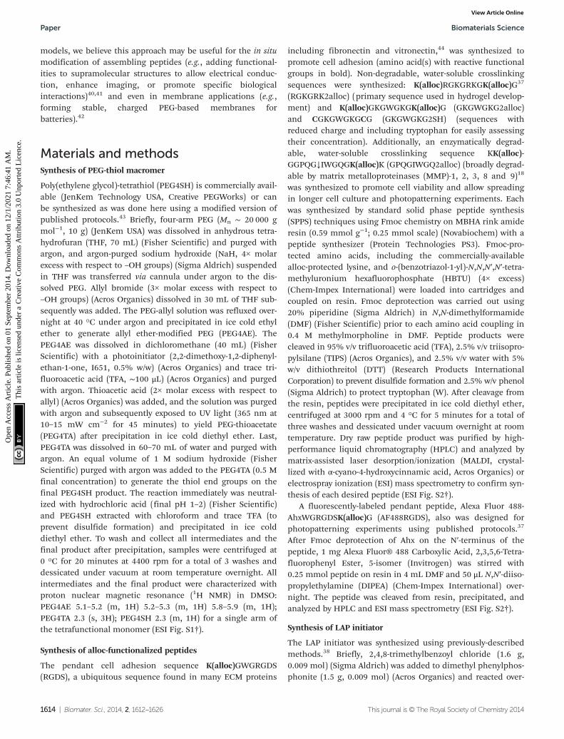
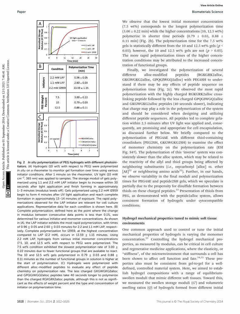
Previously, the general reaction of allyl- and thiol-functiona-lized monomers for hydrogel formation was considered tooslow for gel formation in the presence of cells, which may bedue to a rate-limiting chain transfer step,49 and has beendescribed with limited use in cell culture applications for themodification of synthetic hydrogel matrices with pendantalloc-modified peptide tethers.15,37 Here, we examined water-soluble initiator and monomer compositions to identify cyto-compatible conditions for alloc-based hydrogel formation.Hydrogels were polymerized in situ on a rheometer to monitorpolymerization times of gels formed with different water-soluble photoinitiators (LAP and I2959) and initial monomerconcentrations (7.5, 10, 12.5 wt% with respect to PEG). Twoinitiator concentrations were selected (1.1 and 2.2 mM) tomatch concentrations that have been used to polymerize othertypes of hydrogels in the presence of cells,39 as cell viabilitypreviously has been observed to be sensitive to the concen-tration of LAP owing to robust free radical generation withirradiation at 365 nm.38
The rheological data collected by in situ polymerization ofhydrogels demonstrates the efficiency of the LAP initiator forthe radical reaction of thiol with vinyl functional groups. Theslope of the moduli over time for the 1.1 and 2.2 mM LAP con-ditions becomes approximately 0 after complete gelation,whereas the I2959 continues to slowly increase (slope = 1.5 to5 Pa s−1) (Fig. 2a) indicating a less rapid reaction. While pres-entation of hydrogel moduli (y-axis) on a log scale is typical,we have chosen to present moduli on an absolute (normalized)scale to demonstrate the efficiency of the LAP initiator inachieving complete gelation when compared directly to I2959.Further, the polymerization times of the gels formed using 1.1and 2.2 mM LAP were determined to be approximately 5 and15 times faster than those using I2959 as the initiator (2.60 ±0.03 and 0.96 ± 0.05 min, respectively, vs. 13.59 ± 1.15 min)(Fig. 2b). This order of magnitude difference in polymerizationtime is comparable to differences observed between LAP andI2959 in the polymerization of other functional groups, suchas the chain growth polymerization of PEG-diacrylate with LAP(10 times faster than with I2959),38 and arises from theincreased absorbance of and radical generation by LAP relativeto I2959 at long wavelengths of UV light (365 nm). Movingforward, we focused on the 2.2 mM LAP polymerization con-dition, which provided the most rapid gel formation. However,the 1.1 mM LAP condition may be attractive for investigationsin the future for specific cell culture applications as higherinitiator concentrations can result in lower cell viability.39
In addition to comparing the effect of different initiatingconditions on polymerization rates, the concentration ofmonomers initially present also must be considered. The avail-ability of terminal functional groups for reaction influencesthe time to complete gelation, especially at low concentrationswhere the distance between functional groups is greater and,after reaction of one end group, can decrease the probability ofreaction with a functional group on a different monomer.50
Biomaterials Science Paper
This journal is © The Royal Society of Chemistry 2014 Biomater. Sci., 2014, 2, 1612–1626 | 1617
Ope
n A
cces
s A
rtic
le. P
ublis
hed
on 0
1 Se
ptem
ber
2014
. Dow
nloa
ded
on 1
2/1/
2021
7:4
6:41
AM
. T
his
artic
le is
lice
nsed
und
er a
Cre
ativ
e C
omm
ons
Attr
ibut
ion
3.0
Unp
orte
d L
icen
ce.
View Article Online
We observe that the lowest initial monomer concentration(7.5 wt%) corresponds to the longest polymerization time(3.00 ± 0.22 min) while the higher concentrations (10, 12.5 wt%)polymerize in shorter time periods (0.79 ± 0.03, 0.88 ±0.11 min) (Fig. 2b). The polymerization time for the 7.5 wt%gels is statistically different from the 10 and 12.5 wt% gels (p <0.05); however, the 10 and 12.5 wt% gels are not (p > 0.05).The more rapid polymerization times of the higher concen-tration conditions may be attributed to the increased concen-tration of functional groups.
Finally, we investigated the polymerization of severaldifferent alloc-modified peptides (RGKGRK2alloc,GKGWGKG2alloc, GPQGIWGQ2alloc) with PEG4SH to under-stand if there may be any effects of peptide sequence onpolymerization time (Fig. 2c). We observed the most rapidpolymerization with the highly charged RGKGRK2alloc cross-linking peptide followed by the less charged GPQGIWGQ2allocand GKGWGKG2alloc peptides (40 seconds slower), indicatingthat charge may play a role in the polymerization of the systemand should be considered when designing and utilizingdifferent peptide sequences. All peptides led to complete gela-tion within 2.5 minutes after UV light was applied and, conse-quently, are promising and appropriate for cell encapsulation,as discussed further below. We briefly compared to thepolymerization of PEG4AE with different thiol-containingcrosslinkers (PEG2SH, GKGWGKG2SH) to examine the effectof monomer chemistry on the polymerization rate (ESIFig. S4†). The polymerization of this ‘inverse’ system was con-sistently slower than the alloc system, which may be related tothe reactivity of the allyl and thiol groups being affected byneighboring substituents (i.e., oxycarbonyl [alloc] vs. ether[AE]51 or neighboring amino acids52). Further, in our hands,we observe variability in the final moduli and polymerizationtimes for PEG4AE and peptide2SH gels, which we speculate ispartially due to the propensity for disulfide formation betweenthiols on these charged peptides.32 Presentation of thiols fromPEG, as demonstrated with the peptide2alloc system, allowsconsistent formation of hydrogels under cytocompatibleconditions.
Hydrogel mechanical properties tuned to mimic soft tissueenvironments
One common approach used to control or tune the initialmechanical properties of hydrogels is varying the monomerconcentration.53 Controlling the hydrogel mechanical pro-perties, as measured by modulus, can be critical in cell cultureand regenerative medicine applications, where the elasticity, or“stiffness”, of the microenvironment that surrounds a cell hasbeen shown to affect cell function and fate.54,55 These pro-perties also must be consistent from gel-to-gel for a well-defined, controlled material system. Here, we aimed to estab-lish hydrogel compositions with a range of equilibrium-swollen moduli that mimic different soft tissues. Toward this,we measured the swollen storage moduli (G′) and volumetricswelling ratios (Q) of hydrogels formed from different initial
Fig. 2 In situ polymerization of PEG hydrogels with different photoini-tiators. (A) Hydrogels (10 wt% with respect to PEG) were polymerizedin situ on a rheometer to monitor gel formation over time using variousinitiator conditions. After 1 minute on the rheometer, UV light (10 mWcm−2, 365 nm) was applied to samples. The storage moduli of gels poly-merized using 1.1 and 2.2 mM LAP initiator begin to increase within 30seconds after light application and finish forming in approximately1–3 minutes (modulus levels off ). Gels polymerized using 2.2 mM I2959begin to form 4 minutes after UV light application and reach completeformation in approximately 13–14 minutes of exposure. The rapid poly-merizations observed for the LAP initiator are relevant for cell cultureapplications. Representative data for each condition is shown here. (B)Complete polymerization, defined here as the point where the changein modulus between consecutive data points is less than 0.1%, wasdetermined for various initiator and monomer concentrations. As shownin (A), the LAP initiator exhibits the most rapid polymerization, with timesof 0.96 ± 0.05 and 2.60 ± 0.03 minutes for 2.2 and 1.1 mM LAP, respect-ively. Complete polymerization for I2959, at the highest concentrationcompared to LAP (2.2 mM), occurs in 13.59 ± 1.15 minutes. Using2.2 mM LAP, hydrogels from various initial monomer concentrations(7.5, 10, and 12.5 wt% with respect to PEG) were polymerized. The7.5 wt% condition exhibited the slowest polymerization rate of 3.00 ±0.22 minutes due to fewer functional groups that are available to react.The 10 and 12.5 wt% gels polymerized in 0.79 ± 0.03 and 0.88 ±0.11 minutes as the number of functional groups in solution is higher atthe start of polymerization. (C) Hydrogels were polymerized withdifferent alloc-modified peptides to evaluate any effect of peptidechemistry on polymerization rate. The less charged GKGWGKG2allocand GPQGIWGQ2alloc peptides take 40 seconds longer to polymerizethan the charged RGKGRK2alloc peptide, although this is not as signifi-cant as the effects of weight percent and the type and concentration ofinitiator on polymerization time.
Paper Biomaterials Science
1618 | Biomater. Sci., 2014, 2, 1612–1626 This journal is © The Royal Society of Chemistry 2014
Ope
n A
cces
s A
rtic
le. P
ublis
hed
on 0
1 Se
ptem
ber
2014
. Dow
nloa
ded
on 1
2/1/
2021
7:4
6:41
AM
. T
his
artic
le is
lice
nsed
und
er a
Cre
ativ
e C
omm
ons
Attr
ibut
ion
3.0
Unp
orte
d L
icen
ce.
View Article Online
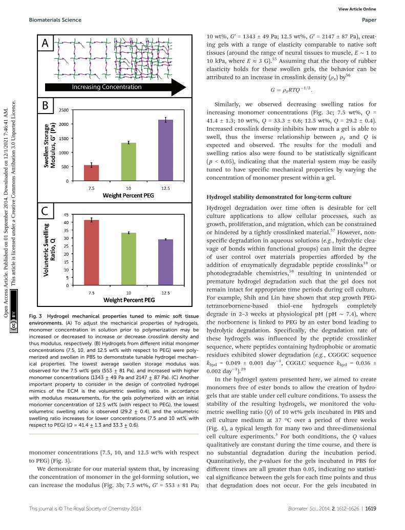
monomer concentrations (7.5, 10, and 12.5 wt% with respectto PEG) (Fig. 3).
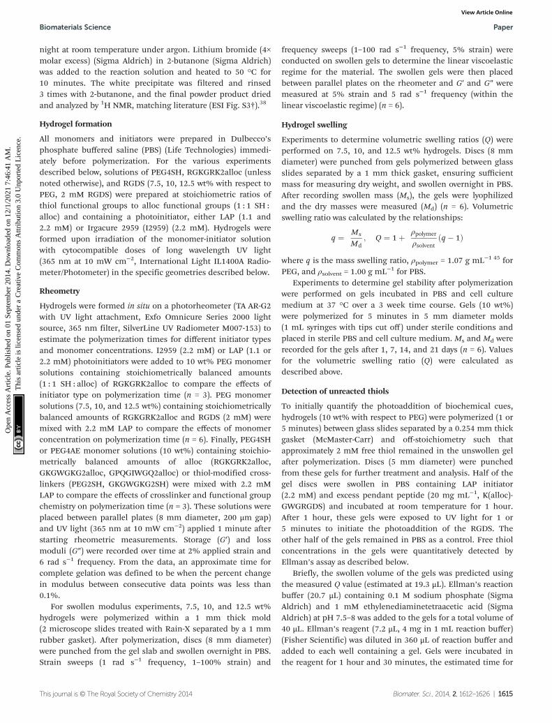
We demonstrate for our material system that, by increasingthe concentration of monomer in the gel-forming solution, wecan increase the modulus (Fig. 3b; 7.5 wt%, G′ = 553 ± 81 Pa;
10 wt%, G′ = 1343 ± 49 Pa; 12.5 wt%, G′ = 2147 ± 87 Pa), creat-ing gels with a range of elasticity comparable to native softtissues (around the range of neural tissues to muscle, E ∼ 1 to10 kPa, where E ≈ 3 G).55 Assuming that the theory of rubberelasticity holds for these swollen gels, the behavior can beattributed to an increase in crosslink density (ρx) by
56
G ¼ ρxRTQ�1=3:
Similarly, we observed decreasing swelling ratios forincreasing monomer concentrations (Fig. 3c; 7.5 wt%, Q =41.4 ± 1.3; 10 wt%, Q = 33.3 ± 0.6; 12.5 wt%, Q = 29.2 ± 0.4).Increased crosslink density inhibits how much a gel is able toswell, thus the inverse relationship between ρx and Q isexpected and observed. The results for the moduli andswelling ratios also were found to be statistically significant(p < 0.05), indicating that the material system may be easilytuned to have specific mechanical properties by varying theconcentration of monomer present within a gel.
Hydrogel stability demonstrated for long-term culture
Hydrogel degradation over time often is desirable for cellculture applications to allow cellular processes, such asgrowth, proliferation, and migration, which can be constrainedor hindered by a tightly crosslinked material.57 However, non-specific degradation in aqueous solutions (e.g., hydrolytic clea-vage of bonds within functional groups) can limit the degreeof user control over materials properties afforded by theaddition of enzymatically degradable peptide crosslinks18 orphotodegradable chemistries,58 resulting in unintended orpremature hydrogel degradation such that the gel does notremain intact for appropriate time periods during cell culture.For example, Shih and Lin have shown that step growth PEG-tetranorbornene-based thiol–ene hydrogels completelydegrade in 2–3 weeks at physiological pH (pH ∼ 7.4), wherethe norbornene is linked to PEG by an ester bond leading tohydrolytic degradation. Specifically, the degradation rate ofthese hydrogels was influenced by the peptide crosslinkersequence, where peptides containing hydrophobic or aromaticresidues exhibited slower degradation (e.g., CGGGC sequencekhyd = 0.049 ± 0.001 day−1, CGGLC sequence khyd = 0.036 ±0.002 day−1).29
In the hydrogel system presented here, we aimed to createmonomers free of ester bonds to allow the creation of hydro-gels that are stable under cell culture conditions. To assess thestability of the resulting hydrogels, we monitored the volu-metric swelling ratio (Q) of 10 wt% gels incubated in PBS andcell culture medium at 37 °C over a period of three weeks(Fig. 4), a typical length for many two and three-dimensionalcell culture experiments.3 For both conditions, the Q valuesqualitatively are constant during the time course, and there isno substantial degradation during the incubation period.Quantitatively, the p-values for the gels incubated in PBS fordifferent times are all greater than 0.05, indicating no statisti-cal significance between the gels for each time points and thusthat degradation does not occur. For the gels incubated in
Fig. 3 Hydrogel mechanical properties tuned to mimic soft tissueenvironments. (A) To adjust the mechanical properties of hydrogels,monomer concentration in solution prior to polymerization may beincreased or decreased to increase or decrease crosslink density andthus modulus, respectively. (B) Hydrogels from different initial monomerconcentrations (7.5, 10, and 12.5 wt% with respect to PEG) were poly-merized and swollen in PBS to demonstrate tunable hydrogel mechan-ical properties. The lowest average swollen storage modulus wasobserved for the 7.5 wt% gels (553 ± 81 Pa), and increased with highermonomer concentrations (1343 ± 49 Pa and 2147 ± 87 Pa). (C) Anotherimportant property to consider in the design of controlled hydrogelmimics of the ECM is the volumetric swelling ratio. In accordancewith modulus measurements, for the gels polymerized with an initialmonomer concentration of 12.5 wt% (with respect to PEG), the lowestvolumetric swelling ratio is observed (29.2 ± 0.4), and the volumetricswelling ratio increases for lower concentrations (7.5 and 10 wt% withrespect to PEG) (Q = 41.4 ± 1.3 and 33.3 ± 0.6).
Biomaterials Science Paper
This journal is © The Royal Society of Chemistry 2014 Biomater. Sci., 2014, 2, 1612–1626 | 1619
Ope
n A
cces
s A
rtic
le. P
ublis
hed
on 0
1 Se
ptem
ber
2014
. Dow
nloa
ded
on 1
2/1/
2021
7:4
6:41
AM
. T
his
artic
le is
lice
nsed
und
er a
Cre
ativ
e C
omm
ons
Attr
ibut
ion
3.0
Unp
orte
d L
icen
ce.
View Article Online
culture medium, when comparing days 1–14, the p-values areall greater than 0.05. However, the day 21 time point is statisti-cally different from the day 1 and 7 time points (p < 0.05), indi-cating a slight change in swelling by 3 weeks. We hypothesizethat nonspecific degradation of the peptide crosslinker couldbe occurring over time in growth medium, which is morecomplex than PBS and contains serum laden with enzymes,resulting in this small but statistically significant increase inswelling. Despite this small swelling change, the hydrogelsremain robust and intact over multiple weeks in culture. Theswelling ratios of hydrogels in PBS versus media also are stat-istically significant for the entire incubation period, which wespeculate results from differences in the composition of PBSand growth media. With this base system, various degradablepeptide crosslinks derived from ECM proteins (e.g.,
GPQG↓IWGQ or IPVS↓LRSG derived from collagen I) can beincorporated within the gels to allow cell-controlled matrixdegradation, where the degradation rate of the matrix can betuned by peptide selection for different applications.18
Biochemical cues spatially patterned within hydrogels
One benefit that photoclick chemistry provides in the designof hydrogels is the ability to control the presentation of bio-chemical cues in space or time.59 Native tissues are dynamicenvironments with gradients and defined regions of biologicalcues occurring at different times, and the ability to capturethis complexity in synthetic systems is important for under-standing and directing cellular processes.25 Here, we studiedthe photoaddition of a model biochemical cue to our material(i) to establish if excess free thiols could be modified afterhydrogel formation and (ii) to demonstrate control over thespatial presentation of these cues. Specifically, an alloc-modi-fied integrin binding peptide (RGDS or AF488RGDS) wascoupled homogenously or in specific regions to hydrogels con-taining free thiols using photopatterning.
While one of our goals was to develop a hydrogel fromaccessible materials, we also aimed to use simple techniquesto characterize this system. Ellman’s assay, which can identifyfree thiols in solution, is one such technique that has beenused to quantify free thiols in materials post-polymeriz-ation.60,61 We have utilized this assay in a non-destructivemethod to quantify free thiols in our hydrogels such that, ifdesired, gels may be rinsed of reagent, treated with tris(2-car-boxyethyl)phosphine (TCEP), rinsed of TCEP, and re-used inadditional studies. In addition to quantifying free thiols, wewanted to demonstrate that Ellman’s reagent also could beused to observe biochemical patterns created in gels with areasonable degree of resolution as an inexpensive and rapidalternative or complementary approach to using a fluore-scently-tagged cue (Fig. 5a).
Toward achieving this, 10 wt% gels (0.254 mm thickbetween glass slides) initially were polymerized off stoichio-metry so that free thiols (2 mM at preparation prior to equili-brium swelling) remained for later modification with thependant RGDS peptide. Adjusting for swelling, the free thiolconcentration at equilibrium was estimated to be roughly∼0.61 ± 0.05 mM, so the free thiol concentration in hydrogelsas measured by Ellman’s assay will be lower than 2 mM. Thefree thiol concentrations of off-stoichiometry gels polymerizedfor 1 and 5 minutes subsequently was determined by Ellman’sassay to be 1.13 ± 0.09 mM and 0.97 ± 0.10 mM, respectively(Fig. 5b, −RGDS condition). The gels polymerized for 1 and5 minutes do not have statistically different thiol concen-trations (p > 0.05), supporting the results in Fig. 2b that the10 wt% gels are completely formed in under one minute.
To initially determine if a model biochemical cue could beadded to these gels, pre-formed gels incubated in RGDSmonomer (20 mg mL−1 ∼20× excess to SH) with LAP (2.2 mM)were exposed to UV light for 1 and 5 minutes. The thiol con-centration after modification was determined for each con-dition by Ellman’s (1 min = 0.01 ± 0.01 mM, 5 min = 0.003 ±
Fig. 4 Hydrogel stability toward long-term cell culture. (A) The thiol–ene system is linked by a carbamate bond between a thiol-functiona-lized PEG macromer and an alloc-functionalized peptide, which bothlack hydrolytically cleavable bonds (e.g., esters). To evaluate gel stabilityfor controlled cell culture over several weeks, 10 wt% hydrogels (withrespect to PEG) were incubated in cell culture medium and PBS for3 weeks and volumetric swelling ratios (Q) measured. (B) The swelling ofgels incubated in PBS did not significantly change (p > 0.05 for allsamples) indicating stability over the time period (Q = 33.4 ± 0.8, 30.8 ±1.3, 30.6 ± 1.3, 30.3 ± 1.5). The swelling of gels incubated in growthmedia slightly increased through the incubation period such that thefirst two and last Q values are different (p < 0.05), although consecutivepoints are not (p > 0.05), which may be attributed partly to non-specificdegradation of the hydrogel by enzymes present in the more complexgrowth medium (Q = 25.1 ± 0.2, 25.6 ± 0.4, 26.8 ± 0.8, 27.6 ± 0.4).
Paper Biomaterials Science
1620 | Biomater. Sci., 2014, 2, 1612–1626 This journal is © The Royal Society of Chemistry 2014
Ope
n A
cces
s A
rtic
le. P
ublis
hed
on 0
1 Se
ptem
ber
2014
. Dow
nloa
ded
on 1
2/1/
2021
7:4
6:41
AM
. T
his
artic
le is
lice
nsed
und
er a
Cre
ativ
e C
omm
ons
Attr
ibut
ion
3.0
Unp
orte
d L
icen
ce.
View Article Online

0.003 mM) (Fig. 5b, +RGDS condition). These concentrationscorrespond to 93.1 and 94.8% modification of the remainingfree thiols and 98.5 and 99.0% total thiol modification, indi-cating high coupling efficiency of the pendant peptide. Thereare slightly fewer free thiols in the gels polymerized for5 minutes indicating that a longer polymerization time resultsin higher conversion of functional groups; however, there is nostatistical significance between the two conditions indicatingthat the effects of longer polymerization are ultimately negli-gible. Hydrogels polymerized off-stoichiometry (2 mM freethiol at preparation) were then incubated in growth medium at37 °C for 3 days to determine if cues could be added atdifferent times during culture. Only trace free thiols wereobserved with Ellman’s assay after this 3-day incubation(0.008 ± 0.002 mM), indicating the formation of disulfideseither with components in the culture medium or betweenfree thiol end groups on PEG. To test this hypothesis, TCEP
(10 mM in PBS) was added to the gels for 1 hour to breakpotential disulfide bonds. Gels subsequently were rinsed, andthe presence of free thiols was detected with Ellman’s (1.54 ±0.09 mM) (ESI Fig. S5†). This recovery of thiols confirms that alarge portion of free thiols post-polymerization were lost to di-sulfide formation upon incubation in culture medium. Whilethe application TCEP could be investigated as an approach toallow temporal photopatterning, reducing agents such as itwill negatively affect cell viability62 and may not be a practicaloption for in situ photopatterning. However, different ortho-gonal chemistries2 could be utilized within this base hydrogelsystem to allow the temporal addition of cues throughoutlong-term cell culture in future investigations.
With the ability to add cues to the matrix after initial for-mation, spatially defined regions of various cues of interestcan be created toward directing the organization and functionof cells in three dimensions.10,22,63 Fluorescently-labeled cues
Fig. 5 Biochemical cues spatially patterned within hydrogels. (A) To create patterns of biochemical cues, hydrogels are polymerized off-stoichi-ometry ([SH] > [alloc]) and incubated with excess pendant RGDS or AF488RGDS peptide. Gels were irradiated through photomasks printed withblack lines or squares for 1 and 5 minutes (left). Samples are subsequently analyzed with fluorescent light or Ellman’s reagent to determine themodification of free thiols with pendant biochemical cues (right). (B) Gels (10 wt% with respect to PEG) were polymerized with 2.2 mM LAP for 1 and5 minutes off stoichiometry (2 mM free thiol at preparation). After equilibrium swelling, the initial free thiol concentration in these gels were 1.13 ±0.09 and 0.97 ± 0.10 mM, respectively, as determined by Ellman’s assay. Only 0.01 ± 0.01 and 0.003 ± 0.003 mM free thiol remained after addingthe RGDS tether indicating the efficient coupling of the model biochemical cue to the hydrogel network. (C) Following the setup shown in (A), arbi-trary patterns (squares, 1600 μm2; lines of different thickness, 200–1000 μm) of a fluorescent peptide (AF488RGDS) were created within pre-formedhydrogels and imaged on a confocal microscope for analysis. Resolution of the pattern is observed in the x-, y-, and z-planes indicating selectivecoupling to only regions of the gel that were exposed to light (scale bar, 200 μm). (D) As a quick and inexpensive alternative to fluorescence, a non-fluorescent pendant peptide (RGDS) was photopatterned (lines of different thickness) into pre-formed hydrogels. Ellman’s reagent was directlyapplied to the top of these gels to identify regions lacking the pendant peptide (yellow) with resolution in the x- and y-planes over short times(<5 min) (scale bar, 1 mm).
Biomaterials Science Paper
This journal is © The Royal Society of Chemistry 2014 Biomater. Sci., 2014, 2, 1612–1626 | 1621
Ope
n A
cces
s A
rtic
le. P
ublis
hed
on 0
1 Se
ptem
ber
2014
. Dow
nloa
ded
on 1
2/1/
2021
7:4
6:41
AM
. T
his
artic
le is
lice
nsed
und
er a
Cre
ativ
e C
omm
ons
Attr
ibut
ion
3.0
Unp
orte
d L
icen
ce.
View Article Online
are typically used to observe biochemical patterns in hydrogel-based matrices with a high degree of resolution; however, thisapproach requires additional expense and time for peptidelabeling and fluorescence imaging. For a rapid and inexpen-sive assessment of patterning, we examined using Ellman’sreagent to observe spatially-defined patterns as a simplealternative or complementary approach for preliminary evalu-ations. Hydrogels photopatterned with the AF488RGDSpeptide demonstrate spatial resolution of cue addition(Fig. 5c) in the x, y, and z-directions for patterns of arbitraryshapes (wide and narrow lines, squares). Next, to test Ellman’sas an alternative to fluorescently-labeled evaluation, non-labeled RGDS was patterned into gels, and the gels wereimaged under a light microscope immediately after the appli-cation of Ellman’s reagent (Fig. 5d). At short time periods(<5 minutes), we observed resolution of the patterns; however,as the products from reaction with Ellman’s reagent diffusedthroughout the gel, the pattern began to disappear (ESIFig. S6†). While Ellman’s reagent is limited by the fastdiffusion of the reaction products, resulting in the short-termobservation of patterns in the x–y plane only, we envisionusing this test in initial or follow-up studies of photopattern-ing in thiol–ene hydrogels because it is easy to use and pro-vides almost instant results. Initially, one could test the abilityto pattern a hydrogel before building or purchasing a moreexpensive fluorescently-tagged peptide. In later experiments,one could quickly confirm that a different peptide or peptidesequence is patterned into the same system without having tobuild another labeled peptide and use an epi-fluorescent orconfocal microscope.
Encapsulated stem cells remain viable and metabolically activewithin patterned and non-patterned hydrogels
Hydrogel systems for cell culture or delivery must not only becytocompatible, but cells also must be able to withstand theirpolymerization conditions for encapsulation within thematrix. PEG, the primary component of the materials pre-sented here, has been used in a variety of hydrogel systemsowing to its bioinert nature, providing a blank slate for thepresentation of peptide sequences or whole proteins to elicitspecific cellular responses.25 Furthermore, cells must be ableto withstand multiple doses of UV light and radical initiatorfor the creation of biochemical patterns within gels to directcell behavior in three dimensions.
To evaluate the cytocompatibility of the initial polymeriz-ation conditions, we encapsulated adult human mesenchymalstem cells, hMSCs, within non-degradable gels (10 wt%,2.2 mM LAP, 2 mM RGDS, 3000 cells μL−1) polymerized fordifferent lengths of time. Specifically, based on our rheometricmeasurements, hydrogels were polymerized for the minimumamount of time required to completely polymerize 10 and12.5 wt% samples (1 minute) and in excess of the minimumamount of time to polymerize 7.5 wt% samples (5 minutes). Inaddition, cell density was kept low to promote primarily cell–matrix interactions and fully understand the limits of cell via-bility in the system when encapsulating a dilute, single-cell
suspension. Cell viability and metabolic activity subsequentlywere evaluated 1 and 3 days after polymerization to determinepolymerization conditions appropriate for the initial encapsu-lation and culture of cells, respectively.
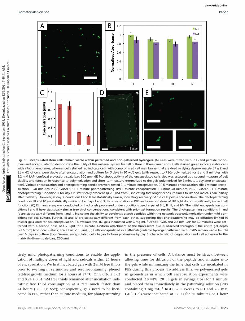
A membrane integrity assay (LIVE/DEAD® Viability/Cytotoxi-city Kit) of cells encapsulated in gels (Fig. 6a) showed a higherpercentage of living cells in gels polymerized for 1 minute (87± 2%) in comparison to 5 minutes (81 ± 4%) at day 3 inculture. While decreased cell viability is observed for the5 minute polymerization condition, which could limit the useof gels with lower modulus in cell culture (e.g., 7.5 wt%), viabi-lity can be rescued by adjustment of experimental parameters,including increased cell–cell contact (i.e., controlling thedensity of encapsulated cells),64 incorporating biomimetic pep-tides that promote additional cell–matrix interactions,20,65 andlower initiator concentration (i.e., reducing concentration ofradicals during polymerization but at some cost to polymeriz-ation time).38 We increased the encapsulation density of cellsin non-degradable gels polymerized for 5 minutes (3000 to30 000 cells μL−1) and demonstrated a corresponding increasein viability (83 ± 2% to 92 ± 1%) (ESI Fig. S7†). Accordingly,cell encapsulation density can be adjusted as appropriate tosupport viability and function depending on the experimentalvariables to be studied and should be considered in experi-mental design when using this system.
The metabolic activity of cells, an indicator of cell viabilityand function, also was monitored 1 and 3 days after encapsula-tion using CellTiter 96. Constant metabolic activity over timewas observed in the gels polymerized for 1 and 5 minutes overthree days (p > 0.05) (Fig. 6b). Initially (D1) the metabolicactivity of the gels polymerized for 5 minutes is statisticallydifferent (p < 0.05) from gels polymerized for only one minute.However, by day 3, the metabolic activity of the gels polymer-ized for 1 and 5 minutes is statistically similar (p > 0.05), indi-cating that the initial effects of the polymerization are mostapparent for longer irradiation time periods but do not impactcell metabolic activity past the initial treatment. Here, theshort-term effects of encapsulation on cell survival appearminimal and similar to that observed in other hydrogelsformed by free radical initiation,38,48 indicating that this newhydrogel system could support cell culture or delivery invarious experimental applications.
Note that all conditions in the metabolic activity experi-ments presented above were normalized to cells encapsulatedin hydrogels with 1 minute of light exposure. While normaliza-tion to encapsulated cells without UV exposure is desirable,the hydrogel system presented cannot be easily formedwithout light. To assess any effect of UV light alone on cellfunction, hMSCs were seeded in 96-well plates and metabolicactivity monitored 1 and 3 days after exposure to UV. Lightexposure did not significantly affect hMSC metabolic activity ateither D1 or D3 post-irradiation (p > 0.05, compared to no UVcontrol) (ESI Fig. S8†). This result is consistent with thereports of others for single doses of UV light at 10 mW cm−2.66
Toward utilizing this system for patterning gels with bio-chemical cues during cell culture, we sought to establish rela-
Paper Biomaterials Science
1622 | Biomater. Sci., 2014, 2, 1612–1626 This journal is © The Royal Society of Chemistry 2014
Ope
n A
cces
s A
rtic
le. P
ublis
hed
on 0
1 Se
ptem
ber
2014
. Dow
nloa
ded
on 1
2/1/
2021
7:4
6:41
AM
. T
his
artic
le is
lice
nsed
und
er a
Cre
ativ
e C
omm
ons
Attr
ibut
ion
3.0
Unp
orte
d L
icen
ce.
View Article Online
tively mild photopatterning conditions to enable the appli-cation of multiple doses of light and radicals within 24 hoursof encapsulation. We first incubated gels with 2 mM free thiolsprior to swelling in serum-free and serum-containing, phenolred-free growth medium for 2 hours at 37 °C. Only 0.26 ± 0.02and 0.24 ± 0.04 mM free thiols remained after incubation indi-cating free thiol consumption at a rate much faster than24 hours (ESI Fig. S5†); consequently, gels need to be incu-bated in PBS, rather than culture medium, for photopatterning
in the presence of cells. A balance must be struck betweenallowing time for diffusion of the peptide and initiator intothe gels while minimizing the time that cells are incubated inPBS during this process. To address this, we polymerized gelsin geometries in which cell encapsulation experiments wereconducted (10 wt%, 20 μL gels in syringe tips) for 1 minuteand placed them immediately in the patterning solution (PBScontaining 3 mg mL−1 RGDS ∼3× excess to SH and 2.2 mMLAP). Gels were incubated at 37 °C for 30 minutes or 1 hour
Fig. 6 Encapsulated stem cells remain viable within patterned and non-patterned hydrogels. (A) Cells were mixed with PEG and peptide mono-mers and encapsulated to demonstrate the utility of this material system for cell culture in three dimensions. Cells stained green indicate viable cellswith intact membranes, whereas cells stained red indicate cells with compromised cell membranes that are dead or dying. Approximately 87 ± 2 and81 ± 4% of cells were viable after encapsulation and culture for 3 days in 10 wt% gels (with respect to PEG) polymerized for 1 and 5 minutes with2.2 mM LAP (confocal projection; scale bar, 200 μm). (B) Metabolic activity of the encapsulated cells also was assessed as a second measure of cellviability and function in response to polymerization and short-term culture (normalized to the gels polymerized for 1 minute 1 day after encapsula-tion). Various encapsulation and photopatterning conditions were tested (I) 1 minute encapsulation, (II) 5 minutes encapsulation, (III) 1 minute encap-sulation + 30 minutes PBS/RGDS/LAP + 1 minute photopatterning, (IV) 1 minute encapsulation + 1 hour 30 minutes PBS/RGDS/LAP + 1 minutephotopatterning. Condition II for day 1 is statistically different (p < 0.05) from I, indicating that longer exposure times to UV and radicals can initiallyaffect viability. However, at day 3, conditions I and II are statistically similar, indicating ‘recovery’ of the cells post-encapsulation. The photopatterningconditions III and IV are statistically similar to I at days 1 and 3; thus, incubation in PBS and a second dose of UV light do not significantly impact cellfunction. (C) Ellman’s assay was conducted on hydrogels processed under conditions used in panel B (I, II, III, and IV). The initial encapsulation con-ditions I and II have statistically similar free thiol concentrations, consistent with prior gel formation results. The photopatterning conditions III andIV are statistically different from I and II, indicating the ability to covalently attach peptides within the network post-polymerization under mild con-ditions for cell culture. Further, III and IV are statistically different from each other, suggesting that photopatterning may be diffusion-limited inthicker gels used for cell encapsulation. To evaluate this, (D) gels incubated with 3 mg mL−1 AF488RGDS and 2.2 mM LAP for 30 minutes were pat-terned with a second dose of UV light for 1 minute. Uniform attachment of the fluorescent cue is observed throughout the entire gel depth(∼1.6 mm) (confocal Z-stack; scale Bar, 200 μm). (E) Cells encapsulated in a MMP-degradable hydrogel patterned with RGDS remain viable (>90%)over 6 days in culture (top). Several encapsulated cells began to form protrusions by day 6, characteristic of degradation and cell adhesion to thematrix (bottom) (scale bars, 200 μm).
Biomaterials Science Paper
This journal is © The Royal Society of Chemistry 2014 Biomater. Sci., 2014, 2, 1612–1626 | 1623
Ope
n A
cces
s A
rtic
le. P
ublis
hed
on 0
1 Se
ptem
ber
2014
. Dow
nloa
ded
on 1
2/1/
2021
7:4
6:41
AM
. T
his
artic
le is
lice
nsed
und
er a
Cre
ativ
e C
omm
ons
Attr
ibut
ion
3.0
Unp
orte
d L
icen
ce.
View Article Online
and 30 minutes, times longer and shorter than the time esti-mated for diffusion of the monomer to the center of the gelassuming Fickian diffusion (td ∼ 65 minutes):
td ¼ L2
D
where L is half the thickness of the unswollen gel(∼0.625 mm) and D the diffusion coefficient (∼10−6 cm2 s−1
based on proteins of similar molecular weight as the RGDSpeptide).67 A second dose of UV light (1 minute) was appliedto covalently link RGDS within the hydrogel. As previouslyobserved, free thiol concentration in gels polymerized for1 and 5 minutes (without patterning) was not statisticallydifferent (p > 0.05) and the patterned gels exhibit significantlylower concentrations of free thiols post-patterning (p < 0.05compared to that after 1 and 5 minute gel formation) at 0.30 ±0.01 and 0.21 ± 0.02 mM, respectively (Fig. 6c). These twophotopatterning conditions have statistically different thiolconcentrations after polymerization (p < 0.05), suggesting thatthe peptide and initiator may not have fully penetrated the gelduring this incubation time. To test this hypothesis, gels(10 wt%, 20 μL in syringe tips, 1 minute polymerization) wereincubated with AF488RGDS (3 mg mL−1) and LAP (2.2 mM) inPBS for 30 minutes, 1 hour, and 1 hour 30 minutes, andexposed to UV light for 1 minute to allow covalent attachmentof the fluorescent peptide. Z-Stack images through the entiregel depth (confocal) indicate consistent patterning of thepeptide through the gel depth for all conditions (Fig. 6d, ESIFig. S9†). We speculate that the slight differences seenbetween the thiol concentrations after patterning by Ellman’sassay (Fig. 5b and 6c) are the result of small variationsbetween batches of PEG-4SH monomer and hydrogels or therelative excesses at which the cues were tagged (20× for proof-of-concept and 3× for patterning in the presence of cells).
To compare the effects of these photopatterning conditionson cell activity and viability, cells encapsulated in non-degrad-able gels (3000 cells μL−1, 1 minute UV exposure) were incu-bated for 30 minutes or 1 hour 30 minutes in PBS containingRGDS and LAP and a second dose of UV light subsequentlywas applied for 1 minute. Cell metabolic activity for thesephotopatterning conditions is statistically similar to the1 minute hydrogel formation condition at days 1 and 3 (p >0.05), indicating that exposure to multiple polymerizations(formation + patterning) has a minimal effect on cell function(Fig. 6b). There appears to be a slight, but not statistically sig-nificant, decrease in metabolic activity for each conditionbetween days 1 and 3. We hypothesize that this negligibledecrease results from minor damage to cells in all cases by theradically-mediated polymerizations, which shows up inreduced metabolic activity at day 3. No statistical difference isobserved between any condition at day 3. Taken together, nospecific effect of the photopatterning process is observed, andthe photopatterning conditions assessed here are appropriatefor use in cell culture.
Finally, toward long-term culture of cells in patterned gels,hMSCs were encapsulated in cell-degradable gels crosslinked
with a MMP-cleavable peptide sequence18 (GPQGIWGQ2alloc)and treated with 3 mg mL−1 RGDS and 2.2 mM LAP in PBS for1 hour (between the minimum and maximum incubationtimes tested for photopatterning) before a second dose of UVlight was applied to photopattern RGDS within the network.After 6 days of culture, cells were stained with the LIVE/DEAD® Viability/Cytotoxicity Kit and imaged on a confocalmicroscope to observe cell viability and any spreading withinthe network. Viability greater than 90% was observed and afew cells exhibited protrusions (Fig. 6e), indicative of adhesionto and degradation of the matrix. Based on these results, thisapproach for cell encapsulation and matrix photopatterning ispromising for future studies to probe stem cell–material inter-actions and direct cell function and fate in vitro.
Conclusions
In summary, we presented a novel hydrogel system formed bythiol–ene photoclick chemistry through reaction of thiol-modi-fied PEG and alloc-modified peptides. Use of the LAP photo-initiator allowed rapid polymerization with cytocompatibledoses of UV light and the formation of hydrogels with appro-priate mechanical properties to mimic soft tissues. Thesehydrogels remain stable in cell culture conditions and encap-sulated cells are viable within the network. Biochemical cueswere selectively patterned within the gels to demonstratespatial control over matrix properties, and cells remainedviable. Further, the monomers used in the design of thissystem may be synthesized using established protocols or com-mercially purchased, making the material accessible for thefacile and consistent formation of robust hydrogels to mimicthe ECM. In the future, this base material may be used withorthogonal click chemistries to allow control over biochemicaland biomechanical properties over days to weeks to study cellresponse to changes in the surrounding environment and pro-vides a useful platform to adapt for a variety of biomaterialsapplications, including cell culture, tissue engineering, anddrug delivery. Specifically, toward application in culture anddirecting hMSC fate, gels could be patterned with individualor multiple biochemical cues in spatially defined regions todrive cellular processes, including adhesion, migration, pro-liferation, or differentiation.68,69
Acknowledgements
This work was supported by the Institutional DevelopmentAward from the National Institutes of Health (P20GM103541),the Pew Charitable Trusts (00026178), a National Science Foun-dation Career Award (DMR-1253906), and the National ScienceFoundation IGERT SBE2 program at the University of Delaware(fellowship to LAS). The authors thank the Delaware Biotech-nology Institute at the University of Delaware for training andaccess to confocal microscopy at the BioImaging Center,Mr. Matthew Rehmann for generously providing hMSCs isolated
Paper Biomaterials Science
1624 | Biomater. Sci., 2014, 2, 1612–1626 This journal is © The Royal Society of Chemistry 2014
Ope
n A
cces
s A
rtic
le. P
ublis
hed
on 0
1 Se
ptem
ber
2014
. Dow
nloa
ded
on 1
2/1/
2021
7:4
6:41
AM
. T
his
artic
le is
lice
nsed
und
er a
Cre
ativ
e C
omm
ons
Attr
ibut
ion
3.0
Unp
orte
d L
icen
ce.
View Article Online
from bone marrow and the cysteine-modified peptide, Prof.Christopher J. Kloxin and Mr Stephen Ma for generously pro-viding photomasks, Prof. Wilfred Chen for use of the auto-mated plate reader, and Mr Eric Macedo for training inpolymer and peptide synthesis techniques.
References
1 H. C. Kolb, M. Finn and K. B. Sharpless, Angew. Chem., Int.Ed., 2001, 40, 2004–2021.
2 W. Xi, T. F. Scott, C. J. Kloxin and C. N. Bowman, Adv.Funct. Mater., 2014, 24(18), 2572–2590.
3 P. M. Kharkar, K. L. Kiick and A. M. Kloxin, Chem. Soc.Rev., 2013, 42, 7335–7372.
4 R. K. Iha, K. L. Wooley, A. M. Nyström, D. J. Burke,M. J. Kade and C. J. Hawker, Chem. Rev., 2009, 109, 5620–5686.
5 M. J. Kade, D. J. Burke and C. J. Hawker, J. Polym. Sci., PartA: Polym. Chem., 2010, 48, 743–750.
6 M. Lomba, L. Oriol, R. Alcalá, C. Sánchez, M. Moros,V. Grazú, J. L. Serrano and J. M. De la Fuente, Macromol.Biosci., 2011, 11, 1505–1514.
7 K. G. Robinson, T. Nie, A. D. Baldwin, E. C. Yang,K. L. Kiick and R. E. Akins, J. Biomed. Mater. Res., Part A,2012, 100, 1356–1367.
8 M. Lutolf and J. Hubbell, Biomacromolecules, 2003, 4, 713–722.
9 J. Zheng, L. A. Smith Callahan, J. Hao, K. Guo,C. Wesdemiotis, R. Weiss and M. L. Becker, ACS MacroLett., 2012, 1, 1071–1073.
10 C. A. DeForest, B. D. Polizzotti and K. S. Anseth, Nat.Mater., 2009, 8, 659–664.
11 D. L. Alge, M. A. Azagarsamy, D. F. Donohue andK. S. Anseth, Biomacromolecules, 2013, 14, 949–953.
12 R. Selvaraj and J. M. Fox, Curr. Opin. Chem. Biol., 2013, 17,753–760.
13 A. D. Baldwin and K. L. Kiick, Polym. Chem., 2013, 4, 133–143.
14 C. E. Hoyle and C. N. Bowman, Angew. Chem., Int. Ed.,2010, 49, 1540–1573.
15 B. D. Polizzotti, B. D. Fairbanks and K. S. Anseth, Biomacro-molecules, 2008, 9, 1084–1087.
16 W. M. Gramlich, I. L. Kim and J. A. Burdick, Biomaterials,2013, 34, 9803–9811.
17 C. R. Nuttelman, M. A. Rice, A. E. Rydholm, C. N. Salinas,D. N. Shah and K. S. Anseth, Prog. Polym. Sci., 2008, 33,167–179.
18 J. Patterson and J. A. Hubbell, Biomaterials, 2010, 31, 7836–7845.
19 B. D. Fairbanks, M. P. Schwartz, A. E. Halevi,C. R. Nuttelman, C. N. Bowman and K. S. Anseth, Adv.Mater., 2009, 21, 5005–5010.
20 M. Lutolf and J. Hubbell, Nat. Biotechnol., 2005, 23, 47–55.21 A. E. Rydholm, S. K. Reddy, K. S. Anseth and
C. N. Bowman, Polymer, 2007, 48, 4589–4600.
22 K. A. Mosiewicz, L. Kolb, A. J. van der Vlies, M. M. Martino,P. S. Lienemann, J. A. Hubbell, M. Ehrbar and M. P. Lutolf,Nat. Mater., 2013, 12, 1072–1078.
23 T. Yang, H. Long, M. Malkoch, E. Kristofer Gamstedt,L. Berglund and A. Hult, J. Polym. Sci., Part A: Polym. Chem.,2011, 49, 4044–4054.
24 J. A. Burdick and W. L. Murphy, Nat. Commun., 2012, 3,1269.
25 M. S. Rehmann and A. M. Kloxin, Soft Matter, 2013, 9,6737–6746.
26 C.-C. Lin, A. Raza and H. Shih, Biomaterials, 2011, 32,9685–9695.
27 C. N. Salinas, B. B. Cole, A. M. Kasko and K. S. Anseth,Tissue Eng., 2007, 13, 1025–1034.
28 J. L. Ifkovits and J. A. Burdick, Tissue Eng., 2007, 13,2369–2385.
29 H. Shih and C.-C. Lin, Biomacromolecules, 2012, 13,2003–2012.
30 H. Shih and C. C. Lin, Macromol. Rapid Commun., 2013, 34,269–273.
31 J. J. Roberts and S. J. Bryant, Biomaterials, 2013, 34,9969–9979.
32 R. Sanchez, M. Riddle, J. Woo and J. Momand, Protein Sci.,2008, 17, 473–481.
33 S. T. Gould, N. J. Darling and K. S. Anseth, Acta Biomater.,2012, 8, 3201–3209.
34 C. A. DeForest, E. A. Sims and K. S. Anseth, Chem. Mater.,2010, 22, 4783–4790.
35 M. M. Kreevoy, E. T. Harper, R. E. Duvall, H. S. WilgusIII and L. T. Ditsch, J. Am. Chem. Soc., 1960, 82, 4899–4902.
36 A. A. Aimetti, R. K. Shoemaker, C.-C. Lin and K. S. Anseth,Chem. Commun., 2010, 46, 4061–4063.
37 C. A. DeForest and K. S. Anseth, Nat. Chem., 2011, 3, 925–931.
38 B. D. Fairbanks, M. P. Schwartz, C. N. Bowman andK. S. Anseth, Biomaterials, 2009, 30, 6702–6707.
39 S. J. Bryant, C. R. Nuttelman and K. S. Anseth, J. Biomater.Sci., Polym. Ed., 2000, 11, 439–457.
40 Z. N. Mahmoud, S. B. Gunnoo, A. R. Thomson,J. M. Fletcher and D. N. Woolfson, Biomaterials, 2011, 32,3712–3720.
41 J. A. Johnson, M. Finn, J. T. Koberstein and N. J. Turro,Macromol. Rapid Commun., 2008, 29, 1052–1072.
42 R. Khurana, J. Schaefer, L. A. Archer and G. W. Coates,J. Am. Chem. Soc., 2014, 136(20), 7395–7402.
43 B. D. Fairbanks, S. P. Singh, C. N. Bowman andK. S. Anseth, Macromolecules, 2011, 44, 2444–2450.
44 E. Ruoslahti and M. D. Pierschbacher, Science, 1987, 238,491–497.
45 S. J. Bryant and K. S. Anseth, in Scaffolding in Tissue Engin-eering, ed. P. X. Ma and J. Elisseeff, Marcel Dekker, Inc.,Boca Raton, FL, 1st edn, 2005, pp. 71–90.
46 S. B. Anderson, C.-C. Lin, D. V. Kuntzler and K. S. Anseth,Biomaterials, 2011, 32, 3564–3574.
47 A. B. Lowe, Polym. Chem., 2010, 1, 17–36.
Biomaterials Science Paper
This journal is © The Royal Society of Chemistry 2014 Biomater. Sci., 2014, 2, 1612–1626 | 1625
Ope
n A
cces
s A
rtic
le. P
ublis
hed
on 0
1 Se
ptem
ber
2014
. Dow
nloa
ded
on 1
2/1/
2021
7:4
6:41
AM
. T
his
artic
le is
lice
nsed
und
er a
Cre
ativ
e C
omm
ons
Attr
ibut
ion
3.0
Unp
orte
d L
icen
ce.
View Article Online
48 S. Gerecht, J. A. Burdick, L. S. Ferreira, S. A. Townsend,R. Langer and G. Vunjak-Novakovic, Proc. Natl. Acad.Sci. U. S. A., 2007, 104, 11298–11303.
49 N. B. Cramer, S. K. Reddy, A. K. O’Brien andC. N. Bowman, Macromolecules, 2003, 36, 7964–7969.
50 J. E. Elliott, M. Macdonald, J. Nie and C. N. Bowman,Polymer, 2004, 45, 1503–1510.
51 V. Zubov, M. V. Kumar, M. Masterova and V. Kabanov,J. Macromol. Sci., Chem., 1979, 13, 111–131.
52 C. N. Salinas and K. S. Anseth, Macromolecules, 2008, 41,6019–6026.
53 M. W. Toepke, N. A. Impellitteri, J. M. Theisen andW. L. Murphy, Macromol. Mater. Eng., 2013, 298, 699–703.
54 D. E. Discher, P. Janmey and Y.-l. Wang, Science, 2005, 310,1139–1143.
55 I. Levental, P. C. Georges and P. A. Janmey, Soft Matter,2007, 3, 299–306.
56 M. P. Schwartz, B. D. Fairbanks, R. E. Rogers, R. Rangarajan,M. H. Zaman and K. S. Anseth, Integr. Biol., 2010, 2, 32–40.
57 G. Raeber, M. Lutolf and J. Hubbell, Biophys. J., 2005, 89,1374–1388.
58 A. M. Kloxin, A. M. Kasko, C. N. Salinas and K. S. Anseth,Science, 2009, 324, 59–63.
59 M. Guvendiren and J. A. Burdick, Curr. Opin. Biotechnol.,2013, 24, 841–846.
60 B. Fejerskov, B. E. Jensen, N. B. Jensen, S.-F. Chong andA. N. Zelikin, ACS Appl. Mater. Interfaces, 2012, 4, 4981–4990.
61 S. Kobel, M. Limacher, S. Gobaa, T. Laroche andM. P. Lutolf, Langmuir, 2009, 25, 8774–8779.
62 X. Ren, Y. J. Lee, H. J. Han and I. S. Kim, Chemosphere,2008, 74, 84–88.
63 S.-H. Lee, J. J. Moon and J. L. West, Biomaterials, 2008, 29,2962–2968.
64 C.-C. Lin and K. S. Anseth, Proc. Natl. Acad. Sci. U. S. A.,2011, 108, 6380–6385.
65 C. R. Nuttelman, M. C. Tripodi and K. S. Anseth, MatrixBiol., 2005, 24, 208–218.
66 M. Guvendiren and J. A. Burdick, Nat. Commun., 2012, 3,792.
67 L. M. Weber, C. G. Lopez and K. S. Anseth, J. Biomed.Mater. Res., Part A, 2009, 90, 720–729.
68 R. A. Marklein and J. A. Burdick, Adv. Mater., 2010, 22,175–189.
69 M. P. Lutolf, P. M. Gilbert and H. M. Blau, Nature, 2009,462, 433–441.
Paper Biomaterials Science
1626 | Biomater. Sci., 2014, 2, 1612–1626 This journal is © The Royal Society of Chemistry 2014
Ope
n A
cces
s A
rtic
le. P
ublis
hed
on 0
1 Se
ptem
ber
2014
. Dow
nloa
ded
on 1
2/1/
2021
7:4
6:41
AM
. T
his
artic
le is
lice
nsed
und
er a
Cre
ativ
e C
omm
ons
Attr
ibut
ion
3.0
Unp
orte
d L
icen
ce.
View Article Online