cardio-vascular system
DESCRIPTION
Cardio-Vascular SystemTRANSCRIPT
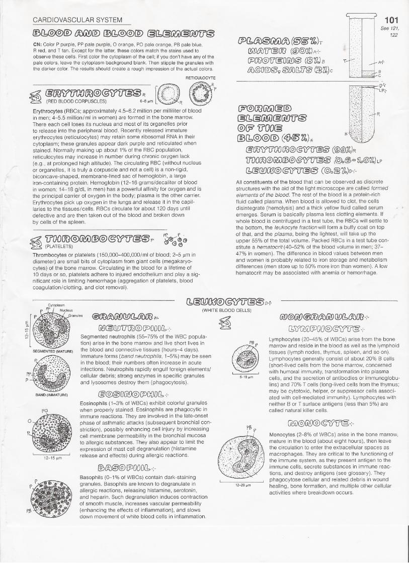
101See 121,
122
CARDIOVASCULAR SYSTEM
fS[b@@@) ~@ (!311@@[Q)§(1.§(1;)@(jIf)'fj@[P[b@@G!iJtJJ(~~"/l,)T
WtAJ'iJ'§(f] (@@~)A-l-
CfJ!XJ@'i]egO@@ (m "IlJB
~@O@)~'i7 ~fAJf1'fl@ ~Lh)c
CN: Color P purple, PP pale purple, 0 orange, PO pale orange, PB pale blue,R red, and T tan. Except for the latter, these colors match the stains used toobserve these cells. First color the cytoplasm of the cell; if you don't have any of thepale colors, leave the cytoplasm background blank. Then stipple the granules withthe darker color. The results should create a rough impression of the actual colors.
RETICULOCYTE
To A":-
6C
.a. &OO~fl()f)OO@@51'fr(g§R L:'~~;~:\'}
'E5 (RED BLOODCORPUSCLES) 6-8 I'm ~ ~.",~.~;..:"R _
Erythrocytes (RBCs; approximately 4.5-6.2 million per milliliter of bloodin men; 4-5.5 million/ml in women) are formed in the bone marrow.There each cell loses its nucleus and most of its organelles priorto release into the peripheral blood. Recenlly released immature
erythrocytes (reticulocytes) may retain some ribosomal RNA in theircytoplasm; these granules appear dark purple and reticulated whenstained. Normally making up about 1% of the RBC population,reticulocytes may increase in number during chronic oxygen lack(e.g., at prolonged high altitude). The circulating RBC (without nucleusor organelles, it is truly a corpuscle and not a cell) is a non-rigid,biconcave-shaped, membrane-lined sac of hemoglobin, a largeiron-containing protein. Hemoglobin (12-16 grams/decaliter of bloodin women; 14-18 g/dL in men) has a powerful affinity for oxygen and isthe principal carrier of oxygen in the body; plasma is the other carrier.Erythrocytes pick up oxygen in the lungs and release it in the capil-laries to the tissues/cells. RBCs circulate for about 120 days untildefective and are then taken out of the blood and broken down
by cells of the spleen.
~@OO(j\!A)&@C311&UiW@(W'U'@@@?1l00§@(1.@@@)C~~Vo)*
@OO)J?'fJGOOO@@'u:'f)@@ (@@lo)R
IJDQOO@Gi!:iJ@@@rf'fJ'[§@ (~@ <=>Vo@'Zlo)LP
f1@(][)(];J@@f?iJ@@ (@o~~)D-:-All constituents of the blood that can be observed as discrete
structures with the aid of the light microscope are called formedelements of the blood. The rest of the blood is a protein-rich
fluid called plasma. When blood is allowed to clot, the cellsdisintegrate (hemolysis) and a thick yellow fluid called serumemerges. Serum is basically plasma less clotting elements. Ifwhole blood is centrifuged in a test tube, the RBCs will settle tothe bottom, the leukocyte fraction will form a buffy coat on topof that, and the plasma, being the lightest, will take up theupper 55% of the total volume. Packed RBCs in a test tube con-stitute a hematocrit (4D-52% of the blood volume in men; 37-47% in women). The difference in blood values between menand women is probably related to iron storage and metabolismdifferences (men store up to 50% more iron than women). A lowhematocrit may be associated with anemia or hemorrhage.
PI' f'.
~~@
Thrombocytes or platelets (150,OOD-400,000/mlof blood; 2-5 11mindiameter) are small bits of cytoplasm from giant cells (megakaryo-cytes) of the bone marrow. Circulating in the blood for a lifetime of10 days or so, platelets adhere to injured endothelium and playa sig-nificant role in limiting hemorrhage (aggregation of platelets, bloodcoagulation/clotting, and clot removal).
(b§Q!J(J\1@@i17Y§@Nr FP" -~ucleus (WHITEBLOODCELLS)
E
I
p. ~':~'i:'" ':Granules @OO~(jI!)01JCb/K)fX3*- .a.~ ~i ';~';... ~ ~@l1D"ilOO@lPOOOLb-'- 'E:':S'I 0L.:.. .;;...: I~ .~:;:,; .. .:~.: ' Segmented neutrophils (55-75% of the WBC popula-
"~: ,;J:::>:~' tion) arise in the bone marrow and live short lives in .' . P8f'SEGMENTED(MATURE) the blood and connective tissues (hours-4 days). .:-':..~'" ".
,... ::". Immature forms (band neutrophils, 1-5%) may be seen :. ':;:;.>",:,'.~':':::~:'::.::.:;.. in the blood; their numbers often increase in acute : :..,;~:~. ,; ;'>::':;~"" infections. Neutrophils rapidly engulf foreign elements/" : .:'.':-
f'P.,t :':.~':'.~;' "., cellular debris; strong enzymes in specific granules ' ,P ,.'. '. and Iysosomes destroy them (phagocytosis).
(if)@(jif)@Gf1{J;)(i!)Q!)[bcJ0[J).,,-
[11:?GY()~GD@(5)J1?@.:-
Lymphocytes (20-45% of WBCs) arise from the bonemarrow and reside in the blood as well as the lymphoidtissues (lymph nodes, thymus, spleen, and so on).Lymphocytes generally consist of about 20% B cells(short-lived cells from the bone marrow, concernedwith humoral immunity, transformation into plasmacells, and the secretion of antibodies or immunoglobu-lins) and 70% T cells (long-lived cells from the thymus;may be cytotoxic, helper, or suppressor cells associ-ated with cell-mediated immunity). Lymphocytes withneither B or T surface antigens (less than 5%) arecalled natural killer cells.
@@@OGlfl@(J)G{JO[1-:-
Eosinophils (1-3% of WBCs) exhibit colorful granuleswhen properly stained. Eosinophils are phagocytic inimmune reactions. They are involved in the late-onset
phase of asthmatic attacks (subsequent bronchial con-striction). possibly enhancing cell injury by increasingcell membrane permeability in the bronchial mucosato allergic substances. They also appear to limit theexpression of mast cell degranulation (histaminerelease and effects) during allergic reactions.
BA~D (IMMATURe)
Monocytes (2-8% of WBCs) arise in the bone marrow,mature in the blood (about eight hours), then leavethe circulation to enter the extracellular spaces asmacrophages. They are critical to the functioning ofthe immune system, as they present antigen to theimmune cells, secrete substances in immune reac-tions, and destroy antigens (see glossary). Theyphagocytose cellular and related debris in woundhealing, bone formation, and multiple other cellularactivities where breakdown occurs.
Basophils (0-1% of WBCs) contain dark-staininggranules. Basophils are known to degranulate inallergic reactions, releasing histamine, serotonin,and heparin. Such degranulation induces contractionof smooth muscle, increases vascular permeability(enhancing the effects of inflammation), and slowsdown movement of white blood cells in inflammation.

CARDIOVASCULAR SYSTEM
CN: Use blue for A, purple for B, red for C, and very light colors for Dand E. (1) Color the titles for systemic and pulmonary circulation, thetwo figures, and the borders bracketing the large illustration. Also colorpurple (representing the transitional state between oxygenation anddeoxygenation) the two capillaries, demonstrating the differencebetween capillary function in the lungs and that in the body. (2) Begin inthe right atrium of the heart and color the flow of oxygen-poor blood (A)into the lungs. After coloring the pulmonary capillary network (B), colorthe oxygen-rich blood (C) that re-enters the heart and is pumped intoand through the systemic circuit.
@~u:'@@[iJ}o fP@@OO @[h@)@@A
~Ol1~W &>l1@)@@)B
@lZi'f@§[ii:!) a 000@00ID{l:,@@@c.
D
.' ". .' .' -.. .' . ..' . ,"
.., E E.
..
c c..
~............... .
D D
Thoracic andabdominal wall
CSYSTEMIC
ARTERY
Gastrointestinaltract
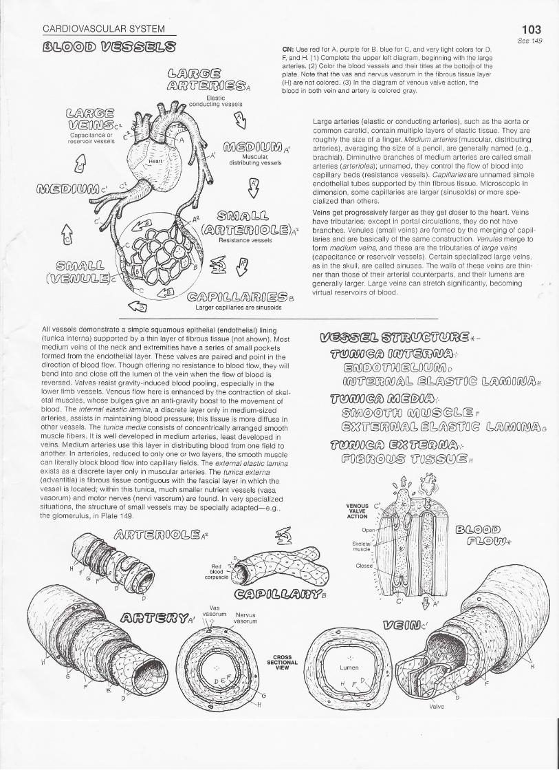
102See 119, lSD, 152
Circulation of blood begins with the heart, which pumps bloodinto arteries and receives blood from veins. Arteriesconduct
blood away from the heart regardless of the amount of oxygen(oxygenation) in that blood. Veins conduct blood toward theheart, regardless of the degree of oxygenation of the blood.Capillaries are networks of extremely thin-walled vesselsthroughout the body tissues that permit the exchange of gasesand nutrients between the vessel interior (vascular space) andthe area external to the vessel (extracellular space). Capillar-ies receive blood from small arteries and conduct blood tosmall veins.
~~&uma@@OOO@QfJf:l:OJ110@fi!)D
Carbon dioxide,waste products
~"6
. . ..... .. . ., .'.
Oxygen.nutrients
..
....'
Carbon dioxide
There are two circuits of blood flow: (1) the pulmonary circuit,which conveys blood from the right side of the heart to thelungs and fresh blood back to the left side of the heart, and(2) the systemic circuit, which conveys blood from the leftheart to the body tissues and returns blood to the right heart.The color red is used universally for oxygenated blood. andthe color blue is used for oxygen-poor blood.
Clearly, not all arterial blood is fully oxygenated (in the pulmo-nary circulation, arteries conduct poorly oxygenated blood tothe lungs). and not all venous blood is oxygen deficient(pulmonary veins conduct oxygenated blood to the heart).
Capillary blood is mixed; it is largely oxygenated on the arterialside of the capillary bed, and it is largely deoxygenated on thevenous side. as a consequence of delivering oxygen to andpicking up carbon dioxide from the tissues it supplies.
One capillary network generally exists belvv'een an artery anda vein. There are exceptions: the portal circulation of the liverinvolves two sets of capillaries between artery and vein (Plate119); the hypophyseal portal system involves two capillarynetworks between artery and vein (Plate 152); and the renalvascular system has a glomerulus and a peritubular capillaryplexus between artery and vein (Plate 150).
CARDIOVASCULAR SYSTEM
OO(b@@[Q)W@@~cglb~
@Ji)@lli>OQDG'!VA'Muscular,
distributing vessels
A'L ~~l%(1~
(~(IDl?@OOO@l1~)A~Resistance vessels
" /
~--/{g~[PDCb{1tmOOOfE@BLarger capillaries are sinusoids
103See 149
CN: Use red for A, purple for B, blue for C, and very light colors for 0,F, and H. (1) Complete the upper left diagram, beginning with the largearteries. (2) Color the blood vessels and their titles at the botto{Tlof theplate. Note that the vas and nervus vasorum in the fibrous tissue layer(H) are not colored. (3) In the diagram of venous valve action, theblood in both vein and artery is colored gray.
Large arteries (elastic or conducting arteries), such as the aorta orcommon carotid, contain multiple layers of elastic tissue. They areroughly the size of a finger. Medium arteries (muscular, distributingarteries). averaging the size of a pencil, are generally named (e.g.,brachial). Diminutive branches of medium arteries are called smallarteries (arterioles); unnamed, they control the flow of blood intocapillary beds (resistance vessels). Capillaries are unnamed simpleendothelial tubes supported by thin fibrous tissue. Microscopic indimension, some capillaries are larger (sinusolds) or more spe-cialized than others,
Veins get progressively larger as they get closer to the heart. Veinshave tributaries; except in portal circulations, they do not havebranches. Venules (small veins) are formed by the merging of capil-laries and are basically of the same construction. Venules merge toform medium veins, and these are the tributaries of large veins(capacitance or reservoir vessels). Certain specialized large veins,as in the skull, are called sinuses. The walls of these veins are thin-ner than those of their arterial counterparts, and their lumens aregenerally larger. Large veins can stretch significantly, becomingvirtual reservoirs of blood.
All vessels demonstrate a simple squamous epithelial (endothelial) lining(tunica intema) supported by a thin layer of fibrous tissue (not shown). Mostmedium veins of the neck and extremities have a series of small pocketsformed from the endothelial layer. These valves are paired and point in thedirection of blood flow. Though offering no resistance to blood flow, they willbend into and close off the lumen of the vein when the flow of blood is
reversed. Valves resist gravity-induced blood pooling, especially in thelower limb vessels. Venous flow here is enhanced by the contraction of skel-etal muscles, whose bulges give an anti-gravity boost to the movement ofblood. The internal elastic lamina, a discrete layer only in medium-sizedarteries, assists in maintaining blood pressure; this tissue is more diffuse inother vessels. The tunica media consists of concentrically arranged smoothmuscle fibers. It is well developed in medium arteries, least developed inveins. Medium arteries use this layer in distributing blood from one field toanother. In arterioles, reduced to only one or two layers, the smooth musclecan literally block blood flow into capillary fields. The external elastic laminaexists as a discrete layer only in muscular arteries. The tunica externa(adventitia) is fibrous tissue contiguous with the fascial layer in which thevessel is located; within this tunica, much smaller nutrient vessels (vasavasorum) and motor nerves (nervi vasorum) are found. In very specializedsituations, the structure of small vessels may be specially adapted-e.g.,the glomerulus. in Plate 149.
CROSSSECTIONAL
VIEW
D
~@1~~"*-1?@Ci!JO@f1)O£i!)(J@(J](jiY)cm-;.
@UID@@Vao@(bDOD~D
O@'[i'@OO@]&l1 @[1,tA)@'iJO@(1~~OUtYJl%E
llQQ@JO@&Ju.YiJ@@)Oif:N
@GMi)@@'U'GOGi!A)OD@@l1:.~F
@~lJ@OO~(JJ(1 @(b,LA)@'IT'O@l1l%(itA)O~l%G
'fl(lfJ(i!)O@(f;) @l)37J'§(IDfi!)(tj-(-
C?O@OO@QD§'fJO@@(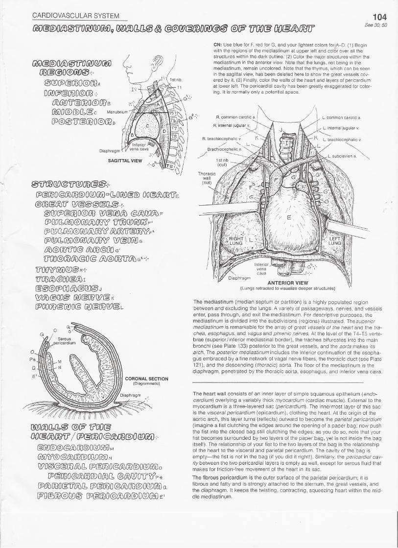
CARDIOVASCULAR SYSTEM'
OO(gf2)OfA)@ffO(i!}Ql)[M)9W~(bl1@ ~ @@f!J@OODli!J@@@£P 1f(}{)(g OO@~OOff
01l{f)[!f)@1?(JfJOO@f§J-;-fP&@O@~OO@OC!D!XA)oCbO~@@)OO~ro'U'E@OO@cfJ'f)W@@@~f1§ -~-
@@7~@OOO@OOW@@iA) @fl0WIft:JF{P1J!J(bU1!iiJ@(j\[)tJ.)OOW'iJOOQD(jj'[J{EF'
~QQI1~@~~W L%OO'iJ§OOWF~[jJQ!J(b@iJ@Ui'!JtJ0cmfW@OG\DGaJ@fX)U'O@ {t;J(1J@{}DG'
'U'(X)@@t%@O@ ~@OOTI'~G"-;-
1fGOWfiCiJG!)@H-{-
'U@&:>@OO&LAjI
~@@IPO{J&>@(!O@ J
W~@@@ Ul'£}@OOW(3K
rPOOOO~@O@U!)§OOQ?@L
Q
E'CORONAL SECTION
(Diagrammatic)
/Wa)(b(b@ @(;? fi'OO&OO@(A){M1f/ CP&OOO@~@)O@(li)-:-
&fA!)@@@~@@OODU\!iiJMG\YAJ\?@@~ro@)OCW@li)N
WO@@&OO/JJf1 [J)@;OOO@(f;J!XJIDOQ!)@o!P@roo@~OO@)O~f1@(f;JWOtll((p*
~<%OOO@'lJtA)(b[P&@O @&JOO@OWGtA)Q
WO(!)tM@Q!)~ ~@OOO@~OO@)OQD@(ve:'
104See 30, 50
CN: Use blue for F, red for G, and your lightest colors for A-D. (1) Beginwith the regions of the mediastinum at upper left and color over all thestructures within the dark outline. (2) Color the major structures within themediastinum in the anterior view. Note that the lungs, not being in themediastinum, remain uncolored. Note that the thymus, which can be seenin the sagittal view, has been deleted here to show the great vessels cov-ered by it. (3) Finally, color the walls of the heart and layers of pericardiumat lower left. The pericardial cavity has been greatly exaggerated for color-ing. It is normally only a potential space.
L. common carotid a.
~/R. brachiocephalic v.
/' ""Brachiocephalic a.
// ---
ANTERIOR VIEW(Lungs retracted to visualize deeper structures)
The mediastinum (median septum or partition) is a highly populated regionbetween and excluding the lungs. A variety of passageways, nerves, and vesselsenter, pass through, and exit the mediastinum. For descriptive purposes, themediastinum is divided into the subdivisions (regions) illustrated. Thesuperiormediastinum is remarkable for the array of great vessels of the heart and the tra-chea, esophagus, and vagus and phrenic nerves. At the level of the T4- T5 verte-brae (superior finferior mediastinal border). the trachea bifurcates into the mainbronchi (see Plate 133) posterior to the great vessels, and the aorta makes its
arch. The posterior mediastinum includes the inferior continuation of the esopha-gus embraced by a fine network of vagal nerve fibers, the thoracic duct (see Plate121). and the descending (thoracic) aorta. The floor of the mediastinum is thediaphragm, penetrated by the thoracic aorta, esophagus, and inferior vena cava.
The heart wall consists of an inner layer of simple squamous epithelium (endo-cardium) overlying a variably thick myocardium (cardiac muscle). External to themyocardium is a three-layered sac (pericardium). The innermost layer of this sacis the visceral pericardium (epicardium), clothing the heart. At the origin of theaortic arch, this layer turns (reflects) outward to become the parietal pericardium(imagine a fist clutching the edges around the opening of a paper bag; now pushthe fist into the closed bag still clutching the edges; as you do so, note that yourfist becomes surrounded by two layers of the paper bag, yet is not inside the bagitself). The relationship of your fist to the two layers of the bag is the relationshipof the heart to the visceral and parietal pericardium. The cavity of the bag isempty-the fist is not in the bag (if you did it right!). Similarly, the pericardial cav-ity between the two pericardiallayers is empty as well, except for serous fluid thatmakes for friction-free movement of the heart in its sac.
The fibrous pericardium is the outer surface of the parietal pericardium; it isfibrous and fatty and is strongly attached to the sternum, the great vessels, andthe diaphragm. 11keeps the twisting, contracting, squeezing heart within the mid-dle mediastinum.
CARDIOVASCULAR SYSTEM
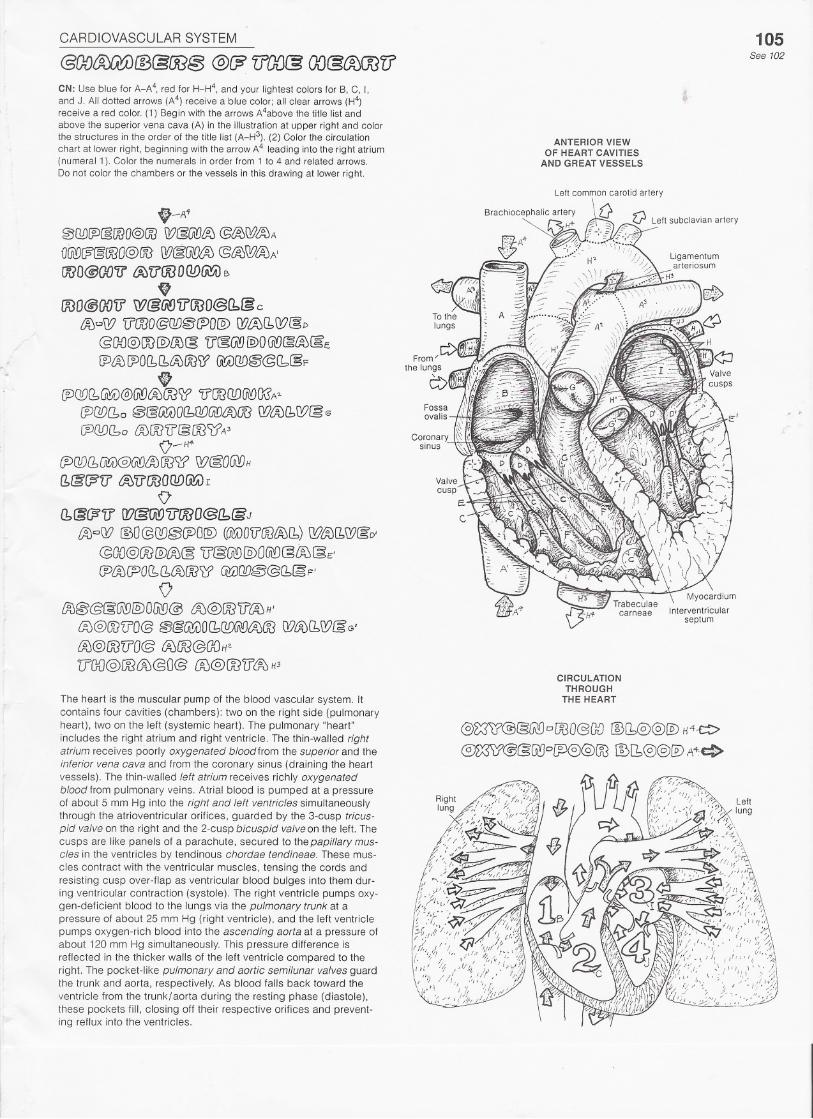
@(){)~lB@ro@ @(F lfOOC3GO@~LfCN: Use blue for A_A4, red for H_H4, and your lightest colors for B, C, I,and J. All dotted arrows (A4) receive a blue color; all clear arrows (H4)receive a red color. (1) Begin with the arrows A4above the title list andabove the superior vena cava (A) in the illustration at upper right and colorthe structures in the order of the title list (A_H3). (2) Color the circulationchart at lower right, beginning with the arrow A4 leading into the right atrium(numeral 1). Color the numerals in order from 1 to 4 and related arrows.Do not color the chambers or the vessels in this drawing at lower right.
"-A~0fY)(j)@OOO@OO\}?@(ji!J8J@%\W~Aa@f?&OOO@OOW~~ @l%WiA)A'OOO@OO'irLA)V'OOOQD~B.OOO@(}OU'W@~'irOOO@(bcg c
~CW 'iJ'OOO&C!DfflrPO@W~I1\}?§j)@I}O@)OO@OO@U'~@)@)OG\!J~~@:E!P8JIJ>OI1I1tA)OO~@A)@@@(1§F.
lPC?D(bOO@(RD~~\(?'iJ'OOODG\I0O\?A?[P(J[)Cbo ~@li!A)O(b{]OCii!J~){X3G?LA)Cb\}?@6
IJJI1DCbo ffiOO'fJ' § 00 CClA3
O-H4-tP(Jj)(b@1)@@J~OOWW@O@HCb&CP'V'l%'U'OOOQD@A)r
0'(1@(P'il fll@jj'f)<[fWO@fb(gJ
fAjcC}?@O@Q[)@(J)O@ (6i'0D'f?@tA)[b)W~!1W&D'@OO@OO@~~ 'i5'§U\9@)OGi'D@{A)§E'CP~(POI1(bl%OO)j> Q0lJD~@(b@ F'
o£m@@§@[§)O!XD@ ~@OO'i?t%H'
~@OO'fJ'O@ @@'@{)O(1@J(iiY)l%OOG?~(1W§ G'
!AJ@[i3ffO@ tJ.JOO@(}{JH"lfOO@OO~@O@iA)@(]]lf60H3
The heart is the muscular pump of the blood vascular system. Itcontains four cavities (chambers); two on the right side (pulmonaryheart). two on the left (systemic heart). The pulmonary "heart"includes the right atrium and right ventricle. The thin-walled rightatrium receives poorly oxygenated blood from the superior and theinferior vena cava and from the coronary sinus (draining the heartvessels). The thin-walled left atrium receives richly oxygenatedblood from pulmonary veins. Atrial blood is pumped at a pressureof about 5 mm Hg into the right and left ventricles simultaneouslythrough the atrioventricular orifices, guarded by the 3-cusp tricus-pid valve on the right and the 2-cusp bicuspid valve on the left. Thecusps are like panels of a parachute, secured to the papillary mus-cles in the ventricles by tendinous chordae tendineae. These mus-cles contract with the ventricular muscles, tensing the cords andresisting cusp over-flap as ventricular blood bulges into them dur-ing ventricular contraction (systole). The right ventricle pumps oxy-gen-deficient blood to the lungs via the pulmonary trunk at apressure of about 25 mm Hg (right ventricle). and the left ventriclepumps oxygen-rich blood into the ascending aorta at a pressure ofabout 120 mm Hg simultaneously. This pressure difference isreflected in the thicker walls of the left ventricle compared to theright. The pocket-like pulmonary and aortic semilunar valves guardthe trunk and aorta, respectively. As blood falls back toward theventricle from the trunk/aorta during the resting phase (diastole),these pockets fill, closing off their respective orifices and prevent-ing reflux into the ventricles.
105See 102
ANTERIOR VIEWOF HEART CAVITIES
AND GREAT VESSELS
CIRCULATIONTHROUGH
THE HEART
@~rl?@§Gi!!J°OOO@GO@(b@@@) H4~
~@@G!J°~OO IDI1@@@)A4~
CARDIOVASCULAR SYSTEM
@~OO@OtA)@@@(i!)@CJ!J@VO@@~@'[f&@A) &JJ'flDO& @@@
106
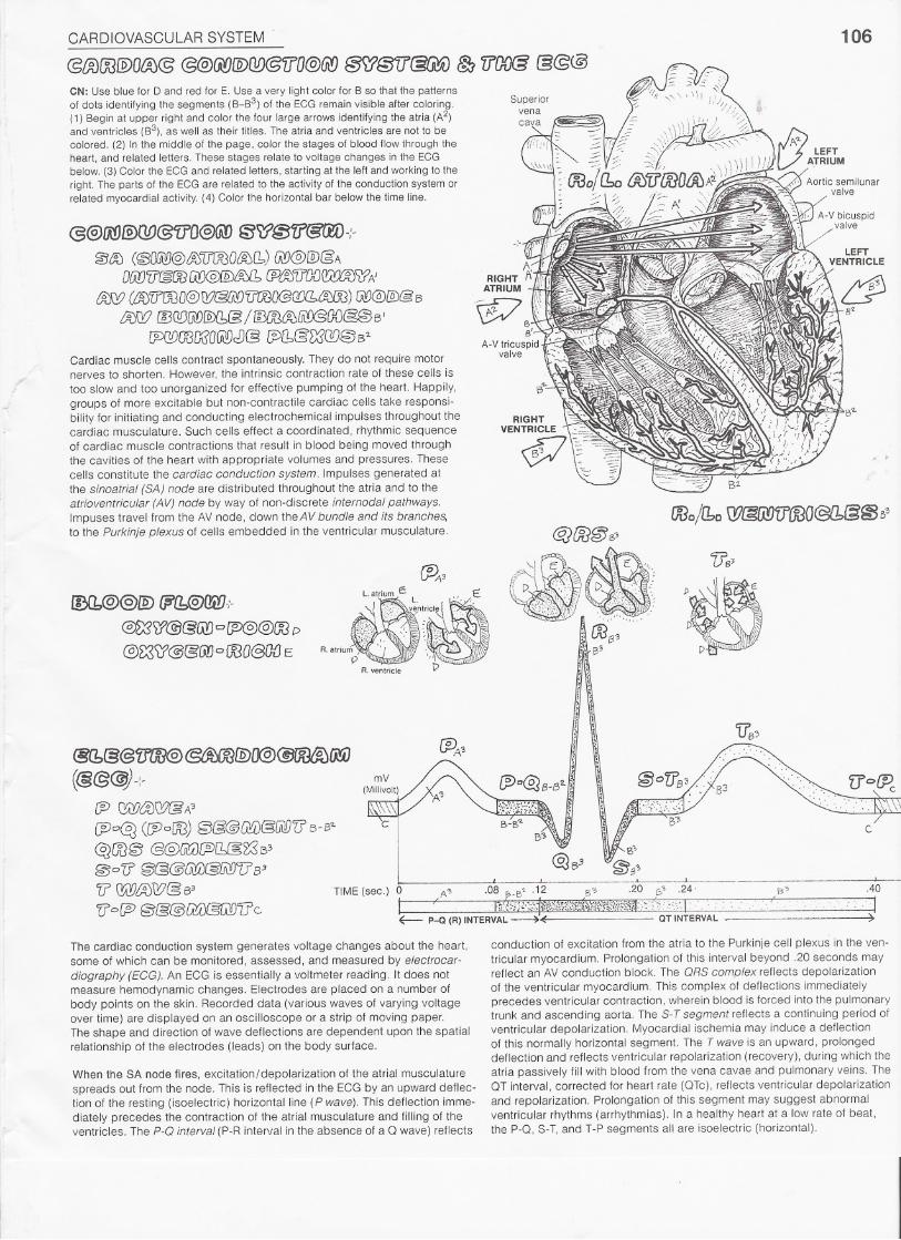
CN: Use blue for 0 and red for E. Use a very light color for B so that the patternsof dots identifying the segments (B-B3) of the ECG remain visible after coloring.(1) Begin at upper right and color the four large arrows identifying the atria (A2)and ventricles (B3), as well as their titles. The atria and ventricles are not to becolored. (2) In the middle of the page, color the stages of blood flow through theheart, and related letters. These stages relate to voltage changes in the ECGbelow. (3) Color the ECG and related letters, starting at the left and working to theright. The parts of the ECG are related to the activity of the conduction system orrelated myocardial activity. (4) Color the horizontal bar below the time line.
@@fM)@O!J@'[f[J@(i.!)f3~~tr@lED-:-@ro (~ffiYJ@&>"iJOOOtmCb)W@[QJ@A
O(jJJ'fl@OOfi[)@@&>11 (jJ~"U'OO(~~)li?A'
!fS:!)(l%V"OOo@W@,i'[)v=roO@m~) @@@)@B
/A)W @Q!)[W[QJ(1@/IDOOtJ;)[ji!J@(]{J§@B'
~mlJl5aGi!!JJJ@1Pl1&lX?(][)@62
Cardiac muscle cells contract spontaneously. They do not require motornerves to shorten. However, the intrinsic contraction rate of these cells is
too slow and too unorganized for effective pumping of the heart. Happily,
groups of more excitable but non-contractile cardiac cells take responsi-bility for initiating and conducting electrochemical impulses throughout thecardiac musculature. Such cells effect a coordinated, rhythmic sequenceof cardiac muscle contractions that result in blood being moved throughthe cavities of the heart with appropriate volumes and pressures. Thesecells constitute the cardiac conduction system. Impulses generated at
the sinoatrial (SA) node are distributed throughout the atria and to theatrioventricular (AV) node by way of non-discrete internodal pathways.Impuses travel from the AV node, down theAV bundle and its branches,to the Purkinje plexus of cells embedded in the ventricular musculature.
ID(b@@lIDfP'fb@f!irJ.;-
@i:3'!f@@@ofP@@roP
@~<i1@&G!J 0 OOO@()(JE
'.
1\RIGHT11
]\
ATRIUM . .'r-£7 S.~ \\B ,
a'A-V tricuspid
valve
@1@@<[]W@~@(}@@~(jriJ
(&@~-:-fP \JA:!J&fi!J&A'(P"'~ (@oW) 0&@Gti)@CiIf)(] B-S"
~m@ @@[li)[JJ(l:'(~~53
0"'V @@@@i)@[j\f)1J'B3'f? QW~W@ 63 TIME(sec.) 0fj' o[p @@@Q;{)@(ill{Jc
A'" .08
..20 s' .24'
..40
aT INTERVAL ~
The cardiac conduction system generates voltage changes about the heart,some of which can be monitored, assessed, and measured by electrocar-diography (EGG). An ECG is essentially a voltmeter reading. It does notmeasure hemodynamic changes. Electrodes are placed on a number ofbody points on the skin. Recorded data (various waves of varying voltageover time) are displayed on an oscilloscope or a strip of moving paper.The shape and direction of wave deflections are dependent upon the spatialrelationship of the electrodes (leads) on the body surface.
When the SA node fires, excitation/depolarization of the atrial musculature
spreads out from the node. This is reflected in the ECG by an upward deflec-tion of the resting (isoelectric) horizontal line (P wave). This deflection imme-diately precedes the contraction of the atrial musculature and filling of theventricles. The P-Q interval (P-R interval in the absence of a 0 wave) reflects
conduction of excitation from the atria to the Purkinje cell plexus in the ven-
tricular myocardium. Prolongation of this interval beyond .20 seconds mayreflect an AV conduction block. The QRS complex reflects depolarizationof the ventricular myocardium. This complex of deflections immediatelyprecedes ventricular contraction, wherein blood is forced into the pulmonarytrunk and ascending aorta. The S- T segment reflects a continuing period of
ventricular depolarization. Myocardial ischemia may induce a deflectionof this normally horizontal segment. The T wave is an upward, prolongeddeflection and reflects ventricular repolarization (recovery), during which the
atria passively fill with blood from the vena cavae and pulmonary veins. TheOT interval, corrected for heart rate (OTc), reflects ventricular depolarizationand repolarization. Prolongation of this segment may suggest abnormalventricular rhythms (arrhythmias). In a healthy heart at a low rate of beat,the P-O, SoT,and T-P segments all are isoelectric (horizontal).

CARDIOVASCULAR SYSTEM'
@@OO@~~OO11~OO{f@OOO@@~ ~@Ot%@ W@OG!J~CN: Use your brightest colors for A, 0, and L. (1) When coloring the arteries,include the broken lines that represent vessels on the posterior surface of theheart. (2) Do the same with the veins. (3) Color the artery in front of the plaque inthe circled view; color the vessel after the plaque a lighter shade of the samecolor or do not color it at all.
@@ro@oo&>oow ~OO'U'@OOO~ ','.
filO@O(Jrr@@OO@(;{)lA)m~A
&AJQ!)@@@C!lA)OOOOOO~(l)@(}{) A'
Gt'£t)LA)OO@O(ii!]~[1 IDOOiA)lA!'J@!X)B
(p@@'[j'@/130@OO D@'i1~OOW@@'iJOOO@QQG.{A)OO
(@)@@@&~@)OOO@) @OOiA)(jlfl@(x)c
Cb(j~ff @@ro@(ID~OOfi1D
~lA'O'i?§OOO@OO OW'il@OOCO@@'U'/KJO@Q£)(1[%)(M
@@@@@@@)O@J@) @JOO§.)(j\!)@(]{)E
G!iJ(][)0@lJDCbaJ@ IDrotAJ(jifJ@O{JE'
@OOO@8D~@'(1§mIDOOt%~@OOF
The coronary arteries form an upside-down crown (L. corona) about or justdeep to the surface of the heart. The arteries lie in grooves, or sulci, oftencovered over by the epicardium and sometimes the myocardium as well.
Both left and right arteries arise from small openings (aortic sinuses) justabove the two aortic semilunar valve cusps. Generally, the left coronaryartery is somewhat larger than the right; during the cardiac cycle. the flowrate through the left is greater in most people than that through the right.There may be considerable variation in the anastomotic pattern of the leftand right arterial branches. These branches terminate in multitudes ofarterioles supplying the vast capillary network among the muscle fibers.The apparent multiple communications among the left and right coronaryarteries notwithstanding, varying degrees of vascular insufficiency occurwhen there is significant obstruction of one or both coronary arteries. Thereis some extra-coronary arterial supply to the heart from the epicardial ves-sels (branches of internal thoracic arteries) and aortic vasa vasorum.
107
ANTERIORVIEW
G\!;t)W@@~OO@)at%CbO@I?~OO@'[f'O@~*
Damage to the intimal layer of coronary arteries can occur with lipiddeposition or inflammation. Platelet aggregation at these sites contributes tothe formation of plaque (cell material, lipid, platelet, fibrin). Plaque builds upwithin the vessels, forming thrombi that occlude the vessels in progressivelygreater degrees. Significantly reduced blood flow to the myocardium(ischemia) can cause sharp pain (angina) to the chest, back. shoulder.and arm as well as permanent damage to the myocardium (infarction).
@c:%OO@)O~@ W~Offi'9~-:-
@OO@0&)LF @~OO@)O~@~ G
~O@[Q)[b@ @l%OO@)O~@ WeH
~l%OO@OINJ~[1 Wo r
{JJ(A!)'fl@@O@@ @~OO[Q)O~@ WcJ
fB@i)&J(1fl. @l%OO@O~@ WoK
@@OO@ IAQ~OO1?~O(iiY)(J{J@ L.
The cardiac veins travel with the coronary arteries, but incompletely. Vastanastomoses of veins occur throughout the myocardium; most drain into theright atrium by way of the coronary sinus. The anterior cardiac veins conductblood directly into the right atrium. Other small veins may drain directly intothe right atrium as well. Some deep (arteriosinusoidal) veins drain directlyinto the atria and ventricles. Extracardiac venous drainage can also occurthrough the vasa vasorum of the vena cavae.
CARDIOVASCULARSYSTEM'
a>OO'il'(gOOO@@@W 'flOO(g GO@~@) E!3'(A'{)(g@fXS
.Thyroid cartilage
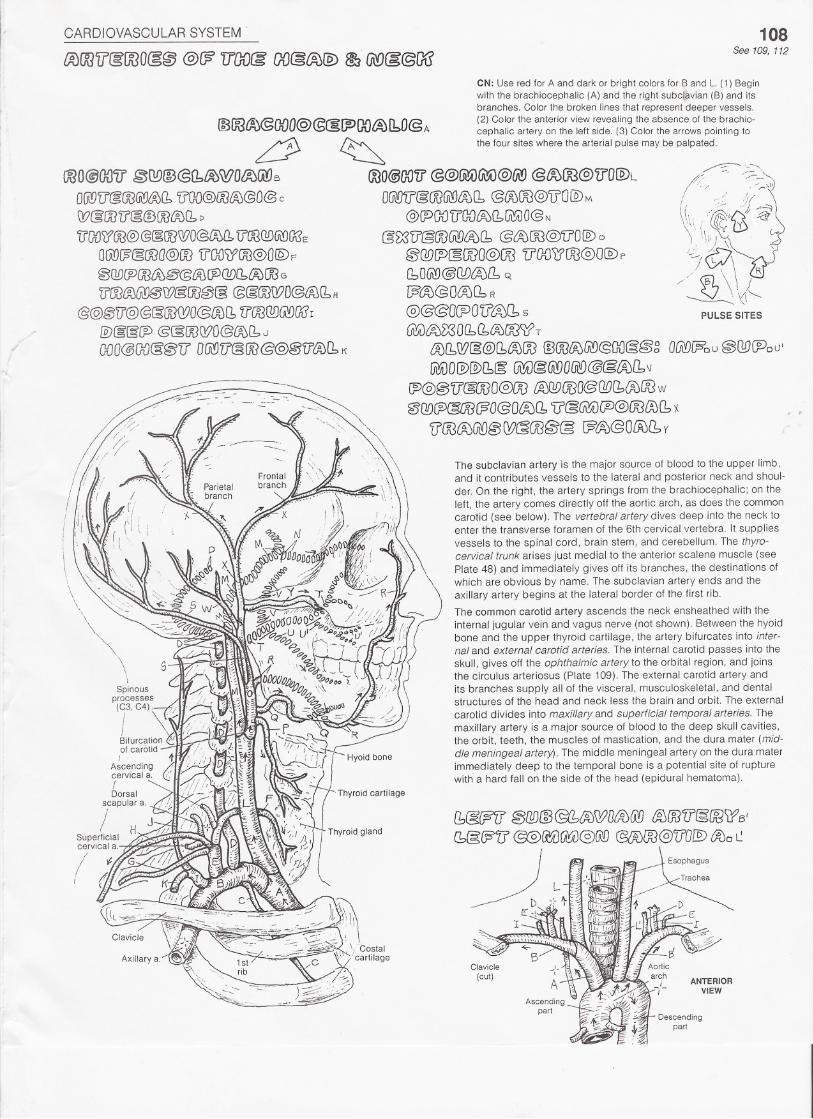
108See 109. 112
CN: Use red for A and dark or bright colors for Band L. (1) Beginwith the brachiocephalic (A) and the right subclavian (B) and itsbranches. Color the broken lines that represent deeper vessels.(2) Color the anterior view revealing the absence of the brachio-cephalic artery on the left side. (3) Color the arrows pointing tothe four sites where the arterial pulse may be palpated.
ANTERIORVIEW
Descendingpart
CARDIOVASCULAR SYSTEM .
OlA!Yij@OO(j1)~(b~ro@ffO@)A
~~V'(gOOO@OO@§ID§(i300fAJI1B(fiJGt!J'illEOOO@OO@@Gi!A)[0iJ(]J]@O@tt;)lYOW@~
G\!A)O@[Q)(1,§@@@@@OOlA)11 t>
IP@@'U'@OOO@OO@@~Cii!A)@(ii!)O@~ITO(ji'!)@E
(Anterior)
Pontinea."',"
Pons
CIRCULUSARTERIOSUS
(Circle of Willis)
Labyrinthine a.
109See 108
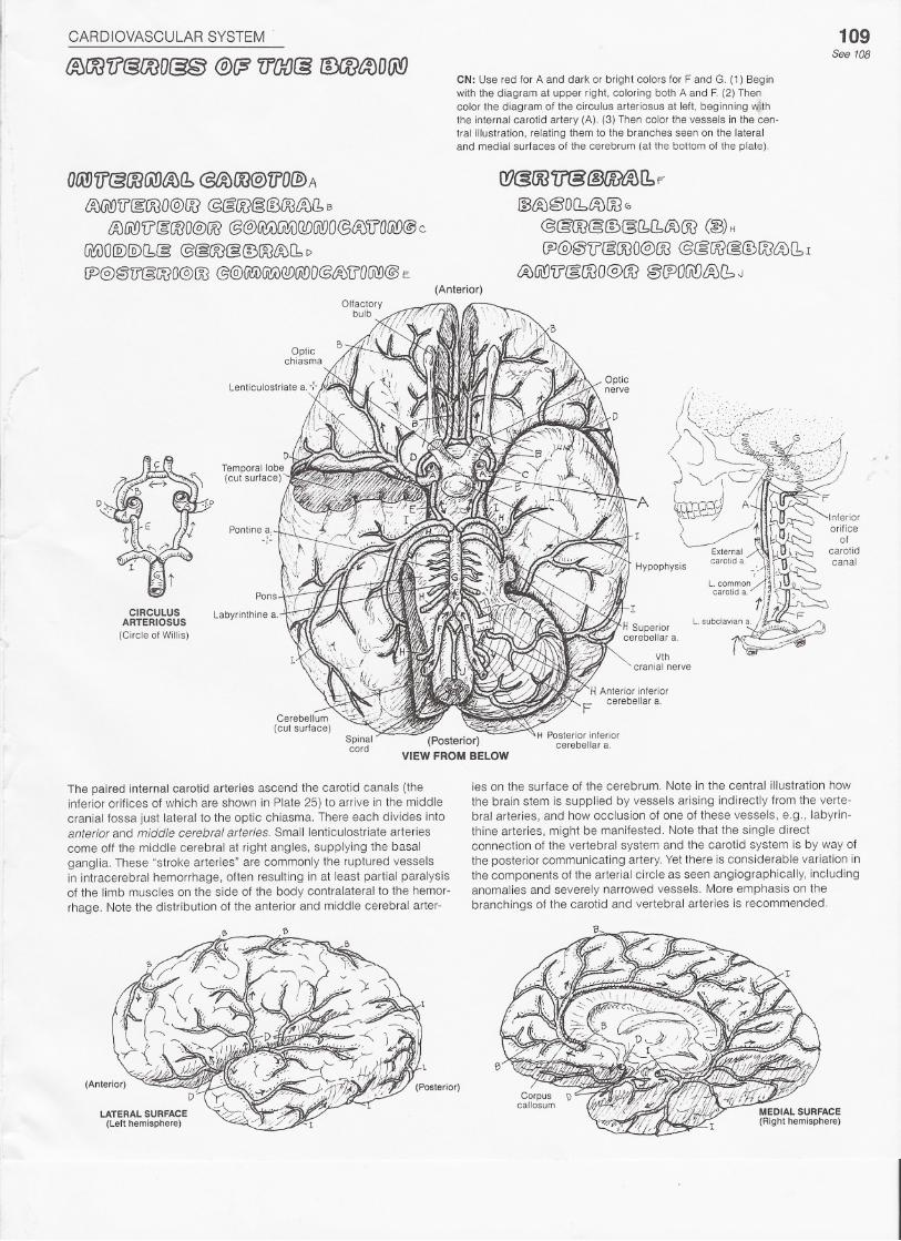
CN: Use red for A and dark or bright colors for F and G. (1) Beginwith the diagram at upper right, coloring both A and F. (2) Thencolor the diagram of the circulus arteriosus at left, beginning withthe internal carotid artery (A). (3) Then color the vessels in the cen-tral illustration. relating them to the branches seen on the lateraland medial surfaces of the cerebrum (at the bottom of the plate).
Q7@OOlr@~I1F~t%@O(btA)OOG
@@[email protected])OO(~) H
CP@@(J'@OOO@OO@@OO§@[X]lAJCbI
~@'[f&roo@oo @CPOGIDt%fbJ
(Posterior)VIEW FROM BELOW
The paired internal carotid arteries ascend the carotid canals (theinferior orifices of which are shown in Plate 25) to arrive in the middlecranial fossa just lateral to the optic chiasma. There each divides intoanterior and middle cerebral arteries. Small lenticulostriate arteriescome off the middle cerebral at right angles, supplying the basalganglia. These "stroke arteries" are commonly the ruptured vesselsin intracerebral hemorrhage, often resulting in at least partial paralysisof the limb muscles on the side of the body contralateral to the hemor-rhage. Note the distribution of the anterior and middle cerebral arter-
LATERAL SURFACE(Left hemisphere)
ies on the surface of the cerebrum. Note in the central illustration howthe brain stem is supplied by vessels arising indirectly from the verte-bral arteries, and how occlusion of one of these vessels, e.g., labyrin-thine arteries, might be manifested. Note that the single directconnection of the vertebral system and the carotid system is by way ofthe posterior communicating artery. Yet there is considerable variation inthe components of the arterial circle as seen angiographically, includinganomalies and severely narrowed vessels. More emphasis on thebranchings of the carotid and vertebral arteries is recommended.
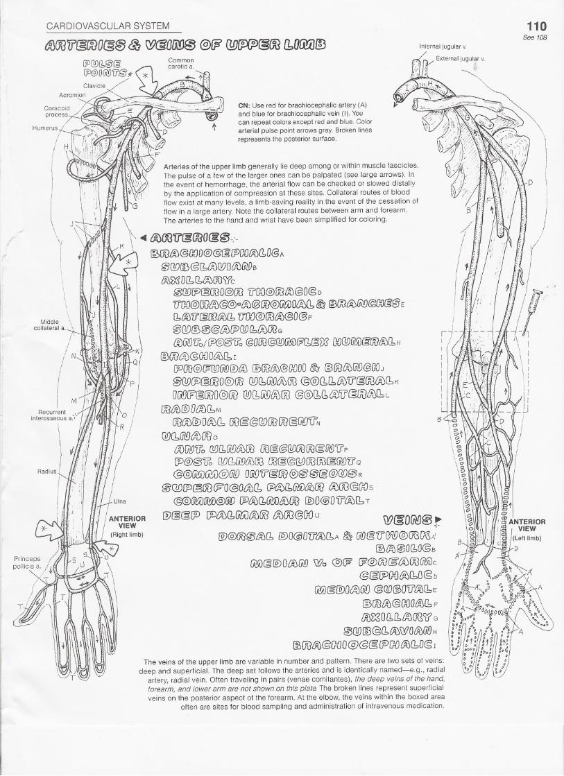
CARDIOVASCULAR SYSTEM 110See 108
Internal jugular v.
CN: Use red for brachiocephalic artery (A)and blue for brachiocephalic vein (I). Youcan repeat colors except red and blue. Colorarterial pulse point arrows gray. Broken linesrepresents the posterior surface.
Arteries of the upper limb generally lie deep among or within muscle fascicles.The pulse of a few of the larger ones can be palpated (see large arrows). Inthe event of hemorrhage, the arterial flow can be checked or slowed distallyby the application of compression at these sites. Collateral routes of bloodflow exist at many levels, a limb-saving reality in the event of the cessation offlow in a large artery. Note the collateral routes between arm and forearm.
\ The arteries to the hand and wrist have been simplified for coloring.
\ ~ £%OO'lf&OOO(g@-:-K
Middlecollateral a.
Recurrent ,interosseous a'-'-
\\\\
[;)OO8J@OOD@@[EfP[XJI1;)(10@A
~QD@@(btA)Q?OtAJGWB
~'iX3 DflJbtJJOO)fc
&BflDrP@OOO@OO'iJ'DD@OO~@O@D'U'ao@roL~)(5@o£Aj@OO@GU) 00;)(1,f£'i@3(ftJ{A)GiY)@(]{)&fB£[biA)V@OOiAJ[bVOO@(fJ(i)@O@F
~(][)@0@tftJ[PGD(b6.)OOG .
{fJ@)'U'o/ (P@~'i?o @OW@@(K;i)@[1@;83DO@)UUJ@~&>11H
@ro~@ODO{A)[b I
CPOO@(?QD(ii1D@)/A)(])OO&@OOOO&J @roffiGiD@[X)J@W!P§OOO@OO@C!:,@t:A;OO@@(111lft.)V'@@iA)CbK
aG\DW@OOO@)OO@)(b@iA)w @@(bCh~lJ@OOV0o:'LOOttJ ID D{ftJ f.b M
OO@@)OlA)(1 OO@@@JOOOO@Gi!OV'N
QDC1Ui'DaJOO0
a)@)'U'o C!Dl1Gi9fA)fjJOO~@(WOOOO(gUl'9'U'P
[P@@'i?oQD!1G\9~(Ji3OO@@(Y)OOOO(g~U'Q@@~Gt:V@@ O&D'i?@OO@@@@@(][J@ R
~QD(P@OO WO@OLA)(b[j)~(bfKA)&JOOtA)(JJ@OOs@@(Ka)~@(jJ) [;JtAj!1@;U&JOO@)O@OU't%[bT
@§@!P (P~Cb1KA)£%@ iAJOO@ODu W&O~@r@)@OO@&J11 @O@O'[l~l1A ~ (i\\)@'[lGW@OOWA'
~l%@O(lO@B
@i)@@)Ol%lADWo @)W ~@ro@'WOO(j\)jik@~JPGD£A)110@D
@i)@'@)O~~ @C!D~O'U'L%(bE
@OOlAJ@OOOl%fbF
LR)~O(1(1 l% (KSl? G
0ODID@(1tAJWOt%GI'9H
fSOOL%@G=OO@@&fPfXJ~[bD(s 1
CARDIOVASCULAR SYSTEM
IAJOO1lcgOOO(g@@W 1fOOcg(b@W~OO CbOrNAJOO
\\ii
\
i\I\
\
\I
I,I;
IjI
N \Descending
gfniCUlara.
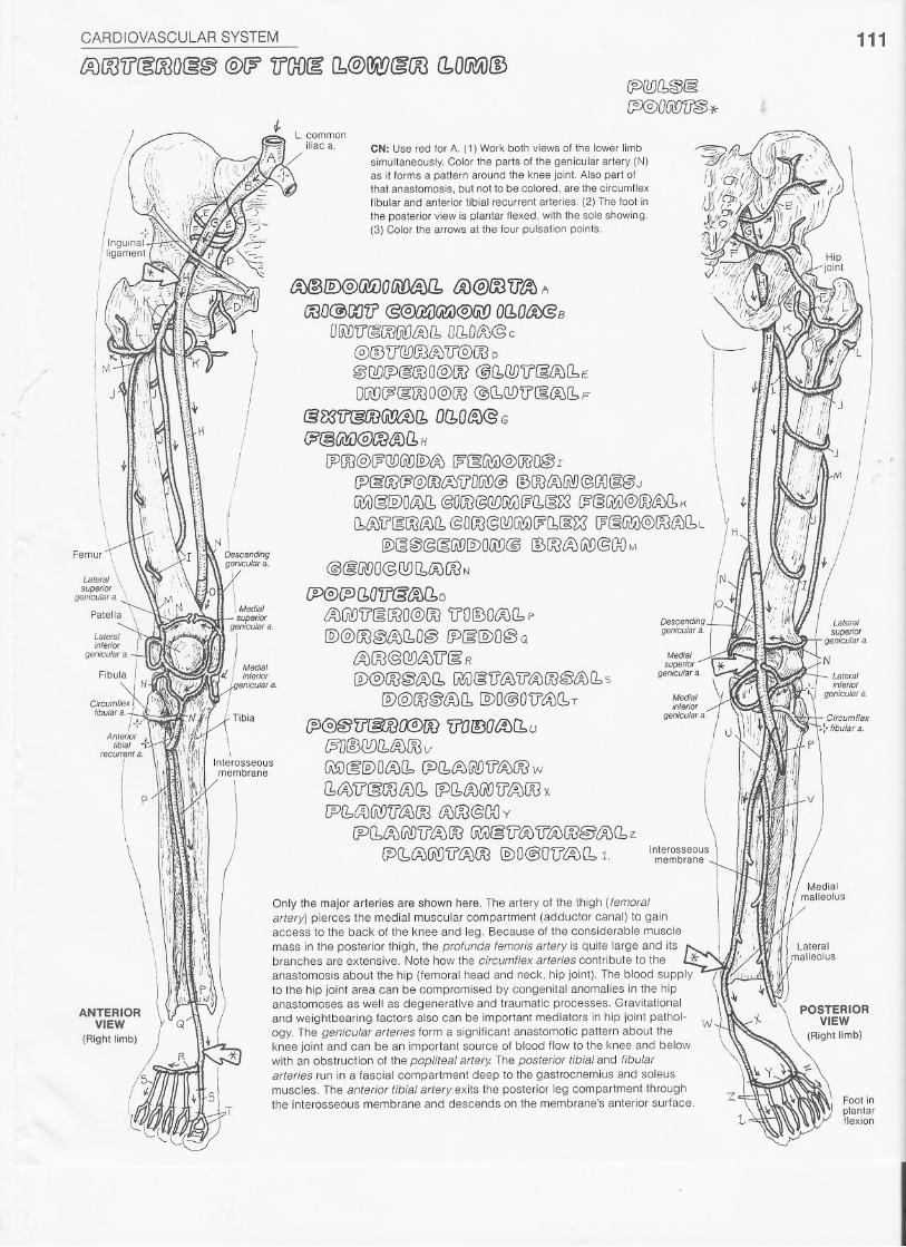
111
L. commoniliac a. CN: Use red for A. (1) Work both views of the lower limb
simultaneously. Color the parts of the genicular artery (N)as it forms a pattern around the knee joint. Also part ofthat anastomosis, but not to be colored, are the circumflexfibular and anterior tibial recurrent arteries. (2) The foot inthe posterior view is plantar flexed, with the sole showing,(3) Color the arrows at the four pulsation points.
~@@arvolW~a. tV@ro'[f~ A
CX3D@(J[J71@~~ 0l10t(€B
OfjJ)'[J'@!XJ(j1f)t%[l,0f10~@ c '\.-@@V~~~V@OODI@Q£){P@@O@OO @CbJYJ'iJ'[gLAJf1e: , ,
O@(p(gOOO@OO@CbQDU'@:OOCbp I .Jlilil&~'ir@roGl!7a){1o~atJJ@G !
~@~@m~~H \~OO@WGDG\!)[V~[?~~@OOO~I \ " f. ~:I
[JJ§rol?@OO~VO@@ @OOO,JIft1)@G{J@@J
\. ;;c'Ii,"
G&!§@)OlA)(1, @OOO@QD[~H?(1~m/?§lA'A)@OO&J(1,K t I';~I'I
[b6J'i?[gOO~11 @OOO@@~0?[b§~ (?(3~@~{fJfbL i H : .)I!/ftJ@)~@@§(ill@)OUlD@ ~OO~GlD@aoM
j' " . ill,;/
@@/ADO@QDl1iAJOON N </;"""",'f"OVO\TO\n n<i'?r-i!/;;I.n 'I, fllilllJ""'1.:::IV'-'~ U 13UVlbO I I , ,;"1 ,
'0"" ; t ".,
LAjGiDV§OOO@OOV'OIDO£AjCbP Descending" ,/ {II'@@OO@/JJf10@(J)g[§)O@Q geniculara, I!.. '. ~~J~
~~@QD00'iJ@: R
ID@OO~lA)[1 [Wl)@U'LA)1J'~ro~~Q,S@)@OO@wf1 @)O@O'[f't:4)(hT
(p~'iJ'@1)(@ro 'ifaIDOaJCbu~D@(!!)f1&>OOV
G\YV@@)OfA)f1IP(1~~),ifLA>OO w
11tA')'U'@OOLA)[b ~[b~(N)U'~OO X
[P(b~U\'!JTI'lA)OO iAJro@G{)y
(Pf1<%~'jj~OO G\'A)§U'OOlf~(]f@LAj(1 z
~Cb(A)U\D1f'6J~IDO@OU'I%(11.
Medialsuperior
geniculara,
Interosseousmembrane
Only the major arteries are shown here. The artery of the thigh (femoralartery) pierces the medial muscular compartment (adductor canal) to gainaccess to the back of the knee and leg. Because of the considerable musclemass in the posterior thigh, the profunda femoris artery is quite large and itsbranches are extensive, Note how the circumflex arteries contribute to the I..!!'anastomosis about the hip (femoral head and neck, hip joint). The blood supplyto the hip joint area can be compromised by congenital anomalies in the hipanastomoses as well as degenerative and traumatic processes. Gravitationaland weightbearing factors also can be important mediators in hip joint pathol-ogy. The genicular arteries form a significant anastomotic pattern about theknee joint and can be an important source of blood flow to the knee and belowwith an obstruction of the popliteal artery The posterior tibial and fibulararteries run in a fascial compartment deep to the gastrocnemius and soleusmuscles. The anterior tibial artery exits the posterior leg compartment throughthe interosseous membrane and descends on the membrane's anterior surface,
w
I
CARDIOVASCULAR SYSTEM
tA)@OOU'~& ~OO~~@OO{g~
~OO@)@lM)O£OOtA)a.@@OO1?t:%A2
@&I1UtAJ@'ilOO[/j)(j\f)C;JL
[b@f?'iJ" @£%@U'OOO@M
@~Cb@G\DO@N
@@Gi!V(;i!;V@GiJO[)O@;~{g)1JO@0
@(lD[J)(gOOO@OO @t)@@@fF!)'[f~mO@ p
OO@'W)LAJCbQ1f@@710@@JCb~OO/ @WlA)OOO~(AORQ,QO~~OOsO@J(?@OOO@OO@i)@@@@'U'@OOO@r
@@@i)~@@J DCbO~@v
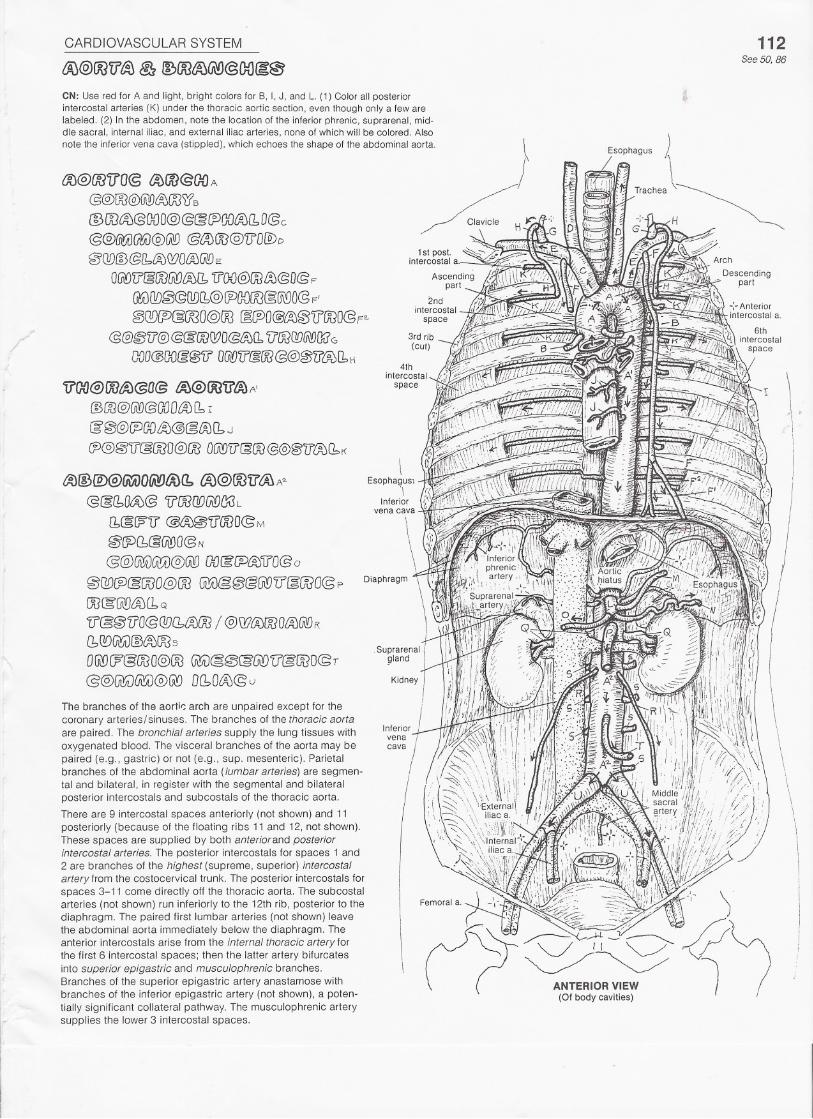
The branches of the aorti~ arch are unpaired except for thecoronary arteries/sinuses. The branches of the thoracic aortaare paired. The bronchial arteries supply the lung tissues withoxygenated blood. The visceral branches of the aorta may bepaired (e.g., gastric) or not (e.g., sup. mesenteric). Parietalbranches of the abdominal aorta (lumbar arteries) are segmen-tal and bilateral, in register with the segmental and bilateralposterior intercostals and subcostals of the thoracic aorta.
There are 9 intercostal spaces anteriorly (not shown) and 11posteriorly (because of the floating ribs 11 and 12, not shown).These spaces are supplied by both anterior and posteriorintercostal arteries. The posterior intercostals for spaces 1 and2 are branches of the highest (supreme, superior) intercostalartery from the costocervical trunk. The posterior intercostals forspaces 3-11 come directly off the thoracic aorta. The subcostalarteries (not shown) run inferiorly to the 12th rib, posterior to thediaphragm. The paired first lumbar arteries (not shown) leavethe abdominal aorta immediately below the diaphragm. Theanterior intercostals arise from the internal thoracic artery forthe first 6 intercostal spaces; then the latter artery bifurcatesinto superior epigastric and musculophrenic branches.Branches of the superior epigastric artery anastamose withbranches of the inferior epigastric artery (not shown), a poten-tially significant collateral pathway. The musculophrenic arterysupplies the lower 3 intercostal spaces.
112See 50. 86
~ndintercostal
space
3rd rib(cut)
4thintercostal
space
\ .ii
III
\Esophagus;
'\Inferior
'vena cava
\
CARDIOVASCULAR SYSTEM
t%m?'iJ&OOOrn:@U'@@&@lf~@OfilDLf@~(jO~~(1ITOO~lj~ @~~1J(g@) @@@~@@
113See 156
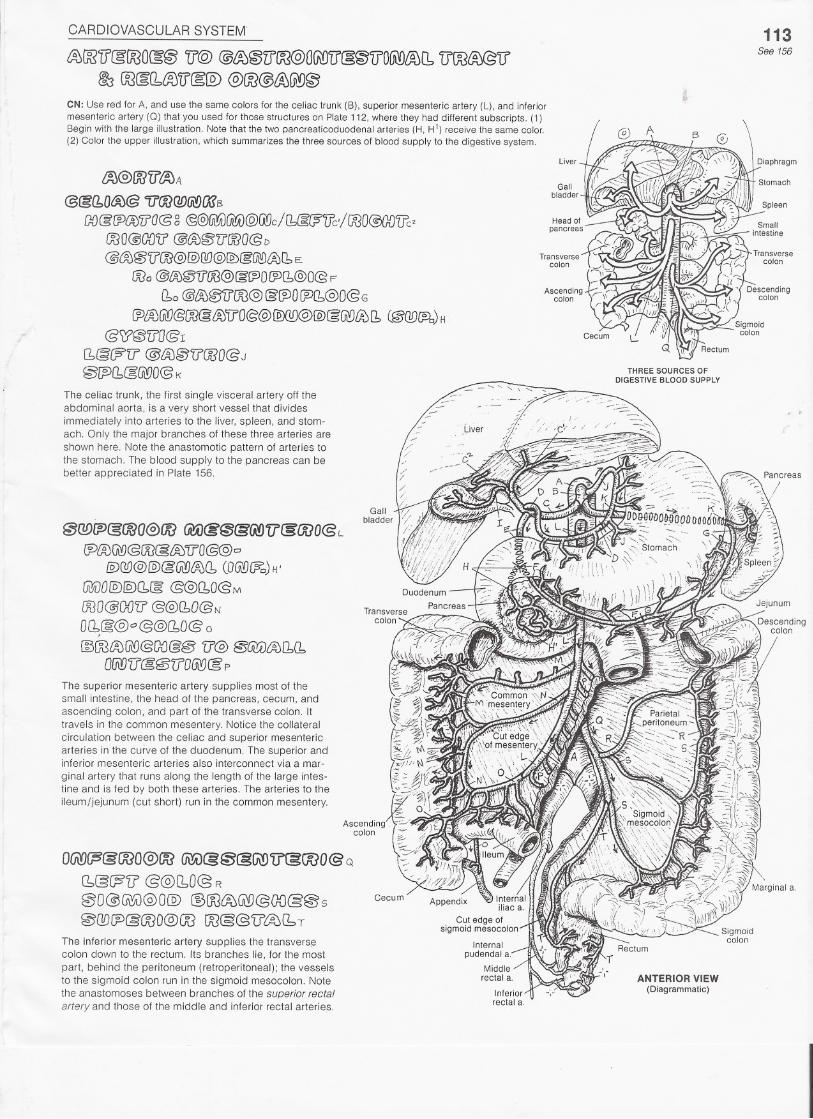
CN: Use red for A, and use the same colors for the celiac trunk (B), superior mesenteric artery (L), and inferiormesenteric artery (Q) that you used for those structures on Plate 112, where they had different subscripts. (1)Begin with the large illustration. Note that the two pancreaticoduodenal arteries (H, H1) receive the same color.(2) Color the upper illustration, which summarizes the three sources of blood supply to the digestive system.
£%@)OOlrt%A
@@(bD~@ 11'@CJ!)(ji'l)(]8B
DiJ&fPCA)'ltO@g @@@JiJ@i)@@)c![b@:l?iJc';OOO@()[J'[fczOOO@GO'U'@@@'[)OOO@D@l%@tJOO@@)UD@I0@,~iftJ(bE
000 @t%~'ifro@@(PO~(b@O@ F
(10 @/%@lrOO@§'(PO[?J!1@O@G
[PLA)(ji!}@OO@I%'i]'O@@@@)@@§GI!J{A)!1 (@OD~o) H
@)J@'t]O@r
[1§{J?'[J @LA)@1t'OOO@J
@fPf1cgWO@ K
The celiac trunk, the first single visceral artery off theabdominal aorta, is a very short vessel that dividesimmediately into arteries to the liver, spleen, and stom-ach. Only the major branches of these three arteries areshown here. Note the anastomotic pattern of arteries tothe stomach. The blood supply to the pancreas can bebetter appreciated in Plate 156.
@Q!)f:)@@O@@@iJ~@&(jyJ1i§roo@ L
~l%Gifl@OO@&>"[f'O@@o
!EXi!J@@@@)tA)(1(O@~) H'
GiJiJO@[§X1[g@@)(10@N\
OOO@OO'U' @@(bO@N
D~@@"'@@(10@ 0§OOmJ@@G{)@§ 'if@ @Gi!iJtA)[1(1
OWJ(]@@5(J'OUi!J@p
o~~@ooo@oo(b@(;S"i? @@(10@ R
00@~@O@) @@lft0@@OO@@s@[][J[P@@O@(K3ro§@'U'~(b T
The inferior mesenteric artery supplies the transvers!3colon down to the rectum. Its branches lie, for the most
part, behind the peritoneum (retroperitoneal); the vesselsto the sigmoid colon run in the sigmoid mesocolon. Notethe anastomoses between branches of the superior rectalartery and those of the middle and inferior rectal arteries.
Liver Diaphragm
Gallbladder
Stomach
Spleen
Smallintestine
Transversecolon
Ascendingcolon
THREE SOURCES OFDIGESTIVE BLOOD SUPPLY
ANTERIOR VIEW(Diagrammatic)
CARDIOVASCULAR SYSTEM
~OO'if(gOOOcg@@f? 1100& jj)@'l1WO@'<$ lP@OOO(A'D~Q!)1NJD
114See 52,53, 61, 111
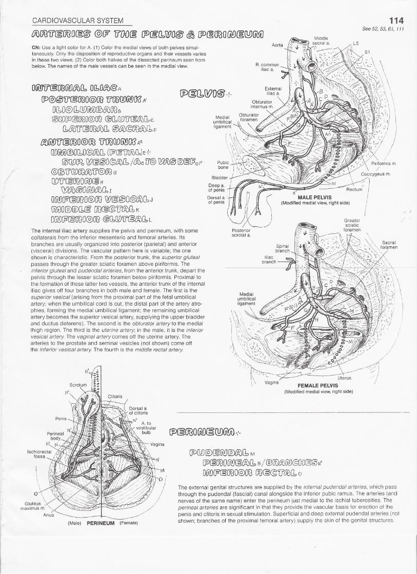
CN: Use a light color for A. (1) Color the medial views of both pelves simul-taneously. Only the disposition of reproductive organs and their vessels variesin these two views. (2) Color both halves of the dissected perineum seen frombelow. The names of the male vessels can be seen in the medial view.
O(i!J'lf@OOGlOt%f1OI1Ol%@ A
~@~lr@OOO@OO 'lJOOOf)fA!)CKSA'
O(bO@!1QD@JD~LAJOO6
@C!D!P@OOO@im@l1QD'iJ[g~(be
[bl%'D'(§OO<%(1 @i%@OOWEbD
~'U'@OOO@OO llOOODfi\!)Q(?A1.
@[0i)@OCbO@{A){1(W&'U'WCb)E-:-
0Gre?, W@@O@£%(b /&00 (]'@ ~@ @§(?op
@(SLrcwrol%U'@OOG
QO(J'@OOOGOO@HWlAJ@O(R{)~(1I
a~w@ooo@ooW§@O@{R)(1J@Ji)O@ffi@' OO@@U~Q, K
OUill@@OOO@@@Cb(1)Lr@~(1L
The internal iliac artery supplies the pelvis and perineum, with somecoilaterals from the inferior mesenteric and femoral arteries. Itsbranches are usually organized into posterior (parietal) and anterior(visceral) divisions. The vascular pattern here is variable; the oneshown is characteristic. From the posterior trunk, the superior glutealpasses through the greater sciatic foramen above piriformis. Theinferior gluteal and pudendal arteries, from the anterior trunk, depart thepelvis through the lesser sciatic foramen below piriformis. Proximal tothe formation of these latter two vessels, the anterior trunk of the internaliliac gives off four branches in both male and female. The first is thesuperior vesical (arising from the proximal part of the fetal umbilicalartery; when the umbilical cord is cut, the distal part of the artery atro-phies, forming the medial umbilical ligament; the remaining umbilicalartery becomes the superior vesical artery, supplying the upper bladderand ductus deferens). The second is the obturator artery to the medialthigh region. The third is the uterine artery; in the male, it is the inferiorvesical artery. The vaginal artery comes off the uterine artery. Thearteries to the prostate and seminal vesicles (not shown) come offthe inferior vesical artery. The fourth is the middle rectal artery.
Bladder
Deepa..ofpenis-iDorsal a. . .of penis .
(lr' (UPosteriorscrotal a.
II
/
(Male) PERINEUM (Female)
FEMALE PELVIS(Modified medial view, right side)
CPM@~G\'£J@)@(1M~@OOOUiYJ@&J(1N/ @~~(ill@CDcg~N'OU\YJW@OOO@OOOO@@'U'&>(10
The external genital structures are supplied by the internal pudendal arteries, which passthrough the pudendal (fascial) canal alongside the inferior pubic ramus. The arteries (andnerves of the same name) enter the perineum just medial to the ischial tuberosities. Theperineal arteries are significant in that they provide the vascular basis for erection of thepenis and clitoris in sexual stimulation. Superficial and deep external pudendal arteries (notshown; branches of the proximalfemoral artery) supply the skin of the genital structures.
CARDIOVASCULAR SYSTEM'
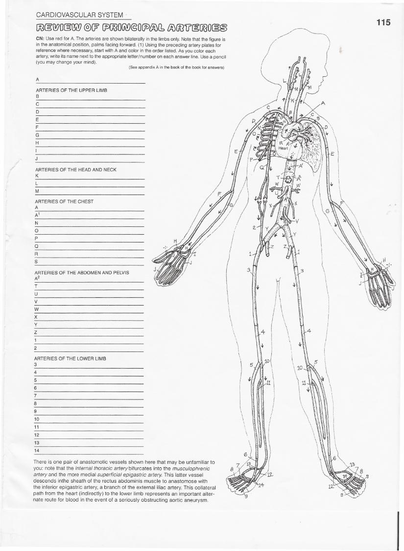
!m@;WOcgW@(? (pOOO~@OCF>t%(1tAJOO'iJ(gOOO(3@CN: Use red for A. The arteries are shown bilaterally in the limbs only. Note that the figure isin the anatomical position, palms facing forward. (1) Using the preceding artery plates forreference where necessary, start with A and color in the order listed. As you color eachartery, write its name next to the appropriate letterlnumber on each answer line. Use a pencil(you may change your mind).
(See appendix A in the back of the book for answers)
A
ARTERIES OF THE UPPER LIMBB
C
D
E
F
G
H
I
J
ARTERIES OF THE HEAD AND NECKK
L
M
ARTERIES OF THE CHESTA
A'
N
oP
QR
S
ARTERIES OF THE ABDOMEN AND PELVISA2
T
U
V
W
X
Y
Z
1
2
\
\\i\\
\\
)
!I
\\
\\
ARTERIES OF THE LOWER LIMB3
4
5
6
7
8
9
10
11
12
13
14
There is one pair of anastomotic vessels shown here that may be unfamiliar toyou: note that the internal thoracic artery bifurcates into the musculophrenicartery and the more medial superficial epigastric artery. This latter vesseldescends inthe sheath of the rectus abdominis muscle to anastomose with
the inferior epigastric artery, a branch of the external iliac artery. This collateralpath from the heart (indirectly) to the lower limb represents an important alter-nate route for blood in the event of a seriously obstructing aortic aneurysm.
115
:3
1
3'iJI
-l-
I
4 (II
5 IwiI
.J.N A111 \III{
/iII
CARDIOVASCULAR SYSTEM
WrgO@!)@ @W 1100& OO@'iA)@) &!JGiY)@@(]\?
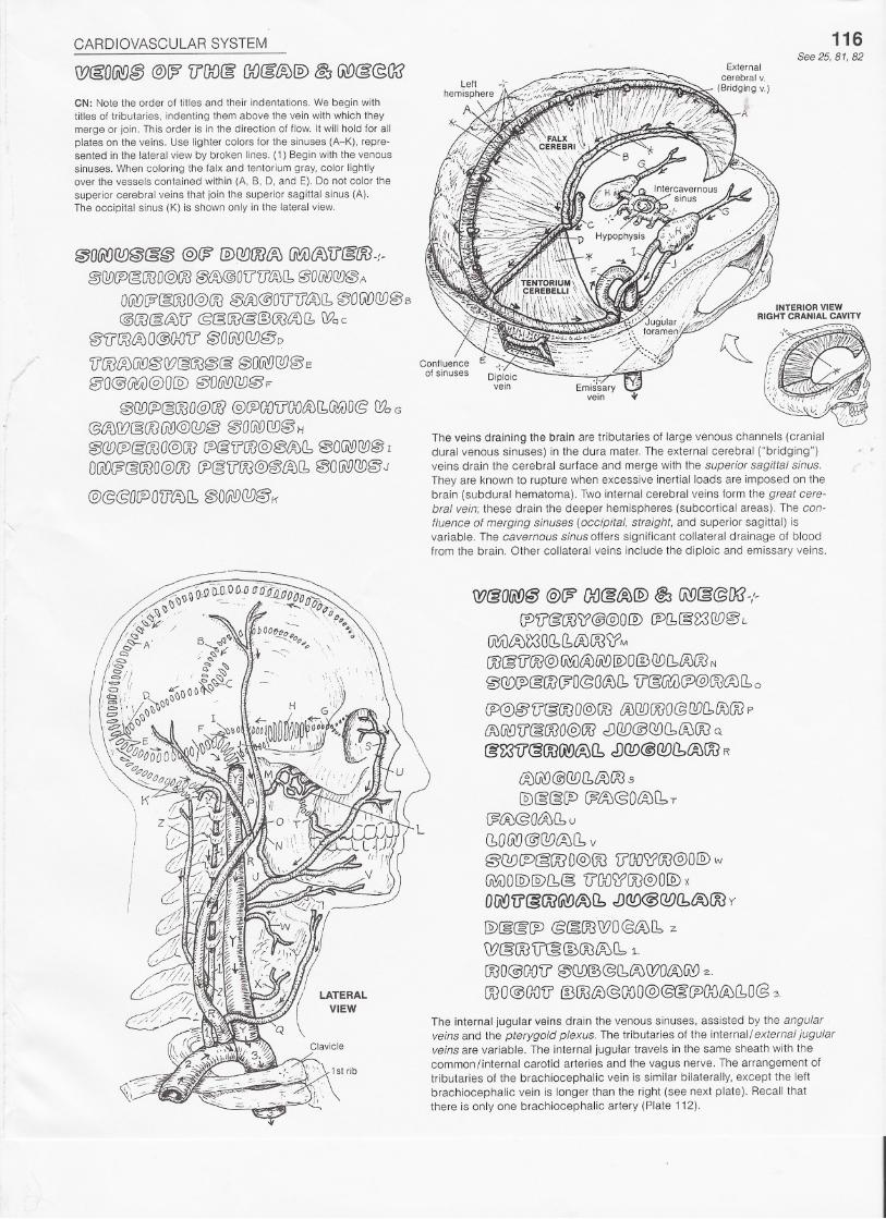
CN: Note the order of titles and their indentations. We begin withtitles of tributaries, indenting them above the vein with which theymerge or join. This order is in the direction of flow. It will hold for allplates on the veins. Use lighter colors for the sinuses (A-K), repre-sented in the lateral view by broken lines. (1) Begin with the venoussinuses. When coloring the falx and tentorium gray, color lightlyover the vessels contained within (A, B, 0, and E). Do not color thesuperior cerebral veins that join the superior sagittal sinus (A).The occipital sinus (K) is shown only in the lateral view.
@06irJ@@@@ @(fi' @C!!JOO/R) U\a)(ftSfl@OO-:~@@(P~OOO@OO~@OU'(JiA)(1, @O(i(QC!!J@A
OGiDW&OOO@@~£AJ@D'U'(JiAJ(1,~OfAD@)08
@(2@tft;)'[f cS§OO@@OO~11 Wo c
@'[flX$iAJO@(NJi]'00G\98D@D
'iJW&V0i'0@(!J~~@@ @OUiYJ@)~E
§'O@(iiYJi)@O@) @O@[l!JgJF
@QD~@OOO@IJ} @~GOl]'GO~[1~O& (}?oG
@tA)Q?@OO~@8D~@OG\iJO!J@Ii
@@~&OOG@OO fP&'ff'OO@&B&JCb@OGi:D8D@I
OGl!)@@OOO@OO/P@U'OO@@{AJ(1,@OG\YJ@@J
@@@O~O()lAJl1 @O~Q!)@11'
116See 25,81,82
The veins draining the brain are tributaries of large venous channels (cranialdural venous sinuses) in the dura mater. The external cerebral ("bridging")veins drain the cerebral surface and merge with the superior sagittal sinus.They are known to rupture when excessive inertial loads are imposed on thebrain (subdural hematoma). Two internal cerebral veins form the great cere-bral vein; these drain the deeper hemispheres (subcortical areas). The con-fluence of merging sinuses (occipital, straight, and superior sagillal) isvariable. The cavernous sinus offers significant collateral drainage of bloodfrom the brain. Other collateral veins include the diploic and emissary veins.
W@Ofil!J~@f? OO@~@ ~ 1A!J(g@/x?-,'-
(P'[(@OO)1@@OID~11§~ [W@L~§,)~O(1,Q,LAJOO)?MOO@U'OO@~lA)lJiY) @)OIDQD[blA)OON
~QO~@roC?O@OcA)11V'@(iU)@@OO(A)(1,o
CP@@'U'@(X]O@OOtA)[l!JOOO@[]£)£1lAJOOp
{l,J@'i]@OOO@OO cDQD@QDCbroOOQ
@W<300!A'D£A)(bcDQD@Q!)Ib~OOR
(A)G\!)@QD[1&JOO5
@)~@(P (;?~@O~[b" T
(J?fA)@O~Cb U
(bOG\!]@@)OJI1 v
~@J~@OO O@)OO'ilIXJWOO@OIDw~O@)@)l1§ 'lfao~oo@O@) x
OU\'{)lr@OO@~Cb.!J@@GDlb£A)OOy
@@&IP @(gOOWO@OJI1z
W§OOV'@IDOO{A)\1,1.
OOO@OOV 0QD@@11ift0Q?OLftJ~~.
OOO@OOU'IDOO~@(x)O@@@(POOlAJ(1,O@~.
The internal jugular veins drain the venous sinuses, assisted by the angularveins and the pterygoid plexus. The tributaries of the internal/external jugularveins are variable. The internal jugular travels in the same sheath with thecommon/internal carotid arteries and the vagus nerve. The arrangement oftributaries of the brachiocephalic vein is similar bilaterally, except the left
brachiocephalic vein is longer than the right (see next plate). Recall thatthere is only one brachiocephalic artery (Plate 112).
CARDIOVASCULAR SYSTEM
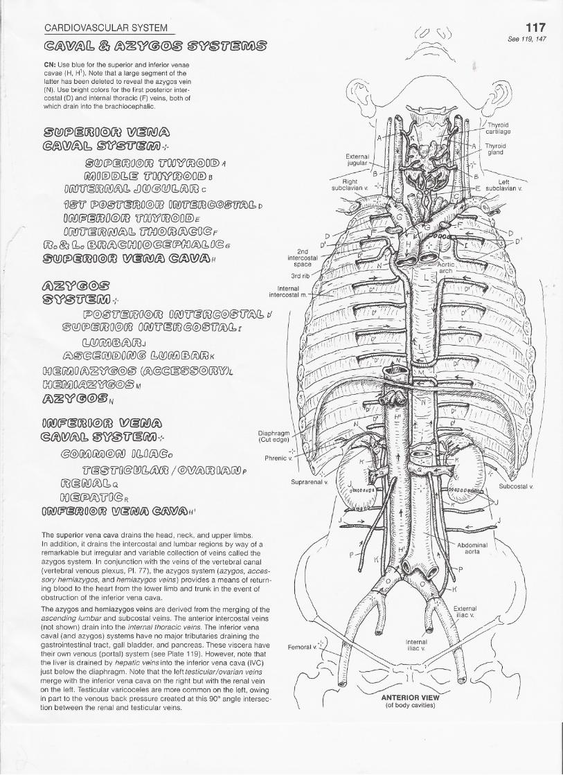
@~~~11 ~ t%~~@@~ ~<iY~V~@',V~CN: Use blue for the superior and inferior venaecavae (H. H1j. Note that a large segment of thelatter has been deleted to reveal the azygos vein(N). Use bright colors for the first posterior inter-costal (0) and internal thoracic (F) veins. both ofwhich drain into the brachiocephalic.
~(Y}£p~OOO@)OOW~~~@l%WtA)[b @¥J@1f(g(j{jiJ-f.
@@[P@OOO@OO'U'OOl:?OO@O@)t1
@;UO@)[Q)I1§7l00):?OO@O@BOW(],§OOU\!JtAJf1dJ(]f)@QOr1~OOc
fJ@ll~@@U'@OOO@OOOfA'VU'@OO@@@U'l%(1D
OGl(J@§£K30@Gf)V[}{)/fOO@O@/:
OWJ'il~OO(ji:!J~(bU'OO@OO(A)@O@F000@j(10 @@{A)@GDO@@@CPGDl%110@G
@Q[){P@OOO@OOW(g~ffi @~\YJ~H
D~CP~OOO@OOQ?@,~~@lA)W~Q" ~g;U"cg~-:.
@@~[jjYii)@~ Dl1000@o
'[J'@@'U'O@[J[J[btA:;OO/ @Wi%OOO/%@poo@(iiYJiAJ(bQ
OO@~<%iJ'O@ROfiWW@OOO@)OO\YJ~fA9/j;j~o/JfJ:JH'
The superior vena cava drains the head. neck, and upper limbs.In addition, it drains the intercostal and lumbar regions by way of aremarkable but irregular and variable collection of veins called theazygos system. In conjunction with the veins of the vertebral canal(vertebral venous plexus, PI. 77). the azygos system (azygos, acces-sory hemiazygos, and hemiazygos veins) provides a means of return-ing blood to the heart from the lower limb and trunk in the event ofobstruction of the inferior vena cava.
The azygos and hemiazygos veins are derived from the merging of theascending lumbar and subcostal veins. The anterior intercostal veins(not shown) drain into the internal thoracic veins. The inferior venacaval (and azygos) systems have no major tributaries draining thegastrointestinal tract, gall bladder, and pancreas. These viscera havetheir own venous (portal) system (see Plate 119). However, note thatthe liver is drained by hepatic veins into the inferior vena cava (IVC)just below the diaphragm. Note that the left testicular lovarian veinsmerge with the inferior vena cava on the right but with the renal veinon the left. Testicular varicoceles are more common on the left, owingin part to the venous back pressure created at this 90° angle intersec-tion between the renal and testicular veins.
117See 119, 147
CARDIOVASCULAR SYSTEM
W§O~@ @ff?U'OO(g[b@W@OO110~OO
ANTERIORVIEW
(Dorsumof foot)
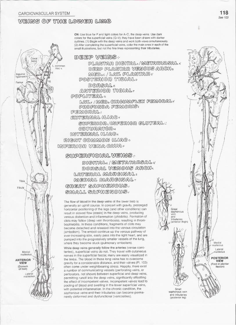
CN: Use blue for P and light colors for A-O, the deep veins. Use darkcolors for the superficial veins (Q-V); they have been drawn with darkeroutlines. (1) Begin with the deep veins and work both views simultaneously.(2) After completing the superficial veins, color the main ones in each of thesmall illustrations, but not the fine lines representing their tributaries.
118See 103
ID~[g(P W~OG\fl~-;-
CP(1iA)U\!J'i}tiJOO@O@Otf/%!1A/ ~~'[f~TI'l%OO~(1,!\ID~~~ [J)Q,{gJW\ilti)OO W@:(AY)@C!D@~OO@OOB
~(g@)cC / (1£%'i)o~(1~~U'~OO c'~@@'[J~OOO@(]] 'iY0I!30~0:. D
@@)OO@/A)(1E~~V'(groo@oo 'iJO@OaJQ"F
lY@rPQ,O'iJ[g~(1 G
f1iAJ'iloHI ~@@)c @OOO@GD~(F(b@L;S@§tii!;i)@OO&)!1H'
[POO@{¥?QD~@)~W§~@OOO@r[?@~@ooro[bJ@mU'@OOGi:!J~(10I10~@ t{
@Q!)[P@X30@OOL/OUi!JW&OOO@ffi)@!1Q!)'i]'@iAJCb~
@~ 'U'ODOOLAYiJ@OOM
OGi'D'i1§OOGlQ/AJ11OCbOlA)@N
OOO@OO'1f@@lA!Ji)~@(jiYJ OQ,Ol%@ 0
O@~§OOO@)OO W(]:G\D~@[f;';R'!}(f0 p
@Q!)/p(gOO(?O@Otro(bW§O~~ -,'.
@O@Olf'~(1Q / @U~V~TI'~OO~~I1~1@)@OO@~11 W@fiir)@W~ ~OO@OOR
f1,tAJ'[J@OO{ftJCbG1!A)~lm@O~tA)(1.s
G\£V@@OfA)[b~~OO@O@tA)(b T
@OO(g'~'il @{fJ(fJOOcg(j\9@W§u
@~~(1.11 @(jJ[ft)fJD(g(j()@QD@v
u
The flow of blood in the deep veins of the lower limb isgenerally an uphill course. In concert with gravity, prolongedhorizontal positioning of the legs (and other conditions) canresult in slowed flow (stasis) in the deep veins, producingvenous distention and inflammation (phlebitis). Formation ofclots may follow (deep vein thrombosis), resulting in throm-bophlebitis. In these conditions, fragments of clots maybecome detached and released into the venous circulation
(embolism). The emboli continue up the venous pathway ofever-increasing size, easily pass into the right heart, and arepumped into the progressively smaller vessels of the lung,where they become stuck (pulmonary embolism).
While deep veins generally follow the arteries (venae comi-tantes), superficial veins do not. They travel with cutaneousnerves in the superficial fascia; many are easily visualized inthe limbs. The blood in these long veins has to overcomegravity for a considerable distance, and their valves (pI. 103)often come under weightbearing stress. Happily, there exista number of communicating vessels (perforating veins, orperforators, not shown) between superficial and deep veins,permitting runoff into the deep veins, significantly offsettingthe effect of incompetent valves. Incompetent valves lead topooling of blood and swelling in the lower superficial veins,with potential inflammation. In the chronic condition, thesaphenous veins and their tributaries can become perma-nently deformed and dysfunctional (varicosities).
\J
Smallsaphenous vein
and tributaries(posterior leg)

CARDIOVASCULAR SYSTEM
oo@~~m@~@oov~~~~~~~~119
See 102. 144
CN: Use blue for I and a dark coli;>rfor J. (1) Color the veins draining the intestines, pancreas,gall bladder, and spleen. There are both left and right gastro-epiploic (0, 01) and gastric (G, G')veins. For the darkly outlined directional arrows adjacent to blood vessels, use the color of theblood vessel. (2) After coloring gray the inferior vena cava (*) and its tributaries (.j.1),the tribu-taries of the superior vena cava (",2), and their directional arrows, color the three large arrows(,,3) identifying anastomotic sites (include the esophageal veins passing posterior to the heart).
DDcglP~'ii'a@CP@OO'ii't%(b@~@'ircg@;i}-.'-@f!!)[J)~ooo@ro[JJ[§@'U'lA)(bA
orww§OOO@)OOGtV@~@:G\D'lf§OOO@B
(p<A)Gi!J@OOcg~'V'O@ c
(bo@~~U'OO@o~(J)O~I1@O@ D
@(P(1~~O@E
000@l%@V'OO@o@CPO(;)(1@O@D'
0QD~§OOO@OO ~§~§U\'Oll@OOO@ F
roo @tA:@TrOOO<sG
[bo @(AY~iJ'OOO@6'
@)Jg5'1]'O@H
(P@OO'tr~(hrQ{J@:[Pb;)'iJO@WOJ ~ UIDO~@1j'~@(7@@J'OG\D~W§Ui!JLm@ffi)W~* /71000IDQ!JU'1AJ1XW*1'iJ'OOOlEJo@@'@fJDfPoW§~.% @(JJ\!JIJJ*'2.
Capillaries of the gastrointestinal tract, gall bladder,pancreas, and spleen are drained by tributaries of thehepatic portal vein. Within the liver, branches of this vein(like those of an artery) discharge blood into capillaries(sinusoids) surrounded by liver cells. These cellsremove digested (molecular) lipids, carbohydrates,amino acids, vitamins, and iron from the sinusoids andstore them, alter their structure, and/or distribute themto the bOdy tissues (and, in the case of unnecessarymolecules or degraded remains of toxic substances,the kidneys). The distribution process begins with theselective release of molecular substances from the liver
cells into the small tributaries of the three hepatic veins.The hepatic veins join the inferior vena cava (IVC)immediately below the diaphragm.
The portal system of veins is so called because ittransports nutrients and other molecules from the firstcapillary network in the intestines directly to the secondcapillary network (sinusoids) of the liver without goingthrough the heart first.
In certain diseases of the liver, the formation of scar
tissue blocks flow through the sinusoids. As a result,blood begins to back up in the portal system. In thechronic condition, the portal vein and its tributariesenlarge significantly. The blood, seeking the path ofleast resistance, finds collateral routes to the heart,enlarging them (varices). These routes (noted byarrows) occur by way of anastomoses between veins ofthe portal system and veins of the inferior caval, supe-rior caval, azygos, and vertebral venous systems. Otheranastomoses exist via a number of retroperitoneal veinsand the umbilical vein (not shown). In the latter vessel,irregular varicose veins can appear on the surface ofthe abdominal wall (caput Medusae; see glossary).
RECTUM
PORTAL VEINAND ITS TRIBUTARIES
(Anterior view, diagrammatic)
CARDIOVASCULAR SYSTEM
OO@WO@W@(?'CPOOO6\O@OCPtAibW(gO~@
VEINS OF THE UPPER LIMBA
B
C
D
E
F
G
H
I
J
K
L
M
N
120
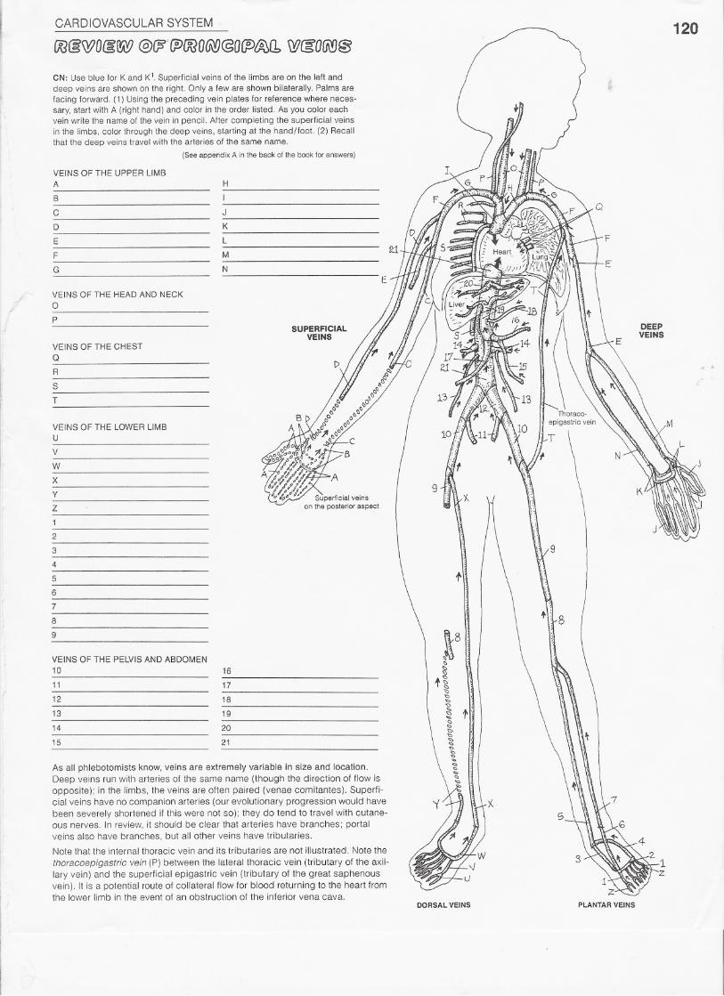
CN: Use blue for K and K1. Superficial veins of the limbs are on the left anddeep veins are shown on the right. Only a few are shown bilaterally. Palms arefacing forward. (1) Using the preceding vein plates for reference where neces-sary, start with A (right hand) and color in the order listed. As you color eachvein write the name of the vein in pencil. After completing the superficial veinsin the limbs, color through the deep veins, starting at the hand/foot. (2) Recallthat the deep veins travel with the arteries of the same name.
(Seeappendix A inthebackof thebookforanswers)
E
VEINS OF THE HEAD AND NECKoP
SUPERFICIALVEINS
DEEPVEINS
VEINS OF THE CHESTQ
R
S
T
VEINS OF THE LOWER LIMBU
V
W
X
y
Z
1
2
3
4
5
6
7
8
9
f
9
VEINS OF THE PELVIS AND ABDOMEN10
11
12
13
14
15
16
17
18
19
20
21
As all phlebotomists know, veins are extremely variable in size and location.Deep veins run with arteries of the same name (though the direction of flow isopposite); in the limbs, the veins are often paired (venae comitantes). Superfi-cial veins have no companion arteries (our evolutionary progression would have
been severely shortened if this were not so); they do tend to travel with cutane-ous nerves. In review, it should be clear that arteries have branches; portalveins also have branches, but all other veins have tributaries.
Note that the internal thoracic vein and its tributaries are not illustrated. Note the
thoracoepigastric vein (P) between the lateral thoracic vein (tributary of the axil-lary vein) and the superficial epigastric vein (tributary of the great saphenousvein). It is a potential route of collateral flow for blood returning to the heart fromthe lower limb in the event of an obstruction of the inferior vena cava.
DORSAL VEINS PLANTAR VEINS